ASM ESCMID Abstracts for Poster Presentations. for website 9.6 · 2019-09-09 · candidate for use...
174
2019 ASM ESCMID ABSTRACTS FOR POSTER PRESENTATIONS Wednesday Presentation Number: W‐01 Publishing Title: Decoding the biosynthesis of α‐glucan glycoconjugates in M. tuberculosis ‐ An approach to fight antimicrobial resistances Author Block: M. Babu Sait, R. Kalscheuer; Heinrich Heine Univ. Düsseldorf, Düsseldorf, Germany Abstract Body: Background: Mycobacterium tuberculosis (Mtb) has an exclusive cell envelope containing complex lipids and glycoconjugates known as the mycolyl‐ arabinogalactan‐peptidoglycan (mAGP) complex. In addition to extracellular cell wall components, mycobacteria also produce cytoplasmic complex oligomeric glycoconjugates such as 3‐O‐methyl mannose polysaccharides and 6‐O‐methylglucose lipopolysaccharides (MGLPs). MGLPs appear to be of critical importance for viability of Mtb as strict essentiality of some genes implicated in MGLP biosynthesis has been suggested from genome‐wide transposon mutant studies. MGLPs have been known to form 1:1 ratio stable complexes with fatty acyl chains and acyl‐CoA thereby regulating the activity of fatty acid synthase I in vitro. They also act as carrier molecules of the newly synthesized fatty acids and protect them from degradation by cytoplasmic enzymes. MGLPs comprise 10‐20 glucose or 6‐O‐methyl glucose units with additional 3‐O‐ methyl glucose units at the non‐reducing end and diglucosylglycerate at the reducing end. Methods: We hypothesize that the enzyme catalyzing diglucosylglycerate formation is a branching enzyme encoded by the Rv3031 gene. Rv3031 is considered to be essential for Mtb growth. We generated two conditional Rv3031 knock‐in mutants (based on two potential stating codons) by inserting a tetracycline‐regulatable synthetic promoter cassette immediately upstream of the start codon of Rv3031employing specialized transduction. We further validated the essentiality of the gene by using the Tet operator regulation system. Results: The knock‐in mutants showed an altered colony morphology when compared to wildtype. With the conditional knock‐in mutants, TetR + /ATc ‐ inhibited the gene expression as the Tet repressor protein (TetR) binds to the operator placed in front of the start codon of the gene. Addition of anhdrotetracycline (ATc) allows gene expression by binding to TetR. We now aim at the characterization of the enzymatic activity of Rv3031 and elucidation of the identity and biological relevance of the pathway products using bioinformatics, molecular genetics, and biochemical approaches. Conclusions: Briefly, considering the key role of these pathway products and enzymes, they might represent a novel drug target in Mtb and also a potential solution to address the problem of multi‐drug resistant strains of Mtb.
Transcript of ASM ESCMID Abstracts for Poster Presentations. for website 9.6 · 2019-09-09 · candidate for use...
2019 ASM ESCMID ABSTRACTS FOR POSTER PRESENTATIONS
Wednesday Presentation Number:
W‐01
Publishing Title: Decoding the biosynthesis of α‐glucan glycoconjugates in M. tuberculosis ‐ An approach to fight antimicrobial resistances
Author Block: M. Babu Sait, R. Kalscheuer; Heinrich Heine Univ. Düsseldorf, Düsseldorf, Germany
Abstract Body:
Background: Mycobacterium tuberculosis (Mtb) has an exclusive cell envelope containing complex lipids and glycoconjugates known as the mycolyl‐arabinogalactan‐peptidoglycan (mAGP) complex. In addition to extracellular cell wall components, mycobacteria also produce cytoplasmic complex oligomeric glycoconjugates such as 3‐O‐methyl mannose polysaccharides and 6‐O‐methylglucose lipopolysaccharides (MGLPs). MGLPs appear to be of critical importance for viability of Mtb as strict essentiality of some genes implicated in MGLP biosynthesis has been suggested from genome‐wide transposon mutant studies. MGLPs have been known to form 1:1 ratio stable complexes with fatty acyl chains and acyl‐CoA thereby regulating the activity of fatty acid synthase I in vitro. They also act as carrier molecules of the newly synthesized fatty acids and protect them from degradation by cytoplasmic enzymes. MGLPs comprise 10‐20 glucose or 6‐O‐methyl glucose units with additional 3‐O‐methyl glucose units at the non‐reducing end and diglucosylglycerate at the reducing end. Methods: We hypothesize that the enzyme catalyzing diglucosylglycerate formation is a branching enzyme encoded by the Rv3031 gene. Rv3031 is considered to be essential for Mtb growth. We generated two conditional Rv3031 knock‐in mutants (based on two potential stating codons) by inserting a tetracycline‐regulatable synthetic promoter cassette immediately upstream of the start codon of Rv3031employing specialized transduction. We further validated the essentiality of the gene by using the Tet operator regulation system. Results: The knock‐in mutants showed an altered colony morphology when compared to wildtype. With the conditional knock‐in mutants, TetR+/ATc‐ inhibited the gene expression as the Tet repressor protein (TetR) binds to the operator placed in front of the start codon of the gene. Addition of anhdrotetracycline (ATc) allows gene expression by binding to TetR. We now aim at the characterization of the enzymatic activity of Rv3031 and elucidation of the identity and biological relevance of the pathway products using bioinformatics, molecular genetics, and biochemical approaches. Conclusions: Briefly, considering the key role of these pathway products and enzymes, they might represent a novel drug target in Mtb and also a potential solution to address the problem of multi‐drug resistant strains of Mtb.
2019 ASM ESCMID ABSTRACTS FOR POSTER PRESENTATIONS
Wednesday Presentation Number:
W‐03
Publishing Title: Development of a Novel Bacteriocin from Lactobacillus plantarum as a Promising New Treatment for Antibiotic Resistant Listeria monocytogenes
Author Block: E. Parlindungan, B. May, O. Jones; RMIT Univ., Melbourne CBD, Australia
Abstract Body:
Background: Listeria monocytogenes is a highly virulent, foodborne pathogen with a fatality rate that can reach as high as 20 to 30%. As such it is a major public health concern, especially in immuno‐compromised individuals, pregnant women, newborn children and the elderly. Currently, treatment with antibiotics is crucial to control infections caused by Listeria, but the issue of antibiotic resistance means treating such infections has become increasingly challenging. A bacteriocin produced by Lactobacillus plantarum B21 is a potential alternative. It was known to inhibit a range of pathogens including Listeria and Clostridia. This study aims to characterise the structure of the B21 bacteriocin and investigate how it exerts its effects on target organisms. Methods: A variety of culture conditions were investigated to find those that would result in high bacterial growth and/or high bacteriocin production. Gas chromatography mass spectrometry (GC‐MS) based metabolomics was utilised to assess the cellular and functional behaviour of L. plantarum B21. Structural analysis of the bacteriocin was undertaken with two‐dimensional nuclear magnetic resonance spectroscopy (NMR). Well diffusion assays and electron microscopy was used to assess the effectiveness of the bacteriocin against a range of target strains of bacteria. Results: A lack of a major carbohydrate source was found to promote both bacteriocin production and long‐term robustness in L. plantarum B21 cultures. GC‐MS data demonstrated significant (p <0.005) production of aspartic acid, glutamic acid and alanine; these compounds may act as biomarkers of cell robustness. NMR indicates the bacteriocin to have a cyclic structure. The electron microscopy data showed cell perforation and blebbing in the target species indicating that the bacteriocin works by damaging the cell wall of target species, such as L. monocytogenes and other closely related bacteria strains, which allows the cell contents to leak out thus causing the cell death. Conclusion: The B21 bacteriocin is "broad‐spectrum" and effective against many Gram‐positive organisms including known pathogens such as Listeria. The compound is safe, pH and temperature stable, and biodegrades quickly in the environment meaning it is hard for target species to develop resistance. The use of bacteriocins such as those produced by the L. plantarum B21 strain to fight strains of bacteria resistant to traditional antibiotics therefore shows great promise in the fight against antibiotic resistant infections.
2019 ASM ESCMID ABSTRACTS FOR POSTER PRESENTATIONS
Wednesday Presentation Number:
W‐04
Publishing Title: Partial characterization of bacteriocin from Bacillus thuringiensis strains obtained from Middle Tennessee
Author Block: F. Alshammari, J. Terrance, E. Anthony; Tennessee State Univ., Nashville, TN
Abstract Body:
Background:Bacillus thuringiensis (Bt) is a Gram‐positive spore forming soil dwelling bacterium that is toxic against insect pests. Bt strains have been reported to also produce many proteins including bacteriocin which is an inhibitory substance to other bacteria. Bacteriocins, originally thought to be produced primarily by Enterobacteriacaea are now known to be produced by many bacterial species. Bacteriocins are active against many Gram‐positive bacteria, though some can also inhibit Gram‐negative species. Methods: In this project, a stock of 66 Bt strains isolated from Middle Tennessee was tested for the presence of bacteriocin. The agar well diffusion method was used to determine if the strains had any bacteriocin activity. The strains were cultured for 24 hours, and the cells were removed by centrifugation. The supernatant was filter‐sterilized and Bacillus cereus (CB154870A), Escherichia coli (CB155065A), Staphylococcus aureus (CB155554A) and Pseudomonas aeruginosa (CB155250A) were used as indicator organisms Results:It was found that 32 of the Bt strains tested produced a bacteriocin. This was evidenced by a clear zone on the plates indicating that a bacteriocin was produced. The bacteriocins were extracted from the Bt strains and characterized based on the sensitivity to heat, various pH values and proteinase k. It was found that bacteriocins were sensitive to low and high pH values and high temperature. No bacteriocin activity was detected after treatment with proteinase K. Conclusions: 32 of the 66 strains of Bt demonstrated the presence of a bacteriocin, and this activity in one strain was due to a non‐protein molecule.
2019 ASM ESCMID ABSTRACTS FOR POSTER PRESENTATIONS
Wednesday Presentation Number:
W‐05
Publishing Title: Purification And Characterization of Subtilin MH1, a New Bacteriocin Produced by Bangladeshi Strain of Bacillus subtilis
Author Block: M. S. H. Hossain, M. M. Hoq; Univ. of Dhaka, Dhaka, Bangladesh
Abstract Body:
Background: Bacteriocins are ribosomally synthesized small peptides which have the potential to be used as an alternative to antibiotics. Recently, research on bacteriocins has received intense attention due to the escalating problems of antibiotic resistance among pathogenic bacteria. The goal of the present study is to characterize a novel bacteriocin from Bacillus subtilis MH1 isolated from the soil sample of Bangladesh. Methods: Deferred antagonism bacteriocin assay and agar well diffusion method was used for bacteriocin assay. Aammonium sulphate precipitation of peptide from culture supernatant followed by reverse phase chromatography with C‐18 column was was for purification. MALDI‐TOF MS was used for molecular weight determination. Results: Deferred antagonism bacteriocin assay and agar well diffusion method suggested that culture supernatant of B. subtilis MH1 has high level of inhibitory activity against Listeria monocytogenes, Staphylococcus aureus. Micrococcus luteus and Bacillus cereus. Subtilin MH1 is generally produced at the mid‐logarithmic phase of growth with optimum temperature of 37°C, pH‐7.0 and 24 h of incubation. Heat stability assay demonstrated that the bacteriocin produced from this strain is highly heat stable and can retain activity up to 100°C. Activity was lost when treated with proteinase K. Subtilin MH1 can be purified by ammonium sulphate precipitation of protein from culture supernatant followed by reverse phase chromatography with C‐18 column. Its molecular mass determined by mass spectrometry was 1700 Da. To the best of our knowledge, this is the first bacteriocin exhibiting such characteristics reported to be produced by B. subtilis. Conclusions:Our study indicates that this bacteriocin may be a potential candidate for use as natural food preservative and an alternative antimicrobial agent. Further studies are required to understand the amino acid sequences, bacteriocin structural genes and associated regulatory elements as well as mode of actions of this bacteriocin.
2019 ASM ESCMID ABSTRACTS FOR POSTER PRESENTATIONS
Wednesday Presentation Number:
W‐06
Publishing Title: Rationalizing the Transport of Trojan Horse Compounds for Crossing the Outer Membrane of Gram Negative Bacteria
Author Block: S. Milenkovic, I. Bodrenko, M. Ceccarelli; Univ. of Cagliari, Cagliari, Italy
Abstract Body:
Background: One of the challenges of modern medicine is to find efficient antibiotics to counteract infections. The main concern is represented by Gram negative species, where antibiotics have to cross the outer membrane for reaching their targets. Porins expressed in the outer membrane control the permeation of small polar molecules and antibiotics. However, Pseudonomas aeruginosa and Acinetobacter baumannii lack the large trimeric porins expressed in enterobacterial species, thus showing low level of susceptibility. Among the possible strategy for resolving the permeation of antibiotics is the use of TonB dependent transporters, such as those expressed to capture iron from the environment. Existing siderophore molecules enriched by anti‐infective properties, so‐called Trojan‐Horse candidates, can be transported efficiently inside the cell. Methods: The high‐resolution structures obtained recently using X‐ray crystallography (holo BauA/ PfeA and apo PiuA/PiuD) have open the way to a more detailed knowledge of transport mechanism. Thanks to the new structures, we have applied molecular simulations in combination with NMR spectroscopy to investigate at molecular level the formation of the molecule‐ion complex in solution, the binding of the complex to the transporter and finally its diffusion along the interior of the transporter. Results: The main outcome is that in general the ion‐siderophore complex in solution is the one recognized also by the transporter, in a precise recognition pocket. Upon the recognition, the binding can provide a unique allosteric signal appearing to activate other regions of the transporters in order to control the self‐transport of the ligand. This effect upon ligand binding promotes a novel idea that the internal diffusion does not require a large conformational change of the transporter, as suggested earlier. Conclusions: We obtained detailed molecular data to understand how to rationalize new siderophore antinfectives able to use transporters to cross the outer membrane. The three fundamental steps are: formation of the ion‐siderophore complex in solution, binding and recognition on the transporter, and internal diffusion. Only the first two steps seem to play a key role in the transport and both depend on subtle interactions ion‐siderophore‐transporter, modulated by selecting precise chemical groups.
2019 ASM ESCMID ABSTRACTS FOR POSTER PRESENTATIONS
Wednesday Presentation Number:
W‐07
Publishing Title: Nano‐mupirocin Characterization as a Potential Candidate for MDR Gonorrhea Treatment
Author Block: A. Cern1, Y. Bavli1, A. Hod1, D. Zilbersheid1, Y. Feinstein1, D. Barasch1, G. Cinamon2, Y. Barenholz1; 1Hebrew Univ., Jerusalem, Israel, 2Rebiotics Rx, Jerusalem, Israel
Abstract Body:
Background: Mupirocin is an antibiotic having a unique mode of action used for the treatment of staphylococci skin infections. It has high protein binding and is rapidly eliminated from the circulation, limiting its use to topical settings.Loading mupirocin into PEGylated nano‐liposomes to form Nano‐mupirocin (NM) protects the drug allowing its parental use against a wider range of bacteria. Mupirocin is highly active against N. gonorrhea for which resistance for all marketed antibiotics is emerging. In order to test the suitability of NM for gonorrhea treatment, in vitro susceptibility of N. gonorrhea strains to mupirocin and in vitro resistance studies were performed. Additionally, the distribution of NM to the vaginal mucus was studied. Methods: Mupirocin MIC against clinical isolates of N. gonorrhea was determined by the agar dilution method (Cern A, Connolly KL, Jerse AE, Barenholz Y. 2018. Antimicrob Agents Chemother:AAC.02377‐17). Resistance assays were performed at IHMA by serial passages and spontaneous mutation frequency (SMF) experiments. Distribution of NM to the vaginal mucus of mice was performed by vaginal swabbing at different time points after NM administration and analysis by LCMS/MS. In addition, NM labelled fluorescently in the lipidic membrane was injected to mice and vaginal mucus collected with swabs was observed by fluorescence microscopy. Results: Mupirocin showed MIC90 value of 0.031 µg/ml. No cross‐resistance with mupirocin was observed for isolates with resistance to either of the comparator antibiotics. Only eight isolates had increased MIC in the resistance passaging experiment, and only one isolate showed minor elevated MIC. In the SMF experiment, no mutants were obtained. Nano‐mupirocin was found in the vaginal mucus of mice at concentrations > MIC. High fluorescence intensity was observed in the mucus of mice injected with labeled NM. Conclusions: The in vitro activity of NM, as well as its distribution to vaginal mucus strongly support the development of NM for the treatment of MDR gonorrhea.
2019 ASM ESCMID ABSTRACTS FOR POSTER PRESENTATIONS
Wednesday Presentation Number:
W‐08
Publishing Title: Development of Efflux Pump Modulators that Reduce Bacterial Load in vivo
Author Block: A. Crooks1, U. Ochsner2, C. DETWEILER1; 1Univ. of Colorado, Boulder, CO, 2Crestone, Inc., Boulder, CO
Abstract Body:
Efflux pumps transport small molecules from the bacterial cytoplasm or periplasm outside the cell. Efflux pump activity is typically increased in antimicrobial resistant (AMR) pathogens by one or multiple mechanisms. Therefore, chemicals that inhibit efflux pumps may re‐sensitize AMR pathogens to clinical antibiotics. From a drug diversity library, we identified three efflux pump modulators (EPMs) that prevent energy‐dependent efflux pump activity in AMR Enterobacteria clinical isolates. The EPMs bind the AcrB subunit of the AcrAB‐TolC efflux system with KDs in the micromolar range and act synergistically with mammalian antimicrobial peptides and with clinical antibiotics to kill bacteria. The EPMs also decrease bacterial colonization in cell culture and in mice. We synthesized and analyzed ~200 analogs of the EPMs and found 11 that reduce bacterial load in cell culture in the sub‐micromolar range and are not apparently toxic in mice. These compounds have potential for early stage antibiotic discovery.

2019 ASM ESCMID ABSTRACTS FOR POSTER PRESENTATIONS
Wednesday Presentation Number:
W‐09
Publishing Title: Novel Enviromimetic Culturing Prototype for Elucidation of Soil Derived Marine Bacteria
Author Block: D. Garcia, G. Plassche, O. Lee, C. Born; Brown Univ., Diane N. Weiss Ctr. for Orthopaedic Trauma Res., Rhode Island Hosp., Providence, RI
Abstract Body:
Background: Since the 2014 warning of the post‐antibiotic era by the World Health Organization, the discovery of antibiotics has become a top priority.1 Pharmacognostic isolation of bacterial‐derived compounds is a highly attractive methodology for the discovery of novel antimicrobial structures. Microbial‐derived bioactive secondary metabolites (BSMs) are low molecular weight compounds that act as defense weapons, aid in communication between organisms, or help in the acquisition of nutrients.2 Unfortunately, agar based mediums are limited, as exhibited by QT‐PCR, which showed that only 2% of all bacteria are culturable via plating.3 Due to this limitation, few new antibiotic‐producing bacteria have been isolated, and no new antibiotics have been translated to the clinical realm in at least 2 decades. In order to address this issue, our team developed enviromimetic cassettes composed of stainless steel meshes, calcium carbonate, chitin, calcium phosphate, polyurethane and polyvinyl acetate. These cassettes mimic structures and conditions found in the natural environment and may allow for the culture of new bacteria. Methods: Stainless steel cassettes were passivated utilizing 4M HCl and 4% nitric acid, and coated different formulations of calcium carbonate, calcium phosphate, and chitin. Additional formulations also utilized polyurethane and polyvinyl acetate instead of passivation. Uncoated stainless steel meshes were utilized as negative controls. Cassette formulations were placed in aliquots of Streptomycetes sp. isolates for 3 weeks and imaged via SEM at 1,000x and 5,000x to determine the level of bacterial adherence. Results: Although all six conditions exhibited some level of bacterial growth, data showed that hydrochloric acid‐passivated meshes coated with calcium phosphate had the most adherence. Additionally, the presence and growth of Streptomycetes sp. on these cassettes occurred faster than on agar plates. Conclusions: The development of enviromimetic cassette prototypes provides a much needed alternative to traditional bacterial culture. These cassettes may facilitate the ability to culture novel marine saprophytic BSMs‐producing bacteria which may lead to new antibiotic discovery. 1. “Antimicrobial Resistance”, 2014 2. Genilloud, 2014 3. Wade, 2002
2019 ASM ESCMID ABSTRACTS FOR POSTER PRESENTATIONS
Wednesday Presentation Number:
W‐10
Publishing Title: Targeting Cardiolipin Microdomains in Bacteria
Author Block: K. A. Hansford, J. J. Dilly, C. K. Davis, I. Lazarova, A. G. Elliott, G. Lowe, O. A. K. Jessop, M. R. L. Stone, M. A. Cooper; Inst. for Molecular BioSci., Brisbane, Australia
Abstract Body:
Background: The predominant anionic lipids in bacterial membranes are phosphatidylglycerol (PG) and cardiolipin (CL), the presence of which provides a selectivity basis for cationic antimicrobial agents targeting bacteria over mammalian cells. CL, found in the bacterial inner membrane, forms lipid microdomains at the cell poles and division septum. It plays a key role in the maintenance of bacterial shape, and the structural regulation of membrane associated proteins. Polycationic antimicrobial compounds can induce phase boundary defects by sequestering anionic lipids such as CL, leading to membrane permeabilization and loss of bacterial function. Compounds targeting CL and CL microdomains might represent a hitherto underexplored strategy in the design of new antimicrobial agents. Methods: The peptide SS‐31 (D‐Arg‐L‐Tyr(2,6‐DiMe)‐L‐Lys‐L‐Phe‐NH2) was N‐acylated with n‐alkyl fatty acids of variable length to generate a series of lipopeptides. SPR was used to characterize compound affinity for model membrane vesicles containing CL and PG. Incorporation of a C‐terminal azido lysine enabled click attachment of a fluorophore. Analogs were assessed for intrinsic antimicrobial potency, and for synergistic activity in vitro against wild‐type and MDR/XDR Gram‐negative (G‐ve) pathogens when partnered with minocycline, rifampicin or clarithomycin. The same peptide series was modified with an additional C‐terminal lysine, enabling Nε‐acylation with vancomycin to generate vancomycin‐peptide conjugates. Results: Lipopeptide analogs of SS‐31 possessed increased affinity for anionic SUVs compared to SS‐31, but the compound series did not possess significant intrinsic antimicrobial activity. However, MDR/XDR G‐ve bacteria were resensitized to either minocycline, rifampicin or clarithromycin in the presence of 1 µg/mL or more of lipopeptide. Lipopeptide conjugation to vancomycin produced highly potent compounds against a panel of Gram‐positive pathogens, including VRSA and VanA enterococci, but no significant G‐ve activity was observed. Confocal imaging of E. coli following incubation with the fluorescent analog bearing the SS‐31 sequence revealed localisation of the peptide at the septal poles, consistent with CL targeting. Conclusions: Modification of SS‐31 produced lipopeptides capable of resensitizing bacteria to antibiotics synergistically or by direct conjugation, possibly through the interaction with CL microdomains leading to increased membrane permeabilization and/or membrane affinity.
2019 ASM ESCMID ABSTRACTS FOR POSTER PRESENTATIONS
Wednesday Presentation Number:
W‐11
Publishing Title: Membrane Active Novel Small Molecule Compound Controls Extra‐intestinal Pathogenic Escherichia coli (ExPEC) Infection in Poultry
Author Block: D. Kathayat, Y. Helmy, L. Deblais, V. Srivastava, G. Closs Jr, G. Rajashekara; The Ohio State Univ., Wooster, OH
Abstract Body:
Background: Extraintestinal pathogenic E. coli (ExPEC) in poultry, also known as avian pathogenic E. coli (APEC), causes high morbidity and mortality of chickens. A recent report has also suggested APEC as a foodborne human uropathogen transmitted through consumption of contaminated poultry products. Further, APEC is also considered as a source of antibiotic resistant genes (ARGs) to human pathogens, including human ExPECs, which makes human infections difficult to treat. Currently, antibiotics are used to control APEC infection in poultry; however, multidrug‐resistant (MDR) APEC strains have been reported worldwide. Therefore, new and potent anti‐APEC therapeutics are critically needed. Methods: Towards this end, we screened a pre‐selected enriched small molecule (SM) library, and identified 11 SMs bactericidal to multiple APEC serotypes, antibiotic‐resistant APECs, and APEC in biofilm. Eight SMs, that are non‐toxic and effective in cultured eukaryotic cells (Caco‐2, HD11, and THP‐1) and wax moth (Galleria mellonella) larvae, were tested in one‐week‐old commercial broiler chickens. Chickens were infected subcutaneously (s/c) with rifampicin resistant (Rifr) APEC O78 (1 x 10⁷ CFU/chicken) and SMs were orally gavaged (1 mg/kg body weight) twice a day starting one day prior to infection to three days post‐infection. Results: Four SMs (GI‐7, GI‐10, GI‐6, and GI‐2) reduced the mortality (42.8 to 71.42%), APEC lesions severity (13.5 to 62%) and APEC load (1.3 to 2.6 logs) in chickens. Further, administration of GI‐7 (40 mg/L and 60 mg/L), a most effective SM, in drinking water for seven days, which simulates the farm practice, also reduced the mortality (up to 84.84%), APEC lesions severity (up to 38%) and APEC load (up to 2.5 logs) in chickens. Additionally, the body weight gain (BWG) of GI‐7 treated chickens was similar to untreated chickens. Confocal and scanning electron microscopy of GI‐7 treated APEC showed bleb‐like structures on the APEC membrane suggesting the membrane affecting mode of GI‐7. Further, the expression of LptD, an outer membrane lipopolysaccharide transporter and a novel antibacterial target, was downregulated (10‐fold) upon treatment of APEC with GI‐7. Conclusions: Our results demonstrate that the GI‐7 can represent a novel anti‐APEC therapeutic; thereby, can be developed as an alternative to currently used antibiotics. Our future studies will be focused on validating the antibacterial target of GI‐7 as well as assessing the GI‐7 efficacy in chickens naturally infected with APEC.
2019 ASM ESCMID ABSTRACTS FOR POSTER PRESENTATIONS
Wednesday Presentation Number:
W‐12
Publishing Title:
Targeting Drug‐Resistant Bacteria with Chelators for Antibacterial Activity and Metallo‐β‐Lactamase Inhibition
Author Block: A. C. Jackson, J. M. Zaengle‐Barone, K. J. Franz; Duke Univ., Durham, NC
Abstract Body:
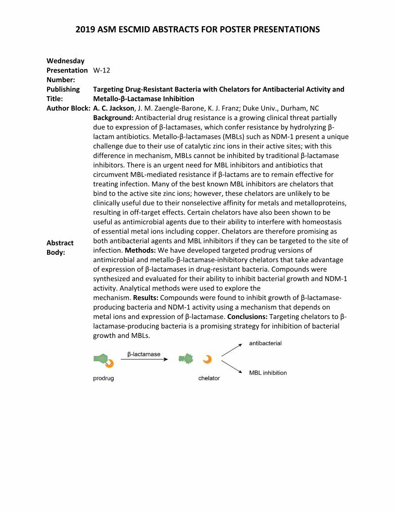
Background: Antibacterial drug resistance is a growing clinical threat partially due to expression of β‐lactamases, which confer resistance by hydrolyzing β‐lactam antibiotics. Metallo‐β‐lactamases (MBLs) such as NDM‐1 present a unique challenge due to their use of catalytic zinc ions in their active sites; with this difference in mechanism, MBLs cannot be inhibited by traditional β‐lactamase inhibitors. There is an urgent need for MBL inhibitors and antibiotics that circumvent MBL‐mediated resistance if β‐lactams are to remain effective for treating infection. Many of the best known MBL inhibitors are chelators that bind to the active site zinc ions; however, these chelators are unlikely to be clinically useful due to their nonselective affinity for metals and metalloproteins, resulting in off‐target effects. Certain chelators have also been shown to be useful as antimicrobial agents due to their ability to interfere with homeostasis of essential metal ions including copper. Chelators are therefore promising as both antibacterial agents and MBL inhibitors if they can be targeted to the site of infection. Methods: We have developed targeted prodrug versions of antimicrobial and metallo‐β‐lactamase‐inhibitory chelators that take advantage of expression of β‐lactamases in drug‐resistant bacteria. Compounds were synthesized and evaluated for their ability to inhibit bacterial growth and NDM‐1 activity. Analytical methods were used to explore the mechanism. Results: Compounds were found to inhibit growth of β‐lactamase‐producing bacteria and NDM‐1 activity using a mechanism that depends on metal ions and expression of β‐lactamase. Conclusions: Targeting chelators to β‐lactamase‐producing bacteria is a promising strategy for inhibition of bacterial growth and MBLs.
2019 ASM ESCMID ABSTRACTS FOR POSTER PRESENTATIONS
Wednesday Presentation Number:
W‐13
Publishing Title: Design, Synthesis and Biological Evaluation of Novel Aspergillomarasmine A (AMA) Analogs as Metallo‐ β‐Lactamase (MBL) Inhibitors
Author Block: K. Koteva, D. Sychantha, C. M. Rotondo, G. D. Wright; McMaster Univ., Hamilton, ON, Canada
Abstract Body:
Background: Bacterial resistance to β‐lactam antibiotics is mainly achieved by enzymatic inactivation involving β‐lactamases. The serine‐β‐lactamase (SBL) enzymes utilize a serine residue to inactivate these drugs while zinc is required for the activity of metallo‐β‐lactamase (MBL) enzymes. As there several SBL inhibitors currently being employed in the clinic, MBLs present the greatest threat to the efficacy of β‐lactam antibiotics as there are no clinically available inhibitors for these enzymes. However, previous studies demonstrated that aspergillomarasmine A (AMA), a fungal natural product, had the capacity to inhibit two clinically relevant MBLs, NDM‐1 and VIM‐2. Although AMA demonstrated the ability of restoring the activity of meropenem against these MBLs, the mechanism of action of AMA remains unknown. Detailed enzymatic studies showed that AMA has unexpectedly weak affinity for MBLs. Therefore, the goal of this study was to develop more potent and broad‐spectrum AMA‐based MBL inhibitors. Methods: Here we report an improved, practical route to AMA analogs via aziridine ring opening on a solid support. Results: Using this approach, we were able to prepare a series of AMA derivatives carrying substitutions on the N‐terminus or with the aspartic acid portion of molecule substituted with different amino acids. All the AMA analogs synthesized using this method were studied using both in vivo and in vitro assays against one of the most widespread MBL, NDM‐1. The structure‐activity relationships of the most promising analogs were further probed through assays involving VIM‐2 and IMP‐7. Conclusion: This study led to the identification of a new compound which was 500‐fold more potent than AMA in vitro. Ultimately, this compound lowered the rescue concentration of AMA in combination with meropenem against strains of carbapenem‐resistant Escherichia coli that produced MBLs previously recalcitrant to inhibition.
2019 ASM ESCMID ABSTRACTS FOR POSTER PRESENTATIONS
Wednesday Presentation Number:
W‐14
Publishing Title: Fragment‐based Carbapenemase Inhibitors
Author Block:
J. Kraemer1, L. Goncalves1, R. Mejdi‐Nitiu1, C. Softley2, K. Zak2, M. Bostock2, G. Popowiz2, C. Grandclaudon3, M. Brönstrup3, M. Sattler2, H. Meyer1; 1Technical Univ. Munich, Munich, Germany, 2Helmhotz Zentrum München, Neuherberg, Germany, 3Helmholtz‐Zentrum für Infektionsforschung GmbH, Braunschweig, Germany
Abstract Body:
Background The spreading of carbapenemases is posing an increasing threat to human health and lives, as these enzymes mediate resistance against all types of ß‐lactam antibiotics. The ß‐lactams are widely applicable and particularly well tolerated, and thus represent the most frequently used and most important class of antibiotics. Moreover, as many carbapenemase‐positive pathogens also express resistances against other classes of antibiotics, treatment options are further limited, which is predominantly seen in NDM‐1 positive pathogens. Therefore, novel treatment options are urgently needed, in particular for Gram‐negative carbapenem‐resistant pathogens. This has been underlined recently in the published WHO priority pathogens list for R&D of new antibiotics, which summarizes several of these pathogens in the Priority 1 group classified as critical. We therefore aim to discover carbapenemase inhibitors for clinical use in combination with last resort ß‐lactam antibiotics, like Meropenem and Imipenem. Methods In our previous work, we have developed an assay platform for MBL‐and carbapenemase inhibitors, which uniquely reflects the pathophysiology of infection and therapy. Utilizing this assay platform, we chose a fragment‐based approach for identifying novel carbapenemase inhibitor phamacophores. Results We identified two novel fragment classes with inhibitory activity in the micromolar range and ligand efficiency up to 0.52 against selected metallo‐carbapenemases (fragment class I) and metallo‐ as well as serine‐carbapenemases (fragment class II), respectively. Both fragment classes show biological activity against priority I clinical isolates in MIC shift experiments. Furthermore, fragment class II exhibits an antibiotic activity independent of carbapenemase inhibition. Co‐crystal structures with IMP‐13 (MBL) and OXA‐48 (SBL) are presented. Conclusions As both fragments bind IMP‐13 in (fragment II) or close to (fragment I) the active side fragment merging offers a promising strategy for compound optimization. The dual mode of action is a valuable starting point for the development of innovative anti‐infective drug (s).
2019 ASM ESCMID ABSTRACTS FOR POSTER PRESENTATIONS
Wednesday Presentation Number:
W‐15
Publishing Title: Overcoming Metallo‐β‐Lactamases Mediated Antibiotic Resistance Through the Use of Zinc Chelating Agent as Metallo‐β‐Lactamase Inhibitor
Author Block: A. M. Somboro, D. G. Amoako, J. O. Sekyere, H. M. Kumalo, R. Khan, L. A. Bester, S. Y. Essack; Univ. of KwaZulu‐Natal, Durban, South Africa
Abstract Body:
Background: Evolution of bacteria producing Metallo‐β‐lactamase (MBL) enzymes capable of hydrolysing clinically available β‐lactams, is of grave concern. Thus far there are no MBL inhibitors (MBLIs) approved for clinical use, hence the WHO ranked MBLs mediated antibiotic resistance as priority pathogens which urgently need new agents. A strategy to inhibit MBLs is to employ molecules that are capable of sequestering/inhibiting the zinc atoms at their active sites. Therefore, we aimed to investigate a zinc chelating agent, 1,4,7‐triazacyclononane (TACN) as potential MBLI. Methods: The study employed a molecular simulation approach to investigate enzyme‐ligand interaction and binding affinity of TACN, coupled with experimental assays to reveal the potentiality of this molecule as an antimicrobial agent. The graphical user interface of Chimera was used as molecular modeling tool, and docking calculations performed using AutoDock Vina software. Combination activity of a β‐lactam antibiotic, meropenem (MEM) with TACN were undertaken through MIC, MBC, synergistic and serum effects and time‐kill kinetics assays using broth microdilution methods. MBL inhibition by TACN was performed through nitrocefin‐based colorimetric assays. MTT assay served to evaluate the cytotoxicity of TACN against HepG2 cells. Results: Computational studies predicted that TACN inhibits MBLs (NDM‐1 and VIM‐2) by targeting their catalytic active site pockets. The theoretical calculations of the binding free energies showed good affinity and stability between ligand‐enzyme complexes. This was supported by the experimental assays where TACN exposure to NDM‐1 exhibited an inhibition constant (Ki) of 0.044 μM with an inactivation constant (kinact) of 0.0406 min‐1 stating the efficient inhibitory property of this molecule and thus holding great promise as MBLI. MEM activity was potentiated against strains of bacteria carrying MBLs, with the restoration of its MICs from 8‐64 mg/L to 0.03 mg/L in the presence of 8 mg/L of TACN. MEM‐TACN combination displayed bactericidal effect with MBC/MIC ratio of ≤4 and synergistic activity with the fractional inhibitory concentration index ranging from ≤0.005 to 0.25. No cytotoxicity at concentrations above the MIC values (IC50=56 mg/L) and no human serum effects on the MICs were observed. Conclusions: TACN efficiently potentiated the antimicrobial property of MEM through the inactivation of zinc atoms at the active side of MBLs, as should

2019 ASM ESCMID ABSTRACTS FOR POSTER PRESENTATIONS
thus be further investigated as a MBLI for clinical use, in combination with carbapenems.
2019 ASM ESCMID ABSTRACTS FOR POSTER PRESENTATIONS
Wednesday Presentation Number:
W‐17
Publishing Title: Bactericidal action of mangostin and celastrol against diverse animal pathogenic bacteria
Author Block: D. Moon; Animal and Plant Quarantine Agency, Gimcheon‐si, Korea, Republic of
Abstract Body:
Background: The alpha mangostins (α‐ MG) and gamma mangostins (γ‐ MG), a natural xanthanoid isolated from the fruit wall of Garcinia mangostana. The α‐ MG and γ‐ MG are broad spectrum bioactive substances containing anti‐inflammatory, anti‐tumor, antibacterial and antifungal activities. Similarly, celastrol also is known to possess multiple pharmacological effects. Recent in vitro studies α‐ MG, γ‐ MG and celastrol has been reported to be an effective antimicrobial and anti‐biofilm effects against bacteria. The aim of this study was to examine the antibacterial activities of mangostins and celastrol against several animal pathogens and antimicrobial resistant bacteria derived from animal. Methods: A total of 3,634 chemical library compounds were obtained from Korean Chemical Bank. Antibacterial activity was evaluated by broth microbilution method against 13 animal pathogens and 3 multidrug‐resistant Staphylococcus aureus. To assess the antibiofilm activity of compounds for Staphylococcus epidermidis, crystal violet‐based biofilm inhibition assay was carried out. Results: Among the compounds, three most promising agents, α‐ MG, γ‐ MG and celastrol showed antibiofilm activity for S. epidermidis. The compounds‐induced decrease in biofilm formation was dose‐dependent based on the results of the inhibition assay. For antibacterial activity, these compounds significantly inhibited bacterial growth of the gram‐positive bacteria range with minium inhibition concentrations 1.25 ‐ 20 µmol/L. Furthermore, the compounds showed a bactericidal effect for methicillin‐resistant S. aureus, linezolid‐resistant S. aureus and quinupristin/dalfopristin‐resistant S. aureus. However, no or low antibacterial activity was observed for Gram‐negative bacteria and Prototheca spp., respectively. Conclusions: These results suggest that α‐ MG, γ‐ MG and celastrol compounds showed the bactericidal activity to diverse animal pathogens and resistant bacteria. New concept of alternative antibiotic may lead to reduce the emergence of antibiotic resistance and increase treatment success rate. However, further research is required to prove the mechanisms of action, toxicity and side effects in animals for drug development.
2019 ASM ESCMID ABSTRACTS FOR POSTER PRESENTATIONS
Wednesday Presentation Number:
W‐18
Publishing Title: Phenoxyacetamide Inhibitors of P. aeruginosa Type III Secretion are Efficacious in a Murine Lung Infection Model
Author Block:
D. T. MOIR1, M. C. Torhan1, S. L. Waidyarachchi1, Y. Yan1, N. G‐Dayanandan1, S. Adhikari1, S. C. Cardinale1, J. Yabut1, T. Murphy2, Z. D. Aron1, T. L. Bowlin1; 1Microbiotix, Inc., Worcester, MA, 2NeoSome Life Sci., LLC, Lexington, MA
Abstract Body:
Background: The Pseudomonas aeruginosa type III secretion system (T3SS) is a clinically important virulence mechanism that translocates protein effector toxins into human cells to facilitate the establishment and dissemination of bacterial infection. We previously identified a highly stereo‐selective phenoxyacetamide (PhA) series of inhibitors of P. aeruginosa T3SS and demonstrated that mutations selected for resistance to PhA map to the needle protein gene, pscF. We now show that compounds in this chemical series significantly improve murine survival in a lethal P. aeruginosa lung infection model. Methods: A standard immunocompetent murine acute pneumonia lung infection model was employed. Balb/C mice were challenged with approximately 107P. aeruginosa strain PA99 CFUs intranasally, which resulted in death of most mice by 72 hr. PhA analogs were formulated in 5% DMSO, 5% Cremophor, potassium phosphate and administered to mice via the intravenous (IV) route. PK parameters from in vivo assays in mice were calculated using WinNonLin. Results: The sodium salt of PhA analog MBX‐4742 was shown to be tolerated IV at 300 mg/kg/day for up to 5 days, and PK studies of a single 50 mg/kg IV dose revealed a Cmax of 50 x EC50, t1/2 of 1 hr, and AUC of 42,000 min*ng/mL. MBX‐4742 administered IV at 50 mg/kg TID for 2 days significantly (p<0.0001) increased the survival of mice challenged with wild‐type PA99, but not mice challenged with a PhA‐resistant mutant of PA99 [pscF(R75H)]. Dose response studies revealed efficacy at 25 and 50 mg/kg but not at 12.5 mg/kg. Dose frequency studies demonstrated efficacy from TID and BID dosing. Conclusions: Intravenous administration of PhA small molecule T3SS inhibitors alone significantly increased the survival of mice challenged with P. aeruginosa intranasally by blocking the T3SS. Acknowledgments: References: Bowlin NO, et al., 2014. Antimicrob Agents Chemother. 58:2211‐20; Williams JD, et al., 2015. Bioorg Med Chem. 23:1027‐43. Financial support: This research was supported in part by NIAID under awards R44 AI068185 and R01 AI099269 and by CARB‐X4500002329.
2019 ASM ESCMID ABSTRACTS FOR POSTER PRESENTATIONS
Wednesday Presentation Number:
W‐19
Publishing Title: Discovery of Novel Penicillin‐binding Protein 3 Inhibitors to Treat Pseudomonas aeruginosa Infections
Author Block:
T. Durand‐Reville, X. Wu, S. Guler, J. Zhang, J. Comita‐Prevoir, M. Sylvester, J. Romero, C. Velez‐Vega, F. Wu, R. Iyer, A. Shapiro, S. Moussa, N. Carter, A. Chen, S. McLeod, J. O'Donnell, A. Miller, R. Tommasi; Entasis Therapeutics, Waltham, MA
Abstract Body:
Background: The emergence of antibiotic resistance, especially in Gram‐negative bacteria, is an urgent threat to public health. The discovery of novel antibacterial agents against these pathogens has been impeded by a fundamental lack of understanding of the molecular drivers governing cellular permeation. To address this gap, we rationally designed a novel series of penicillin‐binding protein 3 (PBP3) inhibitors as a potential therapy for Pseudomonas aeruginosa (P.a.) infections. Methods: Novel diazabicyclooctanone (DBO) analogs were synthesized at Entasis Therapeutics. Binding modes were analyzed by protein crystallography. X‐ray diffraction data were collected at the Advanced Photon Source (Lemont, IL). The PBP acylation rates were measured using a fluorescence anisotropy assay. Porin uptake was assessed using the Titrable Outer Membrane permeability Assay. Susceptibility testing was performed according to CLSI guidelines. Murine efficacy studies were conducted using IACUC approved methods. Results: A series of novel DBO‐based analogs were synthesized. Several co‐crystal structures were solved to support our design hypothesis and engineer PBP3 selectivity from PBP2 which was required for in vivo efficacy. The new PBP3 analogs show acylation rates > 800,000 M‐1.s‐1. Bacterial permeation was optimized concomitantly, and multiple analogs showed permeation through > 8 distinct porins, resulting in < 2 mg/L MICs against wild type P.a. strains and low frequencies of resistance (< 10‐9). MICs were unchanged when tested against a P.a. isogenic panel of 20 strains overexpressing individual beta‐lactamases from all 4 Ambler classes, demonstrating no vulnerability to these enzymes. In vivo efficacy was demonstrated against a MDR P.a. clinical isolate in a murine thigh model with >2 log CFU reduction at 250 mg/kg (q3h). Conclusions: An innovative two‐prong rational design approach led to the discovery of a novel series of DBO analogs with, no beta‐lactamase susceptibility, improved biochemical potency and permeation leading to wild type antibacterial activity and in vivo efficacy. This approach has the potential to deliver a therapy to treat multi‐drug resistant P.a. infections.
2019 ASM ESCMID ABSTRACTS FOR POSTER PRESENTATIONS
Wednesday Presentation Number:
W‐20
Publishing Title:
Targeting Iron Metabolism in Pseudomonas aeruginosa and Klebsiella pneumoniae by Dual Inhibition using Gallium Porphyrin and Gallium Nitrate
Author Block: S‐r. Choi, B. Britigan, P. Narayanasamy; Univ. of Nebraska Med. Ctr., Omaha, NE
Abstract Body:
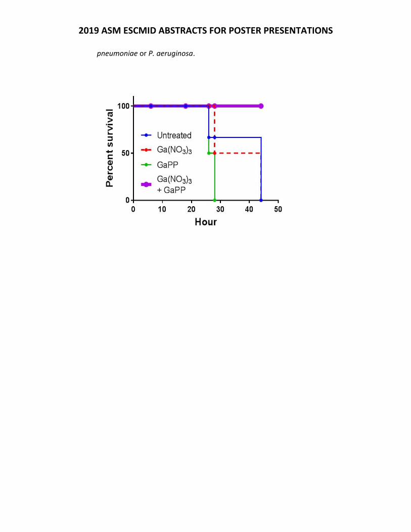
Background: In iron metabolism, Iron‐ and heme‐uptake pathways are promising targets for the development of new antimicrobial agents. Here, nutritional iron starvation leads to inhibition of bacterial growth. For years Gallium (Ga) nitrate, an iron mimetic metal, disrupted iron‐dependent biological processes by binding iron‐utilizing proteins. Recently, Ga porphyrins, heme mimetic complexes, are found to disrupt heme‐utilizing hemeproteins. Hence, we hypothesized that these two Ga compounds may disrupt both bacterial iron/heme acquisition/utilization pathways, and Ga(NO3)3/Ga porphyrin combinations would result in enhanced antimicrobial activity. Methods: Antimicrobial activity of Ga porphyrin (Ga protoporphyrin (GaPP) or Ga mesoporphyrin (GaMP)) alone and in combination with Ga(NO3)3 was evaluated against P. aeruginosa, K. pneumoniae, A. baumannii and MRSA under iron‐depleted condition. Results: The Ga porphyrin/Ga(NO3)3 combination demonstrated substantial synergism against K. pneumoniae, P. aeruginosa and MRSA. Our assays revealed that the GaPP/Ga(NO3)3 combination was bacteriostatic against K. pneumoniae and bactericidal against P. aeruginosa. The biofilm disruption assay confirmed that the GaPP/Ga(NO3)3 combination significantly disrupted K. pneumoniae and P. aeruginosa biofilms on plasma‐coated surfaces. Similarly the GaPP/Ga(NO3)3 combination also increased the survival of C. elegans infected with K. pneumoniae or P. aeruginosa. We also observed 100 % survival of mice on using GaPP/Ga(NO3)3 combination compared to individual Ga salt. Conclusion: Our results demonstrate that GaPP and Ga(NO3)3 had significant synergistic effect by dual inhibition of iron/heme metabolism. The combination significantly disrupted K. pneumoniae and P. aeruginosa growth, biofilms and also increased the survival of C. elegans and mice infected with K.
2019 ASM ESCMID ABSTRACTS FOR POSTER PRESENTATIONS
pneumoniae or P. aeruginosa.
2019 ASM ESCMID ABSTRACTS FOR POSTER PRESENTATIONS
Wednesday Presentation Number:
W‐21
Publishing Title: The Application of Photodynamic Treatment with Methylene Blue against Cryptococcus neoformans and Cryptococcus gattii
Author Block: A. O. Ogundeji, C. H. Pohl, O. M. Sebolai; Univ. of the Free State, Bloemfontein, South Africa
Abstract Body:
Background: Photodynamic treatment (PDT) has emerged as possible alternative treatment strategy against Cryptococcus (C.) neoformans and Cryptococcus (C.) gattii. PDT is often successful when used against microbes that are non‐fermentative because of their oxygen metabolism that is sensitive to photodynamic action. Methods: In this study, we determined the photodynamic action of methylene blue when administered to cryptococcal cells. Also, the cytotoxic effect of PDT on murine macrophage was also determined.Results:In our study, treatment of cells resulted in a significant (p < 0.01) reduction in the growth of cryptococcal cells (81% reduction) as compared with non‐treated cells. The effect of PDT on the treated cells showed a significant (p < 0.05) loss of membrane potential when compared with non‐treated cells, this explains why a significant accumulation of reactive oxygen species was also observed among the treated cells as compared to non‐treated cells. Importantly, PDT was shown to be non‐toxic to the macrophage, but rather enhanced their phagocytic capability which led to phagocytosis of more cryptococcal cells. Conclusions: The results from this study highlight the potential of PDT as an alternative in the control of the growth cryptococcal cells which could be attributed to its effectiveness against respiring microbes.Keywords. Photodynamic treatment; Methylene blue; Cryptococcus; Macrophages; ROS‐mediated membrane damage.
2019 ASM ESCMID ABSTRACTS FOR POSTER PRESENTATIONS
Wednesday Presentation Number:
W‐22
Publishing Title:
Acetylsalicylic Acid (Aspirin) as a Photosensitiser in Photodynamic Treatment against Cryptococcus neoformans and Cryptococcus gattii
Author Block: A. O. Ogundeji, C. H. Pohl, O. M. Sebolai; Univ. of the Free State, Bloemfontein, South Africa
Abstract Body:
Background: We have previously reported on the antimicrobial activity of acetylsalicylic acid (aspirin). There, aspirin (1 mM) inhibited growth of cryptococcal cells via reactive oxygen species (ROS)‐mediated mechanism, which may be a mechanism employed by photodynamic treatment (PDT). Methods:In this current study, we examined the effect of aspirin as a photosensitiser in PDT against cryptococcal cells using a lower concentration than previously reported. In addition to that, the mode of action of PDT using aspirin was determined by looking at the ultrastructure of the cells and accumulation of reactive oxygen species (ROS) analysis following treatment. The cytotoxic effect of PDT with aspirin on murine macrophage growth was also determined. Results:All treated cryptococcal cells showed a significant (p < 0.05) reduction of 97% in growth as compared with non‐treated cells. Interestingly, this reduction was achieved at an aspirin concentration of ½ MIC that was of our previous study. The effect of PDT on the treated cells showed a significant (p < 0.05) accumulation of ROS among the treated cells as compared with non‐treated cells. This in turn explains why we obtained a significant (p < 0.05) reduction of the extracellular matrixes covering the cell wall surfaces with 70% ruptured cells indicating membrane damage . Based on these outcomes, we can deduced that the killings of cryptococcal cells was achieved through ROS‐mediated membrane damage. Also, PDT did not negatively affected the metabolic activity of the macrophage, but in contrast, it aids the macrophages to internalised cryptococcal cells effectively. Conclusions: From our findings, we can conclude that aspirin can be an effective photosensitiser against cryptococcal cells in PDT with a lower concentration, which can minimise the issue of side effect.Keywords: Aspirin; Photosensitisers; Photodynamic treatment; Cryptococcus; Macrophages; Extracellular matrixes; ROS‐mediated membrane damage.
2019 ASM ESCMID ABSTRACTS FOR POSTER PRESENTATIONS
Wednesday Presentation Number:
W‐23
Publishing Title:
Underestimated Potential of Metal‐Complexes as New Antibiotics? ‐ A Look at the Numbers
Author Block: A. Frei, J. Zuegg, M. Blaskovich, M. Cooper; Inst. for Molecular BioSci., St. Lucia, Australia
Abstract Body:
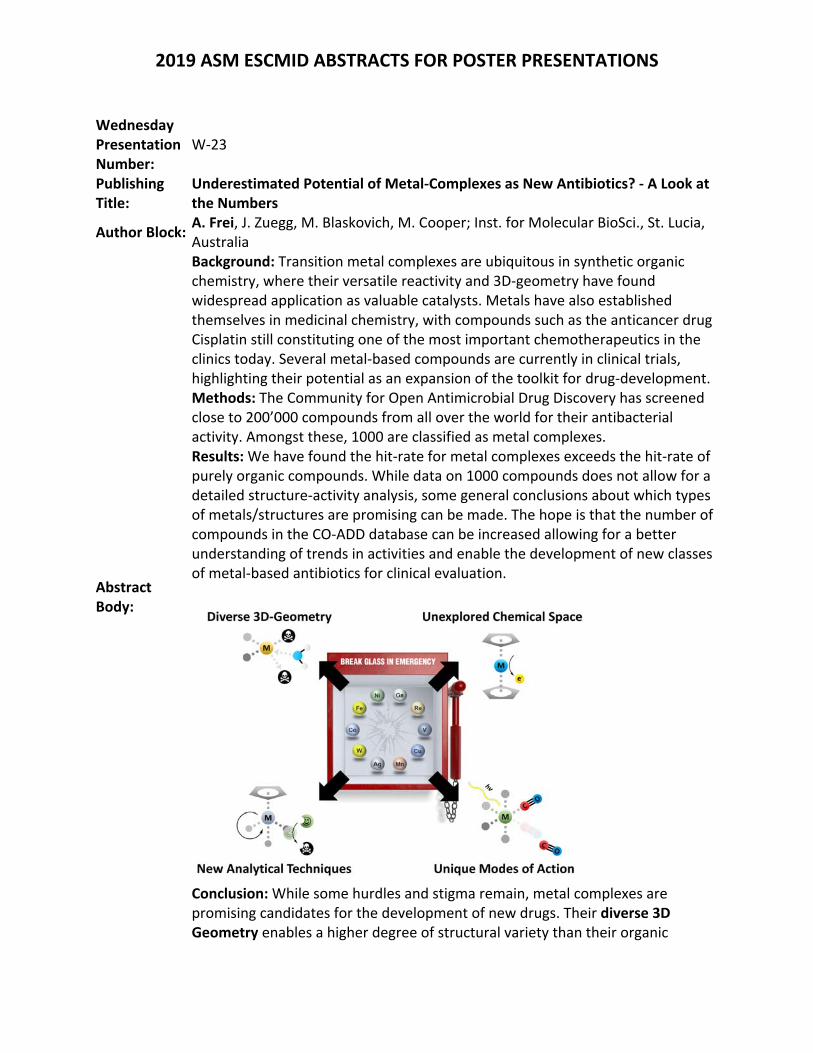
Background: Transition metal complexes are ubiquitous in synthetic organic chemistry, where their versatile reactivity and 3D‐geometry have found widespread application as valuable catalysts. Metals have also established themselves in medicinal chemistry, with compounds such as the anticancer drug Cisplatin still constituting one of the most important chemotherapeutics in the clinics today. Several metal‐based compounds are currently in clinical trials, highlighting their potential as an expansion of the toolkit for drug‐development.Methods: The Community for Open Antimicrobial Drug Discovery has screened close to 200’000 compounds from all over the world for their antibacterial activity. Amongst these, 1000 are classified as metal complexes. Results: We have found the hit‐rate for metal complexes exceeds the hit‐rate of purely organic compounds. While data on 1000 compounds does not allow for a detailed structure‐activity analysis, some general conclusions about which types of metals/structures are promising can be made. The hope is that the number of compounds in the CO‐ADD database can be increased allowing for a better understanding of trends in activities and enable the development of new classes of metal‐based antibiotics for clinical evaluation.
Conclusion: While some hurdles and stigma remain, metal complexes are promising candidates for the development of new drugs. Their diverse 3D Geometry enables a higher degree of structural variety than their organic

2019 ASM ESCMID ABSTRACTS FOR POSTER PRESENTATIONS
counterparts. This diversity also implies an enormous landscape of unexplored chemical space that is available through these compounds. On top of simple target binding, metal complexes also have access to a suite of unique modes of action that are hard or impossible to achieve with organic compounds. Finally, the use of metal compounds enables the use of new analytical techniques to study the in vitro and in vivo behavior of the complex.
2019 ASM ESCMID ABSTRACTS FOR POSTER PRESENTATIONS
Wednesday Presentation Number:
W‐24
Publishing Title: In vivo Proof‐of‐Concept for a Novel Small‐Molecule Inhibitor of Bacterial Lipoprotein Transport Targeting Enterobacteriaceae
Author Block: E. Breidenstein, O. Abdulle, T. Avis, C. Charrier, C. Ciardullo, C. Coward, T. Duffy, N. Khan, C. Mason, P. Meo, D. J. Powell; Summit Therapeutics, Cambridge, United Kingdom
Abstract Body:
Background: Increasing antimicrobial resistance among Gram‐negative bacteria combined with the current antibiotic innovation gap highlights the need for novel agents. Carbapenem‐Resistant Enterobacteriaceae (CRE) and Extended‐Spectrum β‐Lactamase (ESBL)‐producing Enterobacteriaceae have been listed as serious or urgent threats by the World Health Organization (WHO) and US Centers for Disease Control and Prevention (CDC). To address this urgent unmet medical need, Summit Therapeutics has discovered a first‐in‐class new mechanism small‐molecule antibiotic series (DDS‐04) that overcomes all pre‐existing resistance mechanisms. The DDS‐04 series targets the clinically unexploited bacterial LolC/E complex, involved in lipoprotein transport. Our antibiotic series is a precision therapy that has the potential to treat infections caused specifically by Enterobacteriaceae (including bloodstream, respiratory and urinary tract infections). Methods: Efficacy was established for the DDS‐04 series against murine pharmacodynamic (PD) models of urinary tract infection (UTI) (C3H/HeN mice infected with E. coli UTI89), septicaemia (non‐neutropenic CD1 mice infected with E. coli ATCC BAA246) and pneumonia (neutropenic CD1 mice infected with Klebsiella pneumoniae ATCC 43816). Results: Following IV dosing, the DDS‐04 series maintained good Cmax levels in the bloodstream and was distributed to multiple infection sites including the bladder, kidneys and lungs. In vivo efficacy studies demonstrated that the DDS‐04 series significantly reduced the bacterial burden in animal models of UTI, septicaemia and pneumonia. In the UTI model, a significant reduction in colony‐forming units (CFU) was observed in the urine, the bladder and the kidneys. In the septicaemia model, the bacterial burden was below the limit of detection in the blood, the kidneys, the liver, lungs and the spleen. In the pneumonia model, a 4.5 log10 reduction in CFU was observed in the lungs. Conclusions: Our new mechanism, small‐molecule LolC/E inhibitors represent a promising antibiotic class that has the potential to treat infections caused by Enterobacteriaceae. Further development is warranted.
2019 ASM ESCMID ABSTRACTS FOR POSTER PRESENTATIONS
Wednesday Presentation Number:
W‐25
Publishing Title: Potency of Aspergillomarasmine A is Influenced by the Class of the β‐Lactam Antibiotic Partner
Author Block: C. M. Rotondo, D. Sychantha, K. Koteva, G. D. Wright; McMaster Univ., Hamilton, ON, Canada
Abstract Body:
Background: β‐Lactamases are major contributors to bacterial resistance to β‐lactam antibiotics. Two types of β‐lactamases are known, serine‐β‐lactamases (SBLs), which catalyze β‐lactam hydrolysis using a nucleophilic serine residue, and metallo‐β‐lactamases (MBLs), which use Zn2+ co‐factors. While several SBL inhibitors are employed in the clinic, there are no clinically approved MBL inhibitors, representing a significant healthcare gap in treating drug‐resistant infections. Aspergillomarasmine A (AMA), a natural product from the Aspergillus versicolor fungus, exhibited the ability to restore the activity of meropenem against MBL‐producing bacteria. Methods: A comprehensive investigation into the inhibitory potency of AMA was conducted using six β‐lactam antibiotic partners from three subclasses (carbapenem, cephem and penam) and nineteen MBLs from three different subclasses (B1, B2 and B3). The efficacy of AMA was scored based on the minimum concentration needed to inhibit the growth of strains of Escherichia coli and Klebsiella pneumoniae at the susceptibility breakpoint of each β‐lactam antibiotic. In addition, the influence of the β‐lactam partner on the potency of AMA was also probed with intracellular antibiotic accumulation of each β‐lactam antibiotic and the substrate specific zinc dependence of diverse MBLs. Results: Cell‐based assays showed that bacteria expressing NDM‐1 and VIM‐2 of subclass B1 were the most susceptible to the AMA/β‐lactam combinations, whereas bacteria expressing AIM‐1 of subclass B3 were the most resistant. In addition, AMA exhibited the greatest inhibitory potency when paired with carbapenems, and not cephems or penams. Intracellular antibiotic accumulation assays and in vitro enzyme assays demonstrated that the antimicrobial activity of the different β‐lactams did not correlate with outer membrane permeability, drug efflux or substrate specific zinc requirements of the MBLs. Conclusion: The results of this study suggested that an AMA/carbapenem pairing would be the most effective combination for treating infections caused by MBL‐producing bacteria. This is consistent with the high affinity of carbapenems for their targets, compared to the other β‐lactam classes.
2019 ASM ESCMID ABSTRACTS FOR POSTER PRESENTATIONS
Wednesday Presentation Number:
W‐26
Publishing Title: Development of Curated Panels of Neisseria gonorrhoeae Isolates to Facilitate Antimicrobial Testing and Diagnostic Test Development
Author Block: M. Schmerer, H. Liu, K. Gernert, S. St Cyr, E. Kersh, B. Raphael; CDC, Atlanta, GA
Abstract Body:
Background:Neisseria gonorrhoeae (Ng), the causative agent of the disease gonorrhea, has developed resistance to nearly every first line treatment option. Recent reports of treatment failures using dual therapy with azithromycin and ceftriaxone in England and Australia punctuate this point. Fortunately, these cases remain rare, but their presences emphasizes the need for the development of new antimicrobials and point‐of‐care tests. Methods:In support of these efforts, we have developed highly curated Ng isolate panels using data from both antimicrobial susceptibility testing by agar dilution and antimicrobial resistance marker analysis from whole genome sequencing. Results:Through the CDC & FDA Antimicrobial Resistance (AR) Isolate Bank, we currently offer a panel of 50 isolates selected from the Gonococcal Isolate Surveillance Project (GISP) collection from the year 2012. The isolates in this panel represent 9 different Multi‐Locus Sequence Types (MLST STs), 8 out of 10 Department of Health and Human Services (HHS) regions as well as either susceptibility and/or non‐susceptibility to each of PEN, TET, CIP, CFM, CRO, or AZM. Raw whole genome sequence data along with annotated draft assemblies for each of the 50 isolates is available through the National Center for Biotechnology Information (NCBI). More recently, we developed a well‐characterized panel of isolates (N = 14) focusing on the genetic mechanisms of CIP resistance, specifically mutations in the genes gyrA and parC. New panels are under development focusing on various loci associated with azithromycin resistance and penA allele diversity for cephalosporin resistance. Conclusions:We offer these panels as a resource for the scientific community in order to spur the innovation of new antimicrobials to help prevent the emergence of untreatable gonorrhea.
2019 ASM ESCMID ABSTRACTS FOR POSTER PRESENTATIONS
Wednesday Presentation Number:
W‐27
Publishing Title: EnvZ is an iron sensor controlling bacterial virulence gene expression: potential of EnvZ in antimicrobial development
Author Block: Y. ZHANG1, D. GU2, X. ZHOU3, X. XIA4; 1Northwest A&F Univ., Storrs, CT, 2Yangzhou Univ., Yangzhou, China, 3Univ. of Connecticut, Storrs, CT, 4Northwest A&F Univ., Yangling, China
Abstract Body:
Background The development of novel antimicrobial agents is needed to control bacterial infections. Targeting bacterial virulence regulated by environmental signals within hosts is an effective way for antimicrobial treatment. Iron, an essential trace element in bacterial lifecycles, is involved in many metabolic pathways. EnvZ/OmpR, a two‐component regulatory system (TCS), is known to act as a key role in stress response. However, its role in iron sensing for virulence gene regulation in Vibrio parahaemolyticus, a foodborne pathogen, is unknown. Herein, we studied the effects of iron signals on the virulence gene expression by EnvZ/OmpR TCS sensing, aiming to provide new targets for the development of novel antimicrobials. Methods Quantitative Polymerase Chain Reaction (qPCR) was used to determine the expression levels of virulence genes. Electrophoretic Mobility Shift Assay (EMSA) was applied to test if OmpR binds to promoter of downstream gene. Phos‐tag assay was performed to determine the role of iron in phosphorylation of histidine kinases. Results Transcriptomic analysis revealed that expression of virulence genes including type III secretion system1 (T3SS1), type VI secretion system2 (T6SS2) and ompN encoding one of outer membrane proteins, were significantly reduced in wild type compared to EnvZ mutant when grown in LB medium. To identify the signals that activate histidine kinase EnvZ, we added chemicals present in the human intestine to LB medium individually and monitored phosphorylation of EnvZ. The results showed that addition of 2,2'‐bipyridine (iron depletion) in LB inhibited phosphorylation of EnvZ, indicating iron is the signal that activates EnvZ/OmpR. qPCR data showed that expression of ompN, T3SS1 and T6SS2 was significantly down regulated in iron depletion condition compared to LB condition. We further analyzed virulence gene expression in minimum growth medium with/without addition of iron. The results showed that addition of iron promotes the expression of virulence genes in wild type strain but not in the EnvZ mutant by contrast. These results strongly indicated that iron activates virulence gene expression through EnvZ sensing. Finally, EMSA results illustrated that OmpR directly binds the promoter of ompN, T3SS1 and T6SS2. Conclusions This study revealed that EnvZ is the iron sensor controlling virulence gene expression in V. parahaemolyticus. The blockade of iron sensing could be an effective way to reduce virulence gene expression and develop effective antimicrobials to control bacterial infection.
2019 ASM ESCMID ABSTRACTS FOR POSTER PRESENTATIONS
Wednesday Presentation Number:
W‐28
Publishing Title: Naphthoquinone derivatives as new antimicrobial agents against multidrug‐resistant Staphylococcus aureus
Author Block: R. Song, B. Yu, S. Huang, M‐H. KIM; Kent State Univ., Kent, OH
Abstract Body:
Background: The objective of this study was to design and synthesize the derivative of Lawsone (2‐hydroxy‐1,4‐naphthoquinone), a plant‐derived natural product, towards an enhanced antimicrobial activity against multidrug resistant S. aureus (MRSA). Although the antimicrobial activity of Lawsone has been reported, its use as an antimicrobial agent has been limited due to its relatively modest antibacterial efficacy. Here, we have newly synthesized the derivative of Lawsone by tuning the lipophilicity of Lawsone compound to increase membrane permeability, while retaining the activities of redox reaction and metal ion chelation that contribute to the bacterial killing by means of disturbing iron metabolism in bacterial cells. Methods: The derivatives of Lawsone were synthesized by using an organocatalytic three‐component reductive alkylation (TCRA) reaction. A total of 5 compounds of Lawsone derivatives (compounds 6a, 6b, 6c, 6d, and 6e) were synthesized to exhibit a varying degree of lipophilicity and they were characterized by single crystal XRD. The antimicrobial susceptibility of the compounds were determined against drug‐sensitive S. aureus (ATCC 29213) and MRSA (ATCC BAA‐44) from in vitro cell culture. The antimicrobial efficacy of the compound on the resolution of infection was tested using a mouse model of skin wound infection in vivo. Results: We rationally designed derivatives of Lawsone that have varying degrees of lipophilicity and found that compound 6c could exhibit the strongest antimicrobial activity against both drug sensitive and drug resistant strains of S. aureus, which is comparable to that of vancomycin. Importantly, our compound (6c) did not develop a resistance against S. aureus, while the MIC value of Ciprofloxacin started to increase after 3 passages, and the value had increased by a factor of ~65 after 20 passages. The antimicrobial effect of 6c compound was validated in vivo, in which the topical application of 6c compound to the wound of C57BL/6 mice could significantly reduce a bacterial burden in the wound by ~70% over 24 hour. Conclusions: We have successfully synthesized Lawsone derivatives by rational design and validated its efficacy as an antimicrobial agent, without showing a detectable resistance against S. aureus. This supports the therapeutic potential of the 6c compound as an antimicrobials for fighting multi‐drug resistant bacterial infections.
2019 ASM ESCMID ABSTRACTS FOR POSTER PRESENTATIONS
Wednesday Presentation Number:
W‐29
Publishing Title: Exploiting Mycobacterium tuberculosis Cholesterol Metabolism for New Opportunities in Anti‐TB Drug Discovery
Author Block: J. Werman; Stony Brook Univ., Stony Brook, NY
Abstract Body:
Background: King’s Evil, White Plague, Consumption; all names used to identify the pulmonary disease which has plagued humanity and escaped eradication over the last 70,000+ years: Tuberculosis. Mycobacterium tuberculosis, the causative agent of Mtb, currently infects nearly 1/3 of the world’s population. New anti‐mycobacterial agents are in high demand due to the emergence of multi‐drug resistant strains leading to increased mortality rates, and due to the lengthy duration and complexity of treatment regimens required to manage Tb disease. Herein we describe the utilization of a privileged steroid scaffold, azasteroids, based upon the ability of Mtb to modulate their microenvironment and exploit host cholesterol as its sole source of energy during latent infection. Azasteroids are able to strongly syngergize with current front‐line TB treatments as well as function, moderately, as an anti‐mycobacterial in monotherapy, in both in‐vitro and in‐vivo models. Methods: Medicinal chemistry‐based lead optimization of azasteroid anti‐mycobacterials focused on four major in vitro categories: aerobic and anaerobic potency, metabolic stability, cytotoxicity, and specific off target effects. BALB/c mice were used to obtain PK/PD profiles and were tested in a 4‐week acute model of infection to determine a lead compound’s in vivo efficacy in comparison to Bedaquiline and Rifampin. Mechanism of action studies were performed, including whole genome sequencing of resistant mutants, metabolomic response of azasteroids on Mtb and transcriptional profiling. Results: We have discovered a lead compound which shows very good in vitro potency under both aerobic and anaerobic conditions. The lead has shown an extended (>16 hrs) in vitro and in vivo half‐lives allowing for once daily dosing and a possible maintenance therapy regimen due to its high levels of accumulation in the lungs of BALB/c mice over a 5 day PK/PD study ‐ AUC/dose (5 mg/kg) of 563 h*mg/mL. Compound toxicity in mice was not observed at 100 mg/kg over a five‐day MTD and was minimal at doses below 100 mg/kg over four weeks of treatment in an acute model. In vivo efficacy based experiments are on‐going, but preliminary data from an infected mouse model has shown promise for azasteroids as an effective treatment for TB. Conclusions: Azasteroids have high in vivo stability, low toxicity, and excellent bioavailability in hosts, enabling their development as a promising TB drug for TB treatment.
2019 ASM ESCMID ABSTRACTS FOR POSTER PRESENTATIONS
Wednesday Presentation Number:
W‐33
Publishing Title:
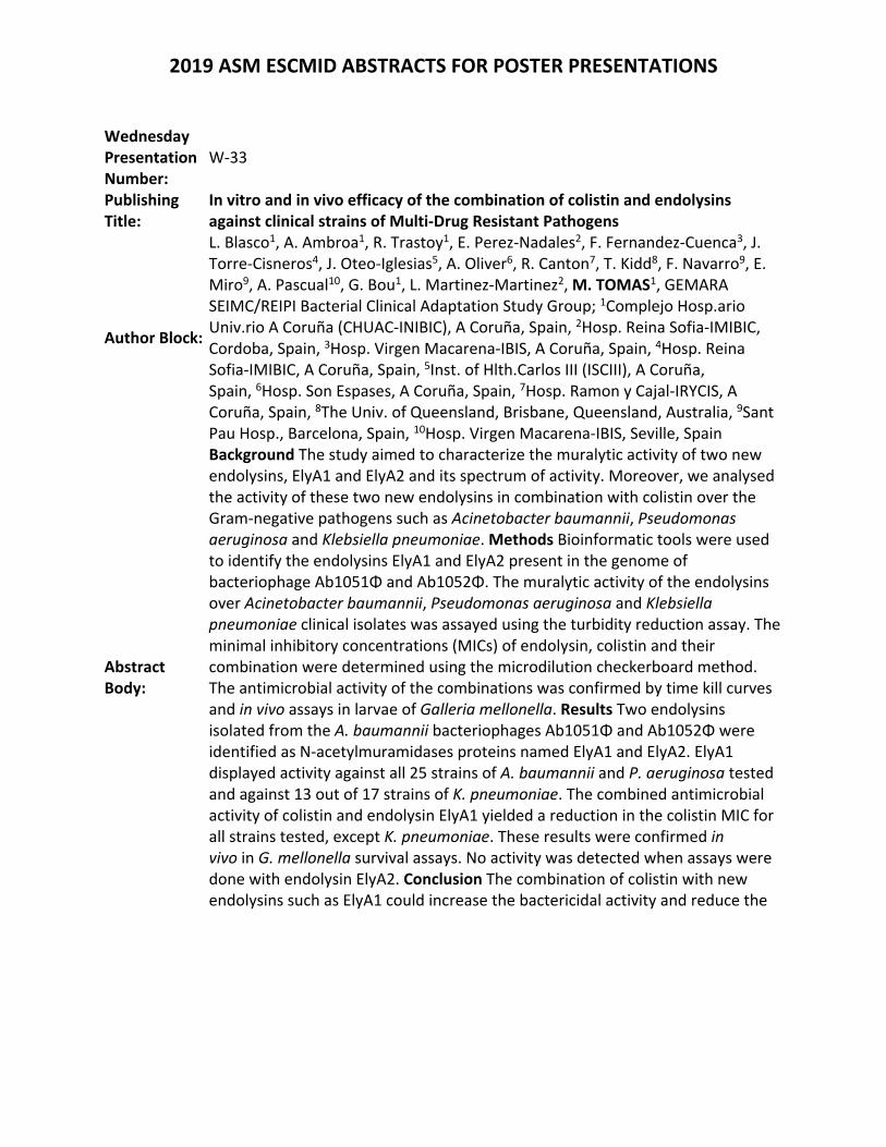
In vitro and in vivo efficacy of the combination of colistin and endolysins against clinical strains of Multi‐Drug Resistant Pathogens
Author Block:
L. Blasco1, A. Ambroa1, R. Trastoy1, E. Perez‐Nadales2, F. Fernandez‐Cuenca3, J. Torre‐Cisneros4, J. Oteo‐Iglesias5, A. Oliver6, R. Canton7, T. Kidd8, F. Navarro9, E. Miro9, A. Pascual10, G. Bou1, L. Martinez‐Martinez2, M. TOMAS1, GEMARA SEIMC/REIPI Bacterial Clinical Adaptation Study Group; 1Complejo Hosp.ario Univ.rio A Coruña (CHUAC‐INIBIC), A Coruña, Spain, 2Hosp. Reina Sofia‐IMIBIC, Cordoba, Spain, 3Hosp. Virgen Macarena‐IBIS, A Coruña, Spain, 4Hosp. Reina Sofia‐IMIBIC, A Coruña, Spain, 5Inst. of Hlth.Carlos III (ISCIII), A Coruña, Spain, 6Hosp. Son Espases, A Coruña, Spain, 7Hosp. Ramon y Cajal‐IRYCIS, A Coruña, Spain, 8The Univ. of Queensland, Brisbane, Queensland, Australia, 9Sant Pau Hosp., Barcelona, Spain, 10Hosp. Virgen Macarena‐IBIS, Seville, Spain
Abstract Body:
Background The study aimed to characterize the muralytic activity of two new endolysins, ElyA1 and ElyA2 and its spectrum of activity. Moreover, we analysed the activity of these two new endolysins in combination with colistin over the Gram‐negative pathogens such as Acinetobacter baumannii, Pseudomonas aeruginosa and Klebsiella pneumoniae. Methods Bioinformatic tools were used to identify the endolysins ElyA1 and ElyA2 present in the genome of bacteriophage Ab1051Φ and Ab1052Φ. The muralytic activity of the endolysins over Acinetobacter baumannii, Pseudomonas aeruginosa and Klebsiella pneumoniae clinical isolates was assayed using the turbidity reduction assay. The minimal inhibitory concentrations (MICs) of endolysin, colistin and their combination were determined using the microdilution checkerboard method. The antimicrobial activity of the combinations was confirmed by time kill curves and in vivo assays in larvae of Galleria mellonella. Results Two endolysins isolated from the A. baumannii bacteriophages Ab1051Φ and Ab1052Φ were identified as N‐acetylmuramidases proteins named ElyA1 and ElyA2. ElyA1 displayed activity against all 25 strains of A. baumannii and P. aeruginosa tested and against 13 out of 17 strains of K. pneumoniae. The combined antimicrobial activity of colistin and endolysin ElyA1 yielded a reduction in the colistin MIC for all strains tested, except K. pneumoniae. These results were confirmed in vivo in G. mellonella survival assays. No activity was detected when assays were done with endolysin ElyA2. Conclusion The combination of colistin with new endolysins such as ElyA1 could increase the bactericidal activity and reduce the
2019 ASM ESCMID ABSTRACTS FOR POSTER PRESENTATIONS
MIC of the antibiotic, thus also reducing the associated toxicity.
2019 ASM ESCMID ABSTRACTS FOR POSTER PRESENTATIONS
Wednesday Presentation Number:
W‐34
Publishing Title: A novel prioritization platform for antifungal drug discovery
Author Block: J. Wuyts1, S. Verdonck1, B. Pauwels2, W. Luyten2, B. Landuyt2, P. Van Dijck1; 1KU Leuven/VIB, Leuven, Belgium, 2KU Leuven, Leuven, Belgium
Abstract Body:
Background: The global AIDS crisis, the use of implants and the higher survival rates of immunocompromised patients has resulted in an increase in invasive fungal infections. Moreover, antifungal drug resistance has compounded these problems, resulting in a need for novel antifungal drugs. Recent efforts have revealed that the vast majority of micro‐organisms is still unable to grow under laboratory conditions, indicating a huge unexplored source of new natural products. Mining this so‐called ‘microbial dark matter’ has recently proven successful in the search for novel antibiotics, but has not yet been used in the search for novel antifungals. Even though previously uncultured micro‐organisms are a rich source of novel antimicrobials, they can still produce known compounds. Therefore, using a high‐throughput de‐replication platform to filter out known or undesirable antimicrobials would decrease costs and time consumption. Methods: In situ and standard cultivation methods were used to isolate over four thousand bacterial strains from soil samples collected in Belgium. These strains were grown in four different fermentation media and the fermentation extracts were screened for antifungal activity against Candida albicans. Fermentation extracts with antifungal activity were prioritized based on the rarity of the producing strain and the signature response profile (SRP) of the extract using impedance spectroscopy. The SRP was obtained by growing C. albicans biofilms on modified microtiter plates with gold electrodes coated on the bottom of the plates. The cells were subjected to a small alternating potential for 24h in the presence of the extract. The measurement of the resulting micro‐current through the biofilm results in an SRP based on multiple frequency components and, using a proprietary data analyzing algorithm, could be used to identify the mode‐of‐action of the compounds in the extracts. Results: We identified 361 strains, belonging to 47 species that have antifungal activity. Several strains are either novel (<97% identity score) or the activity was not reported in literature before. Class specific SRPs were obtained for known antifungal drug classes (e.g. echinocandins) and could be compared to SRPs of the extracts. Conclusions: By comparing the SRPs of unknown compounds with a library of SRPs of known compounds, we were able to obtain the antifungal mode‐of‐action. In this way, we were able to identify several unique SRPs in our fermentation extract library, hinting at compounds with a novel mode‐of‐action.
2019 ASM ESCMID ABSTRACTS FOR POSTER PRESENTATIONS
Wednesday Presentation Number:
W‐38
Publishing Title:
Antifungal Effect of the Extract of Poincianella pluviosa Stem Bark in Combination with Amphotericin B in Cryptococcus spp.
Author Block: G. M. Andriani1, L. F. A. Spoladori1, A. E. B. Morguette1, E. R. Tavares1, J. C. P. Mello2, L. M. Yamauchi1, S. F. Yamada‐Ogatta1; 1State Univ. of Londrina, Londrina, Brazil, 2State Univ. of Maringá, Maringá, Brazil
Abstract Body:
Background: Cryptococcus (neoformans and gattii) are causative agents of cryptococcosis, a life‐threatening fungal infection associated with high morbidity and mortality. The antifungal treatment of this disease is limited due to the number of available drugs, their high toxicity and the emergence of resistant isolates, all of which could be potentially overcome by combinatory chemotherapy. Natural products have attracted considerable interest mainly due to their remarkable antimicrobial activity when used alone or coupled with other compounds. This study evaluated the antifungal effect of the ethyl‐acetate extract (FAc) obtained from Poincianella pluviosa ‘sibipiruna’ in combination with amphotericin B (AmB) in planktonic and sessile cells of reference strains and clinical isolates of Cryptococcus spp. Methods: The effect of compound combinations was evaluated by the two‐dimensional checkerboard microdilution method. The nature of interactions of the compounds was analyzed by the fractional inhibitory concentration (FIC) index (FICI) which is defined as the sum of FICs values of both compounds. FIC was defined as the minimum inhibitory concentration (MIC) of each compound in combination divided by the MIC of the compound used alone. Results: For FAc and AmB combination, FICI values of 0.05, 0.03, 0.13 and 0.04 were detected for C. gattii ATCC 24065, C. neoformans ATCC 66031, C. gattii 840244 and C. neoformans CN12 respectively, indicating a synergistic effect with reduction of the MIC values of AmB 83‐fold and 41‐fold for C. gattii 840244 and C. neoformans CN12 and C. gattii ATCC 24065 and C. neoformans ATCC 66031. The inhibitory effect of the combination evaluated during the formation of biofilms resulted in significant inhibition of the metabolic activity. The treatment exhibited a synergistic effect against all the strains tested with 8‐ fold and 16‐fold sessile MIC values reduction of AmB. Conclusions: These findings highlight the antifungal activity of FAc in combination with AmB in Cryptococcus spp. These combinations may be one of the feasible ways to overcome antimicrobial resistance by reducing drug concentration and side effects.
2019 ASM ESCMID ABSTRACTS FOR POSTER PRESENTATIONS
Wednesday Presentation Number:
W‐39
Publishing Title: Self‐Assembling Peptide Nanofibers that Entrap and Kill Bacteria
Author Block: J. Payne, M. Del Borgo, K. Kulkarni, T. Izoré, A. Fulcher, A. Peleg, M‐I. Aguilar, M. Cryle; Monash Univ., Clayton, Australia
Abstract Body:
Background: We were inspired by our own bodies defense strategies to tackle the challenge of developing new treatments for combating antimicrobial resistance. We found our inspiration in the arsenal of weapons used by our innate immune systems first responders, neutrophils. This being the neutrophil extracellular trap (NET) which is deployed by the self‐sacrificing neutrophil to entangle the invading microbe in a DNA net. This DNA net is embedded with antimicrobials, which helps ensure the invader cannot escape. We created a proof of concept treatment that mimicked NETs by producing self‐assembling nanofibers that are decorated with the glycopeptide antibiotic vancomycin. Methods & Results: Our net‐like fibers form by using the propensity of lipidated tri‐β‐peptides to self‐assemble into nanofibers. To decorate these fibers with antibiotics we directly linked a β3‐peptide to vancomycin. Mixing the vancomycin linked with plain β3‐peptides in different ratios resulted in vancomycin being incorporated into fibers. These fibers had different structures that have been visualized using negative stain electron microscopy and atomic force microscopy. These distinct fibers have different antimicrobial activity against antibiotic resistant clinical strains of Staphylococcus aureus (Methicillin resistant MRSA or Vancomycin intermediate VISA strains) as measured using a micro broth dilution assay. Creating a fluorescently labelled β3‐peptide allowed us to visualize the fibers wrapping S. aureus by stimulated emission depletion microscopy. In addition, surfaces coated with these fibers also reduce the formation of S. aureus biofilms. Conclusions: Inspired by our immune system, we created self‐assembling nanofibers from β3‐peptides embedded with vancomycin. These nanofibers entrap and kill antibiotic resistant S. aureus.
2019 ASM ESCMID ABSTRACTS FOR POSTER PRESENTATIONS
Wednesday Presentation Number:
W‐41
Publishing Title: Bacteriophage Against Multi Drug Resistant Acinatobacter baumannii: Old Remedy in New Time
Author Block: N. Rathor, T. Bahadur, C. K. Thakur, V. D. Bamola, B. K. Das, R. Chaudhry; All India Inst. of Med. Sci., New Delhi, India
Abstract Body:
Background: Multidrug resistant Acinatobacter baumannii ranked the highest priority by WHO in priority pathogens list for Research & Development of new antibiotics in 2017. The growing antibiotic resistance prompted us to explore other strategies to combat the pathogen other than antibiotics. The River Ganga is considered as symbol of reverence in Indian civilization and reservoir of bacteriophages. We explored the possibility of presence of bacteriophages against multidrug resistant (MDR) A. baumannii in the river Ganga water. Methods: The water samples were collected from 5 different Ganga ghats of Kanpur (Parmat Ghat, Bhairav Ghat, Gola Ghat, Sarsaiya Ghat and Bhagwatdas Ghat) and were tested for the presence of bacteriophages against MDR A. baumannii clinical isolates using spot assay and plaque assay. The obtained bacteriophags were further tested for their lytic activity on 50 clinical isolates of MDR A. baumannii. The obtained phages were further subjected to electron microscopy for morphological characterization. Results:The lytic bacteriophages were obtained from each ghat against MDR A. baumannii. The bacteriophages showed their lytic activity on 3 MDR clinical isolates of A. baumannii out of 50 tested. Electron Microscopy revealed hexagonal heads and long tails of bacteriophages. Conclusions:The above data suggested that the obtained bacteriophages are potential lytic agents for MDR A. baumannii ,having hexagonal heads and long tails belonging to order Caudovirales. Therefore, we concluded that the obtained bacteriophages against A. baumannii possess narrow range host activity. However, may be used as potential therapeutic agent on specific host.
2019 ASM ESCMID ABSTRACTS FOR POSTER PRESENTATIONS
Wednesday Presentation Number:
W‐42
Publishing Title: Synergistic Eradication of Methicillin‐resistant Staphylococcus aureus between Staphyloxanthin Photolysis and Silver Nanoparticles
Author Block: S. Jusuf, P‐T. Dong, J. Hui, J‐X. Cheng; Boston Univ., Boston, MA
Abstract Body:
Background: The rise of antibiotic resistant bacteria e.g. methicillin‐resistant staphylococcus aureus (MRSA) has resulted in a widespread search for alternative treatments not reliant on traditional antibiotics. Silver nanoparticles (AgNPs) have long been known to exhibit strong antimicrobial activity against a wide variety of bacterial species. However, the potential application of AgNPs as an alternative has been primarily limited by its toxicity at higher concentrations in order to be effective. As such, there is a considerable interest in finding a way to improve the efficiency of AgNPs to achieve lower effective antimicrobial concentrations in antibiotic resistant strains like MRSA. Methods: Via blue light photolysis of Staphyloxanthin (STX), a carotenoid pigment in MRSA membrane, we were able to synergize STX photolysis with 10 nm silver nanoparticles, thus significantly increasing the potency of silver nanoparticles. The intake of silver nanoparticles by individual cells was quantified through transient microscopy, while a hydroxyl radical and peroxynitrite sensor (HPF) dye was used to quantify the production of reactive oxygen species. Analysis on potential mammalian cell toxicity was quantified through the use of MTT/MTS assays utilizing both HEK and CHO cell lines. Results: The application of blue light and AgNPs was found to significantly enhance the antimicrobial activity of AgNPs, with concentrations as low as 1 μg/mL resulting in the complete eradication of MRSA colonies. The underlying mechanism was unveiled as STX photolysis facilitates the intake of silver nanoparticle into MRSA and increases the susceptibility of MRSA to AgNP induced reactive oxygen species. This synergistic approach significantly reduces the working concentration of silver nanoparticle, well below the toxic threshold of mammalian cells. Conclusions: This approach has been found to be effective on stationary‐phase MRSA in both planktonic and biofilm forms, demonstrating an alternative method of treating MRSA skin infections. By iterating on the composition and surface chemistry of the AgNPs, it is possible to further improve the efficiency of AgNP treatment when combined with blue light exposure. The utilization of AgNPs over common antibiotics can therefore assist in treating and hindering the further development of antibiotic resistant MRSA strains.
2019 ASM ESCMID ABSTRACTS FOR POSTER PRESENTATIONS
Wednesday Presentation Number:
W‐45
Publishing Title: Alpha‐emitting 227Th Complexes for Targeted Cell Death of Pseudomonas aeruginosa
Author Block: L. R. Stromberg, L. M. Lilley, G. L. Wagner, J. M. Kelliher, P. K. Dighe, S. A. Kozimor, H. Mukundan; Los Alamos Natl. Lab., Los Alamos, NM
Abstract Body:
Background: Pseudomonas aeruginosa is a troublesome pathogen as it can affect nearly any tissue in humans, plants or animals. It is estimated that up to 50% of nosocomial infections are associated with the bacteria, 30% of which are multi‐drug resistant. Recently, use of radiometals such as 68Ga (half life~68 min) have emerged for diagnostics and therapeutics. Readily available radiometals with short half‐lives present unique challenges to therapeutic development. Conversely, longer‐lived alpha emitting isotopes are restricted by availability and handling requirements. Methods: The alpha emitter, Thorium‐227 (227Th, half life~18 d), was purified and titrated to pH 5.5 using ammonium acetate buffer. The solution was used to radiolabel the siderophore, Deferoxamine (DFO); or the chelator p‐SCN‐Bn‐DOTA, which was conjugated to Pseudomonas specific antibodies (Abs: for ATCC strains 33355, 33354, BAA‐47). Ab specificity was tested with ELISAs, and affinity of bacteria for DFO(Fe3+) versus radiometal analogs was assessed. To identify initial dosing parameters, 106 bacterial cells were plated with increasing concentrations of 227Th‐doped diffusion disks and inhibited growth after 24 h was evaluated. Minimum inhibitory concentrations over 24 h were assessed in broth using 96‐well plates with 105 CFU∙well‐1, and OD 600 recorded every 10 min. Results: In all strains, bacterial growth was inhibited by the treatments as compared to untreated cells. Growth was severely inhibited by the highest dose (0.4 µCi 227Th), with minor increases in OD seen over 24 h. The next highest dose (0.1 µCi) also inhibited growth, resulting in a slower exponential growth phase, compared to the negative control. Regression analysis between 1‐8 h revealed decreasing growth rates from the buffer control up to 0.4 µCi 227Th, however, little difference was noted between log phase growth at the lower concentrations of 227Th. Conclusions: The dosing experiments indicate the target dosage of 227Th for inhibiting Pseudomonas growth in culture is between 0.05‐0.4 µCi. It is anticipated that chelating 227Th with the siderophore DFO, will result in internalization of the radiometal, and therefore increased cell death, as compared to delivery with 227Th labeled Abs. Further evaluation of these complexes will examine intracellular localization of DFO into cells, as well as the effects of these potential therapeutics in primary human lung cells co‐infected with bacteria.
2019 ASM ESCMID ABSTRACTS FOR POSTER PRESENTATIONS
Wednesday Presentation Number:
W‐47
Publishing Title: Activity of Fractions Obtained from Extract of Fusarium oxysporum Isolated from Senna spectabilis Against Staphylococcus epidermidis in Biofilm and Planktonic Forms
Author Block: G. M. Righetto1, D. M. Selegato2, I. C. Gamboa2, I. L. B. C. Camargo1; 1Univ. of São Paulo, Sao Carlos, Brazil, 2Univ. Estadual Paulista "Júlio de Mesquita Filho", Araraquara, Brazil
Abstract Body:
Background: Although almost all antimicrobial tests use the planktonic forms, biofilms ‐ surface‐attached microorganism community embedded in a polymeric extracellular matrix ‐ are the natural mode of bacterial growth and can be involved in infectious disease process contributing to the antimicrobial resistance (AMR). Conventional antibiotics usually do not adequately penetrate biofilms, resulting in persistent infections that are harder and more expensive to treat. The search for compounds to reduce or eradicate biofilms seems to be particularly promising in natural compounds since there is an enormous amount of unexplored resources available. This study reports polar fractions of Fusarium oxysporum with intense antibiofilm activity. Methods: The fungal extract was prepared by extraction of the liquid media supernatant with ethyl acetate. Following, this extract was fractionated by solid phase extraction using increasingly high methanol percentages (20, 40, 60, 80, and 100%). Each fraction was tested for its antibiofilm and antimicrobial activity against S. epidermidis ATCC 35984, a well‐known biofilm‐forming strain. The antimicrobial activity was assessed determining the minimum inhibitory concentration (MIC) by microdilution method according to the CLSI recommendations. Biofilm reduction was evaluated by incubating the fractions at 512 mg/L with the pre‐formed biofilm, subjecting to crystal violet staining and reading at OD600 nm. The reduction was determined by comparing treated and non‐treated biofilm. Results: We observed antimicrobial activity with MIC = 512 mg/L of the fractions eluted in 40% and 60% and a substantial biofilm reduction was observed for fractions eluted in 20% and 40% of methanol ((75 ± 9)% and (81±11)%, respectively). Molecule identification was conducted by MS/MS Molecular Networking‐based dereplication. The most abundant compounds found were fusaric and dehydrofusaric acids, along with two other minor compounds ‐ 6‐oxo‐fusaric acid and 6‐oxo‐dehydrofusaric acid. Conclusions: Fusaric acid is already known to inhibit quorum sensing in Gram‐negative bacteria. The antibacterial and antibiofilm activities mechanisms of these molecules in S. epidermidis are unknown and should be investigated since it could be a treatment alternative.
2019 ASM ESCMID ABSTRACTS FOR POSTER PRESENTATIONS
Wednesday Presentation Number:
W‐48
Publishing Title: Quantitative In‐Vitro Model for the Visualization and Characterization of Bacterial Biofilms on Medical Implants
Author Block: D. Garcia1, Z. Biviji2, C. Spake1, E. Berns1, C. Born1; 1Brown Univ., Providence, RI, 2Brown Univ., PROVIDENCE, RI
Abstract Body:
Background: Surgical infections pose one of the greatest challenges for clinicians. In addition to the rise of antibiotic‐resistant bacterial strains, adherent bacterial aggregates termed biofilms colonize implant materials and form a protective matrix composed of proteins, exopolysaccharides, and extracellular DNA.1 Once developed, biofilms become largely unhindered by antibiotic therapy, and are virtually impossible to treat without invasive surgical intervention.2 Despite the problem they cause, few studies have delved into their structural characterization and clinical eradication.3 This study aims to validate a visual and quantitative in‐vitro model for the characterization of biofilms on Polyetheretherketone (PEEK) utilizing Pseudomonas aeruginosa and Staphylococcus epidermidis. Methods: P. aeruginosa and S. epidermidis were cultured overnight in tryptic soy broth at 37°C. PEEK disks were inoculated at concentrations of 2x10⁷ CFU/ml. Non‐adherent bacteria were rinsed with phosphate buffered saline and samples collected at time intervals of 4, 8, 12, 16, and 24 hours. Samples were fixed in 10% neutral buffered formalin and stained with dyes specific for biofilm macromolecules: Proteins (SYPRO Ruby), Exopolysaccharides (Concanavalin A 635 Conjugate), and extracellular DNA (TOTO‐1 Iodide). Samples were imaged with an Olympus FV‐1000 MPE Multiphoton Confocal Microscope and Hitachi 2700 Scanning Electron Microscope. Quantification of each macromolecule and changes in levels over time are analyzed via ImageJ. Results: The assay has provided the ability to visualize and quantify individual biofilm components and determine the exact progression of their dynamic formation. Data suggests that biofilm formation is a very dynamic process in which the order of formation and amount of each respective macromolecule changes from pathogen to pathogen. Conclusion: The use of both confocal laser scanning microscopy and scanning electron microscopy has enabled visualization and quantification of spatial‐temporal biofilm development and composition. This novel in‐vitro model allows the study of biofilm formation on implant materials under clinically‐relevant conditions. 1 Rodney M. Donlan, 2001 2 Hannigan DG, Pulos N, Grice EA, Samir M, 2014 3 Darouiche RO, 2004
2019 ASM ESCMID ABSTRACTS FOR POSTER PRESENTATIONS
Wednesday Presentation Number:
W‐49
Publishing Title: Silver Carboxylate‐Eluting Titanium Dioxide Polydimethyl Siloxane Coating Prevents Serratia marcescens Adherence on PEEK
Author Block: D. Garcia, A. Gilmore, R. Bain, C. Born; Brown Univ., Providence, RI
Abstract Body:
Background: Annually, hospital acquired infections affect more than two million patients in the US and cause more than 100,000 deaths.1 Moreover, infection rates in biomedical implants can be as high as 4%, and can cost over $50,000. 2 A rising contributor to these infections is the opportunistic pathogen Serratia marcescens. Infections by S. marcescens were not recognized until the later half of the 20th century3, but are now commonly identified on chronic orthopedic wounds. This study focuses on an antibiotic‐independent antimicrobial coating composed of Titanium dioxide (TiO2) and Polydimethyl siloxane (PDMS) which is doped with silver carboxylate 4, and its ability to kill and prevent S. marcescens adherence on polyetherether ketone (PEEK). By preventing biofilm formation, this study aims to prevent frequent Surgical Site Infections (SSIs).5 Methods: S. Marcescens was cultured in Trypic Soy with 0.05 mg/ml tobramycin overnight at 37°C. 2.5 mm semi‐circular rods of PEEK were coated with various ratios of TiO2:PDMS matrix doped with increasing concentrations silver carboxylate. 100% Ag and uncoated implants served as positive and negative controls. 1x10⁷ CFU /ml S. Marsescens was allowed to adhere for 4 hours, rinsed with Phosphate Buffer (PBS) and allowed to proliferate for 20 hours. PEEK implants were tagged with an anti‐LPS antibody which was conjugated to FITC and imaged at 120x via confocal microscopy. Samples were also visualized via ThermoFisher Scientific Apreo VS SEM at 5,000x. Image analysis was conducted via ImageJ. Dose‐response curves were also conducted for 3 ratios of TiO2:PDMS for 24 hours. Results: This study found that 95% TiO2‐5% PDMS doped with 10X silver carboxylate was optimal for preventing bacterial adherence. Moreover, the dose response curve for S. marcescens concluded that 1X silver concentration was sufficient for killing S. marcescens. Conclusion: This project showed the effectiveness of the TiO2/PDMS coating doped with silver carboxylate at killing and preventing adherence of S. marcescens. This represents a significant step forward in combating biofilm‐forming gram negative bacteria on medical implants. References:1. Taheri et al., 2014 2. Andersson, 2017 3. Huang, 2014 4. Tran et al., 2015. 5. Rabin, 2015
2019 ASM ESCMID ABSTRACTS FOR POSTER PRESENTATIONS
Wednesday Presentation Number:
W‐50
Publishing Title: Eradicating Recalcitrant Biofilms: Combining Tobramycin and Triclosan Kills Multidrug‐resistant Gram Negative and Gram Positive Bacteria
Author Block: A. M. A. Hunt, M. M. Maiden, E. N. Ottosen, C. M. Waters; Michigan State Univ., East Lansing, MI
Abstract Body:
Background: Biofilms are communities of microorganisms embedded in exopolymeric substances that protect them from the immune system and antimicrobial therapy. Because of the recalcitrant nature of biofilms and their high tolerance to antibiotics, patients with diseases like cystic fibrosis and diabetic foot ulcers have recurrent infections without the prospect of biofilm elimination. The search for new and more potent drugs is not only a long process but also a costly one. Therefore, the identification of combinations of antimicrobials that exhibit synergistic effects may offer advantages to traditional single drug use and represents an attractive approach to treat biofilm‐based infections. Methods: From a screen of over 6000 small molecules we identified that the aminoglycoside tobramycin and the antimicrobial triclosan act together to eliminate biofilms of multiple clinical isolates of P. aeruginosa, Burkholderia spp., Staphylococcus aureus, and Acinetobacter baumanii. High throughput assays combined with luminescence cell viability were used to test the killing effect of different concentrations of the compounds. Associations with other aminoglycosides and classes of antibiotics were evaluated, as well as their in vivo activity using a cystic fibrosis and a chronic wound murine biofilm model. Results: In vitro results demonstrated that in combination tobramycin and triclosan killed nearly 100% of all P. aeruginosa strains, including 19 clinical isolates, at the highest concentrations of 250 µg/mL for tobramycin and 100 µM for triclosan, exhibiting 100‐fold more activity than either antimicrobial alone. Moreover, even at a 5‐fold dilution 87% of the biofilm was eradicated. Synergistic effects were confirmed with other aminoglycosides and treated biofilms of Burkholderia spp, S. aureus and A. baumanii. Our results suggest that the killing effect is not a consequence of triclosan’s inhibition of fatty acid production but rather a novel activity of triclosan. In vivo testing showed a significant reduction after just 4 hours of dual treatment compared to either treatment alone in a mouse infected chronic wounds model. Conclusion: The striking antimicrobial capabilities of tobramycin‐triclosan both in vitro and in vivo is a promising approach to eliminate multidrug‐resistant Gram negative and Gram positive biofilms.
2019 ASM ESCMID ABSTRACTS FOR POSTER PRESENTATIONS
Wednesday Presentation Number:
W‐51
Publishing Title:
Amurin Peptide App2‐M1 Eradicates Stenotrophomonas Biofilms Formed on Hemodialysis Catheters in the Setting of Human Infection
Author Block: J. Oh1, M. Noto2, A. Borgmann2, M. Sika2, C. Cassino1, J. Dwyer2, R. Schuch3; 1ContraFect Corp., Yonkers, NY, 2Vanderbilt Univ. Med. Ctr., Nashville, TN, 3ContraFect Corp., Mountain Lakes, NJ
Abstract Body:
Background. S. maltophilia is a multidrug‐resistant Gram‐negative (GN) pathogen, associated with high morbidity and mortality particularly in immunocompromised patients, and is a recognized pathogen for cystic fibrosis patients in the United States. A new class of direct lytic agents (DLAs) called amurin peptides are now under development to address serious, life‐threatening infections caused by GN pathogens. Amurins exhibit notable broad‐spectrum antimicrobial activity against GN pathogens, as well as a range of other hallmark features including the eradication of biofilms formed in vitro. To extend the analysis of amurin activity, peptide App2‐M1 was tested on infected explanted hemodialysis catheters from patients with suspected catheter‐related bloodstream infections. This is the first study to assess amurin activity on biofilms formed in the setting of human disease. Materials/methods: Three infected hemodialysis catheter were removed as part of clinical care, including two catheters from one patient and third catheter from a second patient. Catheter segments were bisected and allotted into different treatment groups (n=3 segments/group) with Aap2‐M1 and buffer control. Clinically relevant concentrations of Aap2‐M1 were used (i.e., 1 and 10 µg/mL). After 4 h treatments, samples were homogenized for quantitative plating on Tryptic Soy Agar and a subset of resulting isolates (n=16) were examined by comparative DNA sequencing of the 16S rRNA gene and used to determine MIC values for Aap2‐M1. Results: Aap2‐M1 eradicated the biofilm at 1 and 10 µg/mL. These findings are consistent with in vitro observations of minimal biofilm eradication concentrations of ~1 µg/mL for various GN pathogens. Sequence analysis of recovered catheter biofilm bacteria revealed the uniform presence of organisms from the genus Stenotrophomonas with Aap2‐M1 MIC values of 2 µg/mL. Conclusions: A clinically relevant concentration of Aap2‐M1 eradicated a Stenotrophomonas biofilm inside hemodialysis catheters in the setting of a human clinical infection. These data provide the first evidence of translation of the previously reported, potent in vitro antibiofilm activity of Aap2‐MI to an ex‐vivo eradication of biofilms formed in the setting of human infection.
2019 ASM ESCMID ABSTRACTS FOR POSTER PRESENTATIONS
Wednesday Presentation Number:
W‐54
Publishing Title: Inhibition and Stimulation of Fungal Biofilms with Low‐Frequency Ultrasound and Extracorporeal Shockwave Therapy
Author Block: T. Hillock1, K. Anderson2, C. Slezak1, O. Kopp1, P. Slezak3; 1Utah Valley Univ., Orem, UT, 2Univ. of Utah Sch. of Med., Salt Lake City, UT, 3Ludwig Boltzmann Inst. für Experimentelle und Klinische Traumatologie, Vienna, Austria
Abstract Body:
Background: Fungal infections associated with biofilms are on a rise. Biofilms are aggregates of microorganisms into colonies protected by extracellular matrices, decreasing susceptibility to antibiotics and the immune system; and are the dominant form of microbial life. Amphotericin B (Amp B) is commonly used in fungal treatments but is highly toxic, leading to mortality rates of 46% and 97% in rhino‐orbital and rhino‐cerebral mucormycosis, respectively. Previous studies suggest the biofilm inhibitory effects of Low‐Frequency Ultrasound (LFUS) and Extracorporeal Shockwave Therapy (ESWT). This study focuses on the combination of LFUS or ESWT with Amp B to lower antifungal concentrations necessary to inhibit biofilms. Methods: Rhizopus oryzae biofilms were grown on plates and treated by LFUS (550 kHz, 0.43 Vpp, 1 W/cm2, 50% duty cycle, 10 minutes) alone, Amp B alone, or LFUS and Amp B combined. Other biofilms were treated with ESWT (Electromagnetic; 0.55 mJ/mm2, 3 Hz, 300 shots) alone, Amp B alone, or ESWT and Amp B. Biofilms were grown on fibrin and treatment commenced in a water bath, mimicking clinical models. Cell viability measured through absorbance. Results: Combination of Amp B (0.03 µg/mL) and LFUS increased inhibition of biofilms by 17% when compared to Amp B alone. ESWT treatment on plates completely dislodged the biofilms; therefore, an accurate reading could not be determined. Fibrin associated biofilms showed very little inhibition towards ESWT alone, but combination of Amp B and ESWT showed higher viability rates when compared to Amp B (0.5 µg/mL) alone groups. The stimulation of biofilm growth suggests a potential protection against Amp B after ESWT. Conclusions: Combination of LFUS and Amp B successfully increased inhibition rates. Combination of Amp B and ESWT suggested protection from Amp B. The stimulation could be the result of a similar or related mechanism seen in eukaryotic cell regeneration with ESWT. Shockwaves stimulate chemical signals by mechanotransduction. It is possible the host immune system is stimulated through the mechanical signals, therefore leading to inhibition of infections. Further studies are necessary and underway.
2019 ASM ESCMID ABSTRACTS FOR POSTER PRESENTATIONS
Wednesday Presentation Number:
W‐55
Publishing Title: Electrohydraulic Shockwaves as a Possible Treatment for Bacterial Biofilms
Author Block: B. Brunetti1, T. Hillock1, A. Frahm1, C. Slezak1, P. Slezak2, O. Kopp1; 1Utah Valley Univ., Orem, UT, 2Ludwig Boltzmann Inst. for Experimental and Clinical Traumatology, Wien, Austria
Abstract Body:
Background: The rise of antibiotic‐resistant bacteria is a global threat. Escherichia coli has evolved resistance and is a major concern, causing the most infections of any gram‐negative bacteria. Staphylococcus aureus, a gram‐positive species, has become highly resistant and extremely pathogenic. Strains like MRSA and VRSA have the highest rate of drug resistance and are the leading cause of chronic bacterial infections via bacterial biofilms on medical devices. Biofilms are an aggregation of microbes that excrete an extracellular matrix providing an ideal environment for gene exchange and quorum sensing. Their complexity hinders the diffusion of antimicrobials. A proposed method to prevent device‐associated infection is shockwave sterilization and therapy. A shockwave is a high‐energy wave causing a sudden change in temperature, pressure and density in the medium. This study investigates the potential disruption of bacterial biofilms by electrohydraulic shockwaves. Methods: E. coli and S. aureus biofilms were grown on polystyrene plates. Biofilms were treated with shockwaves (0.19mJ/mm2, 300 pulses, 3 Hz) in a water bath and compared with those treated with Vancomycin. Cell viability was determined through XTT/menadione absorbance and specific biofilm formation through crystal violet absorbance. Results Successful biofilm formation was obtained with E. coli and S. aureus. Electrohydraulic shockwave treatment has not been widely studied for antimicrobial purposes. This study will discuss the effects of this treatment on two bacterial species and its potential antimicrobial activity. Conclusions: Device‐associated infections are a serious threat to patients’ health. The diminishing effectiveness of antibiotics in treating and preventing infections along with evolution of mass resistance in bacteria have given rise to the term “post‐antibiotic era”. This study evaluates the effect of electrohydraulic shockwaves on bacterial biofilms. This system could be a great alternative to the use of antibiotics, and potentially life‐saving technology that could save billions of dollars.

2019 ASM ESCMID ABSTRACTS FOR POSTER PRESENTATIONS
Wednesday Presentation Number:
W‐56
Publishing Title: In vitro Activity of RP0217 Bacteriophage Cocktail in Treatment of Experimental Chronic Wound Infections Caused by Multidrug‐Resistant Klebsiella oxytoca and Escherichia coli
Author Block:
R. Prakapaitė1, R. Petraitiene1, V. Petraitis1, R. Planciuniene2, P. Gialanella3, R. Grigaleviciute4, P. Kavaliauskas1; 1Inst. of Infectious Diseases and Pathogenic Microbiol., Prienai, Lithuania, 2Lithuanian Univ. of Hlth.Sci., Kaunas, Lithuania, 3Montefiore Hosp., Albert Einstein Coll. of Med., Bronx, NY, 4Biological Res. Ctr., Lithuanian Univ. of Hlth.Sci.. Tilzes str. 18, 47181. Kaunas, Lithuania, Kaunas, Lithuania
Abstract Body:
Background: Chronic wound infections (CWI) caused by biofilm producing multi‐drug resistant (MDR) Enterobacteriales, present a major threat for both, individual patients and the broader health care system. Profound antimicrobial resistance and poor penetration of antimicrobial agents to the site of infection narrows CWI treatment options. Therefore, we aimed to study the in vitro efficacy of investigational RP0217 phage cocktail in the treatment of CWI, caused by MDR Klebsiella oxytoca and Escherichia coli. Methods: RP0217 was formulated from previously well characterized phages infecting clinical strains of K. oxytoca and E. coli. Host specificity and temperature‐depended activity was evaluated using double agar overlay method. A modified Lubbock Chronic Wound Pathogenic Biofilm (LCWPB) in vitro model was used to evaluate antimicrobial activity (log10CFU/mL) of RP0217 against in vitro wound biofilm structures, formed by MDR K. oxytoca and E. coli. Results: RP0217 containing 18 lytic phages (4.0‐8.0 × 10¹⁰ PFU/mL) was capable of infecting ESBL producing K. oxytoca (n=64) and E. coli (n=83) isolates at broad temperature ranges (30–42ºC). After a single application, RP0217 demonstrated significant reduction of K. oxytoca and E. coli burden in comparison to UC (p<0.001) in LCWPB. RP0217 achieved 4 log reductions of biofilm associated MDR K. oxytoca and E. coli in comparison to UC. RP0217 was able to disrupt K. oxytoca and E. coli biofilm integrity after a single application. Finally, RP0217 showed significant efficacy against biofilms in multi‐species LCWPB model by reducing burden of K. oxytoca and E. coli by 6 log (p=0.004) in comparison to UC. Conclusions: RP0217 has demonstrated promising in vitro antimicrobial activity against biofilm associated MDR Enterobacteriales in CWI model. Further studies are needed for better understanding of the antimicrobial potency and applications of RP0217.
2019 ASM ESCMID ABSTRACTS FOR POSTER PRESENTATIONS
Wednesday Presentation Number:
W‐58
Publishing Title: One Potentiator that Disables Resistance from MRSA, MRSE, MDR. P. aeruginosa, MDR E. Coli, and Biofilms
Author Block: C. Rice; Univ. of Oklahoma, Norman, OK
Abstract Body:
Background: Bacteria within a biofilm differ from their planktonic counterparts in several ways, but perhaps the most important difference is the use of EPS as a barrier against antimicrobial agents. Antibiotics that are effective against planktonic bacteria are nearly inert against biofilms and resistant strains, which often result in severe, chronic infections. With a dwindling arsenal of new antibiotics, existing drugs and regimens must be coupled with potentiators and re‐evaluated as combination treatments for biofilms and antibiotic resistant diseases. The ideal potentiator would be a single, dual‐function, compound that disables biofilms and combats antibiotic resistance. We have discovered such a compound: branched polyethyleneimine (BPEI). For wound infections, the anionic EPS matrix binds to and disables topical agents, such as Ag+ ions, cationic polyhexamethylene biguanide (PHMB), and cationic antibiotics. However, when EPS binds cationic BPEIs, the biofilm disperses. Additionally, because our potentiators bind to biofilm EPS and bacterial LPS, their function does not require crossing of membranes. Thus, without disrupting membranes, the potentiators they have a lower chance of eukaryotic toxicity. Methods: Checkerboard assays were used to measure the minimum inhibitory concentration (MIC) and the minimum biofilm eradication concentration (MBEC). Results: Combinations of a β‐lactam and potentiator kill MRSA, MRSE, P. aeruginosa, and E. coli. Our potentiators lower antibiotic MIC values by a factor of 100. Antibiofilm synergy is created with BPEI and oxacillin, the combination treatment completely eliminate the recalcitrant biofilms of antibiotic‐resistant bacteria by 4‐log reduction in CFUs. Conclusions: Biofilms and antimicrobial resistance create substantial technological barriers to treating chronic wound infections. We have developed a series of dual‐function potentiators that disrupt biofilms and also counteract β‐lactam resistance mechanisms in staphylococci, P. aeruginosa, and E. coli. We envision wound treatment with topical application of potentiators to disable biofilms and resistance mechanisms. This mitigates concerns about toxicity (which our data show to be low) and differences in the PK/PD of antibiotics and potentiators. The mechanism of action involves electrostatic forces that are unlikely to result in resistance phenotypes.
2019 ASM ESCMID ABSTRACTS FOR POSTER PRESENTATIONS
Wednesday Presentation Number:
W‐59
Publishing Title: Staphyloxanthin Virulence‐Targeting Phototherapy Platform for Methicillin‐Resistant Staphylococcus aureus Infections
Author Block: J. Hui1, P‐T. Dong1, L. Liang2, M. Taraknath1, J. Li1, Y. Zhan1, S. Jusuf1, C. Zong1, Q. Cui1, J‐X. Cheng1; 1Boston Univ., Boston, MA, 2Jilin Univ., Changchun, China
Abstract Body:
Background: The rapid development of antibiotic resistance increasingly challenges the successful treatment of Staphylococcus aureus infections, thus posing a great threat to the global health. Confronting the emerging post‐antibiotic era, health organizations are calling for unconventional strategies that attack new targets to overcome resistance. Methods: Transient absorption study on MRSA cells triggered our initial discovery that staphyloxanthin (STX), a carotenoid membrane‐bound pigment and a virulence factor, is subject to photolysis by blue light with its photochemistry studied via Raman spectroscopy and mass spectroscopy. Based on the photolysis physics, a high‐fluence pulsed laser was identified to achieve wide‐field, fast, efficient, and deep STX photolysis. Subsequently, detailed membrane disruption mechanisms were unveiled: 1) membrane permeability by SYTOX green, gentamycin‐Texas red; 2) membrane fluidity by DiIC 18, daptomycin‐BODIPY; 3) PBP2a detachment by immunoassay; 4) microdomain formation and membrane remodeling via molecular dynamics simulation. Growth inhibition or eradication of MRSA for laser treatment w/o conventional antibiotics were evaluated by checkerboard and time‐killing assays. STX production and resistance development for antibiotics w/o laser treatment were studied via serial passage. Treatment efficacy was evaluated on MRSA biofilm and in vivo mice skin infection models with phototoxicity evaluated on human cell lines in vitro and mice skin in vivo. Results: STX is the molecular target of photons in the entire blue range. Pulsed laser enables strikingly high STX photolysis efficiency and depth when compared to low‐level light sources. More importantly, after effective STX photolysis, cell membranes were severely disorganized and malfunctioned to defense antibiotics, as its membrane disassembly mechanisms unveiled by increased membrane fluidity, ample membrane permeability, detachment of trans‐membrane proteins, PBP2a. Consequently, increased susceptibility and inhibited resistance development were found to broad classes of antibiotics including penicillins, quinolones, tetracyclines, aminoglycosides, daptomycin, and oxazolidinones. The synergistical therapy, without phototoxicity to the host, was found effective to combat MRSA on clinically relevant models. Conclusions: This work demonstrates a STX virulence‐targeting phototherapy platform and paves a novel way to resensitize several major classes of conventional antibiotics to combat MRSA infections.
2019 ASM ESCMID ABSTRACTS FOR POSTER PRESENTATIONS
Wednesday Presentation Number:
W‐61
Publishing Title: ApmA is a Unique Aminoglycoside Modifying Enzyme Capable of Inactivating Apramycin
Author Block: E. Bordeleau1, P. J. Stogios2, K. Koteva1, A. Savchenko3, G. D. Wright1; 1McMaster Univ., Hamilton, ON, Canada, 2Univ. of Toronto, Toronto, ON, Canada, 3Univ. of Calgary, Calgary, AB, Canada
Abstract Body:
Background: Apramycin is an aminoglycoside antibiotic that has been traditionally used only in veterinary medicine. Recently, it has been proposed to be repurposed for clinical use in humans owing to its effectiveness against major drug resistant pathogens prioritized by the World Health Organization. The goal of this project is to investigate the most recently identified genetic element found to confer apramycin resistance, apmA. Methods: ApmA was produced in Escherichia coli for characterization through antimicrobial susceptibility testing, X‐ray crystallography, and in vitro enzyme kinetics. Apramycin was inactivated by purified ApmA and characterized using nuclear magnetic resonance spectroscopy. Results: ApmA is an acetyl‐CoA‐dependent aminoglycoside acetyltransferase. Unlike most other such enzymes that belong to the canonical GCN5‐superfamily, ApmA shows a high degree of similarity to the left‐handed β‐helix (LβH) acetyltransferases family of proteins. Other enzymes from this family areknown for their inactivation of the antibiotics chloramphenicol and Type A streptogramins. Crystallographic analysis of ApmA confirmed that this enzyme adopts the predicted LβH fold. Structures with various aminoglycosides identify key molecular interactions in the active site. Antimicrobial susceptibility testing and in vitro enzyme assays confirmed that ApmA N‐acetylates apramycin and other aminoglycosides that are clinically important.Conclusions: The work conducted herein has provided the first structural evidence that ApmA belongs to a family of proteins previously unknown to modify and inactivate aminoglycoside antibiotics. With apmA already mobilized on a plasmid, it is possible that this resistance element may make its way into the clinic. Understanding the structural scaffold and mechanism of action for ApmA is critical for developing apramycin as a future antibiotic for human use and circumventing this potential form of clinical resistance.
2019 ASM ESCMID ABSTRACTS FOR POSTER PRESENTATIONS
Wednesday Presentation Number:
W‐62
Publishing Title:
Omadacycline is Not a Substrate for Clinically Relevant β‐Lactamase Enzymes
Author Block: R. E. Mendes1, L. Deshpande1, M. Castanheira1, A. W. Serio2, E. S. Armstrong2, J. N. Steenbergen2, R. K. Flamm1; 1JMI Lab., North Liberty, IA, 2Paratek Pharmaceuticals, King of Prussia, PA
Abstract Body:
Background: Omadacycline was approved by the FDA in October 2018 for treating acute bacterial skin and skin structure infections and community‐acquired bacterial pneumonia in adults. We hypothesized that omadacycline, a novel aminomethylcycline class antimicrobial, is not hydrolyzed by extended‐spectrum β‐lactamases (ESBLs) class enzymes. Crude extracts from isogenic strains expressing ESBL and carbapenemases were prepared and incubated with omadacycline, and stability was evaluated via absorbance assay. Methods: 17 isogenic isolates carrying β‐lactamase genes were included. Isolates producing the following β‐lactamase enzymes were selected: ESBLs (TEM‐2, TEM‐12, SHV‐2, SHV‐5, SHV‐12, CMY‐2, CTX‐M‐14, and CTX‐M‐15) and carbapenemases (KPC‐2, KPC‐3, VIM‐1, VIM‐2, VIM‐6, NDM‐1, and OXA‐48). To confirm that the β‐lactamase enzymes were active, elevated MICs to β‐lactams were obtained. Escherichia coli ATCC 25922 was used as a negative control, and E. coli carrying tet(X), known to modify tetracycline derivatives, including omadacycline, was utilized as a positive control. Absorbance of omadacycline, ampicillin, ceftriaxone, and imipenem were measured in an Ultrospec™ 3300 pro UV/visible spectrophotometer using the most appropriate wavelength for each antimicrobial. The potential presence of hydrolysis was considered when the initial and final absorbance for any given drug over time (delta absorbance; ΔA) was greater than the negative controls. Results: The ΔA for the β‐lactam agents in the presence of ESBL and carbapenemase enzymes ranged from 0.24 up to 0.54, respectively, indicating hydrolysis. The ΔA for these same agents after exposure to the negative control E. coli ATCC 25922 strain crude extract was minimal (0.0262‐0.0499). Omadacycline had ΔA values ≤0.3 after incubation with all 17 β‐lactamase enzymes, indicating that it is not a substrate for hydrolysis; the absorbance values were similar to negative controls. However, when omadacycline was exposed to the positive control enzyme Tet(X), a ΔA of 0.74 was observed over time, indicating molecule modification. Conclusions: Omadacycline is not a substrate of β‐lactamase enzymes, including clinically important ESBLs and carbapenemases, which are the primary β‐lactam resistance mechanisms in Enterobacteriaceae. The hydrolysis of control β‐lactam agents confirmed the presence of β‐lactamase enzymes in each crude extract, while the alteration of omadacycline by Tet(X) confirmed the experiment’s ability to detect modifications in this molecule.
2019 ASM ESCMID ABSTRACTS FOR POSTER PRESENTATIONS
Wednesday Presentation Number:
W‐65
Publishing Title: Mutations in pmrB Confer Cross‐resistance between the LptD Inhibitor POL7080 and Colistin in Pseudomonas aeruginosa
Author Block: K. P. Romano1, T. Warrier1, B. E. Poulsen1, P. H. Nguyen1, A. R. Loftis2, A. Saebi2, B. L. Pentelute2, D. T. Hung1; 1Massachusetts Gen. Hosp., Boston, MA, 2Massachusetts Inst. of Technology, Cambridge, MA
Abstract Body:
Background Pseudomonas aeruginosa poses a major threat to human health due to limited treatment options and its ability to become resistant to antibiotics. POL7080 is a first‐in‐class antibiotic currently in late stage clinical trials with species‐specific activity against P. aeruginosa. POL7080 and its analogues were derived after extensive chemical modification of the cationic antimicrobial peptide protegrin‐1, and subsequently shown to target the lipopolysaccharide transport protein LptD. Methods To determine the resistance mechanisms of POL7080, we performed whole genome sequencing of spontaneously resistant P. aeruginosa PA14 mutants selected by plating mid‐log culture on Lysogeny Broth agar containing the related LptD inhibitor POL7001. Allelic complementation studies confirmed that mutations in the common gene pmrB accounted for the observed resistance phenotypes. To elucidate the mechanism of resistance, we performed expression analysis using RNAseq and qRT‐PCR, combined with confocal microscopy with a fluorescent POL7080 analogue. Results All resistant clones contained mutations in the common gene pmrB, and mutated alleles were dominant over the wildtype allele. Because several mutants contained PmrB substitutions at the same positions reported previously from colistin‐resistant clinical isolates, we measured colistin activity against all mutants and found cross‐resistance between POL7080 and colistin, including the colistin‐resistant clinical strain PA1571. Expression analysis revealed that a signature transcriptional program, involving key LPS modification genes, is induced by LptD inhibitor exposure, and constitutively upregulated in the most resistant mutant isolated in our study. Confocal microscopy of a fluorescent POL7080 analog confirmed significantly reduced antibiotic uptake in this resistant mutant relative to wildtype. Conclusions Here we report a series of pmrB mutations that confer high‐level resistance to POL7080 and moderate cross‐resistance to colistin. Expression analysis and confocal microscopy data support a resistance mechanism in which pmrB mutations drive LPS modification with L‐Ara4N, aligning with the known resistance mechanisms to polymyxins. Altogether, our findings suggest that pre‐existing colistin resistance may limit the utility of POL7080 in a subset

2019 ASM ESCMID ABSTRACTS FOR POSTER PRESENTATIONS
of highly resistant cases of P. aeruginosa, and that if successfully developed, POL7080 exposure could inadvertently drive cross‐resistance to colistin and other polymyxins.
2019 ASM ESCMID ABSTRACTS FOR POSTER PRESENTATIONS
Wednesday Presentation Number:
W‐66
Publishing Title: Rapid Detection of Antibiotic Resistance Genes by T2 Magnetic Resonance Author Block: D. Gamero, C. Steele, J. L. Snyder, T. J. Lowery; T2 Biosystems, Lexington, MA
Abstract Body:
Background: The T2Resistance Panel is based on T2 magnetic resonance (T2MR), a rapid and automatable technology currently employed in the FDA‐cleared T2Bacteria and T2Candida Panels, with a clinically demonstrated time to result of 3‐5 hours. The panel is capable of detecting antibiotic resistance genes directly from blood, without prior culture, including methicillin resistance genes mecA/C; vancomycin resistance genes vanA/B; carbapenemases blaKPC, blaOXA‐48, blaNDM, blaVIM, and blaIMP; AmpC β‐lactamases blaCMY‐2 and blaDHA; and extended spectrum β‐lactamases blaCTX‐M‐
14 and blaCTX‐M‐15. Here we discuss the development of the assay and the high analytical sensitivity for resistance genes found antibiotic resistant isolates in whole blood. Methods: The panel is composed of a multiplex DNA amplification reaction that targets 13 high threat resistance genes and probe‐conjugated superparamagnetic particles that cluster in the presence of their target. The sample processing, amplification, and detection were automated on the T2Dx Instrument. Assay performance was evaluated using EDTA human whole blood spiked with bacterial isolates obtained from the CDC/FDA Antibiotic Resistance Isolate Bank, ATCC, and JMI. These isolates represent a broad range of resistance gene variants. The assay was optimized for high sensitivity and robust performance on the T2Dx Instrument. Analytical sensitivity, or the limit of detection, was screened by testing concentrations < 10 CFU/mL for all targets. Cross‐reactivity was tested with concentrations of 1000 CFU/mL. Common endogenous and exogenous interfering substances relevant to the clinical setting were tested with all resistance gene targets. Results: Preliminary analytical sensitivity was demonstrated to be equal to or less than 10 CFU/mL for all resistance genes, and some targets had a limit of detection as low as 3 CFU/mL. No cross‐reactivity was observed between targets. Interfering substances did not lead to false negative results. Conclusion: The T2Resistance Panel is a rapid and highly sensitive method for the direct‐from‐blood detection of resistance genes. Among the benefits of a culture independent test for antibiotic resistance gene detection is the ability to rapidly respond to outbreaks of antibiotic resistance organisms or the pre‐screening of patients for clinical trial enrollment for novel antibiotics.
2019 ASM ESCMID ABSTRACTS FOR POSTER PRESENTATIONS
Wednesday Presentation Number:
W‐67
Publishing Title: Contribution of CrrB to High Level Polymyxin Resistance and Virulence in Klebsiella pneumoniae
Author Block: T. McConville1, N. Macesic1, M. Annavajhala1, F. Rozenberg1, S. Trent2, C. Herrera2, A‐C. Uhlemann1; 1Columbia Univ. Med. Ctr., New York, NY, 2Univ. of Georgia, Athens, GA
Abstract Body:
Background: Emerging polymyxin resistance (PR) threatens the cornerstone of treatment for carbapenem resistant Enterobacteriaceae (CRE) infections. MgrB disruption is seen most commonly, but missense mutations in crrB are an alternative pathway to PR, often with higher MICs than mgrB mutants. The mechanism by which crrB mutations induce PR and what other phenotypic effects they have, including changes in virulence, remains incompletely understood. Methods: We created gene knockouts (KO) and single nucleotide polymorphism (SNP) insertions in CRE Klebsiella pneumoniae (CRKP) using CRISPR/Cas9. Three crrB SNPs were chosen based on whole genome sequencing of PRKP clinical isolates and were inserted into a susceptible CRKP, NR5452, to create isogenic mutants. A mgrB KO was created as a positive control. We determined susceptibility by broth‐microdilution, characterized lipid A modifications via thin layer chromatography, and gene expression by RNA‐Seq of RNA extracts. We measured virulence in a Galleria mellonella model by injecting each isolate into 60 larvae and monitoring for death over 72 hours. Significance was calculated via a log rank test and Chi‐square analysis. Results: Polymyxin MIC increased from 1 to 256 μg/ml after insertion of the three crrB SNPs and from 1 to 32 μg/ml after mgrB KO. All mutants showed addition of L‐Ara4N to lipid A. Differences in virulence were seen between the mutants and WT in the G. mellonella model. CrrB94 had the highest virulence (killing 53/63), while all other mutants, except crrB87, had significantly higher virulence than the WT, NR5452 (1/60). RNA‐seq analysis showed upregulation of the pmrHFIJKLM operon and a magnesium transport system in ΔmgrB. One of the crrB mutants did not show this pattern, but displayed upregulation of glycerol‐3‐phosphate dehydrogenase, used in glycerol and lipid metabolism. Conclusions: SNPs in crrB induced MICs eight times higher than the ΔmgrB mutant. Despite similar addition of L‐Ara4N to lipid A, different transcriptome and virulence profiles were observed between the crrB SNPs and the ΔmgrB mutant. This suggests that while SNPs in crrB are sufficient to induce PR, they have wide‐ranging phenotypic effects, including means to increase bacterial virulence. More work is needed to elucidate how other mutations in PRKP isolates affect bacterial phenotype.
2019 ASM ESCMID ABSTRACTS FOR POSTER PRESENTATIONS
Wednesday Presentation Number:
W‐68
Publishing Title:
Comprehensive Penicillin‐Binding Protein (PBP) Occupancy Patterns of 23 Drugs in Klebsiella pneumoniae
Author Block: D. Sutaria, N. Shah, A. Ropy, B. Moya, Y. Jiao, X. Tao, J. Zhou, Y. Lang, E. Shin, A. Louie, G. Drusano, J. Bulitta; Univ. of Florida, Orlando, FL
Abstract Body:
Background: Carbapenem‐resistant (CR) K. pneumoniae (KP) isolates are recognized as an urgent global threat to human health. The PBPs are involved in the bacterial cell wall synthesis and remodeling and present the primary targets of all beta‐lactam antibiotics. While PBPs are highly conserved enzymes, there is no fully comprehensive PBP binding dataset for beta‐lactams in KP. To considerably enhance our prior publication on PBP binding for 13 drugs in KP, we generated binding data for 10 additional beta‐lactams and beta‐lactamase inhibitors (i.e. 23 compounds in total). Methods: The PBP binding studies were carried out in KP strain ATCC 43816. Membrane fractions containing PBPs were isolated and binding reactions performed for beta‐lactams at concentrations ranging from 0.0075 to 256 mg/L. Membranes were subsequently labelled with BOCILLIN FL before binding affinities were determined. The IC50s were reported as the lowest beta‐lactam concentration that half‐maximally inhibited the BOCILLIN FL intensity. Principal component analysis (PCA) was performed on log transformed IC50 data to group the 23 compounds according to their PBP occupancy patterns Results: In the PCA, 77% of the variance was explained by the first two vectors. Compounds broadly grouped into three general clusters (plus cefoxitin [FOX]). The first cluster contained strong PBP3 binders. In the second cluster compounds primarily targeted PBPs 2 and 3 or PBPs 2 and 4. The third cluster contained compounds targeting PBP2 or PBPs 2 and 4, but not PBP3. Conclusions: This study provides the first comprehensive PBP binding data for 23 beta‐lactams and beta‐lactamase inhibitors in KP. Insights on these preferred PBP targets will enable us to design and rationally optimize innovative double beta‐lactam and beta‐lactam / beta‐lactamase inhibitor combinations. Acknowledgements: This work was funded by grants R01 AI136803 and R01 AI130185 from NIH/NIAID.
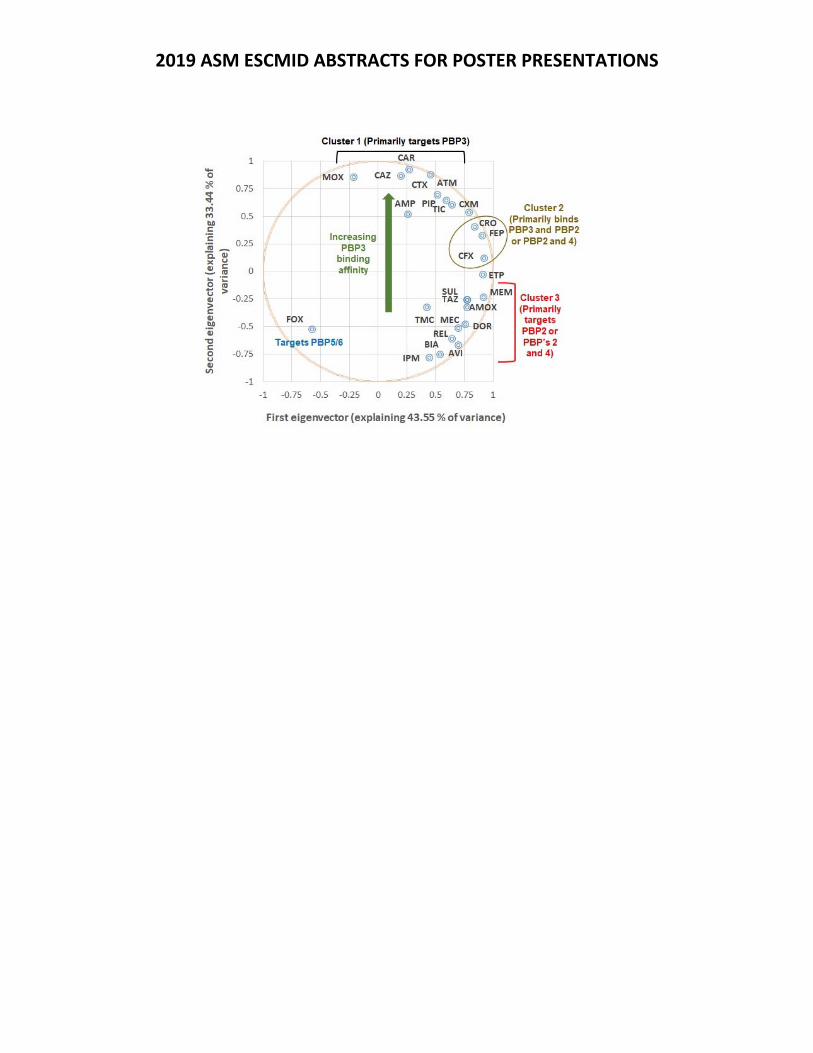
2019 ASM ESCMID ABSTRACTS FOR POSTER PRESENTATIONS
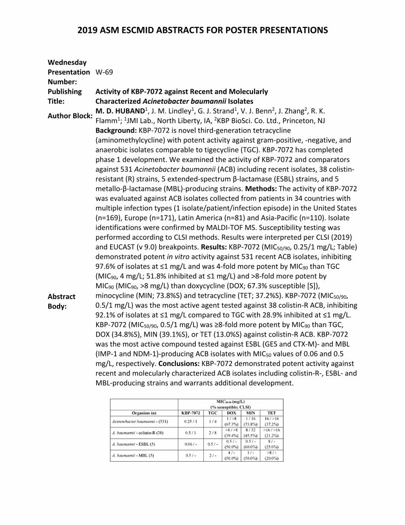
2019 ASM ESCMID ABSTRACTS FOR POSTER PRESENTATIONS
Wednesday Presentation Number:
W‐69
Publishing Title:
Activity of KBP‐7072 against Recent and Molecularly Characterized Acinetobacter baumannii Isolates
Author Block: M. D. HUBAND1, J. M. Lindley1, G. J. Strand1, V. J. Benn2, J. Zhang2, R. K. Flamm1; 1JMI Lab., North Liberty, IA, 2KBP BioSci. Co. Ltd., Princeton, NJ
Abstract Body:
Background: KBP‐7072 is novel third‐generation tetracycline (aminomethylcycline) with potent activity against gram‐positive, ‐negative, and anaerobic isolates comparable to tigecycline (TGC). KBP‐7072 has completed phase 1 development. We examined the activity of KBP‐7072 and comparators against 531 Acinetobacter baumannii (ACB) including recent isolates, 38 colistin‐resistant (R) strains, 5 extended‐spectrum β‐lactamase (ESBL) strains, and 5 metallo‐β‐lactamase (MBL)‐producing strains. Methods: The activity of KBP‐7072 was evaluated against ACB isolates collected from patients in 34 countries with multiple infection types (1 isolate/patient/infection episode) in the United States (n=169), Europe (n=171), Latin America (n=81) and Asia‐Pacific (n=110). Isolate identifications were confirmed by MALDI‐TOF MS. Susceptibility testing was performed according to CLSI methods. Results were interpreted per CLSI (2019) and EUCAST (v 9.0) breakpoints. Results: KBP‐7072 (MIC50/90, 0.25/1 mg/L; Table) demonstrated potent in vitro activity against 531 recent ACB isolates, inhibiting 97.6% of isolates at ≤1 mg/L and was 4‐fold more potent by MIC90 than TGC (MIC90, 4 mg/L; 51.8% inhibited at ≤1 mg/L) and >8‐fold more potent by MIC90 (MIC90, >8 mg/L) than doxycycline (DOX; 67.3% susceptible [S]), minocycline (MIN; 73.8%S) and tetracycline (TET; 37.2%S). KBP‐7072 (MIC50/90, 0.5/1 mg/L) was the most active agent tested against 38 colistin‐R ACB, inhibiting 92.1% of isolates at ≤1 mg/L compared to TGC with 28.9% inhibited at ≤1 mg/L. KBP‐7072 (MIC50/90, 0.5/1 mg/L) was ≥8‐fold more potent by MIC90 than TGC, DOX (34.8%S), MIN (39.1%S), or TET (13.0%S) against colistin‐R ACB. KBP‐7072 was the most active compound tested against ESBL (GES and CTX‐M)‐ and MBL (IMP‐1 and NDM‐1)‐producing ACB isolates with MIC50 values of 0.06 and 0.5 mg/L, respectively. Conclusions: KBP‐7072 demonstrated potent activity against recent and molecularly characterized ACB isolates including colistin‐R‐, ESBL‐ and MBL‐producing strains and warrants additional development.
2019 ASM ESCMID ABSTRACTS FOR POSTER PRESENTATIONS
Wednesday Presentation Number:
W‐70
Publishing Title:
Characterizing Outer Membrane (OM) Permeability and Morphological Changes for Beta‐lactam Antibiotics against Acinetobacter baumannii (Ab)
Author Block:
X. Tao1, N. Shah1, Y. Lang1, J. Zhou1, D. Sutaria1, A. Ropy1, B. Moya1, Y. Jiao1, E. Shin1, A. Louie1, G. Drusano1, H. P. Schweizer2, R. A. Bonomo3, R. E. Lee4, J. B. Bulitta1; 1Univ. of Florida, Orlando, FL, 2Univ. of Florida, Gainesville, FL, 3CASE Western Reserve Univ., Cleveland, FL, 4St Jude Children’s Res. Hosp., Memphis, TN
Abstract Body:
Background: Beta‐lactam antibiotics present are important to combat Ab infections. However, their OM permeability and subsequent morphological changes in response to inactivation of different PBPs remain unknown. Here we sought to develop a novel OM permeability assay that can characterize the rate of OM penetration and to assess the morphological changes of Ab in response to different clinical relevant beta‐lactams. Material/methods: ATCC strain BAA‐2801 at 107.7 CFU/mL was washed six times in PBS. Half of these bacteria were lysed; the other half as intact bacteria. Cell‐free supernatant were obtained at 3, 15, 30, 60 and 120 min after the last wash to determine release of β‐lactamase. The time‐course of extracellular β‐lactam concentrations was determined over up to 120 min in control, lysed and intact bacteria via LC‐MS/MS. Bacterial morphology changes after 3 h of incubation with the respective beta‐lactam were observed using a Zeiss LSM 700 confocal microscope. Images were obtained with BacLight live/dead staining. Results: In lysed bacteria, the hydrolysis was rapid for all drugs. For meropenem and sulbactam, there was no extracellular β‐lactamase activity. Therefore the decline of β‐lactam concentrations in intact bacteria indicated the OM permeability. For others, the extracellular β‐lactamase activity increased linearly when bacteria were incubated in PBS. We compared the simulated extracellular β‐lactamase activity (bold curve) with the observed degradation profile in intact bacteria. When two profiles overlapped, this indicated extremely slow OM permeability. Sulbactam, aztreonam and ceftazidime which predominantly bind PBPs 1a and 3 resulted in filamentous phenotype. In contrast, meropenem (which binding PBP2 and other PBPs) resulted in spherical shaped bacteria. Conclusions: The proposed OM permeability assay for Ab successfully accounted for a time‐dependent release of β‐lactamases and can estimate the OM permeability more precisely.
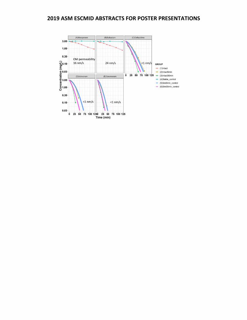
2019 ASM ESCMID ABSTRACTS FOR POSTER PRESENTATIONS
2019 ASM ESCMID ABSTRACTS FOR POSTER PRESENTATIONS
Wednesday Presentation Number:
W‐71
Publishing Title: Bacterial DNA Recombination Regulates the Evolution of Resistance to β‐Lactam Antibiotics
Author Block: L. Zhang1, Q. Su1, N. Cokcetin1, A. Bottomley1, A. Robinson2, A. v. Oijen2, E. Harry1, D. Jin1; 1Univ. of Technology Sydney, Ultimo, Australia, 2Univ. of Wollongong, Wollongong, Australia
Abstract Body:
Antibiotic‐induced bacterial resistance is rising to dangerously high levels in all parts of the world. Many resistance mechanisms have been identified, including mutations that decrease the binding of the drug to its target, modifying antibiotics to resist its action and increased expression of efflux pumps. Recent studies indicate that recombination plays a critical role in DNA repair and genome maintenance that constitutes an important adaptive mechanism of resistance in bacteria. It is well‐known that for fluoroquinolones, evolution of resistance requires an activation of recombinase RecA involved in recombinational DNA repair. However, the importance of bacterial DNA recombination in the emergence of resistance in response to other classes of antibiotics is not understood. Here, contrary to the findings of fluoroquinolones, we found that bacterial DNA recombination can remarkably regulate the evolution of resistance to β‐lactam antibiotics. A single exposure of ampicillin at 10x MIC can kill 99.99% of E. coli. cells, but induces the formation of stable resistance in recA mutant cells after 8 hours exposure. Interestingly, this resistance is associated with increased expression of β‐lactamases. RecA also participates in the classical SOS response pathway. However, investigation morphological changes of mutants involved in the SOS response using super‐resolution microscopy showed that the role of recA in the emergence of resistance was unlikely due to its function in the SOS response. To further understand how recA regulates evolution of resistance, we explored bacterial whole genome sequencing of resistant recA mutant isolates. Results reveal that although the treatment of ampicillin did not induce expected mutations in the β‐lactamases‐associated enzymes genes, interestingly, it induced three single‐base substitution mutations in the pinR gene. PinR is encoded on the cryptic prophage rac, and protein structure prediction found that it shares structural features with serine‐specific active recombinases. ddPCR further confirmed that the higher expression of β‐lactamases was resulted from gene ampC amplification. Thus, this data highlights the importance of DNA recombination in the evolution of antibiotic resistance, and suggests that bacterial evolution of resistance is not mediated by one single recombinase but a novel role for phage‐encoded recombinases as a survival mechanism in response to β‐lactam exposure. Using this knowledge can provide us with new strategies to delay the evolution of resistance to β‐lactams.
2019 ASM ESCMID ABSTRACTS FOR POSTER PRESENTATIONS
Wednesday Presentation Number:
W‐74
Publishing Title: Effect of Polymyxin B alone and in Combination with Ceftazidime/Avibactam plus Aztreonam in the Treatment of Experimental NDM‐1 Klebsiella pneumoniae Pneumonia in Persistently Neutropenic Rabbits
Author Block: V. Petraitis1, R. Petraitiene1, P. Kavaliauskas1, E. Naing1, A. Garcia1, T. Aung1, N. M. Smith2, B. T. Tsuji2, T. J. Walsh1; 1Weill Cornell Med. of Cornell Univ., New York, NY, 2Univ. at Buffalo, State Univ. of New York, Buffalo, NY
Abstract Body:
Background: New Delhi metallo‐β‐lactamase‐1 (NDM‐1) producing Enterobacteriaciae are emerging worldwide as an urgent public health threats. NDM‐1 is able to hydrolyze virtually all β‐lactam antimicrobial agents, including carbapenems, leaving polymyxin B (PMB) as the last‐line option against NDM‐1 producing organisms. Due to rapid acquisition of resistance and dose‐limiting toxicity of PMB, there is an urgent need to develop novel dosing strategies and therapeutics. The objective of this study was to evaluate PMB alone and in combination with ceftazidime/avibactam (CZA) plus aztreonam (ATM) in treatment of experimental pneumonia caused by NDM‐1 Klebsiella pneumoniae in persistently neutropenic rabbits. Material/methods: Female NZW rabbits weighing 2.6‐3.5 kg were used in the study. A well‐characterized K. pneumoniae isolate (blaNDM‐1, blaCTX‐M‐15) was used to establish pneumonia by direct endotracheal inoculation. Four groups were studied: PMB alone, CZA+ATM, CZA+ATM+PMB, and untreated controls (UC). Treatment started 8h post‐inoculation. PMB was initiated with a loading‐dose of 3.5 mg/kg and continued at 1.5 mg/kg IV Q12h. CZA and ATM were administered at 120 mg/kg IV Q8h. Treatment was continued for 7 days in surviving animals. Pulmonary bacterial burden, lung weight, and survival were evaluated for efficacy. Residual organisms at end of study (EOS), were investigated for emergence of resistance as measured by mutation frequency.Results: There was significant reduction in residual bacterial burden and significantly prolonged survival in rabbits treated with CZA+ATM and CZA+ATM+PMB in comparison with PMB and UC (p≤0.01). CZA+ATM+PMB‐treated rabbits also demonstrated significantly lower lung weights vs PMB and UC (p≤0.05). K. pneumoniae recovered at EOS developed resistance to PMB (MIC 4‐128 µg/mL) only in the PMB monotherapy group. Conclusions: This study demonstrates that CZA+ATM and CZA+ATM+PMB are highly active in treatment of experimental CTX‐M‐15‐, NDM‐1‐co‐producing K. pneumoniae pneumonia in persistently neutropenic rabbits. Novel combination dosing strategies may suppress emergence of resistance to PMB.
2019 ASM ESCMID ABSTRACTS FOR POSTER PRESENTATIONS
Wednesday Presentation Number:
W‐76
Publishing Title: Silver‐Carboxylate Eluting Hybrid Matrix Coated Sutures as Preventatives of Surgical Site Infections by Cutibacterium acnes
Author Block: D. Garcia, T. Li, S. Allu, C. Born; Diane N. Weiss Ctr. for Orthopaedic Trauma Res., Providence, RI
Abstract Body:
Background: Post‐operative Cutibacterium acnes (formerly Propionibacterium acnes) surgical site infections (SSIs) have been increasing1. These SSIs can be attributed to the inability to sterilize the pilosebaceous pores where C. acnes resides, and the development of resistance to skin preps, such as Chlorhexidine and Betadine2. Infiltration of C. acnes into the surgical site via the initial incision or suturing technique utilized during surgery3 may lead to biofilm formation and SSIs. SSIs are correlated with longer hospital stays, and revision surgeries, which hinder and disrupt wound healing4. Due to the high use of sutures for transcutaneous fixation, this study evaluated the viability of a silver carboxylate‐eluting titanium dioxide (TiO2)/Polydimethyl siloxane (PDMS) antimicrobial coating on Ethicon Fiberwire, one of the most common high‐tensile sutures in orthopaedics, to inhibit the adherence of, and kill C. acnes. Methods: Kirby Bauer assay was performed on 2 cm sections of Ethicon Fiberwire on a 107 CFU/mL lawn of C. acnes and different ratiometric formulations of the vehicle, and silver carboxylate concentrations. Antimicrobial‐coated suture, Ethicon Coated Vicryl, 100% Ag, and Chloraprep served as positive controls, while vehicle‐only served as negative control. Observations were made at 48, 72, and 96 hours. Zone of Inhibition measurements were quantified with ImageJ. 24hr Dose‐response curves were also generated to determine optimal inhibitory concentration. Results: The silver carboxylate coated sutures were effective at inhibiting C. acnes growth, even at the lowest concentration tested over the course of 96 hours. The commercially available antimicrobial suture, Ethicon Coated Vicryl, provided no protection over the same time. Lastly, the Chloraprep dipped Fiberwire sutures created the largest zone of inhibition. Conclusion: Although Chloraprep was effective at inhibiting C. acnes, the inability of the skin‐prep to penetrate the pilosebaceous glands makes it ineffective at preventing C. acnes colonization. We suggest that the use of silver‐carboxylate coated sutures is an effective alternative to commercially available anti‐microbial sutures in terms of preventing SSIs. References: 1. Portillo, 2013 2. Nakase, 2018 3. Yamakado, 2018 4. Whitehouse, 2002
2019 ASM ESCMID ABSTRACTS FOR POSTER PRESENTATIONS
Wednesday Presentation Number:
W‐77
Publishing Title: Impact of Colistin Exposure on Clinical Outcomes of Critically Ill Patients infected with Carbapenem‐Resistant Pathogens
Author Block:
V. Rognås1, M. O. Karlsson1, L. Leibovici2, Y. Carmeli3, G. L. Daikos4, E. Durante‐Mangoni5, M. Paul6, L. E. Friberg1; 1Uppsala Univ., Uppsala, Sweden, 2Rabin Med. Ctr., Beilinson Hosp., Petah Tikva, Israel, 3Tel Aviv Univ., Ramat‐Aviv, Israel, 4Laikon Gen. Hosp., Athens, Greece, 5Univ. of Campania ‘L Vanvitelli’, Napoli, Italy, 6Rambam Hlth.Care Campus and Technion ‐ Israel Inst. of Technology, Haifa, Israel
Abstract Body:
Background: The AIDA trial in critically ill patients infected with carbapenem resistant pathogens concluded that adding meropenem to colistin did not affect outcomes compared to colistin alone [1]. The aim of this model‐based analysis was to elucidate if colistin plasma exposure has an influence on change in SOFA score and hazard of death. Methods: The study included 406 patients infected with gram‐negative bacteria, identified to be carbapenem resistant (MIC ≥ 2 mg/L) and colistin susceptible (MIC ≤ 2 mg/L). Individual colistin exposures were predicted in all patients based on a developed population pharmacokinetic model, individual dose and plasma concentrations, and creatinine clearance (CrCL). SOFA scores were assessed at onset of disease, and days 1 (randomization), 7 and 14. A Bounded Integer (BI) modelling approach [2] was investigated to describe thetime‐course of SOFA. A parametric proportional‐hazard model was developed for the survival data. Covariates were explored using stepwise covariate model building. Results: The BI model described an underlying decrease in SOFA score during the study period for the typical patient. Significant covariates positively associated with the SOFA score were average colistin concentration during the first 24 hours (Hazard ratio (HR): 1.05 [1.01 1.08] per mg/L increase), and meropenem MIC (HR: 1.08 [1.02 1.14] per log increase in MIC). A generalized log‐logistic distribution best described the survival data. Predictors for time‐to‐death were SOFA score at randomization (HR: 1.18 [1.14 1.24] per score increase), age (HR: 1.02 [1.01 1.03] per year increase) and average colistin concentration during the first 120 hours (HR: 1.18 [1.07 1.28] per mg/L increase). Simulation‐based diagnostics demonstrated adequate performance of both models. Conclusions: The BI model was a successful modelling approach to describe the typical trends and between‐patient variability of the repeated measures of SOFA scores. For both SOFA and death, high colistin exposures were associated with worse outcomes. This finding may however be confounded by kidney function given the high correlation between colistin concentration and CrCL. References: [1] Paul M et al. Colistin alone versus colistin plus meropenem for

2019 ASM ESCMID ABSTRACTS FOR POSTER PRESENTATIONS
treatment of severe infections caused by carbapenem resistant Gram‐negative bacteria: an open‐label, randomised controlled trial. Lancet Infect Dis. 2018 [2] Wellhagen GJ et al. A bounded integer model for rating and composite scale data, AAPS J. 2019.
2019 ASM ESCMID ABSTRACTS FOR POSTER PRESENTATIONS
Wednesday Presentation Number:
W‐78
Publishing Title:
Successful Treatment of Two Patients with Candida auris Candidemia with the Investigational Agent, Oral Ibrexafungerp (formerly SCY‐078), from the CARES Study
Author Block: D. A. Angulo1, D. Juneja2, B. Tarai2; 1SCYNEXIS, Inc., Jersey City, NJ, 2Max Super Specialty Hosp., Saket, New Dehli, India
Abstract Body:
Background: Candida auris is a multidrug‐resistant fungi, associated with high mortality (up to 60%) that can spread from person‐to‐person leading to outbreaks in healthcare facilities. Ibrexafungerp (formerly SCY‐078) is a novel IV/oral glucan synthase inhibitor (triterpenoid) antifungal with activity against Candida, Aspergillus and Pneumocystis, in Phase‐3 of development. In vitro/in vivo studies indicate that ibrexafungerp has activity against Candida auris, including multidrug‐resistant strains. Based on this, an open‐label study to evaluate the efficacy, safety of Ibrexafungerp in patients with Candida auris infections (CARES) (Clinicaltrials.gov NCT03363841) was initiated. Methods: The CARES study is open for enrollment in the U.S. and India. Documented Candida auris infection is required for enrollment. Subjects receive a loading dose of oral ibrexafungerp 750 mg BID during the first 2 days and then subsequent oral ibrexafungerp 750 mg QD for up to 90 days and are followed for 6‐weeks after end of therapy. Results: We present our experience with 2 cases of candidemia due to C. auris enrolled in the CARES study. The first patient, a 54 year‐old male with history of diabetes mellitus (DM), acute ischemic stroke, pyogenic brain abscess and prolonged ICU stay. He developed aspiration pneumonia and septic shock. He we initially treated with, antibiotics and subsequently added fluconazole. C. auris was recovered from a blood culture and antifungal therapy was switched to micafungin. Clinical improvement was observed but blood cultures at day 5 remained positive and ibrexafungerp was initiated once shock resolved. Blood cultures became negative, within 48 hours, and patient continued to improve and completed 17 days of ibrexafungerp. The second patient, a 64 year‐old female with history of DM, chronic kidney disease on hemodialysis, presented with a lower respiratory tract infection and septic shock. She was started on antibiotics and showed improvement but fever persisted. C.auris was isolated from blood cultures and ibrexafungerp was initiated. Blood cultures became negative and completed 22 days of ibrexafungerp. Ibrexafungerp was well‐tolerated by these patients. Conclusions: These cases provide initial evidence of efficacy and safety of ibrexafungerp in the treatment of candidemia caused by Candida auris including patients that have failed previous therapies. Continued enrollment in the CARES study is warranted.
2019 ASM ESCMID ABSTRACTS FOR POSTER PRESENTATIONS
Wednesday Presentation Number:
W‐79
Publishing Title: Results of a Multi‐Centre Randomised Controlled Clinical Trial (RCT) Comparing Minocycline Plus Rifampicin (M+R) to Linezolid (L) Against MRSA Causing Complicated Skin and Skin Structure Infections (cSSSI)
Author Block:
E. Giamarellos‐Bourboulis1, M. O'Hare2, V. Reed2, W. Hope3, A. MacGowan4; 1Natl. and Kapodistrian Univ. of Athens, Athens, Greece, 2Micron Res., Peterborough, United Kingdom, 3Univ. of Liverpool, Liverpool, United Kingdom, 4North Bristol NHS Trust, Bristol, United Kingdom
Abstract Body:
Background: MRSA infection remains a significant healthcare burden. Most clinical trials have focused on intravenous therapies with few conducted on oral therapies except linezolid. Other oral antibacterials are used clinically for MRSA therapy but trials data is lacking. We compared a commonly used combination of M&R to L for patients with MRSA cSSSI. Methods: The primary objective was to demonstrate non‐inferiority between patients treated with M 100mg 12hrly plus R 600mg 24hrly and L 600mg 12hrly in terms of clinical cure at the Test of Cure (ToC) visit. Patients were randomised 2:1 M+R:L. Safety and microbiological eradication were also compared. The design was an open label randomised, multi‐centre study of adult patients requiring oral therapy for cSSSI due to MRSA that was identified either by RT‐PCR and/or growth on chromagar agar. No antibacterial therapy for >24h with an agent active against MRSA was allowed. Patients were assessed at baseline ‐ day 1, day 5 ± 1 and ToC day 14 ± 2. Results: 126 patients were enrolled: 81 treated with M+R, 42 L and 3 untreated. 94 patients clinically evaluable (CE), (59 M+R, 35 L) mean age 71.5 ± 15.7yr, 53.2% male, BMI 25.8 ± 5.8 Kg/m2. All had MRSA isolated, 64(68%) had skin ulcers, 13(14%) wounds and 8(8%) cellulitis. Tenderness, erythema and pain scores were graded as severe in 30‐35% and the area of infection was 55±97cm2. There were no major differences between the two groups. At ToC visit 46/59(78%) of patients receiving M+R were cured and 24/35 (69%) receiving L (P=0.34, percent difference in cure rates 9.4%, 90% CI ‐ 7.2 to 26.8). Microbiological eradication occurred in 80/85(95%), death in 22/123 (18%), CRP was >3 x upper limit of normal at ToC in 25/123(20%) and serious adverse events occurred in 26/123 (21%) with no differences between the two groups. Time of hospitalisation was 9.7 ± 17.8d. Conclusions: The patient population were older and had more risk factors for clinical failure than are present in most cSSSI trials. M+R was non‐inferior to L, in terms of clinical cure, microbiological eradication and safety for cSSSI caused by MRSA. This study was funded by a grant in the LifeSciHealth Priority of the European Commission Seventh Framework Programme (FP7) ‐ Health‐2011‐2.3.1.1. Preserving old antiiotics for the future (AIDA).
2019 ASM ESCMID ABSTRACTS FOR POSTER PRESENTATIONS
Wednesday Presentation Number:
W‐80
Publishing Title: Omadacycline Treatment of Acute Bacterial Skin and Skin Structure Infections in Intravenous Drug Users
Author Block:
C. V. Pollack, Jr1, C. A. Ohl2, S. Chitra3, M. Curran3, L. Garrity‐Ryan4, P. C. McGovern3; 1Thomas Jefferson Univ. Hosp., Philadelphia, PA, 2Wake Forest Sch. of Med., Winston‐Salem, NC, 3Paratek Pharmaceuticals, Inc., King of Prussia, PA, 4Paratek Pharmaceuticals, Inc., Boston, MA
Abstract Body:
Background. Infections in intravenous drug using (IVDU) patients are common and often challenging to treat. Omadacycline (OMC) is a novel aminomethylcycline, a tetracycline class antibiotic approved in the US as once‐daily IV and oral monotherapy for acute bacterial skin and skin structure infections (ABSSSI). Here we report microbiology, efficacy and safety in the pooled IVDU vs non‐IVDU patients from two phase 3 ABSSSI studies (OASIS‐1 and 2). Methods. In OASIS‐1, 655 patients were randomized 1:1 to OMC 100 mg IV q12h × 2 doses then 100 mg IV q24h, or to linezolid (LZD) 600 mg IV q12h. After at least 3 days’ therapy, patients could transition to oral OMC 300 mg q24h or oral LZD 600 mg q12h. In OASIS‐2, 735 patients were randomized 1:1 to OMC 450 mg orally q24h × 2 doses then 300 mg orally q24h, or LZD 600 mg orally q12h. Treatment duration was 7‐14 days in both studies. Efficacy was evaluated 48‐72 hours after first dose (Early Clinical Response [ECR], based on reduction of lesion size by 20% or more) and 7‐14 days after the last dose (Post Therapy Evaluation [PTE], based on Investigator Assessment of Clinical Response). Two‐sided 95% confidence intervals (CI) for the difference (D) in ECR and PTE clinical success rates were calculated. Results. In the combined studies there were 820 IVDU and 527 non‐IVDU patients in the modified intent‐to‐treat population. Wound infection (66%) was more frequent in IVDU patients and cellulitis/erysipelas (56%) was more frequent in non‐IVDU patients. Gram (+) aerobes (82% vs 87%) and S. aureus (66% vs 67%) were the most frequently isolated pathogens in IVDU vs non‐IVDU patients, respectively. ECR success for OMC vs LZD in IVDU patients was 87.4% vs 85.2% [D 2.2, 95% CI: ‐2.5, 7.0] and in non‐IVDU patients was 84.4% vs 81.9% [D 2.5, 95% CI: ‐4.0, 8.9]. PTE success for OMC vs LZD in IVDU patients was 80.7% vs 80.0% [D 0.6, 95% CI: ‐4.8, 6.1] and in non‐IVDU patients was 92.0% vs 85.3% [D 6.7, 95% CI: 1.3, 12.3]. Higher rates of treatment‐emergent adverse events were observed in IVDU (OMC 56.4%, LZD 46.0%) vs non‐IVDU (OMC 43.2%, LZD 34.2%) patients. Nausea and vomiting were higher in OMC‐treated IVDU patients (26.3% and 14.1%) compared to OMC‐treated non‐IVDU patients (15.4% and 7.5%). Conclusion: S. aureus is common in IVDU and non‐IVDU ABSSSI and OMC showed similar efficacy and safety in IVDU and non‐IVDU ABSSSI patients.
2019 ASM ESCMID ABSTRACTS FOR POSTER PRESENTATIONS
Thursday Presentation Number:
T‐01
Publishing Title: Pharmacodynamics of SPR206 in Murine Models of Infection Against Multi‐Drug Resistant (MDR) Gram‐Negative Bacteria
Author Block:
L. Mcentee1, A. Johnson1, N. Farrington1, A. Kirby1, T. Lister2, T. Parr2, N. Cotroneo3, S. Das1, W. Hope1; 1Antimicrobial Pharmacodynamics and Therapeutics, Liverpool, United Kingdom, 2Spero Therapeutics, Cambridge, MA, 3Spero Therapeutics, Cambridge, MD
Abstract Body:
Introduction: The rise of MDR Gram‐negative pathogens has led to the revival of old antibiotics, such as polymyxins. Dose‐limiting toxicity confounds the clinical utility of this class. Novel polymyxin derivatives with reduced toxicity and potent, broad‐spectrum activity against MDR pathoges are needed. SPR206 is a novel polymyxin derivative that demonstrates improved safety compared to polymyxin B and has comparable in vitro antibacterial activity. Methods: Dose finding, dose fractionation, and persistent effect studies were performed in neutropenic CD‐1 mouse thigh and lung models of infection. An ESBL‐producing strain, Klebsiella pneumoniae (SPT 725), was used as the challenge strain. The study endpoint was the bacterial density (CFU/g) in the thigh or lung, 26 hours post infection. SPR206 was administered 2 hours post infection, subcutaneously every 8 hours over a 24‐hour period. The pharmacokinetics were established using dosages of 2.5, 10, 25, and 75 mg/kg q8h and plasma drug concentrations were measured in the 1st and 3rd dosing intervals using LC/MS/MS. The persistent effect of SPR206 was explored in both mouse thigh and lung models of infection, using a single dose over a time course of 50 hours. Dose fractionation experiments were performed in the thigh by fractionating total daily dosages of SPR206 of 25 mg/kg and 75 mg/kg over a 36‐hour period, at q12h, q18h, and q36h regimens. Mice were serially sacrificed at regular intervals post infection; 2, 10, 22, and 36 hours, to identify the time course of the infection. Results: The MIC of SPR206 against K. pneumoniae SPT 725 was 0.125 mg/L. Stasis was acheived in the thigh using 5 mg/kg q8h. SPR206 demonstrated considerable persistent effect in the lung. Logarithmic killing continued at the highest dose of 75 mg/kg long after plasma concentrations dropped below the MIC. In the thigh, SPR206 suppressed growth up to 34 hours post infection at the highest dose of 75 mg/kg. Regrowth occured between 34 and 42 hours post infection. In the dose fractionation studies, 25 mg/kg and 75 mg/kg were not statistically different in a linear mixed effects model (p=0.1187). Conclusions: These studies suggest that AUC:MIC is the dynamically linked variable for SPR206 between drug exposure and antibacterial effect. This is consistent with other antibiotics within the polymyxin class. Further work is needed to identify the magnitude of AUC:MIC for SPR206 that can be used to plan for safe and effective regimens in humans.
2019 ASM ESCMID ABSTRACTS FOR POSTER PRESENTATIONS
Thursday Presentation Number:
T‐02
Publishing Title: Pharmacodynamics of Fosfomycin Against Staphylococcus aureus Studied in an in vitro Pharmacokinetic Model
Author Block: A. Noel, M. Attwood, K. Bowker, A. MacGowan, BS48 4AL; North Bristol NHS Trust, Bristol, United Kingdom
Abstract Body:
Background: Fosfomycin (fos) is undergoing re‐evaluation as a therapeutic agent for multi‐drug resistant Gram‐negative (G‐) and Gram‐positive (G+) bacterial infection. The pharmacodynamics (PD) of fos have recently described for G‐ pathogens in in vivo and in vitro pharmacokinetic (PK) ‐ PD models. Limited information is available on fos PD for G+ pathogens such as Staphylococcus aureus (SA). Here we describe the dominant pharmacodynamic index (PDI) for fos against SA and its size for reduction of bacterial load and changes in population profiles. Methods: A single compartment dilutional in vitro PK model was used to provide fos exposures against 5 strains of SA (fos MICs, 1 (2 strains), 4, 8 or 16mg/L). The fos half‐life modelled was 2.5h. Fos was administered by continuous infusion, dose fractionation (once, twice or three times per 24h) and dose ranging for fos administered three times per 24h. Cmax/MICs from 0 to 120; AUC/MICs from 0 to 750 and T>MIC 0 to 100% were simulated. The primary end point was changed in bacterial load after 24h (d24) fos exposure but changes in SA population profiles were also determined. Results: Log AUC/MIC R2 0.71 and log Cmax/MIC R2 0.69 were related to d24. T>MIC was not related to d24. Cmax/MIC for a 24hr static, ‐1 log drop ‐2 log drop were 3.1±1.7, 4.6±2.4 and 6.2±3.8 respectively. AUC/MIC for a 24h static, ‐1 log drop, and ‐2 log drop were 37.1±18.2, 55.0±27.0 and 79.6±43.2. Emergence of resistance as measured by growth on MICx4 or x8 recovery media was best related to AUC/MIC (R2 0.4860). The AUC/MIC to maximally amplify resistant strains was 16.6. Emergence of resistance as indicated by >2 log growth on MICx8 recover media was suppressed at AUC/MIC ratios of >250 at 48h. Conclusions: The dominant PDI for fos against SA is Cmax/MIC and AUC/MIC. AUC/MIC targets of 24.0‐91.7 are linked to ‐1 log drop in this in vitro model after 24hr exposure. Such targets are suitable for translational modelling with human PK to confirm existing clinical breakpoints and fos dosing regimens to optimally treat SA.
2019 ASM ESCMID ABSTRACTS FOR POSTER PRESENTATIONS
Thursday Presentation Number:
T‐03
Publishing Title: Pharmacodynamics of VNRX‐5133 in Combination with Cefepime Studied in an in vitro Model of Infection
Author Block: A. Noel, K. Bowker, M. Attwood, A. MacGowan, BS48 4AL; North Bristol NHS Trust, Bristol, United Kingdom
Abstract Body:
Background: VNRX‐5133 (VNRX) is a novel β‐lactamase inhibitor with activities against serine and metallo β‐lactamases, being developed with cefepime (CEF). The objective was to define the pharmacodynamics (PD) of VNRX against Enterobacterales with KPC, AmpC, OXA‐48 and CTX‐M β‐lactamases. Methods: An in vitro one compartment dilutional pharmacokinetic model was used. Free drug serum concentrations associated with CEF 2G by 2hr infusion 8hrly were simulated and VNRX given by continuous infusion ‐ concentration range 0.003mg/L‐10mg/L. VNRX was then fractionated at three exposures across the response relationship. Reduction in viable count at 24h (log CFU/mL, d24) was the primary end point. Four clinical strains were used: K. pneumoniae (KP) expressing KPC (CEF/VNRX MIC 1mg/L); KP OXA‐48 (CEF/VNRX MIC 2mg/L); E. coli (EC) CTX‐M (CEF/VNRX MIC 0.25mg/L) and EC AmpC (CEF/VNRX MIC 8mg/L). Results: In VNRX continuous infusion experiments, ≥4 log kill was attained with VNRX concentrations of >0.01mg/L against CTX‐M‐producing E.coli; ≥0.5mg/L against KPC‐producing and OXA‐48‐producing KP; and ≥4mg/L against AmpC‐producing E.coli. Combined analysis of the continuous infusion and dose fractionation simulations were conducted to determine the VNRX pharmacokinetic driver (AUC, Cmax, Time > threshold) for each strain. For the KPC‐producing KP, AUC (R2 0.696) and T>0.25mg/L VNRX (R2 0.718) were best related to d24. For the OXA‐48 producer, AUC (R2 0.672) and T>0.25mg/L (R2 0.941) were best related to d24. For EC producing CTX‐M, AUC (R2 0.744) and T>0.5mg/L (R2 0.616) using a 108 CFU/mL inoculum were best related to d24. Finally, for AmpC producing EC, AUC (R2 0.642) and T>2mg/L (R2 0.520) were best related to d24. The VNRX AUC to produce a static effect at 24h with each strain was 4.0‐5.8mg/L.h and a ‐1 log reduction in count 4.4‐11.2mg/L.h. AUC/MIC, Cmax/MIC and T>MIC/4 could also be related to d24 in a pooled analysis including all four strains, however, curve fit was poor R2<0.55. Conclusions: VNRX was effective in combination with cefepime in producing bacterial clearance from the model for cefepime‐resistant isolates of Enterobacterales with KPC, OXA‐48, CTM‐X and AmpC enzymes. The primary pharmacodynamic driver is AUC or time over threshold ‐ both being closely related to antibacterial effect.

2019 ASM ESCMID ABSTRACTS FOR POSTER PRESENTATIONS
Thursday Presentation Number:
T‐04
Publishing Title:
The Exposure‐Response Relationship of Enmetazobactam, Combined with Cefepime, Is Best Described by fT > CT in a Murine Thigh Infection Model
Author Block: F. Bernhard1, M. Machacek1, P. Warn2, R. Odedra2, S. Sordello2, A. Belley3, P. Knechtle3; 1LYO‐X, Allschwil, Switzerland, 2Evotec, Cheshire, United Kingdom, 3Allecra, Saint‐Louis, France
Abstract Body:
Background. Third‐generation cephalosporin (3GC)‐resistant Enterobacteriaceae are categorized as critical priority pathogens, with extended‐spectrum beta‐lactamases (ESBLs) as main resistance determinants. Enmetazobactam (EMT, formerly AAI101) is a novel ESBL inhibitor developed in combination with cefepime (FEP) targeting 3GC‐resistant Enterobacteriaceae. Here, the PK‐PD index of EMT was assessed in a murine thigh infection model. Methods. A FEP‐resistant CTX‐M‐15‐producing isolate of K. pneumoniae was used in a 26 h neutropenic mouse thigh infection model. EMT was administered in a matrix design of fractionated total dosages of 6, 20, 60, 200 and 600 mg/kg given q4h, q8h, q12h, and q24h. FEP was concomitantly administered at 100 mg/kg q4h. Terminal bioburden was quantified 26 h post infection. PK parameters of EMT were determined in infected animals and exposures from simulated PK profiles expressed as the fraction of free drug above a threshold concentration fT > CT, free‐drug area under the concentration‐time profile fAUC/CT, and free‐drug maximum concentration fCmax/CT, where CT was fixed at 1 µg/ml. Results. Increasing the fractionation of EMT was associated with greater reductions in bioburden for all total doses tested. The exposure‐response (E‐R) relationship determined by regression analysis was best described by fT > CT, followed by fAUC/CT, and fCmax/CT when applying the standard error of the regression (S) as a goodness‐of‐fit measure (Figure). Conclusion. The PK‐PD index for EMT in the neutropenic mouse thigh infection model, in combination with FEP, is fT > CT. These findings corroborate previous studies in a hollow‐fibre infection model. Figure. E‐R relationship of dose‐fractionated EMT combined with FEP. Y‐axes: thigh bioburden as log10 group geometric mean difference to pre‐treatment. X‐axes: EMT exposures as (a) fT > CT, (b) fAUC/CT, and (c) Cmax/CT, with CT = 1 µg/ml.
2019 ASM ESCMID ABSTRACTS FOR POSTER PRESENTATIONS
Thursday Presentation Number:
T‐05
Publishing Title:
Pharmacodynamic Targets of Enmetazobactam, Combined with Cefepime, Against ESBL‐Producing Isolates of K. pneumoniae in a Murine Thigh Infection Model
Author Block: P. Warn1, R. Odedra1, S. Sordello1, M. Machacek2, F. Bernhard2, A. Belley3, P. Knechtle3; 1Evotec, Cheshire, United Kingdom, 2LYO‐X, Allschwil, Switzerland, 3Allecra, Saint‐Louis, France
Abstract Body:
Background. Enmetazobactam (EMT, formerly AAI101) is a novel ESBL inhibitor developed in combination with cefepime (FEP) as carbapenem‐sparing option for the treatment of serious Gram‐negative infections. FEP/EMT has entered phase 3 trials in patients with cUTI/AP. Here, the magnitude of the fraction of free drug above a threshold concentration (%fT > CT), the PK‐PD index for EMT, was assessed in a murine thigh infection model. Methods. Eight FEP‐resistant, ESBL‐producing isolates of K. pneumoniae with FEP/EMT MICs ranging from 0.06 to 2 µg/ml were tested in a neutropenic mouse thigh infection model. EMT dosages of 1, 3.16, 10, 31.6 and 100 mg/kg were administered intravenously q4h. FEP was administered concomitantly at a fixed, non‐effective dose q4h, determined separately for each isolate. Terminal bioburden was quantified 26 h post infection. PK parameters of EMT were determined in infected animals, and the exposure‐response (E‐R) relationship was simulated for the combined set of isolates with the threshold concentration CT fixed at 1 µg/ml. Results. EMT restored the efficacy of FEP against all ESBL‐producing isolates of K. pneumoniae. Sigmoid curve fitting by regression analysis across the combined set of isolates identified PK‐PD targets for stasis and 1‐log10 reduction in bioburden of 5% and 19% fT > 1 µg/ml for the global fit, and 12% and 59% fT > 1 µg/ml for the 80th percentile, respectively. Conclusion. The PK‐PD targets identified here will assist in dose‐selection and breakpoint‐setting for FEP/EMT. Figure. E‐R relationship of EMT, combined with FEP, for the combined set of ESBL‐producing K. pneumoniae isolates. Y‐axis: thigh bioburden as log10 group geometric mean difference to pre‐treatment. X‐axis: EMT exposure as fT > 1 µg/ml. Solid line, best fit; dashed lines, 10th, 20th, 80th, and 90th percentiles. S,
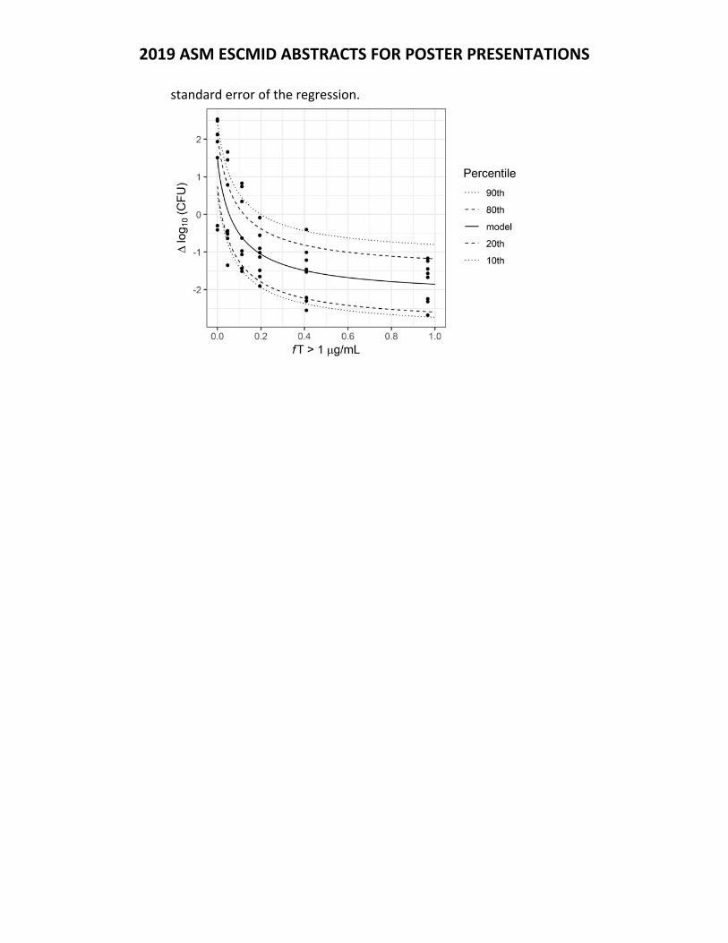
2019 ASM ESCMID ABSTRACTS FOR POSTER PRESENTATIONS
standard error of the regression.
2019 ASM ESCMID ABSTRACTS FOR POSTER PRESENTATIONS
Thursday Presentation Number:
T‐06
Publishing Title:
Pharmacodynamics of Aztreonam Against Escherichia coli and Klebsiella spp Studied in an in vitro Model of Infection
Author Block: A. Noel, M. Attwood, K. Bowker, A. MacGowan, BS10 5NB; BCARE, North Bristol NHS Trust, Bristol, United Kingdom
Abstract Body:
Background: The pharmacodynamics (PD) of aztreonam (AZT) are poorly studied but are an increasingly important area of B.lactam PD investigation. The reasons for this are a) AZT is the only B.lactam of the monobactam class available for clinical use, b) AZT as single or combination therapy has been identified as an old antibiotic worthy of revival, c) other monobactams are presently under development for clinical use. The aim of this study was to define the size of the pharmacodynamic index (PDI) for reduction in bacterial load and risk of emergence of resistance (eor) using an in vitro PK model. Methods: A single compartment dilutional in vitro model was used to model nine T>MIC exposures per strain tested. Three wild type E.coli and three Klebsiella spp, AZT MICs all 0.25mg/L at an initial inoculation of 106 CFU/ml was used. AZT concentrations were based on a serum half‐life of 2.5h. Antibacterial effect was measured by change in viable count over 48h and changes in population profile as assessed by sub‐culture onto recovery media containing AZT at MICx2. MICx4, and MICx8. Results: AZT was bactericidal against E.coli strains producing a ‐4 log drop in initial bacterial load at the highest AZT exposures (T>MIC 100%). The %T>MIC for a 24h static, ‐1 log and ‐2 log in bacterial load was 47.1±3.7%, 52.3±5.9% and 57.7±9.1% respectively. Against Klebsiella spp, AZT was less cidal than against E.coli. The %T>MIC for a 24h static effect, ‐1 log and ‐2 log reduction in Klebsiella bacterial lead was 39.1±6.1%, 53.4±8.4% at 100% respectively. There was no EOR over 48h with any strain. Conclusions: The T>MIC PDI target for AZT based on a static to ‐1 log drop at 24h was 45‐70% for E.coli and Klebsiella spp. Such a target should be valuable for translationally modelling of AZT doses and is longer than equivalent values for Gram‐negative rods in our model with carbapenems (T>MIC target 15‐40%); and cephalosporins (T>MIC target 30‐45%) and more similar to those for penicillins (T>MIC target 30‐60%).
2019 ASM ESCMID ABSTRACTS FOR POSTER PRESENTATIONS
Thursday Presentation Number:
T‐07
Publishing Title: Evaluation of the Pharmacokinetics‐Pharmacodynamics of Oral Avibactam in Combination with Ceftibuten Using a One‐CompartmentIn VitroInfection Model
Author Block: B. D. VanScoy1, A. Mullarkey2, H. Conde1, N. Onufrak1, C. Sable3, J. Trias3, S. M. Bhavnani1, P. G. Ambrose1; 1Inst. for Clinical Pharmacodynamics, Schenectady, NY, 2ICPD, Schenectady, NY, 3Arixa Pharmaceuticals, Palo Alto, CA
Abstract Body:
Background: The development of oral antimicrobial agents active against β‐lactamase‐producing Gram‐negative bacilli fulfills an unmet medical need. The first objective of our study was to determine the magnitude of ceftibuten percent time above MIC (%T>MIC) associated with efficacy when administered alone against wild‐type Enterobacteriaceae. The second objective was to determine the magnitude of the different avibactam pharmacokinetic‐pharmacodynamic (PK‐PD) indices associated with efficacy when administered in combination with ceftibuten against a panel of Enterobacteriaceae known to produce CTX‐M‐15, KPC‐2, and AmpC enzymes. Methods: The 24‐hour one‐compartment in vitro infection model was utilized for all studies. Ceftibuten dose‐ranging studies were completed to identify the magnitude of ceftibuten free‐drug plasma %T>MIC associated with net bacterial stasis when administered every 8 hours (q8h) against a wild‐type Escherichia coli isolate. Ceftibuten/avibactam dose‐ranging studies were completed utilizing a 400 mg ceftibuten dose in combination with a range of avibactam doses q8h (31.3 to 750 mg), against a panel of beta‐lactamase producing Enterobacteriaceae (n=3). The data from the dose‐ranging studies was pooled, and the relationships between change in log10 colony forming units (CFU)/mL from baseline and various avibactam free‐drug plasma PK‐PD indices were evaluated using Hill‐type models to identify the magnitude of individual PK‐PD indices associated with net bacterial stasis, and 1‐ and 2‐log10 CFU/mL reductions from baseline. Results: The magnitude of free‐drug plasma %T>MIC (based on ceftibuten MIC) associated with net bacterial stasis was approximately 45% when administered q8h. The relationship between the change in log10 CFU/mL and the ratio of avibactam area under the free‐drug plasma concentration‐time curve to MIC (determined using a fixed 4 mg/L of avibactam) (AUC:MIC ratio) described the activity of avibactam well (r2 = 0.86). Avibactam free‐drug plasma AUC:MIC ratios of 28.7, 30.8, and 34.2 were associated with net bacterial stasis, and 1‐ and 2‐log10 reductions from baseline, respectively. Conclusions: These data provide insight into the activity of ceftibuten when administered alone as a q8h 400 mg dosing regimen, as well as the magnitude of avibactam free‐drug plasma AUC:MIC ratio required to restore the activity of a ceftibuten 400 mg q8h dose against beta‐lactamase producing

2019 ASM ESCMID ABSTRACTS FOR POSTER PRESENTATIONS
Enterobacteriaceae, which is achieved in combination with 375‐500 mg of avibactam q8h.
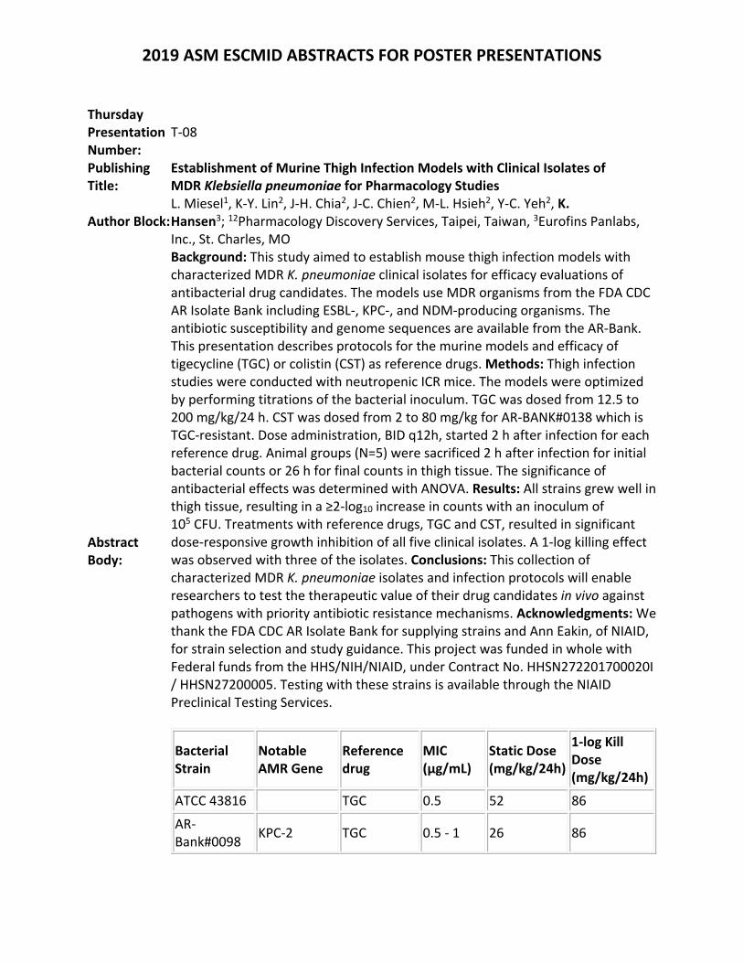
2019 ASM ESCMID ABSTRACTS FOR POSTER PRESENTATIONS
Thursday Presentation Number:
T‐08
Publishing Title:
Establishment of Murine Thigh Infection Models with Clinical Isolates of MDR Klebsiella pneumoniae for Pharmacology Studies
Author Block: L. Miesel1, K‐Y. Lin2, J‐H. Chia2, J‐C. Chien2, M‐L. Hsieh2, Y‐C. Yeh2, K. Hansen3; 12Pharmacology Discovery Services, Taipei, Taiwan, 3Eurofins Panlabs, Inc., St. Charles, MO
Abstract Body:
Background: This study aimed to establish mouse thigh infection models with characterized MDR K. pneumoniae clinical isolates for efficacy evaluations of antibacterial drug candidates. The models use MDR organisms from the FDA CDC AR Isolate Bank including ESBL‐, KPC‐, and NDM‐producing organisms. The antibiotic susceptibility and genome sequences are available from the AR‐Bank. This presentation describes protocols for the murine models and efficacy of tigecycline (TGC) or colistin (CST) as reference drugs. Methods: Thigh infection studies were conducted with neutropenic ICR mice. The models were optimized by performing titrations of the bacterial inoculum. TGC was dosed from 12.5 to 200 mg/kg/24 h. CST was dosed from 2 to 80 mg/kg for AR‐BANK#0138 which is TGC‐resistant. Dose administration, BID q12h, started 2 h after infection for each reference drug. Animal groups (N=5) were sacrificed 2 h after infection for initial bacterial counts or 26 h for final counts in thigh tissue. The significance of antibacterial effects was determined with ANOVA. Results: All strains grew well in thigh tissue, resulting in a ≥2‐log10 increase in counts with an inoculum of 105 CFU. Treatments with reference drugs, TGC and CST, resulted in significant dose‐responsive growth inhibition of all five clinical isolates. A 1‐log killing effect was observed with three of the isolates. Conclusions: This collection of characterized MDR K. pneumoniae isolates and infection protocols will enable researchers to test the therapeutic value of their drug candidates in vivo against pathogens with priority antibiotic resistance mechanisms. Acknowledgments: We thank the FDA CDC AR Isolate Bank for supplying strains and Ann Eakin, of NIAID, for strain selection and study guidance. This project was funded in whole with Federal funds from the HHS/NIH/NIAID, under Contract No. HHSN272201700020I / HHSN27200005. Testing with these strains is available through the NIAID Preclinical Testing Services.
Bacterial Strain
Notable AMR Gene
Reference drug
MIC (µg/mL)
Static Dose (mg/kg/24h)
1‐log Kill Dose (mg/kg/24h)
ATCC 43816 TGC 0.5 52 86
AR‐Bank#0098
KPC‐2 TGC 0.5 ‐ 1 26 86
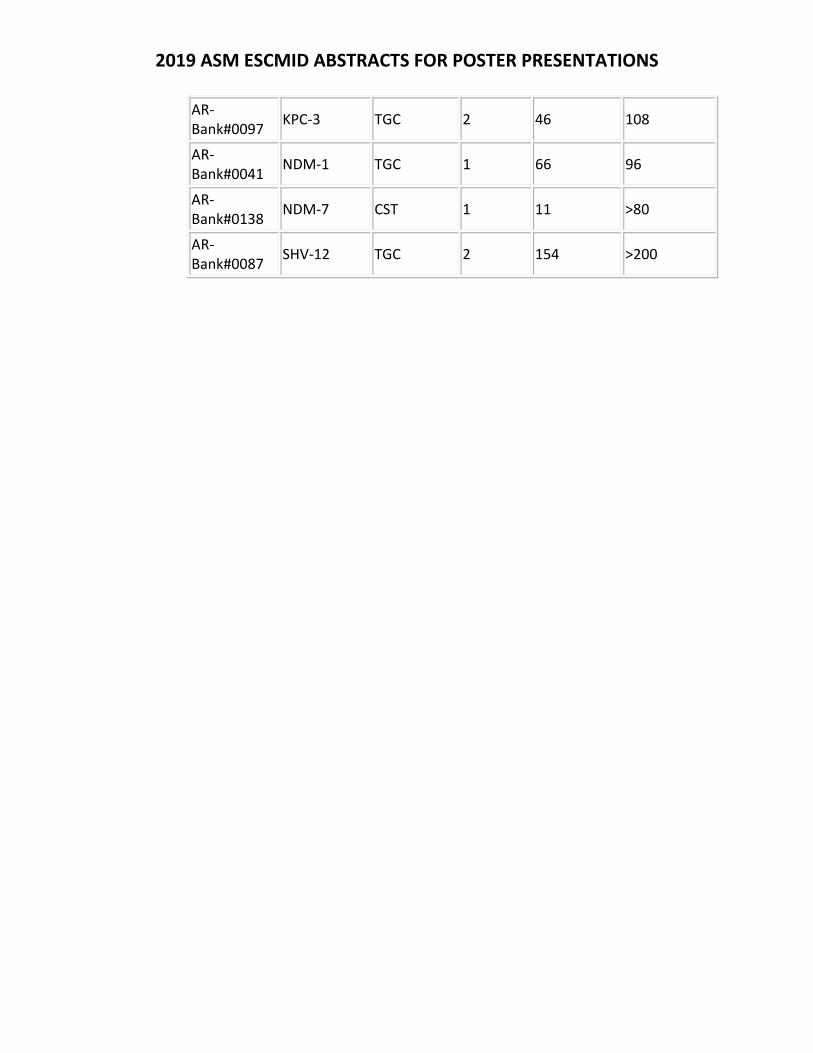
2019 ASM ESCMID ABSTRACTS FOR POSTER PRESENTATIONS
AR‐Bank#0097
KPC‐3 TGC 2 46 108
AR‐Bank#0041
NDM‐1 TGC 1 66 96
AR‐Bank#0138
NDM‐7 CST 1 11 >80
AR‐Bank#0087
SHV‐12 TGC 2 154 >200
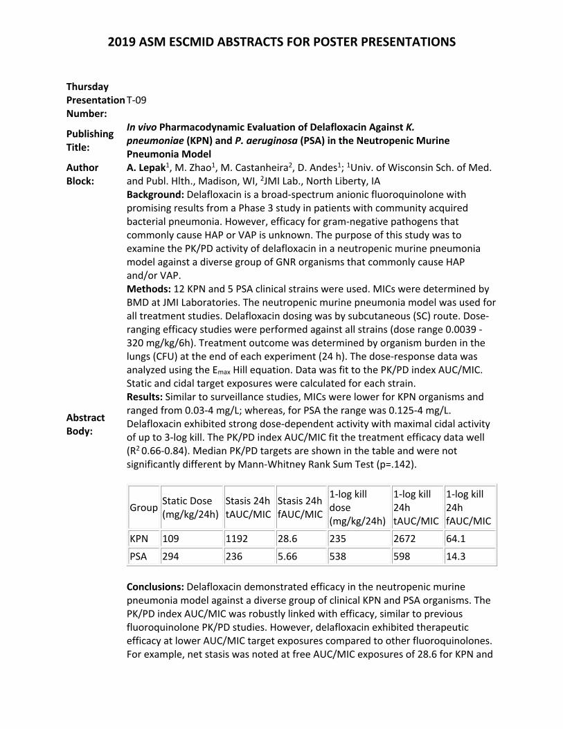
2019 ASM ESCMID ABSTRACTS FOR POSTER PRESENTATIONS
Thursday Presentation Number:
T‐09
Publishing Title:
In vivo Pharmacodynamic Evaluation of Delafloxacin Against K. pneumoniae (KPN) and P. aeruginosa (PSA) in the Neutropenic Murine Pneumonia Model
Author Block:
A. Lepak1, M. Zhao1, M. Castanheira2, D. Andes1; 1Univ. of Wisconsin Sch. of Med. and Publ. Hlth., Madison, WI, 2JMI Lab., North Liberty, IA
Abstract Body:
Background: Delafloxacin is a broad‐spectrum anionic fluoroquinolone with promising results from a Phase 3 study in patients with community acquired bacterial pneumonia. However, efficacy for gram‐negative pathogens that commonly cause HAP or VAP is unknown. The purpose of this study was to examine the PK/PD activity of delafloxacin in a neutropenic murine pneumonia model against a diverse group of GNR organisms that commonly cause HAP and/or VAP. Methods: 12 KPN and 5 PSA clinical strains were used. MICs were determined by BMD at JMI Laboratories. The neutropenic murine pneumonia model was used for all treatment studies. Delafloxacin dosing was by subcutaneous (SC) route. Dose‐ranging efficacy studies were performed against all strains (dose range 0.0039 ‐ 320 mg/kg/6h). Treatment outcome was determined by organism burden in the lungs (CFU) at the end of each experiment (24 h). The dose‐response data was analyzed using the Emax Hill equation. Data was fit to the PK/PD index AUC/MIC. Static and cidal target exposures were calculated for each strain. Results: Similar to surveillance studies, MICs were lower for KPN organisms and ranged from 0.03‐4 mg/L; whereas, for PSA the range was 0.125‐4 mg/L. Delafloxacin exhibited strong dose‐dependent activity with maximal cidal activity of up to 3‐log kill. The PK/PD index AUC/MIC fit the treatment efficacy data well (R2 0.66‐0.84). Median PK/PD targets are shown in the table and were not significantly different by Mann‐Whitney Rank Sum Test (p=.142).
Group Static Dose (mg/kg/24h)
Stasis 24h tAUC/MIC
Stasis 24h fAUC/MIC
1‐log kill dose (mg/kg/24h)
1‐log kill 24h tAUC/MIC
1‐log kill 24h fAUC/MIC
KPN 109 1192 28.6 235 2672 64.1
PSA 294 236 5.66 538 598 14.3
Conclusions: Delafloxacin demonstrated efficacy in the neutropenic murine pneumonia model against a diverse group of clinical KPN and PSA organisms. The PK/PD index AUC/MIC was robustly linked with efficacy, similar to previous fluoroquinolone PK/PD studies. However, delafloxacin exhibited therapeutic efficacy at lower AUC/MIC target exposures compared to other fluoroquinolones. For example, net stasis was noted at free AUC/MIC exposures of 28.6 for KPN and

2019 ASM ESCMID ABSTRACTS FOR POSTER PRESENTATIONS
5.66 for PSA. These results suggest delafloxacin may be a useful addition to the armamentarium for GNR organisms involved in HAP and/or VAP and will be useful for designing dosing regimens for future clinical trials.
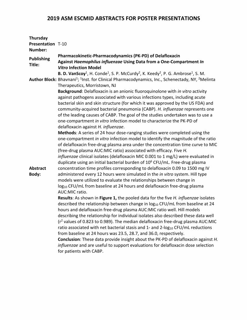
2019 ASM ESCMID ABSTRACTS FOR POSTER PRESENTATIONS
Thursday Presentation Number:
T‐10
Publishing Title:
Pharmacokinetic‐Pharmacodynamics (PK‐PD) of Delafloxacin Against Haemophilus influenzae Using Data from a One‐Compartment In Vitro Infection Model
Author Block: B. D. VanScoy1, H. Conde1, S. P. McCurdy2, K. Keedy2, P. G. Ambrose1, S. M. Bhavnani1; 1Inst. for Clinical Pharmacodynamics, Inc., Schenectady, NY, 2Melinta Therapeutics, Morristown, NJ
Abstract Body:
Background: Delafloxacin is an anionic fluoroquinolone with in vitro activity against pathogens associated with various infections types, including acute bacterial skin and skin structure (for which it was approved by the US FDA) and community‐acquired bacterial pneumonia (CABP). H. influenzae represents one of the leading causes of CABP. The goal of the studies undertaken was to use a one‐compartment in vitro infection model to characterize the PK‐PD of delafloxacin against H. influenzae. Methods: A series of 24 hour dose‐ranging studies were completed using the one‐compartment in vitro infection model to identify the magnitude of the ratio of delafloxacin free‐drug plasma area under the concentration time curve to MIC (free‐drug plasma AUC:MIC ratio) associated with efficacy. Five H. influenzae clinical isolates (delafloxacin MIC 0.001 to 1 mg/L) were evaluated in duplicate using an initial bacterial burden of 106 CFU/mL. Free‐drug plasma concentration time profiles corresponding to delafloxacin 0.09 to 1500 mg IV administered every 12 hours were simulated in the in vitro system. Hill type models were utilized to evaluate the relationships between change in log10 CFU/mL from baseline at 24 hours and delafloxacin free‐drug plasma AUC:MIC ratio. Results: As shown in Figure 1, the pooled data for the five H. influenzae isolates described the relationship between change in log10 CFU/mL from baseline at 24 hours and delafloxacin free‐drug plasma AUC:MIC ratio well. Hill models describing the relationship for individual isolates also described these data well (r2 values of 0.823 to 0.989). The median delafloxacin free‐drug plasma AUC:MIC ratio associated with net bacterial stasis and 1‐ and 2‐log10 CFU/mL reductions from baseline at 24 hours was 23.5, 28.7, and 36.0, respectively. Conclusion: These data provide insight about the PK‐PD of delafloxacin against H. influenzae and are useful to support evaluations for delafloxacin dose selection for patients with CABP.
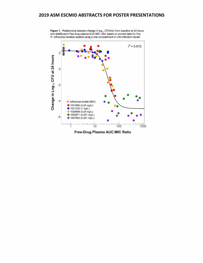
2019 ASM ESCMID ABSTRACTS FOR POSTER PRESENTATIONS
2019 ASM ESCMID ABSTRACTS FOR POSTER PRESENTATIONS
Thursday Presentation Number:
T‐11
Publishing Title:
Evaluation of the Pharmacokinetics‐Pharmacodynamics (PK‐PD) of the Fluorocycline, TP‐6076, Against Acinetobacter baumannii Using a One‐Compartment In Vitro Infection Model
Author Block: B. D. VanScoy1, H. Conde1, G. Giesel1, N. J. Onufrak1, J. Zhou2, S. M. Bhavnani1, P. G. Ambrose1; 1Inst. for Clinical Pharmacodynamics, Inc., Schenectady, NY, 2Tetraphase Pharmaceuticals, Watertown, MA
Abstract Body:
Background: TP‐6076 is a novel synthetic fluorocycline antibiotic in development for the treatment of multi‐drug resistant organisms, including Acinetobacter baumannii. Data from dose‐fractionation studies carried out using a one‐compartment in vitro infection model identified the AUC:MIC ratio as the PK‐PD index associated with TP‐6076 efficacy. In the studies described herein, a one‐compartment in vitro infection model was used to determine the magnitude of free‐drug plasma AUC:MIC ratio associated with net bacterial stasis, and 1‐ and 2‐ log10 CFU/mL reductions from baseline at 24 hours for A. baumannii. Methods: A series of 24 hour dose‐ranging studies were completed using a one‐compartment in vitro infection model to identify the magnitude of the TP‐6076 free‐drug plasma AUC:MIC ratio associated with efficacy. Seven A. baumannii isolates (TP‐6076 MIC values of 0.03 to 0.25 mg/L) were evaluated in duplicate using an initial bacterial burden of 106 CFU/mL. Free‐drug plasma concentration time profiles corresponding to TP‐6076 0.47 to 240 mg IV administered every 12 hours were simulated in the in vitro system. Hill type models were used to evaluate the relationships between change in log10 CFU/mL from baseline at 24 hours and TP‐6076 free‐drug plasma AUC:MIC ratio. Results: Hill models describing the relationship between change in log10 CFU/mL from baseline at 24 hours and free‐drug plasma AUC:MIC ratio for the individual isolates described the data well (r2 values of 0.708 to 0.974). The relationship based on pooled data for the A. baumannii isolates also described the data well (Figure 1). Based on this relationship, the magnitude of TP‐6076 free‐drug plasma AUC:MIC ratio associated with net bacterial stasis and 1‐ and 2‐log10 CFU/mL reductions from baseline at 24 hours was 4.56, 11.2 and 24.7, respectively. Conclusion: These data provide insight about the PK‐PD of TP‐6076 against A. baumannii and will be useful to support for future in vitro and in
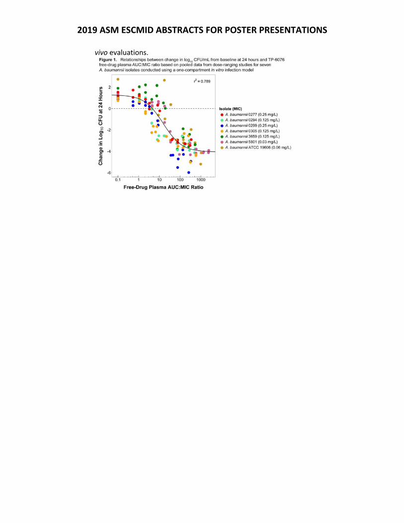
2019 ASM ESCMID ABSTRACTS FOR POSTER PRESENTATIONS
vivo evaluations.
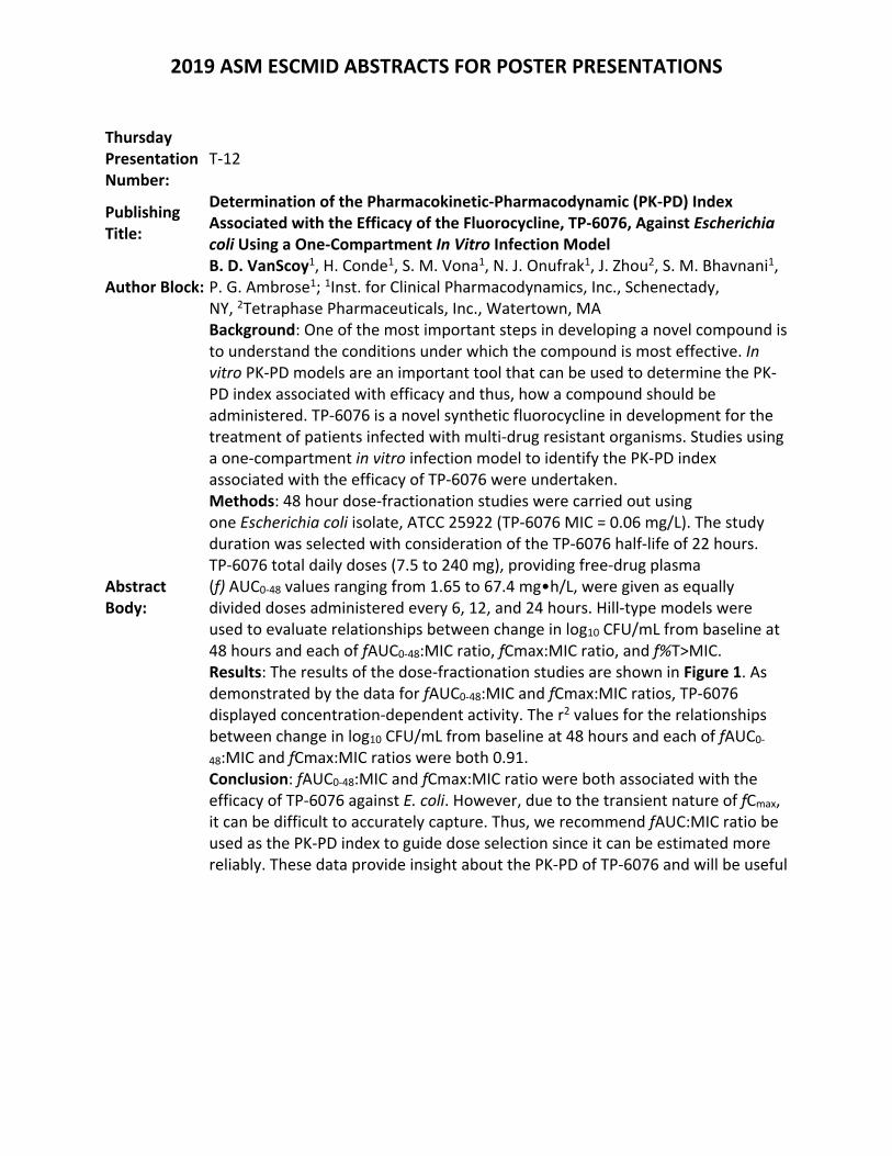
2019 ASM ESCMID ABSTRACTS FOR POSTER PRESENTATIONS
Thursday Presentation Number:
T‐12
Publishing Title:
Determination of the Pharmacokinetic‐Pharmacodynamic (PK‐PD) Index Associated with the Efficacy of the Fluorocycline, TP‐6076, Against Escherichia coli Using a One‐Compartment In Vitro Infection Model
Author Block: B. D. VanScoy1, H. Conde1, S. M. Vona1, N. J. Onufrak1, J. Zhou2, S. M. Bhavnani1, P. G. Ambrose1; 1Inst. for Clinical Pharmacodynamics, Inc., Schenectady, NY, 2Tetraphase Pharmaceuticals, Inc., Watertown, MA
Abstract Body:
Background: One of the most important steps in developing a novel compound is to understand the conditions under which the compound is most effective. In vitro PK‐PD models are an important tool that can be used to determine the PK‐PD index associated with efficacy and thus, how a compound should be administered. TP‐6076 is a novel synthetic fluorocycline in development for the treatment of patients infected with multi‐drug resistant organisms. Studies using a one‐compartment in vitro infection model to identify the PK‐PD index associated with the efficacy of TP‐6076 were undertaken. Methods: 48 hour dose‐fractionation studies were carried out using one Escherichia coli isolate, ATCC 25922 (TP‐6076 MIC = 0.06 mg/L). The study duration was selected with consideration of the TP‐6076 half‐life of 22 hours. TP‐6076 total daily doses (7.5 to 240 mg), providing free‐drug plasma (f) AUC0‐48 values ranging from 1.65 to 67.4 mg•h/L, were given as equally divided doses administered every 6, 12, and 24 hours. Hill‐type models were used to evaluate relationships between change in log10 CFU/mL from baseline at 48 hours and each of fAUC0‐48:MIC ratio, fCmax:MIC ratio, and f%T>MIC. Results: The results of the dose‐fractionation studies are shown in Figure 1. As demonstrated by the data for fAUC0‐48:MIC and fCmax:MIC ratios, TP‐6076 displayed concentration‐dependent activity. The r2 values for the relationships between change in log10 CFU/mL from baseline at 48 hours and each of fAUC0‐
48:MIC and fCmax:MIC ratios were both 0.91. Conclusion: fAUC0‐48:MIC and fCmax:MIC ratio were both associated with the efficacy of TP‐6076 against E. coli. However, due to the transient nature of fCmax, it can be difficult to accurately capture. Thus, we recommend fAUC:MIC ratio be used as the PK‐PD index to guide dose selection since it can be estimated more reliably. These data provide insight about the PK‐PD of TP‐6076 and will be useful
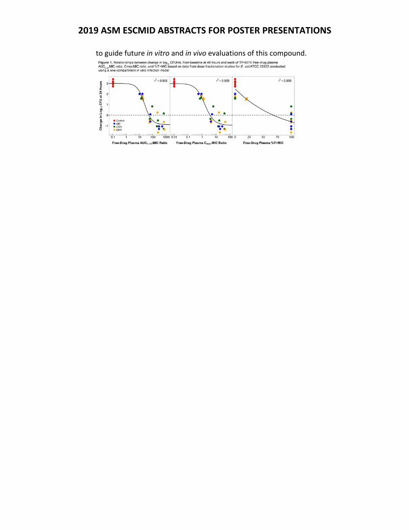
2019 ASM ESCMID ABSTRACTS FOR POSTER PRESENTATIONS
to guide future in vitro and in vivo evaluations of this compound.
2019 ASM ESCMID ABSTRACTS FOR POSTER PRESENTATIONS
Thursday Presentation Number:
T‐13
Publishing Title:
Use of a Hollow‐Fiber In Vitro Infection Model (HFIM) to Determine the Pharmacokinetic‐Pharmacodynamic (PK‐PD) Index Associated with the Efficacy of SMT‐571, a Novel Small Molecule with Activity Against Neisseria gonorrhoeae
Author Block: B. D. VanScoy1, A. I. Carranco1, H. Conde1, N. J. Onufrak1, P. Meo2, S. M. Bhavnani1, P. G. Ambrose1; 1Inst. for Clinical Pharmacodynamics, Inc., Schenectady, NY, 2Summit Therapeutics, Cambridge, United Kingdom
Abstract Body:
Background: The growing prevalence of multi‐drug resistant Neisseria gonorrhoeae is a global concern. The development of on‐therapy N. gonorrhoeae resistance, coupled with the difficulty to evaluate this pathogen using standard conditions, makes the HFIM an ideal tool to evaluate the PK‐PD of potential antimicrobial agents against N. gonorrhoeae. We undertook studies utilizing a HFIM to determine the PK‐PD index associated with the efficacy of SMT‐571, a small molecule with a novel mechanism of action. Methods: 96 hour dose‐fractionation studies were carried out using one N. gonorrhoeae clinical isolate, CDC 0170 (SMT‐571 MIC = 0.25 mg/L) with an initial bacterial burden of 1.0 x 10⁶ CFU/mL. SMT‐571 total doses (2.5, 5 and 10 mg/kg), providing free‐drug plasma AUC0‐96 values ranging from 1.79 to 18.9 mg•h/L, were given as equally divided doses administered every 24, 48, and 96 hours. Hill‐type models were used to evaluate relationships between change in log10 CFU/mL from baseline at 96 hours and each of free‐drug plasma AUC0‐
96:MIC ratio, Cmax0‐96:MIC ratio, Cmin0‐96:MIC ratio, and %T0‐96>MIC. All regimens were compared to ciprofloxacin (500 mg PO) and ceftriaxone (250 mg IM) control regimens. Results: As shown in Figure 1, relationships between change in log10 CFU/mL from baseline at 96 hours and each of free‐drug plasma AUC0‐96:MIC ratio and %T0‐96>MIC best described SMT‐571 efficacy (r2 of 0.91 and 0.94, respectively). The magnitude of SMT‐571 free‐drug plasma AUC0‐96:MIC ratio and %T0‐96>MIC associated with near maximal kill was 55 and 13.8, respectively. The ceftriaxone and ciprofloxacin controls resulted in treatment success and failure, respectively (data not shown). Conclusion: Free‐drug plasma AUC0‐96:MIC ratio and %T0‐96>MIC were both associated with the efficacy of SMT‐571 against the single N. gonorrhoeae isolate studied. These data provide insight about the PK‐PD of SMT‐571 and may help
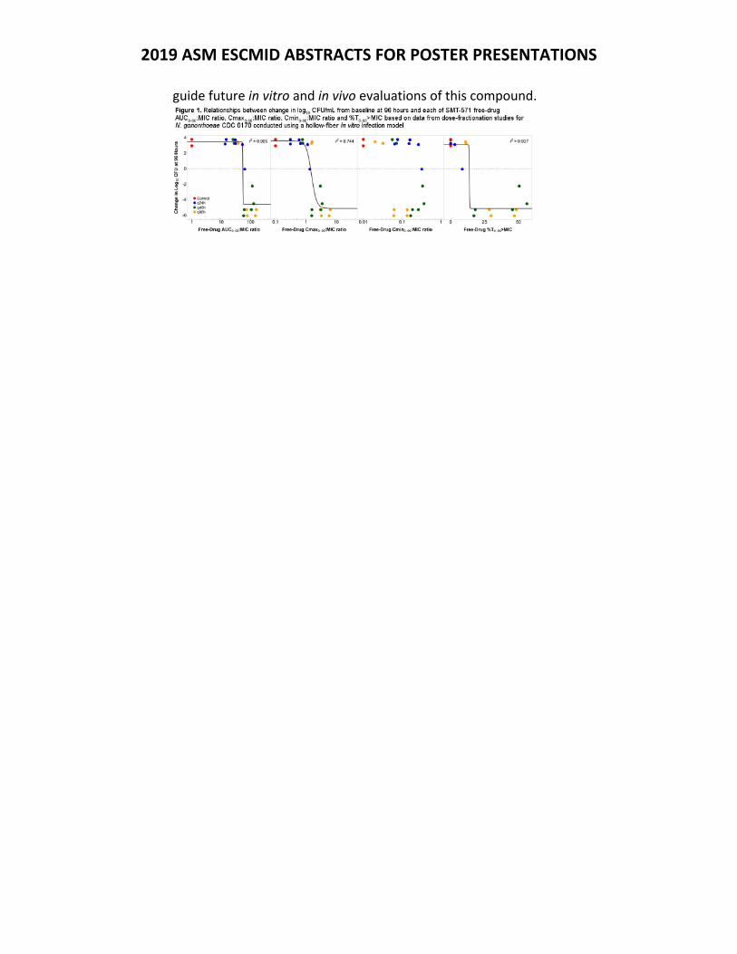
2019 ASM ESCMID ABSTRACTS FOR POSTER PRESENTATIONS
guide future in vitro and in vivo evaluations of this compound.
2019 ASM ESCMID ABSTRACTS FOR POSTER PRESENTATIONS
Thursday Presentation Number:
T‐14
Publishing Title: Pharmacokinetic and Pharmacodynamic Analysis of Cefepime and AAI101 Against Multidrug‐Resistant Enterobacteriaceae
Author Block: A. Johnson1, N. Farrington1, L. McEntee1, P. Knechtle2, S. Biondi2, S. Shapiro2, S. Das1, W. Hope1; 1Univ. of Liverpool, Liverpool, United Kingdom, 2Allecra, Saint‐Louis, France
Abstract Body:
Introduction: AAI101 is being developed in combination with cefepime for the treatment of serious hospital‐acquired Gram‐negative infections. AAI101 is a novel penicillanic acid sulfone ß‐lactamase inhibitor (BLI) with proven activity against extended spectrum ß‐lactamases (ESBLs) as well as some carbapenemases. Methods: A well characterised murine neutropenic lung infection model was used. Three challenge strains were initially identified to perform dose range finding and dose fractionation studies. These included two ESBL‐producing, cefepime‐resistant isolates, IHMA#1093554, IHMA#1280740, and a non‐ESBL‐producing, cefepime susceptible isolate, IHMA#1133791. The study end point was bacterial density in the lung. Dose finding studies were performed by administering cefepime alone as 100 mg/kg q8 + AAI101 q8h intravenously. Dose fractionation experiments were performed by administering the total daily dose of AAI101, when combined with cefepime 100 mg/kg q8h, that produced 20, 40, 60 and 80% of maximal effect every 4, 8 and 12 hours. The PK was estimated using dosages of cefepime/AAI101 25/6.25, 50/25, 100/75 and 200/200 mg/kg q8h. Multiple ESBL producing K. pneumoniae challenge strains (n=4) were then used, covering an MIC range of 0.06‐8mg/L to define the magnitude. Results: Linear mixed effect models showed that regimens of cefepime 100mg/kg q8h in combination with AAI101 2.5mg/kg q4h, 5mg q8h, and 7.5mg q12h were not associated with statistically significant differences in bacterial burden with time (p‐values of 0.9913, 0.9248, 0.9874 respectively). However, a regimen of cefepime 100mg/kg with AAI101 2.5mg/kg q4h, 5mg q8h or 7.5mg q12h were all statistically significantly better than cefepime 100 mg/kg q8h alone with p‐values of 0.0081, 0.0162, 0.029, respectively. The threshold plasma concentration of AAI101 that allowed bacterial regrowth was 0.07mg/L. The 2.5 mg/kg regimen administered q4h resulted in killing for approximately 47% of the dosing interval. In comparison, the 7.5mg/kg regimen administered q12h resulted in killing for 32% of the dosing interval. Conclusions: Both AUC and time>threshold can be used to account for the experimental data. However, fractionation studies suggest T>threshold is a more conservative measure to use for PK‐PD bridging studies and dose identification for clinical use for pneumonia.
2019 ASM ESCMID ABSTRACTS FOR POSTER PRESENTATIONS
Thursday Presentation Number:
T‐15
Publishing Title: Translational PK and PKPD Modeling of Preclinical In‐Vitro and In‐Vivo Studies to Predict Efficacious Human Dose of Apramycin
Author Block:
T. Sou1, E. Liepins2, J. Hansen3, S. Grinberga2, M. Backlund1, O. Ercan1, A. Petersson1, D. Hughes1, M. Tomczak4, M. Urbas4, D. Zabicka5, C. Vingsbo Lundberg3, S. N. Hobbie6, L. E. Friberg1; 1Uppsala Univ., Uppsala, Sweden, 2Latvian Inst. of Organic Synthesis, Riga, Latvia, 3Statens Serum Inst., Copenhagen, Denmark, 4Natl. Med.s Inst., Warsaw, Poland, 5Natl. Med.s Inst., Uppsala, Poland, 6Univ. of Zurich, Zurich, Switzerland
Abstract Body:
Background: Apramycin represents a subclass of aminoglycoside antibiotics that has been shown to evade almost all mechanisms of clinically relevant aminoglycoside resistance. Preclinical profiling has identified apramycin as a candidate for development into a new human therapeutic, in particular for the treatment of carbapenem‐ and colistin‐resistant infections with Enterobacteriaceae and Acinetobacter baumannii. Here we used PK and PKPD modelling to predict an efficacious human dose from preclinical studies with apramycin. Methods: Preclinical pharmacokinetic (PK) data of apramycin were available from four different species (mouse, rat, guinea pig, and dog). PK models were fitted to the data and the parameters were allometrically scaled to humans. In‐vitro time‐kill data and in‐vivo infection studies for four E.coli strains were performed to define PD targets (based on PK/PD index methodology) and for PKPD modelling, describing the time‐course of bacterial growth and killing. Human efficacious doses were predicted using 3 different approaches: (i) from the 95% probability of target attainment (PTA) for stasis and 1‐log kill, (ii) stasis at 24 h based on the PKPD model where predicted human PK profiles drove the killing, and (iii) scaling aminoglycoside doses with typical differences in MIC. Results: From the PK analysis, 1‐compartment models described the data from the preclinical species. Allometric scaling resulted in values for clearance and volume of distribution of 7.07 L/h and 26.8 L, respectively, i.e. similar to typical population PK parameters of gentamicin [1]. From the neutropenic mouse thigh infection models, the required fAUC/MIC targets for stasis and 1‐log kill were 42 and 89, respectively. The PKPD model predicted the time‐kill data well with strain specific differences in susceptibility, maximum bacterial load and resistance development. All three approaches supported an apramycin daily dose of 30 mg/kg/day for a typical adult patient. Conclusions: Translational PK and PKPD modelling was successfully applied to analyse the results from preclinical studies on apramycin. Predictions from the PKPD model, with adjusted in‐vivo bacterial growth, resulted in the same dose prediction of 30 mg/kg as the other methods. References 1. Xuan D, Nicolau DP, Nightingale CH. Population pharmacokinetics of

2019 ASM ESCMID ABSTRACTS FOR POSTER PRESENTATIONS
gentamicin in hospitalized patients receiving once‐daily dosing. Int J Antimicrob Agents. 2004.
2019 ASM ESCMID ABSTRACTS FOR POSTER PRESENTATIONS
Thursday Presentation Number:
T‐16
Publishing Title: Single‐ and Multiple‐Ascending Dose (SAD/MAD) Study Demonstrates the Human Pharmacokinetics (PK) and Tolerability of SPR994 (Tebipenem Pivoxil Hydrobromide), an Oral Carbapenem (CP), at the Predicted Therapeutic Dose
Author Block: P. Eckburg1, A. Jain1, S. Walpole1, G. Moore1, L. Utley2, E. Manyak1, A. Dane3, D. Melnick1; 1Spero Therapeutics, Cambridge, MA, 2Ribon Therapeutics, Cambridge, MA, 3DaneStat Consulting, Macclesfield, United Kingdom
Abstract Body:
Background: SPR994 is an orally available prodrug of tebipenem (TBPM), a CP with activity versus multidrug‐resistant Gram‐negative pathogens, including quinolone‐resistant and extended‐spectrum ß‐lactamase‐producing Enterobacteriaceae. It is under development as an oral alternative to IV antibiotic therapy. We report results of the first‐in‐human SAD/MAD study of SPR994 in healthy adult volunteers, including the PK of TBPM. Materials/Methods: This was a double‐blind, placebo‐controlled trial. TBPM pivoxil hydrobromide (SPR994 tablets), Tebipenem pivoxil free base (Orapenem® fine granules), or placebo (PBO) was administered (n=8/cohort, 3:1 randomization) at a single dose of 100‐900 mg in varying immediate and extended release formulations (14 SAD cohorts) and 300‐600 mg q8h for 14 days (2 MAD cohorts). Concentrations of TBPM in plasma and urine were measured by validated LC‐MS/MS methods. Plasma PK parameters were determined using non‐compartmental methods. Safety assessments included physical exams, electrocardiograms, and serum and urine laboratory tests.
Results: 124 subjects were enrolled in the study. Plasma Cmax and AUC0‐ of TBPM increased in a dose linear and proportional manner across the SAD cohorts. Tmax was observed at 1 h post dose; mean t1/2 in the SAD was 0.93 h. Urinary excretion was the major route of TBPM elimination with 35‐56% of the dose recovered intact in urine. No change in the exposure of TBPM was observed when administered with food. MAD plasma PK characteristics were similar. Overall, SPR994 was well tolerated. In SAD, 20/75 of SPR994 subjects experienced ≥1 treatment‐emergent adverse event (TEAE), all of which were mild. In MAD, 100% of SPR994 and PBO subjects experienced ≥1 TEAE; most were mild, none severe or serious. The most common TEAE was diarrhea (13/87 SPR994 subjects); all cases were mild and transient. TEAEs of increased ALT were observed in 0/75 SPR994 SAD subjects and 3/12 SPR994 MAD subjects. Each case was asymptomatic and reversible with no associated elevation in bilirubin. Conclusions: • SPR994 was well tolerated at the predicted therapeutic dose and exposure levels •

2019 ASM ESCMID ABSTRACTS FOR POSTER PRESENTATIONS
Predictable PK characteristics allow for q8h oral dosing without regard to meals • Further evaluation in a Phase 3 cUTI trial is planned

2019 ASM ESCMID ABSTRACTS FOR POSTER PRESENTATIONS
Thursday Presentation Number:
T‐17
Publishing Title: Phase III Dose Selection for Tebipenem‐Pivoxil Hydrobromide, a Novel Oral Carbapenem
Author Block: S. Das1, M. Laura1, A. Johnson1, N. Farrington1, S. Gore1, A. Kirby1, A. Jain2, D. Melnick2, T. Parr2, W. Hope1; 1Univ. of Liverpool, Liverpool, United Kingdom, 2Spero Therapeutics, Cambridge, MA
Abstract Body:
Background: The PK‐PD relationship for tebipenem the active moiety of tebipenem‐pivoxil hyrdrobromide (TBPM‐PI‐HBr) is well characterized with the PD driver determined as fAUC/MIC*1/tau. Population PK modelling of Phase I clinical data, and probability of target attainment (PTA) analysis against the PK‐PD target was used to select the dose for Phase III. Methods: A population PK model was developed with data from 36 healthy volunteers receiving either a single, or multiple doses of TBPM‐PI‐HBr. The PK of tebipenem was described by a 2‐compartment model and used to simulate different potential clinical dose schedules of TBPM‐PI‐HBr. To account for potential increased variability in patients, additional simulations were conducted with inflated variance of clearance. PTA against the preclinical PK‐PD target was calculated. Results: Figure 1 shows the population PK simulations of different dose regimens overlaid on the MIC distribution for E. coli and K.pneumoniae. A 600mg three times daily regimen (q8h) was predicted to achieve robust coverage with high PTA (>90%) up to an MIC of 0.25mg/L which covers the wild‐type distribution of both strains. High PTA was still predicted when simulations were conducted with inflated variance the PTA. Conclusions: 600mg q8h has been selected as the Phase III dose for TBPM‐PI‐HBr for a clinical trial in patients with cUTI. This dose is well tolerated in healthy volunteers and predicted to achieve adequate PTA against the wild‐type distribution of common UTI pathogens. Figure 1. Simulated PTA of different dose regimens of tebipenem by MIC overlaying the percentage distribution of E. coli (n=101) and K. pneumo (n=208) isolates.
2019 ASM ESCMID ABSTRACTS FOR POSTER PRESENTATIONS
Thursday Presentation Number:
T‐18
Publishing Title:
Pharmacokinetic‐Pharmacodynamic (PK‐PD) Analyses for Efficacy Based on Data from Lefamulin‐Treated Patients Enrolled in Phase 3 Studies for Community‐Acquired Bacterial Pneumonia (CABP)
Author Block:
S. M. Bhavnani1, J. P. Hammel1, N. J. Onufrak1, K. Liolios1, W. W. Wicha2, S. Paukner2, E. Alexander3, J. Schranz3, C. M. Rubino1, S. P. Gelone3, P. G. Ambrose1; 1Inst. for Clinical Pharmacodynamics, Inc., Schenectady, NY, 2Nabriva Therapeutics GmbH, Vienna, Austria, 3Nabriva Therapeutics US, Inc., King of Prussia, PA
Abstract Body:
Background: Lefamulin is a semi‐synthetic intravenous (IV) and oral (PO) pleuromutilin antibiotic with activity against the most common CABP pathogens, including multi‐drug resistant Streptococcus pneumoniae and Staphylococcus aureus. Lefamulin is currently in late‐stage development for the treatment of patients with CABP. PK‐PD relationships for efficacy were evaluated using data from lefamulin‐treated patients with CABP from Phase 3 trials. Methods: Patients received lefamulin 150 mg IV q12h with an optional switch (after at least 6 doses of IV) to 600 mg PO q12h or 600 mg PO q12h. Efficacy endpoints assessed included early clinical response (96 ± 24 hours after the first dose of test article), investigator‐assessed clinical response at end of therapy (EOT), test of cure (TOC) and late follow‐up (LFU), and microbiological response at EOT, TOC, and LFU. Using a population PK model for lefamulin developed using Phase 1, 2 and 3 data and plasma PK data from patients, Day 1 free‐drug plasma and total‐drug ELF area under the concentration‐time curve (AUC) were determined. Relationships between efficacy endpoints and each of lefamulin Day 1 free‐drug plasma and total‐drug ELF AUC:MIC ratio were assessed among evaluable patients and subsets with baseline pathogens of interest using chi‐square tests or Fisher’s exact tests for categorical independent variables and logistic regression for continuous independent variables. Results: Successful response across efficacy endpoints ranged from 85.4 to 93.5% among 92 evaluable patients and the subset of 54 patients with S. pneumoniae at baseline. Results of PK‐PD analyses for efficacy failed to demonstrate statistically significant and biologically plausible univariable relationships between efficacy endpoints and AUC:MIC ratio. The limited sample size and number of failures potentially hindered the identification of PK‐PD relationships for efficacy. However, all patients with S. pneumoniae and S. aureus at baseline achieved free‐drug plasma and total‐drug ELF AUC:MIC ratios that were above non‐clinical AUC:MIC ratio targets associated with 1‐ and 2‐log10 CFU reductions from baseline for the same pathogens [ICAAC 2015, Poster A‐037]. These findings suggested that

2019 ASM ESCMID ABSTRACTS FOR POSTER PRESENTATIONS
free‐drug plasma and total‐drug ELF AUC:MIC ratios achieved among lefamulin‐treated patients were on the plateau of non‐clinical PK‐PD relationships for efficacy. Conclusions: Results of these analyses provide support for the lefamulin dosing regimens of 150 mg IV q12h and 600 mg PO q12h for the treatment of adult patients with CABP.
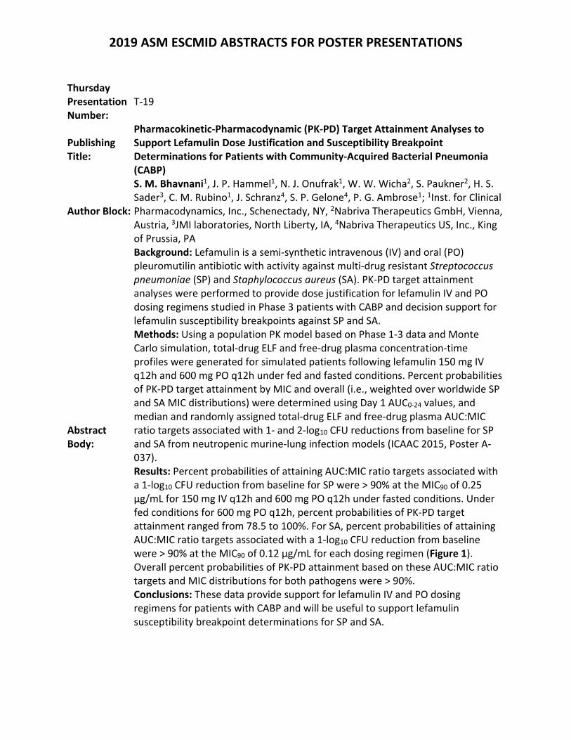
2019 ASM ESCMID ABSTRACTS FOR POSTER PRESENTATIONS
Thursday Presentation Number:
T‐19
Publishing Title:
Pharmacokinetic‐Pharmacodynamic (PK‐PD) Target Attainment Analyses to Support Lefamulin Dose Justification and Susceptibility Breakpoint Determinations for Patients with Community‐Acquired Bacterial Pneumonia (CABP)
Author Block:
S. M. Bhavnani1, J. P. Hammel1, N. J. Onufrak1, W. W. Wicha2, S. Paukner2, H. S. Sader3, C. M. Rubino1, J. Schranz4, S. P. Gelone4, P. G. Ambrose1; 1Inst. for Clinical Pharmacodynamics, Inc., Schenectady, NY, 2Nabriva Therapeutics GmbH, Vienna, Austria, 3JMI laboratories, North Liberty, IA, 4Nabriva Therapeutics US, Inc., King of Prussia, PA
Abstract Body:
Background: Lefamulin is a semi‐synthetic intravenous (IV) and oral (PO) pleuromutilin antibiotic with activity against multi‐drug resistant Streptococcus pneumoniae (SP) and Staphylococcus aureus (SA). PK‐PD target attainment analyses were performed to provide dose justification for lefamulin IV and PO dosing regimens studied in Phase 3 patients with CABP and decision support for lefamulin susceptibility breakpoints against SP and SA. Methods: Using a population PK model based on Phase 1‐3 data and Monte Carlo simulation, total‐drug ELF and free‐drug plasma concentration‐time profiles were generated for simulated patients following lefamulin 150 mg IV q12h and 600 mg PO q12h under fed and fasted conditions. Percent probabilities of PK‐PD target attainment by MIC and overall (i.e., weighted over worldwide SP and SA MIC distributions) were determined using Day 1 AUC0‐24 values, and median and randomly assigned total‐drug ELF and free‐drug plasma AUC:MIC ratio targets associated with 1‐ and 2‐log10 CFU reductions from baseline for SP and SA from neutropenic murine‐lung infection models (ICAAC 2015, Poster A‐037). Results: Percent probabilities of attaining AUC:MIC ratio targets associated with a 1‐log10 CFU reduction from baseline for SP were > 90% at the MIC90 of 0.25 µg/mL for 150 mg IV q12h and 600 mg PO q12h under fasted conditions. Under fed conditions for 600 mg PO q12h, percent probabilities of PK‐PD target attainment ranged from 78.5 to 100%. For SA, percent probabilities of attaining AUC:MIC ratio targets associated with a 1‐log10 CFU reduction from baseline were > 90% at the MIC90 of 0.12 µg/mL for each dosing regimen (Figure 1). Overall percent probabilities of PK‐PD attainment based on these AUC:MIC ratio targets and MIC distributions for both pathogens were > 90%. Conclusions: These data provide support for lefamulin IV and PO dosing regimens for patients with CABP and will be useful to support lefamulin susceptibility breakpoint determinations for SP and SA.
2019 ASM ESCMID ABSTRACTS FOR POSTER PRESENTATIONS
2019 ASM ESCMID ABSTRACTS FOR POSTER PRESENTATIONS
Thursday Presentation Number:
T‐20
Publishing Title:
Pharmacokinetic‐Pharmacodynamic (PK‐PD) Analyses for Alanine Aminotransferase (ALT) Using Phase 2 and 3 Data from Lefamulin‐Treated Patients
Author Block:
S. M. Bhavnani1, M. Trang1, D. R. George1, K. Liolios1, W. W. Wicha2, S. A. Villano3, D. S. Stein4, J. H. Lewis5, C. M. Rubino1, J. Schranz4, S. P. Gelone4, P. G. Ambrose1, J. P. Hammel1; 1Inst. for Clinical Pharmacodynamics, Inc., Schenectady, NY, 2Nabriva Therapeutics GmbH, Vienna, Austria, 3Villano Consulting Services, LLC, Havertown, PA, 4Nabriva Therapeutics US, Inc., King of Prussia, PA, 5Georgetown Univ. Med. Ctr., Washington, VA
Abstract Body:
Background: Lefamulin, a semi‐synthetic IV and PO pleuromutilin, is in late‐stage clinical development for the treatment of patients with CABP. Mild, transient, reversible and asymptomatic elevations of hepatic aminotransferases, without bilirubin elevations were observed in some patients. PK‐PD relationships for ALT were evaluated using Phase 2 and 3 data from lefamulin‐treated patients. Methods: Data were obtained from patients who received IV and/or PO lefamulin in a Phase 2 ABSSSI study and 2 Phase 3 CABP studies. Repeated measures multiple linear regression was used to evaluate factors predictive of ALT, including lefamulin AUC measures prior to each ALT, with interactions and covariates selected stepwise. Using the final model, percent probabilities of ALT elevation >1, 1.5, 2, 3, 5, and 10 x ULN post‐baseline up to 2 days after the end of therapy were calculated among analysis patients at fixed post‐baseline lefamulin AUC values, and among simulated patients after IV and PO dosing regimens. Results: The final model (n=653 patients) included baseline BMI, GGT, and AST (all increased), and age (decreased), as factors predictive of increased ALT (p ≤ 0.0002). Increased prior cumulative AUC was associated with increased ALT, with positive slopes estimated for baseline AST ≤ 59 U/L among males and ≤ 35 U/L among females (p <0.0001 for each interaction). However, across fixed average daily AUC values, the model‐predicted impact of lefamulin on clinically relevant ALT elevation thresholds was minimal (Figure 1A). Percent probabilities were within 1.65% when comparing simulated and observed patients after IV and PO dosing regimens (Figure 1B). Conclusion: While a covariate‐adjusted relationship between increased ALT and increased lefamulin AUC was found, model‐predicted ALT elevation endpoints across fixed lefamulin AUC values, or among simulated patients after administration of lefamulin IV or PO dosing regimens relative to observed patients, were minimal.
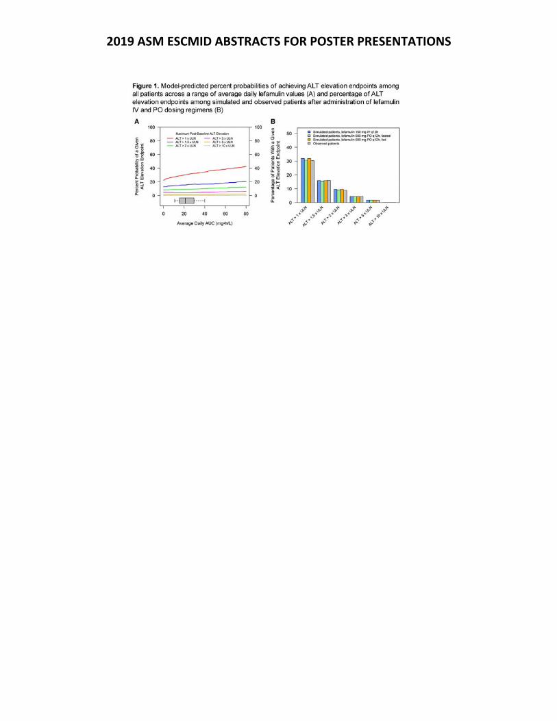
2019 ASM ESCMID ABSTRACTS FOR POSTER PRESENTATIONS
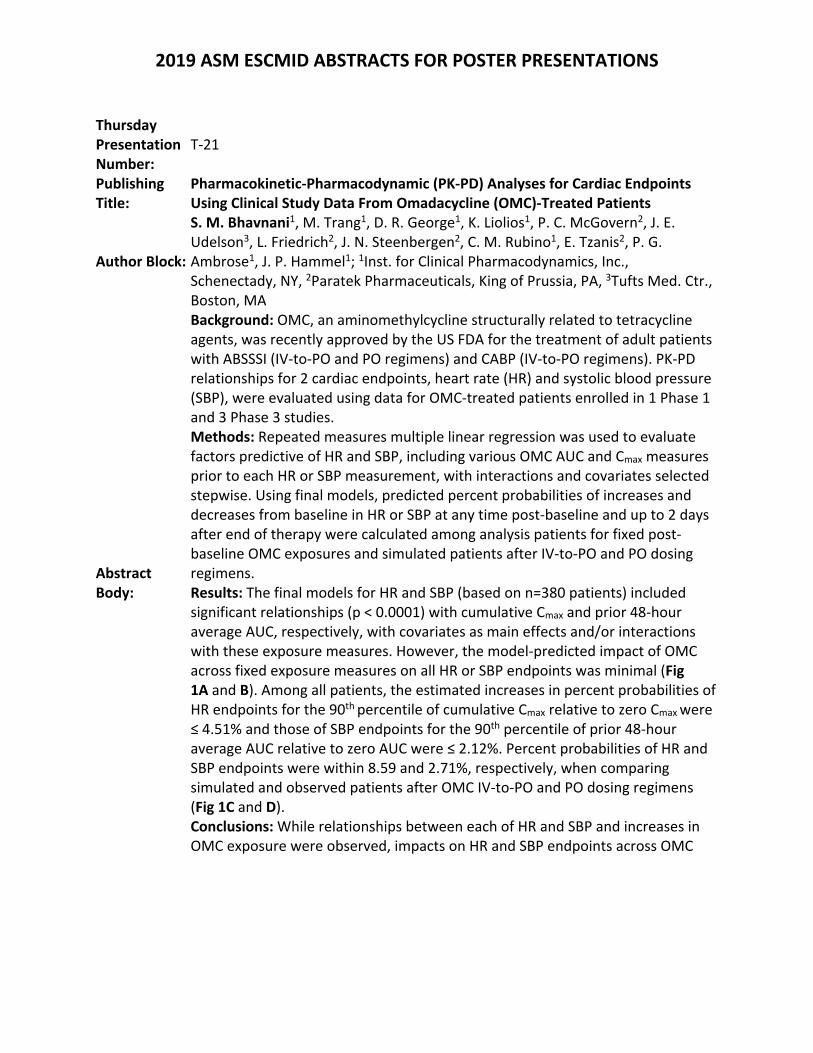
2019 ASM ESCMID ABSTRACTS FOR POSTER PRESENTATIONS
Thursday Presentation Number:
T‐21
Publishing Title:
Pharmacokinetic‐Pharmacodynamic (PK‐PD) Analyses for Cardiac Endpoints Using Clinical Study Data From Omadacycline (OMC)‐Treated Patients
Author Block:
S. M. Bhavnani1, M. Trang1, D. R. George1, K. Liolios1, P. C. McGovern2, J. E. Udelson3, L. Friedrich2, J. N. Steenbergen2, C. M. Rubino1, E. Tzanis2, P. G. Ambrose1, J. P. Hammel1; 1Inst. for Clinical Pharmacodynamics, Inc., Schenectady, NY, 2Paratek Pharmaceuticals, King of Prussia, PA, 3Tufts Med. Ctr., Boston, MA
Abstract Body:
Background: OMC, an aminomethylcycline structurally related to tetracycline agents, was recently approved by the US FDA for the treatment of adult patients with ABSSSI (IV‐to‐PO and PO regimens) and CABP (IV‐to‐PO regimens). PK‐PD relationships for 2 cardiac endpoints, heart rate (HR) and systolic blood pressure (SBP), were evaluated using data for OMC‐treated patients enrolled in 1 Phase 1 and 3 Phase 3 studies. Methods: Repeated measures multiple linear regression was used to evaluate factors predictive of HR and SBP, including various OMC AUC and Cmax measures prior to each HR or SBP measurement, with interactions and covariates selected stepwise. Using final models, predicted percent probabilities of increases and decreases from baseline in HR or SBP at any time post‐baseline and up to 2 days after end of therapy were calculated among analysis patients for fixed post‐baseline OMC exposures and simulated patients after IV‐to‐PO and PO dosing regimens. Results: The final models for HR and SBP (based on n=380 patients) included significant relationships (p < 0.0001) with cumulative Cmax and prior 48‐hour average AUC, respectively, with covariates as main effects and/or interactions with these exposure measures. However, the model‐predicted impact of OMC across fixed exposure measures on all HR or SBP endpoints was minimal (Fig 1A and B). Among all patients, the estimated increases in percent probabilities of HR endpoints for the 90th percentile of cumulative Cmax relative to zero Cmax were ≤ 4.51% and those of SBP endpoints for the 90th percentile of prior 48‐hour average AUC relative to zero AUC were ≤ 2.12%. Percent probabilities of HR and SBP endpoints were within 8.59 and 2.71%, respectively, when comparing simulated and observed patients after OMC IV‐to‐PO and PO dosing regimens (Fig 1C and D). Conclusions: While relationships between each of HR and SBP and increases in OMC exposure were observed, impacts on HR and SBP endpoints across OMC
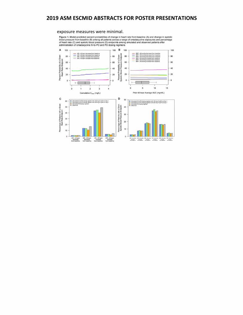
2019 ASM ESCMID ABSTRACTS FOR POSTER PRESENTATIONS
exposure measures were minimal.
2019 ASM ESCMID ABSTRACTS FOR POSTER PRESENTATIONS
Thursday Presentation Number:
T‐22
Publishing Title:
Dalbavancin Activity against Contemporary Gram‐Positive Clinical Isolates from Pneumonia in Hospitalized Patients and Lower Respiratory Tract Infections from the International Dalbavancin Evaluation of Activity (IDEA) Surveillance Program
Author Block: D. Debabov1, U. Rappo2, A. Suen2, V. Mas Casullo2; 1Allergan plc, Irvine, CA, 2Allergan plc, Madison, NJ
Abstract Body:
Background: Dalbavancin is approved in the US and Europe for acute bacterial skin and skin structure infections, exhibits broad spectrum activity against clinically important Gram‐positive pathogens, and has a terminal half‐life of 14.4 days. Dalbavancin intrapulmonary concentrations exceed the MIC90 of Streptococcus pneumoniae (SP) and Staphylococcus aureus (SA) after a single 1500‐mg IV infusion; dalbavancin has also been used off‐label for the treatment of MRSA pneumonia. We assessed dalbavancin activity against recent (2015–2017) Gram‐positive clinical isolates from pneumonia in hospitalized patients (PHP) and from lower respiratory tract infections (LRTI). Methods: Susceptibilities for dalbavancin and commonly used antimicrobials were tested using CLSI broth microdilution methods on 11,042 Gram‐positive isolates (5944 isolates from PHP; 5098 isolates from LRTI) from the IDEA Surveillance Program. Susceptibilities for antimicrobials were interpreted using CLSI guidelines. Results: The most common organisms were SA (47%) and SP (49%). SA was isolated only from patients with PHP. Dalbavancin, teicoplanin, vancomycin and telavancin showed complete activity (100.0% susceptible) against SA; daptomycin and linezolid showed >99.9% activity. Dalbavancin MIC90 was identical to telavancin MIC90, 8‐fold more potent than daptomycin, and 16‐fold more potent than vancomycin. Linezolid and teicoplanin were also active. All SP isolates were susceptible to vancomycin and linezolid. Dalbavancin MIC90 against SP from both PHP and LRTI was 0.015 μg/mL, 32‐fold more potent than vancomycin (Table). Conclusions: This study demonstrated potent in vitro activity of dalbavancin against contemporary US and European Gram‐positive clinical isolates from PHP and LRTI. These data suggest that dalbavancin may emerge as an important therapeutic option for these infections.
Table. Dalbavancin, daptomycin, vancomycin, and telavancin MIC90 for Staphylococcus aureus and Streptococcus pneumoniae isolated from PHP and LRTI
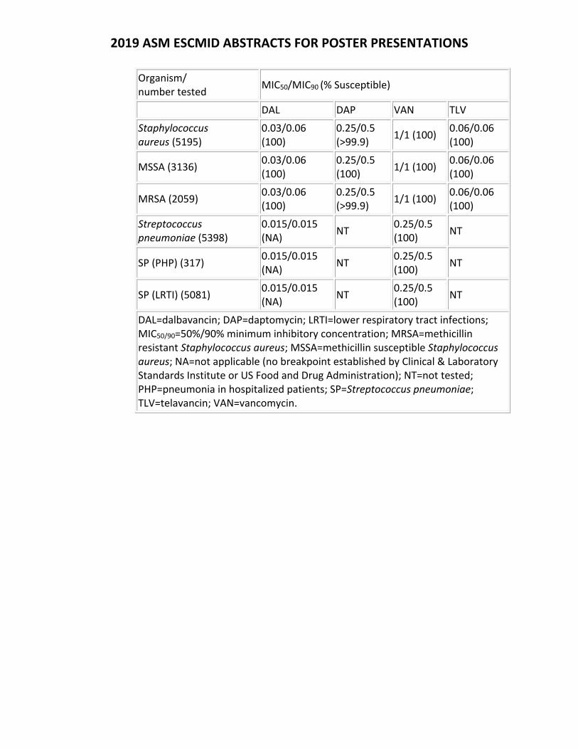
2019 ASM ESCMID ABSTRACTS FOR POSTER PRESENTATIONS
Organism/ number tested
MIC50/MIC90 (% Susceptible)
DAL DAP VAN TLV
Staphylococcus aureus (5195)
0.03/0.06 (100)
0.25/0.5 (>99.9)
1/1 (100) 0.06/0.06 (100)
MSSA (3136) 0.03/0.06 (100)
0.25/0.5 (100)
1/1 (100) 0.06/0.06 (100)
MRSA (2059) 0.03/0.06 (100)
0.25/0.5 (>99.9)
1/1 (100) 0.06/0.06 (100)
Streptococcus pneumoniae (5398)
0.015/0.015 (NA)
NT 0.25/0.5 (100)
NT
SP (PHP) (317) 0.015/0.015 (NA)
NT 0.25/0.5 (100)
NT
SP (LRTI) (5081) 0.015/0.015 (NA)
NT 0.25/0.5 (100)
NT
DAL=dalbavancin; DAP=daptomycin; LRTI=lower respiratory tract infections; MIC50/90=50%/90% minimum inhibitory concentration; MRSA=methicillin resistant Staphylococcus aureus; MSSA=methicillin susceptible Staphylococcus aureus; NA=not applicable (no breakpoint established by Clinical & Laboratory Standards Institute or US Food and Drug Administration); NT=not tested; PHP=pneumonia in hospitalized patients; SP=Streptococcus pneumoniae; TLV=telavancin; VAN=vancomycin.
2019 ASM ESCMID ABSTRACTS FOR POSTER PRESENTATIONS
Thursday Presentation Number:
T‐23
Publishing Title: Chloroquine & Primaquine Diphosphate as Possible Alternative Drugs for the Treatment of Cryptococcal Infection
Author Block: L. U. Madu, C. H. Pohl, O. M. Sebolai; Univ. of the Free State, Bloemfontein, South Africa
Abstract Body:
Background: Cryptococcal meningitis is one of the leading causes of mortality in persons living with HIV/AIDS, especially in Sub‐Saharan Africa. Treatment of this infection with amphotericin B and fluconazole is unfortunately associated with clinical failure. In the quest for alternative treatment, several compounds have been considered. Thus, this study investigates the action of chloroquine (CQ) and primaquine (PQ) diphosphate on the growth of Cryptococcus. Methods: Sensitivity of cryptococcal strains towards CQ and PQ was determined. Mode of action of the two drugs in killing Cryptococcus was investigated. Lastly, the ability of CQ and PQ to sensitize macrophages (host cells) was also determined. Results: After 48 h maximum time exposure, the direct effect of CQ and PQ on eight cryptococcal isolates showed a distinctive growth reduction ranging from 50 ‐ 89% (p < 0.01) and 38 ‐ 74% (p < 0.01) respectively, with CQ exerting a greater inhibitory effect. The minimum inhibitory concentrations (MICs) were determined to be 50 µM for CQ and 60 µM for PQ. These drugs at their MICs possibly inhibited cryptococcal growth by adversely affecting the mitochondria. We observed the loss of mitochondrial membrane potential (MMP) with CQ and PQ accounting for 43 ‐ 60% (p < 0.01) and 52 ‐ 65% (p < 0.01) respectively compared to the non‐treated cells. To the point, the loss of MMP resulted from a significant dislodging of cytochrome c (p < 0.05) from the mitochondria and overproduction of reactive oxygen species (p < 0.01), leaving the non‐fermentative isolates with no other means of energy production. CQ and PQ also induced disruption of the cell wall integrity of these fungi by creating pores (p < 0.05), causing substantial leakage of cellular components (p < 0.05) and ultimately led to cell death after 24 h. Concerning the macrophages, both drugs did not negatively affect macrophages as we recorded a broad therapeutic index of 20:1. Remarkably, CQ and PQ significantly enhanced the phagocytic efficiency of macrophages against Cryptococcus by a maximum of 27% (p < 0.05) and 32% (p < 0.05) respectively at ½ their MICs. Conclusions: Based on this study, CQ and PQ can serve as candidate drugs that can control the growth of Cryptococcus.
2019 ASM ESCMID ABSTRACTS FOR POSTER PRESENTATIONS
Thursday Presentation Number:
T‐24
Publishing Title:
Repurposing Mefloquine and Mefloquine Analogs to Treat Human Fungal Infections
Author Block: M. C. Montoya1, D. J. Krysan2; 1Univ. of Rochester Sch. of Med. and Dentistry, Rochester, NY, 2Univ. of Iowa Carver Coll. of Med., Iowa City, IA
Abstract Body:
Background: Treatment of invasive fungal infections is an increasingly difficult challenge. Currently there are only three classes of antifungal drugs, to which many human fungal pathogens have inherent or acquired resistance. The development of novel antifungal therapeutics can be expedited by repurposing FDA‐approved drugs. Mefloquine (MEF), a known anti‐malarial, has previously been found to have modest antifungal activity. We explore the antifungal activity of MEF and MEF analog molecules 2450, 4377, 13480, and 305758 as potential therapeutics for invasive fungal infections caused by Candida and Cryptococcus species. Methods: MEF analogs were obtained from the National Cancer Institute Developmental Therapeutics Program. Minimum inhibitory concentration (MIC) against susceptible reference strains and antifungal‐resistant clinical isolates of Cryptococcus neoformans, Candida albicans, Candida glabrata, and Candida auris were determined using Clinical & Laboratory Standards Institute methods. Interactions between clinical antifungals and analogs were characterized using the fractional inhibitory concentration index (FICI). Mitochondrial membrane potential was assayed with MitoTracker Red CMXRos. Vacuoles were stained with FM4‐64. Capsule was induced with DMEM and stained with India ink. Filamentation was induced with 1% FBS. Human toxicity was determined by LDH release and XTT metabolism assays. Results: MEF MICs ranged from 32‐128 µg/ml. MEF analog MICs ranged from 1‐8 µg/ml in susceptible strains and 2‐16 µg/ml in multi‐drug resistant strains. FICIs identified additive relationships with clinical antifungals. Though alone they are fungistatic, the combination of MEF or MEF analogs with fluconazole is fungicidal. Mechanistic studies indicate analogs disrupt of capsule formation, filamentation, mitochondrial membrane potential, and cause vacuolar defects. Toxicity to human cells was minimal. Conclusions: Initial structure‐activity studies of the MEF scaffold identified candidate molecules with good in vitro activity against susceptible and resistant strains of Candida and Cryptococcus. The antifungal activity and drug‐like properties of the MEF scaffold make it an attractive candidate for further investigation.
2019 ASM ESCMID ABSTRACTS FOR POSTER PRESENTATIONS
Thursday Presentation Number:
T‐25
Publishing Title:
Repurposing a neurodegenerative drug to overcome Gram‐negative antibiotic‐resistant bacterial sepsis
Author Block:
D. M. P. De Oliveira1, L. Bohlmann1, T. Conroy2, K. A. Hansford3, R. Bolisetti3, I. M. El‐Deeb2, A. Tan4, T. Rivera‐Hernandez1, S. Brouwer1, T. J. Kidd1, A. J. Cork1, M‐D. Phan1, G. M. Cook5, D. L. Paterson6, A. G. McEwan1, M. A. Schembri1, M. A. T. Blaskovich3, C. A. McDevitt4, M. P. Jennings2, M. von Itzstein2, M. J. Walker1; 1The Univ. of Queensland, Brisbane, Australia, 2Inst. for Glycomics, Gold Coast, Australia, 3Inst. for Molecular BioSci., Brisbane, Australia, 4Peter Doherty Inst. for Infection and Immunity, Melbourne, Australia, 5Univ. of Otago, Dunedin, New Zealand, 6UQ Ctr. for Clinical Res. and Australian Infectious Diseases Res. Ctr., Brisbane, Australia
Abstract Body:
Due to the marginal incentives of the current pharmaceutical R&D model for antibiotic development and discovery, repurposing existing non‐antimicrobial drugs in to direct‐antimicrobial or potentiator compounds represents a viable alternative to de novo drug discovery, favourably reducing the time, cost and risk associated with drug innovation. Here we report the ability of the safe‐for‐human use ionophore PBT2 (2‐(dimethylamino) methyl‐5, 7‐dichloro‐8‐hydroxyquinoline) to restore antibiotic sensitivity in polymyxin‐resistant, ESBL producing, carbapenem‐resistant Gram‐negative human pathogens. PBT2 is a once‐a‐day orally bioavailable hydroxyquinoline ionophore, which is able to mediate the transfer of metal ions such as zinc, iron and copper across biological membranes. PBT2 has progressed to Phase 2 human clinical trials for the treatment of Huntington’s and Alzheimer’s disease, with once‐daily oral doses of 250 mg generally safe and well tolerated when administered for periods of 6 to 24 months. Our data shows that PBT2 disrupts bacterial cellular homeostasis and resensitizes Klebsiella pneumoniae, Escherichia coli, Acinetobacter baumannii and Pseudomonas aeruginosa to last‐resort polymyxin class antibiotics, including a ‘next generation’ polymyxin derivative, FADDI‐287, developed to overcome the nephrotoxic side effects of the parent compound. We were unable to select for mutants resistant to PBT2+colistin, PBT2+polymyxin B or PBT2+FADDI‐287 in bacteria containing either the mcr‐1 gene or mgrB mutation mediating polymyxin resistance. Using a highly invasive K. pneumoniae strain engineered for polymyxin resistance through mgrB mutation, we successfully demonstrated the efficacy of PBT2+polymyxins in vivo for the treatment of Gram‐negative sepsis. The emerging level of polymyxin resistance in carbapenem‐resistant, ESBL‐producing Gram‐ negative bacteria is severely limiting patient treatment options. Here, we present a new treatment modality to safely break antibiotic resistance in high‐priority polymyxin‐resistant Gram‐negative pathogens.
2019 ASM ESCMID ABSTRACTS FOR POSTER PRESENTATIONS
Thursday Presentation Number:
T‐26
Publishing Title: Cannabidiol is a Remarkably Active Gram‐Positive Antibiotic
Author Block: M. A. T. Blaskovich1, A. Kavanagh1, S. Ramu1, B. Zhang1, M. Sampson2, M. Callahan2, M. Thurn2; 1The Univ. of Queensland, St Lucia, Australia, 2Botanix Pharmaceuticals Ltd, Northbridge WA, Australia
Abstract Body:
Background: Infections caused by drug‐resistant Gram‐positive bacteria affect millions of people and cause tens of thousands of deaths in North America alone. New antimicrobial agents are urgently needed, particularly novel structural classes with new mechanisms of action that can overcome resistant strains. Cannabidiol, the main non‐psychoactive component of cannabis, has found increasing attention for a range of medical conditions, including epilepsy and inflammation. Here, we assess the antimicrobial activity of synthetically‐produced cannabidiol, free from isolation‐dependent impurities that may confound biological testing results obtained with plant extracts. Material/methods: Cannabidiol was tested in a suite of standard antimicrobial assays, starting with broth microdilution assays against a range of aerobic and anaerobic Gram positive bacteria. Time‐kill, resistance induction, and biofilm disruption experiments were also conducted, along with assessment of in vivo activity against MRSA in several murine models. Results: Cannabidiol was remarkably effective at killing a range of Gram‐positive (but not Gram‐negative) bacteria, with broth microdilution MICs similar to clinical antibiotics such as vancomycin and daptomycin. Notably, activity was retained against‐resistant strains of S. aureus (MRSA, VISA, VRSA), Streptococcus pneumoniae (MDR) and E. faecalis (VRE). Cannabidiol was bactericidal, showed low levels of propensity to induce resistance, and was active against MRSA biofilms. Conclusions: Cannabidiol possesses surprisingly effective activity as an antibiotic, comparable to widely used antibiotics for Gram positive infections such as vancomycin and daptomycin, but with retention of activity against bacteria that have become resistant to these drugs. Given cannabidiol’s documented anti‐inflammatory effects, extensive safety data in humans, and potential for oral delivery, it is a promising new antibiotic. The combination of inherent antimicrobial activity and potential to reduce damage caused by the inflammatory response to infections is particularly attractive.
2019 ASM ESCMID ABSTRACTS FOR POSTER PRESENTATIONS
Thursday Presentation Number:
T‐28
Publishing Title: Nano‐mupirocin: A 2‐Week Repeat Dose Intravenous Toxicity and Toxicokinetic Study Followed by a 7‐Day Recovery Period in Rats
Author Block: A. Cern1, G. Cinamon2, Y. Barenholz1; 1Hebrew Univ., Jerusalem, Israel, 2Rebiotics Rx, Jerusalem, Israel
Abstract Body:
Background: Nano‐mupirocin (NM) is a PEGylated nano‐liposomal formulation of mupirocin. Mupirocin has a unique mode of action: inhibition of isoleucyl tRNA synthase, not shared by other antibiotics. Yet, due to rapid metabolism and high protein binding, its current use is limited to topical administration. NM overcomes these limitations and enables efficacy of mupirocin by the parenteral route as demonstrated in animal models (necrotizing fasciitis, osteomyelitis, endocarditis and pneumonia). Here we present a 2‐week repeated dose toxicity and toxicokinetic (TK) study in rats performed at ITR laboratories Canada Inc. Methods: NM and control buffer were administered to rats by slow IV (10, 30 and 100 mg/kg) or IM injection (10.5 mg/kg) on Days 1, 4, 7, 9, 11 and 14. Monitored parameters included; mortality, clinical signs, food consumption and body weight. Blood samples were collected for TK on Days 1 and 14. Some animals were left to recover for additional 7 days.At necropsy, blood samples were collected for hematology, clinical chemistry and coagulation analysis and organs were subjected to gross pathology and histopathological evaluation. TK samples were analyzed for mupirocin levels by an LCMS method. Results: There were neither mortalities nor clinical signs noted during the study. There were no effects on body weight, food consumption, hematology and coagulation parameters. No macroscopic changes at necropsy. At the end of the treatment period, there was increase in cholesterol values that is related to the cholesterol of the liposomal formulation. This increase was resolved by the end of the recovery period. Based on the absence of any adverse findings, the No Observable Adverse Effect Level (NOAEL) was determined to be the highest dose assessed (100 mg/kg). The TK data demonstrated NM to result in dose proportionality increase in exposure to mupirocin with the increase of NM doses and demonstrated no accumulation following 3 times/ week dosing. Conclusions: The present study demonstrated a safe profile for NM and a good systemic exposure. These pre‐clinical observations further support the potential of NM to be safely used in humans at doses high enough to eradicate bacterial infections.
2019 ASM ESCMID ABSTRACTS FOR POSTER PRESENTATIONS
Thursday Presentation Number:
T‐29
Publishing Title: Nano‐mupirocin for systemic treatment of invasive Staphylococcus aureus infections
Author Block: O. Goldmann1, A. Cern2, W. Weiss3, Y. Barenholz2, E. Medina1; 1Helmholtz Ctr. for Infection Res., Braunschweig, Germany, 2Hebrew Univ., Jerusalem, Israel, 3UNT Hlth.Sci. Ctr., Fort Worth, TX
Abstract Body:
Background: Mupirocin is an antibiotic with a unique mode of action used for the treatment of staphylococci skin infections. It has high protein binding and is rapidly eliminated from the circulation, limiting its use to topical settings.Loading mupirocin into PEGylated nano‐liposomes to form Nano‐mupirocin (NM) protects the drug and potentially allows parenteral use against a wider range of infections. Here we present the in vivo activity of NM in mice osteomyelitis (OM) and pneumonia models caused by S. aureus. Methods: For the OM model, C57BL/6 female mice were inoculated with 106 CFU of S. aureus strain 6850 via a lateral tail vein. Three groups of mice were treated IV on day 3 of infection with 50 mg/kg of either free‐mupirocin, or NM, or with blank liposomes and then IP on days 4, 5, 6 and 7. On day 8, mice were sacrificed and bacteria were enumerated in liver, kidneys and tibia.For the neutropenic lung infection model, female CD‐1 mice were rendered neutropenic by cyclophosphamide. Mice were inoculated intranasally with 5 x 10⁷ CFU of MRSA (UNT141‐3) suspension. Groups of mice were treated IV at either 2 h (QD) or 2, 10 and 18 h (TID) with NM, free mupirocin (50 mg/kg or 75 mg/kg) or blank liposomes. Mice were euthanized at 24 h of infection and bacteria were enumerated in lungs. Results: In the OM model, treatment with NM was significantly more efficacious at reducing the bacterial loads than free mupirocin. Furthermore, signs of morbidity, like body weight loss and systemic inflammation were also significantly lower. Mupirocin was found in the tibia and kidney only when it was administered as NM (not as free mupirocin).In the neutropenic mouse lung infection model (without the activity of the innate immune response), both free mupirocin and NM lowered the load of MRSA in the lungs. Yet, administration of NM resulted in lung titers that were 1.08 ‐ 2.28 log10 CFU lower than those observed for free mupirocin treated mice and this reduction was significant for the 75 mg/kg QD dose. Conclusions: Treatment with NM results in an effective systemic delivery of mupirocin to infected sites leading to robust bacterial killing in S. aureus models.
2019 ASM ESCMID ABSTRACTS FOR POSTER PRESENTATIONS
Thursday Presentation Number:
T‐30
Publishing Title: In vitro Activity of Mupirocin and Nano‐mupirocin Against Gram‐positive Clinical Isolates
Author Block: A. Cern1, G. Cinamon2, Y. Barenholz1; 1Hebrew Univ., Jerusalem, Israel, 2Rebiotics Rx, Jerusalem, Israel
Abstract Body:
Background: Mupirocin is an antibiotic used for the treatment of superficial staphylococci skin infections. It has high protein binding and is rapidly eliminated from the circulation, limiting its use to topical settings.Loading mupirocin into PEGylated nano‐liposomes to form Nano‐mupirocin (NM) protects the drug and potentially allows it to be used parenterally against a wider range of bacteria. Here we present two studies performed at Public Health England (PHE) and IHMA. The studies included primary profiling of mupirocin and NM activity, assessment of bactericidal activity and resistance assays. Methods: PHE tested mupirocin MIC of 101 E. faecalis and 115 E. faecium isolates resistant to vancomycin by agar dilution, and used vancomycin and linezolid as comparators.INMA performed: a) primary profiling of mupirocin and NM activity, and comparators by broth microdilution against a collection of 167 Gram‐positive recent clinical isolates of E. faecalis (7), E. faecium (7), MRSA (51), MSSA (51) S. pneumonia (25) and S. pyogenes (26), harboring key resistance phenotypes. b) assessment of NM and mupirocin bactericidal activity by minimal bactericidal concentration (MBC) 3) resistance assays by serial passages and spontaneous mutation frequency (SMF) experiments. Results: Mupirocin and NM MIC's against most clinical isolates tested were in the range of 0.25‐1 µg/ml. The activity of mupirocin and NM was unrelated to the resistant to comparator antibiotics.Mupirocin and NM MBCs were similar to the MICs established for S. pneumoniae and for six MRSA clinical isolates indicating bactericidal activity. High MBCs were found for S. aureus ATCC 29213 (≥16 µg/ml), E. faecium (≥64 µg/ml), and S. pyogenes (≥4 µg/ml) indicating non‐bactericidal effect against these isolates.Mupirocin and NM MICs were stable over time in the resistance passaging experiment, especially for MRSA isolates, and very few confirmed mutants were obtained. Resistant mutants were also uncommon in the SMF experiments for mupirocin and for NM. Conclusions: NM holds promise for the parenteral treatment of MDR Gram‐positive infections.
2019 ASM ESCMID ABSTRACTS FOR POSTER PRESENTATIONS
Thursday Presentation Number:
T‐31
Publishing Title: Nano‐mupirocin in Mice Necrotizing Fasciitis Model
Author Block: A. Cern1, A. Michael‐Gayegob2, Y. Bavli1, A. Maly2, A. E. Moses2, Y. Barenholz1; 1Hebrew Univ., Jerusalem, Israel, 2Hadassah Hebrew Univ. Med. Ctr., Jerusalem, Israel
Abstract Body:
Background: Mupirocin, an antibiotic with a unique mechanism of action (inhibition of isoleucyl‐tRNA synthetase) is limited to topical use due to its rapid systemic elimination and high protein binding. Loading mupirocin into PEGylated nano‐liposomes to form Nano‐mupirocin (NM) protects the drug and potentially allows it to be used parenterally against a wider range of bacteria and infections. The activity of NM in a necrotizing fasciitis model in mice was studied and described herein. Methods: Female BALB/c mice aged 3‐4 weeks were injected SC with ~1x10⁸ CFU, M14 Group A streptococcus (GAS) which resulted in superficial skin lesions. Two studies will be described: 1) Treatment (free mupirocin or NM) was administered IV either prophylactically or 5 h after infection. This study included histopathology of the skin lesions taken 48 h after infection. 2) A dose response study of a single IV dose of 1.1‐56.5 mg/kg NM administered 1 h after infection. In addition, skin lesions were extracted and tested for mupirocin content using HPLC method. Results: NM efficacy was demonstrated when administered prophylactically or 5 h after the infection compared to the free drug as demonstrated by the disease state and survival. Histopathology of the wounds demonstrated viable tissue in the NM groups vs necrotic tissue in the free mupirocin and untreated groups.In the dose response study, 100% mortality in the untreated group occurred 48 h after the bacterial challenge vs no mortality at this time point in the NM groups. In the 1.1 and 5.65 mg/kg groups, mortality started to occur only 72 h after the bacterial challenge.Bio‐distribution to the skin lesions was tested. No mupirocin was found in the free mupirocin group vs 4.7 µg/ml mupirocin 24 h after administration of NM. Conclusions: NM administered parenterally suggest a potential treatment for GAS systemic infections.
2019 ASM ESCMID ABSTRACTS FOR POSTER PRESENTATIONS
Thursday Presentation Number:
T‐32
Publishing Title:
QPX7728: In Vitro Activity in Combination with Meropenem against Carbapenem Resistant Enterobacteriaceae (CRE)
Author Block:
O. Lomovskaya, K. Nelson, D. Rubio‐Aparicio; Qpex Biopharma, Inc, San Diego, CA
Abstract Body:
Background: QPX7728 (QPX) is a novel cyclic boronic acid derived ultra‐broad‐spectrum inhibitor of both serine and metallo‐beta‐lactamases (MBL). We evaluated in vitro activity of QPX in combination with meropenem and comparators against a panel of clinical isolates of CRE, including those with permeability defects. Methods: 598 clinical isolates of Enterobacteriaceae (ENT), including 555 carbapenemase (CP) producing strains [class A, KPC, class B, MBL (NDM, VIM, IMP), class D, OXA‐48] and 43 non‐CP CRE (meropenem MIC > 2µg/ml), were tested by the reference broth microdilution method. Meropenem was tested alone and in combination with QPX (1‐16 µg/ml). The presence of specific CPs was determined by PCR and whole genome sequencing. Sequence analysis of ompK35/Omp36 and OmpF/OmpC in a subset of strains was performed. Results: QPX decreased MER MIC in a dose‐dependent manner: 70% and >95% were inhibited by MER ≤ 8 µg/ml with QPX at 1 µg/ml and 4‐16 µg/ml, respectively. MER combination with QPX at 8 µg/ml was the most active combination tested (Table). QPX significantly increased the potency of MER against all types of CRE: decrease in MER MIC50 and MIC90 ranged from 1 to >512‐fold and from 4 to 128‐fold, respectively, in the presence of QPX. Based on MIC90 the highest increase in potency (128‐512‐fold) was observed against KPC‐ and MBL‐ producing strains with fully functional porin OmpK36 and OXA‐48‐producing strains. Defects in OmpK36 decreased potency of MER+QPX against KPC producers to a lesser degree than MER+VAB as well as to compared to MBL producers (4‐fold vs 32‐fold based on MIC50). Overall, MER+QPX retained excellent potency against the majority of strains, including those with combination of CP production and reduced permeability. Conclusions: Combination of QPX7728 with MER against CRE has an attractive microbiological profile and warrants further investigation. (This project has been funded in whole or in part with Federal funds from the Department of Health and Human Services; Office of the Assistant Secretary for Preparedness and Response; Biomedical Advanced Research and Development Authority (BARDA), under OTA number HHSO100201600026C.)
MIC50/MIC90 (µg/ml)
Beta‐Lactamases
Number
MER MER+QPXMER+VAB
CAZ CAZ+AVI
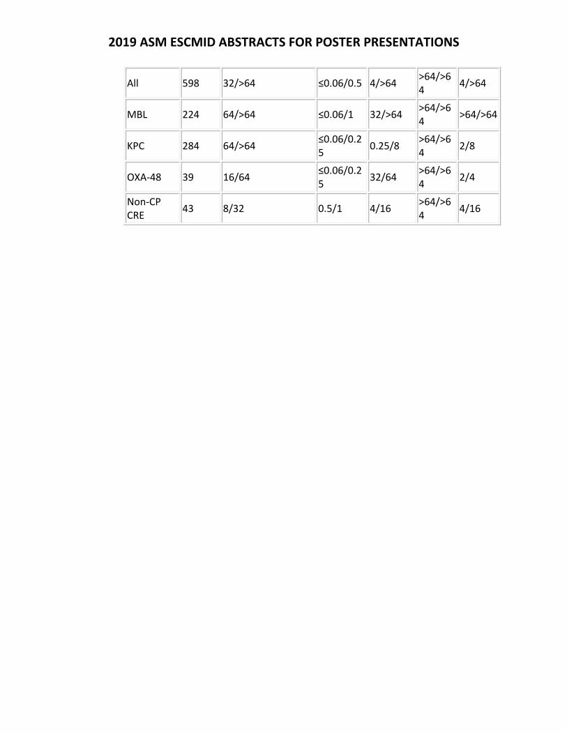
2019 ASM ESCMID ABSTRACTS FOR POSTER PRESENTATIONS
All 598 32/>64 ≤0.06/0.5 4/>64 >64/>64
4/>64
MBL 224 64/>64 ≤0.06/1 32/>64 >64/>64
>64/>64
KPC 284 64/>64 ≤0.06/0.25
0.25/8 >64/>64
2/8
OXA‐48 39 16/64 ≤0.06/0.25
32/64 >64/>64
2/4
Non‐CP CRE
43 8/32 0.5/1 4/16 >64/>64
4/16
2019 ASM ESCMID ABSTRACTS FOR POSTER PRESENTATIONS
Thursday Presentation Number:
T‐33
Publishing Title:
Addressing the Elephant in the Room: Identifying Key Areas of Opportunity and Unmet Need to Support the Development of Therapies for HABP/VABP Due toCarbapenem‐Resistant Organisms
Author Block: N. Bardhan1, N. T. Antunes2, D. W. Holman2; 1Decision Resources Group, Gurugram, India, 2Decision Resources Group, Burlington, MA
Abstract Body:
Background: Hospital‐acquired and ventilator‐associated bacterial pneumonia (HABP/VABP) are the most prevalent infections among patients admitted to the ICU. Members of the Enterobacterales, Pseudomonas aeruginosa and Acinetobacter baumannii are key causative gram‐negative pathogens in HABP/VABP that are becoming increasingly resistant to carbapenems. The current treatment of HABP/VABP due to carbapenem‐resistant organisms (CROs) includes older generic drugs, such as polymyxins, colistin, tigecycline, and aminoglycosides that, while inexpensive, are often associated with safety/tolerability concerns. Newer drugs such as ceftazidime/avibactam and meropenem/vaborbactam offer some improvements in safety and tolerability but come at a premium price. The goal of this project was to identify key areas of opportunity and unmet need for developers of new therapies for the treatment of HABP/VABP due to CROs. Methods: We surveyed 91 ID specialists from US and Europe in February 2019 and an adaptive choice‐based conjoint (ACBC) module was included to evaluate conscious and unconscious prescribing drivers and physician’s willingness to make tradeoffs across seven clinical and nonclinical attributes, including drug price. Results/conclusions: Analysis of data identified several attributes as key influencers/hidden opportunities, including ACM (all‐cause mortality) rate at 28 days, incidence of nephrotoxicity, source of clinical data and price per day. This allowed us to model the impact of the ACBC analysis findings and create user‐defined TPPs with varying performance across different attributes, helping us to evaluate their impact on the share of preference and prescribing likelihood for each TPP included in these market scenarios.
2019 ASM ESCMID ABSTRACTS FOR POSTER PRESENTATIONS
Thursday Presentation Number:
T‐34
Publishing Title: Anti‐Gonorrhoeal Ribosome Rescue Inhibitors; Susceptible Sexually Transmitted Pathogens and in vivo Proof of Concept
Author Block:
Z. D. Aron1, M. T. Torhan1, S. Cardinale1, M. Cabrera Goss2, S. Kwasny1, L. Soileau3, A. E. Jerse3, K. L. Connolly4, R. J. Suchland5, D. G. Edmondson6, S. J. Norris6, M. Butler1, T. Opperman1, T. Bowlin1, K. Keiler7; 1Microbiotix, Worcester, MA, 2Penn State Univ., University Park, PA, 3Uniformed Services Univ., BETHESDA, MD, 4Uniformed Services Univ., Bethesda, MD, 5Univ. of Washington, Seattle, WA, 6Univ. of Texas Hlth.Sci. Ctr., Houston, TX, 7Penn State Univ., Worcester, PA
Abstract Body:
Background: Bacterial translation is plagued by transcription errors, mRNA damage and translational frameshifts that lead to non‐stop ribosome complexes (nsRCs), preventing release of protein products and inhibiting further translation. Recovery of nsRCs in Neisseria gonorrhoeae is mediated by trans‐translation, an essential process conserved across all sequenced bacterial genomes. We previously identified small molecule inhibitors of trans‐translation that act as broad‐spectrum antibiotics. Here we provide an overview of their SAR and ADME, describe their spectrum of activity towards sexually transmitted pathogens, and demonstrate the in vivo efficacy of a lead compound, MBX‐4132, against MDR murine N. gonorrhoeae infections. Methods: Analogs were synthesized using published methods. Potency was determined through a previously described luciferase reporter system for trans‐translation as well as MIC determination vs. various pathogens using published methods. In vitro ADMET assays were used to estimate pharmacokinetic properties of analogs. Pharmocokinetic parameters from in vivo assays were calculated using WinNonLin. Efficacy was determined using the published vaginal murine model. Results: Evaluation of the spectrum of activity of MBX‐4132 revealed activity against both Chlamydia trachomatis and Treponema pallidum. Furthermore, in vivo studies of MBX‐4132 demonstrated a highly statistically significant effect in murine gonococcal infections, with 70‐90% clearance at a single dose of 10 mg/kg and highly significant dose response. Conclusions: This work provides both a key in vivo proof of concept and establishes a spectrum of activity applicable to the empirical treatment of sexually transmitted infections (STIs). Acknowledgments/ References: References: Ramadoss, N. S., et. al., 2013, PNAS 110:10282‐10287. Connolly, K. L., et. al. 2019, AAC e01644‐18. This research was supported in part by NIAID under awards R43AI113993 and R01AI132276.
2019 ASM ESCMID ABSTRACTS FOR POSTER PRESENTATIONS
Thursday Presentation Number:
T‐35
Publishing Title: Early Clinical Response and Clinical Stability as Predictors of Overall Clinical Response in Community‐acquired Bacterial Pneumonia
Author Block: J. Ramirez1, E. Tzanis2, M. Curran2, B. Noble2, A. Manly2, C. Kirsch2, S. Chitra2, P. McGovern2; 1Univ. of Louisville, Louisville, KY, 2Paratek Pharmaceuticals, Inc., King of Prussia, PA
Abstract Body:
Background. Clinical stability informs treatment decisions including oral switch and hospital discharge in patients with community‐acquired bacterial pneumonia (CABP). Early clinical response (ECR) is an FDA‐defined endpoint in registrational trials of CABP, that could also potentially inform treatment decisions. The Omadacycline for Pneumonia Treatment In the Community (OPTIC) phase 3 trial demonstrated non‐inferiority of omadacycline (OMC), a novel aminomethylcycline antibiotic, to moxifloxacin (MOX) for treatment of CABP. We describe the performance of ECR and clinical stability as well as its performance with an investigator‐assessment of clinical response (IACR) at the post treatment evaluation (PTE). Methods. A multicenter, randomized, double‐blind study compared OMC with MOX for treatment of CABP in adults. Patients received OMC or MOX IV treatment ≥3 days, then could transition to oral treatment. Total treatment duration was 7‐14 days. ECR and clinical stability were assessed 72‐120 hours after administration of first dose. ECR was defined as survival, no use of rescue antibiotics, and improvement in ≥2 CABP symptoms (cough, sputum production, pleuritic chest pain, dyspnea) with no worsening of other CABP symptoms; clinical stability was based on improvement in vital signs. IACR was assessed at PTE (5‐10 days after last dose); success was defined as survival with resolution of signs and symptoms of the infection such that further antibacterial therapy was not necessary. Results. Most patients achieved ECR (OMC: 81.1%, MOX: 82.7%) and clinical stability (OMC: 88.9%, MOX: 89.3%) during the 72‐120‐hour window. Both ECR and clinical stability exhibited high concordance (>70%) with clinical success at PTE. ECR and clinical stability demonstrated high sensitivity (>80%) and positive predictive value (>90%) for clinical success at PTE, whereas negative predictive value was poor for both assessments (<50%). Conclusions. Patients with CABP achieve both ECR and clinical stability at high rates 72‐120 hours after treatment initiation. ECR and clinical stability are good predictors of clinical success at PTE but poor predictors of clinical failure at PTE. Further studies are needed to validate the clinical utility of the ECR assessment.
2019 ASM ESCMID ABSTRACTS FOR POSTER PRESENTATIONS
Thursday Presentation Number:
T‐36
Publishing Title:
Bayesian Adaptive Dose‐Response Studies in Complicated and Uncomplicated Urinary Tract Infection
Author Block: M. Curran, M. Ciesielska, R. Noble, P. B. Eckburg, A. Manley, E. Tzanis, P. C. McGovern; Paratek Pharmaceuticals, Inc., King of Prussia, PA
Abstract Body:
Background: Adaptive design methods, based on pre‐specified rules and data accrual, have become prevalent in clinical development. In Phase 2, elucidation of the dose‐response function and incorporating Bayesian methods are ideal in preventing subsequent dose changes or failure in Phase 3 due to inappropriate dose selection. Compared with traditional study designs these methods incorporate scientifically driven decision rules in the design stage, which aim to enhance the use of evidence at the decision gates, resulting in a more efficient study. Methods: We present 2 statistical designs for proof‐of‐concept and dose selection Phase 2 studies of omadacycline (OMC) for treating adult females with uncomplicated urinary tract infection (uUTI), or adult females with acute pyelonephritis (AP). Both are randomized, double‐blind, adaptive, dose‐response studies with 3 interim analyses. During both studies, an unblinded Data Monitoring Committee (DMC) monitored safety, tolerability, and efficacy using pre‐defined decision rules based on Bayesian logistics regression to initiate or drop OMC treatment group(s) or modify the randomization ratio. Response criteria were targeted toward estimating the probability that the clinical success rate (proportion of patients) for each dose group was within 10% of the comparator group. If the probability exceeded 80% for a dose group, recruitment for that group was increased to improve precision of the estimate. Dose group enrollment could be stopped based on safety and tolerability.Results: The uUTI study was designed to enroll 220 patients into 1 of 3 OMC dose groups or a single nitrofurantoin dose. The AP study was designed to enroll 200 patients into 1 of 4 OMC dose groups or levofloxacin. Initial patient allocation to treatment groups was equal. Scientific challenges included expected dose‐response relationships, i.e. monotonicity, and grouping of doses to improve decision making. Operational challenges were similar in both studies and included fast enrollment rates, few patients with clean data for microbiological endpoints, and the lag between data collection and data availability for DMC meetings. Conclusions: Bayesian adaptive designs in Phase 2 dose selection studies allow more patients to enroll in the more effective and tolerable dosing groups, maximizing precision and value of evidence utilizing the most efficient sample size. Modeling and simulation efforts upfront, along with knowing the strength of evidence requirement, can aid in mitigating scientific and operational challenges.
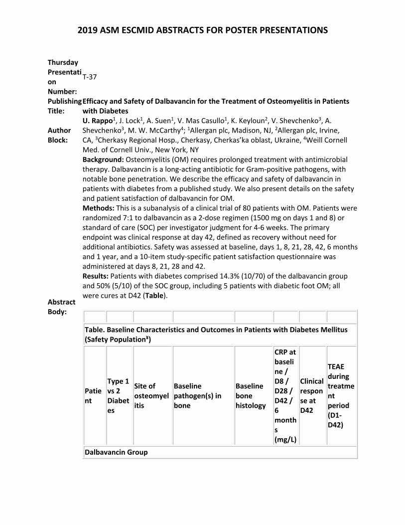
2019 ASM ESCMID ABSTRACTS FOR POSTER PRESENTATIONS
Thursday Presentation Number:
T‐37
Publishing Title:
Efficacy and Safety of Dalbavancin for the Treatment of Osteomyelitis in Patients with Diabetes
Author Block:
U. Rappo1, J. Lock1, A. Suen1, V. Mas Casullo1, K. Keyloun2, V. Shevchenko3, A. Shevchenko3, M. W. McCarthy4; 1Allergan plc, Madison, NJ, 2Allergan plc, Irvine, CA, 3Cherkasy Regional Hosp., Cherkasy, Cherkas’ka oblast, Ukraine, 4Weill Cornell Med. of Cornell Univ., New York, NY
Abstract Body:
Background: Osteomyelitis (OM) requires prolonged treatment with antimicrobial therapy. Dalbavancin is a long‐acting antibiotic for Gram‐positive pathogens, with notable bone penetration. We describe the efficacy and safety of dalbavancin in patients with diabetes from a published study. We also present details on the safety and patient satisfaction of dalbavancin for OM. Methods: This is a subanalysis of a clinical trial of 80 patients with OM. Patients were randomized 7:1 to dalbavancin as a 2‐dose regimen (1500 mg on days 1 and 8) or standard of care (SOC) per investigator judgment for 4‐6 weeks. The primary endpoint was clinical response at day 42, defined as recovery without need for additional antibiotics. Safety was assessed at baseline, days 1, 8, 21, 28, 42, 6 months and 1 year, and a 10‐item study‐specific patient satisfaction questionnaire was administered at days 8, 21, 28 and 42. Results: Patients with diabetes comprised 14.3% (10/70) of the dalbavancin group and 50% (5/10) of the SOC group, including 5 patients with diabetic foot OM; all were cures at D42 (Table).
Table. Baseline Characteristics and Outcomes in Patients with Diabetes Mellitus (Safety Population¥)
Patient
Type 1 vs 2 Diabetes
Site of osteomyelitis
Baseline pathogen(s) in bone
Baseline bone histology
CRP at baseline / D8 / D28 / D42 / 6 months (mg/L)
Clinical response at D42
TEAE during treatment period (D1‐D42)
Dalbavancin Group
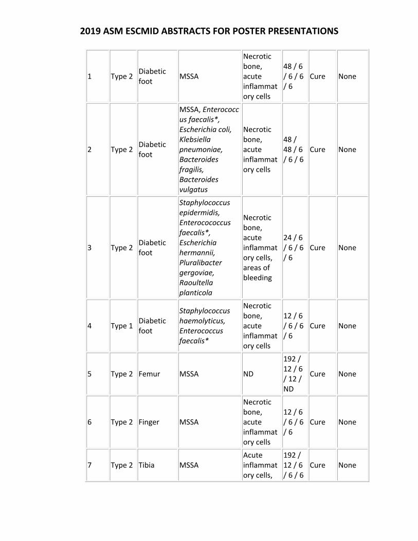
2019 ASM ESCMID ABSTRACTS FOR POSTER PRESENTATIONS
1 Type 2 Diabetic foot
MSSA
Necrotic bone, acute inflammatory cells
48 / 6 / 6 / 6 / 6
Cure None
2 Type 2 Diabetic foot
MSSA, Enterococcus faecalis*, Escherichia coli, Klebsiella pneumoniae, Bacteroides fragilis, Bacteroides vulgatus
Necrotic bone, acute inflammatory cells
48 / 48 / 6 / 6 / 6
Cure None
3 Type 2 Diabetic foot
Staphylococcus epidermidis, Enterocococcus faecalis*, Escherichia hermannii, Pluralibacter gergoviae, Raoultella planticola
Necrotic bone, acute inflammatory cells, areas of bleeding
24 / 6 / 6 / 6 / 6
Cure None
4 Type 1 Diabetic foot
Staphylococcus haemolyticus, Enterococcus faecalis*
Necrotic bone, acute inflammatory cells
12 / 6 / 6 / 6 / 6
Cure None
5 Type 2 Femur MSSA ND
192 / 12 / 6 / 12 / ND
Cure None
6 Type 2 Finger MSSA
Necrotic bone, acute inflammatory cells
12 / 6 / 6 / 6 / 6
Cure None
7 Type 2 Tibia MSSA Acute inflammatory cells,
192 / 12 / 6 / 6 / 6
Cure None
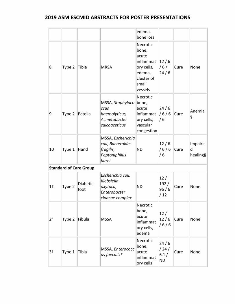
2019 ASM ESCMID ABSTRACTS FOR POSTER PRESENTATIONS
edema, bone loss
8 Type 2 Tibia MRSA
Necrotic bone, acute inflammatory cells, edema, cluster of small vessels
12 / 6 / 6 / 24 / 6
Cure None
9 Type 2 Patella
MSSA, Staphylococcus haemolyticus, Acinetobacter calcoaceticus
Necrotic bone, acute inflammatory cells, vascular congestion
24 / 6 / 6 / 6 / 6
Cure Anemia§
10 Type 1 Hand
MSSA, Escherichia coli, Bacteroides fragilis, Peptoniphilus harei
ND 12 / 6 / 6 / 6 / 6
Cure Impaired healing§
Standard of Care Group
1‡ Type 2 Diabetic foot
Escherichia coli, Klebsiella oxytoca, Enterobacter cloacae complex
ND
12 / 192 / 96 / 6 / 12
Cure None
2€ Type 2 Fibula MSSA
Necrotic bone, acute inflammatory cells, edema
12 / 12 / 6 / 6 / 6
Cure None
3† Type 1 Tibia MSSA, Enterococcus faecalis*
Necrotic bone, acute inflammatory cells
24 / 6 / 24 / 6.1 / ND
Cure None
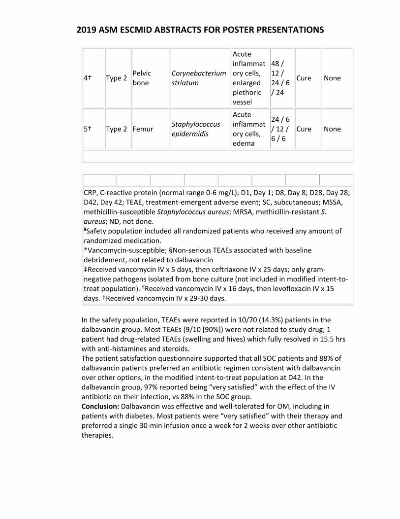
2019 ASM ESCMID ABSTRACTS FOR POSTER PRESENTATIONS
4† Type 2 Pelvic bone
Corynebacterium striatum
Acute inflammatory cells, enlarged plethoric vessel
48 / 12 / 24 / 6 / 24
Cure None
5† Type 2 Femur Staphylococcus epidermidis
Acute inflammatory cells, edema
24 / 6 / 12 / 6 / 6
Cure None
CRP, C‐reactive protein (normal range 0‐6 mg/L); D1, Day 1; D8, Day 8; D28, Day 28; D42, Day 42; TEAE, treatment‐emergent adverse event; SC, subcutaneous; MSSA, methicillin‐susceptible Staphylococcus aureus; MRSA, methicillin‐resistant S. aureus; ND, not done. ¥Safety population included all randomized patients who received any amount of randomized medication. *Vancomycin‐susceptible; §Non‐serious TEAEs associated with baseline debridement, not related to dalbavancin ‡Received vancomycin IV x 5 days, then ce riaxone IV x 25 days; only gram‐negative pathogens isolated from bone culture (not included in modified intent‐to‐treat population). €Received vancomycin IV x 16 days, then levofloxacin IV x 15 days. †Received vancomycin IV x 29‐30 days.
In the safety population, TEAEs were reported in 10/70 (14.3%) patients in the dalbavancin group. Most TEAEs (9/10 [90%]) were not related to study drug; 1 patient had drug‐related TEAEs (swelling and hives) which fully resolved in 15.5 hrs with anti‐histamines and steroids. The patient satisfaction questionnaire supported that all SOC patients and 88% of dalbavancin patients preferred an antibiotic regimen consistent with dalbavancin over other options, in the modified intent‐to‐treat population at D42. In the dalbavancin group, 97% reported being “very satisfied” with the effect of the IV antibiotic on their infection, vs 88% in the SOC group. Conclusion: Dalbavancin was effective and well‐tolerated for OM, including in patients with diabetes. Most patients were “very satisfied” with their therapy and preferred a single 30‐min infusion once a week for 2 weeks over other antibiotic therapies.
2019 ASM ESCMID ABSTRACTS FOR POSTER PRESENTATIONS
Thursday Presentation Number:
T‐38
Publishing Title:
The Novel β‐lactamase Inhibitor Enmetazobactam is More Potent than Tazobactam against ESBL‐producing Enterobacteriaceae
Author Block:
A. Belley1, I. Morrissey2, S. Hawser3, M. Huband4, K. Papp‐Wallace5, R. A. Bonomo5, P. Knechtle1; 1Allecra Therapeutics SAS, Saint Louis, France, 2IHMA Europe Sarl, Monthey, Switzerland, 3IHMA Europe Sarl, 1870 Monthey, Switzerland, 4JMI Lab. Inc, North Liberty, IA, 5Louis Stokes Cleveland Dept. of Veteran Affairs Med. Ctr., Cleveland, OH
Abstract Body:
Background: New carbapenem‐sparing therapies are needed for infections caused by extended‐spectrum β‐lactamase (ESBL)‐producing Enterobacteriaceae. Cefepime (FEP) combined with the novel ESBL inhibitor enmetazobactam (EMT, formerly AAI101) is in phase 3 development for the treatment of adult patients with cUTI/AP. In this study, the in vitro activity of EMT relative to tazobactam (TZB) was compared against ESBL‐producing‐ and FEP‐resistant Enterobacteriaceae.Methods: Broth microdilution MIC assays were performed following CLSI guidelines. Isolates tested were Escherichia coli isogenic strains (15) expressing ESBLs (CTX‐M, SHV, TEM); clinical isolates (CI) of E. coli (109) and Klebsiella pneumoniae (102) expressing diverse ESBLs with or without AmpC and/or OXA‐48; and CI of Enterobacteriaceae (41) resistant to FEP (MIC ≥16 µg/ml) but susceptible to meropenem (MIC ≤1 µg/ml). Results: Against the 15 isogenic strains of ESBL‐producing E. coli, addition of 4 or 8 µg/ml of EMT reduced the MIC90 value of FEP 128‐fold from 16 to 0.12 µg/ml (Table). The MIC90 of piperacillin‐EMT (8 µg/ml) was ≥32‐fold lower than piperacillin‐TZB (256 µg/ml). Against the ESBL‐producing E. coli and K. pneumoniae CI, MIC90 values for FEP‐EMT were ≥1,024‐fold (0.12 µg/ml) and ≥128‐fold (1 µg/ml) lower, respectively, than the FEP MIC90 value alone (>64 µg/ml) for both species. FEP‐TZB was less potent against the ESBL‐producing K. pneumoniae CI, with an MIC90 of 8 µg/ml. Against the FEP‐resistant Enterobacteriaceae, FEP‐EMT exhibited an MIC90 of 1 µg/ml. FEP‐EMT exhibited similar potency as meropenem against all organism groups.Conclusions: EMT exhibits more potent inhibitory activity than TZB against ESBL‐producing Enterobacteriaceae. Continued development of FEP‐EMT as empiric therapy in settings where ESBLs with or without AmpC and/or OXA‐48 are prevalent is warranted.
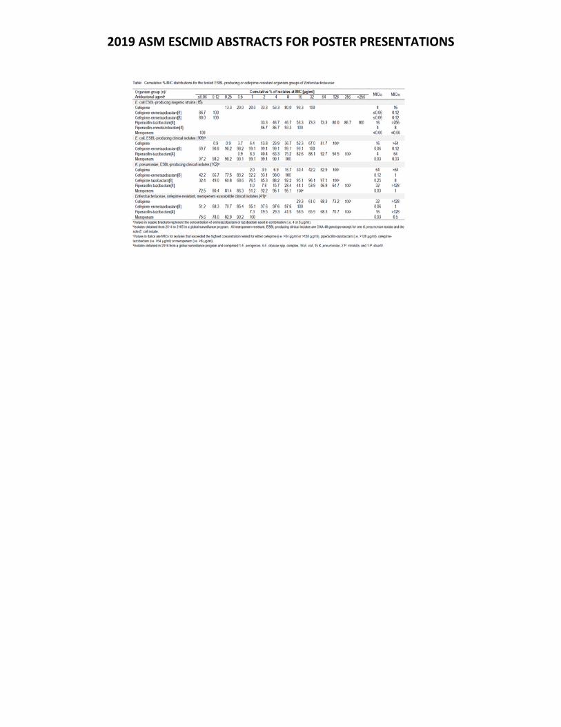
2019 ASM ESCMID ABSTRACTS FOR POSTER PRESENTATIONS
2019 ASM ESCMID ABSTRACTS FOR POSTER PRESENTATIONS
Thursday Presentation Number:
T‐39
Publishing Title: Development of an Experimental Model of Bacterial Meningoencephalitisto Facilitate Antimicrobial Drug Development for Neonates
Author Block: N. Farrington1, L. McEntee1, A. Johnson1, A. Kirby1, S. Gore1, R. Greenberg2, S. Das1, W. Hope1; 1Univ. of Liverpool, Liverpool, United Kingdom, 2Duke Clinical Res. Inst., Durham, NC
Abstract Body:
Background: The aim of this project was to develop and characterize a rabbit model of meningoencephalitis that can be used to develop new antibiotics for MDR and XDR bacterial diseases for neonates. Methods: Male New Zealand White rabbits were used. The challenge strain was Pseudomonas aeruginosa ATCC 27853. The inoculum was administered intrathecally via the cistern under general anaesthesia. Antimicrobial treatment was initiated 6 hours post infection. The duration of the experimental period was 30 hours. Meropenem was administered intravenously at 5, 10 and 30 mg/kg q8h. Blood samples to estimate plasma concentrations were taken throughout the first dosing interval. Animals were sacrificed 10h and 30hr post infection. Terminal CSF samples were collected. The brain was dissected and submitted for quantitative cultures and histological analysis. The meninges were collected and frozen for PK analyses. PK‐PD modelling was performed using Pmetrics. Results: The bacterial density progressively increased in the cerebrum and CSF of untreated controls. There was progressive meningoencephalitis with florid inflammation 30 hours post inoculation. There was a dose‐response relationship in both cerebrum and CSF. Near maximal activity was observed with a regimen of 30mg/kg q8h, which also limited tissue damage at both 10 and 30 hours post infection. A three‐compartment PK‐PD (central and peripheral compartments, and CSF) model described the PK‐PD data well. The model was used to link drug exposure in the plasma, CSF and the resultant antibacterial activity of meropenem within the CSF. Conclusions: The rabbit model of neonatal meningoencephalitis is a valuable new tool to assess the pharmacodynamics of antimicrobial agents for neonatal infection. The pharmacodynamics of meropenem has been described and serves as a benchmark for evaluation of new antimicrobial agents for potential use in neonates. Such an approach provides a new pathway for the accelerated development of antibiotics for a special population and and enables new treatment options to be available to address unmet medical need.
2019 ASM ESCMID ABSTRACTS FOR POSTER PRESENTATIONS
Thursday Presentation Number:
T‐40
Publishing Title: Assigning Potency Values to USP Antibiotic Reference Standards: Best Practices
Author Block: J. Fringer1, H. Tu1, L. James1, S. Krishna C2, S. Walfish1, M. Samaddar2, R. Chakrabarti2, F. Atouf1; 1US Pharmacopeia, Rockville, MD, 2US Pharmacopeia, Hyderabad, India
Abstract Body:
Background: A key step in the development of new antibiotics is assigning potency values using USP antibiotic reference standards. The potency values of USP antibiotic reference standards are traceable to primary international reference standards. In 2017, USP became aware of a discrepancy between its Nystatin reference standard and an international standard, a 13.6% difference between the label claim and laboratory data. An incorrect potency assignment can affect the outcome of release testing for antibiotic lots; therefore, USP responded by quickly mobilizing a team of scientists who investigated the entire portfolio of USP antibiotic reference standards to ensure traceability to international standards. Methods: Microbiological assays used for potency determination are found in USP General Chapter <81>: Antibiotics‐Microbial Assays, recently revised. In the cylinder plate assay, cylinders containing antibiotics are placed on a lawn of microorganisms. (For example, Penicillin G is tested against Staphylococcus aureus.) The USP antibiotic standard diffuses into the lawn and forms a no‐growth zone, the diameter of which is plotted as a standard curve versus concentration. Antibiotics with unknown potency are tested against the standard curve, resulting in a relative potency in the form of units or μg of activity per mg. Results: The result of the investigation was institution of new testing requirements for all USP antibiotic reference standards, which include a triangulated testing scheme of new reference standard lots against a primary WHO antibiotic reference standard and a European Pharmacopeia secondary reference standard. The goal of this strategy is to preserve the traceability of antibiotic potency values over time. In addition, USP has optimized the capabilities of microbiological methods in order to reduce variability. Conclusions: USP has taken stewardship of its mission to promote quality and protect public health by strengthening its oversight of antibiotic reference standard testing and stability. Because of its more stringent requirements for antibiotic testing by incorporating the usage of primary international standards, the value assignment for USP antibiotic reference standards have increased confidence levels.
2019 ASM ESCMID ABSTRACTS FOR POSTER PRESENTATIONS
Thursday Presentation Number:
T‐41
Publishing Title: Apidaecins: Linear Peptide Antibiotics for the Treatment of Serious Gram‐Negative Infections
Author Block: D. Knappe1, A. Gallinat1, R. Hoffmann2, D. Joseph‐McCarthy3; 1EnBiotix, GmbH, Leipzig, Germany, 2Univ. Leipzig, Leipzig, Germany, 3EnBiotix, Inc., Boston, MA
Abstract Body:
Background: Apidaecins are linear peptide antibiotics (LPAs) with a novel mechanism of action that are aimed at the treatment of serious, drug‐resistant Gram‐negative infections. Apidaecin 1b (GNNRPVYIPQPRPPHPRL‐OH), isolated from Apis melifera (honey bee), is a linear, proline‐rich antimicrobial peptide. Unlike most other classes of antimicrobial peptides (AMPs), apidaecins selectively enter bacterial cells without membrane disruption. As a result, they exhibit no mammalian cytotoxicity or hemolytic activity. Apidaecins exert their antibacterial activity through inhibition of protein translation, by binding to the exit tunnel of existing 70S bacterial ribosome which blocks the dissociation of release factors and by preventing the assembly of additional bacterial ribosome. Methods: MICs were determined under standard CLSI methodology in cation‐adjusted Mueller Hinton Broth. Proteolytic stability was evaluated in plasma, serum, and bronchoalveolar lavage of several mammalian species. Results: MICs across a set of Gram‐negative strains including MDR, efflux‐defective, and colistin‐resistant strains are quite promising; e.g., MICs in 25% diluted medium for Api137 were ≤0.125 to 8 μg/ml vs. Eco (n=8) and ≤0.125 to 0.5 μg/ml vs. Kpn (n=6). In the limited studies undertaken we have seen correlation between MICs in 25% diluted medium and in vivo efficacy. The frequency of resistance at 4x MIC for Api137 vs. Eco was similar to that for colistin. In addition, the effect of serum on the MICs is minimal, there is only a small MIC shift in an SmbA knockout Eco strain, and there is no evidence of cross‐resistance with strains resistant to ribosome‐targeting antibiotics. Finally, the proteolytic stability was higher overall in non‐rodent matrices. Conclusions: Taken together with previously reported encouraging in vivo results (in sepsis, thigh infection, and urinary tract infection (UTI) models), this further in vitro characterization of apidaecins supports their continued development as therapeutics, initially for complicated UTIs with a view toward expansion into complicated intra‐abdominal infections and ventilated‐pneumonias.
2019 ASM ESCMID ABSTRACTS FOR POSTER PRESENTATIONS
Thursday Presentation Number:
T‐42
Publishing Title: In vitro Activity of KTU286, a Novel Butanehydrazide Derivative with Antimicrobial Activity Against Drug Resistant Staphylococcus aureus with Defined Resistance Mechanisms
Author Block:
P. Kavaliauskas1, V. Mickevicius2, R. Petraitiene1, I. Dobtsis3, P. Gialanella4, V. Petraitis1; 1Weill Cornell Med. of Cornell Univ., New York, NY, 2Kaunas Univ. of Technology, Dept. of Organic Chemistry, Kaunas, Lithuania, 3Mem. Slone Kettering Cancer Ctr., New York, NY, 4Montefiore Hosp., Albert Einstein Coll. of Med., Bronx, NY
Abstract Body:
Introduction: Infections caused by drug resistant (DR) Staphylococcus aureus (S. aureus) pose a threat to public health. The emergence of resistance shortens therapeutic options and worsens patient’s prognosis. New antimicrobial agents are needed to overcome this problem. We therefore aimed to characterize in vitro antimicrobial activity of new butanehydrazide derivative KTU286 against MDR S. aureus strains with defined resistance mechanisms. Methods: Antimicrobial activity of KTU286 was evaluated against panel of MRSA (SCmecA+), VRSA (vanA+) and pan‐susceptible (PS) S. aureus strains by using broth microdilution method. Concentration and time‐depended killing was evaluated by using spectrophotometric methods. KTU286 biofilm disrupting activity was evaluated by using crystal violet assay. Murine LD50 was estimated using in silico models. Results: KTU286 showed considerably good (>50 µM) antimicrobial activity against all tested strains. The MIC100 for S. aureus ranged from 0.5 to 16 µg/mL (1.4‐45.9 µM). KTU286 demonstrated concentration depended (1‐8 µg/mL) antibacterial activity against MRSA strains harboring SCmecA. MIC for VRSA strains (vanA+) was 4 µg/mL. KTU286 showed activity against pan‐susceptible S. aureus strains (0.5‐2 µg/mL). KTU286 decreased viability of biofilm associated MRSA, VRSA, and PS S. aureus strains (p<0.05) as well as it was able to affect structural integrity of matured biofilms after exposition for 24 hours in comparison to untreated control. The estimated LD50 was 1000 mg/kg. Conclusion: KTU286 showed in vitro antimicrobial activity against DR S. aureus with different mechanisms of resistance. Further studies are needed for better understanding of antimicrobial activity, safety, synergistic relationship, and therapeutic potency of KTU286.
2019 ASM ESCMID ABSTRACTS FOR POSTER PRESENTATIONS
Thursday Presentation Number:
T‐43
Publishing Title: Renal Biomarkers to Monitor Functional and Degenerative Changes in the Kidney of Rats and Dogs Administered an Optimized Arylomycin
Author Block: S. T. Laing, J. Brumm, N. Stagg, G. Morrow, O. Zuniga, P. Katavolos, W. Proctor, T. S. Zabka; Genentech, South San Francisco, CA
Abstract Body:
Background. Chemically optimized arylomicin derivatives (LepBi) are potent inhibitors of the essential Gram‐negative type 1 signal peptidase and promising therapeutics for the treatment of multi‐drug resistant infections. Administration of a LepBi to dogs and rats for 7 days resulted in nephrotoxicity at high exposures. Emerging urinary renal tubular biomarkers of kidney function and injury were investigated as potential translational monitoring tools. Methods. Urine was collected at baseline (dogs only) and following 7‐days administration of LepBi at 0, 50, 100, and 150 mg/kg/day. The following biomarkers were normalized to urine creatinine and compared to microscopic findings and standard kidney labs: clusterin (CLU), osteopontin (OPN), kidney injury molecule 1 (KIM1), albumin (ALB), cystatin C (CYSC), alpha‐1‐microglobulin/bikunin precursor (AMBP), N‐acetyl‐beta‐D‐glucosaminidase (NAG), and/or retinol binding protein 4 (RBP4). Results. In rats administered 50 mg/kg and dogs at all doses, the predominant microscopic finding was non‐adverse eosinophilic inclusions in tubules without alteration of serum urea nitrogen or creatinine, or altered urinalysis. Tubular inclusions were associated with a trend of increased urinary tubular function markers, particularly CYSC (rat) and AMBP (dog). Early tubular injury in the dog (all doses) was associated with minor elevations of CLUS. Rats administered ≥ 100 mg/kg had severe tubular degeneration with elevated serum BUN, creatinine, and urinary casts that correlated with robust elevations of the tubular injury markers (CLU, OPN, KIM1, NAG) as well as augmented elevations of tubular functional biomarkers (ALB, AMBP, CYSC). Conclusions. These data suggest that LepBi‐induced renal tubular inclusions and degeneration may be monitored using distinct combinations of urinary renal tubular biomarkers in conjunction with standard labs. Considerations for a composite score, as described in the Clinical Qualification of these biomarkers, will be discussed.
2019 ASM ESCMID ABSTRACTS FOR POSTER PRESENTATIONS
Thursday Presentation Number:
T‐44
Publishing Title: QPX7728: Resistance Selection, Prevention, and Molecular Mechanisms in Mutants of KPC‐producing Klebsiella pneumoniae
Author Block: O. Lomovskaya, K. Nelson, D. Rubio‐Aparicio; Qpex Biopharma, Inc, San Diego, CA
Abstract Body:
Background: QPX7728 (QPX) is a new ultra‐broad‐spectrum beta‐lactamase inhibitor based on a cyclic boronic acid pharmacophore capable of inhibiting numerous serine (classes A, C, D) and metallo‐beta‐lactamases (class B), including class A, B and D carbapenemases. As a part of systematic resistance studies, we evaluated the selection of mutants with reduced sensitivity to the combination of QPX with meropenem using KPC‐producing strains of CRE and characterized the selected mutations. Methods: Fifteen strains of KPC‐producing Klebsiella pneumoniae with pre‐existing resistance mechanisms (mutations in porins OmpK35 and OmpK36, upregulated AcrAB) were selected from a worldwide collection of isolates. Resistance studies were performed with MER at 8 µg/ml and QPX varied from 0.5 to 8 µg/ml. Some of the strains were also used in resistance studies using ceftazidime‐avibactam (C/A) at 2x ‐ 8x the MIC (with avibactam [AVI] fixed at 4 µg/ml). Results: MER at 8 μg/ml and QPX at 2‐4 μg/ml suppressed the drug‐resistance mutation frequency to <1 × 10‐9 in all strains. Mutants selected at lower drug concentrations showed phenotypes associated with previously described carbapenem resistance mechanisms, including ompK36 inactivation in mutants selected from OmpK36 proficient strains, and an increased blaKPC gene copy number in strains with partially functional or non‐functional OmpK36. No mutations in the coding region of blaKPC were identified. In contrast, C/A only selected mutations in blaKPC in all KPC‐producing strains. The majority of these mutants did not have an increase in MER/QPX MICs Conclusions: These data indicate that selection of mutants with reduced sensitivity to MER/QPX from KPC‐producing Klebsiella pneumoniae is associated with previously described mechanisms involving porin mutations that largely impact MER and the increase in the blaKPC gene copy number and not amino acid changes in the KPC enzyme; the latter was routinely observed for mutants selected with C/A. of note, KPC mutations that affect the potency of C/A have minimal effect on potency of MER/QPX. (This project has been funded in whole or in part with Federal funds from the Department of Health and Human Services; Office of the Assistant Secretary for Preparedness and Response; Biomedical Advanced Research and Development Authority (BARDA), under OTA number HHSO100201600026C.)
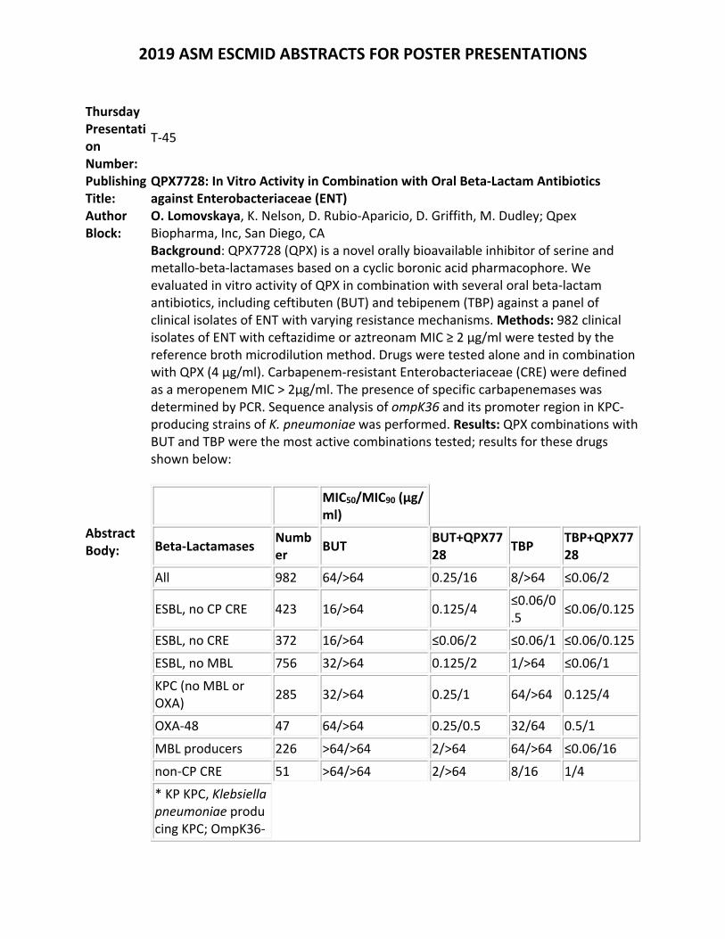
2019 ASM ESCMID ABSTRACTS FOR POSTER PRESENTATIONS
Thursday Presentation Number:
T‐45
Publishing Title:
QPX7728: In Vitro Activity in Combination with Oral Beta‐Lactam Antibiotics against Enterobacteriaceae (ENT)
Author Block:
O. Lomovskaya, K. Nelson, D. Rubio‐Aparicio, D. Griffith, M. Dudley; Qpex Biopharma, Inc, San Diego, CA
Abstract Body:
Background: QPX7728 (QPX) is a novel orally bioavailable inhibitor of serine and metallo‐beta‐lactamases based on a cyclic boronic acid pharmacophore. We evaluated in vitro activity of QPX in combination with several oral beta‐lactam antibiotics, including ceftibuten (BUT) and tebipenem (TBP) against a panel of clinical isolates of ENT with varying resistance mechanisms. Methods: 982 clinical isolates of ENT with ceftazidime or aztreonam MIC ≥ 2 µg/ml were tested by the reference broth microdilution method. Drugs were tested alone and in combination with QPX (4 µg/ml). Carbapenem‐resistant Enterobacteriaceae (CRE) were defined as a meropenem MIC > 2µg/ml. The presence of specific carbapenemases was determined by PCR. Sequence analysis of ompK36 and its promoter region in KPC‐producing strains of K. pneumoniae was performed. Results: QPX combinations with BUT and TBP were the most active combinations tested; results for these drugs shown below:
MIC50/MIC90 (µg/ml)
Beta‐Lactamases Number
BUT BUT+QPX7728
TBP TBP+QPX7728
All 982 64/>64 0.25/16 8/>64 ≤0.06/2
ESBL, no CP CRE 423 16/>64 0.125/4 ≤0.06/0.5
≤0.06/0.125
ESBL, no CRE 372 16/>64 ≤0.06/2 ≤0.06/1 ≤0.06/0.125
ESBL, no MBL 756 32/>64 0.125/2 1/>64 ≤0.06/1
KPC (no MBL or OXA)
285 32/>64 0.25/1 64/>64 0.125/4
OXA‐48 47 64/>64 0.25/0.5 32/64 0.5/1
MBL producers 226 >64/>64 2/>64 64/>64 ≤0.06/16
non‐CP CRE 51 >64/>64 2/>64 8/16 1/4
* KP KPC, Klebsiella pneumoniae producing KPC; OmpK36‐
2019 ASM ESCMID ABSTRACTS FOR POSTER PRESENTATIONS
F, full‐copy functional OmpK36; OmpK36‐DEF, partially functional or non‐functional OmpK36 due to insertions or decreased expression.
Based on MIC90, potency of BUT and TBP against these strains was increased >8‐fold and >32‐fold, respectively. Neither BUT or TBP alone had any relevant activity against KPC, OXA‐48, MBL‐producing strains or against non‐carbapenemase producing (CP) CRE. In general, TBP/QPX was numerically more potent than BUT/QPX with the exception of strains with a non‐functional or partially functional porin OmpK36; defects in OmpK36 decreased potency of BUT/QPX to a lesser degree compared to TBP/QPX. QPX potentiated activity of BUT and TBP to a similar degree against KPC‐producers with a functional OmpK36 and against OXA‐48 producers. QPX also enhanced activity of both drugs in strains producing MBLs. Conclusions: Combinations of QPX7728 with oral beta‐lactam antibiotics have an attractive microbiological profile and warrant further investigation. (This project has been funded in whole or in part with Federal funds from the Department of Health and Human Services; Office of the Assistant Secretary for Preparedness and Response; Biomedical Advanced Research and Development Authority (BARDA), under OTA number HHSO100201600026C.)
2019 ASM ESCMID ABSTRACTS FOR POSTER PRESENTATIONS
Thursday Presentation Number:
T‐46
Publishing Title: The Pyranopyridine Efflux Pump Inhibitor (EPI) MBX‐4191 as an Adjunctive to Tetracycline Antibiotics for Treating MDR Enterobacteriaceae Infections
Author Block: T. J. OPPERMAN, 016051, S. M. Kwasny1, R. Barber1, M. Carlton1, S. C. Cardinale1, W. J. Weiss2, T. L. Bowlin1, Z. D. Aron1; 1Microbiotix, Inc, Worcester, MA, 2Univ. of North Texas Hlth.Sci. Ctr., Fort Worth, TX
Abstract Body:
Background: Infections caused by multi‐drug resistant (MDR) Enterobacteriaceae are becoming increasingly difficult to treat with existing antibiotics. Efflux pumps of the Resistance Nodulation Division Efflux (RND) family play a major role in MDR and virulence. Here we present the in vitro and in vivo activities of MBX‐4191, a pyranopyridine (PyPy) EPI that is a potent inhibitor of the major RND efflux pump (AcrB) in the Enterobacteriaceae and potentiates the antibacterial activity of tetracycline antibiotics against these organisms. Methods: MBX‐4191 was synthesized using published methods. Time kill assays were performed using standard methods. MIC90s were measured using the CLSI protocol for broth dilution MICs. In vitro ADMET assays (cytotoxicity, solubility, murine liver microsome stability (MLMS), serum binding) were used to prioritize analogs for in vivo studies. PK parameters from in vivo assays in mice were calculated using WinNonLin. A murine model of sepsis was used to demonstrate in vivo efficacy of MBX‐4191 in combination with minocycline (MIN). Results: MBX‐4191 potentiated the bactericidal activity of MIN (4 µg/ml) against E. coli and K. pneumoniae in time kill assays, and reduced the MIC90 of tetracycline antibiotics (MIN, eravacycline, and omadacycline) against MDR panels of E. coli and K. pneumoniae by 8‐32 fold. The in vitro ADMET profile of MBX‐4191 was suitable for in vivo studies [aqueous solubility ≥ 100 µM), CC50 = 47 µM, and MLMS (97 % remaining after 30 min)]. In a murine PK study, MBX‐4191 achieved high levels of exposure (AUC = 26,000 hr*ng/mL at 10 mg/kg IV) and was well‐tolerated by mice after a single intravenous or intraperitoneal dose (MTD ≥200 mg/kg). In the murine sepsis infection model, MBX‐4191 rescued the activity of MIN against a MIN‐resistant strain of K. pneumonia (MIC MIN = 32 µg/ml). Conclusion: MBX‐4191 has demonstrated in vivo proof‐of‐principle as an adjunctive therapy to MIN.
2019 ASM ESCMID ABSTRACTS FOR POSTER PRESENTATIONS
Thursday Presentation Number:
T‐47
Publishing Title: In‐vivo Efficacy of Apramycin Against Enterobacteriaceae and A.baumannii
Author Block: J. U. Hansen1, E. Leipins2, S. Grinberga2, S. N. Hobbie3, C. Vingsbo Lundberg1; 1Statens Serum Inst., Copenhagen, Denmark, 2Latvian Inst. of Organic Synthesis, Riga, Latvia, 3Univ. of Zurich, Zürich, Switzerland
Abstract Body:
Background: Apramycin is an aminoglycoside recently described to have a broad activity against multidrug‐, carbapenem‐ and aminoglycoside‐resistant Enterobacteriaceae and A. baumannii. In this study, we evaluate the in vivo efficacy of apramycin in murine infection models with E. coli, K. pneumoniae and A. baumannii. Methods: A single dose apramycin was evaluated in a septicemia and a pneumonia model in female mice inoculated with aminoglycoside‐susceptible E. coli (EN121, APR MIC=4‐8, GEN MIC=0.5‐1) or K. pneumoniae (EN124, APR MIC 1‐2, GEN MIC= 0.25). Next we established mouse models of thigh infection and cUTI in female mice using gentamicin‐resistant E. coli (EN591, APR MIC=4‐8, GEN MIC>256) and A. baumannii (EN592, APR MIC=4, GEN MIC>256). Mice were treated with a single or multiple doses of apramycin, vehicle or control antibiotics and bacterial loads in relevant compartments were quantified. Apramycin PK and elimination was evaluated in healthy and infected mice. Results: Against septicemia, apramycin reduced the bacterial load by more than 4 log compared to vehicle controls, and 2.3 (EN121) or 3.5 (EN124) log compared to start of treatment. The ED50 of apramycin was 9.1 mg/kg (EN121) or 12.7 mg/kg (EN124). The ED50 of gentamicin was 1.1 mg/kg (EN121) or 3.2 mg/kg (EN124). In the pneumonia model, the ED50 against EN124 was 26 mg/kg. The pharmacokinetic profile of gentamicin and apramycin in mice was similar, and thus the efficacy of apramycin and gentamicin was comparable and reflected the relative MICs of the two drugs against these two bacterial isolates Apramycin is eliminated renally with high drug concentrations achieved in urine, bladder and kidney. Against gentamicin‐resistant E. coli EN591 cUTI, the apramycin ED50 in kidney was 0.8 mg/kg and the bacterial burden was reduced more than 3 log. A single dose of apramycin was also efficacious against aminoglycoside‐resistant isolates in the thigh infection model, whereas gentamicin had no effect. Conclusion: In mice, the efficacy of apramycin is comparable to gentamicin and reflects the MIC of these drugs. Further, apramycin is efficacious against MDR clinical isolates that are untreatable with gentamicin in mice.
2019 ASM ESCMID ABSTRACTS FOR POSTER PRESENTATIONS
Thursday Presentation Number:
T‐48
Publishing Title: Time‐kill Kinetics of ACX‐362E against Clostridioides difficile
Author Block: B. Murray1, C. Wolfe1, A. Marra1, M. H. Silverman2, C. Pillar1, D. Shinabarger1; 1Micromyx, Inc., Kalamazoo, MI, 2Acurx, LLC, White Plains, NY
Abstract Body:
Background: ACX‐362E is a novel DNA polymerase IIIC inhibitor in clinical development for the treatment of Clostridioides difficile infections. The bactericidal activity of ACX‐362E was evaluated against actively‐growing C. difficile by determining the MIC and the minimum bactericidal concentration (MBC) against three C. difficile isolates. Time‐kill kinetics assays were additionally performed against the three C. difficile isolates with metronidazole and vancomycin as comparators.Methods: ACX‐362E and comparator susceptibility testing was conducted per CLSI published methodology (M11). MBC values were obtained by determining the viable count of each inoculum immediately following inoculation of MIC plates and comparing that to the viable count in the MIC well and three wells above the MIC post‐incubation. The MBC was then defined as the lowest concentration of agent to demonstrate a 99% killing of bacteria (CLSI M26‐A). Time‐kill kinetics were determined using methods described by CLSI (M26‐A) used to evaluate the bactericidal activity of test agents. All compounds were tested in the assay at 8X, 16X and 32X the broth microdilution MIC and viable counts were determined at 2, 6, 24 and 48 hours.Results: ACX‐362E had MIC values of 1 µg/mL against C. difficile MMX 5680 (ribotype 027), BAA 1382 (ribotype 078), and BAA 1875 (ribotype 078), respectively, and corresponding MBC:MIC ratios of 1, 2 and >8. MBC:MIC ratios for metronidazole were 1, 2 and 2 and for vancomycin against the same three C. difficile isolates they were 1, 2 and 1, respectively. When the time‐kill kinetics of ACX‐362E were evaluated against two isolates of C. difficile, bactericidal activity was observed at 24 and 48 hr at 16X and 32X the MIC. Against the third isolate, ACX‐362E did not demonstrate the ≥3 log10 CFU/mL killing required for bactericidal activity but bacterial levels were reduced by >2 log10 CFU/mL at the later time points at the 16X and 32X the MIC concentrations. Conclusions: Against two of three isolates, the bactericidal activity of ACX‐362E was observed to occur at concentrations similar to its inhibitory activity, as demonstrated by MBC:MIC ratios and reflected in time‐kill kinetics assays. This ability bodes well for the therapeutic potential of ACX‐362E, the first DNA polymerase IIIC inhibitor to enter human trials. to suppress and kill C. difficile.
2019 ASM ESCMID ABSTRACTS FOR POSTER PRESENTATIONS
Thursday Presentation Number:
T‐49
Publishing Title: In Vitro Activity of the Novel Antibacterial Agent ACX‐362E against Clostridioides difficile
Author Block: B. Murray1, A. Marra1, M. Silverman2, C. Pillar1, D. Shinabarger1; 1Micromyx, Inc., Kalamazoo, MI, 2Acurx, LLC, White Plains, NY
Abstract Body:
Background: ACX‐362E is a novel DNA polymerase IIIC inhibitor in clinical development for the treatment of Clostridioides difficile infections. Therefore, it is important to show that toxin‐producing and epidemic strains of C. difficile are susceptible to ACX‐362E. In this study, the in vitro activity of ACX‐362E was evaluated against a panel of 104 isolates of C. difficile, including those with relevant ribotypes (e.g. 027) and those producing Toxin A/B. Methods: The agar dilution method per CLSI (M11‐A8) was used to determine MIC values in this study; metronidazole, vancomycin and fidaxomicin were the comparator agents (M100‐S28). Of the 104 C. difficile isolates tested, 30 were of different ribotypes and 36 produced at least one of the A/B toxins. Results: The activity of ACX‐362E and comparators against 104 C. difficile isolates is summarized below:
ACX‐362E MTZ VAN FDX
MIC range: 1 ‐ 8 0.25 ‐ 16 0.5 ‐ 4 0.015 ‐ 1
MIC50: 4 0.5 1 0.12
MIC90: 4 1 2 0.25
The MIC50/90 values for ACX‐362E against the panel of C. difficile isolates overall was 4/4 µg/mL; when parsed against those characterized by ribotype (n=30) or those that were toxin producers (n=36), the MIC50/90 values were 2/4 and 4/4 µg/mL, respectively. The comparators metronidazole, vancomycin and fidaxomicin had MIC90 values of 1, 2 and 0.25 µg/mL, respectively, against this set of C. difficile isolates, and similar MIC90 values were observed against the 30 different ribotypes or the 36 isolates that produced toxin. Conclusions: Overall, the results of this study indicated activity for ACX‐362E that was similar to that of the comparators evaluated, with a narrow MIC range, even with 30 isolates being of different ribotypes and another 36 isolates being toxin‐producers. In addition, four isolates of the epidemic strain ribotypes 027 and 078 demonstrated ACX‐362E sensitivities similar to those of other ribotypes. These data support the continued development of ACX‐362E, the first DNA polymerase IIIC to enter human trials, for the treatment of C. difficile infections.
2019 ASM ESCMID ABSTRACTS FOR POSTER PRESENTATIONS
Thursday Presentation Number:
T‐50
Publishing Title:
Pre‐Clinical Development of Corallopyronin A against Helminths, STIs and Staphylococci
Author Block:
K. Pfarr1, A. Schiefer1, A. Krome1, S. Kehraus2, S. Hüttel3, R. Jansen4, G. M. König2, C. Keller5, J. Rupp6, M. Hubner1, R. Muller7, K. Wagner2, T. Hesterkamp8, M. Stadler9, A. Hoerauf10; 1Univ. Hosp. Bonn, Bonn, Germany, 2Univ. of Bonn, Bonn, Germany, 3Helmholtz Ctr. for Infection Res., Braunschweig, Germany, 4Helmholtz Ctr. for Infection Res., Bonn, Germany, 5Univ. of Marburg, Marburg, Germany, 6Univ. of Lubeck, Lubeck, Germany, 7Helmholtz Inst. for Pharmaceutical Res., Saarland, Germany, 8German Ctr. for Infection Res. (DZIF), Braunschweig, Germany, 9Helmholtz Ctr. for Infection Res., Braunschweig, Germany, 10DZIF partner site Bonn‐Cologne, Bonn, Germany
Abstract Body:
Background Corallopyronin A (CorA) inhibits bacterial DNA‐dependent RNA polymerase by binding to the switch region, not the active site, making it effective against rifampicin‐resistant Staphylococcus aureus. CorA also kills Gram‐negative Wolbachia, endobacteria of nematodes that cause the poverty‐related diseases lymphatic filariasis and river blindness. Depletion of these essential endosymbionts results in worm sterility and death. We have demonstrated CorA activity against Rickettsia spp., Chlamydia trachomatis, and multi‐resistant S. aureus. Methods Standard ADME and investigative toxicity/safety assays according to EMA guidelines. Results No red flags have arisen during the pre‐clinical PK/PD and ADMET studies. Compared to rifampicin, CorA does not alter expression of CYP450s and CYP3A4 induction is eight‐fold lower. CorA is stable in plasma >240 min and is slowly metabolized via phase I reactions in human microsomes (t1/2 >45 min) into oxidation products with no glucuronidation. Only 3/72 off‐target profiles required follow‐up: EC50 for the A3 and PPARγ receptors and COX1 are >1000‐fold higher than that of CorA (0.013 µM). No micronuclei or phototoxicity were seen. CorA does not inhibit hERG. Heterologous expression in Myxococcus xanthus and a simplified fermentation process for cost‐effective industrial‐scale production have been achieved. An optimized downstream process yields CorA at >90% purity, confirmed by NMR and HPLC, and has been formally accepted by the German Federal Institute for Drugs and Medical Devices (BfArM). Conclusions CorA is a novel solution to several Global Health targets in the UN Sustainable Development Goals and WHO Priority Pathogen List requiring new antibiotics.

2019 ASM ESCMID ABSTRACTS FOR POSTER PRESENTATIONS
We have funding to produce GMP CorA, complete non‐GLP and GLP studies, and will submit an IND for clinical phase by 2023.
2019 ASM ESCMID ABSTRACTS FOR POSTER PRESENTATIONS
Thursday Presentation Number:
T‐51
Publishing Title:
Establishment of Preliminary Breakpoints for the Disk Diffusion Susceptibility Testing of Gepotidacin and Neisseria gonorrhoeae
Author Block: C. Case1, A. Marra1, D. Shinabarger1, C. Jakielaszek2, N. Scangarella‐Oman2, C. Pillar1; 1Micromyx, Kalamazoo, MI, 2GlaxoSmithKline Pharmaceuticals, Collegeville, PA
Abstract Body:
Background: Gepotidacin (GEP) is a novel topoisomerase type II inhibitor undergoing clinical development for the treatment of gonorrhea. This study was conducted to determine preliminary gepotidacin disk diffusion (DD) breakpoints for Neisseria gonorrhoeae (NG) with both MAST and Becton Dickinson (BD) manufactured 10 μg GEP disks relative to MICs obtained by the agar dilution (AD) reference method. Methods: A total of 123 non‐duplicate clinical NG isolates of diverse geographic origin, including a subset of laboratory‐derived mutants (n=6) and clinical isolates (n=2) with elevated GEP MIC values, were tested concurrently by AD and DD for GEP and ciprofloxacin (CIP) in accordance with Clinical and Laboratory Standards Institute (CLSI) guidelines M02, M07 and M100. Two lots of 10 µg GEP disks (BD and MAST) were tested. The correlation between AD and DD data was determined by linear regression. Preliminary breakpoints for GEP DD testing was determined in accordance with CLSI M23 by error‐rate bounding (ERB) analysis based on preliminary AD breakpoints of ≤1/2/≥4 µg/mL for susceptible(S)/intermediate(I)/resistant(R). Results: DD zone diameters (ZD) and AD MIC values for GEP and CIP were within CLSI quality control ranges. 57.7% of the tested NG were CIP‐R. GEP had an overall MIC50/90 of 0.25/1 µg/mL and MIC values of 8‐32 µg/mL against the 8 NG with elevated GEP MIC values pre‐selected to challenge the disk. A strong linear correlation was observed between GEP ZD (BD: R=‐0.9048, MAST: R=‐0.9104), CIP ZD (R=‐0.9762) and AD MIC values. Acceptable categorical agreement between GEP ZD and AD MIC values was observed by ERB for preliminary disk breakpoints of ≥ 27/22‐26/≤ 21 mm for S/I/R, respectively; no very major or major errors and only 2.4% and 1.6% minor errors were observed for GEP MAST and BD disks, respectively. Conclusions: GEP ZD correlated well with AD MIC values against NG. Preliminary breakpoints of ≥ 27/22‐26/≤ 21 mm for S/I/R are suitable for GEP in vitro susceptibility testing of NG. Both preliminary AD and DD breakpoints are subject to adjustment as necessary based on additional data collected during Phase 3 clinical trials.

2019 ASM ESCMID ABSTRACTS FOR POSTER PRESENTATIONS
Thursday Presentation Number:
T‐52
Publishing Title: Impacts of Interpersonal Variation on Susceptibility to Clostridioides difficile Infection
Author Block: A. I. Lerma1, K. Farrell2, T. A. Auchtung1, R. A. Britton2, A. Haag2, J. M. Auchtung1; 1Univ. of Nebraska‐Lincoln, Lincoln, NE, 2Baylor Coll. of Med., Houston, TX
Abstract Body:
Background: Infections caused by multi‐drug resistant bacteria have increased substantially over the past 30 years. Widespread antibiotic use not only selects for drug‐resistant bacteria but can also lead to disruption of the microbiome and loss of resistance to infection with gastrointestinal pathogens. Although it is known that interpersonal variation can influence susceptibility to antibiotic‐mediated disruption, we do not fully understand the mechanisms that underlie this variation. To better understand this variation, we have been using complementary in vitro (human fecal minibioreactor arrays, MBRAs) and in vivo (humanized microbiota [HMb] mouse) models to evaluate the impacts of different classes of antibiotics on multiple configurations of the human gut microbiome. Methods: Human fecal communities cultured in MBRAs and colonizing HMbmice of three different genetic backgrounds were treated with different classes of broad and narrow‐spectrum antibiotics. Changes in microbial composition and levels of bile salts were evaluated with 16S rRNA gene sequencing and LC‐MS, respectively. Susceptibility to Clostridioides difficile infection was measured through selective plating, monitoring mouse body mass, and Vero cell toxin assay. Results: Microbial community disruption varied in response to antibiotic treatment across fecal communities, with the most robust changes caused by clindamycin. Similarly, proportions of primary and secondary bile salts were only shifted in clindamycin‐treatment communities. Single antibiotic treatments were not sufficient to produce severe disease in the HMbmouse models. Severe disease was only observed following treatment with a five antibiotic cocktail followed by intraperitoneal injection of clindamycin and was limited to mice of the C57Bl/6J background. Conclusions: Differences in initial microbiome composition and model system are significant determinants of final microbiome composition, with each model preserving distinct subsets of the starting community. Interpersonal microbiome variation influenced overall community stability and susceptibility to disruption by different classes of antibiotics. Severity of disease varied amongst the three HMbmouse strains; future studies are planned to investigate the potential role that host‐specific immune responses play in the severity of C. difficile‐associated disease.
2019 ASM ESCMID ABSTRACTS FOR POSTER PRESENTATIONS
Thursday Presentation Number:
T‐53
Publishing Title: Epidemic isolates ofClostridioides difficileexhibit increased virulencein vivodespite having similar phenotypes to non‐epidemic isolatesin vitro
Author Block: M. E. Pulse1, J. Vitucci2, L. Tabor‐Simecka2, W. J. Weiss1, J. W. Simecka1; 1UNT System Coll. of Pharmacy, Fort Worth, TX, 2UNT Hlth.Sci. Ctr., Fort Worth, TX
Abstract Body:
Background: There is conflicting evidence about the hypervirulence of epidemic C. difficile ribotypes, as in vitro studies have shown that sporulation rates and toxin production of ribotype‐027 isolates were similar to other clinical isolates. Therefore, to see if any of these differences could impact disease, we conducted studies that compared the in vitro profiles to the in vivo virulence of 7 non‐epidemic and 6 epidemic isolates. Methods: In vitro growth and sporulation rates were determined anaerobically with vegetative and sporulation broth media over 72 hours, and toxin A and B concentrations determined by ELISA (tgcBIOMICS). In vitro spore adhesion assays with 2 intestinal epithelial cell lines were conducted over a 3‐hr period following inoculation. Female C57 mice were given antibiotic‐supplemented water for 5 days before infection and orally infected with 6.0 log10 spores. Male Golden Syrian hamsters were infected with spore titers ranging from 800 to 30,000 spores, and administered 10 mg/kg clindamycin SC 24 hr after infection. Feces were collected daily from mouse and hamster cages, and survival census recorded for 7‐14 days post‐infection. Results: In vitro growth and sporulation rates, as well as associated toxin titers of 48 hr cultures were not comparably different between the epidemic and non‐epidemic isolates. However, epidemic spores adhered to both intestinal cell lines at a 5% higher rate than non‐epidemic spores over a 3‐hr incubation period. In the mouse model, mortality rates for mice infected with epidemic isolates ranged from 15 – 30%, while the mortality rate for non‐epidemic infected mice was 5 – 20%. Even though mouse fecal‐associated CFU counts were similar, between 1.5 ‐ 2.5x more toxin A and B were associated with the feces of epidemic infected mice between 2 to 10 days after infection. The LD50 value for the epidemic infected hamsters was 3.56 log10 spores, while for non‐epidemic infected hamsters it was 3.97 log10 spores. Additionally, mean Toxin A and B titers were 2 to 4x higher in the feces collected from epidemic infected hamsters. Conclusion: The results of these studies demonstrate elevated toxin expression by the epidemic isolates in vivo is the primary factor contributing to the enhanced virulence of epidemic C. difficile isolates in both animal CDI models, which could not be confirmed in vitro.
2019 ASM ESCMID ABSTRACTS FOR POSTER PRESENTATIONS
Thursday Presentation Number:
T‐54
Publishing Title:
SPR741 in Combination with Minocycline Increases Antibacterial Efficacy in vitro and in vivo against XDR‐Acinetobacter baumannii in Models of Pulmonary and Wound Infection
Author Block: Y. A. Alamneh1, V. Antonic1, B. Garry1, M. Pucci2, R. Abu‐Taleb1, J. P. Shearer1, S. T. Demons1, T. Lister2, D. V. Zurawski1; 1Walter Reed Army Inst. of Res., Silver Spring, MD, 2Spero Therapeutics, Inc., Cambridge, MA
Abstract Body:
Background: Acinetobacter baumannii is a significant health problem for civilians and the Warfighter. A. baumannii makes up about 10% of all nosocomial infections, and has >50% mortality rates in the ICU. The World Health Organization has identified A. baumannii as a priority pathogen with an urgent need to develop novel therapeutics. Based on previous data, we hypothesized that a combination of SPR741 (potentiator) and minocycline would increase efficacy against XDR strains of A. baumannii. Methods: MICs of SPR741 and minocycline alone and in combination were determined against a diversity set of A. baumannii strains. Time‐kill assays were performed according to standard methods. A murine pulmonary model of infection was used to evaluate the activity of SPR741 and minocycline in combination against AB5075, an XDR‐A. baumannii strain. Mice were treated with either sterile saline, minocycline alone, SPR741 alone, or the combination at 4 hours post‐infection, and subsequently, BID for 3 days. Survival was ascertained on Day 7, and bacterial load was measured via CFU/g on Day 2. Similar conditions were used for our wound model of infection except, a bioluminescent strain was used, and the bacterial inoculum was delivered into a 6 mm punch biopsy wound. Results: The combination of SPR741 and minocycline was efficacious against XDR‐A. baumannii strains with >16‐fold reduction of MIC. Mice treated with the SPR741/minocycline combination had a >80% survival rate, with statistical significance when compared to all other groups (P < 0.0001). In contrast, mice treated with minocycline alone, only had a 30% survival rate, and no mice survived when SPR741 was used alone. When CFU/g of lung tissue was determined, a 4‐5 log10 reduction of bacterial burden was observed with the drug combination. In the wound model, a significant reduction of bacterial burden was observed as measured via an IVIS system. Conclusions: SPR741 and minocycline are a potent combination against susceptible A. baumannii, and this is highlighted by efficacy in two separate models of infection. We are now assessing other Gram‐negative bacteria.
2019 ASM ESCMID ABSTRACTS FOR POSTER PRESENTATIONS
Thursday Presentation Number:
T‐55
Publishing Title:
Combined use of the Ab105‐2фΔCI lytic mutant phage with different antibiotics in clinical isolates of multiresistant Acinetobacter baumannii
Author Block:
L. Blasco1, M. Lopez1, L. Fernandez‐Garcia1, A. Ambroa1, I. Bleriot1, R. Trastoy1, J. Ramos‐Vivas2, T. Coenye3, J. Cisneros4, J. Pachon4, G. Bou1, M. TOMAS1, GEMARA SEIMC/REIPI Bacterial Clinical Adaptation Study Group; 1Complejo Hosp.ario Univ.rio A Coruña (CHUAC‐INIBIC), A Coruña, Spain, 2IDIVAL‐Marques de Valdecilla, Santander, Spain, 3Ghent Univ., Ghent, Belgium, 4IBiS. Hosp. Univ.rio Virgen del Rocío/CSIC/Univ. of Seville, Seville, Spain
Abstract Body:
Background In this work, we evaluated the antimicrobial activity of antibiotics in combination with a lytic mutated phage Ab105‐2∆CI, derived from the temperate phage Ab105‐2(PRJNA422585) in A.baumannii. Methods The Ab clinical strain Ab107 GEIH‐2000 was used as the host for infection with the temperate phage Ab105‐2 and the lytic mutant phage Ab105‐2∆CI.An infection curve was done with the temperate phage Ab105‐2 and the lytic mutant phage Ab105‐2∆CI at MOI 0.1,MOI 1 and MOI 10, for 6 h.One Step Growth Curve was done as described by Hyman and Abedon(2009).The rate of phage resistance mutant was established as described previously by Lopes et al.(2018). Killing curves were done combining the phage Ab105‐phi2ΔCI (MOI1‐10) with antibiotics meropenem, imipenem and doxiciclin at 1/8 and ¼ of its MIC. Survival assay in larvae of G. mellonela was done with the combination of the lytic mutant Ab105‐phi2ΔCI and meropenem and imipenem at MIC1/4 and MOI10. Results The infection curves showed the difference between the temperate and the lytic phages. The one step growth curve established a latent period of 30 min and a burst size of 21.5 PFU/cell. The emergence of phage resistant mutants in presence of each antibiotic was reduced in almost 1 log when compared with the phage alone. The time kill curves of the mutant lytic phage in combination with meropenem and imipenem showed reduction in the CFU/ml in all the combinations at 6 h but only the combination of meropenem at ¼ MIC and phage at MOI10 maintained the synergic effect at 24h, and in the case of imipenem this was maintained for MOI1 and MOI10 in combination with ¼ of the MIC at 24h. In the survival assay in G. mellonella the higher rate of survival was obtained when the phage at MOI10 was combined with ¼ MIC for imipenem. Conclusion The combination of the lytic phage Ab1052ΔCI(Engineeringed‐phage) with antibiotics for which the strains of Ab are resistant, allows to reduce their MIC to values associated with sensitivity and synergy.
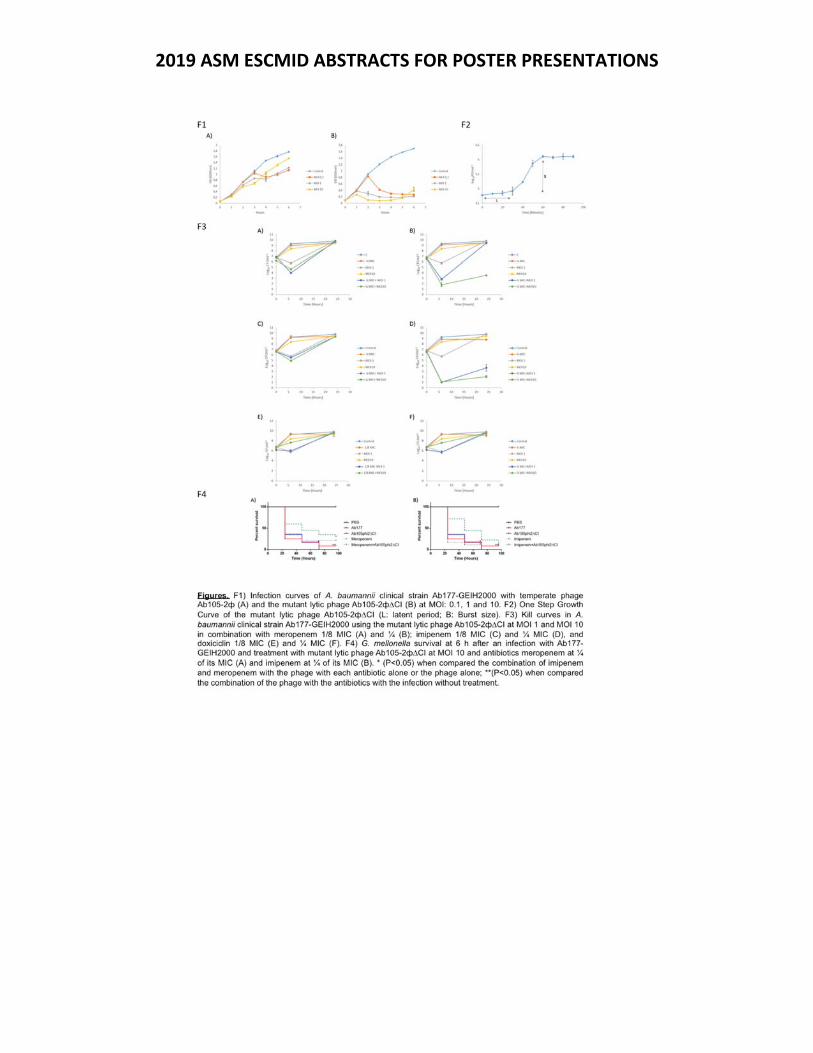
2019 ASM ESCMID ABSTRACTS FOR POSTER PRESENTATIONS
2019 ASM ESCMID ABSTRACTS FOR POSTER PRESENTATIONS
Thursday Presentation Number:
T‐56
Publishing Title: Anti‐virulence Biaryl Hydroxyketones as Potentiators of Antibiotic Efficacy Author Block: M. Shoham, J. BenArie, D. Blum, Y. Shoham; Q2Pharma, Ltd., Haifa, Israel
Abstract Body:
Background: Anti‐virulence agents represent an alternative or adjuvant to antibiotic therapy. These small‐molecule agents disarm pathogens of disease‐causing toxins without killing them, thereby eliminating survival pressure to develop resistance. Additional beneficial properties include biofilm inhibition and potentiation of antibiotic efficacy. Material/methods: Small‐molecule anti‐virulence agents F12 and F19 block staphylococcal transcription factor AgrA from binding to its promoter. Consequently, toxin expression is inhibited, thus preventing host cell damage by various Gram‐positive pathogens including MRSA, VRSA, Staphylococcus epidermidis, Streptococcus pyogens and Streptococcus pneumoniae. Results: Evidence of in‐vivo efficacy came from an MRSA bacteremia/sepsis study in mice. Treatment with 30 mg/kg F19 IP bid provided complete protection of the animals. All 10 animals treated with F19 survived by the end of the experiment, whereas untreated animals had 70% mortality. Potentiation of antibiotic efficacy was observed in vitro with beta‐lactam antibiotics and fluoroquinolones. For example, addition of 1 mg/L F19 reduced the MIC of nafcillin and cephalothin from 60 and 40 to 1 mg/L. In an MRSA murine wound infection model the combination treatment of F19 + cephalothin resulted in a reduction of bacterial load in the blood that was better than mono therapy by cephalothin by about 100‐fold, and even 10‐fold better than mono therapy with vancomycin, a current standard of care. The synergy with antibiotics to which pathogens are resistant in mono therapy may facilitate the reintroduction of obsolete antibiotics into the clinic. F12 exhibits efficacy against a Gram‐negative pathogen, clinical isolate M2 of Acinetobacter baumannii, as evidenced in vitro by cell lysis inhibition assays in immune system cells. In‐vivo efficacy was demonstrated in insect larvae survival assays. Conclusion: Biaryl hydroxyketone compounds could be used alone or in combination with antibiotics to prevent and treat infections of various Gram‐positive pathogens. Compound F12 could be used against some Gram‐negative pathogens.
2019 ASM ESCMID ABSTRACTS FOR POSTER PRESENTATIONS
Thursday Presentation Number:
T‐57
Publishing Title: Bacteriophage & Bacteriophage‐Derived Nontraditional Antibiotic Clinical Pipeline Analysis
Author Block: C‐L. L. Furr; Bacteriophage & Drug Dev. Consultants, LLC, Boone, NC
Abstract Body:
Background. Undisputedly, the global public health crisis due to antimicrobial resistant (AMR) bacteria is here. The speed at which AMR develops and spreads among bacterial populations exceeds the capacity of the biomedical community to develop new, effective antibiotics. Traditional (small molecule) antibiotic innovation is largely limited to derivatives of established classes. Discovery and development of novel classes are urgently needed to contest AMR and, paradoxically, nontraditional antibiotics (NTAB) are all around us. With eons of mortal combat, bacteria and bacteriophage (phage) co‐evolution pursuit and evasion strategies endure without succumbing to resistance. With considerable potential, phage and phage‐derived NTAB development is a burgeoning field; the clinical pipeline is presented here. Methods. Publicly available information was analyzed to identify phage and phage‐derived NTAB in the clinical pipeline between 1 May 2012 to 01 May 2019. Sources included ClinicalTrials.gov registry, publications, reports, press releases, and websites in English. Results. Public information revealed 21 phage and/or phage‐derived NTAB companies. Three companies have completed clinical trials, and most are advancing multiple therapies toward licensure. The 12 phage (8 natural, 3 engineered, 1 natural and engineered) and 11 phage‐derived (7 lysins, 2 nanocarriers, 1 amurin, 1 phage‐display small domain antibody) therapies in the pipeline target a variety of pathogens, and are in “early” development (9 discovery/preclinical, 10 clinical [3 phase I, 2 phase I/II, 5 phase II]). In the next 18 months, 7 trials are slated to begin. ClinicalTrials.gov retrieved 14 registered studies of 9 phages and 5 lysins (6 completed, 2 recruiting, 1 not yet recruiting, 5 terminated/no longer available/unknown). PubMed search recovered 10 published clinical trials of 8 phage and 2 lysin therapies, respectively, against UTI, CF, and severe infections. Conclusions. From 21 companies, the current clinical pipeline of phage (natural, engineered) and phage‐derived (4 types) NTAB target a range of bacterial pathogens, including AMR forms. Ten clinical studies have been published, 10 therapies are in clinical trials, 7 studies are to start within 18 months, and 11 therapies are advancing toward the clinic. The phage therapy clinical pipeline is expanding but it is woefully insufficient to offset AMR; more NTAB candidates are urgently needed.
2019 ASM ESCMID ABSTRACTS FOR POSTER PRESENTATIONS
Thursday Presentation Number:
T‐60
Publishing Title: Specificity of β‐lactam Enhancement of Cationic Peptide Antibacterial Activity in Daptomycin Resistant MRSA
Author Block: C. Lew1, N. Mishra2, A. Bayer2, W. Rose1; 1Univ. of Wisconsin‐Madison, Madison, WI, 2LA BioMed. Res. Inst., Torrance, CA
Abstract Body:
Background: Although the clinical usefulness of β‐lactam antibiotics in gram‐positive bacteria has ben altered following the emergence of methicillin‐resistant Staphylococcus aureus (MRSA), these antibiotics can still play a vital role in treatment of these infections by supplementing the activity of cationic peptides. This includes enhancing the activity of adjunct cationic peptide antibiotics, such as daptomycin (DAP), and modulating the activity of innate antibacterial host defense peptides (HDP). Although the improvement of in activity has been well documented, little is known about the discrepancies in mechanism with different β‐lactams and different cationic peptides. Methods: The membrane characteristics of nine DAP susceptible/ DAP resistant clinically derived strain pairs were analyzed pre‐ and post‐ exposure individually to a panel of six distinct β‐lactams (nafciliin, meropenem, cloxacillin, ceftriaxone, cefaclor, and cefoxitin). Two of these strain pairs were used in time‐kill analysis with either clinically relevant concentrations of DAP or the HDP LL‐37 and the panel of β‐lactams. Results: Exposure to β‐lactams resulted in decreased membrane surface charge, decreased membrane fluidity, and increased cell wall thickness. Distinct β‐lactams altered one or more of the physiological characteristics to variable extents. In the time‐kill analysis, DAP combinations showed more potent improvement of activity in DAP susceptible strains, however LL‐37 combinations had greater synergy in DAP resistant strains. Conclusions: The diversity in response to different β‐lactam antibiotics suggests discrete interactions with the bacterial cell membrane and cell wall. As the selected cationic peptide antibacterial actions both involve perturbation of the cell wall and membrane, it is likely that the mechanism of enhancement of activity may be specific for certain β‐lactams. Further, variation in strain response to DAP and LL‐37 combination treatments implies distinct mechanisms of β‐lactam enhancement of cationic peptide activity.
2019 ASM ESCMID ABSTRACTS FOR POSTER PRESENTATIONS
Thursday Presentation Number:
T‐65
Publishing Title:
Estimating the treatment of carbapenem resistant Enterobacteriaceae (CRE) infections in the United States (US): Implications for approved anti‐CRE agents and drug development.
Author Block: C. CLANCY1, B. Potoski1, D. Buehrle2, M. Nguyen1; 1Univ. of Pittsburgh, Pittsburgh, PA, 2VA Pittsburgh, Pittsburgh, PA
Abstract Body:
Background. The development of antibiotics with anti‐CRE activity has been designated as a top medical priority by healthcare organizations globally. However, financial challenges faced by companies introducing new anti‐CRE agents to the clinic raise concerns about the viability of the marketplace. Our objectives were to estimate the positioning of new anti‐CRE antibiotics (ceftazidime‐avibactam (CA), meropenem‐vaborbactam (MV), plazomicin (PLZ)) at US hospitals, current usage of new agents against CRE infections, and the size of the anti‐CRE market. Methods. We made estimates using results from an on‐line survey of hospital‐based, Society of Infectious Diseases Pharmacists members (218 respondents, 41 states; Nov‐Dec 2018), and data on antibiotic prescriptions and numbers of CRE infections in the US from IQVIA and DRIVE‐AB, respectively. Results. CA, MV and PLZ were formulary restricted or non‐formulary but available at 84%, 68% and 31% of respondents’ hospitals, respectively. A new agent was positioned as first‐line against CRE pneumonia, bacteremia, abdominal, and urinary tract infections at 87%, 90%, 83% and 56% of respondents’ hospitals, respectively. From Feb 2018‐Jan 2019, an estimated 7,941 CRE infections were treated with a new agent in the US (~23% (range: 16%‐42%) of all CRE infections). In different models, use of a new agent exceeded that of parenteral polymyxin against CRE infections as of Nov 2017‐Dec 2018. As of Jan 2019, new agents were estimated to treat 35% (23%‐62%) of CRE infections in which they were expected to be first‐line agents. Based on combined 12 month sales data for CA, MV and PLZ through Jan 2019, we estimate the current US CRE market for new agents is ~$305 M ($170 M‐$465 M). Conclusions. New anti‐CRE agents are available with restrictions on use at most US hospitals, and they are positioned as first‐line against most CRE infections. Uptake against CRE infections has been steady, but usage remains significantly less than would be expected. Therefore, there is room for growth in use of anti‐CRE antibiotics. However, the size of the market may not support several drugs, unless agents expand their niche beyond CRE and/or clinicians are willing to use them empirically. Crucial unanswered questions are: How does pricing impact anti‐CRE agent use? What concerns or behavioral factors explain the discordance between anti‐CRE agent positioning and usage? What considerations would justify clinicians using a new agent empirically? How would broader coverage (e.g., NDMs, CR‐Acinetobacter) change usage and market size?
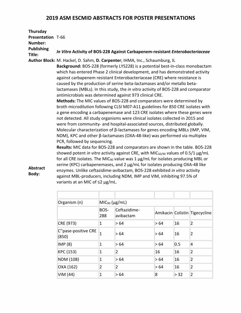
2019 ASM ESCMID ABSTRACTS FOR POSTER PRESENTATIONS
Thursday Presentation Number:
T‐66
Publishing Title:
In Vitro Activity of BOS‐228 Against Carbapenem‐resistant Enterobacteriaceae
Author Block: M. Hackel, D. Sahm, D. Carpenter; IHMA, Inc., Schaumburg, IL
Abstract Body:
Background: BOS‐228 (formerly LYS228) is a potential best‐in‐class monobactam which has entered Phase 2 clinical development, and has demonstrated activity against carbapenem resistant Enterobacteriaceae (CRE) where resistance is caused by the production of serine beta‐lactamases and/or metallo beta‐lactamases (MBLs). In this study, the in vitro activity of BOS‐228 and comparator antimicrobials was determined against 973 clinical CRE. Methods: The MIC values of BOS‐228 and comparators were determined by broth microdilution following CLSI M07‐A11 guidelines for 850 CRE isolates with a gene encoding a carbapenemase and 123 CRE isolates where these genes were not detected. All study organisms were clinical isolates collected in 2015 and were from community‐ and hospital‐associated sources, distributed globally. Molecular characterization of β‐lactamases for genes encoding MBLs (IMP, VIM, NDM), KPC and other β‐lactamases (OXA‐48‐like) was performed via multiplex PCR, followed by sequencing. Results: MIC data for BOS‐228 and comparators are shown in the table. BOS‐228 showed potent in vitro activity against CRE, with MIC50/90 values of 0.5/1 µg/mL for all CRE isolates. The MIC90 value was 1 µg/mL for isolates producing MBL or serine (KPC) carbapenemases, and 2 µg/mL for isolates producing OXA‐48 like enzymes. Unlike ceftazidime‐avibactam, BOS‐228 exhibited in vitro activity against MBL‐producers, including NDM, IMP and VIM, inhibiting 97.5% of variants at an MIC of ≤2 µg/mL.
Organism (n) MIC90 (µg/mL)
BOS‐288
Ceftazidime‐avibactam
Amikacin Colistin Tigecycline
CRE (973) 1 > 64 > 64 16 2
C''pase‐positive CRE (850)
1 > 64 > 64 16 2
IMP (8) 1 > 64 > 64 0.5 4
KPC (153) 1 2 16 16 2
NDM (108) 1 > 64 > 64 16 2
OXA (162) 2 2 > 64 16 2
VIM (44) 1 > 64 8 > 32 2
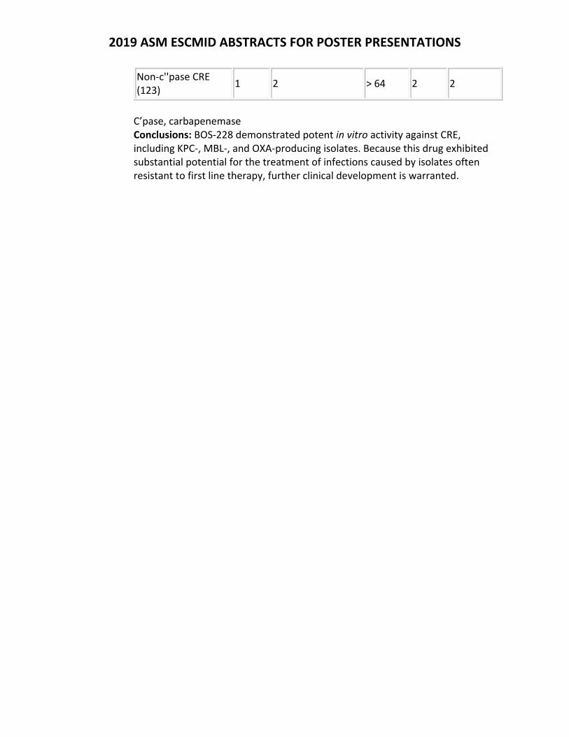
2019 ASM ESCMID ABSTRACTS FOR POSTER PRESENTATIONS
Non‐c''pase CRE (123)
1 2 > 64 2 2
C’pase, carbapenemase Conclusions: BOS‐228 demonstrated potent in vitro activity against CRE, including KPC‐, MBL‐, and OXA‐producing isolates. Because this drug exhibited substantial potential for the treatment of infections caused by isolates often resistant to first line therapy, further clinical development is warranted.
2019 ASM ESCMID ABSTRACTS FOR POSTER PRESENTATIONS
Thursday Presentation Number:
T‐67
Publishing Title:
Ceftobiprole Activity against Pathogens Causing Bacterial Skin and Skin Structure Infections in the United States (2016‐2018)
Author Block: L. R. Duncan1, K. Hamed2, J. I. Smart2, R. E. Mendes1, M. A. Pfaller1, R. K. Flamm1; 1JMI Lab., North Liberty, IA, 2Basilea Pharmaceutica, Basel, Switzerland
Abstract Body:
Background: Ceftobiprole medocaril, the prodrug of ceftobiprole (BPR), is an advanced cephalosporin approved in many European and non‐European countries for treating adults with community‐ and hospital‐acquired pneumonia (excluding ventilator‐associated pneumonia). Ceftobiprole medocaril is not approved in the United States (USA) but has qualified infectious disease product status and is being evaluated in two phase 3 clinical trials—in patients with acute bacterial skin and skin structure infections and in patients with Staphylococcus aureus bacteremia. Here, BPR and comparator activity was evaluated against recent clinical isolates collected in the USA from skin and skin structure infections (SSSIs). Methods: Over 7,300 isolates were collected from patients with SSSIs at 32 US medical centers from 2016‐2018. Susceptibility to BPR and comparator agents was tested using current CLSI methods. CLSI and EUCAST interpretive criteria followed current guidelines. Results: The major SSSI species and pathogen groups included S. aureus (53.3%), Enterobacteriaceae (23.3%), Pseudomonas aeruginosa (7.2%), β‐hemolytic streptococci (BHS; 6.4%), Enterococcus spp. (3.9%), and coagulase‐negative staphylococci (CoNS; 2.5%). BPR was highly active against S. aureus (MIC50/90, 0.5/1 mg/L; 99.8% susceptible [S] by EUCAST criteria). MIC50/90 values increased only 2‐fold (99.4%S) against the methicillin‐resistant S. aureus (MRSA) subset (41.7%). BPR exhibited potent activity against other Gram‐positive cocci, including BHS (MIC50/90, 0.015/0.03 mg/L), Enterococcus faecalis (MIC50/90 values, 0.5/2 mg/L), and CoNS (MIC50/90, 0.5/1 mg/L). The overall susceptibility of Enterobacteriaceae to BPR was 85.0% (98.0%S for the non‐ESBL‐phenotype subset), and 74.0% of P. aeruginosa isolates were inhibited by BPR at ≤4 mg/L. As expected, BPR was largely inactive against Enterococcus faecium and ESBL‐phenotype Enterobacteriaceae. Conclusions: BPR was highly active against most clinical isolates from the major Gram‐positive and Gram‐negative SSSI pathogen groups collected in the USA during 2016‐2018. The broad‐spectrum activity of BPR, including potent activity against MRSA, supports its further evaluation for this potential indication. Acknolwedgements: This project has been funded in whole or in part with federal funds from the Department of Health and Human Services; Office of the Assistant Secretary for Preparedness and Response; Biomedical Advanced Research and Development Authority, under Contract No. HHSO100201600002C.
2019 ASM ESCMID ABSTRACTS FOR POSTER PRESENTATIONS
Thursday Presentation Number:
T‐68
Publishing Title:
Prevalence of Antimicrobial Resistance in OropharyngealNeisseria meningitidisCarriage Isolates Among Patients Attending an STD Clinic in Columbus, Ohio
Author Block:
M. Bhat1, A. N. Turner2, J. A. Bazan3, A. Carter2, B. Snyder4, M. Brown2, J. J. Kwiek1; 1Dept. of Microbiol., The Ohio State Univ., Columbus, OH, 2Div. of Infectious Diseases, The Ohio State Univ., Columbus, OH, 3Div. of Infectious Diseases, The Ohio State Univ. & Sexual Hlth.Clinic, Columbus Publ. Hlth., Columbus, OH, 4Ohio Univ. Heritage Coll. of Osteopathic Med., Columbus, OH
Abstract Body:
Background: Background:Though rarely reported, antimicrobial drug resistance in Neisseria meningitidisis an essential public health concern in both developed and developing countries due to the severity of disease. There is a paucity of antimicrobial susceptibility data for oropharyngeal (OP) Neisseria meningitidiscarriage isolates in patients attending STD clinics. To document the prevalence of reduced susceptibility among Nm patients, we performed antimicrobial susceptibility testing (AST) on OP Nm isolates collected between January and July 2018 from patients seeking care at a large and urban STD clinic in Columbus, Ohio (USA). Methods: Patients reporting oral sex in the last 12 months are screened for OP gonorrhea usingnucleic acid amplification testing (NAAT) and cultured using modified Thayer Martin media. Analytical profile index (API)‐NH and PCR was used to distinguish between gonorrhea and Nm. A total of 100 Nm isolates underwent AST using ETEST strips (Biomerieux) on Mueller‐Hinton agar + 5% sheep blood. When available, minimum inhibitory concentration (MIC) breakpoints were categorized according to CLSI, EUCAST or CA‐SFM standards. Results: Over half (N=56, 56%) of Nm isolates displayed intermediate susceptibility to benzyl‐penicillin (MIC range 0.12 ‐ 0.25 μg/mL), while 29% (N=29) were resistant (MIC ≥ 0.5 μg/mL). β‐lactamase production was not observed in any Nm isolates by nitrocefin disk. Forty‐percent (N=40) of the Nm isolates showed reduced susceptibility to ciprofloxacin, whereas 26% (N=26) were resistant to ceftriaxone and 60% (N=60) were resistant to trimethoprim‐sulfamethoxazole. Twenty‐six percent of the Nm isolates (N=26) were resistant to azithromycin, and of these fourteen had MIC values greater than 256 μg/mL. More than 95% of isolates showed susceptibility to tetracycline N= 95 (95%) and rifampicin N=96 (96%). Some isolates were highly resistant to cefixime (N=4) and gentamicin (N=1) were not included in the resistant group due to lack of CLSI recommendation for them. Conclusions: Preliminary outcome indicates high proportion of resistance to benzyl‐penicillin, ciprofloxacin, trimethoprim‐sulfamethoxazole, and azithromycin among OP Nm carriage isolates which may warrant concern about current STD treatment modalities. Further inter‐rater reliability assessment of data will be done to demonstrate consistencyby multiple readers. Additional

2019 ASM ESCMID ABSTRACTS FOR POSTER PRESENTATIONS
studies are needed to determine predictors of OP colonization with drug‐resistant Nm isolates in patients attending STD clinics.
2019 ASM ESCMID ABSTRACTS FOR POSTER PRESENTATIONS
Thursday Presentation Number:
T‐69
Publishing Title:
Cefepime‐Zidebactam (WCK 5222) Activity against Clinical Isolates of Non‐Fermentative Gram‐Negative Bacilli Collected Worldwide in 2018
Author Block: H. S. SADER1, C. G. Carvalhaes2, L. R. Duncan2, S. J. R. Arends2, R. E. Mendes2, M. Castanheira2; 1JMI Lab., NORTH LIBERTY, IA, 2JMI Lab., North Liberty, IA
Abstract Body:
Background: Zidebactam (ZID) is a bicyclo‐acyl hydrazide with a dual mechanism of action: selective binding to gram‐negative (GN) penicillin‐binding protein 2 and β‐lactamase inhibition. Cefepime (FEP)‐ZID is in clinical development at 2g/1g q8 hours dosage. Methods: A total of 3,711 non‐fermentative GN bacilli (NF‐GNB) were collected by the 2018 SENTRY Antimicrobial Surveillance Program from medical centers in the United States (USA; n=1,667; 68 centers), Europe (EUR; n=1,311; 38 centers), Asia‐Pacific (APAC; n=465; 16 centers) and Latin America (LATAM; n=268; 8 centers) in 2018. Susceptibility testing was performed in a central laboratory (JMI Laboratories) by a reference broth microdilution method against FEP‐ZID (1:1 ratio) and comparators. A FEP‐ZID susceptible (S) breakpoint of ≤64mg/L has been proposed based on pharmacokinetic/pharmacodynamic target attainment and was applied. The FEP S breakpoint of ≤8mg/L (CLSI, high dose) was also applied to FEP‐ZID for comparison. Results: FEP‐ZID exhibited potent activity against Pseudomonas aeruginosa (PSA; MIC50/90, 1/4 mg/L) with 99.0% (APAC) to 100.0% (LATAM) of isolates inhibited at ≤8 mg/L. PSA S rates for ceftazidime‐avibactam, ceftolozane‐tazobactam, piperacillin‐tazobactam, and meropenem were 95.0%, 94.9%, 76.3%, and 76.5%, respectively. Against Acinetobacter baumannii‐calcoaceticus, percentages inhibited at ≤8/≤64 mg/L of FEP‐ZID were 73.4/99.4% in USA, 44.9/99.2% in EUR, 59.1/100.0% in APAC and 29.8/100.0% in LATAM, and MEM S rates were 69.8%, 24.2%, 43.5% and 8.3% in USA, EUR, APAC, and LATAM, respectively. FEP‐ZID inhibited 76.7% (EUR) to 100.0% (LATAM) of Stenotrophomonas maltophilia isolates at ≤8 mg/L and 99.4% (USA) to 100.0% (EUR, APAC, and LATAM) at ≤64 mg/L. Among Burkholderia cepacia, FEP‐ZID MIC >16 mg/L was observed only in the APAC region. Conclusion: FEP‐ZID showed potent in vitro activity against contemporary NF‐GNB collected worldwide. FEP‐ZID may represent a valuable therapeutic option for these difficult‐to‐treat organisms.
2019 ASM ESCMID ABSTRACTS FOR POSTER PRESENTATIONS
Thursday Presentation Number:
T‐70
Publishing Title:
Antimicrobial Activity of Cefepime‐Zidebactam (WCK‐5222) against Clinical Isolates of Carbapenem‐Resistant Enterobacterales Collected Worldwide in 2018
Author Block: H. S. SADER1, R. K. Flamm2, J. M. Streit2, T. B. Doyle2, M. Castanheira2; 1JMI Lab., NORTH LIBERTY, IA, 2JMI Lab., North Liberty, IA
Abstract Body:
Background: Zidebactam is a bicyclo‐acyl hydrazide with a dual mechanism of action: selective gram‐negative penicillin‐binding protein (PBP) 2 binding and β‐lactamase inhibition. Cefepime‐zidebactam is in clinical development at 2g/1g q8 hours dosage. We evaluated the in vitro activity of cefepime‐zidebactam against contemporary clinical isolates of carbapenem‐resistant Enterobacterales (CRE). Methods: A total of 14,500 Enterobacterales isolates were collected by the SENTRY Antimicrobial Surveillance Program worldwide in 2018 and 200 (1.4%) were categorized as CRE (resistant to meropenem, imipenem, or meropenem per EUCAST criteria). CRE isolates were from 54 medical centres in 14 countries located in Europe (n=81), the United States (n=63), Latin America (n=40), and Asia‐Pacific region (APAC; n=16). Susceptibility testing was performed in a central laboratory by a reference broth microdilution method against cefepime‐zidebactam (1:1 ratio) and comparators. All CRE isolates were characterized by whole genome sequencing. Results: The most common CRE species were Klebsiella pneumoniae (74.0%), Enterobacter cloacae (11.5%), and Serratia marcescens (5.0%). Isolates were mainly from bloodstream infections (30.5%), pneumonia (25.0%), and urinary tract infections (18.5%). Cefepime‐zidebactam was the most active agent, with MIC50/90 of 0.5/2 mg/L and highest MIC value of 8 mg/L. Tigecycline was the most active comparator (MIC50/90, 1/2 mg/L; 77.0% susceptible [S]), followed by ceftazidime‐avibactam (MIC50/90, 1/>32 mg/L; 76.5%S), colistin (MIC50/90, 0.25/>8 mg/L; 74.9%S), and amikacin (MIC50/90, 8/>32 mg/L; 56.5%S). Cefepime‐zidebactam was active against CRE isolates from all regions, and isolates from APAC exhibited slightly lower cefepime‐zidebactam MIC values (MIC50/90, 0.25/1 mg/L) compared to other regions (MIC50/90, 0.5‐1/2 mg/L). Susceptibility to ceftazidime‐avibactam ranged from 0.0% in APAC, 72.5% in Latin America, 82.7% in Europe, and 90.5% in the United States. Conclusion: Cefepime‐zidebactam demonstrated potent in vitro activity against contemporary (2018) CRE isolates collected worldwide. Antimicrobial agents currently available for clinical use exhibited limited activity against CRE, emphasizing the urgent need for novel agents to treat infections caused by these multidrug‐resistant organisms.
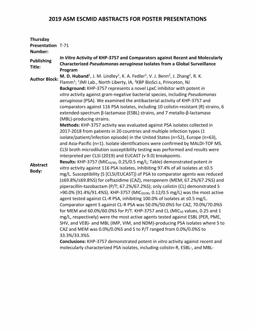
2019 ASM ESCMID ABSTRACTS FOR POSTER PRESENTATIONS
Thursday Presentation Number:
T‐71
Publishing Title:
In Vitro Activity of KHP‐3757 and Comparators against Recent and Molecularly Characterized Pseudomonas aeruginosa Isolates from a Global Surveillance Program
Author Block: M. D. Huband1, J. M. Lindley1, K. A. Fedler1, V. J. Benn2, J. Zhang2, R. K. Flamm1; 1JMI Lab., North Liberty, IA, 2KBP BioSci.s, Princeton, NJ
Abstract Body:
Background: KHP‐3757 represents a novel LpxC inhibitor with potent in vitro activity against gram‐negative bacterial species, including Pseudomonas aeruginosa (PSA). We examined the antibacterial activity of KHP‐3757 and comparators against 116 PSA isolates, including 10 colistin‐resistant (R) strains, 6 extended‐spectrum β‐lactamase (ESBL) strains, and 7 metallo‐β‐lactamase (MBL)‐producing strains. Methods: KHP‐3757 activity was evaluated against PSA isolates collected in 2017‐2018 from patients in 20 countries and multiple infection types (1 isolate/patient/infection episode) in the United States (n=52), Europe (n=63), and Asia‐Pacific (n=1). Isolate identifications were confirmed by MALDI‐TOF MS. CLSI broth microdilution susceptibility testing was performed and results were interpreted per CLSI (2019) and EUCAST (v 9.0) breakpoints. Results: KHP‐3757 (MIC50/90, 0.25/0.5 mg/L; Table) demonstrated potent in vitro activity against 116 PSA isolates, inhibiting 97.4% of all isolates at ≤0.5 mg/L. Susceptibility (S [CLSI/EUCAST]) of PSA to comparator agents was reduced (≤69.8%/≤69.8%S) for ceftazidime (CAZ), meropenem (MEM; 67.2%/67.2%S) and piperacillin‐tazobactam (P/T; 67.2%/67.2%S); only colistin (CL) demonstrated S >90.0% (91.4%/91.4%S). KHP‐3757 (MIC50/90, 0.12/0.5 mg/L) was the most active agent tested against CL‐R PSA, inhibiting 100.0% of isolates at ≤0.5 mg/L. Comparator agent S against CL‐R PSA was 50.0%/50.0%S for CAZ, 70.0%/70.0%S for MEM and 60.0%/60.0%S for P/T. KHP‐3757 and CL (MIC50 values, 0.25 and 1 mg/L, respectively) were the most active agents tested against ESBL (PER, PME, SHV, and VEB)‐ and MBL (IMP, VIM, and NDM)‐producing PSA isolates where S to CAZ and MEM was 0.0%/0.0%S and S to P/T ranged from 0.0%/0.0%S to 33.3%/33.3%S. Conclusions: KHP‐3757 demonstrated potent in vitro activity against recent and molecularly characterized PSA isolates, including colistin‐R, ESBL‐, and MBL‐
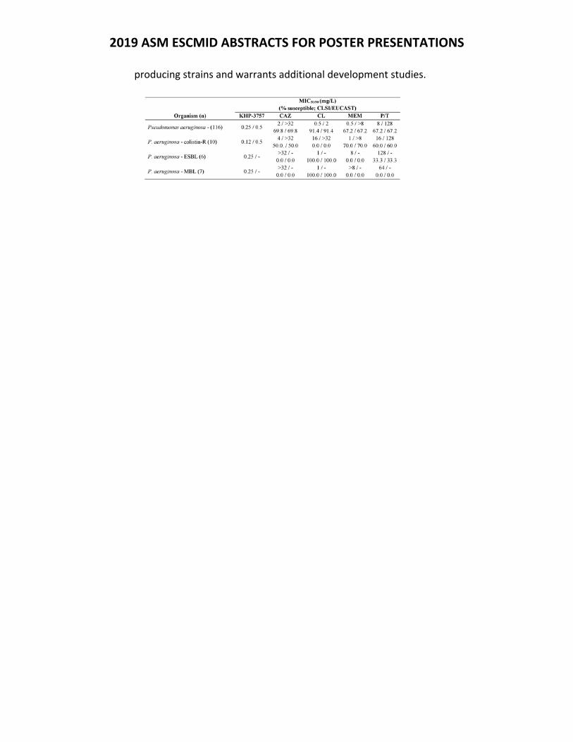
2019 ASM ESCMID ABSTRACTS FOR POSTER PRESENTATIONS
producing strains and warrants additional development studies.
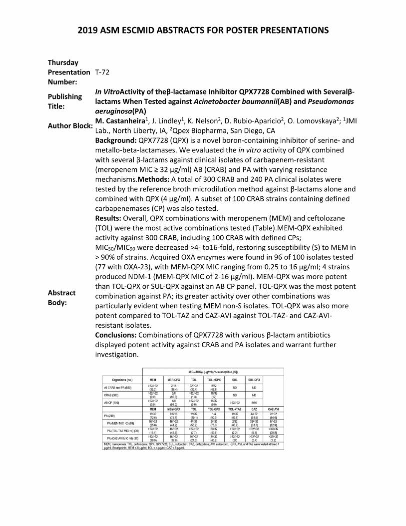
2019 ASM ESCMID ABSTRACTS FOR POSTER PRESENTATIONS
Thursday Presentation Number:
T‐72
Publishing Title:
In VitroActivity of theβ‐lactamase Inhibitor QPX7728 Combined with Severalβ‐lactams When Tested against Acinetobacter baumannii(AB) and Pseudomonas aeruginosa(PA)
Author Block: M. Castanheira1, J. Lindley1, K. Nelson2, D. Rubio‐Aparicio2, O. Lomovskaya2; 1JMI Lab., North Liberty, IA, 2Qpex Biopharma, San Diego, CA
Abstract Body:
Background: QPX7728 (QPX) is a novel boron‐containing inhibitor of serine‐ and metallo‐beta‐lactamases. We evaluated the in vitro activity of QPX combined with several β‐lactams against clinical isolates of carbapenem‐resistant (meropenem MIC ≥ 32 µg/ml) AB (CRAB) and PA with varying resistance mechanisms.Methods: A total of 300 CRAB and 240 PA clinical isolates were tested by the reference broth microdilution method against β‐lactams alone and combined with QPX (4 µg/ml). A subset of 100 CRAB strains containing defined carbapenemases (CP) was also tested. Results: Overall, QPX combinations with meropenem (MEM) and ceftolozane (TOL) were the most active combinations tested (Table).MEM‐QPX exhibited activity against 300 CRAB, including 100 CRAB with defined CPs; MIC50/MIC90 were decreased >4‐ to16‐fold, restoring susceptibility (S) to MEM in > 90% of strains. Acquired OXA enzymes were found in 96 of 100 isolates tested (77 with OXA‐23), with MEM‐QPX MIC ranging from 0.25 to 16 µg/ml; 4 strains produced NDM‐1 (MEM‐QPX MIC of 2‐16 µg/ml). MEM‐QPX was more potent than TOL‐QPX or SUL‐QPX against an AB CP panel. TOL‐QPX was the most potent combination against PA; its greater activity over other combinations was particularly evident when testing MEM non‐S isolates. TOL‐QPX was also more potent compared to TOL‐TAZ and CAZ‐AVI against TOL‐TAZ‐ and CAZ‐AVI‐resistant isolates. Conclusions: Combinations of QPX7728 with various β‐lactam antibiotics displayed potent activity against CRAB and PA isolates and warrant further investigation.
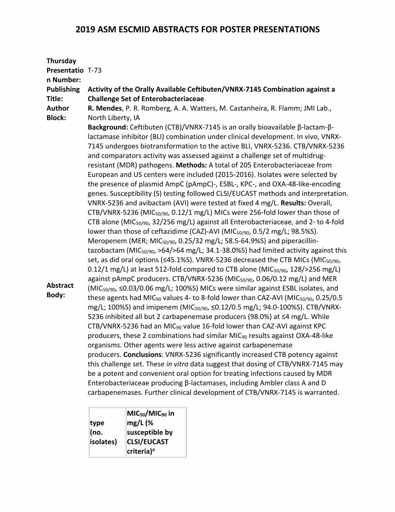
2019 ASM ESCMID ABSTRACTS FOR POSTER PRESENTATIONS
Thursday Presentation Number:
T‐73
Publishing Title:
Activity of the Orally Available Ceftibuten/VNRX‐7145 Combination against a Challenge Set of Enterobacteriaceae
Author Block:
R. Mendes, P. R. Romberg, A. A. Watters, M. Castanheira, R. Flamm; JMI Lab., North Liberty, IA
Abstract Body:
Background: Ceftibuten (CTB)/VNRX‐7145 is an orally bioavailable β‐lactam‐β‐lactamase inhibitor (BLI) combination under clinical development. In vivo, VNRX‐7145 undergoes biotransformation to the active BLI, VNRX‐5236. CTB/VNRX‐5236 and comparators activity was assessed against a challenge set of multidrug‐resistant (MDR) pathogens. Methods: A total of 205 Enterobacteriaceae from European and US centers were included (2015‐2016). Isolates were selected by the presence of plasmid AmpC (pAmpC)‐, ESBL‐, KPC‐, and OXA‐48‐like‐encoding genes. Susceptibility (S) testing followed CLSI/EUCAST methods and interpretation. VNRX‐5236 and avibactam (AVI) were tested at fixed 4 mg/L. Results: Overall, CTB/VNRX‐5236 (MIC50/90, 0.12/1 mg/L) MICs were 256‐fold lower than those of CTB alone (MIC50/90, 32/256 mg/L) against all Enterobacteriaceae, and 2‐ to 4‐fold lower than those of ceftazidime (CAZ)‐AVI (MIC50/90, 0.5/2 mg/L; 98.5%S). Meropenem (MER; MIC50/90, 0.25/32 mg/L; 58.5‐64.9%S) and piperacillin‐tazobactam (MIC50/90, >64/>64 mg/L; 34.1‐38.0%S) had limited activity against this set, as did oral options (≤45.1%S). VNRX‐5236 decreased the CTB MICs (MIC50/90, 0.12/1 mg/L) at least 512‐fold compared to CTB alone (MIC50/90, 128/>256 mg/L) against pAmpC producers. CTB/VNRX‐5236 (MIC50/90, 0.06/0.12 mg/L) and MER (MIC50/90, ≤0.03/0.06 mg/L; 100%S) MICs were similar against ESBL isolates, and these agents had MIC90 values 4‐ to 8‐fold lower than CAZ‐AVI (MIC50/90, 0.25/0.5 mg/L; 100%S) and imipenem (MIC50/90, ≤0.12/0.5 mg/L; 94.0‐100%S). CTB/VNRX‐5236 inhibited all but 2 carbapenemase producers (98.0%) at ≤4 mg/L. While CTB/VNRX‐5236 had an MIC90 value 16‐fold lower than CAZ‐AVI against KPC producers, these 2 combinations had similar MIC90 results against OXA‐48‐like organisms. Other agents were less active against carbapenemase producers. Conclusions: VNRX‐5236 significantly increased CTB potency against this challenge set. These in vitro data suggest that dosing of CTB/VNRX‐7145 may be a potent and convenient oral option for treating infections caused by MDR Enterobacteriaceae producing β‐lactamases, including Ambler class A and D carbapenemases. Further clinical development of CTB/VNRX‐7145 is warranted.
type (no. isolates)
MIC50/MIC90 in mg/L (% susceptible by CLSI/EUCAST criteria)a
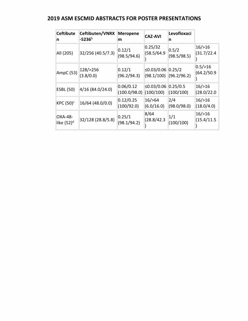
2019 ASM ESCMID ABSTRACTS FOR POSTER PRESENTATIONS
Ceftibuten
Ceftibuten/VNRX‐5236b
Meropenem
CAZ‐AVI Levofloxacin
All (205) 32/256 (40.5/7.3)0.12/1 (98.5/94.6)
0.25/32 (58.5/64.9)
0.5/2 (98.5/98.5)
16/>16 (31.7/22.4)
AmpC (53) 128/>256 (3.8/0.0)
0.12/1 (96.2/94.3)
≤0.03/0.06(98.1/100)
0.25/2 (96.2/96.2)
0.5/>16 (64.2/50.9)
ESBL (50) 4/16 (84.0/24.0) 0.06/0.12 (100.0/98.0)
≤0.03/0.06(100/100)
0.25/0.5 (100/100)
16/>16 (28.0/22.0
KPC (50)c 16/64 (48.0/0.0) 0.12/0.25 (100/92.0)
16/>64 (6.0/16.0)
2/4 (98.0/98.0)
16/>16 (18.0/4.0)
OXA‐48‐like (52)d
32/128 (28.8/5.8)0.25/1 (98.1/94.2)
8/64 (28.8/42.3)
1/1 (100/100)
16/>16 (15.4/11.5)
2019 ASM ESCMID ABSTRACTS FOR POSTER PRESENTATIONS
Thursday Presentation Number:
T‐74
Publishing Title:
Lefamulin Activity Against Gram‐Positive Pathogens Collected in the 2017 Global SENTRY Antimicrobial Surveillance Program
Author Block: S. Paukner1, S. P. Gelone2, S. J. R. Arends3, H. S. Sader3; 1Nabriva Therapeutics GmbH, Vienna, Austria, 2Nabriva Therapeutics, Inc., King of Prussia, PA, 3JMI Lab., North Liberty, IA
Abstract Body:
Background: Lefamulin, a novel pleuromutilin antibiotic that inhibits bacterial protein synthesis, is in post‐phase 3 clinical development for IV and oral treatment of community‐acquired (CA) bacterial pneumonia. We investigated lefamulin and comparator in vitro activity against a contemporary global set of gram‐positive pathogens. Methods: Unique isolates from patients (n=4337; 36.8% USA, 38.8% Europe, 13.1% Asia Pacific, 11.3% Latin America; 34 countries, 98 sites) were collected from CA respiratory tract infections (40.0%), pneumonia from hospitalized patients (13.6%), bloodstream infections (23.2%), skin and soft tissue infections (18.7%), and other infections (4.5%). Testing was done by broth microdilution (CLSI) and susceptibility was determined via CLSI (2019) breakpoints. Results: Lefamulin displayed potent antibacterial activity against S. pneumoniae (MIC50/90 0.12/0.25 µg/mL; Table); all isolates were inhibited at ≤1 µg/mL and were largely susceptible to moxifloxacin (98.0‐100%) and amoxicillin‐clavulanic acid (88.5‐94.8%). In contrast, only 38.2‐76.2% and 44.5‐71.9% were susceptible to azithromycin and oral penicillin, respectively, with the lowest susceptibility rates in the Asia‐Pacific region. Lefamulin also showed potent activity against S. aureus (including MRSA; both, MIC50/90 0.06/0.12 µg/mL) and coagulase‐negative Staphylococcus spp., which generally showed reduced susceptibility rates to azithromycin and moxifloxacin. The activity of lefamulin was unaffected by resistance to other antibiotic classes. Conclusions: Lefamulin demonstrated potent activity against this collection of gram‐positive pathogens compared with comparators, including against antimicrobial‐resistant isolates. Lefamulin may offer a new option for empiric treatment of infections caused by these organisms, particularly in countries with
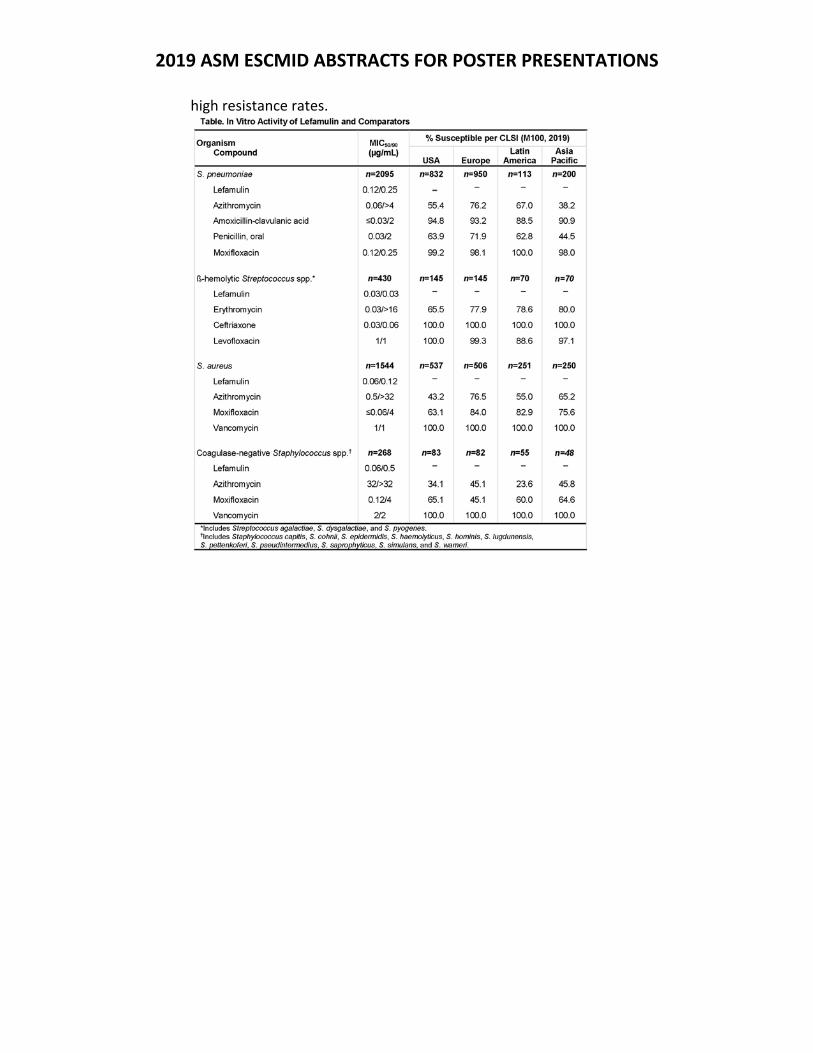
2019 ASM ESCMID ABSTRACTS FOR POSTER PRESENTATIONS
high resistance rates.
2019 ASM ESCMID ABSTRACTS FOR POSTER PRESENTATIONS
Thursday Presentation Number:
T‐75
Publishing Title: The burden of resistance among urinary tract isolates of Escherichia coli in the United States in 2017
Author Block: I. Critchley1, N. Cotroneo1, K. Sulham1, D. Melnick1, R. Mendes2; 1Spero Therapeutics, Cambridge, MA, 2JMI Lab., North Liberty, IA
Abstract Body:
Background: Urinary tract infections (UTI’s) caused by Escherichia coli (EC) have been historically managed with oral agents such as the fluoroquinolones and trimethoprim‐sulfamethoxazole (TMP‐SMX). Use of these agents is being compromised by the increase in extended‐spectrum‐β‐lactamase (ESBL)‐producing organisms that show co‐resistance. Management of UTI’s caused by ESBL‐producing organisms is challenging due to the limited options available outside the hospital. This study assessed the prevalence of ESBL‐phenotypes and genotypes among UTI EC collected in the US in 2017 and the impact of co‐resistance. Methods: 1831 EC from UTIs in the SENTRY Surveillance Program were evaluated for susceptibility to various agents. Isolates were collected from medical centers geographically distributed across the US Census regions, centrally tested, and results interpreted according to CLSI criteria. Isolates that met ESBL phenotypic screening criteria had their genome sequenced and screened for β‐lactamase genes.Results: Among the 1831 EC, national prevalence of ESBL phenotypes was 15.7% with regional differences ranging from 10.5% in West North Central to 30% in Mid Atlantic. All EC were susceptible (S) to meropenem (MER). Among all EC resistance (R) rates for cefuroxime, levofloxacin (LEV) and TMP‐SMX were 15.9%, 24.3% and 32.1%, respectively. R to LEV ranged from 18% in Mountain region to 38.1% in Mid Atlantic. R to TMP‐SMX ranged from 26.8% in East North Central to 43.5% in Mid Atlantic. Among ESBL EC R was higher; LEV R ranged from 52.6% in East North Central to 82.4% in East South Central, while TMP‐SMX R ranged from 36.8% in West North Central to 74.2% in Mid Atlantic. Among the CTX‐M‐15 producing EC, R rates for LEV and TMP‐SMX were 81.5% and 69.5%, respectively. Among OXA‐1/30 producing EC, R rates for LEV, TMP‐SMX and MER were 98.9%, 78.3% and 0%, respectively. Conclusions: LEV and TMP‐SMX exhibit R rates ≥24% against all EC from UTI in the US. Co‐resistance for LEV and TMP‐SMX was higher among ESBL phenotypes (≥59.2%), confirmed CTX‐M‐15 (≥69.5%) and OXA‐1/30 (≥78.3%) genotypes. All EC UTI isolates were S to MER suggesting that new oral agents with the spectrum and potency of the carbapenems address an unmet need for new options to treat co‐resistant ESBL EC from UTI.
2019 ASM ESCMID ABSTRACTS FOR POSTER PRESENTATIONS
Thursday Presentation Number:
T‐77
Publishing Title:
Longitudinal Analysis of Antibiotics Clinical Pipeline
Author Block: C. J. Lepore, W. Kim, K. Talkington; The Pew Charitable Trusts, Washington, DC
Abstract Body:
Background: In 2014, The Pew Charitable Trusts’ antibiotic resistance project began tracking the pipeline of small molecule antibiotics in clinical development to inform public policies aimed at spurring innovation of new antibiotics. Each pipeline update is a “snapshot in time,” comparing current data against prior assessments. Of particular interest are candidates that have potential activity against globally‐recognized priority pathogens, including Gram‐negative ESKAPE pathogens. Methods: Characterization of each candidate was conducted from 2014 ‐ 2018 through collection of publicly‐available clinical trial registries, company websites and press releases, peer‐reviewed literature, and conference presentations. For each candidate, the following inputs were collected: clinical development phase, organization(s) developing the candidate, drug class and target, and expected activity against Gram‐negative ESKAPE pathogens, US Centers for Disease Control and Prevention’s (CDC) urgent or World Health Organization’s (WHO) critical threat pathogens. Results: Of the 67 antibiotics that have been in some stage of clinical development between 2014 through 2018, 20 new candidates have entered the pipeline and ten products have been approved, while 17 have been either suspended or discontinued. An additional seven candidates have stalled. Of 19 candidates representing novel chemical classes, over half either stalled or were discontinued, compared to less than a third attrition observed for candidates based on prior discoveries. Furthermore, just 18 of the 67 compounds potentially addresses at least one of WHO’s recognized critical threats: carbapenem‐resistant Enterobacteriaceae (CRE), A. baumannii (CRAB), and P. aeruginosa (CRPA). Of the 14 still‐active or approved drugs during this time period, the majority have potential activity against most CRE (13), but few have potential to address CRAB (4) and CRPA (3). None of the ten approvals since 2014 are indicated against CRE, CRAB, or CRPA on their FDA drug labels. Conclusions: The antibiotics pipeline has remained stagnant over the past five years and is insufficient to address the growing public health threat of antibiotic resistance. This longitudinal trend analysis highlights key gaps that still exist in the pipeline today, particularly limited novel candidates for addressing multi‐drug resistant Gram‐negative ESKAPE pathogens.
2019 ASM ESCMID ABSTRACTS FOR POSTER PRESENTATIONS
Thursday Presentation Number:
T‐78
Publishing Title: Department of Defense Small Molecule Antibiotic Development Effort Coordination ‐ The Steering Committee Approach
Author Block:
D. V. Zurawski1, J. Rohde1, M. Khraiwesh1, M. MacDonald1, M. Kreishman‐Deitrick1, S. M. Noble1, P. T. McGann1, B. E. Swierczewski1, S. D. Tyner1, N. L. Michael1, K. L. Lawrence2, S. Zumbrun3, R. Panchal3, D. Jirage4, M. P. Simons5, A. Horstman‐Smith6, P. Waterman7, C. C. Black1; 1Walter Reed Army Inst. of Res., Silver Spring, MD, 2U.S. Army Med. Materiel Agency, Fort Detrick, MD, 3US Army Med. Res. Inst. of Infectious Diseases, Frederick, MD, 4Military Infectious Disease Res. Program, Silver Spring, MD, 5Naval Med. Res. Ctr., Silver Spring, MD, 6Defense Threat Reduction Agency, Fort Belvoir, VA, 7Office of Sci. and Technology Policy, District of Columbia, DC
Abstract Body:
The Presidential Initiative to Combat Antibiotic‐Resistant Bacteria (CARB) was established in 2015 to address the systemic effects of multidrug‐resistant (MDR) bacteria. The WRAIR Center for Infectious Disease Research (CIDR), Experimental Therapeutics (ET) in conjunction with the Bacterial Diseases Branch (BDB), was charged to execute a specified CARB task to discover and develop novel small molecule antibiotics (AB). The mandate reorganized existing WRAIR capabilities ‐ medicinal and synthetic chemistry, drug metabolism and disposition test systems, and in vitro/vivo biological test systems ‐ into a gated‐tier, resource‐sparing test paradigm. The Multidrug‐Resistant Organism Repository and Surveillance Network (MRSN) provides fully characterized, military healthcare system‐derived MDR isolates that populate the AB test systems. This suite of capabilities are utilized by ET and BDB investigators as well as pharmaceutical industry, government, and academic partners, to advance military‐relevant AB candidates that meet a target product profile of efficacy against MDR Gram‐negative, ESKAPE pathogens. In parallel, the Defense Threat Reduction Agency (DTRA) and their DoD‐intramural preclinical AB development partner USAMRIID have a requirement to discover and advance AB effective against Gram negative biothreat agents. Activity against biothreats enhances an AB candidate’s preclinical data package when approaching the Biomedical Advanced Research and Development Authority (BARDA) for clinical transition funding considerations. External partners would greatly benefit from a ‘one‐stop‐shop’ AB consortium of DoD funders (DTRA) with the science and technology base (WRAIR and USAMRIID), and advanced development (USAMMDA and BARDA) subject matter experts that can advise and direct AB project advancement utilizing diverse DoD funding, contract vehicles, competencies and test systems. We propose the creation of a small molecule AB Steering Committee (SC) with these intra‐agency partners to evaluate new candidate drugs. The Next Generation Antibiotic (NGAB) SC would have multi‐institute tech base

2019 ASM ESCMID ABSTRACTS FOR POSTER PRESENTATIONS
representation, as well as funder and advanced development input and oversight. A draft SC charter is currently under evaluation, and the NGAB‐SC will bring together all of these DoD agencies to facilitate AB development from preclinical to advanced development.
2019 ASM ESCMID ABSTRACTS FOR POSTER PRESENTATIONS
Thursday Presentation Number:
T‐79
Publishing Title:
The Shared Platform for Antibiotic Research and Knowledge: A Collaborative Tool to Spark Innovation in Gram‐Negative Antibiotic Discovery
Author Block: K. R. Prosen, 20004, C. J. Lepore, W. KIm, A. Coukell; The Pew Charitable Trusts, Washington, DC
Abstract Body:
Background: The Shared Platform for Antibiotic Research and Knowledge (SPARK) is an free, open‐access collaboration tool launched by The Pew Charitable Trusts in September 2018 to help spur development of antibiotics targeting Gram‐negative ESKAPE pathogens. Gram‐negative bacteria can cause severe infections that are difficult or impossible to treat with currently available antibiotics due to their innate defense mechanisms. SPARK provides a tool for scientists to share and exchange research ideas, deposit and access data, and perform analyses based on physiochemical properties, antibacterial susceptibility, and biochemical activity. Methods: Pew assessed antibiotics discovery programs to identify outreach targets, and subsequently engaged organizations to explore opportunities for data contributions. This outreach effort is supplemented with extracting data from peer‐reviewed publications. Experimental data and metadata are collated and standardized by data scientists, followed by curation by microbiologists, detailing experimental methods, targets, and bacterial strain genotypes and phenotypes. Results: The SPARK online community is rapidly growing, with more than 500 users registered. To date, ~70,000 MIC and ~1,800 IC50 curated data points are available, enabling users to parse data by susceptibility, species‐ or strain‐specific activity, or molecular target. Two pharmaceutical companies have provided data from their LpxC and gyrase inhibitor programs, as well as a target‐agnostic efflux panel screening. Further contributions from industry, academia, and other discovery programs are in progress. Conclusions: Initial data contributions from industry demonstrate willingness and ability to share previously unreleased data. The antibiotics discovery community is actively utilizing SPARK data and tools, with plans to present their studies soon. Furthermore, other data‐sharing initiatives have expressed interest in utilizing SPARK as a component for their respective endeavors (e.g., Joint Programming Initiative on Antimicrobial Resistance’s Virtual Research Institute). As SPARK continues to grow, it will serve as a key resource to inform physiochemical properties affecting Gram‐negative accumulation and permeation, and to ultimately catalyze discovery of novel antibiotics.
2019 ASM ESCMID ABSTRACTS FOR POSTER PRESENTATIONS
Thursday Presentation Number:
T‐80
Publishing Title:
In Vivo Activity of QPX7728 in Combination with Meropenem against Carbapenem‐resistant K. pneumoniae, A. baumannii, and P. aeruginosa
Author Block: M. Sabet1, Z. Tarazi1, D. GRIFFITH1, J. Loutit2; 1Qpex Biopharma, SAN DIEGO, CA, 20, SAN DIEGO, CA
Abstract Body:
Background: The emergence of resistance to β‐lactam antibiotics due to the increasing variety of β‐lactamase enzymes has become a major clinical issue. A new boron based beta‐lactamase inhibitor, QPX7728, with in vitro inhibitory activity against major members of Class A, B, C, and D beta‐lactamases has been discovered. The objective of these studies was to demonstrate the in vivo efficacy of meropenem in combination with QPX7728 in mouse models of pulmonary and thigh infection due to carbapenem‐resistant gram‐negative organisms. Methods: Six K. pneumoniae, four A. baumannii, two P. aeruginosa and one E. cloacae strain with meropenem MICs ranging between 8 ‐ 512 mg/L were used. Neutropenic mice were infected with ~105 CFU/lung or ~106 CFU/thigh. Intraperitoneal treatments with meropenem ± QPX7728 were initiated 2 hours post‐infection every two hours for 24 hours. Mice were sacrificed after 24 hours of treatment and colony counts in tissue determined. Results: For all strains, treatment with meropenem + QPX7728 produced significantly lower bacterial counts in tissues compared to the meropenem alone in both infection models. Data from representative strains are shown below:
Strain ModelMeropenem MIC (µg/mL)
Compound Change in Log CFU/thigh or lung @ 24h
Alonewith QPX7728
(4 µg/mL)
K. pneumoniae KP1099
Lung 128 ≤0.06 Mero 300 mg/kg
+ 0.64
Mero 300 mg/kg + QPX7728 50 mg/kg
‐1.69
K. pneumoniae KP1099
Thigh 128 ≤0.06 Mero 300 mg/kg
+ 1.89
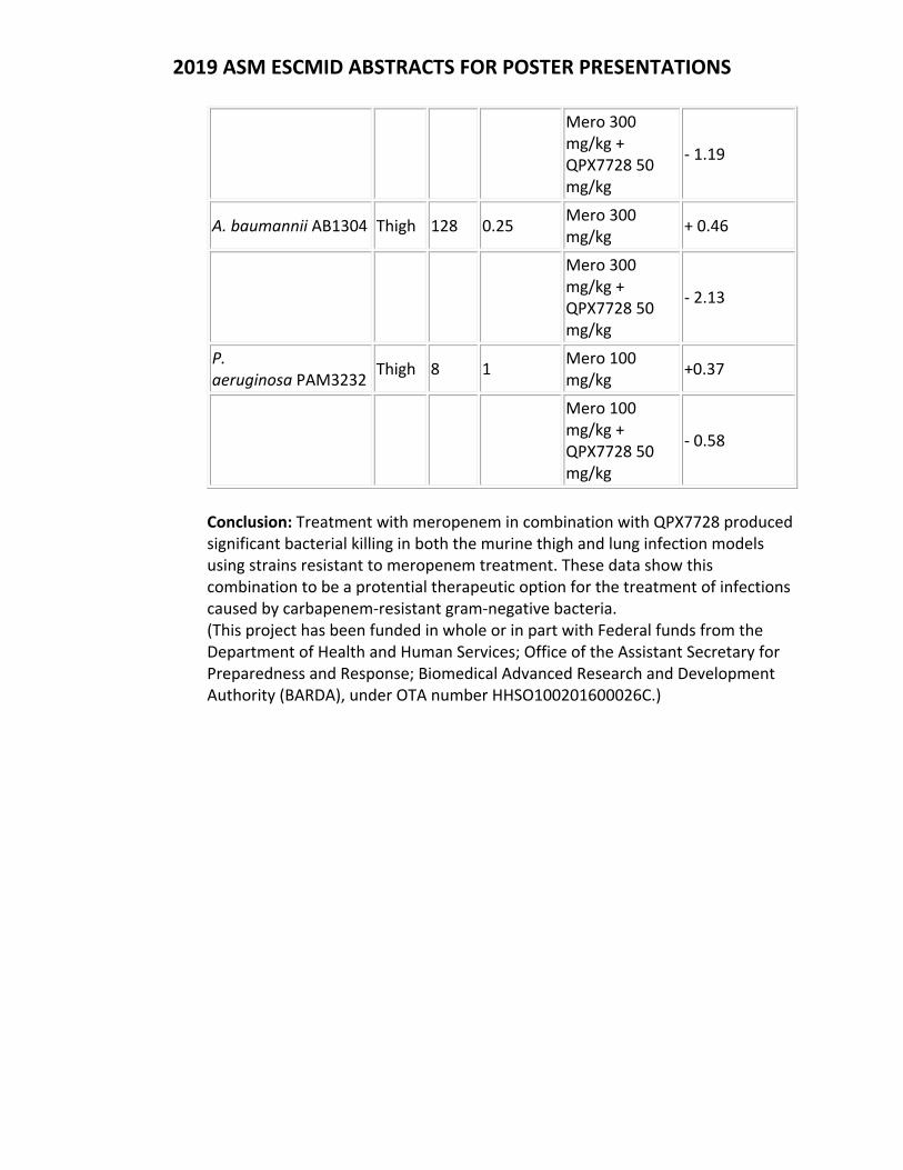
2019 ASM ESCMID ABSTRACTS FOR POSTER PRESENTATIONS
Mero 300 mg/kg + QPX7728 50 mg/kg
‐ 1.19
A. baumannii AB1304 Thigh 128 0.25 Mero 300 mg/kg
+ 0.46
Mero 300 mg/kg + QPX7728 50 mg/kg
‐ 2.13
P. aeruginosa PAM3232
Thigh 8 1 Mero 100 mg/kg
+0.37
Mero 100 mg/kg + QPX7728 50 mg/kg
‐ 0.58
Conclusion: Treatment with meropenem in combination with QPX7728 produced significant bacterial killing in both the murine thigh and lung infection models using strains resistant to meropenem treatment. These data show this combination to be a protential therapeutic option for the treatment of infections caused by carbapenem‐resistant gram‐negative bacteria. (This project has been funded in whole or in part with Federal funds from the Department of Health and Human Services; Office of the Assistant Secretary for Preparedness and Response; Biomedical Advanced Research and Development Authority (BARDA), under OTA number HHSO100201600026C.)
2019 ASM ESCMID ABSTRACTS INDEX
W‐ Wednesday poster presentation T‐ Thursday poster presentation
Abdulle, O. W-24 Abu-Taleb, R. T-54 Adhikari, S. W-18 Aguilar, M.-I.W-39 Alamneh, Y. A. T-54 Alexander, E. T-18 Allu, S. W-76 Alshammari, F. W-04 Ambroa, A. T-55, W-33 Ambrose, P. G. T-07, T-10, T-11, T-12, T-13, T-18, T-19, T-20, T-21 Amoako, D. G. W-15 Anderson, K. W-54 Andes, D. T-09 Andriani, G. M. W-38 Angulo, D. A. W-78 Annavajhala, M. W-67 Anthony, E. W-04 Antonic, V. T-54 Antunes, N. T. T-33 Arends, S. J. R. T-69 Arends, S. J. R. T-74 Armstrong, E. S. W-62 Aron, Z. D. T-34, T-46, W-18 Atouf, F. T-40 Attwood, M. T-02, T-03, T-06 Auchtung, J. M. T-52 Auchtung, T. A. T-52 Aung, T. W-74 Avis, T. W-24 Babu Sait, M. W-01 Backlund, M. T-15 Bahadur, T. W-41 Bain, R. W-49 Bamola, V. D. W-41 Barasch, D. W-07 Barber, R. T-46 Bardhan, N. T-33 Barenholz, Y. T-28, T-29, T-30, T-31, W-07 Bavli, Y. T-31, W-07
Bayer, A. T-60 Bazan, J. A. T-68 Belley, A. T-04, T-05, T-38 BenArie, J. T-56 Benn, V. J. T-71, W-69 Bernhard, F. T-04, T-05 Berns, E. W-48 Bester, L. A. W-15 Bhat, M. T-68 Bhavnani, S. M. T-07, T-10, T-11, T-12, T-13, T-18, T-19, T-20, T-21 Biondi, S. T-14 Biviji, Z. W-48 Black, C. C. T-78 Blasco, L. T-55, W-33 Blaskovich, M. W-23 Blaskovich, M. A. T. T-25, T-26 Bleriot, I. T-55 Blum, D. T-56 Bodrenko, I. W-06 Bohlmann, L. T-25 Bolisetti, R. T-25 Bonomo, R. A. T-38, W-70 Bordeleau, E. W-61 Borgmann, A. W-51 Born, C. W-09, W-48, W-49, W-76 Bostock, M. W-14 Bottomley, A. W-71 Bou, G. T-55, W-33 Bowker, K. T-02, T-03, T-06 Bowlin, T. T-34 Bowlin, T. L. T-46, W-18 Breidenstein, E. W-24 Britigan, B. W-20 Britton, R. A. T-52 Brönstrup, M. W-14 Brouwer, S. T-25 Brown, M. T-68 Brumm, J. T-43 Brunetti, B. W-55
Buehrle, D. T-65 Bulitta, J. W-68 Bulitta, J. B. W-70 Butler, M. T-34 Cabrera Goss, M. T-34 Callahan, M. T-26 Camargo, I. L. B. C. W-47 Canton, R. W-33 Cardinale, S. C. W-18 Cardinale, S. T-34 Cardinale, S. C. T-46 Carlton, M. T-46 Carmeli, Y. W-77 Carpenter, D. T-66 Carranco, A. I. T-13 Carter, A. T-68 Carter, N. W-19 Carvalhaes, C. G. T-69 Case, C. T-51 Cassino, C. W-51 Castanheira, M. T-09, T-69, T-70, T-72, T-73, W-62 Ceccarelli, M. W-06 Cern, A. T-28, T-29, T-30, T-31, W-07 Chakrabarti, R. T-40 Charrier, C. W-24 Chaudhry, R. W-41 Chen, A. W-19 Cheng, J.-X.W-42, W-59 Chia, J.-H.T-08 Chien, J.-C.T-08 Chitra, S. T-35, W-80 Choi, S.-R.W-20 Ciardullo, C. W-24 Ciesielska, M. T-36 Cinamon, G. T-28, T-30, W-07 Cisneros, J. T-55 CLANCY, C. T-65 Closs Jr, G. W-11 Coenye, T. T-55 Cokcetin, N. W-71
Comita-Prevoir, J. W-19 Conde, H. T-07, T-10, T-11, T-12, T-13 Connolly, K. L. T-34 Conroy, T. T-25 Cook, G. M. T-25 Cooper, M. W-23 Cooper, M. A. W-10 Cork, A. J. T-25 Cotroneo, N. T-01 Cotroneo, N. T-75 Coukell, A. T-79 Coward, C. W-24 Cowling, B. T-58 Critchley, I. T-75 Crooks, A. W-08 Cryle, M. W-39 Cui, Q. W-59 Curran, M. T-35, T-36, W-80 Daikos, G. L. W-77 Dane, A. T-16 Das, B. K. W-41 Das, S. T-01 Das, S. T-14, T-17, T-39 Davis, C. K. W-10 Debabov, D. T-22 Deblais, L. W-11 Del Borgo, M. W-39 Demons, S. T. T-54 De Oliveira, D. M. P. T-25 Deshpande, L. W-62 DETWEILER, C. W-08 Dighe, P. K. W-45 Dilly, J. J. W-10 Dobtsis, I. T-42 Dong, P.-T.W-42, W-59 Doyle, T. B. T-70 Drusano, G. W-68, W-70 Dudley, M. T-45 Duffy, T. W-24 Duncan, L. R. T-67, T-69 Durand-Reville, T. W-19 Durante-Mangoni, E. W-77 Dwyer, J. W-51 Eckburg, P. T-16 Eckburg, P. B. T-36
Edmondson, D. G. T-34 El-Deeb, I. M. T-25 Elliott, A. G. W-10 Ercan, O. T-15 Essack, S. Y. W-15 Farrell, K. T-52 Farrington, N. T-01 Farrington, N. T-14, T-39 Farrington, N. T-17 Fedler, K. A. T-71 Feinstein, Y. W-07 Fernandez-Cuenca, F. W-33 Fernandez-Garcia, L. T-55 Flamm, R. T-73 Flamm, R. K. T-67, T-70, T-71, W-62, W-69 Frahm, A. W-55 Franz, K. J. W-12 Frei, A. W-23 Friberg, L. E. T-15, W-77 Friedrich, L. T-21 Fringer, J. T-40 Fulcher, A. W-39 Furr, C.-L.L. T-57 Gallinat, A. T-41 Gamboa, I. C. W-47 Gamero, D. W-66 Garcia, A. W-74 Garcia, D. W-09, W-48, W-49, W-76 Garrity-Ryan, L. W-80 Garry, B. T-54 G-Dayanandan, N. W-18 Gelone, S. P. T-18, T-19, T-20, T-74 GEMARA SEIMC/REIPI Bacterial Clinical Adaptation S, t. T-55, W-33 George, D. R. T-20, T-21 Gernert, K. W-26 Gialanella, P. T-42, W-56 Giamarellos-Bourboulis, E. W-79 Giesel, G. T-11 Gilmore, A. W-49 Goldmann, O. T-29
Goncalves, L. W-14 Gore, S. T-17, T-39 Grandclaudon, C. W-14 Greenberg, R. T-39 Griffith, D. T-45, T-80 Grigaleviciute, R. W-56 Grinberga, S. T-15, T-47 GU, D. W-27 Guler, S. W-19 Haag, A. T-52 Hackel, M. T-66 Hamed, K. T-67 Hammel, J. P. T-18, T-19, T-20, T-21 Hansen, J. T-15 Hansen, J. U. T-47 Hansen, K. T-08 Hansford, K. A. T-25, W-10 Harry, E. W-71 Harwell, J. T-58 Hawser, S. T-38 Helmy, Y. W-11 Herrera, C. W-67 Hesterkamp, T. T-50 Hillock, T. W-54, W-55 Hobbie, S. N. T-15, T-47 Hod, A. W-07 Hoerauf, A. T-50 Hoffmann, R. T-41 Holman, D. W. T-33 Hope, W. T-01, T-14, T-17, T-39, W-79 Hoq, M. M. W-05 Horstman-Smith, A. T-78 Hossain, M. S. H. W-05 Hsieh, M.-L.T-08 Hu, Y. J. T-58 Huang, S. W-28 Huband, M. T-38 Huband, M. D. T-71, W-69 Hubner, M. T-50 Hughes, D. T-15 Hui, J. W-42, W-59 Hung, D. T. W-65 Hunt, A. M. A. W-50 Hüttel, S. T-50
Iyer, R. W-19 Izoré, T. W-39 Jackson, A. C. W-12 Jain, A. T-16, T-17 Jakielaszek, C. T-51 James, L. T-40 Jansen, R. T-50 Jennings, M. P. T-25 Jerse, A. E. T-34 Jessop, O. A. K. W-10 Jiao, Y. W-68, W-70 Jin, D. W-71 Jirage, D. T-78 Johnson, A. T-01 Johnson, A. T-14, T-17, T-39 Jones, O. W-03 Joseph-McCarthy, D. T-41 Juneja, D. W-78 Jusuf, S. W-42, W-59 Kalscheuer, R. W-01 Karlsson, M. O. W-77 Katavolos, P. T-43 Kathayat, D. W-11 Kavaliauskas, P. T-42, W-56, W-74 Kavanagh, A. T-26 Keedy, K. T-10 Kehraus, S. T-50 Keiler, K. T-34 Keller, C. T-50 Kelliher, J. M. W-45 Kersh, E. W-26 Keyloun, K. T-37 Khan, N. W-24 Khan, R. W-15 Khraiwesh, M. T-78 Kidd, T. W-33 Kidd, T. J. T-25 KIM, M.-H.W-28 Kim, W. T-77, T-79 Kirby, A. T-01 Kirby, A. T-17, T-39 Kirsch, C. T-35 Knappe, D. T-41 Knechtle, P. T-04, T-05, T-14, T-38
König, G. M. T-50 Kopp, O. W-54, W-55 Koteva, K. W-13, W-25, W-61 Kozimor, S. A. W-45 Kraemer, J. W-14 Kreishman-Deitrick, M. T-78 Krishna C, S. T-40 Krome, A. T-50 Krysan, D. J. T-24 Kulkarni, K. W-39 Kumalo, H. M. W-15 Kwasny, S. T-34 Kwasny, S. M. T-46 Kwiek, J. J. T-68 Laing, S. T. T-43 Landuyt, B. W-34 Lang, Y. W-68, W-70 Laura, M. T-17 Lawrence, K. L. T-78 Lazarova, I. W-10 Lee, O. W-09 Lee, R. E. W-70 Leibovici, L. W-77 Leipins, E. T-47 Lepak, A. T-09 Lepore, C. J. T-77, T-79 Lerma, A. I. T-52 Lew, C. T-60 Lewis, J. H. T-20 Li, J. W-59 Li, T. W-76 Liang, L. W-59 Liepins, E. T-15 Lilley, L. M. W-45 Lin, K.-Y.T-08 Lindley, J. T-72 Lindley, J. M. T-71, W-69 Liolios, K. T-18, T-20, T-21 Lister, T. T-01 Lister, T. T-54 Liu, H. W-26 Lock, J. T-37 Loftis, A. R. W-65 Lomovskaya, O. T-32, T-44, T-45, T-72 Lopez, M. T-55
Louie, A. W-68, W-70 Loutit, J. T-80 Lowe, G. W-10 Lowery, T. J. W-66 Luyten, W. W-34 MacDonald, M. T-78 Macesic, N. W-67 MacGowan, A. T-02, T-03, T-06, W-79 Machacek, M. T-04, T-05 Madu, L. U. T-23 Maiden, M. M. W-50 Maly, A. T-31 Manley, A. T-36 Manly, A. T-35 Manyak, E. T-16 Marra, A. T-48, T-49, T-51 Martinez-Martinez, L. W-33 Mas Casullo, V. T-22, T-37 Mason, C. W-24 May, B. W-03 McCarthy, M. W. T-37 McConville, T. W-67 McCurdy, S. P. T-10 McDevitt, C. A. T-25 Mcentee, L. T-01, T-14, T-39 McEwan, A. G. T-25 McGann, P. T. T-78 McGovern, P. T-35 McGovern, P. C. T-21, T-36, W-80 McLeod, S. W-19 Medina, E. T-29 Mejdi-Nitiu, R. W-14 Mello, J. C. P. W-38 Melnick, D. T-16, T-17, T-75 Mendes, R. T-73, T-75 Mendes, R. E. T-67, T-69, W-62 Meo, P. T-13, W-24 Meyer, H. W-14 Michael, N. L. T-78 Michael-Gayegob, A. T-31 Mickevicius, V. T-42 Miesel, L. T-08 Milenkovic, S. W-06

Miller, A. W-19 Miro, E. W-33 Mishra, N. T-60 MOIR, D. T. W-18 Montoya, M. C. T-24 Moon, D. W-17 Moore, G. T-16 Moore, M. T-58 Morguette, A. E. B. W-38 Morrissey, I. T-38 Morrow, G. T-43 Moses, A. E. T-31 Moussa, S. W-19 Moya, B. W-68, W-70 Mukundan, H. W-45 Mullarkey, A. T-07 Muller, R. T-50 Murphy, T. W-18 Murray, B. T-48, T-49 Naing, E. W-74 Narayanasamy, P. W-20 Navarro, F. W-33 Nelson, K. T-32, T-44, T-45, T-72 Nguyen, M. T-65 Nguyen, P. H. W-65 Noble, B. T-35 Noble, R. T-36 Noble, S. M. T-78 Noel, A. T-02, T-03, T-06 Norris, S. J. T-34 Noto, M. W-51 Ochsner, U. W-08 Odedra, R. T-04, T-05 O'Donnell, J. W-19 Ogundeji, A. O. W-21, W-22 Oh, J. W-51 O'Hare, M. W-79 Ohl, C. A. W-80 Oijen, A. v. W-71 Oliver, A. W-33 Onufrak, N. T-07 Onufrak, N. J. T-11, T-12, T-13, T-18, T-19 Opperman, T. T-34 OPPERMAN, T. J. T-46
Oteo-Iglesias, J. W-33 Ottosen, E. N. W-50 Pachon, J. T-55 Panchal, R. T-78 Papp-Wallace, K. T-38 Parlindungan, E. W-03 Parr, T. T-01 Parr, T. T-17 Pascual, A. W-33 Paterson, D. L. T-25 Paukner, S. T-18, T-19, T-74 Paul, L. T-58 Paul, M. W-77 Pauwels, B. W-34 Payne, J. W-39 Peleg, A. W-39 Pentelute, B. L. W-65 Perez-Nadales, E. W-33 Petersson, A. T-15 Petraitiene, R. T-42, W-56, W-74 Petraitis, V. T-42, W-56, W-74 Pfaller, M. A. T-67 Pfarr, K. T-50 Phan, M.-D.T-25 Pillar, C. T-48, T-49, T-51 Planciuniene, R. W-56 Plassche, G. W-09 Pohl, C. H. W-21, W-22 Pohl, C. H. T-23 Pollack, C. V. W-80 Popowiz, G. W-14 Potoski, B. T-65 Poulsen, B. E. W-65 Powell, D. J. W-24 Prakapaitė, R. W-56 Proctor, W. T-43 Prosen, K. R. T-79 Pucci, M. T-54 Pulse, M. E. T-53 Rajashekara, G. W-11 Ramirez, J. T-35 Ramos-Vivas, J. T-55 Ramu, S. T-26 Raphael, B. W-26 Rappo, U. T-22, T-37
Rathor, N. W-41 Reed, V. W-79 Rice, C. W-58 Righetto, G. M. W-47 Rivera-Hernandez, T. T-25 Robinson, A. W-71 Rognås, V. W-77 Rohde, J. T-78 Romano, K. P. W-65 Romberg, P. R. T-73 Romero, J. W-19 Ropy, A. W-68, W-70 Rose, W. T-60 Rotondo, C. M. W-13, W-25 Rozenberg, F. W-67 Rubino, C. M. T-18, T-19, T-20, T-21 Rubio-Aparicio, D. T-32, T-44, T-45, T-72 Rupp, J. T-50 Sabet, M. T-80 Sable, C. T-07 Sader, H. S. T-19, T-69, T-70, T-74 Saebi, A. W-65 Sahm, D. T-66 Samaddar, M. T-40 Sampson, M. T-26 Sattler, M. W-14 Savchenko, A. W-61 Scangarella-Oman, N. T-51 Schembri, M. A. T-25 Schiefer, A. T-50 Schmerer, M. W-26 Schranz, J. T-18, T-19, T-20 Schuch, R. W-51 Schweizer, H. P. W-70 Sebolai, O. M. T-23, W-21, W-22 Sekyere, J. O. W-15 Selegato, D. M. W-47 Serio, A. W. W-62 Shah, N. W-68, W-70 Shapiro, A. W-19 Shapiro, S. T-14 Shearer, J. P. T-54

Shevchenko, A. T-37 Shevchenko, V. T-37 Shin, E. W-68, W-70 Shinabarger, D. T-48, T-49, T-51 Shoham, M. T-56 Shoham, Y. T-56 Sika, M. W-51 Silverman, M. T-49 Silverman, M. H. T-48 Simecka, J. W. T-53 Simons, M. P. T-78 Slezak, C. W-54, W-55 Slezak, P. W-54, W-55 Smart, J. I. T-67 Smith, N. M. W-74 Snyder, B. T-68 Snyder, J. L. W-66 Softley, C. W-14 Soileau, L. T-34 Somboro, A. M. W-15 Song, R. W-28 Sordello, S. T-04, T-05 Sou, T. T-15 Spake, C. W-48 Spoladori, L. F. A. W-38 Srivastava, V. W-11 Stadler, M. T-50 Stagg, N. T-43 St Cyr, S. W-26 Steele, C. W-66 Steenbergen, J. N. T-21, W-62 Stein, D. S. T-20 Stogios, P. J. W-61 Stone, M. R. L. W-10 Strand, G. J. W-69 Streit, J. M. T-70 Stromberg, L. R. W-45 Su, Q. W-71 Suchland, R. J. T-34 Suen, A. T-22, T-37 Sulham, K. T-75 Sutaria, D. W-68, W-70 Swierczewski, B. E. T-78 Sychantha, D. W-13, W-25 Sylvester, M. W-19
Tabor-Simecka, L. T-53 Talkington, K. T-77 Tan, A. T-25 Tao, X. W-68, W-70 Tarai, B. W-78 Taraknath, M. W-59 Tarazi, Z. T-80 Tavares, E. R. W-38 Terrance, J. W-04 Thakur, C. K. W-41 Thurn, M. T-26 TOMAS, M. T-55, W-33 Tomczak, M. T-15 Tommasi, R. W-19 Torhan, M. C. W-18 Torhan, M. T. T-34 Torre-Cisneros, J. W-33 Trang, M. T-20, T-21 Trastoy, R. T-55, W-33 Trent, S. W-67 Trias, J. T-07 Tsuji, B. T. W-74 Tu, H. T-40 Turner, A. N. T-68 Tyner, S. D. T-78 Tzanis, E. T-21, T-35, T-36 Udelson, J. E. T-21 Uhlemann, A.-C.W-67 Urbas, M. T-15 Utley, L. T-16 Van Dijck, P. W-34 VanScoy, B. D. T-07, T-10, T-11, T-12, T-13 Velez-Vega, C. W-19 Verdonck, S. W-34 Villano, S. A. T-20 Vingsbo Lundberg, C. T-15, T-47 Vitucci, J. T-53 Vona, S. M. T-12 von Itzstein, M. T-25 Wagner, G. L. W-45 Wagner, K. T-50 Waidyarachchi, S. L. W-18 Walfish, S. T-40 Walker, M. J. T-25
Walpole, S. T-16 Walsh, T. J. W-74 Wang, S. T-58 Warn, P. T-04, T-05 Warrier, T. W-65 Waterman, P. T-78 Waters, C. M. W-50 Watters, A. A. T-73 Weiss, W. T-29 Weiss, W. J. T-46, T-53 Werman, J. W-29 Wicha, W. W. T-18, T-19, T-20 Wolfe, C. T-48 Wright, G. D. W-13, W-25, W-61 Wu, F. W-19 Wu, X. W-19 Wuyts, J. W-34 XIA, X. W-27 Yabut, J. W-18 Yamada-Ogatta, S. F. W-38 Yamauchi, L. M. W-38 Yan, Y. W-18 Yeh, Y.-C.T-08 Yu, B. W-28 Zabicka, D. T-15 Zabka, T. S. T-43 Zaengle-Barone, J. M. W-12 Zak, K. W-14 Zhan, Y. W-59 Zhang, B. T-26 Zhang, J. T-71, W-69 Zhang, J. W-19 Zhang, L. W-71 ZHANG, Y. W-27 Zhao, M. T-09 Zhou, J. T-11, T-12 Zhou, J. W-68, W-70 ZHOU, X. W-27 Zhou, X. T-58 Zilbersheid, D. W-07 Zong, C. W-59 Zuegg, J. W-23 Zumbrun, S. T-78 Zuniga, O. T-43 Zurawski, D. V. T-54, T-78


![[4] involute ASM(high-intensity gear design system)](https://static.fdocument.org/doc/165x107/6196bb6fd0016a40897c2c34/4-involute-asmhigh-intensity-gear-design-system.jpg)