ANALYSIS OF IMMUNOLOGICAL RESPONSES OF MURINE … fileviii 4.3 Summary 87 CHAPTER 5 ROLE OF...
44
i ANALYSIS OF IMMUNOLOGICAL RESPONSES OF MURINE MACROPHAGES TO HYDROXYAPATITE By VELLORE KANNAN GOPINATH DOCTOR OF PHILOSOPHY UNIVERSITI SAINS MALAYSIA February / 2006
Transcript of ANALYSIS OF IMMUNOLOGICAL RESPONSES OF MURINE … fileviii 4.3 Summary 87 CHAPTER 5 ROLE OF...
i
ANALYSIS OF IMMUNOLOGICAL RESPONSES OF MURINE
MACROPHAGES TO HYDROXYAPATITE
By
VELLORE KANNAN GOPINATH
DOCTOR OF PHILOSOPHY
UNIVERSITI SAINS MALAYSIA
February / 2006
ii
Acknowledgments
I would like to express my deepest gratitude to my supervisor A.P Dr.
Mustafa Musa for his guidance. I also would like to take this opportunity to
thank co-supervisor Prof. A.R. Samsudin and Dr. W. Sosroseno for their
help during this project. I would also like to express my gratitude to A.P Dr.
Syed Hatim Noor for his assistance in statistical analysis. I would take this
opportunity to thank School of Material and Mineral Resources Engineering,
Universiti Sains Malaysia, Malaysia for preparing hydroxyapatite used in
this study. I would like to express my appreciations to the facilities provides
for conducting this research in Crainofacial Laboratory, School of Dental
Sciences, Immunology Laboratory, School of Medical Sciences and
INFORMM, USM.
iii
CONTENT
PAGE
Acknowledgments ii
Table of contents iii
List of Figures ix
List of abbreviations xii
Abstrak xvi
Abstract xviii
CHAPTER 1- INTRODUCTION
Bone
replacement materials 1
Hydroxyapatit
e 6
Hydroxyapatit
e as bone substitute or 11
replacement material
In vivo studies
with hydroxyapatite 12
iv
In vitro studies
on hydroxyapatite 16
Complexity in
phagocytosis 21
1.3.1 Receptors for phagocytosis 24
1.3.2 Role of signaling pathways during 24
engulfing
1.3.3 Role of protein kinase C in phagocytosis 25
1.3.4 Coupling inflammation to phagocytosis 26
1.3.5 Cytokine and chemokine production in 26
phagocytosis
1.3.6 Regulatory effect of nitric oxide on 28
phagocytosis
Hydroxyapatit
e and Macrophages 30
Biocompatibilit
y of hydroxyapatite 30
Correlation
between hydroxyapatite 32
particles and cytokine production
Correlation
between hydroxyapatite 35
v
particles and nitric oxide production
Correlation between hydroxyapatite 38
particles and protein kinase C
Objectives
of the study 40
CHAPTER 2 - MATERIALS AND METHODS
2.1 Materials 42
2.1.1 Murine Macrophages 42
2.1.2 Particles 42
2.1.2 (a) Hydroxyapatite 42
2.1.2 (b) Latex bead 43
2.1.3 Reagents 44
2.1.4 Antibodies 44
2.1.5 Kits and Laboratory equipments 44
2.2 Methods 45
2.2.1 Thawing frozen cells 45
2.2.2 Cell culture 45
2.2.3 Cell counting and evaluation of 46
viable cells
2.2.4 Cryopreservation 47
2.2.5 Phagocytic Index determination 47
2.2.6 Cytochalasin B assay 48
vi
2.2.7 Colchicine assay 49
2.2.8 Transmission Electron Microscopy 50
2.2.9 Protein Kinase C Assay 51
2.2.10 Bisindolylmaleimide assay 52
2.2.11 Nitric oxide assay 53
2.2.12 SDS Polyacrylamide Gel 53
Electrophoresis
2.2.13 Western Blotting 55
2.2.14 L-NIL assay 56
2.2.15 L-arginine assay 57
2.2.16 Cytokine assay 57
2.2.17 Neutralization of Interlukin-1β 59
and Tumor necrosis factor-α
2.2.18 Statistical analysis 60
CHAPTER 3 HYDROXYAPATITE-INDUCED PHAGOCYTIC ACTIVITY
BY MURINE MACROPHAGES
3.1 Introduction 61
3.2 Experimental design & results 62
3.2.1 Measuring hydroxyapatite 62
induced phagocytosis
vii
3.2.2 HA induced phagocytosis under 65
transmission electron microscopy
3.2.3 Role of actin-filament polymerization 69
in phagocytosis
3.2.4 Role of microtubule-disrupting agent 69
in phagocytosis
3.2.5 Estimation of hydroxyapatite-induced 72
protein kinase C production
3.2.6 The effect of protein kinase C inhibitor 75
on hydroxyapatite-induced phagocytosis
3.3 Summary 77
CHAPTER 4 ROLE OF NITRIC OXIDE IN HYDROXYAPATITE-
INDUCED PHAGOCYTOSIS BY MURINE MACROPHAGE
4.1 Introduction 78
4.2 Experimental design & results 78
4.2.1 Measuring hydroxyapatite 78
induced nitric oxide production
4.2.2 Expression of inducible nitric oxide 81
synthase by western blot analysis
4.2.4 Role of exogenous L-arginine 83
4.2.5 Selective inhibition of inducible 83
nitric oxide synthase
viii
4.3 Summary 87
CHAPTER 5 ROLE OF INTERLEUKIN-1β AND TUMOR NECROSIS
FACTOR-α ON HYDROXYAPATITE-INDUCED PHAGOCYTOSIS BY
MURINE MACROPHAGES
5.1 Introduction 88
5.2 Experimental design & results 89
5.2.1 Measuring hydroxyapatite induced 89
Interlukin-1β and Tumor necrosis
factor-α during phagocytosis
5.2.2 The effect of neutralization of 95
Interlukin-1β and Tumor necrosis
factor-α with antibodies on
hydroxyapatite-induced phagocytosis
5.3 Summary 98
CHAPTER 6 - DISCUSSION AND SUMMARY 99
Limitations and Recommended Future work 117
REFERENCES 118
Appendices 136
Scientific publications
ix
Scientific presentations
Awards
Abstracts
List of reagents and equipments
Ethical approval
List of Figures
Page
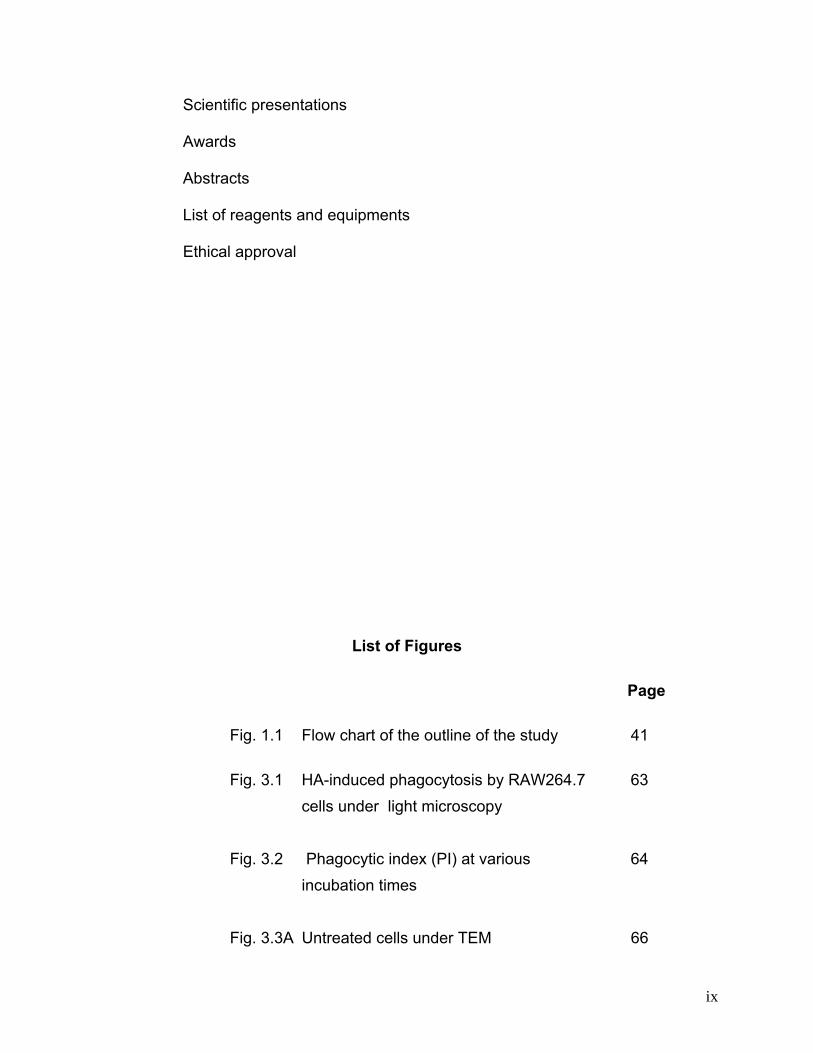
Fig. 1.1 Flow chart of the outline of the study 41
Fig. 3.1 HA-induced phagocytosis by RAW264.7 63 cells under light microscopy Fig. 3.2 Phagocytic index (PI) at various 64 incubation times Fig. 3.3A Untreated cells under TEM 66
x
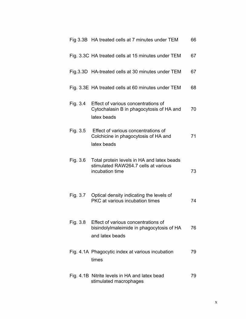
Fig 3.3B HA treated cells at 7 minutes under TEM 66 Fig. 3.3C HA treated cells at 15 minutes under TEM 67 Fig.3.3D HA-treated cells at 30 minutes under TEM 67 Fig. 3.3E HA treated cells at 60 minutes under TEM 68 Fig. 3.4 Effect of various concentrations of
Cytochalasin B in phagocytosis of HA and 70 latex beads
Fig. 3.5 Effect of various concentrations of Colchicine in phagocytosis of HA and 71
latex beads Fig. 3.6 Total protein levels in HA and latex beads
stimulated RAW264.7 cells at various incubation time 73
Fig. 3.7 Optical density indicating the levels of
PKC at various incubation times 74
Fig. 3.8 Effect of various concentrations of bisindolylmaleimide in phagocytosis of HA 76
and latex beads Fig. 4.1A Phagocytic index at various incubation 79 times Fig. 4.1B Nitrite levels in HA and latex bead 79 stimulated macrophages
xi
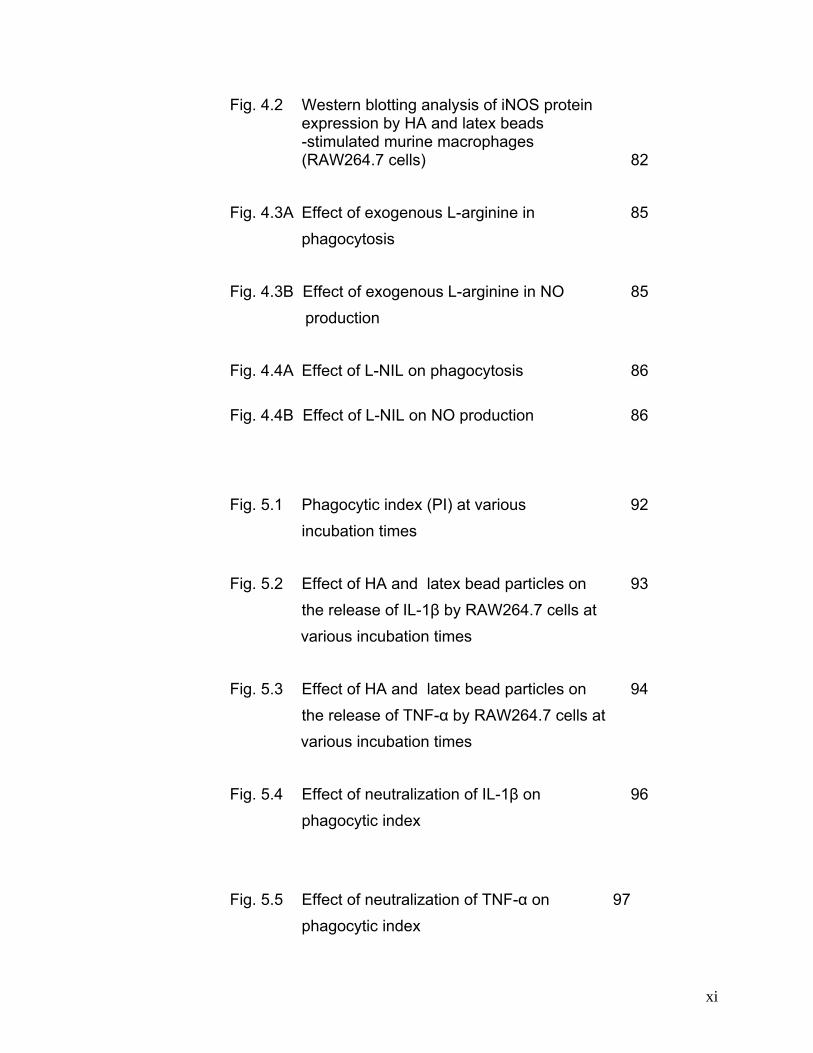
Fig. 4.2 Western blotting analysis of iNOS protein expression by HA and latex beads -stimulated murine macrophages (RAW264.7 cells) 82
Fig. 4.3A Effect of exogenous L-arginine in 85 phagocytosis Fig. 4.3B Effect of exogenous L-arginine in NO 85 production Fig. 4.4A Effect of L-NIL on phagocytosis 86
Fig. 4.4B Effect of L-NIL on NO production 86
Fig. 5.1 Phagocytic index (PI) at various 92 incubation times Fig. 5.2 Effect of HA and latex bead particles on 93 the release of IL-1β by RAW264.7 cells at various incubation times Fig. 5.3 Effect of HA and latex bead particles on 94 the release of TNF-α by RAW264.7 cells at various incubation times Fig. 5.4 Effect of neutralization of IL-1β on 96 phagocytic index Fig. 5.5 Effect of neutralization of TNF-α on 97
phagocytic index
xii
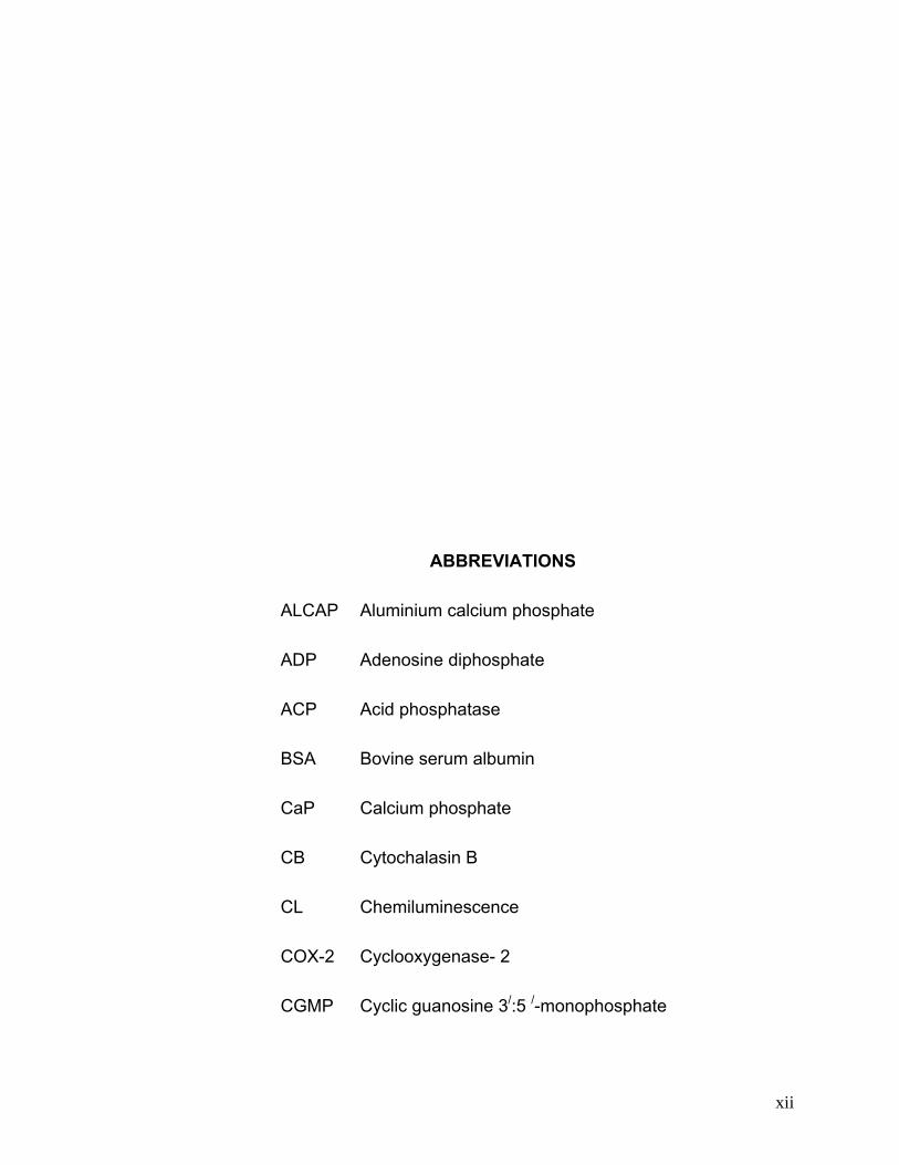
ABBREVIATIONS
ALCAP Aluminium calcium phosphate
ADP Adenosine diphosphate
ACP Acid phosphatase
BSA Bovine serum albumin
CaP Calcium phosphate
CB Cytochalasin B
CL Chemiluminescence
COX-2 Cyclooxygenase- 2
CGMP Cyclic guanosine 3/:5 /-monophosphate
xiii
CMF Cell movement factors
CR Complement receptors
DNA Deoxyribonucleic acid
DBM Demineralized bone matrix
DMEM Dulbecco’s modified Eagle’s medium
DMSO Dimethyl Sulfoxide
eNOS Endothelial nitric oxide synthase
ECW Extracellular matrix
FCS Fetal calf serum
g grams
HA Hydroxyapatite
HAC Hydroxyapatite crystals
IgG Immunoglobulin G
iNOS Inducible NOS
IL Interleukin
INFγ Interferon gamma
ICE IL-1β converting enzyme
KDa Kilodalton
LDH Lactate dehydrogenase
xiv
LSP Large size polyethylene
LTCP Large particle size tricalcium phosphate
LPS Lipopolysaccharide
L-NIL L-N6-(1-iminoethyl)lysine hydrochloride)
M Molar
μm micrometer
mg milligram
μg microgram
μl microliter
ml milliliter
μM micromolar
mM millimolar
mRNA Messenger ribonucleic acid
MDA Malondialdehyde
MSUM Monosodium urate monohydrate
MMP-1 Metalloproteases-1
NOS Nitric oxide synthase
NMA NG-methyl-L-arginine
NO Nitric oxide
xv
nNOS Neural NOS
nm nanometer
nM nanomolar
ng nanogram
NCP Nitrocellulose membrane
OD Optical density
PBS Phosphate buffered saline
pg picogram
PSG Pollen starch granules
PGE2 Prostaglandin E2
PLC Phospholipase C
PKC Protein Kinase C
PI Phagocytic index
PMN Polymorphonuclear leukocytes
RAW Murine macrophage cell line
SDS Sodium dodecyl sulfate
SEM Scanning electron microscopy
SPE Submicron polyethylene
STCP Small particle size tricalcium phosphate
xvi
SiC Silicon Carbide
Ti Titanium
Tris-Hcl Tris(hydroxymethyl)aminomethane-
hydrochloride
TCP Tricalcium phosphate
TEM Transmission electron microscopy
TNF Tumor necrosis factor
TGF-β1 Transforming growth factor beta 1
TLRs Toll like receptors
VD3 Vitamin D3
Abstrak
Analisis gerak balas imunologi makrofaj murin terhadap hidroksiapatit
Tujuan kajian ini adalah untuk menganalisis tindak balas
immunologi hidroksiapatit (HA) ke atas sel RAW264.7 dengan
menentukan fagositosis terangsang HA. Parameter imunologi
yang dikaji termasuk peranan polimerisasi mikrotubul dan aktin,
protein kinase-C (PKC), nitrik oksida (NO), interleukin-1β (IL-
1β) dan faktor nekrosis tumor-α (TNF- α).
Keputusan menunjukkan bahawa HA difagositosis oleh sel
warisan makrofaj (sel RAW264.7) pada tempoh eraman yang
xvii
berbeza. Peningkatan adalah selaras dengan peningkatan PI
di dalam sel yang telah dirangsang dengan HA tempoh
eraman. Analisis TEM telah menunjukkan bahawa partihel HA
telah ditelan oleh sel dan berada di dalam vakuol sel
berkenaan. Cytochalasin B atau colchicine didapati secara
signifikan merencat aktiviti fagositosis sel ke atas kedua-dua
partikel HA dan manik lateks dalam bentuk bergantung dos. Sel
yang dirangsang pula menghasilkan enzim PKC seawal awal
fagositosis iaitu pada masa 7 minit. Sel yang telah dirangsang
dengan Bisindolylmaleimide didapati labih sedikit partikel dalam
bentuk menelan bergantung dos. Penghasilan NO daripada
sel-sel yang dirangsang oleh HA adalah lebih rendah
berbanding sel yang dirangsang oleh manik lateks. Ekspresi
iNOS di dalam kedua-dua sel yang dirangsang oleh manik
lateks dan juga HA telah dikesan pada 7, 15, 30 dan 60 minit
tempoh eraman. L-arginin meningkatkan kedua-dua fagositosis
dan dan penghasilan NO oleh sel yang dirangsang oleh HA
tetapi sebaliknya L-NIL merencat aktiviti tersebut. HA
merangsang sel-sel untuk merembes kedua-dua IL-1β dan
TNF- α dalam bentuk bergantung masa. Dengan kehadiran
antibodi anti-murine IL-1β dan TNF- α aktiviti fagositosis oleh
sel RAW264.7 yang dirangsang oleh HA didapati dikurangkan
dengan signifikan.Olehitu, keputusan kajian ini mencadangkan
xviii
bahawa partikel HA boleh merangsang aktiviti fagositosis
makrofaj murine (RAW264.7 cells) melalui mekanisme yang
berkait dengan polimerisasi aktin dan miktotubul, yang mungkin
berperantarakan enzim PKC. NO juga mungkin memainkan
peranan penting dalam fagositosis terangsang HA oleh sel-sel
RAW264.7 yang mungkin juga bergantung kepada IL-1β dan
TNF- α.
Abstract
The aim of the present study was to analyze the immunological
response of hydroxyapatite (HA) to RAW264.7 cells by
determining HA-induced phagocytosis. Immunological
parameters included in this study were the role of
polymerization of actin and microtubule, protein kinase-C
(PKC), nitric oxide (NO), interleukin-1β (IL-1β) and tumor
necrosis factor-α (TNF-α).
The results showed that HA were phagocytosed by murine
macrophage cell line (RAW264.7 cells) at different incubation
time. Increased PI of HA-treated cells were paralleled with
increased period of incubation. TEM (Transmission electron
xix
microscopy) analysis showed that HA particles were engulfed
by the cells and located within cell vacuoles. Cytochalasin B
or/and colchicine significantly inhibited phagocytic activity of the
cells to both HA particles and latex bead in a dose-dependent
fashion. Stimulated cells produced PKC enzyme right at the
early stage of phagocytosis at 7 minutes. Pre-treated cells with
Bisindolylmaleimide ingested fewer particles in a dose
dependent fashion. NO production was less by HA stimulated
cells than by latex bead-stimulated cells. Inducible nitric oxide
synthase (iNOS) expression in both latex bead- and HA-
stimulated cells was observed at 7, 15, 30 and 60 minutes of
incubation time. L-arginine enhanced but L-NIL inhibited both
phagocytosis and NO production by HA-stimulated cells. HA
stimulated the cells to release both IL-1 β and TNF-α in a time-
dependent fashion. In the presence of anti-murine IL-1 β and
TNF-α, HA-induced phagocytic activity by RAW264.7 cells was
significantly reduced. Therefore, the results of the present study
suggest that HA particles may induce phagocytic activity of
murine macrophages (RAW264.7 cells) in an actin and
microtubule polymerization dependent mechanism, which may
be mediated through PKC enzyme. NO may play a crucial role
in HA-induced phagocytosis by RAW264.7 cells which may
also depend on IL-1 β and TNF-α.

xx
1
CHAPTER 1
INTRODUCTION
1.1 Bone Replacement Materials
Effective repair of bone defects of the skull and facial bones
secondary to traumatic, inflammatory, neoplastic, or iatrogenic
lesions had always been a challenging problem to surgeons.
The first attempt to use bone for cranial reconstruction was
made as far back as 1670 by Van Meekren (Van Meekren,
1682). In 1821, Von Walther performed the first autologous
bone graft (Von Walther, 1821), and in 1867 Ollier
emphasized the role of periosteum in bone regeneration
(Ollier, 1867).
Bone grafting is intended to bridge bone defects there by
establishing native bone architecture. One of the most
effective methods of bridging bone defects is by the use of
autograft. However several limiting factors such as operative
time, blood loss, postoperative pain, length of hospital stay,
and cost are the factors, which are not in favor of autografts.
On the other hand fresh or frozen allografts are available in
different forms with strong mechanical properties. However,
these have the disadvantage of inducing bacterial and viral
2
infection in the recipient due to the infected graft, which may
lead to the possibility of graft rejection. All these facts forced
the researches to indulge in developing the synthetic
materials for bone replacements (Ilan and Ladd, 2002).
Demineralized Bone Matrix (DBM) is an allograft, which is
used commonly nowadays and is available as dry, moldable
or injectable forms. It is in the form of powder, paste, or putty,
using a carrier that renders it suitable for placement. However
the demineralization leads to denaturation of the protein
matrix in the bone because of chemical and radiation
treatment resulting in weakening of the bone and the bone
formation potential. Growth factors might survive processing,
although their actual presence is not very clear (Ilan and
Ladd, 2002).
Endobon is a bovine-derived ceramic consisting of cancellous
bone, sintered and processed to eliminate all but the mineral
components (Werber et al., 2000). This material lacks in
immunogenicity and has a poor remolding capacity. It is
currently used in the treatment of craniofacial fractures and
bone defects. Coralline HA (Pro Osteon, Interpore Cross
International, Irvine, CA) is coral that is thermo chemically
treated with ammonium phosphate, which demonstrates
3
porosity similar to bone and finds its application in the
treatment of fractures (Wolfe et al., 1999).
Alloplastic materials are synthetic materials that are used in
reconstructive dental and orthopedic surgery. The source from
which these materials are derived were of non-human, non-
animal, and hence, non-organic. Since these materials are of
non-organic source they are easily available for reconstructive
work and have an added advantage of avoiding donor scar
and infection in the recipient as a result of contaminated graft
from the donors. However for the alloplastic material to be
clinically successful, it must be biocompatible, implying an
acceptable interaction between the host and the implant
material. These alloplastic materials such as solid silicone,
polytetrafluoroethylene, polyethylene and acrylic when
implanted into defected bone, they are generally encapsulated
by a fibrous tissue, which is initiated by a host inflammatory
response (Anderson and Miller, 1984). As a result of this
response, the implant materials do not adhere to bone and
this is a critical problem in their use in bone repair.
Bioactive ceramics that spontaneously bind to and integrate
with bone in the living body have been investigated (Kokubo
et al., 2003). Various types of bioactive ceramics have been
developed over the last three decades. Among these the main
4
bioactive ceramics used clinically are hydroxyapatite (HA)
(Jarcho et al., 1977), HA micro crystals (Fukuchi et al., 1995),
Calcium phosphate (CaP) ceramic (Benahmed et al., 1996b)
etc.
Macrophage actively phagocytosed the HA micro crystals.
However, no damage was observed in macrophages exposed
to HA micro crystals by transmission electron microscopy
(TEM). Macrophage in the presence of HA micro crystals
showed less acid phosphatase (ACP) and lactate
dehydrogenase (LDH) activity and higher intracellular calcium
content than those in the presence of calcined HA and
alumina. HA micro crystals as well sintered HA were reported
to have excellent biocompatibility to macrophages (Fukuchi et
al., 1995).
Studies have been conducted on an ultrastructural
scale to determine the specific behavior of human monocytes
with regard to CaP ceramic (Benahmed et al., 1996b).
Phagocytosis of CaP coincided with autophagy and
accumulation of residual bodies in the cells. Addition of
HILDA/LIF leukemia inhibitory factor to these cultures induced
a very marked decrease in phagocytotic activity on CaP
crystals. Autophagy was reduced, and residual bodies were
absent
5
Studies on the evaluation of the importance of particle
characteristics on cytokine production by human monocytes
in vitro demonstrated that needle shaped particles induced
larger production of tumor necrosis factor-α (TNF-α),
interleukin-6 (IL-6) and interleukin-10 (IL-10) by cells as
compared to spherical and indifferent shaped particles
(Laquerriere et al., 2003b). To a less extent, the smallest
particles induced an increase in the expression and
production of the cytokines.
In view of these limitations in using autogenous and allogenic
bones as replacement materials, alloplastic implant materials
offer an excellent substitute in soft and hard tissue
replacement and repair (Eppley, 1999). The ranges of
materials included are dimethylsiloxane (Silicone),
polytetrafluoroethylene, polyethylene, polyseters, polyamides,
acrylic, metals, cynoacrylate adhesives and calcium
phosphates.
Ilan and Ladd (2002) described several minerals/ ceramics
such as HA, corraline HA granules or blocks, calcium sulfate
pellets, β-tricalcium phosphate, bioactive glass, polymer
implant with bioactive glass were osteoconductive solid
formulations that were used as bone implant material.
6
1.2 Hydroxyapatite (HA)
HA ceramic was found to have chemical compositions closely
resembling that of mineral phase of natural bone (Liu, 1998).
Implants composed of CaP have been available as bone
replacement/augmentation materials for more than 20 years
(Jarcho, 1981). CaP materials are not osteoinductive by
themselves, but they do provide a physical substrate on to
which new bone from adjacent surfaces may be deposited
and guided into area occupied by the material (Alexander,
1987).
Currently available CaP materials were manufactured as HA
with a chemical formula of Ca10(PO4)6(OH)2. It can be
manufactured either as ceramic or as non ceramic apatites
and can be formed into a wide variety of physical
configurations (Yuen et al., 1994). Ceramic HA were made
from crystals that are sintered at high temperatures into a
hard nonresorbable solid. They were available as dense
granules or blocks in the early 1980s and were used in
maxillofacial reconstruction, particularly alveolar ridge
augmentation (Frame et al., 1987). The dense form of the
granules was prone for migration before significant fibro-
osseous in growth, and the dense blocks were difficult to
7
shape and were prone for extrusion. Therefore, dense HA
were replaced by a different physical structure.
Porous HA were based on the structure of marine corals
(calcium carbonate skeleton), which have interconnecting
porosity of a size varying from 50 to 200 μm that permitted
fibrovascular and osseous growth with the potential for cell-
mediated resorption and osseous replacement (Holmes and
Hagler, 1988). Of the available porous HA forms, the granules
have achieved the greatest current use as an augmentation
material for the craniofacial skeleton (Byrd et al., 1993). The
block forms were used primarily as an interpositional graft
material in facial skeletal osteotomies (Salyer and Hall, 1989).
In dentistry, HA is used as a bone replacement material in
surgical work. Study undertaken to evaluate the histological
effect of HA–collagen on twenty-four proximal periodontal
defects in mongrel dogs revealed that the implanted
biomaterial promoted cementogenesis of the demineralized
root surfaces and established a strong interdigitation between
the root surface and the gingival connective tissue fibers
(Minabe et al., 1988). Similar investigations on the histological
response of HA on infrabony periodontal pockets in humans
by Galgut et.al (1990) showed the healing response that vary
between specimens and between sites within the same
8
specimen at 22, 40 and 60 weeks after placement of implant.
HA finds its application as a bone implant material in clinical
dentistry particularly in the fields of periodontology, oral
surgery and endodontic work.
Although HA is well suited as biomaterial to replace
autogenous bone in skeletal reconstruction it has been
observed that this biomaterial when come in intimate contact
with the tissues of the body, might initiate several complicated
biological reactions such as the release of chemotactic
mediators and growth factors that might elicit and sustain
inflammatory responses at the implant site (David et al., 1964,
Bloom and Bennett, 1966, Nathan et al., 1971).
It had been found that neutrophils, monocytes and
macrophages invaded the tissues surrounding the implant.
This was controlled and directed by chemotactic or
chemokinetic agents that included complement factors,
lymphokines, platelet factors, leukotrienes and bacterial
fragments (Remes and Williams, 1990). Studies have shown
that in the case of ceramic implants, different calcium
phosphate powder stimulated the activation of complement
involving phagocytes, which were attracted to the
inflammatory site (Klein et al., 1983). It was known that
biomaterials such as metallic and ceramic implants could
9
cause sensitivity during exposure to the tissues, with
attraction and activation of macrophages (Bosetti et al., 1999).
Previous studies indicated that a wide variety of particles
could prime the macrophages to give a marked increase in
their oxidative response (Myrvik et al., 1993).
The effects of HA particulate debris on the production of
cytokines like Interleukin-1β (IL1β), IL-6, TNF-α on human
fibroblasts demonstrated that HA and HA/TCP particles were
capable of stimulating the expression and secretion of
cytokines and proteases that enhanced bone resorption,
suggesting that particulate debris from implants using these
coating also might increase osteolysis and loosening of the
implant (Ninomiya et al., 2001). During pro-inflammation,
macrophages release pro-inflammatory cytokines like IL1β
and TNF-α. These Cytokines have been identified in the
preprosthetic tissues of patients with implants (Goodman et
al., 1998, Catelas et al., 2003). It has also been observed that
HA activated human monocytes in vitro stimulated the
production of IL-1β and TNF-α (Laquerriere et al., 2003a).
However it is not clear whether the released cytokines would
act in an autocrine fashion to regulate HA induced
macrophage activity.
10
Studies have demonstrated that nitric oxide (NO) has negative
regulatory role in cytoskeletal assembly, pseudopodia
formation, phagocytosis and adherence of murine
macrophages in association with the ADP– ribosylation of
actin on a laminin substratum. NO, depending on the extent
and duration of its production, resulted in reversible inhibition
of macrophage function, thereby protecting the tissue
microenvironment from harmful effect of activated
macrophages (Jun et al., 1996). Studies by Ke et.al (2001)
have postulated that cyclic guanosine’ 5’–monophosphate
(cGMP) and subsequent ca2+/calmodulin might be key
regulators of actin reorganization in NO – stimulated RAW
264.7 cells.
Macrophages, when involved in phagocytosis, release NO but
it is still unclear weather this NO will upregulate or
downregulate HA-induced macrophage functions. In vivo,
macrophages migrate to and phagocyte the implanted HA
(Rahbek et al., 2005). It is assumed that macrophages would
release NO when phagocytosed by HA. It is also possible that
this gaseous molecule would also regulate the levels of HA
induced macrophage phagocytic activity.
Protein kinase C (PKC) is required at the earliest stage of
particle internalization since inhibition of PKC blocks the
11
formation of actine filaments beneath the site of particle
binding (Allen and Aderem, 1995). PKCs are key participants
in numerous signaling pathways to the actin cytoskeleton and
the nucleus, including signals stimulated by hormones,
cytokines, and adhesion, suggesting multiple level of
regulation of phagocytic efficiency (Kikkawa et al., 1989, Zhu
et al., 2001). However there was no data to show that PKC
might play a role in HA induced macrophage phagocytic
activity.
1.2.1 HA as Bone Substitute or Replacement Material
CaP biomaterials are used as an augmentation material in
reconstructive surgery to bridge surgical bone defects
(LeGeros, 2002). Commercially available CaP biomaterials
differ in origin (natural or synthetic), composition (HA, beta-
tricalcium phosphate and biphasic CaP) and physical forms
(particulates, blocks, cements, coating on metal implants,
composites with polymers) and in its physicochemical
properties.
The properties which are in favor of CaP biomaterials include
the similarity in composition to bone mineral, bioactivity
(ability to form bone apatite like material or carbonate HA on
their surface), ability to promote cellular function and
expression leading to formation of a uniquely strong bone
12
CaP biomaterial interface and osteoconductivity (ability to
provide the appropriate scaffold or template for bone
formation) (LeGeros, 2002).
It had been observed that CaP biomaterials in its three-
dimensional geometry was able to bind and concentrate
endogenous bone morphogenetic proteins in circulation, and
might become osteoinductive (capable of osteogenesis), and
could be the effective carriers of bone cell seeds (LeGeros,
2002).
Other advantages of HA over autografts and allografts are
unlimited supply, easy sterilization and storage (Bucholz,
2002). In view of the above advantages, CaP biomaterials find
their potential use in tissue engineering for regeneration of
hard tissues.
1.2.2 In Vivo Studies with HA
Various researches have investigated HA, when this material
is implanted into the tissue monocytes/ macrophages, are
attracted to the implant site (Heymann et al., 1999). Analysis
of retrieved tissues from animal models (Rahbek et al., 2005)
and in humans have confirmed phagocytosis of HA particles
(Bloebaum et al., 1994).
13
The in vivo effect of the phagocytosis of pure HA particles and
HA/dichloromethylene bisphosphonate (clodranate)
suspension were investigated by TEM and standard
chemiluminescence (CL) assays following intra peritoneumal
injection in rats (Hyvonen and Kowolik, 1992). Macrophages
were harvested at 12, 24, 48 and 96 hours. HA was
completely phagocytosed by 24 hours and HA reacted with
clodronate was completely phagocytosed by 48 hours. From
48 hours onwards HA dissolution was observed in the
phagosomes of the cell in the two groups. Clodronate seemed
to exhibit an inhibitory effect on the phagocytic activity and an
enhancement of the chemiluminescence production by the
cells in this model, indicating that it was modifying the
inflammatory cell response
In a similar study, HA-induced macrophage interactions were
investigated after implantation into spongy bone of the distal
femur of rabbits (Muller-Mai et al., 1990). The specimen was
examined under TEM and scanning electron microscopy
(SEM) following transverse fracture in the interface. It was
observed that the implants displayed considerable changes in
the surface morphology caused by leaching, corrosion and
active resorption by osteoclast-like cells. Macrophages were
also involved in cleaning the surface via phagocytosis of loose
14
implant particles. Subsequent mineralizations of these areas
were also observed. In vivo mechanisms of HA ceramic
degradation by osteoclast cells were investigated by TEM
(Wenisch et al., 2003). The results revealed that the
osteoclasts mediated the degradation of HA ceramic
implanted into the sheep bone by simultaneous resorption and
phagocytosis.
In vivo animal studies were done to see the effect of
phagocytosable particles of HA on bone in-growth in the
bilaterally implanted harvest chamber in the proximal tibial
metaphyses of 13 mature rabbits (Wang et al., 1994). The
results of this study showed that after 6 weeks, HA particles
were incorporated within the matrix of ingrown new bone and
there was no evidence of granuloma formation or
inflammation. Thus, HA particles, which were small enough to
be phagocytosed by macrophages, had no adverse effect on
bone in-growth. Balla et.al (1991) also studied the effect of HA
in relation to the histology of furcation perforations created in
the mandibular and maxillary premolars and molars of six
rhesus monkeys. They observed that after 6 months HA
treated tooth revealed that perforated defect was filled with ill-
oriented fibrous connective tissue and globules of HA with
minimal inflammation and bone resorption.
15
Van Blitterswijk et.al (1985) have studied the events at the HA
implant material/tissue interface in the middle ear of the rat.
The results suggested that resorption of the implant material
occurred by mono and multinuclear phagocyte activity.
Resorption decreased 6 months after the operation, possibly
due to the decreasing number of phagocytes at the interface
and the increasing amount of bone in the micropores.
Studies were carried out on the effect of HA in human
periodontal tissues, dental pulp, and following tooth
implantations. The biocompatibility of HA crystals implanted
into infrabony periodontal defects in human was also studied
(Benque et al., 1985). They demonstrated that the
biocompatibility was accompanied by a normal fibrogenesis
and apparent osteogenesis after 6 years. Filled extraction
sockets in miniature swine treated in a similar manner with
apatite were also studied.
Noguchi (1989) demonstrated osteodentin bridging in 6
months following direct pulp capping with HA on experimental
exposure of pulp in teeth, which were to be extracted for
orthodontic reasons. It was observed that in the deeper region
of the dentin bridge, tubular dentin was formed newly. Both
osteodentin and tubular dentin fused tightly without any
organelles in the border between them.
16
The importance of crystal size of bioceramic on bone
formation in human periodontal lesions was studied (Frank et
al., 1991). The results suggested that micro sized HA
generated significant amount of peripheral bone formation in 6
months when implanted into human periodontal lesions
Yu et.al (2003) also reported 15-months follow-up after
treating a case of combined endodontic-peridontic lesion on a
mandibular first molar by intentional replantation and
application of HA. They found that tooth was clinically and
radiographically healthy and functioned well at the end of the
follow-up period.
1.2.3 In vitro Studies on HA
Various researchers have investigated HA in vitro which help
us to understand the response of the materials better in
various experimental designs.
Interaction of human monocyte and monocyte derived
macrophages to HA, tricalcium phosphate (TCP), and
aluminum calcium phosphate (ALCAP) were investigated
(Ross et al., 1996). The data from these experiments
suggested that monocytes and macrophages were capable of
adhering to the surface of HA, TCP and ALCAP in an in vitro
environment for over a 7 day period. However, long-term
incubation of the ceramic capsules with macrophages
17
revealed that the cells experienced a gradual disassociation
phenomenon. Similar studies on the interrelationship of
various biomaterials such as HA, titanium (Ti), large size
polyethylene (LSP), submicron polyethylene (SPE), large
particle size tricalcium phosphate (LTCP) and small particle
size tricalcium phosphate (STCP) towards human
monocytes/macrophages suggested that regardless of the
biomaterial used, all experimental groups experienced
remarkable phagocytosis in the first two phases (24, 48 hours)
(Carr et al., 1999).
Human monocytes placed on the surface of HA and biphasic
calcium phosphate (BCP) tablets in the presence of vitamin
D3 (VD3) and interferon gamma (INF gamma) showed
monocytes being influenced by soluble factors (vitamins,
cytokines) in initiating the degradation process on biomaterial
(Benahmed et al., 1996a).
Bosetti et.al (1999) investigated on the biological reaction of
the macrophages to natural apatite (heat treated bovine
bone), synthetic apatite (HA), and three types of alumina as
control. Ultrastructural observation and electron microscopic
analysis showed that the macrophages grown in the presence
of natural and synthetic apatite were seen with features of
healthy cells while macrophages grown in the presence of
18
alumina seemed to be negatively affected. Biocompatibility of
biomaterials (titanium, mixed particle size polyethylene (MPE),
ultra high molecular weight polyethylene, mixed particle size
of tricalcium phosphate, and hydroxyapatite) in response to
RAW macrophages was evaluated at 24, 48 and 72 hours
using biochemical markers (Johnston et al., 1999). At 24
hours there was an increase in catalase levels and no initial
cell membrane damage was observed by malondialdehyde
(MDA) assay, but in 48 and 72 hours, cellular injury occurred
in all treatment groups as evidenced by lactate
dehydrogenase (LDH) levels.
HA-induced neutrophil interactions were studied under TEM
and CL by Dowsett et.al (1997). The TEM results confirmed
the functional integrity of the neutrophils, particularly those
phagocytosing HA particles up to 24 hours. Based on these
results it was demonstrated that human peripheral blood
neutrophils could be maintained in a fully functional state with
respect to the respiratory bust and morphology at least for 24
hours.
In an other identical report the interaction between human
neutrophils and pure HA particles were assessed in vitro by
TEM and CL (Hyvonen and Kowolik, 1991). Neutrophils
convincingly phagocytosed HA particles within 15 minutes and
19
a high CL response was elicited by the zymosan-stimulated
CL reaction. Clodronate (dichloromethylene bisphosphonate)
alone appeared to have little effect on the cell morphology or
CL. When HA was combined with clodronate, phagocytosis
was more rapid, and the zymosan-stimulated CL was 50% of
that of the HA group suggesting the anti-inflammatory role of
clodronate.
Various researchers studied the in vitro response of fibroblast
cells to HA. Gregoire et.al (1987) studied the in vitro effect of
synthetic granular HA on cultured fibroblastic cells (L929,
human bone and gingival cells). Phagocytosis of synthetic
granular HA particles resulted in morphological cell changes
which were demonstrated by microscopic examinations.
In a similar study human fibroblasts were incubated in the
presence of hydroxyapatite or calcium hydroxide (Ca (OH) 2)
(Alliot-Licht et al., 1994). With Ca (OH) 2, the cells exhibited
alteration in morphology, DNA synthesis, alkaline
phosphatase activity and protein synthesis, in accordance
with the necrosis observed when Ca (OH) 2 was used as a
pulp-capping agent. However with HA, phagocytic activity of
pulpal fibroblast towards HA was seen. As a consequence,
DNA synthesis was affected with inhibitory effect on alkaline
phosphatase activity, which correlated with clinical
20
observation where reparative dentin bridge was observed
directly on the pulp tissues when HA was used as a pulp-
capping agent.
Further reports on fibroblast interactions with finely ground
powder of synthetic HA by Evans (1991) suggested a
reduction in total growth rate and mitotic rate of the cells and
increase in the number of pycnotic cells. The effect was dose
related and any occurred with small particles. The small
particles appeared to either adhere to the cells or
phagocytosed by them. The toxic effect was assumed to be
physical rather than chemical.
The ability of monosodium urate monohydrate (MSUM), HA
and diamond crystals to stimulate phagocytosis, degranulation
and secretion of cell movement factors (CMF) from
polymorphonuclear leukocytes (PMN) were also assessed
(Swan et al., 1990). The ability of each crystal to absorb PMN
derived enzymes and CMF was also compared. MSUM
crystals stimulated greater enzyme release and generation of
CMF than HA; in contrast, HA crystals exhibited greater
absorption of PMN products. Diamond crystals clearly
interacted with PMN, but they did not stimulate degranulation
or CMF production.
21
The influence of HA particle size (0.5-3.0, 37-63, 177-250 and
420-841 micron) on osteoblasts was also investigated in-vitro
at 1 hour, 3 hours, 1day, 3 days and 7 days (Sun et al., 1998).
It was observed that adding HA to the culture reduced the
osteoblast cell count. Transforming growth factor-beta1 (TGF-
β1) concentrations in culture decreased significantly with the
addition of HA particles. Prostaglandin E2 (PGE2)
concentration in the medium increased significantly. The
changes in TGF-β1 and PGE2 concentration were more
significant and persisted longer in smaller-particle groups.
Thus, the above in-vitro studies highlighted the HA
interactions with various cells such as human monocytes,
macrophage, neutrophils, fibroblast, polymorphonuclear
leukocytes, and osteoblast.
1.3 Complexity in Phagocytosis
The term phagocytosis means engulfing or ingesting
particulate materials and microorganisms by phagocytic cells
which form an essential component of the innate immune
system (Stossel, 1999).
The phagocyte-microbe interaction had been widely
investigated which was accompanied by intracellular signals
that triggered various cellular processes such as cytoskeletal
rearrangement, alteration in membrane trafficking, activation
22
of microbial killing mechanisms, production of pro-and anti-
inflammatory cytokines and chemokines, activation of
apoptosis, and production of molecules required for efficient
antigen presentation of the adaptive immune system (Aderem
and Underhill, 1999). Even the fundamental processes of
internalizing particles were to be proceeded through a variety
of distinct molecular and morphological processes.
This complex phagocytic mechanism involves many
underlying principles such as the involvement of many
receptors and many signaling molecules have been described
as key signaling molecules in phagocytic responses. In one
recent study, Garin et.al (2001) purified macrophage
phagosomes containing latex beads and identified more than
140 proteins associated with the phagosome by two-
dimensional electrophoresis and mass spectrometry. These
investigators identified many proteins which were not
previously known to be associated with phagosomes, as well
as many novel proteins.
Phagocytosis occurs in four stages (Underhill and Ozinsky,
2002). First, many different receptors recognize microbes and
phagocytosis is usually mediated simultaneously by multiple
receptors. Second, different microbe-recognition receptors
induce different signaling pathways and their signals interact
23
cooperatively (and sometime destructively) to mediate
ultimate response to particles. Third, microbe recognition is
coupled (either directly through phagocytic receptors or
indirectly through co-receptors) to inflammatory responses
that in turn affect the efficiency of particle internalization by
phagocyte or neighboring phagocytes. Fourth, many
pathogenic microbes actively attempt to regulate the
mechanisms of phagocytosis to evade destruction.
Phagocytosis is an inherently complex process that requires
coordinated activation of signaling leading to events as
diverse as actin remodeling, alterations on membrane
trafficking, particles engulfment, microbial killing and
production of appropriate inflammatory mediators that direct
the adaptive immune response (Underhill and Ozinsky, 2002).
The consequences of phagocytosis vary and they depend on
the identity of the microbial target and many factors that
modulate the activation state of the phagocyte. Many proteins
have been identified that play important role during
phagocytosis. It is important to integrate these molecules into
pathways that account for the diversity of phagocytic
responses.
24
1.3.1 Receptors for Phagocytosis
Phagocytes express a broad spectrum of receptors that
participate in particle recognition and internalization (Underhill
and Ozinsky, 2002). Some of these receptors are capable of
transmitting intercellular signals that trigger phagocytosis
while other receptors appear primarily to participate in binding
or to increase the efficiency of internalization. The main
classes of phagocytic receptors that participate in
phagocytosis of microbes include Fc-receptors, complement
receptors, various integrins, scavenger receptors, and
mannose receptor.
1.3.2 Role of Signaling Pathways during Engulfing
Particle internalization is accompanied by activation of many
signaling pathways that together coordinate rearrangement of
the actin cytoskeleton, extension of the plasma membrane,
and engulfment. Number of signaling molecules including
actin binding proteins, membrane traffic regulators, ion
channels, kinases and lipases are activated during
phagocytosis of complex particles (such as opsonized
bacteria) and may contribute for the efficient internalization
(Underhill and Ozinsky, 2002). However, certain signaling
molecules stand out as participants both in phagocytosis and
in many other signaling pathways. Phosphoinositide 3-kinase

![OPEN ACCESS International Journal of Molecular Sciences...hair growth [2].Platelet-derived growth factor (PDGF) isoforms reportedlyinduce and maintain theanagen phase of the murine](https://static.fdocument.org/doc/165x107/60f85444d7faee31306fdb0e/open-access-international-journal-of-molecular-sciences-hair-growth-2platelet-derived.jpg)

![Case Report Initial Biological Evaluations of [18F]KS-7-51 to … · 2020. 9. 22. · and initial biological evaluations of [18F]KS-7-51, a p-fluoroethoxy phenyl derivative in a murine](https://static.fdocument.org/doc/165x107/601e58f23cdaba46814221b9/case-report-initial-biological-evaluations-of-18fks-7-51-to-2020-9-22-and.jpg)