Amyloid β 1-40 (human) ELISA - DRG Diagnostics
21
User´s Manual Legal Manufacturer: Distributed by: DRG Instruments GmbH, Germany Division of DRG International, Inc Frauenbergstr. 18, D-35039 Marburg Telefon: +49 (0)6421-17000 Fax: +49-(0)6421-1700 50 Internet: www.drg-diagnostics.de E-mail: [email protected] Amyloid β 1-40 (human) ELISA EIA-5231 96
Transcript of Amyloid β 1-40 (human) ELISA - DRG Diagnostics
User s Manual
Legal Manufacturer: Distributed by:
DRG Instruments GmbH, Germany Division of DRG International, Inc Frauenbergstr. 18, D-35039 Marburg Telefon: +49 (0)6421-17000 Fax: +49-(0)6421-1700 50 Internet: www.drg-diagnostics.de E-mail: [email protected]
Amyloid β 1-40 (human) ELISA
EIA-5231
96
DRG Amyloid β 1-40 (human) ELISA EIA-5231
Vers. 3.0 2012/05 – mhe/vk - 1 -
Contents / Inhaltsverzeichnis 1 INTRODUCTION.......................................................2 2 PRINCIPLE OF THE TEST .......................................2 3 WARNINGS AND PRECAUTIONS ...........................3 4 REAGENTS ..............................................................4 5 SPECIMEN COLLECTION AND PREPARATION.....6 6 ASSAY PROCEDURE ..............................................6 7 EXPECTED NORMAL VALUES................................8 8 QUALITY CONTROL ................................................8 9 PERFORMANCE CHARACTERISTICS....................9 10 LIMITATIONS OF USE ...........................................10 11 LEGAL ASPECTS...................................................10 12 REFERENCES / LITERATURE...............................11 1 EINLEITUNG...........................................................12 2 TESTPRINZIP.........................................................12 3 VORSICHTSMAßNAHMEN ....................................12 4 BESTANDTEILE DES KITS ....................................13 5 PROBENVORBEREITUNG ....................................15 6 TESTDURCHFÜHRUNG ........................................16 7 ERWARTETE WERTE............................................18 8 QUALITÄTS-KONTROLLE .....................................18 9 ASSAY CHARACTERISTIKA..................................18 10 GRENZEN DES TESTS..........................................19 11 RECHTLICHE GRUNDLAGEN ...............................19 12 REFERENZEN / LITERATUR .................................19 SYMBOLS USED WITH DRG ELISAS ...........................20
DRG Amyloid β 1-40 (human) ELISA EIA-5231
Vers. 3.0 2012/05 – mhe/vk - 2 -
1 INTRODUCTION
1.1 Intended Use The DRG Amyloid β 1-40 (human) ELISA (Abeta-40 ELISA) is an enzyme immunoassay for the quantitative in vitro measurement of human Amyloid β peptide 1-40 in serum, plasma and CSF.
1.2 Summary and Explanation The pathological hallmarks of Alzheimer’s disease (AD) are amyloid plaques, neurofibrillary tangles, synaptic degeneration and neuronal loss (1). Amyloid plaques are mainly composed of amyloid-beta (Aβ) 40 and 42 peptides derived from the proteolytic cleavage of amyloid precursor protein (APP) by β-site APP cleavage enzyme 1 (BACE1) (2,3) and the γ-secretase (4). The endosome and the endocytic pathway have been proposed as possible sites for the β and γ cleavage sites of APP (5), and the resulting Aβ peptides are secreted by both neuronal and non-neuronal cells (6). Recently, soluble forms of Aβ have been implicated in neurotoxicity (7), and may correlate better with cognition than amyloid plaque burden (8). As Aβ is considered to play an early and pivotal role in AD pathogenesis, it may be a useful tool in diagnosing AD in the preclinical/early stages, as well as for monitoring potential Aβ modifying therapies (9). While human CSF Aβ levels have mostly shown reduction with disease progression (10), much of the data on human plasma Aβ remain unclear. Studies have shown that subjects who exhibit overproduction of Aβ such as individuals with familial forms of AD and Down’s syndrome have higher plasma Aβ levels compared to controls (11). In addition to variability found with age and disease severity (12), Aβ plasma levels are further modulated by production in large peripheral organs such as skeletal muscle, liver, kidney, skin, and lung (13,14), movement from brain interstitial fluid into blood stream (15), and peripheral uptake and clearance of Aβ in the liver by presence of low density lipoprotein receptor-related protein-1 (16) and receptors for advanced glycation end products (RAGE) (17). This may explain why much of the plasma Aβ data in cross-sectional studies do not show significant differences between sporadic AD and controls. Longitudinal studies seem more promising, showing initially elevated Aβ42 levels in those that eventually develop AD in the future (18). Furthermore, subjects who manifest sporadic AD within the time frame of the study start with higher plasma Aβ42 levels, and the Aβ42/Aβ40 ratios also appear to be significantly different from those who remain asymptomatic (19).
2 PRINCIPLE OF THE TEST The DRG Abeta-40 ELISA Kit is a solid phase enzyme-linked immunosorbent assay (ELISA) based on the sandwich principle. The microtiter wells are coated with a monoclonal [mouse] antibody directed towards a unique antigenic site on the Abeta-40 peptide. An aliquot of patient sample containing endogenous Abeta-40 peptide is incubated in the coated well with enzyme conjugate, which is a biotinylated anti-Abeta-40 antibody. After incubation the unbound conjugate is washed off. Finally, Enzyme Complex, which is streptavidin conjugated with horseradish peroxidase is added, and after incubation, unbound enzyme complex is washed off. The amount of bound peroxidase is proportional to the concentration of Abeta-40 peptide in the sample. Having added the substrate solution, the intensity of colour developed is proportional to the concentration of Abeta-40 in the patient sample.
English
DRG Amyloid β 1-40 (human) ELISA EIA-5231
Vers. 3.0 2012/05 – mhe/vk - 3 -
3 WARNINGS AND PRECAUTIONS 1. This kit is for in vitro diagnostic use only. For professional use only. 2. All reagents of this test kit which contain human serum or plasma have been tested and confirmed
negative for HIV I/II, HBsAg and HCV by FDA approved procedures. All reagents, however, should be treated as potential biohazards in use and for disposal.
3. Before starting the assay, read the instructions completely and carefully. Use the valid version of the package insert provided with the kit. Be sure that everything is understood.
4. The microplate contains snap-off strips. Unused wells must be stored at 2 °C to 8 °C in the sealed foil pouch and used in the frame provided.
5. Pipetting of samples and reagents must be done as quickly as possible and in the same sequence for each step.
6. Use reservoirs only for single reagents. This especially applies to the substrate reservoirs. Using a reservoir for dispensing a substrate solution that had previously been used for the conjugate solution may turn solution colored. Do not pour reagents back into vials as reagent contamination may occur.
7. Mix the contents of the microplate wells thoroughly to ensure good test results. Do not reuse microwells. 8. Do not let wells dry during assay; add reagents immediately after completing the rinsing steps. 9. Allow the reagents to reach room temperature (21 °C to 26°C) before starting the test. Temperature will
affect the absorbance readings of the assay. However, values for the patient samples will not be affected.
10. Never pipet by mouth and avoid contact of reagents and specimens with skin and mucous membranes. 11. Do not smoke, eat, drink or apply cosmetics in areas where specimens or kit reagents are handled. 12. Wear disposable latex gloves when handling specimens and reagents. Microbial contamination of
reagents or specimens may give false results. 13. Handling should be done in accordance with the procedures defined by an appropriate national
biohazard safety guideline or regulation. 14. Do not use reagents beyond expiry date as shown on the kit labels. 15. All indicated volumes have to be performed according to the protocol. Optimal test results are only
obtained when using calibrated pipettes and microtiterplate readers. 16. Do not mix or use components from kits with different lot numbers. It is advised not to exchange wells of
different plates even of the same lot. The kits may have been shipped or stored under different conditions and the binding characteristics of the plates may result slightly different.
17. Avoid contact with Stop Solution containing 0.5 M H2SO4. It may cause skin irritation and burns. 18. Some reagents may contain Proclin 300, BND and/or MIT as preservatives. In case of contact with eyes
or skin, flush immediately with water. 19. TMB substrate has an irritant effect on skin and mucosa. In case of possible contact, wash eyes with an
abundant volume of water and skin with soap and abundant water. Wash contaminated objects before reusing them. If inhaled, take the person to open air.
20. Chemicals and prepared or used reagents have to be treated as hazardous waste according to the national biohazard safety guideline or regulation.
21. For information on hazardous substances included in the kit please refer to Material Safety Data Sheets. Material Safety Data Sheets for this product are available upon request directly from DRG.
DRG Amyloid β 1-40 (human) ELISA EIA-5231
Vers. 3.0 2012/05 – mhe/vk - 4 -
4 REAGENTS
4.1 Reagents provided 1. Microtiterwells, 12 x 8 (break apart) strips, 96 wells;
Wells coated with anti-Abeta-40 antibody (mouse monoclonal). 2. Standard (Standard 0-6), 7 vials, (lyophilized), 1 mL each,
Concentration: 0 - 20 - 50 - 125 - 250 - 500 - 750 pg/mL see „Reagent Preparation“ Contain non-mercury preservative.
3. Control Low & High, 2 vials, (lyophilized) 1 mL each, see „Reagent Preparation“ For control values and ranges please refer to vial label or QC-Datasheet. Contain non-mercury preservative.
4. Assay Buffer, 1 vial, 14 mL, ready to use, Contains non-mercury preservative.
5. Enzyme Conjugate, 1 vial, 7 mL, ready to use, Anti-Abeta-40 antibody conjugated to horseradish peroxidase; Contains non-mercury preservative.
6. Enzyme Complex, 1 vial, 14 mL, ready to use, Streptavidin conjugated to horseradish peroxidase; Contains non-mercury preservative.
7. Substrate Solution, 1 vial, 14 mL, ready to use, Tetramethylbenzidine (TMB).
8. Stop Solution, 1 vial, 14 mL, ready to use, contains 0.5 M H2SO4, Avoid contact with the stop solution. It may cause skin irritations and burns.
9. Wash Solution, 1 vial, 30 mL (40X concentrated), see „ Reagent Preparation “.
Note: Additional Assay Buffer for sample dilution is available upon request.
4.2 Materials required but not provided − A microtiter plate calibrated reader (450 ± 10 nm) (e.g. the DRG Instruments Microtiter Plate Reader). − Calibrated variable precision micropipettes. − Absorbent paper. − Distilled or deionized water − Timer − Semi logarithmic graph paper or software for data reduction
4.3 Storage Conditions When stored at 2 °C to 8 °C unopened reagents will retain reactivity until expiration date. Do not use reagents beyond this date. Opened reagents must be stored at 2 °C to 8 °C. Microtiter wells must be stored at 2 °C to 8 °C. Once the foil bag has been opened, care should be taken to close it tightly again. Opened kits retain activity for two months if stored as described above.
DRG Amyloid β 1-40 (human) ELISA EIA-5231
Vers. 3.0 2012/05 – mhe/vk - 5 -
4.4 Reagent Preparation Bring all reagents and required number of strips to room temperature prior to use. Standard / Control Reconstitute the lyophilized content with 1 mL distilled water and let stand for 10 minutes in minimum. Mix the standards and controls several times before use. Note: The reconstituted standards and controls can be stored for 1 week at 2 °C to 8 °C.
For longer storage, reconstituted controls should be apportioned and stored at –20 °C. Wash Solution Add distilled water to the 40X concentrated Wash Solution. Dilute 30 mL of concentrated Wash Solution with 1170 mL distilled water to a final volume of 1200 mL. The diluted Wash Solution is stable for 2 weeks at room temperature.
4.5 Disposal of the Kit The disposal of the kit must be made according to the national regulations. Special information for this product is given in the Material Safety Data Sheets (see chapter 13).
4.6 Damaged Test Kits In case of any severe damage to the test kit or components, DRG has to be informed in writing, at the latest, one week after receiving the kit. Severely damaged single components should not be used for a test run. They have to be stored until a final solution has been found. After this, they should be disposed according to the official regulations.
DRG Amyloid β 1-40 (human) ELISA EIA-5231
Vers. 3.0 2012/05 – mhe/vk - 6 -
5 SPECIMEN COLLECTION AND PREPARATION Serum, plasma (EDTA-, heparin- or citrate plasma) and spinal cord fluid (CSF) can be used in this assay. Do not use haemolytic, icteric or lipaemic specimens. Please note: Samples containing sodium azide should not be used in the assay.
5.1 Specimen Collection Serum: Collect blood by venipuncture (e.g. Sarstedt Monovette # 02.1388.001), allow to clot, and separate serum by centrifugation at room temperature. Do not centrifuge before complete clotting has occurred. Patients receiving anticoagulant therapy may require increased clotting time. Plasma: Whole blood should be collected into centrifuge tubes containing anti coagulant and centrifuged immediately after collection. (E.g. for EDTA plasma Sarstedt Monovette – red cap - # 02.166.001; for Heparin plasma Sarstedt Monovette – orange cap - # 02.165.001; for Citrate plasma Sarstedt Monovette – green cap - # 02.167.001.) CSF: Collect CSF by lumbar puncture.
5.2 Specimen Storage and Preparation Specimens should be capped and may be stored for up to 1 day at 2 °C to 8 °C prior to assaying. Specimens held for a longer time (up to 12 months) should be frozen only once at -20 °C prior to assay. Thawed samples should be inverted several times prior to testing.
5.3 Specimen Dilution CSF: CSF should be diluted at least 50-fold with Assay Buffer before analysis. For the calculation of the concentrations this dilution factor has to be taken into account. Example: Dilution 1:50: 5 µL CSF + 245 µL Assay Buffer Serum and plasma: If in an initial assay, a specimen is found to contain more than the highest standard, the specimens can be diluted with Assay Buffer and reassayed as described in Assay Procedure. For the calculation of the concentrations this dilution factor has to be taken into account. Example: a) dilution 1:10: 25 µL sample + 225 µL Assay Buffer (mix thoroughly) b) dilution 1:100: 25 µL dilution a) 1:10 + 225 µL Assay Buffer (mix thoroughly).
6 ASSAY PROCEDURE
6.1 General Remarks − All reagents and specimens must be allowed to come to room temperature before use. All reagents must
be mixed without foaming. − Once the test has been started, all steps should be completed without interruption. − Use new disposal plastic pipette tips for each standard, control or sample in order to avoid cross
contamination − Absorbance is a function of the incubation time and temperature. Before starting the assay, it is
recommended that all reagents are ready, caps removed, all needed wells secured in holder, etc. This will ensure equal elapsed time for each pipetting step without interruption.
− As a general rule the enzymatic reaction is linearly proportional to time and temperature.
DRG Amyloid β 1-40 (human) ELISA EIA-5231
Vers. 3.0 2012/05 – mhe/vk - 7 -
6.2 Test Procedure Each run must include a standard curve. 1. Secure the desired number of Microtiter wells in the holder. 2. Dispense 50 µL Assay Buffer into each well. 3. Dispense 100 µL of each Standard, Control and sample with new disposable tips into appropriate
wells. 4. Dispense 50 µL Enzyme Conjugate into each well.
Thoroughly mix for 10 seconds. It is important to have a complete mixing in this step. 5. Incubate for 180 minutes at room temperature on a plate shaker with ~ 700 rpm. 6. Briskly shake out the contents of the wells.
Rinse the wells 4 times with diluted Wash Solution (400 µL per well). Strike the wells sharply on absorbent paper to remove residual droplets. Important note: The sensitivity and precision of this assay is markedly influenced by the correct performance of the washing procedure!
7. Add 100 µL of Enzyme Complex to each well. 8. Incubate for 30 minutes at room temperature. 9. Rinse the wells 4 times with diluted Wash Solution (400 µL per well). Strike the wells sharply on
absorbent paper to remove residual droplets. 10. Add 100 µL of Substrate Solution to each well. 11. Incubate for 20 minutes at room temperature. 12. Stop the enzymatic reaction by adding 100 µL of Stop Solution to each well. 13. Determine the absorbance (OD) of each well at 450 ± 10 nm with a microtiter plate reader.
It is recommended that the wells be read within 10 minutes after adding the Stop Solution.
6.3 Calculation of Results 1. Calculate the average absorbance values for each set of standards, controls and patient samples. 2. Using linear graph paper, construct a standard curve by plotting the mean absorbance obtained from
each standard against its concentration with absorbance value on the vertical (Y) axis and concentration on the horizontal (X) axis.
3. Using the mean absorbance value for each sample determine the corresponding concentration from the standard curve.
4. Automated method: The results in the IFU have been calculated automatically using a 4 PL (4 Parameter Logistics) curve fit. 4 Parameter Logistics is the preferred method. Other data reduction functions may give slightly different results.
5. The concentration of the samples can be read directly from this standard curve. Samples with concentrations higher than that of the highest standard have to be further diluted or reported as > 750 pg/mL. For the calculation of the concentrations this dilution factor has to be taken into account.
6.3.1 Example of Typical Standard Curve The following data is for demonstration only and cannot be used in place of data generations at the time of assay.
Standard Optical Units (450 nm)
Standard 0 (0 pg/mL) 0.09 Standard 1 (20 pg/mL) 0.14 Standard 2 (50 pg/mL) 0.22 Standard 3 (125 pg/mL) 0.47 Standard 4 (250 pg/mL) 0.77 Standard 5 (500 pg/mL) 1.51 Standard 6 (750 pg/mL) 2.27
DRG Amyloid β 1-40 (human) ELISA EIA-5231
Vers. 3.0 2012/05 – mhe/vk - 8 -
7 EXPECTED NORMAL VALUES It is strongly recommended that each laboratory should determine its own normal and abnormal values. In a study conducted with apparently normal healthy adults, using the DRG Abeta-40 ELISA the following values are observed:
Population Valid N
Age (years)
Median (pg/mL)
Mean (pg/mL)
5% - 95% Percentile
apparently healthy males and females 40 25-52 144.0 135.1 72.8 – 203.1
The results are in good agreement with published normal values (80 - 300 pg/mL).
8 QUALITY CONTROL Good laboratory practice requires that controls be run with each calibration curve. A statistically significant number of controls should be assayed to establish mean values and acceptable ranges to assure proper performance. It is recommended to use control samples according to state and federal regulations. The use of control samples is advised to assure the day to day validity of results. Use controls at both normal and pathological levels. The controls and the corresponding results of the QC-Laboratory are stated in the QC certificate added to the kit. The values and ranges stated on the QC sheet always refer to the current kit lot and should be used for direct comparison of the results. It is also recommended to make use of national or international Quality Assessment programs in order to ensure the accuracy of the results. Employ appropriate statistical methods for analysing control values and trends. If the results of the assay do not fit to the established acceptable ranges of control materials patient results should be considered invalid. In this case, please check the following technical areas: Pipetting and timing devices; photometer, expiration dates of reagents, storage and incubation conditions, aspiration and washing methods. After checking the above mentioned items without finding any error contact your distributor or DRG directly.
DRG Amyloid β 1-40 (human) ELISA EIA-5231
Vers. 3.0 2012/05 – mhe/vk - 9 -
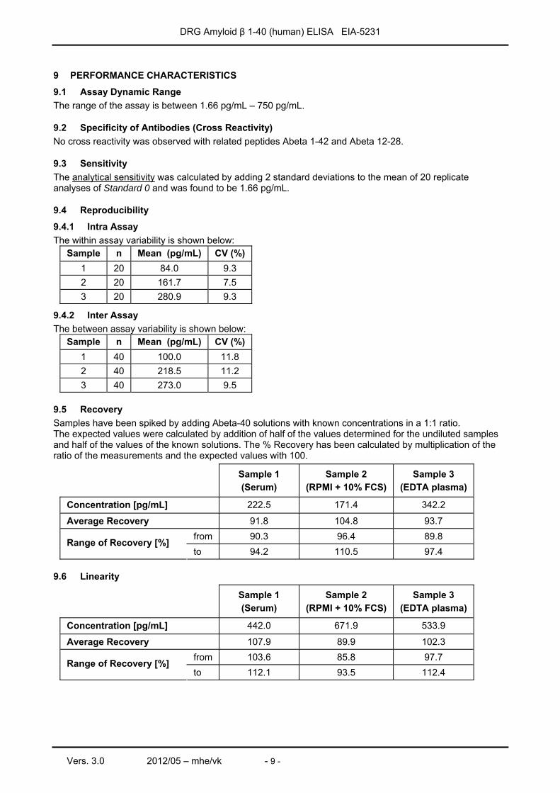
9 PERFORMANCE CHARACTERISTICS
9.1 Assay Dynamic Range The range of the assay is between 1.66 pg/mL – 750 pg/mL.
9.2 Specificity of Antibodies (Cross Reactivity) No cross reactivity was observed with related peptides Abeta 1-42 and Abeta 12-28.
9.3 Sensitivity The analytical sensitivity was calculated by adding 2 standard deviations to the mean of 20 replicate analyses of Standard 0 and was found to be 1.66 pg/mL.
9.4 Reproducibility
9.4.1 Intra Assay The within assay variability is shown below:
Sample n Mean (pg/mL) CV (%) 1 20 84.0 9.3 2 20 161.7 7.5 3 20 280.9 9.3
9.4.2 Inter Assay The between assay variability is shown below:
Sample n Mean (pg/mL) CV (%) 1 40 100.0 11.8 2 40 218.5 11.2 3 40 273.0 9.5
9.5 Recovery Samples have been spiked by adding Abeta-40 solutions with known concentrations in a 1:1 ratio. The expected values were calculated by addition of half of the values determined for the undiluted samples and half of the values of the known solutions. The % Recovery has been calculated by multiplication of the ratio of the measurements and the expected values with 100.
Sample 1 (Serum)
Sample 2 (RPMI + 10% FCS)
Sample 3 (EDTA plasma)
Concentration [pg/mL] 222.5 171.4 342.2
Average Recovery 91.8 104.8 93.7 from 90.3 96.4 89.8 Range of Recovery [%] to 94.2 110.5 97.4
9.6 Linearity
Sample 1 (Serum)
Sample 2 (RPMI + 10% FCS)
Sample 3 (EDTA plasma)
Concentration [pg/mL] 442.0 671.9 533.9
Average Recovery 107.9 89.9 102.3 from 103.6 85.8 97.7 Range of Recovery [%] to 112.1 93.5 112.4
DRG Amyloid β 1-40 (human) ELISA EIA-5231
Vers. 3.0 2012/05 – mhe/vk - 10 -
10 LIMITATIONS OF USE Reliable and reproducible results will be obtained when the assay procedure is performed with a complete understanding of the package insert instruction and with adherence to good laboratory practice. Any improper handling of samples or modification of this test might influence the results.
10.1 Interfering Substances Haemoglobin (up to 4 mg/mL), Bilirubin (up to 0.5 mg/mL) and Triglyceride (up to 30 mg/mL) have no influence on the assay results. The assay contains reagents to minimize interference of HAMA and heterophilic antibodies. However, extremely high titers of HAMA or heterophilic antibodies may interfere with the test results.
10.2 Drug Interferences Until today no substances (drugs) are known to us, which have an influence to the measurement of Abeta-40 in a sample.
10.3 High-Dose-Hook Effect No hook effect was observed in this test up to 96,000 pg/mL of Abeta-40.
11 LEGAL ASPECTS
11.1 Reliability of Results The test must be performed exactly as per the manufacturer’s instructions for use. Moreover the user must strictly adhere to the rules of GLP (Good Laboratory Practice) or other applicable national standards and/or laws. This is especially relevant for the use of control reagents. It is important to always include, within the test procedure, a sufficient number of controls for validating the accuracy and precision of the test. The test results are valid only if all controls are within the specified ranges and if all other test parameters are also within the given assay specifications. In case of any doubt or concern please contact DRG.
11.2 Therapeutic Consequences Therapeutic consequences should never be based on laboratory results alone even if all test results are in agreement with the items as stated under point 11.1. Any laboratory result is only a part of the total clinical picture of a patient. Only in cases where the laboratory results are in acceptable agreement with the overall clinical picture of the patient should therapeutic consequences be derived. The test result itself should never be the sole determinant for deriving any therapeutic consequences.
11.3 Liability Any modification of the test kit and/or exchange or mixture of any components of different lots from one test kit to another could negatively affect the intended results and validity of the overall test. Such modification and/or exchanges invalidate any claim for replacement. Claims submitted due to customer misinterpretation of laboratory results subject to point 11.2. are also invalid. Regardless, in the event of any claim, the manufacturer’s liability is not to exceed the value of the test kit. Any damage caused to the test kit during transportation is not subject to the liability of the manufacturer.
DRG Amyloid β 1-40 (human) ELISA EIA-5231
Vers. 3.0 2012/05 – mhe/vk - 11 -
12 REFERENCES / LITERATURE 1. Price D.L., Sisodia S.S.: Mutant genes in familial Alzheimer's disease and transgenic models.
Annu. Rev. Neurosci.; 1998; 21:479–505. 2. Sinha S. et al.: Purification and cloning of amyloid precursor protein beta-secretase from human brain.
Nature; 1999; 402: 537–540. 3. Vassar R. et al.: Beta-secretase cleavage of Alzheimer's amyloid precursor protein by the
transmembrane aspartic protease BACE. Science 1999; 286:735–741.
4. De Strooper B.: Aph-1, Pen-2, and Nicastrin with Presenilin generate an active gamma-Secretase complex. Neuron; 2003; 38:9–12.
5. Small S.A., Gandy S.: Sorting through the cell biology of Alzheimer's disease: intracellular pathways to pathogenesis. Neuron 2006; 52:15–31.
6. Selkoe D.J.: Clearing the Brain's Amyloid Cobwebs. Neuron; 2001; 32:177–180.
7. Lesne S. et al.: A specific amyloidbeta protein assembly in brain impairs memory. Nature; 2006; 440:352-357.
8. McLean C.A. et al.: Soluble pool of Abeta amyloid as a determinant of severity of neurodegeneration in Alzheimer's disease. Ann. Neurol.; 1999; 46:860–866.
9. Galasko D.: Biomarkers for Alzheimer's disease-clinical needs and application. J Alzheimers Dis 2005; 8:339–346.
10. Jensen M. et al.: Cerebrospinal fluid A beta42 is increased early in sporadic Alzheimer's disease and declines with disease progression. Ann. Neurol.; 1999; 45:504–511.
11. Iwatsubo T.: Amyloid beta protein in plasma as a diagnostic marker for Alzheimer's disease. Neurobiol. Aging; 1998; 19:161–163.
12. Giedraitis V. et al.: The normal equilibrium between CSF and plasma amyloid beta levels is disrupted in Alzheimer's disease. Neurosci. Lett.; 2007; 427:127–131.
13. Vassar R. et al.: Beta-secretase cleavage of Alzheimer's amyloid precursor protein by the transmembrane aspartic protease BACE. Science; 1999; 286:735–741.
14. Sinha S. et al.: Purification and cloning of amyloid precursor protein beta-secretase from human brain. Nature; 1999; 402:537–540.
15. Shibata M. et al.: Clearance of Alzheimer's amyloid-ss(1–40) peptide from brain by LDL receptor-related protein-1 at the blood-brain barrier. J. Clin. Invest. 2000; 106:1489–1499.
16. Deane R. et al.: LRP/amyloid beta-peptide interaction mediates differential brain efflux of Abeta isoforms. Neuron; 2004; 43:333–344.
17. Deane R. et al.: RAGE mediates amyloid-beta peptide transport across the blood-brain barrier and accumulation in brain. Nat. Med.; 2003; 9:907–913.
18. Mayeux R. et al.: Plasma A[beta]40 and A[beta] 42 and Alzheimer's disease: relation to age, mortality, and risk. Neurology; 2003; 61:1185–1190.
19. Graff-Radford N.R. et al.: Association of low plasma Abeta42/Abeta40 ratios with increased imminent risk for mild cognitive impairment and Alzheimer disease. Arch.Neurol.; 2007; 64:354–362.
DRG Amyloid β 1-40 (human) ELISA EIA-5231
Vers. 3.0 2012/05 – mhe/vk - 12 -
1 EINLEITUNG Der DRG Abeta-40 ELISA wird zur quantitativen Bestimmung von Abeta-40 in Serum, Plasma und CSF eingesetzt.
2 TESTPRINZIP Der DRG Abeta-40 ELISA ist ein Festphasen-Enzymimmunoassay, der auf der Sandwichtechnik basiert. Die Wells der Mikrotiterplatten sind mit einem monoklonalen Antikörper beschichtet, der gegen eine definierte Antikörper-Bindungsstelle des Abeta-40-Moleküls gerichtet ist. Die Proben werden in die beschichteten Wells gegeben und zusammen mit einem Assay Buffer und einem Enzyme Conjugate inkubiert. Das Enzyme Conjugate enthält einen monoklonalen anti-Abeta-40-Antikörper, der mit Biotin konjugiert ist. Es wird ein Sandwichkomplex gebildet. Das nicht gebundene Konjugat wird durch Waschen der Wells entfernt. Anschließend wird ein Enzyme Complex zugegeben, welcher ein Streptavidin-Peroxidase-Konjugat enthält. Nach einem Waschschritt wird die Substratlösung zugegeben und die Farbentwicklung nach einer definierten Zeit gestoppt. Die Intensität der gebildeten Farbe ist proportional der Abeta -40-Konzentration in der Probe. Die Extinktion wird bei 450 nm mit einem Mikrotiterplattenleser gemessen.
3 VORSICHTSMAßNAHMEN − Dieser Kit ist nur zum in vitro diagnostischen Gebrauch geeignet. − Nur die gültige, im Testkit enthaltene, Arbeitsanleitung verwenden. − Informationen zu im Kit enthaltenen gefährlichen Substanzen entnehmen Sie bitte dem
Materialsicherheitsdatenblatt. − Alle Bestandteile dieses Testkits, die humanes Serum oder Plasma enthalten, wurden mit FDA-geprüften
Methoden auf HIV I/II, HbsAg und HCV getestet und als negativ bestätigt. Jedoch sollten alle Bestandteile im Umgang und bei der Entsorgung wie mögliche Gefahrenstoffe betrachtet werden.
− Der Kontakt mit der Stop Solution sollte vermieden werden, da sie 0.5 M H2SO4 enthält. Schwefelsäure kann Hautreizungen und Verbrennungen verursachen.
− Nicht mit dem Mund pipettieren und den Kontakt von Kitbestandteilen und Proben mit Haut und Schleimhäuten vermeiden.
− In den Bereichen, in denen Proben oder Kitbestandteile verwendet werden, nicht rauchen, essen oder Kosmetika verwenden.
− Beim Umgang mit Proben oder Reagenzien Einweg-Latexhandschuhe tragen. Die Verunreinigung von Reagenzien oder Proben mit Mikroben kann zu falschen Ergebnissen führen.
− Der Gebrauch sollte gemäß der Vorschriften einer entsprechenden nationalen Gefahrenstoff-Sicherheitsrichtlinie erfolgen.
− Reagenzien nicht nach dem auf dem Kit-Etikett angegebenen Verfallsdatum verwenden. − Alle im Kit-Protokoll angegebenen Mengen müssen genau eingehalten werden. Optimale Ergebnisse
können nur durch Verwendung kalibrierter Pipetten und Mikrotiterplatten-Lesegeräte erreicht werden. − Komponenten von Kits mit unterschiedlichen Lotnummern nicht untereinander vertauschen. Es wird
empfohlen, keine Wells von verschiedenen Platten zu verwenden, auch nicht, wenn es sich um das gleiche Lot handelt. Die Kits können unter anderen Bedingungen gelagert oder versendet worden sein, so dass die Bindungscharakteristik der Platten leicht unterschiedlich ausfällt.
− Chemikalien und zubereitete oder bereits benutzte Reagenzien müssen gemäß den nationalen Gefahrenstoffvorschriften wie gefährlicher Abfall behandelt werden.
− Materialsicherheitsdatenblätter für dieses Produkt sind auf Anfrage direkt von der Firma DRG Instruments GmbH erhältlich. Die Materialsicherheitsdatenblätter entsprechen den Verordnungen der EU-Richtlinie 91/155 EC.
Deutsch
DRG Amyloid β 1-40 (human) ELISA EIA-5231
Vers. 3.0 2012/05 – mhe/vk - 13 -
4 BESTANDTEILE DES KITS
4.1 Kitinhalt 1. Microtiterwells, 96 Wells, 12 x 8 Wells (einzeln brechbar);
Mit anti-Abeta-40-Antikörper (Maus monoklonal) beschichtet. 2. Standard (Standard 0-6), 7 Fläschchen, lyophilisiert; je 1 mL;
Konzentrationen: 0 - 20 - 50 - 125 - 250 - 500 - 750 pg/mL. Enthält quecksilberfreies Konservierungsmittel.
3. Control Low & High (Kontrolle), 2 Fläschchen (lyophilisiert), je 1 mL; Siehe „Vorbereitung der Reagenzien“. Kontrollwerte und –bereiche entnehmen Sie bitte dem Fläschchenetikett oder dem QC-Datenblatt. Enthält quecksilberfreies Konservierungsmittel.
4. Assay Buffer (Assaypuffer), 1 Fläschchen, 14 mL, gebrauchsfertig; Enthält quecksilberfreies Konservierungsmittel.
5. Enzyme Conjugate (Enzymkonjugat), 1 Fläschchen, 7 mL, gebrauchsfertig Anti-Abeta-40-Antikörper, mit Biotin konjugiert, Enthält quecksilberfreies Konservierungsmittel.
6. Enzyme Complex (Enzymkomplex), 1 Fläschchen, 14 mL, gebrauchsfertig Streptavidin, mit Peroxidase konjugiert, Enthält quecksilberfreies Konservierungsmittel.
7. Substrate Solution (Substratlösung), 1 Fläschchen, 14 mL, gebrauchsfertig; Substratlösung TMB.
8. Stop Solution (Stopplösung), 1 Fläschchen, 14 mL, gebrauchsfertig; enthält 0,5 M H2SO4, Kontakt mit der Stopplösung vermeiden! Kann Hautreizungen und Verbrennungen verursachen.
9. Wash Solution (Waschlösung), 1 Fläschchen, 30 mL, 40X konzentriert; Siehe „Vorbereitung der Reagenzien“.
Anmerkung: Zusätzliches Assay Buffer zur Probenverdünnung ist auf Anfrage erhältlich.
4.2 Nicht im Kit enthaltene aber erforderliche Geräte und Materialien − Kalibriertes Mikrotiterplattenlesegerät mit 450 ± 10 nm Filter), (z.B. das DRG Instruments
Mikrotiterplattenlesegerät) − Kalibrierte variable Präzisions-Mikropipette − Saugfähiges Papier − Destilliertes oder deioniseirtes Wasser
4.3 Lagerung und Haltbarkeit des Kits Die ungeöffneten Reagenzien behalten bei Lagerung um 2 °C - 8 °C ihre Reaktivität bis zum Verfallsdatum. Nach dem Verfallsdatum die Reagenzien nicht mehr verwenden. Nach dem Öffnen sollten alle Reagenzien bei 2 °C - 8 °C gelagert werden. Die Mikrotiterwells sollten bei 2 °C - 8 °C gelagert werden. Der einmal geöffnete Folienbeutel sollte stets sehr sorgfältig wieder verschlossen werden. Unter den beschriebenen Lagerbedingungen behalten geöffnete Kits zwei Monate ihre Reaktivität.
DRG Amyloid β 1-40 (human) ELISA EIA-5231
Vers. 3.0 2012/05 – mhe/vk - 14 -
4.4 Vorbereitung der Reagenzien Alle Reagenzien sowie die benötigte Anzahl von Wells sollen vor dem Gebrauch auf Raumtemperatur gebracht werden. Standard / Control Rekonstituieren Sie den lyophilisierten Inhalt der Fläschchen mit 1 mL destilliertem Wasser und lassen Sie die Fläschchen mindestens 10 Minuten ruhen. Vor Gebrauch die Standards und Kontrollen mehrmals vorsichtig schütteln. Achtung: Die rekonstituierten Standards und Kontrollen können bis zu 7 Tagen bei 2 °C – 8 °C gelagert
werden. Für längere Lagerung müssen die rekonstituierten Standards und Kontrollen portioniert und bei -20 °C eingefroren werden.
Wash Solution Die 40-fach konzentrierte Waschlösung (30 mL) mit 1170 mL destilliertem Wasser auf ein Gesamtvolumen von 1200 mL verdünnen. Die verdünnte Waschlösung ist bei Raumtemperatur für 2 Wochen stabil.
4.5 Entsorgung des Kits Die Entsorgung des Kits muss gemäß den nationalen gesetzlichen Vorschriften erfolgen. Spezielle Informationen für dieses Produkt finden Sie im Materialsicherheitsdatenblatt, Kapitel 13.
4.6 Beschädigte Testkits Im Falle einer starken Beschädigung des Testkits oder der Komponenten muss die Firma DRG in schriftlicher Form spätestens eine Woche nach Erhalt des Kits informiert werden. Stark beschädigte Einzelkomponenten sollten nicht für den Testlauf verwendet werden. Sie müssen gelagert werden bis eine endgültige Lösung gefunden wurde. Danach sollten Sie gemäß den offiziellen Richtlinien entsorgt werden.
DRG Amyloid β 1-40 (human) ELISA EIA-5231
Vers. 3.0 2012/05 – mhe/vk - 15 -
5 PROBENVORBEREITUNG Serum, Plasma (EDTA-, Heparin- oder Citratplasma) und Cerebrospinalflüssigkeit (CSF) können in diesem Test als Probenmaterial eingesetzt werden. Lipämische, ikterische und/oder stark hämolysierte Proben sollten nicht verwendet werden. Achtung: Proben, die Natriumazid enthalten, sollten nicht verwendet werden.
5.1 Probenentnahme Serum: Blut durch Venenpunktion entnehmen (z.B. mit Sarstedt Monovette # 02.1388.001), gerinnen lassen und das Serum durch Zentrifugation bei Raumtemperatur abtrennen. Vor der Zentrifugation muss die Gerinnung vollständig abgeschlossen sein. Bei Patienten, die Antikoagulantien erhalten, kann die Gerinnungszeit länger dauern. Plasma: Die Blutentnahme erfolgt mit Röhrchen, die ein Antikoagulanz enthalten. Das Plasma wird als Überstand nach einer Zentrifugation gewonnen. (z.B.: für EDTA-Plasma Sarstedt Monovette – roter Deckel - # 02.166.001; für Heparinplasma Sarstedt Monovette – oranger Deckel - # 02.165.001; für Zitratplasma Sarstedt Monovette – grüner Deckel - # 02.167.001.) CSF: CSF durch Lumbalpunktion entnehmen.
5.2 Probenaufbewahrung Proben sollten stets gut verschlossen sein und können vor Testbeginn bis zu einem Tag bei 2 °C - 8 °C gelagert werden. Für eine längere Aufbewahrung müssen die Proben eingefroren bei –20 °C bis zum Testbeginn gelagert werden. Nur einmal einfrieren! Aufgetaute Proben sollten vor Testbeginn vorsichtig und ohne Schaumbildung durchmischt werden.
5.3 Probenverdünnung CSF: CSF muss vor der Messung mindestens 50-fach mit Assay Buffer verdünnt werden. Die Verdünnung muss bei der Berechnung der Konzentration beachtet werden. Beispiel: Verdünnung 1:50 5 µL CSF + 245 µL Assay Buffer Serum und Plasma: Wenn in einem ersten Testdurchlauf bei einer Probe eine Konzentration höher als der höchste Standard gefunden wird, kann diese Probe mit Assay Buffer weiter verdünnt und nochmals bestimmt werden. Die Verdünnung muss bei der Berechnung der Konzentration beachtet werden. Beispiel: a) Verdünnung 1:10: 25 µL Probe + 225 µL Assay Buffer gründlich mischen) b) Verdünnung 1:100: 25 µL Verdünnung a) 1:10 + 225 µL Assay Buffer (gründlich mischen).
DRG Amyloid β 1-40 (human) ELISA EIA-5231
Vers. 3.0 2012/05 – mhe/vk - 16 -
6 TESTDURCHFÜHRUNG
6.1 Allgemeine Hinweise Alle Reagenzien und Proben müssen vor Gebrauch auf Raumtemperatur gebracht und gut durchgemischt werden. Dabei sollte Schaumbildung vermieden werden. Wenn die Testdurchführung einmal begonnen wurde, muss sie ohne Unterbrechung zu Ende geführt werden. Für jeden Standard, jede Kontrolle oder Probe eine neue Plastikspitze verwenden, um Verschleppungen zu vermeiden. Die Optische Dichte ist abhängig von Inkubationszeit und Temperatur. Deshalb ist es notwendig, vor Beginn der Testdurchführung alle Reagenzien in einen arbeitsbereiten Zustand zu bringen, die Deckel der Fläschchen zu öffnen, alle benötigten Wells in den Halter zu setzen. Nur eine solche Vorbereitung garantiert gleiche Zeiten für jeden Pipettiervorgang ohne Pausen. Als generelle Regel gilt, dass die enzymatische Reaktion linear proportional zu Zeit und Temperatur ist.
6.2 Testdurchführung Jeder Lauf muss eine Standardkurve beinhalten. 1. Die benötigte Anzahl Wells in der Halterung befestigen. 2. 50 µL Assay Buffer in jedes Well geben. 3. Je 100 µL Standards, Control und Probe mit neuen Plastikspitzen in die entsprechenden Wells geben. 4. 50 µL Enzyme Conjugate in jedes Well geben.
Für 10 Sekunden gut schütteln. Es ist sehr wichtig, in diesem Schritt eine komplette Durchmischung zu erreichen.
5. 180 Minuten bei Raumtemperatur auf einem Plattenschüttler bei ~ 700 rpm inkubieren. 6. Den Inhalt der Wells kräftig ausschütteln. Wells 4-mal mit verdünnter Wash Solution (400 µL/Well)
waschen. Verbleibende Flüssigkeit durch Ausklopfen der Wells auf saugfähigem Papier entfernen. Achtung: Die Sensitivität und Präzision dieses Assays wird erheblich beeinflusst von der korrekten Durchführung des Waschschrittes!
7. 100 µL Enzyme Complex in jedes Well geben. 8. 30 Minuten bei Raumtemperatur auf einem Plattenschüttler bei ~ 700 rpm inkubieren. 9. Den Inhalt der Wells kräftig ausschütteln. Wells 4-mal mit verdünnter Wash Solution (400 µL/Well)
waschen. Verbleibende Flüssigkeit durch Ausklopfen der Wells auf saugfähigem Papier entfernen 10. 100 µL Substrate Solution in jedes Well geben. 11. 20 Minuten bei Raumtemperatur inkubieren. 12. Die enzymatische Reaktion durch Zugabe von 100 µL Stop Solution in jedes Well abstoppen. 13. Die Optische Dichte bei 450 ± 10 nm mit einem Mikrotiterplatten-Lesegerät innerhalb von 10 Minuten
nach Zugabe der Stop Solution bestimmen.
DRG Amyloid β 1-40 (human) ELISA EIA-5231
Vers. 3.0 2012/05 – mhe/vk - 17 -
6.3 Ergebnisermittlung 1. Die durchschnittlichen Werte der Optischen Dichte (OD) für jedes Set von Standards, Controls und
Patientenproben bestimmen. 2. Eine Standardkurve ermitteln durch Auftragen der mittleren Optischen Dichte jedes Standards gegen die
Konzentration, wobei der OD-Wert auf der vertikalen (Y) Achse und die Konzentration auf der horizontalen (X) Achse eingetragen wird.
3. Unter Verwendung der mittleren OD wird für jede Probe die entsprechende Konzentration aus der Standardkurve ermittelt.
4. Automatische Methode: Die in der Arbeitsanleitung ermittelten Werte wurden automatisch mit Hilfe der 4 Parameter Gleichung (4PL, 4 Parameter Logistics, 4 Parameter Rodbard) bestimmt. Andere Auswertungsfunktionen können leicht abweichende Werte ergeben.
5. Die Konzentration der Proben kann direkt von der Standardkurve abgelesen werden. Proben, die eine höhere Konzentration als die des höchsten Standards enthalten, müssen verdünnt werden. Dieser Verdünnungsfaktor muss bei der Berechnung der Konzentration beachtet werden.
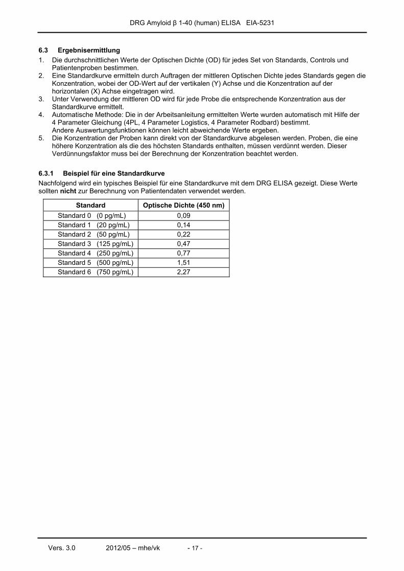
6.3.1 Beispiel für eine Standardkurve Nachfolgend wird ein typisches Beispiel für eine Standardkurve mit dem DRG ELISA gezeigt. Diese Werte sollten nicht zur Berechnung von Patientendaten verwendet werden.
Standard Optische Dichte (450 nm) Standard 0 (0 pg/mL) 0,09 Standard 1 (20 pg/mL) 0,14 Standard 2 (50 pg/mL) 0,22 Standard 3 (125 pg/mL) 0,47 Standard 4 (250 pg/mL) 0,77 Standard 5 (500 pg/mL) 1,51 Standard 6 (750 pg/mL) 2,27
DRG Amyloid β 1-40 (human) ELISA EIA-5231
Vers. 3.0 2012/05 – mhe/vk - 18 -
7 ERWARTETE WERTE Es wird empfohlen, dass jedes Labor seine eigenen normalen und abnormalen Werte ermittelt. Die Normwertermittlung für Abeta-40 Peptide wurde mit offensichtlich gesunden Personen durchgeführt.
Probanten Anzahl Alter (Jahre)
Mittelwert (pg/mL)
Median (pg/mL)
5% - 95% Percentile
Männer und Frauen 40 25-52 144,0 135,1 72,8 – 203,1
Die Ergebnisse stimmen gut mit veröffentlichten Normbereichen überein (80-300 pg/mL).
8 QUALITÄTS-KONTROLLE Es wird empfohlen, die Kontrollproben gemäß den nationalen gesetzlichen Bestimmungen einzusetzen. Durch die Verwendung von Kontrollproben wird eine Tag-zu-Tag Überprüfung der Ergebnisse erzielt. Es sollten Kontrollen sowohl mit normalem als auch pathologischem Level eingesetzt werden. Die Kontrollen mit den entsprechenden Ergebnissen des QC-Labors sind im QC-Zertifikat, das dem Kit beiliegt, aufgeführt. Die im QC-Blatt angegebenen Werte und Bereiche beziehen sich stets auf die aktuelle Kitcharge und sollten zum direkten Vergleich der Ergebnisse verwendet werden. Es wird ebenfalls empfohlen, an nationalen oder internationalen Qualitätssicherungs-Programmen teilzunehmen, um die Genauigkeit der Ergebnisse zu sichern. Es sollten geeignete statistische Methoden zur Analyse von Kontroll-Werten und Trends angewendet werden. Wenn die Ergebnisse des Assays nicht mit den angegebenen Akzeptanzbereichen des Kontrollmaterials übereinstimmen, sollten die Patientenergebnisse als ungültig eingestuft werden. In diesem Fall überprüfen Sie bitte die folgenden Bereiche: Pipetten und Zeitnehmer, Photometer, Verfallsdatum der Reagenzien, Lagerungs- und Inkubationsbedingungen, Absaug- und Waschmethode. Sollten Sie nach Überprüfung der vorgenannten Bereiche keinen Fehler erkannt haben, setzen Sie sich bitte mit Ihrem Lieferanten oder direkt mit der Firma DRG in Verbindung.
9 ASSAY CHARACTERISTIKA
9.1 Messbereich Der Messbereich des Testes liegt zwischen 1,66 – 750 pg/mL.
9.2 Spezifität der Antikörper (Kreuzreaktivität) Die Daten entnehmen Sie bitte der ausführlichen englischen Arbeitsanleitung.
9.3 Analytische Sensitivität Die analytische Sensitivität, definiert als Mittelwert plus der zweifachen Standardabweichung des Standards 0 (n = 20), beträgt 1,66 pg/mL. Die Daten zu:
9.4 Reproduzierbarkeit (Präzision) 9.5 Wiederfindung 9.6 Linearität entnehmen Sie bitte der ausführlichen englischen Arbeitsanleitung.
DRG Amyloid β 1-40 (human) ELISA EIA-5231
Vers. 3.0 2012/05 – mhe/vk - 19 -
10 GRENZEN DES TESTS Jede unsachgemäße Behandlung von Proben oder Modifikationen dieses Tests können die Ergebnisse beeinflussen.
10.1 Interferenzen Hämoglobin (bis zu 4 mg/mL), Bilirubin (bis zu 0.5 mg/mL) und Triglyceride (bis zu 30 mg/mL) haben keinen Einfluss auf das Testergebnis. Der Test enthält Reagenzien, um Interferenzen mit HAMA oder heterophilen Antikörpern zu minimieren. Dennoch ist es möglich, dass ein sehr hoher Titer von HAMA oder heterophilen Antikörpern das Testergebnis beeinflusst.
10.2 Beeinflussung durch Medikamente Uns sind bislang keine Stoffe (Medikamente) bekannt geworden, deren Einnahme die Messung des Abeta-40-Gehaltes der Probe beeinflussen würde.
10.3 High-Dose-Hook Effekt Ein Hook Effekt tritt bei Proben mit bis zu 96.000 pg/mL Abeta-40 nicht auf.
11 RECHTLICHE GRUNDLAGEN
11.1 Zuverlässigkeit der Ergebnisse Der Test muss exakt gemäß der Testanleitung des Herstellers abgearbeitet werden. Darüber hinaus muss der Benutzer sich strikt an die Regeln der GLP (Good Laboratory Practice) oder andere eventuell anzuwendende Regeln oder nationale gesetzliche Vorgaben halten. Dies betrifft besonders den Gebrauch der Kontrollreagenzien. Es ist sehr wichtig, bei der Testdurchführung stets eine ausreichende Anzahl Kontrollen zur Überprüfung der Genauigkeit und Präzision mitlaufen zu lassen. Die Testergebnisse sind nur gültig, wenn alle Kontrollen in den vorgegebenen Bereichen liegen, und wenn alle anderen Testparameter die vorgegebenen Spezifikationen für diesen Assay erfüllen. Wenn Sie bezüglich eines Ergebnisses Zweifel oder Bedenken haben, setzen Sie sich bitte mit der Firma DRG in Verbindung.
11.2 Therapeutische Konsequenzen Therapeutische Konsequenzen sollten keinesfalls nur aufgrund von Laborergebnissen erfolgen, selbst dann nicht, wenn alle Testergebnisse mit den in 11.1. genannten Voraussetzungen übereinstimmen. Jedes Laborergebnis ist nur ein Teil des klinischen Gesamtbildes eines Patienten. Nur in Fällen, in denen die Laborergebnisse in akzeptabler Übereinstimmung mit dem allgemeinen klinischen Bild des Patienten stehen, sollten therapeutische Konsequenzen eingeleitet werden. Das Testergebnis allein sollte niemals als alleinige Grundlage für die Einleitung therapeutischer Konsequenzen dienen.
11.3 Haftung Jegliche Veränderungen des Testkits und/oder Austausch oder Vermischung von Komponenten unterschiedlicher Chargen von einem Testkit zu einem anderen, können die gewünschten Ergebnisse und die Gültigkeit des gesamten Tests negativ beeinflussen. Solche Veränderungen und/oder Austausch haben den Ausschluss jeglicher Ersatzansprüche zur Folge. Reklamationen, die aufgrund von Falschinterpretation von Laborergebnissen durch den Kunden gemäß Punkt 11.2. erfolgen, sind ebenfalls abzuweisen. Im Falle jeglicher Reklamation ist die Haftung des Herstellers maximal auf den Wert des Testkits beschränkt. Jegliche Schäden, die während des Transports am Kit entstanden sind, unterliegen nicht der Haftung des Herstellers.
12 REFERENZEN / LITERATUR Angaben zu den Referenzen entnehmen Sie bitte der ausführlichen englischen Arbeitsanleitung.
DRG Amyloid β 1-40 (human) ELISA EIA-5231
Vers. 3.0 2012/05 – mhe/vk - 20 -
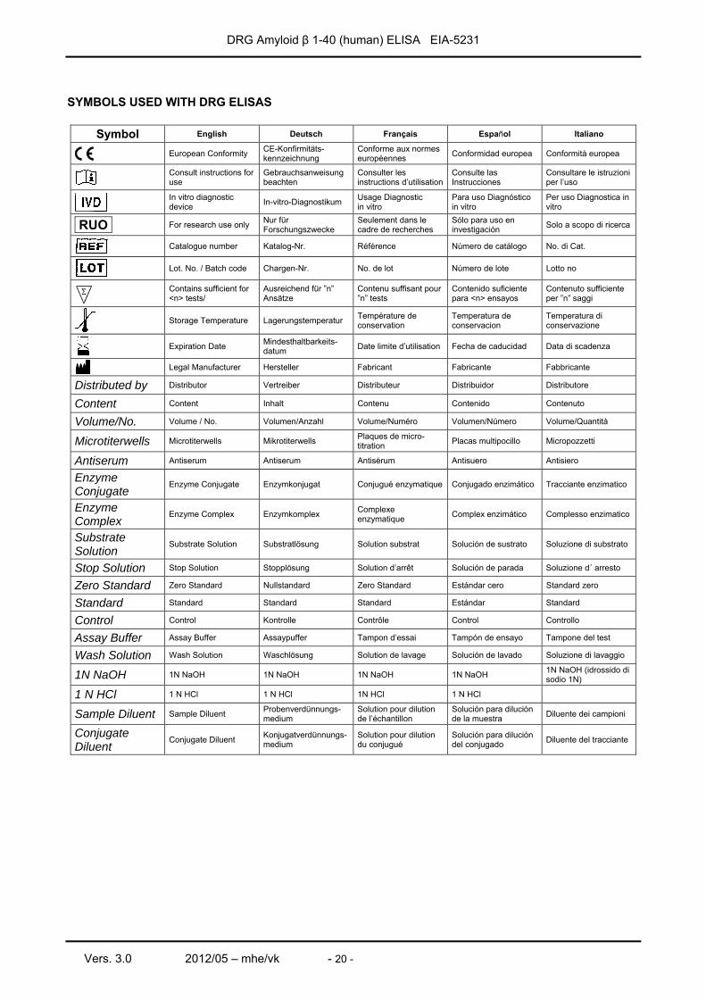
SYMBOLS USED WITH DRG ELISAS
Symbol English Deutsch Français Español Italiano
European Conformity CE-Konfirmitäts-kennzeichnung
Conforme aux normes européennes Conformidad europea Conformità europea
Consult instructions for use
Gebrauchsanweisung beachten
Consulter les instructions d’utilisation
Consulte las Instrucciones
Consultare le istruzioni per l’uso
In vitro diagnostic device In-vitro-Diagnostikum Usage Diagnostic
in vitro Para uso Diagnóstico in vitro
Per uso Diagnostica in vitro
RUO For research use only Nur für Forschungszwecke
Seulement dans le cadre de recherches
Sólo para uso en investigación Solo a scopo di ricerca
Catalogue number Katalog-Nr. Référence Número de catálogo No. di Cat.
Lot. No. / Batch code Chargen-Nr. No. de lot Número de lote Lotto no
Contains sufficient for <n> tests/
Ausreichend für ”n” Ansätze
Contenu suffisant pour ”n” tests
Contenido suficiente para <n> ensayos
Contenuto sufficiente per ”n” saggi
Storage Temperature Lagerungstemperatur Température de
conservation Temperatura de conservacion
Temperatura di conservazione
Expiration Date Mindesthaltbarkeits-datum Date limite d’utilisation Fecha de caducidad Data di scadenza
Legal Manufacturer Hersteller Fabricant Fabricante Fabbricante
Distributed by Distributor Vertreiber Distributeur Distribuidor Distributore
Content Content Inhalt Contenu Contenido Contenuto
Volume/No. Volume / No. Volumen/Anzahl Volume/Numéro Volumen/Número Volume/Quantità
Microtiterwells Microtiterwells Mikrotiterwells Plaques de micro-titration Placas multipocillo Micropozzetti
Antiserum Antiserum Antiserum Antisérum Antisuero Antisiero
Enzyme Conjugate
Enzyme Conjugate Enzymkonjugat Conjugué enzymatique Conjugado enzimático Tracciante enzimatico
Enzyme Complex
Enzyme Complex Enzymkomplex Complexe enzymatique Complex enzimático Complesso enzimatico
Substrate Solution
Substrate Solution Substratlösung Solution substrat Solución de sustrato Soluzione di substrato
Stop Solution Stop Solution Stopplösung Solution d’arrêt Solución de parada Soluzione d´ arresto
Zero Standard Zero Standard Nullstandard Zero Standard Estándar cero Standard zero
Standard Standard Standard Standard Estándar Standard
Control Control Kontrolle Contrôle Control Controllo
Assay Buffer Assay Buffer Assaypuffer Tampon d’essai Tampón de ensayo Tampone del test
Wash Solution Wash Solution Waschlösung Solution de lavage Solución de lavado Soluzione di lavaggio
1N NaOH 1N NaOH 1N NaOH 1N NaOH 1N NaOH 1N NaOH (idrossido di sodio 1N)
1 N HCl 1 N HCl 1 N HCl 1N HCl 1 N HCl
Sample Diluent Sample Diluent Probenverdünnungs-medium
Solution pour dilution de l’échantillon
Solución para dilución de la muestra Diluente dei campioni
Conjugate Diluent
Conjugate Diluent Konjugatverdünnungs-medium
Solution pour dilution du conjugué
Solución para dilución del conjugado Diluente del tracciante