Altered gene dosage confirms the genetic interaction between FIAT and αNAC
6
Altered gene dosage confirms the genetic interaction between FIAT and αNAC Bahareh Hekmatnejad a,b , Vice Mandic a,b , Vionnie W.C. Yu a,b,1 , Omar Akhouayri a,b,2 , Alice Arabian a , René St-Arnaud a,b,c,d, ⁎ a Research Unit, Shriners Hospital for Children – Canada, Montreal, Quebec H3G 1A6, Canada b Department of Human Genetics, McGill University, Montreal, Quebec H3A 2T5, Canada c Department of Surgery, McGill University, Montreal, Quebec H3A 2T5, Canada d Department of Medicine, McGill University, Montreal, Quebec H3A 2T5, Canada abstract article info Article history: Accepted 4 January 2014 Available online 17 January 2014 Keywords: Factor inhibiting ATF4-mediated transcription (FIAT) Nascent polypeptide associated complex and coregulator alpha (αNAC) Bone Cortical porosity Factor inhibiting ATF4-mediated transcription (FIAT) interacts with Nascent polypeptide associated complex and coregulator alpha (αNAC). In cultured osteoblastic cells, this interaction contributes to maximal FIAT-mediated inhibition of Osteocalcin (Ocn) gene transcription. We set out to demonstrate the physiological relevance of this interaction by altering gene dosage in compound Fiat and Naca (encoding αNAC) heterozygous mice. Com- pound Naca +/- ; Fiat +/- heterozygous animals were viable, developed normally, and exhibited no significant dif- ference in body weight compared with control littermate genotypes. Animals with a single Fiat allele had reduced Fiat mRNA expression without changes in the expression of related family members. Expression of the osteocyte differentiation marker Dmp1 was elevated in compound heterozygotes. Static histomorphometry parameters were assessed at 8 weeks of age using microcomputed tomography (μCT). Trabecular measurements were not different between genotypes. Cortical thickness and area were not affected by gene dosage, but we measured a significant increase in cortical porosity in compound heterozygous mice, without changes in biomechanical pa- rameters. The bone phenotype of compound Naca +/- ; Fiat +/- heterozygotes confirms that FIAT and αNAC are part of a common genetic pathway and support a role for the FIAT/αNAC interaction in normal bone physiology. © 2014 Elsevier B.V. All rights reserved. 1. Introduction The leucine zipper protein, FIAT (factor inhibiting ATF4-mediated transcription), forms inactive heterodimers with the transcription fac- tor ATF4 and inhibits its binding to DNA (Yu et al., 2005, 2008). Because ATF4 is a key regulator of osteoblast biology (Yang et al., 2004), the net result of the binding of FIAT to ATF4 is an inhibition of osteoblast func- tion. Over-expressing FIAT in osteoblasts of transgenic mice leads to reduced Osteocalcin (Ocn) gene transcription and osteopenia with re- duced bone mineral density, lowered trabecular volume, and decreased bone strength (Yu et al., 2005). Reciprocally, inhibition of Fiat expres- sion using siRNA-mediated knockdown leads to increased ATF4 activity and results in higher Ocn transcription, type I collagen synthesis, and mineralization (Yu et al., 2009b). FIAT was initially cloned using a yeast two-hybrid screen for proteins interacting with αNAC (Nascent polypeptide associated complex and coregulator alpha, encoded by the Naca gene), a transcriptional coregulator of gene expression in bone cells (Akhouayri et al., 2005; Meury et al., 2010; Yu et al., 2005, 2006). This interaction was indepen- dently confirmed using over-expression of epitope-tagged proteins in heterologous cell systems (Yoshida et al., 2005). Our recent work has confirmed that endogenous, post-translationally modified αNAC func- tionally interacts with the FIAT protein in osteoblastic cells to maximally repress ATF4-mediated Ocn gene transcription (Hekmatnejad et al., 2014). We set out to provide evidence of the physiological relevance of these findings through manipulation of Fiat and Naca dosage in ge- netically modified mice, with particular focus on skeletal development. The Fiat gene (NM_178935.4) is located on the X chromosome and is ubiquitously expressed at the mRNA level (Nogami et al., 2004), but its protein expression pattern has only been studied extensively in bone Gene 538 (2014) 328–333 Abbreviations: FIAT, factor inhibiting ATF4-mediated transcription; αNAC, Nascent polypeptide associated complex and coregulator alpha; Ocn, Osteocalcin; Dmp1, Dentin matrix protein-1; μCT, microcomputed tomography; ATF4, Activating transcription factor 4; siRNA, silencing ribonucleic acid; skNAC, skeletal isoform of αNAC; UTR, untranslated region; PGK-neo-HsvTk, phosphoglycerate kinase-neomycin-herpes simplex virus thymi- dine kinase; ES, embryonic stem; PCR, polymerase chain reaction; IRES-lacZ-neo, internal ribosome entry site-lacZ-neomycin; RT-qPCR, Reverse transcription-quantitative polymerase chain reaction; ROI, region of interest; TV, tissue volume; BV, bone volume; BV/TV, bone volume/tissue volume; SMI, structure model index; S.E.M, standard error of the mean; RANKL, receptor activating nuclear factor kappa ligand; XCI, X chromosome in- activation; ICER, inducible cAMP early repressor; CHOP, CCAAT/enhancer-binding protein homologous protein; OVX, ovariectomized; Wt, wild-type; Mut, mutant. ⁎ Corresponding author at: Research Unit, Shriners Hospital for Children – Canada, 1529 Cedar Avenue, Montreal, Quebec H3G 1A6, Canada. Tel.: +1 514 282 7155; fax: +1 514 842 5581. E-mail address: [email protected] (R. St-Arnaud). 1 Current address: Harvard Stem Cell Institute, Massachusetts General Hospital, Boston, MA 02114, USA. 2 Current address: Laboratoire de Génétique — Neuroendocrinologie et Biotechnologie, Faculté des Sciences Ibn Tofaïl, Kénitra, Morocco. 0378-1119/$ – see front matter © 2014 Elsevier B.V. All rights reserved. http://dx.doi.org/10.1016/j.gene.2014.01.009 Contents lists available at ScienceDirect Gene journal homepage: www.elsevier.com/locate/gene
Transcript of Altered gene dosage confirms the genetic interaction between FIAT and αNAC
Gene 538 (2014) 328–333
Contents lists available at ScienceDirect
Gene
j ourna l homepage: www.e lsev ie r .com/ locate /gene
Altered gene dosage confirms the genetic interaction between FIATand αNAC
Bahareh Hekmatnejad a,b, Vice Mandic a,b, Vionnie W.C. Yu a,b,1, Omar Akhouayri a,b,2,Alice Arabian a, René St-Arnaud a,b,c,d,⁎a Research Unit, Shriners Hospital for Children – Canada, Montreal, Quebec H3G 1A6, Canadab Department of Human Genetics, McGill University, Montreal, Quebec H3A 2T5, Canadac Department of Surgery, McGill University, Montreal, Quebec H3A 2T5, Canadad Department of Medicine, McGill University, Montreal, Quebec H3A 2T5, Canada
Abbreviations: FIAT, factor inhibiting ATF4-mediatedpolypeptide associated complex and coregulator alpha; Omatrix protein-1; μCT, microcomputed tomography; ATF44; siRNA, silencing ribonucleic acid; skNAC, skeletal isoforegion; PGK-neo-HsvTk, phosphoglycerate kinase-neomycdine kinase; ES, embryonic stem; PCR, polymerase chain rribosome entry site-lacZ-neomycin; RT-qPCR, Reverpolymerase chain reaction; ROI, region of interest; TV, tisBV/TV, bone volume/tissue volume; SMI, structure modethemean; RANKL, receptor activating nuclear factor kappaactivation; ICER, inducible cAMP early repressor; CHOP, CChomologous protein; OVX, ovariectomized; Wt, wild-type⁎ Corresponding author at: Research Unit, ShrinersHosp
Cedar Avenue, Montreal, Quebec H3G 1A6, Canada. Tel.: +842 5581.
E-mail address: [email protected] (R. St-A1 Current address: Harvard Stem Cell Institute, Massach
MA 02114, USA.2 Current address: Laboratoire de Génétique— Neuroen
Faculté des Sciences Ibn Tofaïl, Kénitra, Morocco.
0378-1119/$ – see front matter © 2014 Elsevier B.V. All rhttp://dx.doi.org/10.1016/j.gene.2014.01.009
a b s t r a c t
a r t i c l e i n f oArticle history:Accepted 4 January 2014Available online 17 January 2014
Keywords:Factor inhibiting ATF4-mediated transcription(FIAT)Nascent polypeptide associated complex andcoregulator alpha (αNAC)BoneCortical porosity
Factor inhibiting ATF4-mediated transcription (FIAT) interactswith Nascent polypeptide associated complex andcoregulator alpha (αNAC). In cultured osteoblastic cells, this interaction contributes to maximal FIAT-mediatedinhibition of Osteocalcin (Ocn) gene transcription. We set out to demonstrate the physiological relevance ofthis interaction by altering gene dosage in compound Fiat and Naca (encoding αNAC) heterozygous mice. Com-poundNaca+/−; Fiat+/− heterozygous animalswere viable, developed normally, and exhibited no significant dif-ference in bodyweight comparedwith control littermate genotypes. Animalswith a single Fiat allele had reducedFiatmRNA expression without changes in the expression of related familymembers. Expression of the osteocytedifferentiation marker Dmp1 was elevated in compound heterozygotes. Static histomorphometry parameterswere assessed at 8 weeks of age using microcomputed tomography (μCT). Trabecular measurements were notdifferent between genotypes. Cortical thickness and area were not affected by gene dosage, but we measured asignificant increase in cortical porosity in compound heterozygous mice, without changes in biomechanical pa-rameters. The bone phenotype of compound Naca+/−; Fiat+/− heterozygotes confirms that FIAT and αNAC arepart of a common genetic pathway and support a role for the FIAT/αNAC interaction in normal bone physiology.
© 2014 Elsevier B.V. All rights reserved.
1. Introduction
The leucine zipper protein, FIAT (factor inhibiting ATF4-mediatedtranscription), forms inactive heterodimers with the transcription fac-tor ATF4 and inhibits its binding to DNA (Yu et al., 2005, 2008). BecauseATF4 is a key regulator of osteoblast biology (Yang et al., 2004), the net
transcription; αNAC, Nascentcn, Osteocalcin; Dmp1, Dentin, Activating transcription factorrm of αNAC; UTR, untranslatedin-herpes simplex virus thymi-eaction; IRES-lacZ-neo, internalse transcription-quantitativesue volume; BV, bone volume;l index; S.E.M, standard error ofligand; XCI, X chromosome in-AAT/enhancer-binding protein; Mut, mutant.ital for Children – Canada, 15291 514 282 7155; fax: +1 514
rnaud).usetts General Hospital, Boston,
docrinologie et Biotechnologie,
ights reserved.
result of the binding of FIAT to ATF4 is an inhibition of osteoblast func-tion. Over-expressing FIAT in osteoblasts of transgenic mice leads toreduced Osteocalcin (Ocn) gene transcription and osteopenia with re-duced bonemineral density, lowered trabecular volume, and decreasedbone strength (Yu et al., 2005). Reciprocally, inhibition of Fiat expres-sion using siRNA-mediated knockdown leads to increased ATF4 activityand results in higher Ocn transcription, type I collagen synthesis, andmineralization (Yu et al., 2009b).
FIATwas initially cloned using a yeast two-hybrid screen for proteinsinteracting with αNAC (Nascent polypeptide associated complex andcoregulator alpha, encoded by the Naca gene), a transcriptionalcoregulator of gene expression in bone cells (Akhouayri et al., 2005;Meury et al., 2010; Yu et al., 2005, 2006). This interaction was indepen-dently confirmed using over-expression of epitope-tagged proteins inheterologous cell systems (Yoshida et al., 2005). Our recent work hasconfirmed that endogenous, post-translationally modified αNAC func-tionally interactswith the FIAT protein in osteoblastic cells tomaximallyrepress ATF4-mediated Ocn gene transcription (Hekmatnejad et al.,2014). We set out to provide evidence of the physiological relevanceof these findings through manipulation of Fiat and Naca dosage in ge-netically modified mice, with particular focus on skeletal development.
The Fiat gene (NM_178935.4) is located on theX chromosome and isubiquitously expressed at the mRNA level (Nogami et al., 2004), but itsprotein expression pattern has only been studied extensively in bone
329B. Hekmatnejad et al. / Gene 538 (2014) 328–333
cells (Yu et al., 2009a). The Fiat gene, like themajority of X-linked genes,is subject to random inactivation of one allele in females (Lyon, 1999).Therefore, approximately 50% of the cells in Fiatmutant heterozygotesshould express the mutant allele, and the other half express normal al-lele. TheNaca gene (NC_000076.6)maps to chromosome 10 (Yotov andSt-Arnaud, 1996b) and encodes the related proteins αNAC and skNACthrough differential splicing of its large third exon (Yotov and St-Arnaud, 1996a). Global targeting of the Naca gene in conventionalknockoutmice results in early embryonic lethality (Akhouayri et al., un-published), and we had to use knock-in mutagenesis to confirm thephysiological role ofαNAC in bone tissue (Meury et al., 2010). However,mice heterozygous for the conventional Naca knockout mutationhave no detectable phenotype and their skeletal development is normal(Pellicelli et al., 2014), therefore allowing gene dosage alteration incompound heterozygous animals.
We crossed heterozygous Fiat-deficient mice with heterozygousNaca-deficient mice to generate double heterozygous Naca and Fiat fe-male mice. Femoral bone microstructure was examined usingmicrocomputed tomography (μCT). While trabecular bone was not af-fected by altered gene dosage for Naca and Fiat, compound Naca+/−;Fiat+/− heterozygousmice displayed significantly higher cortical poros-ity with increased expression of the osteocyte differentiation marker,Dentin matrix protein-1 (Dmp1). This phenotype confirms that FIATand αNAC are part of a common genetic pathway and supports a rolefor FIAT/αNAC interactions in bone physiology.
2. Materials and methods
2.1. Generation of Naca; Fiat compound heterozygous mutant mice
A 9.6 kb genomic fragment spanning the 5′UTR, exon 1, and the firstintron of the mouse Fiat allele was used in the construction of thetargeting vector (Fig. 1A). A 34 bp LoxP sequencewas inserted upstreamfrom exon 1 and a 6.4 kb PGK-neo-HsvTK cassette flanked by twoLoxP sites was introduced within intron 1. The backbone vector was
SOUTHERN-BSOUTHERN-B
8.5Kb7.6Kb
Wt Mut
E1 E3 E2 MUTANT ALLMUTANT ALL
Xba I 7659bp
E1 E3 E2 Xb
a I
8486bp
WILD-TYPE ALWILD-TYPE AL
αNAC Knock-out B
A
IRES-LacZ-neo
5’
E1PGK-neo
EcoRI
2.5 kb6.4 kb
LoxP LoxP
Fig. 1. Targeted disruption of themurine Fiat andNaca genes by homologous recombination. A.PGK-neo cassette flanked by two LoxP sites was introduced within intron 1. B. Schematic drawiblack box)was inserted at theATG start sitewithin exon2. The diagnosticwild-type 8.5 kb andmcells (left) or tail snips from the first germline transmission litter.
pBluescript. The targeting vector was linearized and electroporatedinto R1 embryonic stem (ES) cells (Nagy et al., 1993) that were subse-quently cultured in the presence of G418. Positive clones were identi-fied using Southern blotting and PCR. Blastocyst injection into C57BL/6was performed according to standard protocols (Hogan et al., 1994).Chimeras were bred with a general deleter Cre transgenic strain(CMV-Cre) (Su et al., 2002) to delete the selection cassette.
The αNAC targeting vector was constructed using a genomic Nacaclone (Meury et al., 2010) by inserting an IRES-LacZ-Neo cassette atthe start codon within exon 2 to create a null allele (Fig. 1B). Genetargeting and production of chimeric mice were performed accordingto standard protocols (Hogan et al., 1994).
All animal procedures were reviewed and approved by the McGillInstitutional Animal Care and Use Committee and followed the guide-lines of the Canadian Council on Animal Care. Mice were kept in an en-vironmentally controlled barrier animal facility with a 12-h light, 12-hdark cycle and were fed mouse chow and water ad libitum. Naca+/−
males were crossed to Fiat+/− females to obtain control (Naca+/+;Fiat+/+,Naca+/−; Fiat+/+, Naca+/+; Fiat+/−) and test (Naca+/−;Fiat+/−) genotypes.
2.2. Genotyping
Initial homologous recombination in ES cells and germline transmis-sion of the mutant Naca allele were confirmed using Southern blottingwith diagnosticwild-type 8.5 kb andmutant 7.6 kbXbaI restriction frag-ments (Fig. 1B). For subsequent routine genotyping, DNAwas preparedfrom tail snips (Laird et al., 1991) and analyzed in two independent po-lymerase chain reactions (PCR) for the presence of the Fiat and Nacawild-type and targeted alleles. For each allele, three primers were used.
Fiat primers: 5′-TGAATAGGCTTCCAGCCCAAAGG-3′; 5′-CCAAGAGTCCCAAAAGAGCATG-3′, 5′-CCTTCTGGGGCTTGTAGTCTC-3′.Naca primers:5′-GTGTTCGAATTGGCCAATGACAAGAC-3′, 5′-CTTTCTTCCCCGAAGAGTCCC-3′, 5′-TCGGAATACTTAGACTAGGCGGA-3′.
LOT LOT
Wt Mut
E8 E7 E6 E5 E4 ELE ELE
Xb
a I
E8 E7 E6 E5 E4 Xb
a I
LELE LELE
3’
EcoRI
7.1 kb
LoxP
Fiat targeting vector. A LoxP sequence (triangle) was inserted upstream from exon 1, and ang of the wild-type (Wt) andmutant (Mut) Naca alleles. An IRES-LacZ-neo cassette (gray-utant 7.6 kb XbaI restriction fragments are indicated in the Southernblot from targeted ES
4 5 6 7 814.5
15.5
16.5
17.5
18.5
19.5
20.5
Age (weeks)
Wei
gh
t (g
)
Naca+/+; Fiat+/+
Naca+/-; Fiat+/+
Naca+/+; Fiat+/-
Naca+/-; Fiat+/-
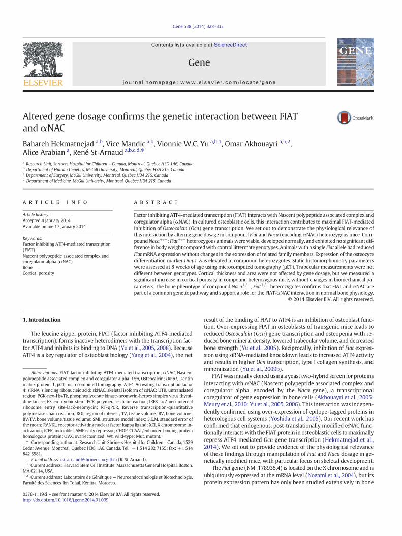
Fig. 2. Fiat and Naca gene dosage alterations do not affect weight gain. No changes wereobserved between Naca+/−; Fiat+/− compound heterozygous mice and control groups.
330 B. Hekmatnejad et al. / Gene 538 (2014) 328–333
2.3. Real-time reverse transcription quantitative PCR (RT-qPCR)
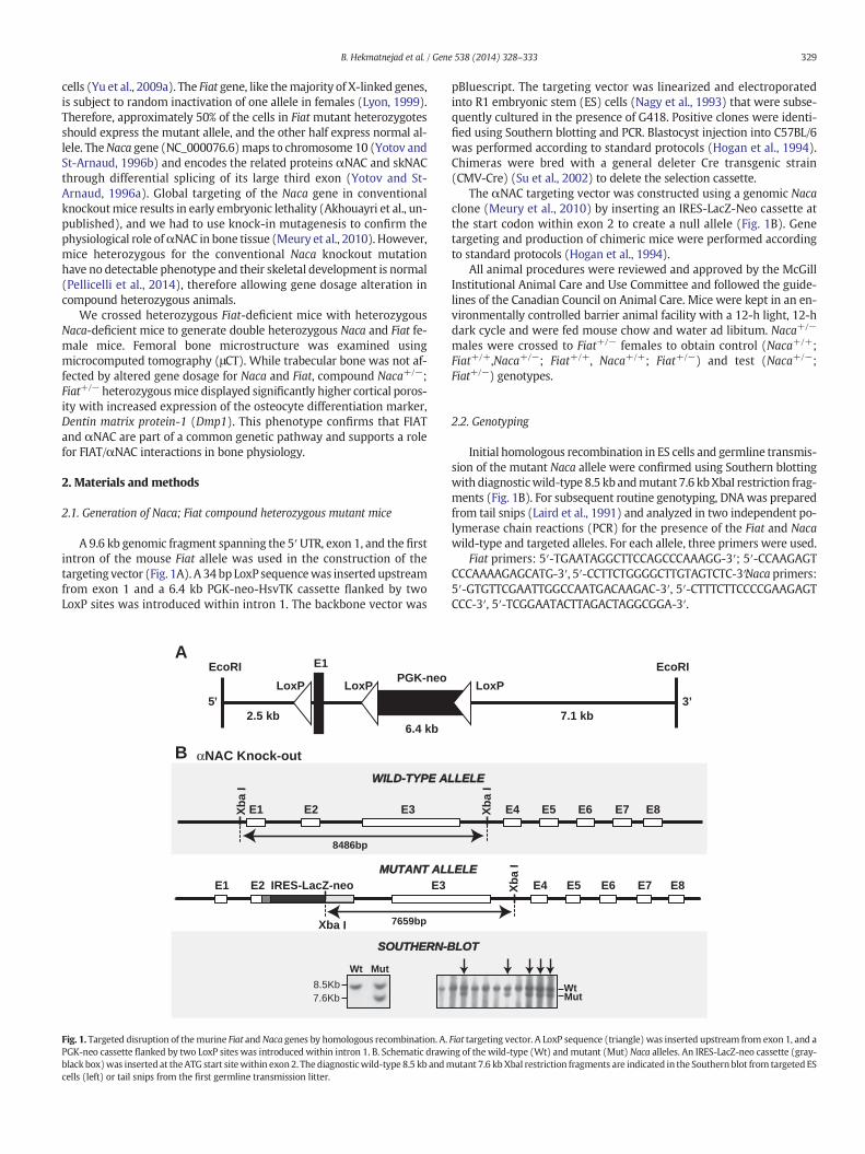
Calvaria from 8-week-old mice were collected into RNAlater solu-tion (Ambion, Austin, TX) andwere stored at−20 °C until further proc-essed. RNA was extracted with TRIzol (Invitrogen) following themanufacturer's instructions. One microgram of RNA was reverse-transcribed into cDNA using the High Capacity cDNA Archive Kit asper the manufacturer's recommendations (Applied Biosystems). Real-time PCR amplification was performed using the TaqMan® UniversalPCR Master Mix (Applied Biosystems) on a 7500 instrument (AppliedBiosystems) and specific Assay-On-demand TaqMan® assays for Fiat,α-taxilin, β-taxilin, Naca, Ocn, and Dentin matrix protein-1 (Dmp1). Rela-tive quantification of mRNA was performed according to the compara-tive Ct method with 18S ribosomal RNA as endogenous control (ABIPRISM 7700 Sequence Detector User Bulletin [2], PE Applied Biosystems1997). Fourteen to sixteen samples per genotype were assayed induplicates.
2.4. Sample preparation
Femurs were collected and cleaned of soft tissue, fixed overnight in4% paraformaldehyde, washed with 1× phosphate buffer saline, anddehydrated in 50%, then 70% ethanol. Samples were stored in 70% etha-nol at 4 °C until scanning.
2.5. Static histomorphometry
Microcomputed tomography was performed using the SkyScan/Bruker 1172 high-resolution μCT scanner of the McGill UniversityHealth Center Orthopaedic Research division. The parameters were 5μm pixel size, 50 kV, 194 μA, 0.5 mm Al filter, angular rotation step of0.45°, and an exposure time of 500 ms with a total scan duration of45 min. Three-frame averaging was used to improve the signal-to-noise ratio. After scanning, 3D microstructural image data was recon-structed using the manufacturer's software (Skyscan NRecon). Thering artifact correction was 4, and the beam hardening correction wasset at 30%.
Structural indiceswere calculated using Skyscan CT Analyzer (CTAn)software. The regions of interest (ROIs) for trabecular microarchitecturalvariables were defined starting at 0.639 mm proximal to the growthplate of each femur and extending 1.475 mm in the direction of thediaphysis where trabeculae were no longer visible. An upper thresholdof 255 and a lower adaptive threshold of 50were used to delineate eachpixel as bone or non-bone, and the following parametersweremeasured:tissue volume (TV, in mm3), trabecular bone volume (BV, in mm3), bonevolume/tissue volume (BV/TV, %), trabecular number (1/mm), trabecularseparation (in μm), trabecular thickness (in μm), structure model index(SMI), and connectivity density (1/mm3).
To quantify cortical bone microstructure, ROI for each animal waschosen starting at 4.42 mm (900 lines, each line being 0.005mm) distalto the growth plate and 1 mm in height. Cortical bone measurementsconsisted of cross-sectional total and cortical area, cortical thickness,percent total cortical porosity, percent open porosity, and percentclosed porosity.
2.6. Biomechanical parameters
In order to assess mechanical integrity, right femora were loaded tofailure in a three-point bending assay using an Instron model 5943single column table frame machine. Tests were carried out in theposterior-to-anterior direction at a loading rate of 0.05mm/s and a sup-port distance of 7 mm. From the resulting load vs. displacement curves,structural properties were calculated to describe the specific aspects ofthe tissue's mechanical behavior under load. Those properties includedstiffness (N/mm), maximum force at failure (N), displacement at maxi-mum force (mm), and maximum displacement at failure/ultimate
displacement (mm). Stiffnesswas defined as the slope of the elastic por-tion of the load/displacement curve, and the maximum force was de-fined as the largest load value registered during the tests.
2.7. Statistical analysis
Kolmogorov–Smirnov tests were employed to confirm normal dis-tribution of the variables in each dataset. Data were expressed asmean ± S.E.M. Statistical differences were calculated using Student'st-test or analysis of variance with posthoc tests. P b 0.05 was acceptedas statistically significant.
3. Results
3.1. Gross phenotype
Female mice of all control genotypes (Naca+/+; Fiat+/+, Naca+/−;Fiat+/+, Naca+/+; Fiat+/−) and compound heterozygous (Naca+/−;Fiat+/−) mice were viable and born at the expected Mendelian ratio.Mice developed normally and exhibited no obvious behavioral or phys-ical phenotype. Furthermore, Naca+/−; Fiat+/− compound heterozy-gous female mice fed on a regular diet showed no differences in sizeor body weight when compared with control littermate genotypes, atbirth or up to 8 weeks of age (Fig. 2 and data not shown).
3.2. Gene expression monitoring
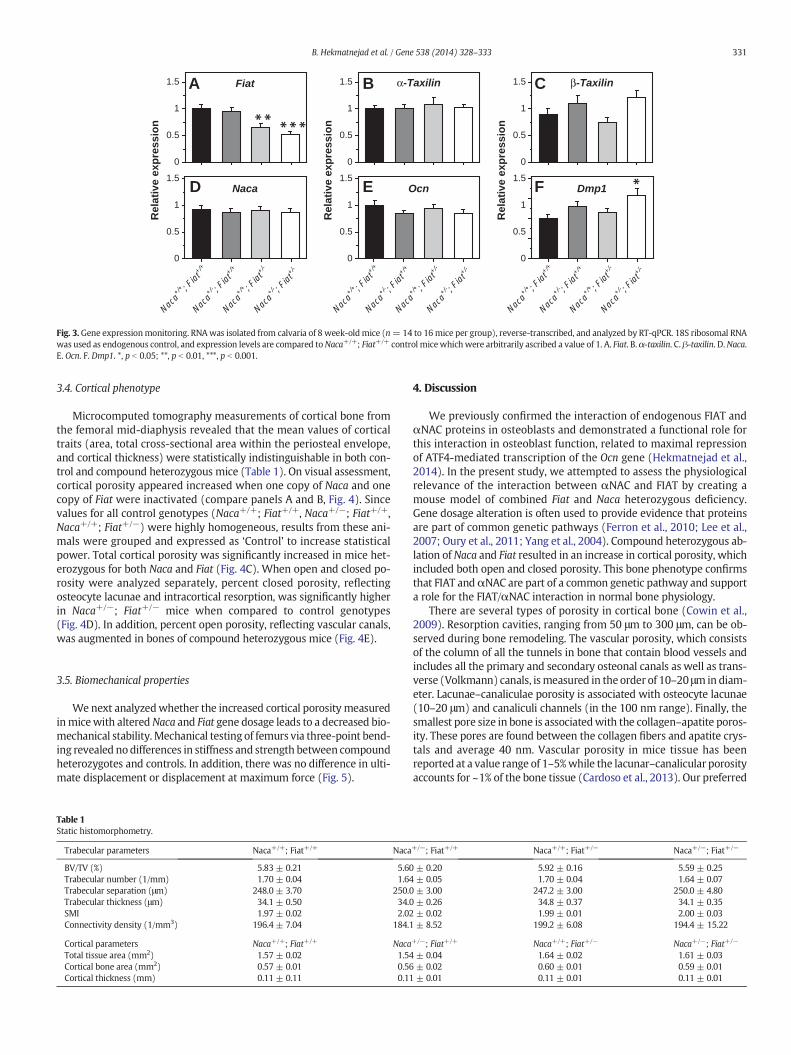
Animals with a single Fiat allele had reduced Fiat mRNA expres-sion without changes in the expression of the related family mem-bers, α-taxilin and β-taxilin (Nogami et al., 2004) (Figs. 3A–C). We didnot measure an impact of Naca gene dosage on Naca expression(Fig. 3D). Surprisingly, the mRNA levels of the best characterized tran-scriptional target of FIAT and αNAC, Ocn, were not affected when oneallele of either or both genes, Fiat and Naca, were inactivated (Fig. 3E).However, expression of the osteocyte differentiation marker Dmp1was increased in Naca+/−; Fiat+/− compound heterozygotes (Fig. 3F).
3.3. Absence of trabecular phenotype
To evaluate bone structural histology, femurs from compound het-erozygous and control females were compared by 3D analysis usingμCT. Trabecular histomorphometric parameters were evaluated at thedistal metaphysis. There were no significant effect of heterozygousNaca and Fiat deficiency on trabecular bone volume, number, thickness,or separation and other trabecular parameters assessed for any of thegenotypes (Table 1).
0
0.5
1
1.5 Fiat α-Taxilin β-Taxilin
0
0.5
1
1.5Naca Ocn Dmp1
A B C
D E F
Rel
ativ
e ex
pre
ssio
n0
0.5
1
1.5
0
0.5
1
1.5
Rel
ativ
e ex
pre
ssio
n
0
0.5
1
1.5
0
0.5
1
1.5
Rel
ativ
e ex
pre
ssio
n
Naca+/
+ ; Fiat
+/+
Naca+/
- ; Fiat
+/+
Naca+/
+ ; Fiat
+/-
Naca+/
- ; Fiat
+/-
Naca+/
+ ; Fiat
+/+
Naca+/
- ; Fiat
+/+
Naca+/
+ ; Fiat
+/-
Naca+/
- ; Fiat
+/-
Naca+/
+ ; Fiat
+/+
Naca+/
- ; Fiat
+/+
Naca+/
+ ; Fiat
+/-
Naca+/
- ; Fiat
+/-
Fig. 3.Gene expressionmonitoring. RNAwas isolated from calvaria of 8 week-oldmice (n=14 to 16mice per group), reverse-transcribed, and analyzed by RT-qPCR. 18S ribosomal RNAwas used as endogenous control, and expression levels are compared toNaca+/+; Fiat+/+ control micewhichwere arbitrarily ascribed a value of 1. A. Fiat. B.α-taxilin. C. β-taxilin. D.Naca.E. Ocn. F. Dmp1. *, p b 0.05; **, p b 0.01, ***, p b 0.001.
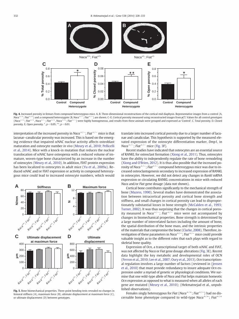
331B. Hekmatnejad et al. / Gene 538 (2014) 328–333
3.4. Cortical phenotype
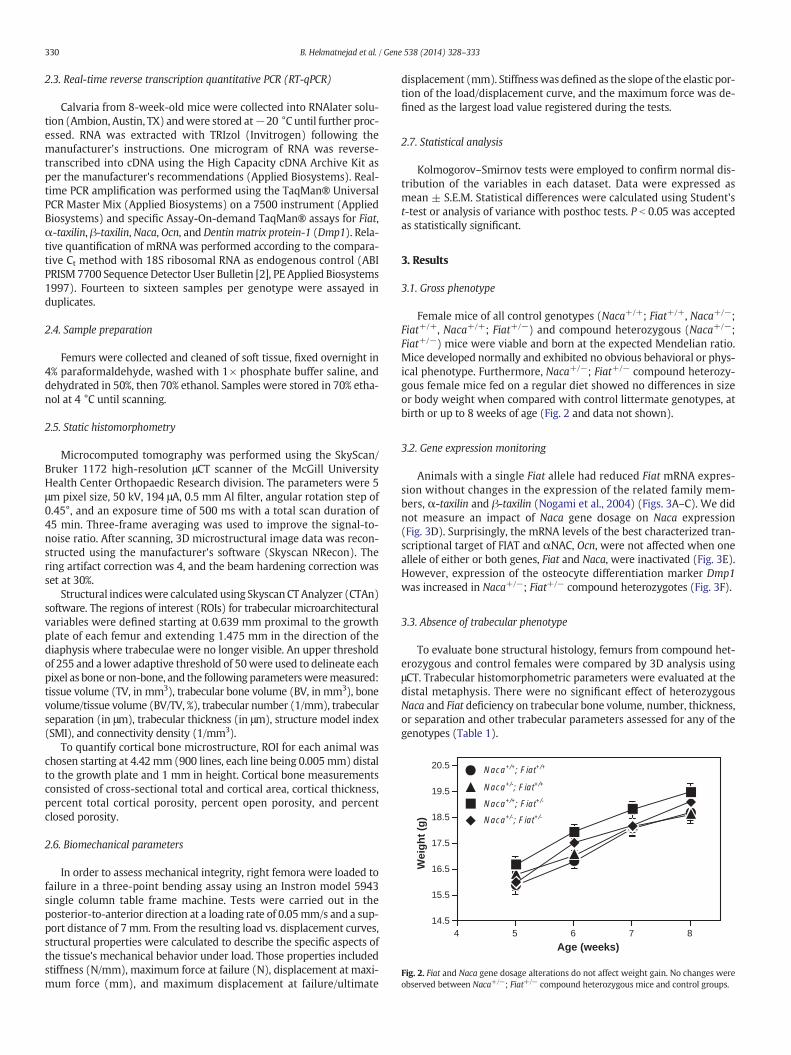
Microcomputed tomography measurements of cortical bone fromthe femoral mid-diaphysis revealed that the mean values of corticaltraits (area, total cross-sectional area within the periosteal envelope,and cortical thickness) were statistically indistinguishable in both con-trol and compound heterozygous mice (Table 1). On visual assessment,cortical porosity appeared increased when one copy of Naca and onecopy of Fiat were inactivated (compare panels A and B, Fig. 4). Sincevalues for all control genotypes (Naca+/+; Fiat+/+, Naca+/−; Fiat+/+,Naca+/+; Fiat+/−) were highly homogeneous, results from these ani-mals were grouped and expressed as ‘Control’ to increase statisticalpower. Total cortical porosity was significantly increased in mice het-erozygous for both Naca and Fiat (Fig. 4C). When open and closed po-rosity were analyzed separately, percent closed porosity, reflectingosteocyte lacunae and intracortical resorption, was significantly higherin Naca+/−; Fiat+/− mice when compared to control genotypes(Fig. 4D). In addition, percent open porosity, reflecting vascular canals,was augmented in bones of compound heterozygous mice (Fig. 4E).
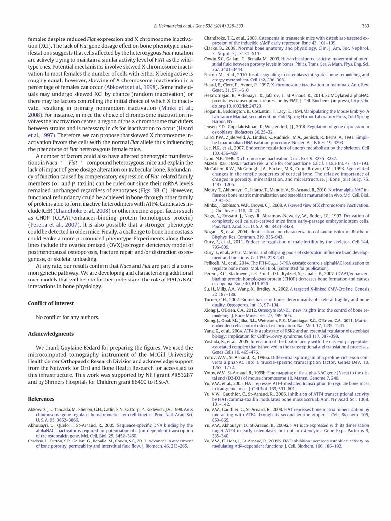
3.5. Biomechanical properties
Wenext analyzedwhether the increased cortical porosity measuredinmicewith alteredNaca and Fiat gene dosage leads to a decreased bio-mechanical stability. Mechanical testing of femurs via three-point bend-ing revealed nodifferences in stiffness and strength between compoundheterozygotes and controls. In addition, there was no difference in ulti-mate displacement or displacement at maximum force (Fig. 5).
Table 1Static histomorphometry.
Trabecular parameters Naca+/+; Fiat+/+ Naca
BV/TV (%) 5.83 ± 0.21 5.60Trabecular number (1/mm) 1.70 ± 0.04 1.64Trabecular separation (μm) 248.0 ± 3.70 250.0Trabecular thickness (μm) 34.1 ± 0.50 34.0SMI 1.97 ± 0.02 2.02Connectivity density (1/mm3) 196.4 ± 7.04 184.1
Cortical parameters Naca+/+; Fiat+/+ NacaTotal tissue area (mm2) 1.57 ± 0.02 1.54Cortical bone area (mm2) 0.57 ± 0.01 0.56Cortical thickness (mm) 0.11 ± 0.11 0.11
4. Discussion
We previously confirmed the interaction of endogenous FIAT andαNAC proteins in osteoblasts and demonstrated a functional role forthis interaction in osteoblast function, related to maximal repressionof ATF4-mediated transcription of the Ocn gene (Hekmatnejad et al.,2014). In the present study, we attempted to assess the physiologicalrelevance of the interaction between αNAC and FIAT by creating amouse model of combined Fiat and Naca heterozygous deficiency.Gene dosage alteration is often used to provide evidence that proteinsare part of common genetic pathways (Ferron et al., 2010; Lee et al.,2007; Oury et al., 2011; Yang et al., 2004). Compound heterozygous ab-lation of Naca and Fiat resulted in an increase in cortical porosity, whichincluded both open and closed porosity. This bone phenotype confirmsthat FIAT andαNAC are part of a common genetic pathway and supporta role for the FIAT/αNAC interaction in normal bone physiology.
There are several types of porosity in cortical bone (Cowin et al.,2009). Resorption cavities, ranging from 50 μm to 300 μm, can be ob-served during bone remodeling. The vascular porosity, which consistsof the column of all the tunnels in bone that contain blood vessels andincludes all the primary and secondary osteonal canals as well as trans-verse (Volkmann) canals, ismeasured in the order of 10–20 μmin diam-eter. Lacunae–canaliculae porosity is associated with osteocyte lacunae(10–20 μm) and canaliculi channels (in the 100 nm range). Finally, thesmallest pore size in bone is associatedwith the collagen–apatite poros-ity. These pores are found between the collagen fibers and apatite crys-tals and average 40 nm. Vascular porosity in mice tissue has beenreported at a value range of 1–5%while the lacunar–canalicular porosityaccounts for ~1% of the bone tissue (Cardoso et al., 2013). Our preferred
+/−; Fiat+/+ Naca+/+; Fiat+/− Naca+/−; Fiat+/−
± 0.20 5.92 ± 0.16 5.59 ± 0.25± 0.05 1.70 ± 0.04 1.64 ± 0.07± 3.00 247.2 ± 3.00 250.0 ± 4.80± 0.26 34.8 ± 0.37 34.1 ± 0.35± 0.02 1.99 ± 0.01 2.00 ± 0.03± 8.52 199.2 ± 6.08 194.4 ± 15.22
+/−; Fiat+/+ Naca+/+; Fiat+/− Naca+/−; Fiat+/−
± 0.04 1.64 ± 0.02 1.61 ± 0.03± 0.02 0.60 ± 0.01 0.59 ± 0.01± 0.01 0.11 ± 0.01 0.11 ± 0.01
0.0
0.2
0.4
0.6
0.8
% T
ota
l po
rosi
ty
0.00
0.02
0.04
0.06
0.08
% C
lose
d p
oro
sity
Naca+/+; Fiat+/+ Naca+/-; Fiat+/-
Control Compound Heterozygote
0.0
0.2
0.4
0.6
0.8
% O
pen
po
rosi
ty
Control Compound Heterozygote
Control Compound Heterozygote
A
C D E
B
Fig. 4. Increased porosity in femurs from compound heterozygous mice. A, B. Three-dimensional reconstructions of the cortical mid diaphysis. Representative images from a control (A,Naca+/+; Fiat+/+) and a compound heterozygote (B,Naca+/−; Fiat+/−) are shown. C–E. Cortical porosity measured using reconstructed images from μCT. Values for all control genotypes(Naca+/+; Fiat+/+, Naca+/−; Fiat+/+, Naca+/+; Fiat+/−) were highly homogeneous, and results from these animals were grouped and expressed as ‘Control’. C. Total porosity. D. Closedporosity. E. Open porosity. *, p b 0.05; **, p b 0.01.
332 B. Hekmatnejad et al. / Gene 538 (2014) 328–333
interpretation of the increased porosity inNaca+/−; Fiat+/−mice is thatlacunar–canalicular porosity was increased. This is based on the emerg-ing evidence that impaired αNAC nuclear activity affects osteoblastmaturation and osteocyte number in vivo (Meury et al., 2010; Pellicelliet al., 2014). Mice with a knock-in mutation that reduces the nucleartranslocation of αNAC have osteopenia with a reduced volume of im-mature, woven-type bone characterized by an increase in the numberof osteocytes (Meury et al., 2010). In addition, FIAT protein expressionhas been localized to osteocytes in adult mice (Yu et al., 2009a). Re-duced αNAC and/or FIAT expression or activity in compound heterozy-gous mice could lead to increased osteocyte numbers, which would
Stiffness
N/m
m
0
20
40
60
80Maximum force
0
4
8
12
N
Ultimate displacementat maximun force
0
100
200
300
400
µm
Ultimate displacement
0
200
400
600
800
µm
Naca+/
+ ; Fiat
+/+
Naca+/
- ; Fiat
+/+
Naca+/
+ ; Fiat
+/-
Naca+/
- ; Fiat
+/-
Naca+/
+ ; Fiat
+/+
Naca+/
- ; Fiat
+/+
Naca+/
+ ; Fiat
+/-
Naca+/
- ; Fiat
+/-
Naca+/
+ ; Fiat
+/+
Naca+/
- ; Fiat
+/+
Naca+/
+ ; Fiat
+/-
Naca+/
- ; Fiat
+/-
Naca+/
+ ; Fiat
+/+
Naca+/
- ; Fiat
+/+
Naca+/
+ ; Fiat
+/-
Naca+/
- ; Fiat
+/-
A B
C D
Fig. 5. Bone biomechanical properties. Three-point bending tests revealed no changes infemoral stiffness (A), maximum force (B), ultimate displacement at maximum force (C),or ultimate displacement (D) between genotypes.
translate into increased cortical porosity due to a larger number of lacu-nae and canaliculae. This hypothesis is supported by the measured ele-vated expression of the osteocyte differentiation marker, Dmp1, inNaca+/−; Fiat+/− mice (Fig. 3F).
Recent studies have indicated that osteocytes are an essential sourceof RANKL for osteoclast formation (Xiong et al., 2011). Thus, osteocyteshave the ability to independently regulate the rate of bone remodeling(Xiong and O'Brien, 2012). It is thus also possible that the increased po-rosity ofNaca+/−; Fiat+/− compound heterozygousmicewas due to in-creased osteoclastogenesis secondary to increased expression of RANKLin osteocytes. However, we did not detect any changes in Rankl mRNAexpression or circulating RANKL concentrations in mice with reducedNaca and/or Fiat gene dosage (data not shown).
Cortical bone contributes significantly to the mechanical strength ofbone (Mazess, 1990). Several studies have demonstrated the associa-tion between intracortical porosity and cortical bone strength andstiffness, and small changes in cortical porosity can lead to dispropor-tionately substantial losses in bone strength (McCalden et al., 1993;Turner, 2002). It was thus surprising that the changes in cortical poros-ity measured in Naca+/−; Fiat+/− mice were not accompanied bychanges in biomechanical properties. Bone strength is determined bya large number of interrelated factors including the amount of bone,the spatial distribution of the bone mass, and the intrinsic propertiesof thematerials that compromise the bone (Clarke, 2008). Therefore, in-vestigation of these parameters in Naca+/−; Fiat+/−mice could providevaluable insight as to the different roles that each plays with regard toskeletal bone quality.
Expression of Ocn, a transcriptional target of both αNAC and FIAT,was not affected byNaca or Fiat gene dosage alterations (Fig. 3E). Recentdata highlight the key metabolic and developmental roles of OCN(Ferron et al., 2010; Lee et al., 2007; Oury et al., 2013).Ocn transcription-al regulation involves a large number of factors (reviewed in (Jensenet al., 2010) that must provide redundancy to insure adequate Ocn ex-pression under a myriad of genetic or physiological conditions. We sur-mise that onewild-type allele of Naca and Fiat helps maintain homeoticOcn expression as opposed to what is measured when all alleles of eachgene are mutated (Meury et al., 2010); (Hekmatnejad et al., unpub-lished observations).
Females singly heterozygous for Fiat (Naca+/+; Fiat+/−) had no dis-cernable bone phenotype compared to wild-type Naca+/+; Fiat+/+
333B. Hekmatnejad et al. / Gene 538 (2014) 328–333
females despite reduced Fiat expression and X chromosome inactiva-tion (XCI). The lack of Fiat gene dosage effect on bone phenotypic man-ifestations suggests that cells affected by the heterozygous Fiatmutationare actively trying tomaintain a similar activity level of FIAT as thewild-type ones. Potential mechanisms involve skewed X chromosome inacti-vation. In most females the number of cells with either X being active isroughly equal; however, skewing of X chromosome inactivation in apercentage of females can occur (Abkowitz et al., 1998). Some individ-uals may undergo skewed XCI by chance (random inactivation) orthere may be factors controlling the initial choice of which X to inacti-vate, resulting in primary nonrandom inactivation (Minks et al.,2008). For instance, in mice the choice of chromosome inactivation in-volves the inactivation center, a region of the X chromosome that differsbetween strains and is necessary in cis for inactivation to occur (Heardet al., 1997). Therefore, we can propose that skewed X chromosome in-activation favors the cells with the normal Fiat allele thus influencingthe phenotype of Fiat heterozygous female mice.
A number of factors could also have affected phenotypic manifesta-tions inNaca+/−; Fiat+/− compound heterozygousmice and explain thelack of impact of gene dosage alteration on trabecular bone. Redundan-cy of function caused by compensatory expression of Fiat-related familymembers (α- and β-taxilin) can be ruled out since their mRNA levelsremained unchanged regardless of genotypes (Figs. 3B, C). However,functional redundancy could be achieved in bone through other familyof proteins able to form inactive heterodimerswith ATF4. Candidates in-clude ICER (Chandhoke et al., 2008) or other leucine zipper factors suchas CHOP (CCAAT/enhancer-binding protein homologous protein)(Pereira et al., 2007). It is also possible that a stronger phenotypecould be detected in oldermice. Finally, a challenge to bonehomeostasiscould evoke a more pronounced phenotype. Experiments along thoselines include the ovariectomized (OVX)/estrogen deficiency model ofpostmenopausal osteoporosis, fracture repair and/or distraction osteo-genesis, or skeletal unloading.
At any rate, our results confirm that Naca and Fiat are part of a com-mon genetic pathway. We are developing and characterizing additionalmicemodels that will help to further understand the role of FIAT/αNACinteractions in bone physiology.
Conflict of interest
No conflict for any authors.
Acknowledgments
We thank Guylaine Bédard for preparing the figures. We used themicrocomputed tomography instrument of the McGill UniversityHealth Center Orthopaedic Research Division and acknowledge supportfrom the Network for Oral and Bone Health Research for access aid tothis infrastructure. This work was supported by NIH grant AR53287and by Shriners Hospitals for Children grant 86400 to R.St-A.
References
Abkowitz, J.L., Taboada, M., Shelton, G.H., Catlin, S.N., Guttorp, P., Kiklevich, J.V., 1998. An Xchromosome gene regulates hematopoietic stem cell kinetics. Proc. Natl. Acad. Sci.U. S. A. 95, 3862–3866.
Akhouayri, O., Quelo, I., St-Arnaud, R., 2005. Sequence-specific DNA binding by thealphaNAC coactivator is required for potentiation of c-Jun-dependent transcriptionof the osteocalcin gene. Mol. Cell. Biol. 25, 3452–3460.
Cardoso, L., Fritton, S.P., Gailani, G., Benalla, M., Cowin, S.C., 2013. Advances in assessmentof bone porosity, permeability and interstitial fluid flow. J. Biomech. 46, 253–265.
Chandhoke, T.K., et al., 2008. Osteopenia in transgenic mice with osteoblast-targeted ex-pression of the inducible cAMP early repressor. Bone 43, 101–109.
Clarke, B., 2008. Normal bone anatomy and physiology. Clin. J. Am. Soc. Nephrol.3 (Suppl. 3), S131–S139.
Cowin, S.C., Gailani, G., Benalla, M., 2009. Hierarchical poroelasticity: movement of inter-stitial fluid between porosity levels in bones. Philos. Trans. Ser. A Math. Phys. Eng. Sci.367, 3401–3444.
Ferron, M., et al., 2010. Insulin signaling in osteoblasts integrates bone remodeling andenergy metabolism. Cell 142, 296–308.
Heard, E., Clerc, P., Avner, P., 1997. X-chromosome inactivation in mammals. Ann. Rev.Genet. 31, 571–610.
Hekmatnejad, B., Akhouayri, O., Jafarov, T., St Arnaud, R., 2014. SUMOylated alphaNACpotentiates transcriptional repression by FIAT. J. Cell. Biochem. (in press), http://dx.doi.org/10.1002/jcb.24729.
Hogan, B., Beddington, R., Costantini, F., Lacy, E., 1994. Manipulating theMouse Embryo. ALaboratory Manual, second edition. Cold Spring Harbor Laboratory Press, Cold SpringHarbor, NY.
Jensen, E.D., Gopalakrishnan, R., Westendorf, J.J., 2010. Regulation of gene expression inosteoblasts. Biofactors 36, 25–32.
Laird, P.W., Zijderveld, A., Linders, K., Rudnicki, M.A., Jaenisch, R., Berns, A., 1991. Simpli-fied mammalian DNA isolation procedure. Nucleic Acids Res. 19, 4293.
Lee, N.K., et al., 2007. Endocrine regulation of energy metabolism by the skeleton. Cell130, 456–469.
Lyon, M.F., 1999. X-chromosome inactivation. Curr. Biol. 9, R235–R237.Mazess, R.B., 1990. Fracture risk: a role for compact bone. Calcif. Tissue Int. 47, 191–193.McCalden, R.W., McGeough, J.A., Barker, M.B., Court-Brown, C.M., 1993. Age-related
changes in the tensile properties of cortical bone. The relative importance ofchanges in porosity, mineralization, and microstructure. J. Bone Joint Surg. 75,1193–1205.
Meury, T., Akhouayri, O., Jafarov, T., Mandic, V., St-Arnaud, R., 2010. Nuclear alpha NAC in-fluences bonematrix mineralization and osteoblast maturation in vivo. Mol. Cell. Biol.30, 43–53.
Minks, J., Robinson, W.P., Brown, C.J., 2008. A skewed view of X chromosome inactivation.J. Clin. Invest. 118, 20–23.
Nagy, A., Rossant, J., Nagy, R., Abramow-Newerly, W., Roder, J.C., 1993. Derivation ofcompletely cell culture-derived mice from early-passage embryonic stem cells.Proc. Natl. Acad. Sci. U. S. A. 90, 8424–8428.
Nogami, S., et al., 2004. Identification and characterization of taxilin isoforms. Biochem.Biophys. Res. Commun. 319, 936–943.
Oury, F., et al., 2011. Endocrine regulation of male fertility by the skeleton. Cell 144,796–809.
Oury, F., et al., 2013. Maternal and offspring pools of osteocalcin influence brain develop-ment and functions. Cell 155, 228–241.
Pellicelli, M., et al., 2014. The PTH-Galpha S-PKA cascade controls alphaNAC localization toregulate bone mass. Mol. Cell Biol. (submitted for publication).
Pereira, R.C., Stadmeyer, L.E., Smith, D.L., Rydziel, S., Canalis, E., 2007. CCAAT/enhancer-binding protein homologous protein (CHOP) decreases bone formation and causesosteopenia. Bone 40, 619–626.
Su, H., Mills, A.A., Wang, X., Bradley, A., 2002. A targeted X-linked CMV-Cre line. Genesis32, 187–188.
Turner, C.H., 2002. Biomechanics of bone: determinants of skeletal fragility and bonequality. Osteoporos. Int. 13, 97–104.
Xiong, J., O'Brien, C.A., 2012. Osteocyte RANKL: new insights into the control of bone re-modeling. J. Bone Miner. Res. 27, 499–505.
Xiong, J., Onal, M., Jilka, R.L., Weinstein, R.S., Manolagas, S.C., O'Brien, C.A., 2011. Matrix-embedded cells control osteoclast formation. Nat. Med. 17, 1235–1241.
Yang, X., et al., 2004. ATF4 is a substrate of RSK2 and an essential regulator of osteoblastbiology; implication for Coffin–Lowry syndrome. Cell 117, 387–398.
Yoshida, K., et al., 2005. Interaction of the taxilin family with the nascent polypeptide-associated complex that is involved in the transcriptional and translational processes.Genes Cells 10, 465–476.
Yotov, W.V., St-Arnaud, R., 1996a. Differential splicing-in of a proline-rich exon con-verts alphaNAC into a muscle-specific transcription factor. Genes Dev. 10,1763–1772.
Yotov, W.V., St-Arnaud, R., 1996b. Fine mapping of the alpha-NAC gene (Naca) to the dis-tal end (D2-D3) of mouse chromosome 10. Mamm. Genome 7, 240.
Yu, V.W., et al., 2005. FIAT represses ATF4-mediated transcription to regulate bone massin transgenic mice. J. Cell Biol. 169, 591–601.
Yu, V.W., Gauthier, C., St-Arnaud, R., 2006. Inhibition of ATF4 transcriptional activityby FIAT/gamma-taxilin modulates bone mass accrual. Ann. NY Acad. Sci. 1068,131–142.
Yu, V.W., Gauthier, C., St-Arnaud, R., 2008. FIAT represses bone matrix mineralization byinteracting with ATF4 through its second leucine zipper. J. Cell. Biochem. 105,859–865.
Yu, V.W., Akhouayri, O., St-Arnaud, R., 2009a. FIAT is co-expressed with its dimerizationtarget ATF4 in early osteoblasts, but not in osteocytes. Gene Expr. Patterns 9,335–340.
Yu, V.W., El-Hoss, J., St-Arnaud, R., 2009b. FIAT inhibition increases osteoblast activity bymodulating Atf4-dependent functions. J. Cell. Biochem. 106, 186–192.