The C2 domain and altered ATP-binding loop phosphorylation at ...
39
The C2 domain and altered ATP-binding loop phosphorylation at Ser 359 mediate the redox- dependent increase in Protein Kinase C-δ activity Jianli Gong a , Yongneng Yao a , Pingbo Zhang b , Barath Udayasuryan d , Elena V. Komissarova a , Ju Chen c , Sivaraj Sivaramakrishnan d , Jennifer E. Van Eyk b , and Susan F. Steinberg a Department of Pharmacology, Columbia University, New York, NY a ; Department of Medicine, Johns Hopkins School of Medicine, Baltimore, MD b ; UCSD School of Medicine, La Jolla, CA c ; Department of Cell and Dev. Biology, Dept. of Biomedical Engineering, University of Michigan, Ann Arbor, MI d . Running Head: Mechanism for redox-dependent changes in PKCδ activity #Address correspondence to: Susan F. Steinberg, M.D., Department of Pharmacology, College of Physicians and Surgeons, Columbia University, 630 West 168 Street, New York, NY 10032. Tel: 212-305-4297; E-mail: [email protected] Materials and Methods section word count: 1271 Introduction, Results, and Discussion sections combined word count: 4981 MCB Accepted Manuscript Posted Online 9 March 2015 Mol. Cell. Biol. doi:10.1128/MCB.01436-14 Copyright © 2015, American Society for Microbiology. All Rights Reserved. on February 9, 2018 by guest http://mcb.asm.org/ Downloaded from
Transcript of The C2 domain and altered ATP-binding loop phosphorylation at ...
The C2 domain and altered ATP-binding loop phosphorylation at Ser359 mediate the redox-
dependent increase in Protein Kinase C-δ activity
Jianli Gonga, Yongneng Yaoa, Pingbo Zhangb, Barath Udayasuryand,
Elena V. Komissarovaa, Ju Chenc, Sivaraj Sivaramakrishnand,
Jennifer E. Van Eykb, and Susan F. Steinberga
Department of Pharmacology, Columbia University, New York, NY a; Department of Medicine, Johns
Hopkins School of Medicine, Baltimore, MD b; UCSD School of Medicine, La Jolla, CA c; Department of
Cell and Dev. Biology, Dept. of Biomedical Engineering, University of Michigan, Ann Arbor, MI d.
Running Head: Mechanism for redox-dependent changes in PKCδ activity
#Address correspondence to: Susan F. Steinberg, M.D., Department of Pharmacology, College of
Physicians and Surgeons, Columbia University, 630 West 168 Street, New York, NY 10032.
Tel: 212-305-4297; E-mail: [email protected]
Materials and Methods section word count: 1271
Introduction, Results, and Discussion sections combined word count: 4981
MCB Accepted Manuscript Posted Online 9 March 2015Mol. Cell. Biol. doi:10.1128/MCB.01436-14Copyright © 2015, American Society for Microbiology. All Rights Reserved.
on February 9, 2018 by guest
http://mcb.asm
.org/D
ownloaded from
ABSTRACT
PKCδ's diverse roles in cellular growth, survival, and injury have been attributed to stimulus-
specific differences in PKCδ signaling responses. PKCδ exerts membrane-delimited actions in cells
activated by agonists that stimulate phosphoinositide hydrolysis. PKCδ is released from membranes as a
Tyr313-phosphorylated enzyme that displays a high level of lipid-independent activity and altered
substrate specificity during oxidative stress. This study identifies an interaction between PKCδ's Tyr313-
phosphorylated hinge region and its phosphotyrosine-binding C2 domain that controls PKCδ's
enzymology indirectly by decreasing phosphorylation in the kinase domain ATP-positioning loop at
Ser359. We show that WT-PKCδ displays a strong preference for substrates with serine as the
phosphoacceptor residue at the active site when it harbors phosphomimetic or bulky substitutions at
Ser359. In contrast, PKCδ-S359A displays lipid-independent activity towards substrates with either a
serine or threonine as the phosphoacceptor residue. Additional studies in cardiomyocytes show that
oxidative stress decreases Ser359 phosphorylation on native PKCδ and that PKCδ-S359A overexpression
increases basal levels of phosphorylation on substrates with both phosphoacceptor site serine and
threonine residues. Collectively, these studies identify a C2 domain-pTyr313 docking interaction that
controls ATP-positioning loop phosphorylation as a novel, dynamically regulated, and physiologically
relevant structural determinant of PKCδ catalytic activity.
on February 9, 2018 by guest
http://mcb.asm
.org/D
ownloaded from
Protein kinase C-δ (PKCδ) is a serine/threonine kinase that plays a key role in signal transduction
pathways that control a wide range of cellular responses. PKCδ contains a highly conserved C-terminal
catalytic domain and N-terminal regulatory C1 and C2 domains. The C1 domain binds lipids and anchors
full-length PKCδ to membranes. The functional role of the C2 domain has remained more elusive. While
C2 domains of conventional PKC (cPKC) isoforms function as calcium-regulated membrane-targeting
modules, the PKCδ C2 domain is a topological variant that does not coordinate calcium or bind lipids (1).
Rather, it has been characterized as a protein-protein interaction motif (2, 3), with recent evidence that the
PKCδ-C2 domain is a phosphotyrosine binding motif that binds the consensus sequence (Y/F)-(S/A)-
(V/I)-pY-(Q/R)-X-(Y/F) (4).
PKCδ is allosterically activated by lipids (diacylglycerol [DAG] or phorbol esters such as phorbol
12-myristate 13-acetate [PMA]) that bind to the C1 domain. PKCδ also is dynamically regulated as a
result of tyrosine phosphorylation by Src (5, 6). We previously showed that oxidative stress releases
PKCδ from membranes, actives Src, and induces a global increase in PKCδ phosphorylation at Tyr313 and
Tyr334 in both soluble and particulate subcellular compartments (7). These residues in the V3 hinge region
of human PKCδ correspond to Tyr311 and Tyr332 in rodent PKCδ. In contrast, PMA does not increase Src
activity, but rather delivers the enzyme in an active conformation to Src-enriched caveolae membranes
where a low level of basal Src activity is sufficient to promote PKCδ phosphorylation at Tyr313, but not
Tyr334 (8). Since PKCδ is phosphorylated by Src in vitro at both Tyr313 and Tyr334 (9), a mechanism that
might account for the selective PMA-dependent PKCδ phosphorylation at Tyr313 but not Tyr334 has never
been obvious.
Stimulus-induced increases in PKCδ-Tyr313 phosphorylation have been implicated in several
PKCδ-dependent cellular responses (10, 11). We previously showed that Tyr313 phosphorylation
influences PKCδ activity toward cardiac troponin I (cTnI, the “inhibitory” subunit of the troponin
complex and a physiologically important PKCδ substrate in cardiomyocytes (9)). cTnI contains several
phosphorylation clusters that exert distinct effects on cardiac contraction. PKCδ phosphorylates cTnI at
Ser23/Ser24 when allosterically-activated by PS/PMA; studies in detergent-extracted single
on February 9, 2018 by guest
http://mcb.asm
.org/D
ownloaded from
cardiomyocytes link cTnI-Ser23/Ser24 phosphorylation to a decrease in tension at submaximum - but not
maximum – calcium concentrations. PKCδ acquires cTnI-Thr144 kinase activity - it phosphorylates cTnI
at both Ser23/Ser24 and Thr144, leading to a decrease in maximum tension and cross-bridge kinetics (i.e., a
different functional response) - when tyrosine phosphorylated by Src. Additional studies showing that the
Src-dependent acquisition of cTnI-Thr144 kinase activity is completely abrogated by a Y313F substitution
implicates Tyr313-phosphorylation as the mechanisms underlying the Src-dependent increase in PKCδ
activity (9).
The structural basis for Tyr313 phosphorylation-dependent changes in PKCδ's enzymology is not
obvious. This study builds upon the intriguing observation that Tyr313 resides in a PKCδ-C2 domain
consensus-binding motif (VGI-Y313-QGF) (4) to show that the C2 domain interacts with the Tyr313-
phosphorylated V3 region and that this interaction controls PKCδ catalytic activity indirectly by
regulating phosphorylation at Ser359, a novel phosphorylation site in the Gly-rich ATP-positioning loop
(G-loop, also known as the phosphate binding P-loop) of the kinase domain.
MATERIALS AND METHODS
Materials - PKCδ and PKCδ-pTyr334 antibodies were from Santa Cruz Biotechnology. Mouse monoclonal
anti-GFP 3E6 was from Invitrogen. Anti-Flag was from Sigma. All other antibodies were from Cell
Signaling Technology. The specificity of all anti-PKCδ antibodies (including phosphorylation site-
specific antibodies that specifically recognize phosphorylation at Thr507, Tyr313, and Tyr334) has been
validated (9). Src was from Invitrogen-Life Technologies. CREBtide and other peptide substrates were
from Anaspec. PMA was from Sigma. Other chemicals were reagent grade.
Plasmids and Cell lines – Mutant constructs of the PKCδ-EGFP expression plasmid (human sequence
with enhanced GFP fused to its C terminus (9)) were generated by PCR and validated by sequencing. N-
terminal Flag-tagged versions of PKCδ, PKCδ-ΔC2 (Δ1-123) and PKCδ-S359A also were generated and
inserted into pcDNA3.1. All results in Figs 2 and 3 with EGFP-tagged enzymes were replicated with
on February 9, 2018 by guest
http://mcb.asm
.org/D
ownloaded from
Flag-tagged enzymes. Expression vectors were introduced into HEK293 cells (maintained in DMEM with
10% FBS) using the Effectene transfection reagent (Qiagen) according to manufacturer’s instructions.
Cells were lysed after 24 h in homogenization buffer containing 20 mM Tris-Cl (pH 7.5), 0.05 mM
EDTA, 0.5 mM dithiothreitol, 0.2% Triton X-100, 5 μg/ml aprotinin, 5 μg/ml leupeptin, 5 μg/ml
benzamidine, 1 mM phenylmethylsulfonyl fluoride, and 5 μM pepstatin A.
Cardiomyocytes were isolated from the hearts of 2-day-old Wistar rats and infected with
adenoviral vectors that drive expression of WT-PKCδ or PKCδ-S359A according to methods published
previously (12). Total cell lysates and fractions enriched in myofibrollar proteins were prepared
according standard methods described in previous publications (7, 13).
Preparation of Caveolae Membranes – Caveolae membrane proteins were isolated according to a
detergent-free purification scheme that was described previously (14).
In Vitro Kinase Assays (IVKAs) and Western Blotting - IVKAs were performed with PKCδ
immunoprecipitated with anti-GFP or anti-Flag from 150 μg of starting cell extract according to methods
described previously (9). Incubations were performed for 30 min at 30°C in 110 μl reaction buffer
containing 30 mM Tris-Cl, pH 7.5, 5.45 mM MgCl2, 0.65 mM EDTA, 0.65 mM EGTA, 0.1 mM DTT,
1.09 mM sodium orthovanadate, 0.1 μM calyculin, 0.55 μM PKI, 217 mM NaCl, 3.6% glycerol, 89 μg/ml
phosphatidylserine plus 175 nM PMA, and [γ-32P]ATP (10 μCi, 66 μM), with 4 μg of Tn complex
(consisting of equimolar cTnI, cTnT, and cTnC) as substrate. Immunoblotting on lysates or
immunoprecipitated PKCδ was according to methods described previously or manufacturer's instructions.
In each figure, each panel represents the results from a single gel (exposed for a uniform duration);
detection was with enhanced chemiluminescence. 32P-incorporation was quantified by PhosphoImager
and PKCδ phosphorylation was normalized to PKCδ protein levels.
Peptide kinase assays were carried out in a similar manner in 200 μl of a reaction mixture
containing 26 mM Tris, pH 7.5, 5 mM MgCl2, 0.6 mM EGTA, 0.6 mM EDTA, 0.5 μM PKI, 10 μM PP1,
0.25 mM DTT and 89 μg/ml phosphatidylserine plus 175 nM PMA, and 50 μM of peptide substrate as
on February 9, 2018 by guest
http://mcb.asm
.org/D
ownloaded from
indicated in Fig 5. Reactions were initiated by the addition [γ-32P]ATP (10 μCi, 66 μM) and were
performed in quadruplicate at 30°C for 15 min. Assays were terminated by placing samples on ice
followed by centrifugation at 1,500 g for 10 min at 4°C. 40 μl of each supernatant was spotted onto
phosphocellulose filter papers (P-81) which was dropped immediately into water, washed (five times for
5 min), and counted for radioactivity.
Multiple reaction monitoring (MRM) phosporylation of PKCδ. WT-PKCδ and PKCδ-ΔC2 enzymes
were heterologously overexpressed in HEK293 cells, immunoprecipitated for IVKAs, and then subjected
to in gel digestion as described previously (15). Briefly, WT-PKCδ and PKCδ-ΔC2 bands separated by
SDS-PAGE were excised, cut into 1 mm pieces, and washed 3 times with 50% acetonitrile/25 mM
ammonium bicarbonate for 15 min with shaking. Gel pieces were incubated with 25 mM ammonium
bicarbonate +10 mM dithiothreitol for 60 min at 55 ºC, washed with acetonitrile (ACN), then incubated
with 25 mM ammonium bicarbonate + 55 mM iodoacetamide (freshly made) for 30 min in the dark. Gel
pieces were then washed with ACN, dried, rehydrated with 10 ng/μl trypsin (Promega, sequencing grade)
in 25 mM ammonium bicarbonate, and then placed on ice for 30 min. Excess trypsin was removed and
then 20 μL of 25 mM ammonium bicarbonate was added. Samples were digested at 37 ºC for 18 hr. The
liquid was transferred to a clean tube. The peptides were extracted twice using 50% ACN + 0.1% TFA for
20 min at 25 ºC with shaking. The final peptide mixture was subjected to C18 reversed-phase
chromatography in C18 ZipTips (Millipore) using 0.1% TFA. The peptides were then eluted two times
with 20 μl of 70% ACN, 0.1% TFA and 95% ACN, 0.1% TFA. The combined solution was dried using a
vacuum centrifuge SpeedVac (Thermo Electron) and frozen at -80ºC until analysis.
MRM assays were developed, optimized, and validated using the nano-LC/MS/MS system, 4000
QTRAP hybrid triple quadrupole/linear IT mass spectrometer (AB SCIEX). Peptides obtained from the
in-gel digestion of PKCδ proteins were reconstituted in 20 μl of 0.1% formic acid. Peptides were
separated by an Eksigent Tempo nano-LC system (Eksigent Technology) onto a BioBasic C18 reverse-
phase PicoFrit column (300 Å, 5 μm, 75 μm ×10 cm, 15 μm tip, New Objective). Peptides were eluted
on February 9, 2018 by guest
http://mcb.asm
.org/D
ownloaded from
36-min linear gradient from 5 to 40% B (mobile phase A: 2% v/v ACN containing 0.1% v/v formic acid;
mobile phase B: 98% v/v ACN containing 0.1% v/v formic acid) at 500 nl/min flow rate on the 4000
QTRAP MS system, with the NanoSpray source spray head coupled to a distal coated PicoTip fused silica
spray tip (360 μm od, 75 μm id, 15 μm diameter emitter orifice; New Objective). Samples were analyzed
using the following settings curtain gas (CUR):15; collision gas (CAD): high; ion spray voltage (IS): 2.5
kV; ion source gas1 (GS1): 25; ion source gas 2 (GS2): 0; resolution Q1 and Q3: unit; heater interface
temperature: 150 ºC. Each sample was run in triplicate. Peak detection and quantification of peak area
was determined with Multiquant software version 2.0 and inspected manually to ensure correct peak
identification and quantification. Quantification of PKCδ phosphosites was carried out using a ratio of the
peak area to gel band densitometry.
FRET sensor data - The C2 domain of PKCδ (123aa) was cloned from full-length human PKCδ cDNA.
All constructs contain an N-terminal FLAG tag immediately followed by mCitrine. In sequence behind
the mCitrine are the C2 domain of PKCδ, 10 nm ER/K α helix linker (16), a TEV-protease site
(ENLYFQ), mCerulean, and a 13 amino acid peptide sequence containing a Tyr residue that when
phosphorylated corresponds to a C2 domain consensus binding site. FRET sensors were generated with
either an optimal binding sequence MALYSIYQPYVFA (4), the native PKCδ SEPVGIY313QGFEKK
sequence, or these sequences with Tyr→Phe substitutions (MALYSIFQPYVFA and
SEPVGIFQGFEKK) as controls. All domains and flurophores are linked with 3-4 [Gly-Ser-Gly] repeats
to allow rotational flexibility and are cloned between unique restriction sites. A C2 domain H62D
mutation was generated by site-directed-mutagenesis (QuikChange, Stratagene). These constructs were
cloned between unique restriction sites in pBiex-1 vector and used for expression in sf9 cells. Proteins
were purified as described previously (17). FRET spectra for recombinant protein were acquired on a
FluoroMax-4 fluorometer (Horiba Scientific). Samples were excited at 430nm (spectral band pass (bp 8
nm) and emission was scanned from 450 to 650 nm (bp 4nm). All samples were suspended in a buffer
composed of 50 mM Tris-HCl, 10 mM MgCl2, 10% glycerol and 2.5 mM DTT. Recombinant Src kinase
on February 9, 2018 by guest
http://mcb.asm
.org/D
ownloaded from
was added to the reaction samples and incubated at 30oC for 30 minutes before spectra were taken. The
samples also contain 0.05mg/mL BSA to limit non-specific surface adsorption of sensor proteins.
RESULTS
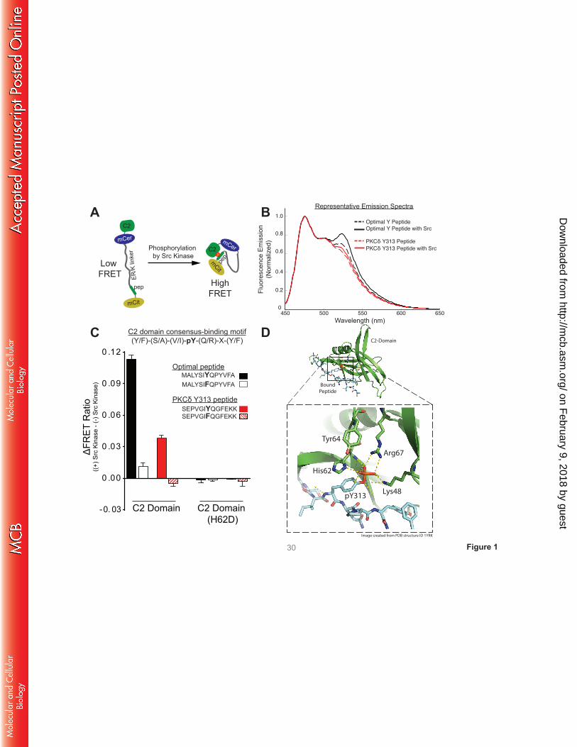
A FRET-based sensor detects a C2 domain interaction with a pTyr313-bearing peptide. Since
traditional bi-molecular approaches are unlikely to be an effective approach to detect a cis interaction
between the C2 domain and the pTyr313 motif (which is predicted to be facilitated by the tethering of the
two domains within full-length PKCδ), we generated FRET-based sensors consisting of a single
polypeptide with (N to C terminus) the C2 domain, mCerulean (as FRET donor), an ER/K linker (18), a
13-residue peptide based upon either a previously described optimal pTyr binding motif (4) or sequence
flanking Tyr313 in PKCδ, and mCitrine (as FRET acceptor). These sensors are designed to primarily report
on the strength of the interaction between the C2 domain and the peptide (higher FRET = stronger
interaction, Fig 1A (16)). Sensors were purified and subjected to in vitro kinase assays with recombinant
Src. Figs 1B and 1C show that the FRET signals for sensors bearing either the optimal pTyr binding
motif or the Tyr313-bearing peptide increase as a result of in vitro phosphorylation by Src. Two sets of
control sensors were generated to establish the specificity of the FRET signals (Fig 1C). First, studies
with sensors containing Tyr→Phe substitutions at each peptide’s phosphorylation site establish that the
increase in FRET signal is due to phosphorylation at the designated tyrosine residue in the peptide.
Second, studies with sensors containing H62D substitutions in the C2 domain pTyr binding pocket (that
disrupts the C2 domain-pTyr interaction; see schematic in Fig 1D (4)) point to a phosphorylation-
dependent interaction with the C2 domain. These results indicate that the Src-dependent increases in
FRET are due to C2 domain interactions with Tyr-bearing peptides, providing direct evidence for an
interaction between the C2 domain and the pTyr313 motif in the V3 hinge of PKCδ.
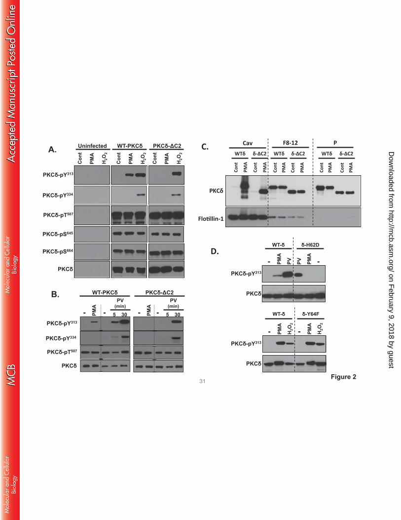
The C2 domain is required for in vivo PMA-dependent PKCδ-Tyr313 phosphorylation. We used a
PKCδ-C2 domain deletion construct (PKCδ-ΔC2) to examine the consequences of C2 domain-dependent
on February 9, 2018 by guest
http://mcb.asm
.org/D
ownloaded from
interactions in a cellular context. Fig 2A shows that WT-PKCδ and PKCδ-ΔC2 are expressed at similar
levels in HEK293 cells and both enzymes undergo similar maturational phosphorylations at the activation
loop (Thr507) and C-terminal (Ser645 and Ser664) phosphorylation sites. These 'priming' site
phosphorylations are stable modifications that are not altered by PMA or H2O2. H2O2 and pervanadate
promote WT-PKCδ and PKCδ-ΔC2 phosphorylation at Tyr313 and Tyr334 (Fig 2A and 2B). In each case,
Tyr313 and Tyr334 phosphorylation is via a Src-dependent mechanism that is inhibited by PP1 (7). In
contrast, PMA induces a Src-dependent increase in WT-PKCδ phosphorylation at Tyr313, but not Tyr334.
PMA does not increase PKCδ-ΔC2 phosphorylation at Tyr313 (Figs 2A and 2B).
Control studies indicate that the effect of the C2 domain deletion to prevent PMA-dependent
Tyr313 phosphorylation is not due to a defect in PKCδ targeting to Src-enriched caveolae membranes. Fig
2C shows that WT-PKCδ and PKCδ-ΔC2 both translocate to the caveolae membrane fraction. These
results indicate that some other mechanism must account for the selective defect in PMA-dependent
PKCδ-ΔC2-Tyr313 phosphorylation. Therefore, we considered whether a C2 domain deletion disrupts the
docking interaction that protects the phosphorylation site at Tyr313 (but not Tyr334) from
dephosphorylation by cellular phosphatases. This mechanism could account for the puzzling observation
that Src phosphorylates PKCδ in vitro at both Tyr313 and Tyr334, but PKCδ is phosphorylated via a Src-
dependent mechanism at Tyr313 - and not Tyr334 - in PMA-treated cells. Indeed, Fig 2D shows that a
PKCδ-H62D substitution in the C2 domain pY binding site (4) completely abrogates PMA-dependent
Tyr313 phosphorylation, whereas Tyr313 phosphorylation is not influenced by a Y64F substitution at an
adjacent site in the C2 domain that does not contribute to pY binding.
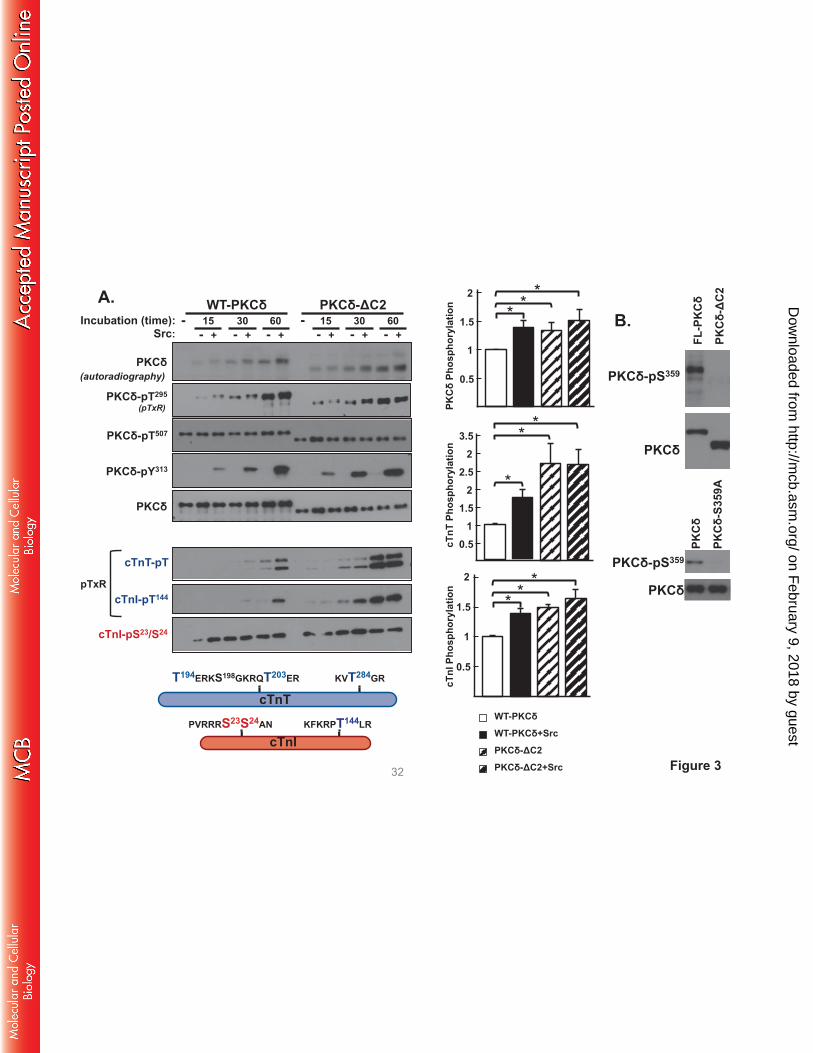
PKCδ-ΔC2 displays a high level of cTnI-Thr144 and cTnT kinase activity that is not influenced by
Src. We performed in vitro kinase assays (IVKAs) without and with Src in buffers containing
[γ32P]ATP, PS/PMA, and cardiac troponin (cTn) complex (consisting of equimolar cTnI, cardiac troponin
T [cTnT], and cardiac troponin C [cTnC] as substrate) to determine whether a C2 domain deletion alters
PKCδ’s in vitro enzymology. Fig 3 shows that WT-PKCδ executes a time-dependent autophosphorylation
on February 9, 2018 by guest
http://mcb.asm
.org/D
ownloaded from
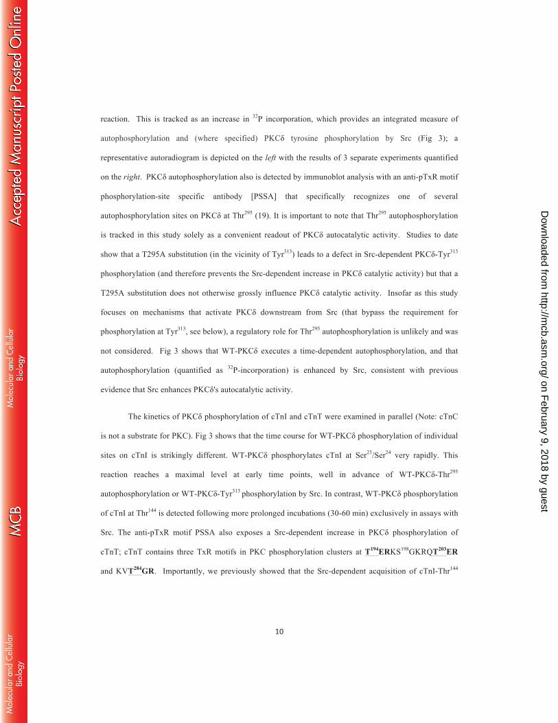
reaction. This is tracked as an increase in 32P incorporation, which provides an integrated measure of
autophosphorylation and (where specified) PKCδ tyrosine phosphorylation by Src (Fig 3); a
representative autoradiogram is depicted on the left with the results of 3 separate experiments quantified
on the right. PKCδ autophosphorylation also is detected by immunoblot analysis with an anti-pTxR motif
phosphorylation-site specific antibody [PSSA] that specifically recognizes one of several
autophosphorylation sites on PKCδ at Thr295 (19). It is important to note that Thr295 autophosphorylation
is tracked in this study solely as a convenient readout of PKCδ autocatalytic activity. Studies to date
show that a T295A substitution (in the vicinity of Tyr313) leads to a defect in Src-dependent PKCδ-Tyr313
phosphorylation (and therefore prevents the Src-dependent increase in PKCδ catalytic activity) but that a
T295A substitution does not otherwise grossly influence PKCδ catalytic activity. Insofar as this study
focuses on mechanisms that activate PKCδ downstream from Src (that bypass the requirement for
phosphorylation at Tyr313, see below), a regulatory role for Thr295 autophosphorylation is unlikely and was
not considered. Fig 3 shows that WT-PKCδ executes a time-dependent autophosphorylation, and that
autophosphorylation (quantified as 32P-incorporation) is enhanced by Src, consistent with previous
evidence that Src enhances PKCδ's autocatalytic activity.
The kinetics of PKCδ phosphorylation of cTnI and cTnT were examined in parallel (Note: cTnC
is not a substrate for PKC). Fig 3 shows that the time course for WT-PKCδ phosphorylation of individual
sites on cTnI is strikingly different. WT-PKCδ phosphorylates cTnI at Ser23/Ser24 very rapidly. This
reaction reaches a maximal level at early time points, well in advance of WT-PKCδ-Thr295
autophosphorylation or WT-PKCδ-Tyr313 phosphorylation by Src. In contrast, WT-PKCδ phosphorylation
of cTnI at Thr144 is detected following more prolonged incubations (30-60 min) exclusively in assays with
Src. The anti-pTxR motif PSSA also exposes a Src-dependent increase in PKCδ phosphorylation of
cTnT; cTnT contains three TxR motifs in PKC phosphorylation clusters at T194ERKS198GKRQT203ER
and KVT284GR. Importantly, we previously showed that the Src-dependent acquisition of cTnI-Thr144
on February 9, 2018 by guest
http://mcb.asm
.org/D
ownloaded from
kinase activity is abrogated by a Y313F substitution on PKCδ (9); a Y313F also abrogates the Src-
dependent increase in TxR motif phosphorylation on cTnT (data not shown).
A similar kinetic analysis of the in vitro kinase activity of PKCδ-ΔC2 shows the following: [1]
PKCδ-ΔC2 exhibits a time-dependent increase in autophosphorylation, detected as an increase in 32P-
incorporation and phosphorylation at Thr295. Quantification, based upon 32P-incorporation, shows that
PKCδ-ΔC2 autophosphorylates to a higher level that WT-PKCδ; PKCδ-ΔC2 autophosphorylation is not
influenced by Src. [2] PKCδ-ΔC2 phosphorylates cTnI at Ser23/Ser24, but unlike WT-PKCδ displays a
high level of Src-independent cTnI-Thr144 and cTnT-TxR motif activity. These results suggest that the C2
domain functions to limit PKCδ-dependent cTnI-Thr144 and cTnT-TxR motif phosphorylation.
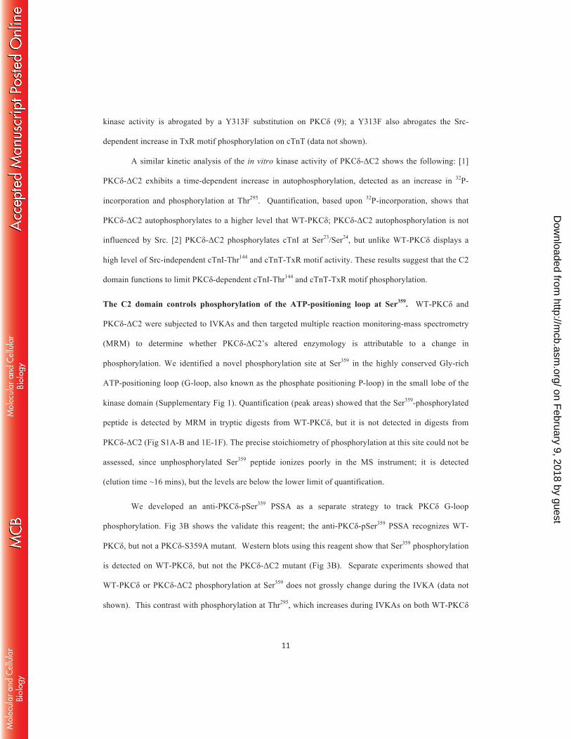
The C2 domain controls phosphorylation of the ATP-positioning loop at Ser359. WT-PKCδ and
PKCδ-ΔC2 were subjected to IVKAs and then targeted multiple reaction monitoring-mass spectrometry
(MRM) to determine whether PKCδ-ΔC2’s altered enzymology is attributable to a change in
phosphorylation. We identified a novel phosphorylation site at Ser359 in the highly conserved Gly-rich
ATP-positioning loop (G-loop, also known as the phosphate positioning P-loop) in the small lobe of the
kinase domain (Supplementary Fig 1). Quantification (peak areas) showed that the Ser359-phosphorylated
peptide is detected by MRM in tryptic digests from WT-PKCδ, but it is not detected in digests from
PKCδ-ΔC2 (Fig S1A-B and 1E-1F). The precise stoichiometry of phosphorylation at this site could not be
assessed, since unphosphorylated Ser359 peptide ionizes poorly in the MS instrument; it is detected
(elution time ~16 mins), but the levels are below the lower limit of quantification.
We developed an anti-PKCδ-pSer359 PSSA as a separate strategy to track PKCδ G-loop
phosphorylation. Fig 3B shows the validate this reagent; the anti-PKCδ-pSer359 PSSA recognizes WT-
PKCδ, but not a PKCδ-S359A mutant. Western blots using this reagent show that Ser359 phosphorylation
is detected on WT-PKCδ, but not the PKCδ-ΔC2 mutant (Fig 3B). Separate experiments showed that
WT-PKCδ or PKCδ-ΔC2 phosphorylation at Ser359 does not grossly change during the IVKA (data not
shown). This contrast with phosphorylation at Thr295, which increases during IVKAs on both WT-PKCδ
on February 9, 2018 by guest
http://mcb.asm
.org/D
ownloaded from
and PKCδ-ΔC2 (Fig 3A). The Thr295-phosphorylated peptide also was detected in the MRM studies,
where the quantification showed 37% higher level of the Thr295-phosphorylated peptide in tryptic digests
from PKCδ-ΔC2 than in the digest from WT-PKCδ (Fig S1A-D).
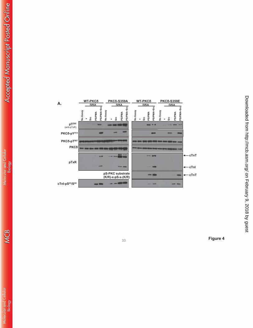
The C2 domain regulates PKCδ catalytic activity indirectly by controling Ser359 phosphorylation.
PKCδ mutants harboring single residue substitutions at position 359 (either on the WT-PKCδ or PKCδ-
ΔC2 backbone) were subjected to IVKAs to examine their autocatalytic and Tn kinase activities.
Preliminary studies showed that Ser359 substitutions do not grossly alter PKCδ expression, ‘priming’ site
(Thr507, Ser645, Ser664) phosphorylation, or cTnI-Ser23/Ser24 kinase activity (data not shown), suggesting
that the PKCδ G-loop is a relatively flexible structure that accomodates a range of amino acid
substitutions at position 359.
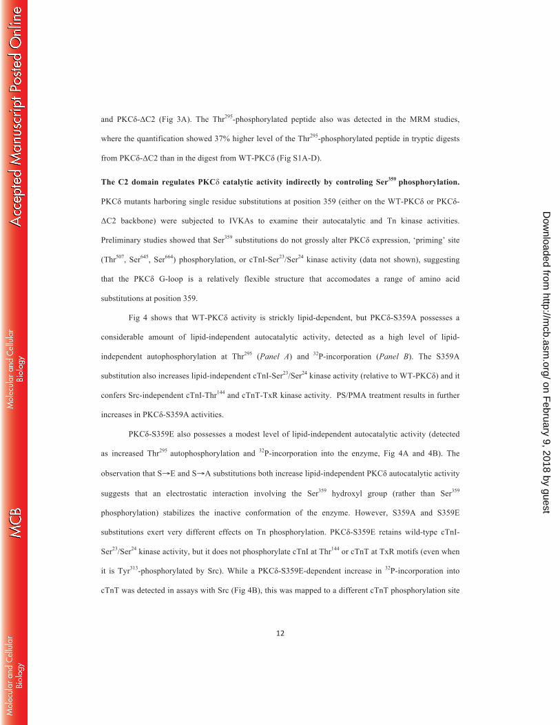
Fig 4 shows that WT-PKCδ activity is strickly lipid-dependent, but PKCδ-S359A possesses a
considerable amount of lipid-independent autocatalytic activity, detected as a high level of lipid-
independent autophosphorylation at Thr295 (Panel A) and 32P-incorporation (Panel B). The S359A
substitution also increases lipid-independent cTnI-Ser23/Ser24 kinase activity (relative to WT-PKCδ) and it
confers Src-independent cTnI-Thr144 and cTnT-TxR kinase activity. PS/PMA treatment results in further
increases in PKCδ-S359A activities.
PKCδ-S359E also possesses a modest level of lipid-independent autocatalytic activity (detected
as increased Thr295 autophosphorylation and 32P-incorporation into the enzyme, Fig 4A and 4B). The
observation that S→E and S→A substitutions both increase lipid-independent PKCδ autocatalytic activity
suggests that an electrostatic interaction involving the Ser359 hydroxyl group (rather than Ser359
phosphorylation) stabilizes the inactive conformation of the enzyme. However, S359A and S359E
substitutions exert very different effects on Tn phosphorylation. PKCδ-S359E retains wild-type cTnI-
Ser23/Ser24 kinase activity, but it does not phosphorylate cTnI at Thr144 or cTnT at TxR motifs (even when
it is Tyr313-phosphorylated by Src). While a PKCδ-S359E-dependent increase in 32P-incorporation into
cTnT was detected in assays with Src (Fig 4B), this was mapped to a different cTnT phosphorylation site
on February 9, 2018 by guest
http://mcb.asm
.org/D
ownloaded from
that is recognized by an anti-PKC substrate motif PSSA (that recognizes pS flanked by R or K at -2 and
+2 positions; Fig 4A); cTnT’s PKC phosphorylation cluster contains a site (T194ERKS198GKRQT203ER)
that conforms to this motif.
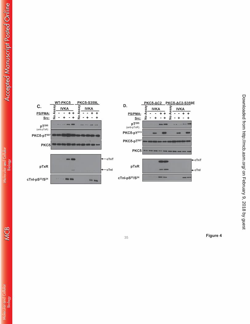
The antithetical effects of S359A and S359E substitutions on PKCδ’s TxR kinase activity could
suggest a role for a negative charge (e.g., phosphorylation) at this site. However, Fig 4C shows that a
position 359 bulky/uncharged Leu substitution also abrogates cTnI-Thr144 and cTnT-TxR motif
phosphorylation, without disrupting PKCδ autocatalytic activity or cTnI-Ser23/Ser24 kinase activity. These
results argue that the steric bulk of a position 359 phosphate group (rather than the negative charge)
disrupts PKCδ activity toward cTnI-Thr144 and cTnT-TxR motifs.
We introduced a S359E substitution into the PKCδ-ΔC2 backbone to test whether the C2 domain
controls PKCδ catalytic activity indirectly by regulating Ser359 phosphorylation. Fig 4D shows that PKCδ-
ΔC2 and PKCδ-ΔC2-S359E display similar cTnI-Ser23/Ser24 kinase activities. PKCδ-ΔC2 (which is not
detectably phosphorylated at Ser359) displays high levels of cTnI-Thr144 and cTnT-TxR motif activity.
cTnI-Thr144 and cTnT-TxR motif phosphorylation is completely abrogated by a S359E substitution in the
PKCδ-ΔC2 backbone.
These studies (summarized in Table 1) implicate Ser359 as a novel PKCδ phosphorylation site that
plays a heretofore unrecognized role to control phosphorylation of TxR motifs on cTnI and cTnT (but not
cTnI phosphorylation at Ser23/Ser24). However, these newer results also raise a question regarding the
mechanism underlying our original observation that Src increases WT-PKCδ’s in vitro TxR activity; Src-
phosphorylated WT-PKCδ acquires TxR activity without any gross change in its Ser359-phosphorylation
status in the IVKAs (an artificial context that does not include cellular phosphatases). The simplest -
most parsimonious – explanation for this finding is that WT-PKCδ TxR activity arises from a pool of
enzyme that is recovered without G-loop Ser359 phosphorylation. This formulation is consistent with [a]
the MRM-MS studies that detect, but could not quantify, the unphoshorylated Ser359 peptide and [b] the
mutagenesis studies showing that TxR activity is completely abrogated by the phosphomimetic or bulky
Ser359 substitutions, indicating that an unphosphorylated - or alanine substituted - Ser359 in the G-loop is
on February 9, 2018 by guest
http://mcb.asm
.org/D
ownloaded from
necessary for TxR activity. In fact, the data in Fig 3 showing that WT-PKCδ displays a very low level of
Src-independent TxR activity when incubations are pushed to very prolonged intervals suggest that TxR
activity reflects both the fraction of total enzyme that is recovered without Ser359 phosphorylation and its
overall catalytic activity – and that Src increases WT-PKCδ’s TxR activity by enhancing its catalytic
activity (independent of any effect on Ser359 phosphorylation). We performed additional studies with the
PKCδ-Y313F mutant enzyme (which prevents phosphorylation by Src but does not influence G loop
Ser359 phosphorylation; Fig 7C) to test this hypothesis. The observation that a Y313F substitution
decreases PKCδ autophosphorylation at Thr295 (53±7% relative to WT-PKCδ; p<0.05, n=3) indicates that
a Src driven conformational change - that may or may not involving the C2 domain-pTyr313 docking
interaction – contributes to the mechanism for maximal activation of WT-PKCδ.
A S359A substitution influences the phosphoacceptor preference of PKCδ. While PKCδ is generally
viewed as a Ser/Thr kinase, substrate sequence logos suggest a clear preference for substrates with a Ser
at the phosphoacceptor site (P-site). Of note, structural studies of PKA complexed with ATP and PKI (an
inhibitor peptide that docks to the active site) place the Ser in the GKGSFG sequence at the tip of the G-
loop in close proximity to the P-site on substrates (Fig 5A). Our observation that a S359A substitution (at
the tip of the G-loop of PKCδ) selectively increases phosphorylation of TxR (Thr-directed) motifs on
cTnI and cTnT provided a rationale to test whether post-translational modifications (PTMs) at Ser359
specifically regulate substrate phosphorylation at Thr (but not Ser) residues. We compared WT-PKCδ and
PKCδ-S359A activity toward short peptide substrates based upon the cTnI phosphorylation site at
position 144 - with either Thr or Ser at the P-site. It is worth noting that IVKAs in the previous sections
tracked WT-PKCδ phosphorylation of proteins in the troponin complex; these assays are performed at
limiting/submicromolar substrate concentrations since practical issues related to cost and protein
insolubility preclude the use higher concentrations of protein substrates. In contrast, peptide kinase assays
are performed at a considerably higher substrate concentration (50 μM) and expose a low level of cTnI-
Thr144 phosphorylation by PKCδ. However, Fig 5B shows that WT-PKCδ displays a considerably higher
on February 9, 2018 by guest
http://mcb.asm
.org/D
ownloaded from
level of activity toward the cTnI-Ser144 peptide. Moreover, the S359A substitution increases peptide
kinase activity. PKCδ-S359A phosphorylates both cTnI-Thr144 and cTnI-Ser144 peptides; it does not
discriminate between these peptides. Similar results were obtained in assays with peptides based upon the
Ser133 phosphorylation site in CREB, which is flanked by a different phosphorylation motif (Fig 5C). In
this case, WT-PKCδ and PKCδ-S359A display high levels of activity toward the CREB-Ser133 peptide. A
S133T substitution markedly decreases peptide phosphorylation by WT-PKCδ, but not PKCδ-S359A.
These results implicate PTMs at Ser359 in PKCδ’s G-loop as a mechanism that regulates PKCδ’s P-site
specificity.
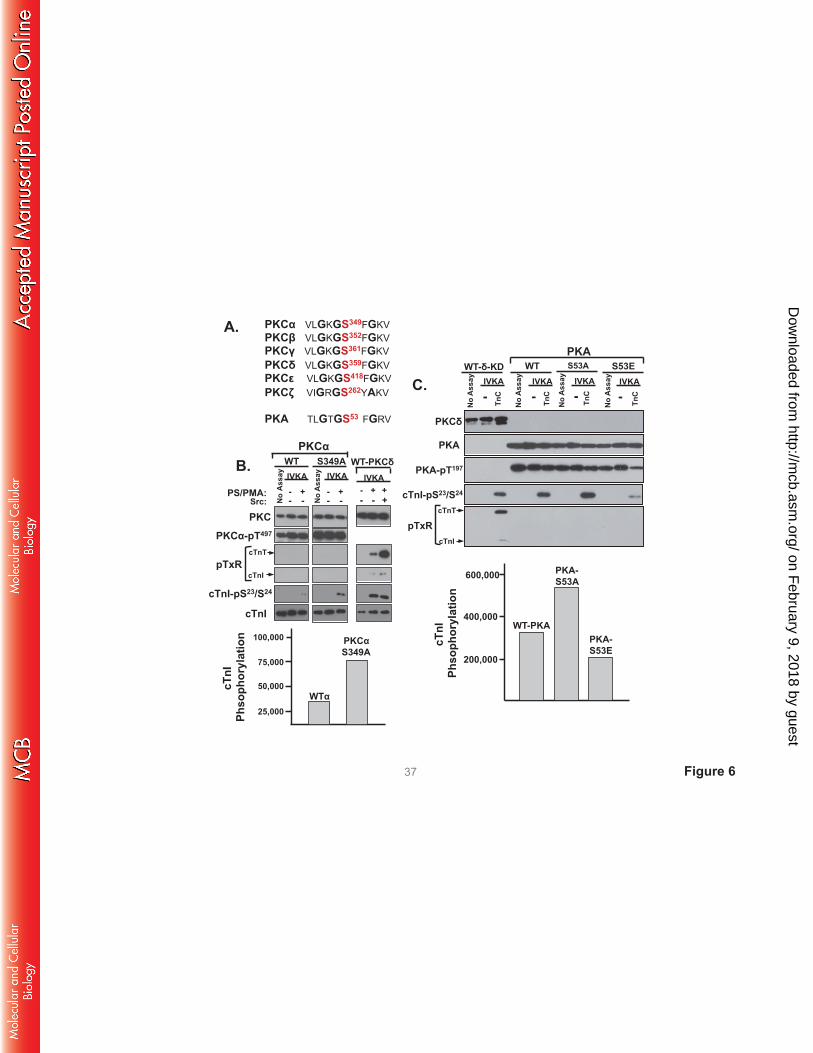
G-loop regulation of PKCα and PKA activities. The G-loop Ser359 phosphorylation site in PKCδ is
highly conserved in other PKCs and in PKA (Fig 6). Since there is recent evidence that this G-loop site is
post-translationally modified in PKCα and PKA (20, 21) and the functional role of this site in PKCα and
PKA remains uncertain, we examined whether G-loop substitutions influence PKCα or PKA catalytic
activity. Figs 6B and 6C show that single residue substitutions in the G-loops of PKCα (at Ser349) or PKA
(at Ser53) do not influence enzyme expression or priming/activation loop phosphorylation. WT-PKCα
displays a modest amount of activity toward cTnI-Ser23/Ser24 in assays with PS/PMA (Fig 6B). The
S349A substitution increases PKCα’s lipid-dependent cTnI-Ser23/Ser24 kinase activity, without imparting
activity toward cTnI-Thr144 or cTnT-TxR motifs (and it does not confer lipid-independent catalytic
activity). Control studies performed in parallel show that a similar level of cTnI-Ser23/Ser24
phosphorylation by lipid-/Src-activated PKCδ is associated with considerable amounts of cTnI-Thr144 and
cTnT-TxR activity. Fig 6C shows that a S53A substitution increases - and a S53E substitution decreases -
cTnI-Ser23/Ser24 phosphorylation by PKA. WT and mutant PKA constructs do not phosphorylate TxR
sites on cTnI or cTnT. Collectively, these studies implicate G-loop PTMs as general regulators of PKCδ,
PKCα, and PKA activity. In each case, S→A substitutions increase - and S→E substitutions decrease -
activity. However, the detailed nature of the regulatory control is kinase-specific. The G-loop S→A
substitution confers TxR motif activity on PKCδ, but not PKCα or PKA.
on February 9, 2018 by guest
http://mcb.asm
.org/D
ownloaded from
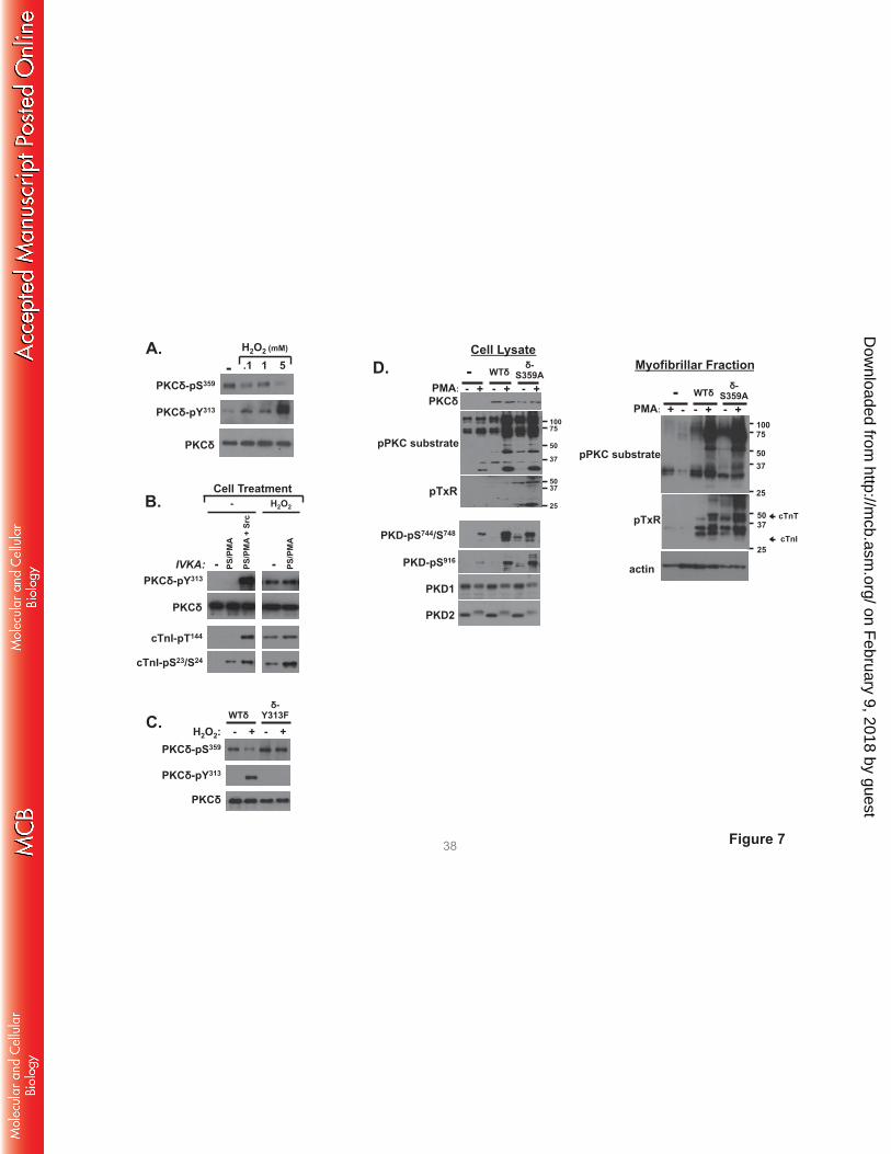
H2O2 decreases PKCδ-Ser359 phosphorylation in cardiomyocytes; PKCδ-S359A displays an
activated phenotype in cardiomyocytes. We examined whether PKCδ-Ser359 phosphorylation is
regulated in cardiomyocytes. Fig 7A shows that endogenous PKCδ is recovered from cardiomyocytes as
a Ser359 phosphorylated enzyme and that PKCδ-Ser359 phosphorylation decreases in association with the
H2O2-dependent increase in PKCδ phosphorylation at Tyr313. PKCδ-pSer359 immunoreactivity is reduced
by 74±4% relative to levels in resting cardiomyocytes following treatment with 5 mM H2O2 (n=4,
p<0.05). Fig 7B shows that the reciprocal H2O2-dependent changes in PKCδ-Tyr313 and Ser359
phosphorylation are associated with the acquisition of lipid-independent cTnI-Ser23/Ser24 and cTnI-Thr144
kinase activities. Fig 7C uses a heterologous overexpression strategy to establish a link between the
H2O2-dependent changes in Tyr313 and Ser359 phosphorylation. Fig 7C shows that the H2O2-dependent
decrease in Ser359 phosphorylation is abrogated by a Y313F substitution.
We used an overexpression strategy with WT-PKCδ and PKCδ-S359A followed by
immunoblotting with the anti-PKC substrate PSSA (as a general screen for target protein
phosphorylation), anti-pTxR (a specific screen for phosphorylation at Thr phosphoacceptor sites), and
antibodies that recognize activating phosphorylations on protein kinase D (PKD, a PKCδ-activated
effector (22)) to examine the functional importance of Ser359 in a cellular context. Immunoblotting
studies on cell lysates show that WT-PKCδ or PKCδ-S359A overexpression leads to an increase in PMA-
dependent responses (Fig 7D). However, PKCδ-S359A uniquely acts as a constitutively-activated
enzyme to enhance basal anti-PKC substrate PSSA immunoreactivity and phosphorylation/activation of
PKD (Fig 7D). PKCδ-S359A overexpression also increases substrate phosphorylation at TxR motifs.
However, pTxR motif immunoreactivity was detected on only a limited number of proteins in cell lysates.
Therefore, we also assessed phosphorylation in the myofibrillar fraction. Fig 7D shows that WT-PKCδ
and PKCδ-S359A overexpression leads to a marked increase in basal- and PMA-dependent anti-PKC
substrate PSSA immunoreactivity in the myofibrillar fraction: PKC substrate motif phosphorylation is
higher in cardiomyocytes that overexpress PKCδ-S359A, compared to cardiomyocytes that overexpress
WT-PKCδ. The myofibrillar fractions also contain considerable amounts of pTxR immunoreactivity.
on February 9, 2018 by guest
http://mcb.asm
.org/D
ownloaded from
pTxR immunoreactivity (including on a band that comigrates with cTnT) is considerably higher in the
myofibrillar fraction from cardiomyocytes that overexpress PKCδ-S359A, compared to WT-PKCδ.
DISCUSSION
PKCδ was originally described as a signaling kinase that mediates growth factor-dependent
cellular responses. The original allosteric model of PKCδ activation focused on translocation events that
deliver the enzyme in an active conformation to DAG-enriched membranes. This model assumes that the
cellular actions of PKCδ are membrane-delimited and that PKCδ’s catalytic activity is an inherent
property of the enzyme that is not altered by the activation process. However, subsequent studies exposed
alternative lipid-independent mechanisms for PKCδ activation that result in the generation of distinct
molecular forms of PKCδ with altered catalytic properties in various subcellular compartments (not just
in DAG-enriched membranes). We previously reported that oxidative stress leads to the activation of Src
and accumulation of a Tyr313-phosphorylated form of PKCδ - with altered catalytic properties - in
cardiomyocytes (7, 9). This study identifies the mechanism that links Tyr313 phosphorylation to changes
in catalytic activity by showing that the Tyr313-phosphorylated hinge region functions as a docking site for
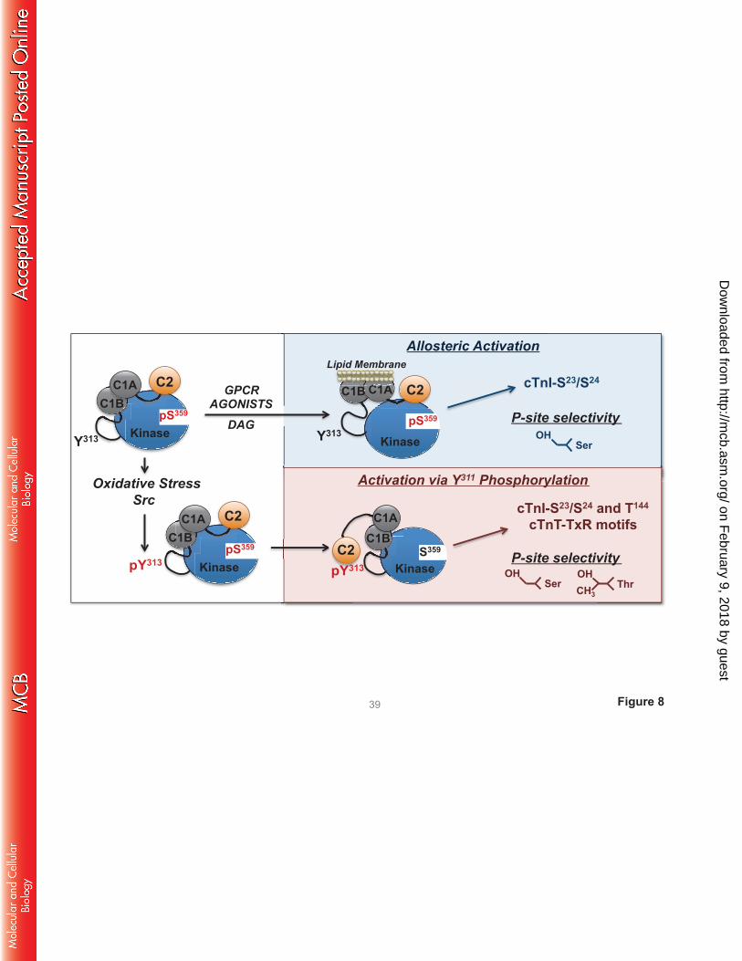
the pTyr binding C2 domain of PKCδ (schematized in Fig 8).
We used a PKCδ-ΔC2 deletion mutant to examine the functional consequences of C2 domain-
mediated docking interactions. First, we showed that full-length PKCδ phosphorylates cTnI-Thr144 and
cTnT-TxR motifs when Tyr313-phosphorylated by Src. In contrast, PKCδ-ΔC2 possesses a high level of
Src-independent cTnI-Thr144 and cTnT-TxR motif activity. These results suggest that the C2 domain
functions as an autoinhibitory regulator of certain full-length PKCδ activities - and that a docking
interaction between the C2 domain and the Tyr313-phosphorylated hinge region induces a global
conformational change that relieves this C2 domain-mediated autoinhibitory constraint. Second, we
showed that the C2 domain regulates PKCδ activity by controlling phosphorylation at Ser359. WT-PKCδ
displays a high level of Ser359 phosphorylation. Studies using two different experimental approaches
(multiple reaction monitoring-mass spectrometry and Western blotting with a PSSA) show that PKCδ-
on February 9, 2018 by guest
http://mcb.asm
.org/D
ownloaded from
ΔC2 is recovered from cells without Ser359 phosphorylation, indicating that Ser359 phosphorylation is
disrupted by the C2 domain deletion. Mutagenesis studies implicate this Ser359 phosphorylation defect as
the mechanism underlying PKCδ-ΔC2’s altered catalytic activity. These results suggest that the C2
domain in the inactive enzyme is positioned to protect Ser359 from cellular phosphatases – and that a C2
domain deletion or a mechanism that displaces the C2 domain from this site (such as the docking
interaction with the Tyr313-phosphorylated hinge region) decreases Ser359 phosphorylation. This model is
supported by cell based studies showing that the H2O2-dependent increase in Tyr313 phosphorylation is
associated with a decrease in Ser359 phosphorylation on WT-PKCδ - but not the PKCδ-Y313F mutant.
The G-loop sequence is highly conserved in many protein kinases. Structural studies of PKA and
several PKCs show that the G-loop forms a flexible clamp that orients the γ-phosphate of ATP for
transfer to substrate; localized changes in the position of this loop influence the kinetics of nucleotide
binding and the phosphoryl transfer reaction. The Ser in the GKGSFG sequence in PKA is strategically
positioned at the tip of the G-loop at the entrance to the ATP-binding cleft, in close proximity to the P-site
on substrates (Fig 6A). This site in the G-loop of several other kinases has been implicated in the control
of catalytic activity. For example, CDK1 (a regulator of the eukaryotic cell cycle) is maintained in an
inactive conformation by inhibitory phosphorylations at Y15 and to a lesser extent the adjacent T14 in its
ATP-binding GEGTYG motif; dephosphorylation of these sites at the onset of mitosis leads to enzyme
activation (23). The non-oncogenic c-ABL proto-oncogene is activated by a G-loop GGGQ(Y253F)G
substitution (24); point mutations in Bcr-Abl (the constitutively active tyrosine kinase that drives
malignant transformation in chronic myeloid leukemia) that alter G-loop Y253 phosphorylation confer
drug-resistance and enhanced oncogenicity (25, 26). Finally, there is evidence that c-Raf is maintained in
a basal autoinhibited state through G-loop autophosphorylation and that c-Raf becomes activated (leading
to a paradoxical activation of the mitogen-activated protein kinase pathway) by ATP-competitive Raf
inhibitors that prevent G-loop autoinhibitory phosphorylation (27). However, post-translational
modifications at the G-loop that regulate PKC (or PKA) activities have never been reported. This study
identifies G-loop regulation of two key aspects of PKCδ’s enzymology:
on February 9, 2018 by guest
http://mcb.asm
.org/D
ownloaded from
[1] Phosphomimetic or bulky substitutions at Ser359 inhibit PKCδ phosphorylation of TxR motifs
on cTnI and cTnT, but not cTnI phosphorylation at Ser23/Ser24. Ser359 phosphorylation could in theory
influence PKCδ’s substrate-specificity indirectly, by restructure the catalytic pocket to accommodate a
wider range of phosphorylation motifs or by modulating docking interactions that control substrate
alignment in the catalytic pocket. However, experiments with short peptide substrates harboring single
residue P-site substitutions show that a phosphomimetic substitution at position 359 prevents
phosphorylation of Thr-phosphoacceptor sites (presumably due to steric clash with the Thr methyl group);
Ser-phosphoacceptor sites are not affected. Hence, while recent literature has focused on consensus
phosphorylation motifs (residues that are either preferred or deselected at positions flanking the P-site on
a particular substrate) as the key determinant of enzyme-substrate specificity, our studies show that PKCδ
activity also can vary depending upon whether the P-site is a Ser or Thr residue. The novelty of this
observation deserves emphasis. While Chen et al. recently identified the “DFG+1” residue in the
activation segment as a structural determinant that defines the inherent P-site specificity of various
Ser/Thr kinases (28), a dynamically regulated post-translational modification that can alter P-site
specificity in a stimulus-specific manner is unprecedented. This study implicates ATP-binding loop
phosphorylation at Ser359 as a mechanism that dynamically regulates the P-site specificity of PKCδ.
[2] A S359A substitution imparts a high level of lipid-independent catalytic activity. The PKCδ-
S359A mutant is a constitutively active enzyme; it acts (much like the lipid-independent form of PKCδ
that accumulates in cells subjected to oxidative stress) to phosphorylate substrates throughout the cell, not
just on lipid membranes. This suggests that the ATP-positioning loop site participates in an autoinhibitory
intramolecular interaction and that this intramolecular interaction is dynamically controlled by G-loop
phosphorylation. This result is consistent with recent structural evidence that the basal catalytic activity of
PKCβII is limited by an autoinhibitory interaction between the ATP binding site and the C1 domain (29).
Several aspects of our study have implications for the pathogenesis and/or treatment of clinical
disease. First, the observation that phosphomimetc and bulky hydrophobic amino acid substitutions at
Ser359 produce similar changes in PKCδ activity indicates that regulation is due to steric hindrance from
on February 9, 2018 by guest
http://mcb.asm
.org/D
ownloaded from
the bulky phosphate group, rather than negative charge, at this site. This suggests that other bulky
substitutions might have similar consequences. This may be pertinent to the diabetic state, where both
PKC and increased protein O-GlcNAcylation contribute to cardiac dysfunction (30). Studies to date
suggest that hyperglycemia activates PKC by inducing oxidative stress or increasing de novo DAG
synthesis. However, the evidence that Ser349 in PKCα’s G-loop is a target for O-GlcNAcylation (addition
of a bulky sugar moiety (21)) suggests that PTMs directed at the G-loop might also underlie
hyperglycemic-induced changes in PKC isoform activity.
Second, while PKCδ-ΔC2 was used in this study as a molecular tool to interrogate the structural
determinants that regulate PKCδ catalytic activity, this construct resembles a truncated PKCδ isoform that
is expressed in a developmentally-regulated manner as a result of alternative splicing in mouse testis (31).
Since disease-specific changes in PKCδ mRNA splicing have recently been identified in certain heart
failure phenotypes (32, 33), a truncated PKCδ isoform might be a component of the altered cardiac
transcriptome that contributes to pathological cardiac stress responses.
Finally, this study identifies a protein-protein interaction involving the C2 domain and the Tyr313-
phosphorylated hinge region that calibrates PKCδ’s substrate specificity by controlling phosphorylation at
Ser359. This mechanism is prominent during oxidative stress, where Src activation leads to an increase in
PKCδ-Tyr313 phosphorylation; PKCδ-Tyr313 phosphorylation is not a feature of G protein-coupled
receptor-activated signaling responses. Stimulus-specific differences in PKCδ phosphorylation at Tyr313
and Ser359 that impact on substrate specificity provide a very plausible explanation for PKCδ’s diverse
roles in both the pathogenesis of ischemia-reperfusion injury and as a mediator of ischemic
preconditioning (a cardioprotective mechanism that mitigates ischemic injury (34)). Stimulus-specific
signaling “modes” for PKCδ that link to functionally distinct cellular responses could also explain why
recent efforts to bring a PKCδ inhibitor compound to the clinic for the treatment of ischemic cardiac
injury met with failure; the trial involved a PKCδ inhibitor that was not designed to discriminate between
pools of PKCδ with distinct phosphorylation patterns at Tyr313 and Ser359. Our results suggest that an
inhibitor compound designed to prevent the C2 domain-pTyr313 interaction (and/or Ser359
on February 9, 2018 by guest
http://mcb.asm
.org/D
ownloaded from
dephosphorylation) might selectively block the redox-activated PKCδ-dependent signaling responses that
contribute to ischemic injury and offer significant therapeutic advantage.
ACKNOWLEDGEMENTS
This work was supported by HL77860 (S.F.S) and grants to the Johns Hopkins Innovation Proteomics
Center in Heart Failure (HHSN268201000032C and NHLBI-HV-10-05, J.V.E.). S.S. was supported by
1DP2 CA186752-01 award from the NIH, and the AHA Scientist Development Grant (13SDG14270009).
REFERENCES
1. Stahelin RV, Digman MA, Medkova M, Ananthanarayanan B, Rafter JD, Melowic HR,
Cho W. 2004. Mechanism of diacylglycerol-induced membrane targeting and activation of
protein kinase Cδ. J Biol.Chem 279: 29501-12.
2. Dekker LV, Parker PJ. 1997. Regulated binding of the protein kinase C substrate GAP-43 to the
V0/C2 region of protein kinase C-δ. J Biol Chem 272:12747-12753.
3. Lopez-Lluch G, Bird MM, Canas B, Godovac-Zimmerman J, Ridley A, Segal AW, Dekker
LV. 2001. Protein kinase Cδ-C2-like domain is a binding site for actin and enables actin
redistribution in neutrophils. Biochem.J 359:39-47.
4. Benes CH, Wu N, Elia AE, Dharia T, Cantley LC, Soltoff SP. 2005. The C2 domain of PKCδ
is a phosphotyrosine binding domain. Cell 121:271-280.
5. Konishi H, Yamauchi E, Taniguchi H, Yamamoto T, Matsuzaki H, Takemura Y, Ohmae K,
Kikkawa U, Nishizuka Y. 2001. Phosphorylation sites of protein kinase Cδ in H2O2-treated cells
and its activation by tyrosine kinase in vitro Proc.Natl.Acad.Sci.U.S.A 98:6587-6592.
6. Steinberg SF. 2008. Structural basis of protein kinase C isoform function. Physiol Rev. 88:1341-
1378.
on February 9, 2018 by guest
http://mcb.asm
.org/D
ownloaded from
7. Rybin VO, Guo J, Sabri A, Elouardighi H, Schaefer E, Steinberg SF. 2004. Stimulus-specific
differences in protein kinase C-δ localization and activation mechanisms in cardiomyocytes. J
Biol Chem 279:19350-19361.
8. Rybin VO, Guo J, Gertsberg Z, Feinmark SJ, Steinberg SF. 2008. Phorbol 12-myristate 13-
acetate-dependent protein kinase C-δ-Tyr313 phosphorylation in cardiomyocyte caveolae J Biol
Chem 283:17777-17788.
9. Sumandea MP, Rybin VO, Hinken AC, Wang C, Kobayashi T, Harleton E, Sievert G,
Balke CW, Feinmark SJ, Solaro RJ, Steinberg SF. 2008. Tyrosine phosphorylation modifies
PKCδ-dependent phosphorylation of cardiac troponin I. J Biol Chem 283:22680-22689.
10. Lu W, Finnis S, Xiang C, Lee HK, Markowitz Y, Okhrimenko H, Brodie C. 2007.
Tyrosine313 is phosphorylated by c-Abl and promotes the apoptotic effect of PKCδ in glioma
cells. Biochem.Biophys.Res.Commun. 352:431-436.
11. Nakashima H, Frank GD, Shirai H, Hinoki A, Higuchi S, Ohtsu H, Eguchi K, Sanjay A,
Reyland ME, Dempsey PJ, Inagami T, Eguchi S. 2008. Novel role of protein kinase C-δ Tyr313
phosphorylation in vascular smooth muscle cell hypertrophy by angiotensin II. Hypertension
51:232-238.
12. Rybin VO, Sabri A, Short J, Braz JC, Molkentin JD, Steinberg SF. 2003. Cross-regulation of
novel protein kinase C (PKC) isoform function in cardiomyocytes. Role of PKC-epsilon in
activation loop phosphorylations and PKC-delta in hydrophobic motif phosphorylations. J Biol
Chem 278:14555-14564.
13. Aprigliano O, Rybin VO, Pak E, Robinson RB, Steinberg SF. 1997. β1-and β2-adrenergic
receptors exhibit differing susceptibility to muscarinic accentuated antagonism. Am J Physiol
272:H2726-2735.
14. Rybin VO, Xu X, Lisanti MP, Steinberg SF. 2000. Differential targeting of β-adrenergic
receptor subtypes and adenylyl cyclase to cardiomyocyte caveolae: A mechanism to functionally
regulate the cAMP signaling pathway. J Biol Chem 275:41447-41457.
on February 9, 2018 by guest
http://mcb.asm
.org/D
ownloaded from
15. Zhang P, Kirk JA, Ji W, dos Remedios CG, Kass DA, Van Eyk JE, Murphy AM. 2012.
Multiple reaction monitoring to identify site-specific troponin I phosphorylated residues in the
failing human heart. Circulation 126:1828-1837.
16. Sivaramakrishnan S, Spudich JA. 2011. Systematic control of protein interaction using a
modular ER/K alpha-helix linker. Proc Natl Acad Sci U S A 108:20467-20472.
17. Ritt M, Guan JL, Sivaramakrishnan S. 2013. Visualizing and manipulating focal adhesion
kinase regulation in live cells. J Biol Chem 288:8875-8886.
18. Sivaramakrishnan S, Spink BJ, Sim AY, Doniach S, Spudich JA. 2008. Dynamic charge
interactions create surprising rigidity in the ER/K alpha-helical protein motif. Proc Natl Acad Sci
U S A 105:13356-13361.
19. Rybin VO, Guo J, Harleton E, Feinmark SJ, Steinberg SF. 2009. Regulatory
autophosphorylation sites on protein kinase C-delta at Thr141 and Thr295. Biochemistry 48:4642-
4651.
20. Seidler J, Adal M, Kubler D, Bossemeyer D, Lehmann WD. 2009. Analysis of
autophosphorylation sites in the recombinant catalytic subunit alpha of cAMP-dependent kinase
by nano-UPLC-ESI-MS/MS. Analytical and bioanalytical chemistry 395:1713-1720.
21. Robles-Flores M, Melendez L, Garcia W, Mendoza-Hernandez G, Lam TT, Castaneda-
Patlan C, Gonzalez-Aguilar H. 2008. Posttranslational modifications on protein kinase C
isozymes. Effects of epinephrine and phorbol esters. Biochim Biophys Acta 1783:695-712.
22. Ozgen N, Obreztchikova M, Guo J, Elouardighi H, Dorn GW, Wilson BA, Steinberg SF.
2008. Protein kinase D links Gq-coupled receptors to cAMP response element-binding protein
(CREB)-Ser133 phosphorylation in the heart. J Biol Chem 283:17009-17019.
23. Norbury C, Blow J, Nurse P. 1991. Regulatory phosphorylation of the p34cdc2 protein kinase
in vertebrates. EMBO J 10:3341-3349.
24. Allen PB, Wiedemann LM. 1996. An activating mutation in the ATP binding site of the ABL
kinase domain. J Biol Chem 271:19585-19591.
on February 9, 2018 by guest
http://mcb.asm
.org/D
ownloaded from
25. Skaggs BJ, Gorre ME, Ryvkin A, Burgess MR, Xie Y, Han Y, Komisopoulou E, Brown LM,
Loo JA, Landaw EM, Sawyers CL, Graeber TG. 2006. Phosphorylation of the ATP-binding
loop directs oncogenicity of drug-resistant BCR-ABL mutants. Proc.Natl.Acad.Sci.U.S.A
103:19466-19471.
26. Griswold IJ, MacPartlin M, Bumm T, Goss VL, O'Hare T, Lee KA, Corbin AS, Stoffregen
EP, Smith C, Johnson K, Moseson EM, Wood LJ, Polakiewicz RD, Druker BJ, Deininger
MW. 2006. Kinase domain mutants of Bcr-Abl exhibit altered transformation potency, kinase
activity, and substrate utilization, irrespective of sensitivity to imatinib. Mol Cell Biol 26:6082-
6093.
27. Holderfield M, Merritt H, Chan J, Wallroth M, Tandeske L, Zhai H, Tellew J, Hardy S,
Hekmat-Nejad M, Stuart DD, McCormick F, Nagel TE. 2013. RAF inhibitors activate the
MAPK pathway by relieving inhibitory autophosphorylation. Cancer cell 23:594-602.
28. Chen C, Ha BH, Thevenin AF, Lou HJ, Zhang R, Yip KY, Peterson JR, Gerstein M, Kim
PM, Filippakopoulos P, Knapp S, Boggon TJ, Turk BE. 2014. Identification of a major
determinant for serine-threonine kinase phosphoacceptor specificity. Mol Cell 53:140-147.
29. Leonard TA, Rozycki B, Saidi LF, Hummer G, Hurley JH. 2011. Crystal structure and
allosteric activation of protein kinase C-βII. Cell 144:55-66.
30. McLarty JL, Marsh SA, Chatham JC. 2013. Post-translational protein modification by O-
linked N-acetyl-glucosamine: Its role in mediating the adverse effects of diabetes on the heart.
Life Sci 92:621-627.
31. Kawaguchi T, Niino Y, Ohtaki H, Kikuyama S, Shioda S. 2006. New PKCδ family members,
PKCδIV, δV, δVI, and δVII are specifically expressed in mouse testis. FEBS Lett 580:2458-
2464.
32. Song HK, Hong SE, Kim T, Kim do H. 2012. Deep RNA sequencing reveals novel cardiac
transcriptomic signatures for physiological and pathological hypertrophy. PloS one 7:e35552.
on February 9, 2018 by guest
http://mcb.asm
.org/D
ownloaded from
33. Ricci M, Xu Y, Hammond HL, Willoughby DA, Nathanson L, Rodriguez MM, Vatta M,
Lipshultz SE, Lincoln J. 2012. Myocardial alternative RNA splicing and gene expression
profiling in early stage hypoplastic left heart syndrome. PloS one 7:e29784.
34. Steinberg SF. 2012. Cardiac actions of protein kinase C isoforms. Physiology (Bethesda)
27:130-139.
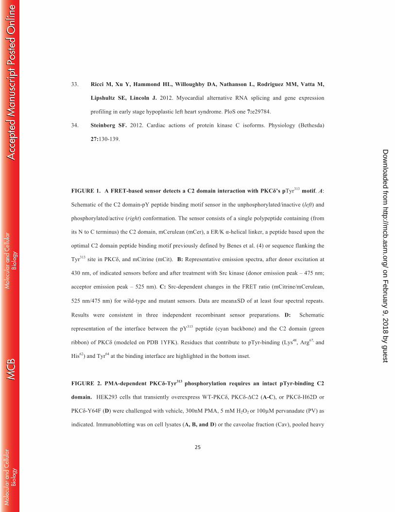
FIGURE 1. A FRET-based sensor detects a C2 domain interaction with PKCδ’s pTyr313 motif. A:
Schematic of the C2 domain-pY peptide binding motif sensor in the unphosphorylated/inactive (left) and
phosphorylated/active (right) conformation. The sensor consists of a single polypeptide containing (from
its N to C terminus) the C2 domain, mCerulean (mCer), a ER/K α-helical linker, a peptide based upon the
optimal C2 domain peptide binding motif previously defined by Benes et al. (4) or sequence flanking the
Tyr313 site in PKCδ, and mCitrine (mCit). B: Representative emission spectra, after donor excitation at
430 nm, of indicated sensors before and after treatment with Src kinase (donor emission peak – 475 nm;
acceptor emission peak – 525 nm). C: Src-dependent changes in the FRET ratio (mCitrine/mCerulean,
525 nm/475 nm) for wild-type and mutant sensors. Data are mean±SD of at least four spectral repeats.
Results were consistent in three independent recombinant sensor preparations. D: Schematic
representation of the interface between the pY313 peptide (cyan backbone) and the C2 domain (green
ribbon) of PKCδ (modeled on PDB 1YFK). Residues that contribute to pTyr-binding (Lys48, Arg67, and
His62) and Tyr64 at the binding interface are highlighted in the bottom inset.
FIGURE 2. PMA-dependent PKCδ-Tyr313 phosphorylation requires an intact pTyr-binding C2
domain. HEK293 cells that transiently overexpress WT-PKCδ, PKCδ-ΔC2 (A-C), or PKCδ-H62D or
PKCδ-Y64F (D) were challenged with vehicle, 300nM PMA, 5 mM H2O2 or 100µM pervanadate (PV) as
indicated. Immunoblotting was on cell lysates (A, B, and D) or the caveolae fraction (Cav), pooled heavy
on February 9, 2018 by guest
http://mcb.asm
.org/D
ownloaded from
gradient fractions (F8-13), and the insoluble pellet (P) that are separated by a detergent-free caveolae
purification scheme (C). It should be noted that the bands representing WT-PKCδ and PKCδ-ΔC2 were
aligned for presentation purposes in Panels A and B; the increased electrophoretic mobility due to the C2
domain deletion was observed in all experiments and can be observed in Panel C. All results were
replicated in 3-6 separate experiments on separate culture preparations. Each panel shows results from a
single gel exposed for a uniform duration.
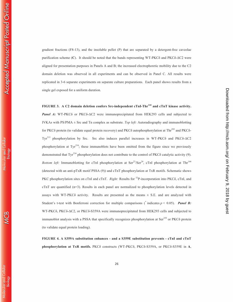
FIGURE 3. A C2 domain deletion confers Src-independent cTnI-Thr144 and cTnT kinase activity.
Panel A: WT-PKCδ or PKCδ-ΔC2 were immunoprecipitated from HEK293 cells and subjected to
IVKAs with PS/PMA ± Src and Tn complex as substrate. Top left: Autoradiography and immunoblotting
for PKCδ protein (to validate equal protein recovery) and PKCδ autophosphorylation at Thr295 and PKCδ-
Tyr313 phosphorylation by Src. Src also induces parallel increases in WT-PKCδ and PKCδ-ΔC2
phosphorylation at Tyr334; these immunoblots have been omitted from the figure since we previously
demonstrated that Tyr334 phosphorylation does not contribute to the control of PKCδ catalytic activity (9).
Bottom left: Immunoblotting for cTnI phosphorylation at Ser23/Ser24, cTnI phosphorylation at Thr144
(detected with an anti-pTxR motif PSSA (9)) and cTnT phosphorylation at TxR motifs. Schematic shows
PKC phosphorylation sites on cTnI and cTnT. Right: Results for 32P-incorporation into PKCδ, cTnI, and
cTnT are quantified (n=3). Results in each panel are normalized to phosphorylation levels detected in
assays with WT-PKCδ activity. Results are presented as the means ± S.E. and are analyzed with
Student’s t-test with Bonferroni correction for multiple comparisons (* indicates p < 0.05). Panel B:
WT-PKCδ, PKCδ-ΔC2, or PKCδ-S359A were immunoprecipitated from HEK293 cells and subjected to
immunoblot analysis with a PSSA that specifically recognizes phosphorylation at Ser359 or PKCδ protein
(to validate equal protein loading).
FIGURE 4. A S359A substitution enhances - and a S359E substitution prevents - cTnI and cTnT
phosphorylation at TxR motifs. PKCδ constructs (WT-PKCδ, PKCδ-S359A, or PKCδ-S359E in A,
on February 9, 2018 by guest
http://mcb.asm
.org/D
ownloaded from
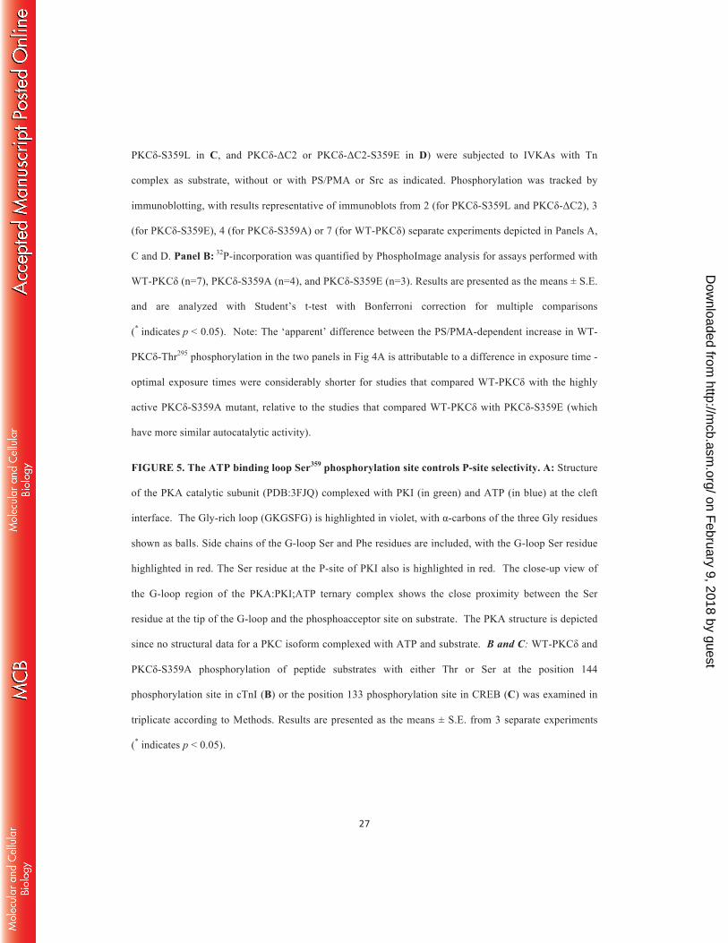
PKCδ-S359L in C, and PKCδ-ΔC2 or PKCδ-ΔC2-S359E in D) were subjected to IVKAs with Tn
complex as substrate, without or with PS/PMA or Src as indicated. Phosphorylation was tracked by
immunoblotting, with results representative of immunoblots from 2 (for PKCδ-S359L and PKCδ-ΔC2), 3
(for PKCδ-S359E), 4 (for PKCδ-S359A) or 7 (for WT-PKCδ) separate experiments depicted in Panels A,
C and D. Panel B: 32P-incorporation was quantified by PhosphoImage analysis for assays performed with
WT-PKCδ (n=7), PKCδ-S359A (n=4), and PKCδ-S359E (n=3). Results are presented as the means ± S.E.
and are analyzed with Student’s t-test with Bonferroni correction for multiple comparisons
(* indicates p < 0.05). Note: The ‘apparent’ difference between the PS/PMA-dependent increase in WT-
PKCδ-Thr295 phosphorylation in the two panels in Fig 4A is attributable to a difference in exposure time -
optimal exposure times were considerably shorter for studies that compared WT-PKCδ with the highly
active PKCδ-S359A mutant, relative to the studies that compared WT-PKCδ with PKCδ-S359E (which
have more similar autocatalytic activity).
FIGURE 5. The ATP binding loop Ser359 phosphorylation site controls P-site selectivity. A: Structure
of the PKA catalytic subunit (PDB:3FJQ) complexed with PKI (in green) and ATP (in blue) at the cleft
interface. The Gly-rich loop (GKGSFG) is highlighted in violet, with α-carbons of the three Gly residues
shown as balls. Side chains of the G-loop Ser and Phe residues are included, with the G-loop Ser residue
highlighted in red. The Ser residue at the P-site of PKI also is highlighted in red. The close-up view of
the G-loop region of the PKA:PKI;ATP ternary complex shows the close proximity between the Ser
residue at the tip of the G-loop and the phosphoacceptor site on substrate. The PKA structure is depicted
since no structural data for a PKC isoform complexed with ATP and substrate. B and C: WT-PKCδ and
PKCδ-S359A phosphorylation of peptide substrates with either Thr or Ser at the position 144
phosphorylation site in cTnI (B) or the position 133 phosphorylation site in CREB (C) was examined in
triplicate according to Methods. Results are presented as the means ± S.E. from 3 separate experiments
(* indicates p < 0.05).
on February 9, 2018 by guest
http://mcb.asm
.org/D
ownloaded from
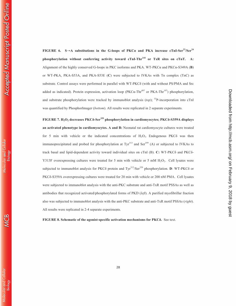
FIGURE 6. S→A substitutions in the G-loops of PKCα and PKA increase cTnI-Ser23/Ser24
phosphorylation without conferring activity toward cTnI-Thr144 or TxR sites on cTnT. A:
Alignment of the highly conserved G-loops in PKC isoforms and PKA. WT-PKCα and PKCα-S349A (B)
or WT-PKA, PKA-S53A, and PKA-S53E (C) were subjected to IVKAs with Tn complex (TnC) as
substrate. Control assays were performed in parallel with WT-PKCδ (with and without PS/PMA and Src
added as indicated). Protein expression, activation loop (PKCα-Thr497 or PKA-Thr197) phosphorylation,
and substrate phosphorylation were tracked by immunoblot analysis (top); 32P-incorporation into cTnI
was quantified by PhosphorImager (bottom). All results were replicated in 2 separate experiments.
FIGURE 7. H2O2 decreases PKCδ-Ser359 phosphorylation in cardiomyocytes; PKCδ-S359A displays
an activated phenotype in cardiomyocytes. A and B: Neonatal rat cardiomyocyte cultures were treated
for 5 min with vehicle or the indicated concentrations of H2O2. Endogenous PKCδ was then
immunoprecipitated and probed for phosphorylation at Tyr313 and Ser359 (A) or subjected to IVKAs to
track basal and lipid-dependent activity toward individual sites on cTnI (B). C: WT-PKCδ and PKCδ-
Y313F overexpressing cultures were treated for 5 min with vehicle or 5 mM H2O2. Cell lysates were
subjected to immunoblot analysis for PKCδ protein and Tyr313/Ser359 phosphorylation. D: WT-PKCδ or
PKCδ-S359A overexpressing cultures were treated for 20 min with vehicle or 200 nM PMA. Cell lysates
were subjected to immunoblot analysis with the anti-PKC substrate and anti-TxR motif PSSAs as well as
antibodies that recognized activated/phosphorylated forms of PKD (left). A purified myofibrillar fraction
also was subjected to immunoblot analysis with the anti-PKC substrate and anti-TxR motif PSSAs (right).
All results were replicated in 2-4 separate experiments.
FIGURE 8. Schematic of the agonist-specific activation mechanisms for PKCδ. See text.
on February 9, 2018 by guest
http://mcb.asm
.org/D
ownloaded from
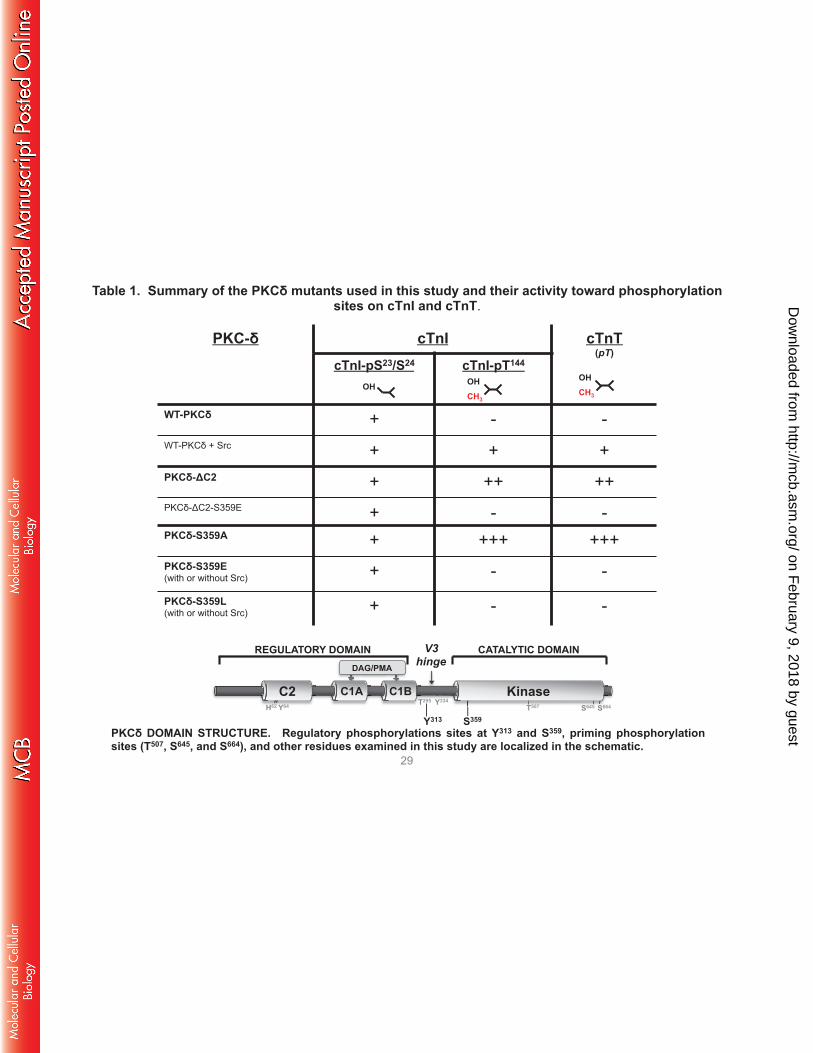
PKC-δ cTnI cTnT (pT)
cTnI-pS23/S24 cTnI-pT144
WT-PKCδ + - - WT-PKCδ + Src + + + PKCδ-ΔC2 + ++ ++ PKCδ-ΔC2-S359E + - - PKCδ-S359A + +++ +++ PKCδ-S359E (with or without Src) + - -
PKCδ-S359L (with or without Src) + - -
OH CH3
OH CH3
OH
Table 1. Summary of the PKCδ mutants used in this study and their activity toward phosphorylation sites on cTnI and cTnT.
29
V3 hinge
Kinase C2 C1A C1B
REGULATORY DOMAIN CATALYTIC DOMAIN DAG/PMA
S359 Y313 T507 S645 S664
Y334 T295 H62 Y64
PKCδ DOMAIN STRUCTURE. Regulatory phosphorylations sites at Y313 and S359, priming phosphorylation sites (T507, S645, and S664), and other residues examined in this study are localized in the schematic.
on February 9, 2018 by guest
http://mcb.asm
.org/D
ownloaded from
His62
Arg67
Lys48pY313
Image created from PDB structure ID 1YRK
C2-Domain
BoundPeptide
Tyr64
450 500 550 600 650
Optimal Y PeptideOptimal Y Peptide with Src
0
0.2
0.4
0.6
0.8
Figure 1 30
on February 9, 2018 by guest
http://mcb.asm
.org/D
ownloaded from
Uninfected PKCδ-ΔC2
PKCδ
PKCδ-pT507
PKCδ-pS645
PKCδ-pS664
PKCδ-pY313
WT-PKCδ A.
B.
PKCδ-pY313
PKCδ
PKCδ-pT507
PKCδ-ΔC2
PMA
WT-PKCδ PV
(min) 5 30 - - PM
A
PV (min)
5 30 - -
Con
t
PMA
H2O
2
Figure 2
Con
t
PMA
H2O
2
Con
t
PMA
H2O
2
PKCδ-pY334
PKCδ-pY334
PKCδ-pY313
PKCδ
PMA
-
δ-H62D WT-δ
PV
PMA
- PV
PKCδ-pY313
PKCδ
PMA
-
δ-Y64F WT-δ
H2O
2
PMA
- H2O
2
31
on February 9, 2018 by guest
http://mcb.asm
.org/D
ownloaded from
(autoradiography)
PKCδ
PKCδ-pT507
PKCδ-pT295
(pTxR)
PKCδ-pY313
PKCδ
cTnI-pS23/S24
cTnI-pT144
WT-PKCδ
Src: Incubation (time): -
- + 15
- + 30
- + 60 -
- + 15
- + 30
- + 60
PKCδ-ΔC2
cTnT-pT
cTnI PVRRRS23S24AN KFKRPT144LR
cTnT T194ERKS198GKRQT203ER KVT284GR
pTxR
Figure 3 32
WT-PKCδ
WT-PKCδ+Src
PKCδ-ΔC2
PKCδ-ΔC2+Src
cTnT
Pho
spho
ryla
tion
cTnI
Pho
spho
ryla
tion
0.5
1.5
1
2
0.5
1.5 1
2 2.5
3.5 2
0.5
1.5
1
2
PKCδ
Phos
phor
ylat
ion
FL-P
KCδ
PKCδ-pS359
PKCδ
A. B.
PKCδ-Δ
C2
*
*
* *
* *
* *
*
PKCδ
PKCδ-pS359
PKCδ
PKCδ-
S359
A
on February 9, 2018 by guest
http://mcb.asm
.org/D
ownloaded from
- PS/P
MA
WT-PKCδ PKCδ-S359A PS
/PM
A+S
rc
Src
No
Ass
ay
IVKA
- PS/P
MA
PS/P
MA
+Src
Src
No
Ass
ay
PKCδ
PKCδ-pT507
pT295
(anti-pTxR)
PKCδ-pY313
cTnI-pS23/S24
pTxR
-
WT-PKCδ PKCδ-S359E
Src
No
Ass
ay
- Src
PS/P
MA
PS/P
MA
+Src
PS/P
MA
PS/P
MA
+Src
No
Ass
ay
IVKA IVKA IVKA
pS-PKC substrate (K/R)-x-pS-x-(K/R) cTnT
A.
cTnI
cTnT
Figure 4 33
on February 9, 2018 by guest
http://mcb.asm
.org/D
ownloaded from
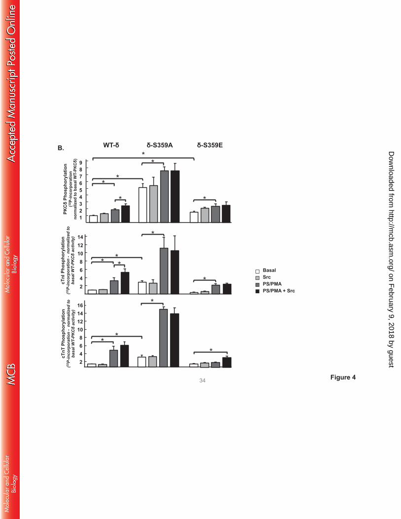
Figure 4 34
WT-δ δ-S359A δ-S359E
Basal Src PS/PMA PS/PMA + Src
PKCδ
Phos
phor
ylat
ion
(32 P
-inco
rpor
atio
n
norm
aliz
ed to
bas
al W
T-PK
Cδ)
cTnI
Pho
spho
ryla
tion
(32 P
-inco
rpor
atio
n -
norm
aliz
ed to
ba
sal W
T-PK
Cδ
activ
ity)
*
12 14
2 4 6 8
10 * *
*
*
*
16
2
6 4
8 10 12 14
* *
cTnT
Pho
spho
ryla
tion
(32 P
-inco
rpor
atio
n -
norm
aliz
ed to
ba
sal W
T-PK
Cδ
activ
ity)
1
3 2
4 5 6 7 8 9
* *
*
*
*
*
*
B.
on February 9, 2018 by guest
http://mcb.asm
.org/D
ownloaded from
D. C.
cTnT
cTnI
PS/PMA: Src:
PKCδ
PKCδ-pT507
pT295
(anti-pTxR)
PKCδ-pY313
cTnI-pS23/S24
pTxR
PKCδ-ΔC2 PKCδ-ΔC2-S359E
No
Ass
ay
No
Ass
ay
-
IVKA -
+ -
- +
+ +
-
IVKA -
+ -
- +
+ +
PKCδ
PKCδ-pT507
pT295 (anti-pTxR)
cTnI-pS23/S24
pTxR
PS/PMA: Src:
WT-PKCδ PKCδ-S359L
No
Ass
ay
No
Ass
ay
-
IVKA -
+ -
- +
+ +
-
IVKA -
+ -
- +
+ +
cTnT
cTnI
Figure 4 35
on February 9, 2018 by guest
http://mcb.asm
.org/D
ownloaded from
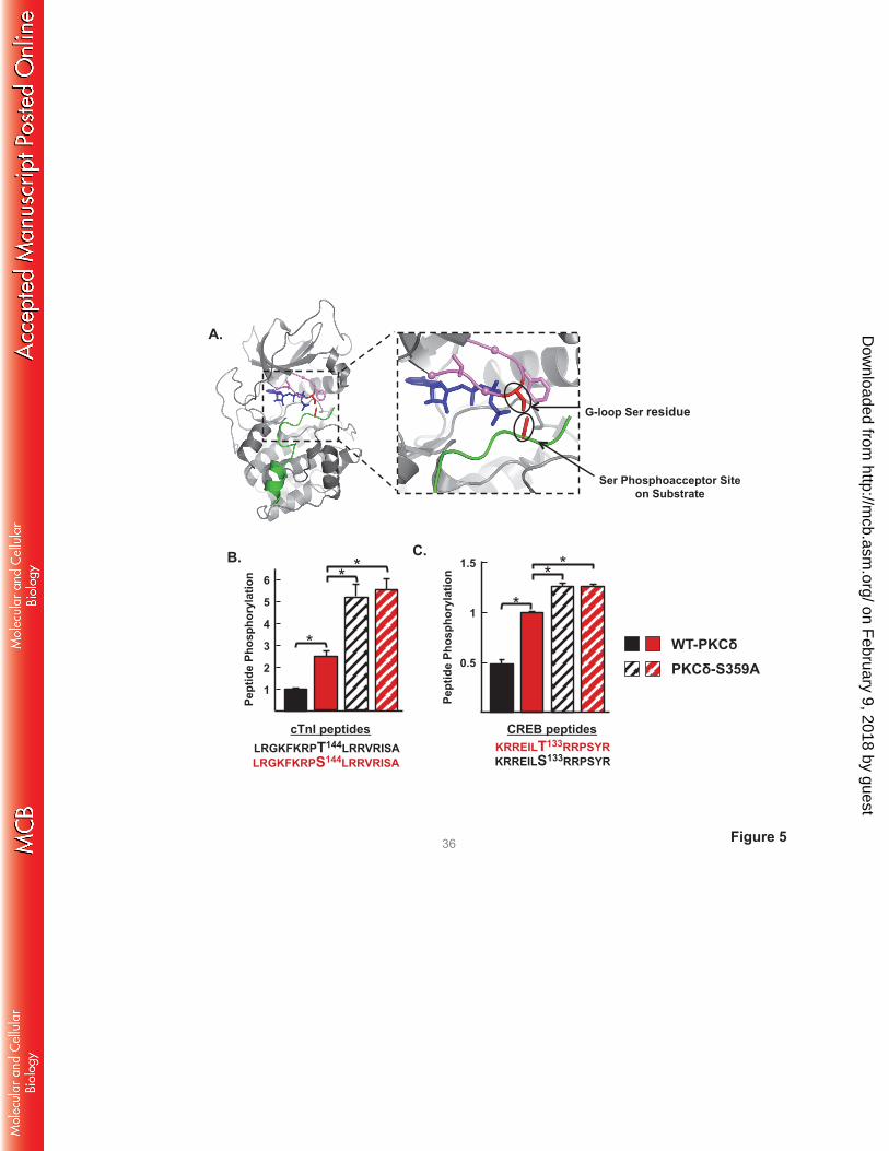
Figure 5
Pept
ide
Phos
phor
ylat
ion
B.
LRGKFKRPT144LRRVRISA LRGKFKRPS144LRRVRISA
cTnI peptides
C.
KRREILS133RRPSYR KRREILT133RRPSYR
CREB peptides
Ser Phosphoacceptor Site on Substrate
G-loop Ser residue
A.
36
1
3
2
4
WT-PKCδ PKCδ-S359A
5
6
Pept
ide
Phos
phor
ylat
ion
0.5
1
1.5 * *
*
*
* *
on February 9, 2018 by guest
http://mcb.asm
.org/D
ownloaded from
No
Ass
ay
No
Ass
ay
No
Ass
ay
No
Ass
ay
PKCα VLGKGS349FGKV PKCβ VLGKGS352FGKV PKCγ VLGKGS361FGKV PKCδ VLGKGS359FGKV PKCε VLGKGS418FGKV PKCζ VIGRGS262YAKV PKA TLGTGS53 FGRV
PKC
PKCα-pT497
cTnI-pS23/S24
WT IVKA
S349A IVKA
WT-PKCδ IVKA
PS/PMA: + - + - + - + Src: - - - - - - + N
o A
ssay
No
Ass
ay
cTnI
PKCα B.
cTnT pTxR
25,000
50,000
75,000
PKCα S349A
100,000
WTα
cTnI
P
hsop
hory
latio
n
200,000
400,000
600,000
WT-PKA
PKA- S53A
PKA- S53E
A.
PKCδ
cTnI-pS23/S24
-
WT
TnC
IVKA
pTxR
-
S53A
TnC
IVKA
-
S53E
TnC
IVKA
- TnC
WT-δ-KD
PKA
PKA-pT197
cTnT
cTnI
IVKA C.
PKA
cTnI
cTnI
P
hsop
hory
latio
n
Figure 6 37
on February 9, 2018 by guest
http://mcb.asm
.org/D
ownloaded from
PKD-pS744/S748
PKD-pS916
pPKC substrate
PKD1
PKD2
PKCδ
WTδ δ-
S359A - PMA: + - + - + -
D.
PKCδ
PKCδ-pS359
PKCδ-pY313
H2O2 (mM)
- .1 1 5
A.
Figure 7
pPKC substrate
pTxR
WTδ δ-
S359A - PMA: - + + - + -
Cell Lysate Myofibrillar Fraction
50 37
25
100 75
50 37
25
cTnT
cTnI
50
37
100 75
50 37
25
pTxR
PKCδ
cTnI-pT144
cTnI-pS23/S24
- H2O2
Cell Treatment
PS/P
MA
PS/P
MA
+ Sr
c
- PS/P
MA
- PKCδ-pY313
IVKA:
38
PKCδ-pS359
PKCδ
PKCδ-pY313
H2O2: - + - +
δ- Y313F WTδ C.
B.
actin
on February 9, 2018 by guest
http://mcb.asm
.org/D
ownloaded from
Allosteric Activation
Activation via Y311 Phosphorylation
Src
Y313 Kinase
C1A
C1B
pY313 Kinase
pS359
pS359
GPCR AGONISTS
DAG
Oxidative Stress
pY31
C2
C1A
C1B
C2
P-site selectivity
cTnI-S23/S24
Y313 Kinase
C1A C1B CCCCC1C A1AAA
Lipid Membrane
pS359 OH
Ser
C2
pY313 Kinase S359
C1A
C1B
C2 P-site selectivity
cTnI-S23/S24 and T144 cTnT-TxR motifs
OH Ser
OH Thr CH3
P
c
Figure 8 39
on February 9, 2018 by guest
http://mcb.asm
.org/D
ownloaded from

![Regulation of Insulin Secretion II MPB333_Ja… · 2 Glucose stimulated insulin secretion (GSIS) [Ca2+] i V m ATP ADP K ATP Ca V GLUT2 mitochondria GK glucose glycolysis PKA Epac](https://static.fdocument.org/doc/165x107/5aebd7447f8b9ae5318e3cc6/regulation-of-insulin-secretion-ii-mpb333ja2-glucose-stimulated-insulin-secretion.jpg)