Adipose rabbit mesenchymal stem cells for the treatment of ... · PDF fileΗ...
19
Adipose rabbit mesenchymal stem cells for the treatment of the chronic scar tissue of the vocal cords Dr. Vasiliki E Kalodimou, Head of Flow Cytometry-Research and Regenerative Medicine Department, IASO-Maternity Hospital, Athens-Greece E-mail: [email protected]
Transcript of Adipose rabbit mesenchymal stem cells for the treatment of ... · PDF fileΗ...
Adipose rabbit mesenchymal stem cells for the treatment
of the chronic scar tissue of the vocal cords
Dr. Vasiliki E Kalodimou,
Head of Flow Cytometry-Research and Regenerative Medicine Department,
IASO-Maternity Hospital, Athens-Greece
E-mail: [email protected]
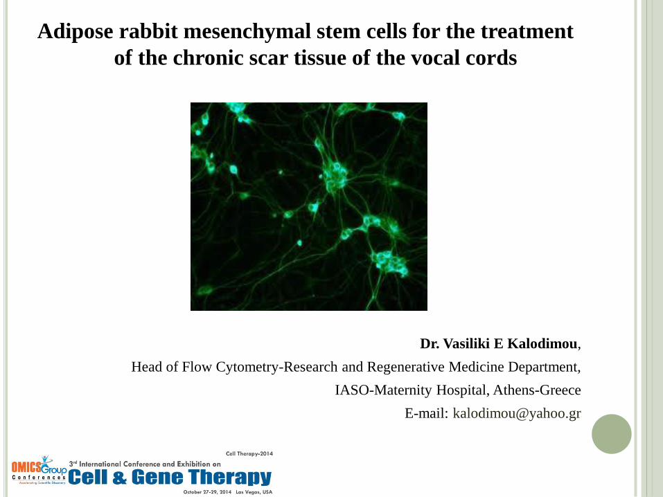
Adipose-derived stem cells (ADSCs), represents a promising approach to future cell-based therapies.
These cells can be readily harvested in large numbers with low donor-site morbidity.
Mesenchymal stem cells are characterized by great “plasticity ", i.e. have the ability to differentiate
to form various cell types.
For this reason it can be used in regenerative medicine to regenerate tissues and organs.
Figure 1: Mesenchymal stem cell differentiation: The MSC's can differentiate into fat, cartilage and bone cells.
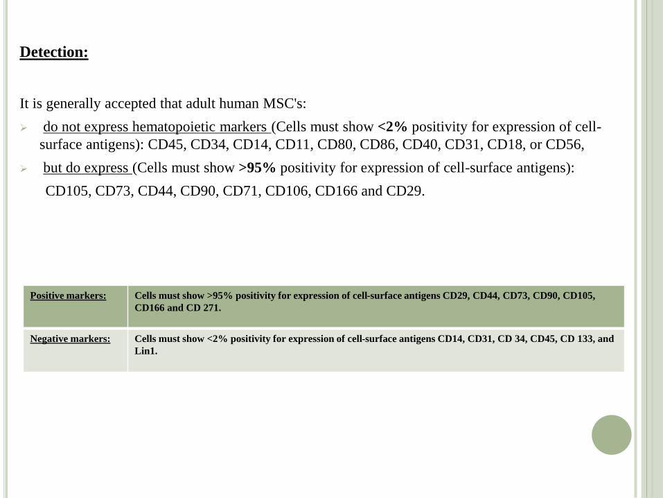
Detection:
It is generally accepted that adult human MSC's:
do not express hematopoietic markers (Cells must show <2% positivity for expression of cell-
surface antigens): CD45, CD34, CD14, CD11, CD80, CD86, CD40, CD31, CD18, or CD56,
but do express (Cells must show >95% positivity for expression of cell-surface antigens):
CD105, CD73, CD44, CD90, CD71, CD106, CD166 and CD29.
Positive markers: Cells must show >95% positivity for expression of cell-surface antigens CD29, CD44, CD73, CD90, CD105,
CD166 and CD 271.
Negative markers: Cells must show <2% positivity for expression of cell-surface antigens CD14, CD31, CD 34, CD45, CD 133, and
Lin1.
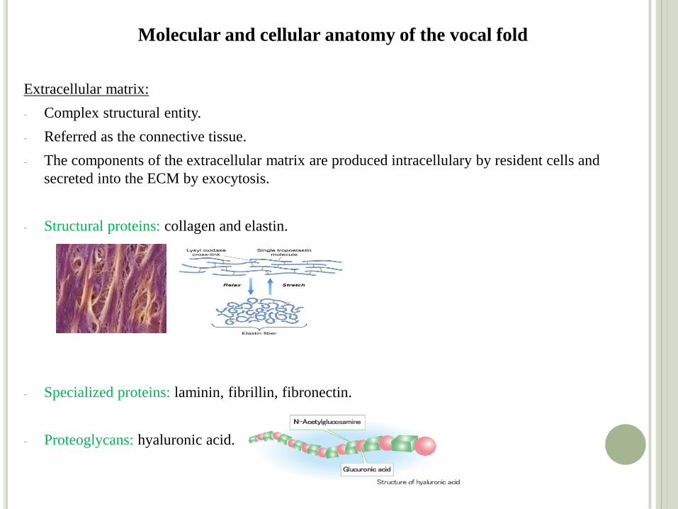
Molecular and cellular anatomy of the vocal fold
Extracellular matrix:
- Complex structural entity.
- Referred as the connective tissue.
- The components of the extracellular matrix are produced intracellulary by resident cells and
secreted into the ECM by exocytosis.
- Structural proteins: collagen and elastin.
- Specialized proteins: laminin, fibrillin, fibronectin.
- Proteoglycans: hyaluronic acid.
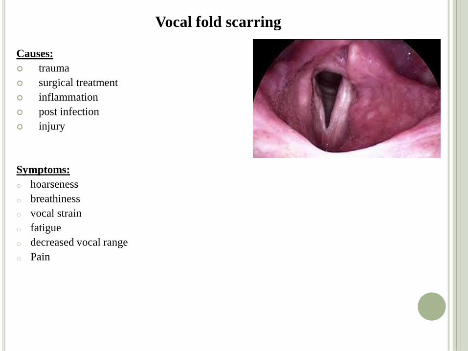
Vocal fold scarring Causes:
trauma
surgical treatment
inflammation
post infection
injury
Symptoms:
o hoarseness
o breathiness
o vocal strain
o fatigue
o decreased vocal range
o Pain
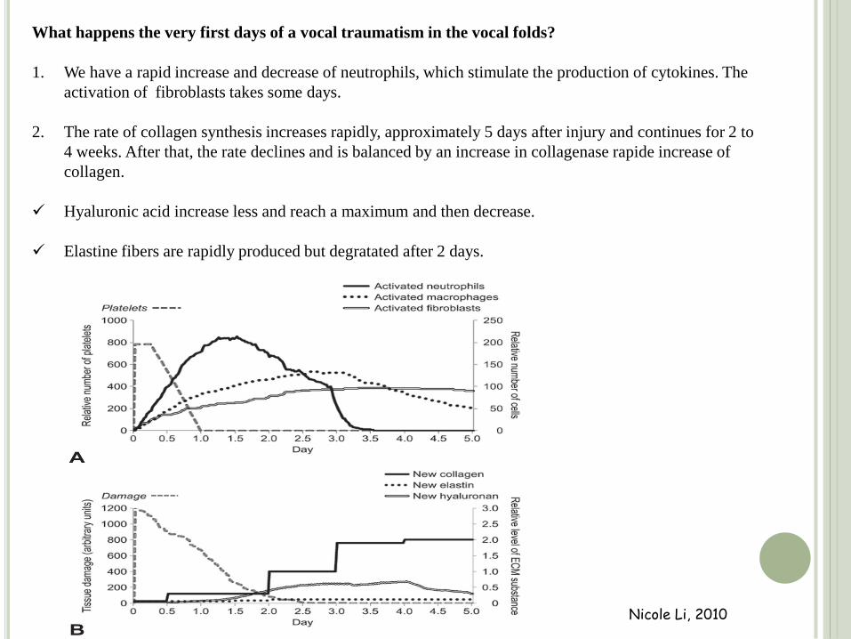
Nicole Li, 2010
What happens the very first days of a vocal traumatism in the vocal folds?
1. We have a rapid increase and decrease of neutrophils, which stimulate the production of cytokines. The
activation of fibroblasts takes some days.
2. The rate of collagen synthesis increases rapidly, approximately 5 days after injury and continues for 2 to
4 weeks. After that, the rate declines and is balanced by an increase in collagenase rapide increase of
collagen.
Hyaluronic acid increase less and reach a maximum and then decrease.
Elastine fibers are rapidly produced but degratated after 2 days.
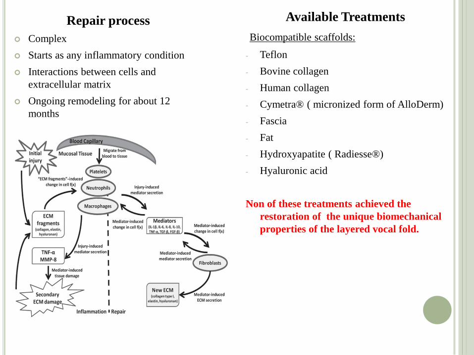
Repair process
Complex
Starts as any inflammatory condition
Interactions between cells and
extracellular matrix
Ongoing remodeling for about 12
months
Available Treatments
Biocompatible scaffolds:
- Teflon
- Bovine collagen
- Human collagen
- Cymetra® ( micronized form of AlloDerm)
- Fascia
- Fat
- Hydroxyapatite ( Radiesse®)
- Hyaluronic acid
Non of these treatments achieved the
restoration of the unique biomechanical
properties of the layered vocal fold.
Animal study
Aim:
To evaluate the effects of injection of autologous adipose mesenchymal stem cells in a
chronic vocal fold scar in rabbits.
Method:
We used 105 New Zealand rabbits around 3 Kg.
Surgical protocol:
The vocal folds of 8 rabbits (16 vocal folds) served as controls.
Under general anesthesia (xylazine, ketamine, diazepam and glycopyrrolate) and direct
laryngoscopy we created a trauma to the vocal fold in the remaining 97 rabbits. The lamina
propria was injured bilaterally with micro scissors by localized resection of the middle
portion of the vocal fold down to the vocal ligament under direct laryngoscopy (endoscope
0, 4mm) with video monitoring.
Post operative follow up:
We perform direct laryngoscopy under general anesthesia at 15 days, 1 month and 3 months
to investigate morphologic changes including surface irregularities, granulation tissue
formation, and atrophy.
Seven (7) rabbits died immediately post operatively and thirteen (13) rabbits died
within one month
slide 9 -video äçìéïõñãéá ïõëçò.mp4
After 18 months we assessed the scar to the vocal folds.
slide 10 - video ïõëç ìåôá áðï 18 ìçíåò.mp4
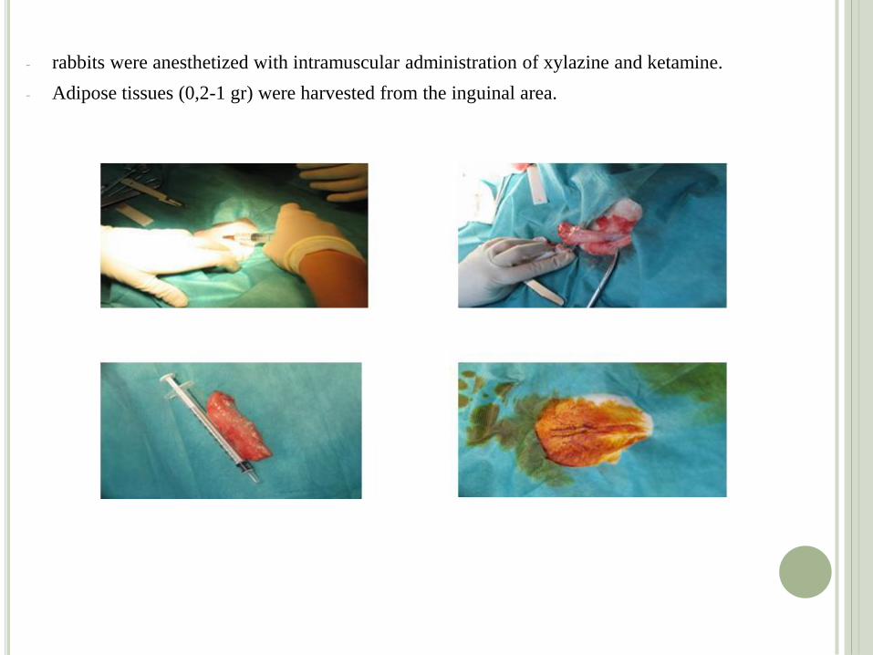
- rabbits were anesthetized with intramuscular administration of xylazine and ketamine.
- Adipose tissues (0,2-1 gr) were harvested from the inguinal area.
Few hours after isolation we proceed with the infusion of 0,1 ml adipose mesenchymal
stem cells into both scarred vocal folds, under sedation and direct laryngoscopy.
slide 12-video åê÷õóç.mp4
We infused hyaluronic acid in 5 rabbits to compare our results with stems cells to
those with the actual treatments proposed.
We infuse 0,1 ml of hyaluronic acid to each scarred vocal fold 18 months after the
trauma of the vocal folds.
slide 13-video åê÷õóç õáëïõñïíéêïõ.mp4
The identification process
Observing the adherent cells under a microscope to study their morphological
characteristics.
Identifying cell surface antigens by flow cytometry (CD 34, CD 44, CD 105, CD 106 and
CD271 specific for rabbits).
We used 7-AAD to asses the viability of the cells which were 98-100%.
Concentrations were 100.000 cells /ml.
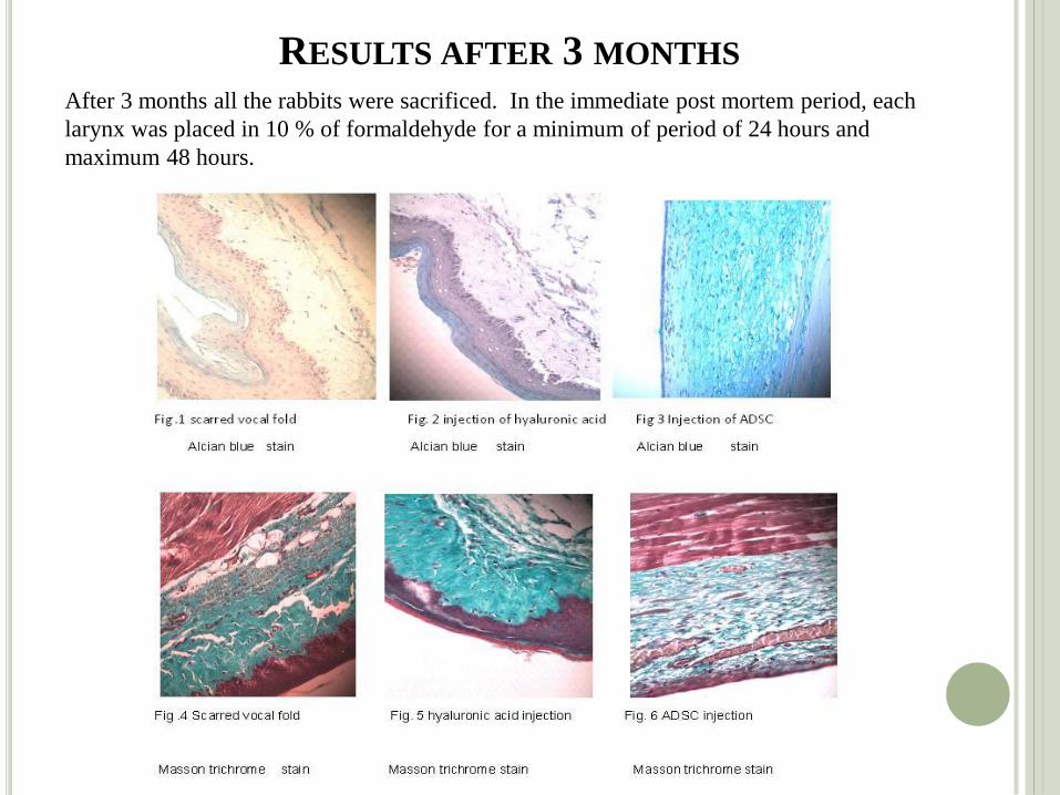
RESULTS AFTER 3 MONTHS After 3 months all the rabbits were sacrificed. In the immediate post mortem period, each
larynx was placed in 10 % of formaldehyde for a minimum of period of 24 hours and
maximum 48 hours.
Lamina propria:
The thickness of the lamina propria is doubled when we have a scar in the vocal fold. This thickness
is preserved when we inject hyaluronic acid. In the contrary, when we inject adipose stem cells,
the thickness of the lamina propria tends to return back to normal values.
Collagen:
In the scar, the fibers of collagen are more, they occupy all the full thickness of the lamina propria
and are disorganized. This structure doesn’t seem to change when we inject hyaluronic acid
Instead, when we inject adipose stem cells the fibers of collagen are less, and organized in a laminar
way, like in a normal vocal fold
Hyaluronic acid:
In a scarred vocal fold, the amount of hyaluronic acid is reduced. Although we inject gel hyaluronic
acid in scarred vocal folds, after three months it seems that it doesn’t integrated in the vocal fold.
When we inject stem cells, the amount of hyaluronic acid returns back to normal.
Benefits:
In past studies the implementation of adipose mesenchymal stem cells had been done 2-5 days after the creation of the vocal cord injury. This means that the implant is in the midst of intense inflammatory reaction. In this study the implantation conducted 18 months after the injury of vocal cord.
Here we compared the effects of:
a) Implanting adipose mesenchymal stem cells
b) infusion of hyaluronic acid &
c) no treatment in the chronic scar tissue.
The study was conducted with a rabbit as an animal model, whose structure of the vocal cords is more like the human vocal chords.
In our study the implanted adipose mesenchymal stem cells derived from autologous fat while in all other studies the implantation derived from bone marrow and the placenta.
The advantages of our method are:
1. easy
2. painless preparation &
3. greater stability
Acknowledgements
Dr. V. Angelou, ENT-Head and neck surgeon, Consultant in “Phoniatric and
Applied Laryngology Center”.
A. Papalois, (PhD, KGSJ), Director of Experimental & Research Center
ELPEN.
A. Zaharioudaki, (DVM, MLAS), Experimental & Research Center ELPEN /
Veterinary department .


![Acute cold exposure-induced down-regulation of CIDEA, cell ... · Cidea is particularly expressed at high levels in the mitochondria of mice brown adipose tissue (BAT) [4]. BAT is](https://static.fdocument.org/doc/165x107/601c3e8d8d3edd79416a1a23/acute-cold-exposure-induced-down-regulation-of-cidea-cell-cidea-is-particularly.jpg)