
0DWHULDO (6, IRU&KHP&RPP From Localization conjugated ... · 6 αjj’, and αkk’ are the...
14
1 Supplementary information for The Second-Harmonic Generation Intensification Derived From Localization conjugated π –orbital in O 2 2- Yunxia Song 丄,†, ‡ , Min Luo 丄,*,† , Fei Liang §, ‡ , Ning Ye *,† , Zheshuai Lin § † Key Laboratory of Optoelectronic Materials Chemistry and Physics, Fujian Institute of Research on the Structure of Matter, Chinese Academy of Sciences, Fuzhou, Fujian, 350002, P. R. China § Center for Crystal Research and Development, Technical Institute of Physics and Chemistry, Chinese Academy of Sciences, Beijing, 100190, China. ‡ University of Chinese Academy of Sciences, Beijing 100049, China Email: [email protected] Email: [email protected] Electronic Supplementary Material (ESI) for ChemComm. This journal is © The Royal Society of Chemistry 2018
Transcript of 0DWHULDO (6, IRU&KHP&RPP From Localization conjugated ... · 6 αjj’, and αkk’ are the...
1
Supplementary information for
The Second-Harmonic Generation Intensification Derived
From Localization conjugated π –orbital in O22-
Yunxia Song 丄,†, ‡, Min Luo 丄,*,†, Fei Liang§, ‡, Ning Ye*,†, Zheshuai Lin§
†Key Laboratory of Optoelectronic Materials Chemistry and Physics, Fujian Institute
of Research on the Structure of Matter, Chinese Academy of Sciences, Fuzhou, Fujian, 350002, P. R. China
§Center for Crystal Research and Development, Technical Institute of Physics and
Chemistry, Chinese Academy of Sciences, Beijing, 100190, China.
‡ University of Chinese Academy of Sciences, Beijing 100049, China
Email: [email protected]: [email protected]
Electronic Supplementary Material (ESI) for ChemComm.This journal is © The Royal Society of Chemistry 2018
2
Table of contents
Sections Titles pages
Section S1. Materials and Methods (Synthesis, Instrumentation, and Computational Details)
S2–S6
Table S1. Crystallographic Data for K3[V(O2)2O]CO3 S7
Table S2. Atomic Coordinates and Equivalent Isotropic Displacement Parameters for K3[V(O2)2O]CO3
S7
Table S3. Selected Bond Distances (Å) for K3[V(O2)2O]CO3 S8
Table S4 The NLO effects of carbonates with coplannar [CO3] groups S9
Table S5 Atom-cutting analysis and calculated SHG coefficients (pm/V) for K3[V(O2)2O](CO3) and K3VO3(CO3) at 1064 nm
S9
Figure S1. Photograph of K3[V(O2)2O](CO3) crystal S10
Figure S2. X-ray powder diffraction patterns of K3[V(O2)2O]CO3. (a) crystal sample and (b) simulation results
S10
Figure S3. TGA and DTA curves of K3[V(O2)2O]CO3 S11
Figure S4. (a)Diffuse reflectance and (b) transmittance spectrum of K3[V(O2)2O]CO3 S11
Figure S5 The calculated band gap of K3[V(O2)2O]CO3. S12
References
S13
3
Section S1. Materials and Methods
Synthesis and determination of sample.
All of the chemicals were of analytical grade from commercial sources and used without further purification. K2CO3 (99.8%), V2O5 (99.0%) and H2O2 (30%) were purchased from Adamas. V2O5 (0.5g), K2CO3 (2.5g) and 1.5ml H2O2(30%) were dissolved in 20 ml distilled water. The solution was transfered to a refrigerator and
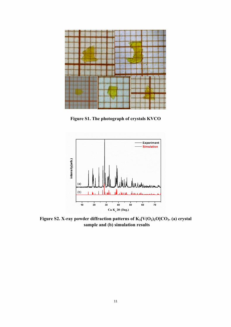
vaporized at 5-10℃. After about several weeks, yellow plate-like crystals were
obtained (See Figure S1).
Single crystal X-ray diffraction data were collected at room temperature on a Bruker Smart APEX II CCD area detector with graphite-monochromatic Mo K α radiation (λ = 0.71073 Å). A yellow plate-like crystal was mounted on a glass fiber with epoxy for structure determination. The intensity data sets were corrected with the ω -scan technique. The data were integrated with the CrystalClear program. The intensities were corrected for Lorentz polarization, air absorption, and absorption attributable to the variation in the path length through the detector faceplate. Absorption corrections were also applied based on the Multiscan technique. The structure was solved with direct methods, refined with difference Fourier maps and full-matrix least-squares fitting on F2 with SHELXL- 971. Moreover, all nonhydrogen atoms were refined with anisotropic thermal parameters. The structure was verified using the ADDSYM algorithm from the program PLATON2, and no higher symmetries were found. The details of the crystallographic data and structure refinement information for K3[V(O2)2O]CO3 are listed in Table S1. Atomic coordinates and isotropic displacement coefficients are listed in Table S1, and bond lengths are listed in Table S2.
Powder X-ray Diffraction.
X-ray diffraction patterns of polycrystalline materials were obtained on a Rigaku Dmax2500 powder X-ray diffractometer by using Cu Kα radiation (λ=1.540598 Å) at room temperature in the angular range of 2θ = 5-75° with a scan step width of 0.05° and a fixed time of 0.2s.
Thermal Analysis.
Thermogravimetric analyses (TGA) and differential scanning calorimetry (DSC) were measured on a NETZCH STA 449F3. Reference (Al2O3) and crystal samples (3-10 mg) were enclosed in Al2O3 crucibles and heated from room temperature to 1100 °C at a rate of 10 °C/min under a constant flow of nitrogen gas.
Diffuse reflectance and transmittance spectroscopy
UV-vis-NIR spectrophotometer of the powder sample was measured at room temperature in the range of 200–2500 nm with BaSO4 as the standard of 100%
4
reflectance. The reflectance spectrum was transformed into the absorbance spectrum using the Kubelka–Munk function3,4. The transmittance spectrum was measured from 400 to 600 nm using an unpolished KVCO crystal.
Second-Harmonic Generation.
Polycrystalline second-harmonic generation (SHG) signals were measured using the method source by Kurtz and Perry5. Since SHG efficiencies are known to depend strongly on particle size, polycrystalline samples were ground and sieved into the following particle size ranges: 25-45, 45-62, 62-75, 75-109, 109-150, and 150-212 μm. The samples were pressed between glass microscope cover slides and secured with tape in 1-mm thick aluminum holders containing an 8-mm diameter hole. The samples were then placed in a light-tight box and irradiated with a pulsed laser. The measurements were performed with a Q-switched Nd:YAG laser at 1064nm. A cutoff filter was used to limit background flash-lamp light on the sample, and an interference filter (530±10nm) was used to select the second harmonic for detection with a photomultiplier tube attached to a RIGOL DS1052E 50-MHz oscilloscope.
Computational Descriptions.
First-Principles Calculations. The first-principles calculations are performed by the psuedopotential6 methods
implemented in the CASTEP package7 based on the density functional theory (DFT).8 The optimized norm-conserving pseudopotentials9 are used to simulate ion-electron interactions for all constituent elements. A kinetic energy cutoff of 660 eV is chosen with Monkhorst-Pack k-point meshes (3×3×5) spanning less than 0.04/Å3 in the Brillouin zone.10 The cell parameters and the atomic positions in the unit cell of KVCO are fully fixed. The convergence threshold for SCF tolerance is set as 1.0×10⁻⁹ eV/atom. Based on the experimental geometry structure, the imaginary part of the dielectric function is calculated and the real part of the dielectric function is determined using the Kramers–Kronig transform,11 and then the refractive indices n and the birefringence ∆n are obtained. The shortest SHG output wavelength λPM is calculated based on the dispersion curves of refractive index (e.g., no and ne), satisfying the condition of no (2λPM) = ne (λPM). The second order susceptibility χ(2) and SHG coefficient dij (dij = 1/2 χ(2)) is calculated using an expression originally proposed by Rashkeev et al12 and developed by Lin et al:13
𝜒𝑖𝑗𝑘= 𝜒𝑖𝑗𝑘(𝑉𝐸) + 𝜒𝑖𝑗𝑘(𝑉𝐻) + 𝜒𝑖𝑗𝑘(𝑡𝑤𝑜𝑏𝑎𝑛𝑑𝑠) (1)
where (VE), (VH) and (two bands) denote the contributions from virtual-𝜒𝑖𝑗𝑘 𝜒𝑖𝑗𝑘 𝜒𝑖𝑗𝑘
electron processes, virtual-hole processes and two band processes, respectively. In
detail, the formulae for calculating (VE), (VH) and (two bands) are as 𝜒𝑖𝑗𝑘 𝜒𝑖𝑗𝑘 𝜒𝑖𝑗𝑘
follows:

5
𝜒𝑖𝑗𝑘(𝑉𝐸) =
𝑒3
2ℏ2𝑚3∑𝑣𝑐𝑐'∫𝑑3𝑘
4𝜋3𝑃(𝑖𝑗𝑘)𝐼𝑚[𝑝 𝑖
𝑣𝑐𝑝𝑗𝑐𝑐'𝑝
𝑘𝑐'𝑣]( 1
𝜔 3𝑐𝑣𝜔
2𝑣𝑐'
+2
𝜔 4𝑣𝑐𝜔𝑐'𝑣
), (2)
𝜒𝑖𝑗𝑘(𝑉𝐻) =
𝑒3
2ℏ2𝑚3∑𝑣𝑣'𝑐∫𝑑3𝑘
4𝜋3𝑃(𝑖𝑗𝑘)𝐼𝑚[𝑝 𝑖
𝑣𝑣'𝑝𝑗𝑣'𝑐𝑝
𝑘𝑐𝑣]( 1
𝜔 3𝑐𝑣𝜔
2𝑣'𝑐
+2
𝜔 4𝑣𝑐𝜔𝑐𝑣'
), (3)
𝜒𝑖𝑗𝑘(𝑡𝑤𝑜𝑏𝑎𝑛𝑑𝑠) =𝑒3
ℏ2𝑚3∑𝑣𝑐∫𝑑3𝑘
4𝜋3𝑃(𝑖𝑗𝑘)
𝐼𝑚[𝑝 𝑖𝑣𝑐𝑝
𝑗𝑐𝑣(𝑝 𝑘
𝑣𝑣 ‒ 𝑝𝑘𝑐𝑐)]
𝜔 5𝑣𝑐
(4)
Here, i, j and k are Cartesian components; v and v’ denote VB, and c and c’ denote CB. P(ijk) denotes full permutation. It is noted that the refractive indices and SHG coefficients can be accurately obtained by DFT in principle because these optical properties are determined by the virtual electronic excited processes, which are described by the first and second order perturbations on the ground state wave functions, respectively.
Furthermore, it should be emphasized that the generalized gradient approximation (GGA) method with PBE functional14 usually heavily underestimates the energy bandgap Eg. Herein, the scissors-corrected15 GGA method is employed to calculate the optical properties, where the scissors operator is set as the difference between the experimental and GGA bandgaps. This self-consistent ab initio approach has been proven to be an efficient way for the investigation of linear and NLO properties in many types of NLO materials without introducing any experimental parameter.16 Meanwhile, the linear response method is employed to obtain the phonon dispersion of crystal.17 The LO-TO phonon frequency splitting at the Γ-point is also included in the phonon calculations. The dispersion separation of 0.01/Å3 is adopted to make sure the good convergence.
In the real-space atom-cutting technique,13 the contribution of ion A to the nth-order susceptibility (denoted as χ(n)(A)) is obtained by cutting all ions except A from the original wave functions χ(n)(A) = χ(n)(all ions except A are cut). In this technique, the “cutting” charge densities are set to be spherical. In fact, this strategy to calculate the respective contribution of ions or groups to the SHG coefficients is not very rigorous, since in the real situation the charge densities around the concerning ions or groups are not spherical. Therefore, as the spherical charge densities are “cut”, some charge densities belonging to the concerning ions or groups are very likely to be untouched. It is the repeated calculations of these left charge densities that make the sum of ligand contributions often exceed the total dij.
The Anionic Group Theory Calculation. The macroscopic second-order susceptibility χ (2) could be expressed by Eq. 3 according to the anionic group theory.
(5)i'j'k'
(2 (2)ii' jj' kk'
p i'j'k'(P), ijk
FxV
)
where F is the correction factor of the localized field, V is the volume of the unit cell,αii’,
6
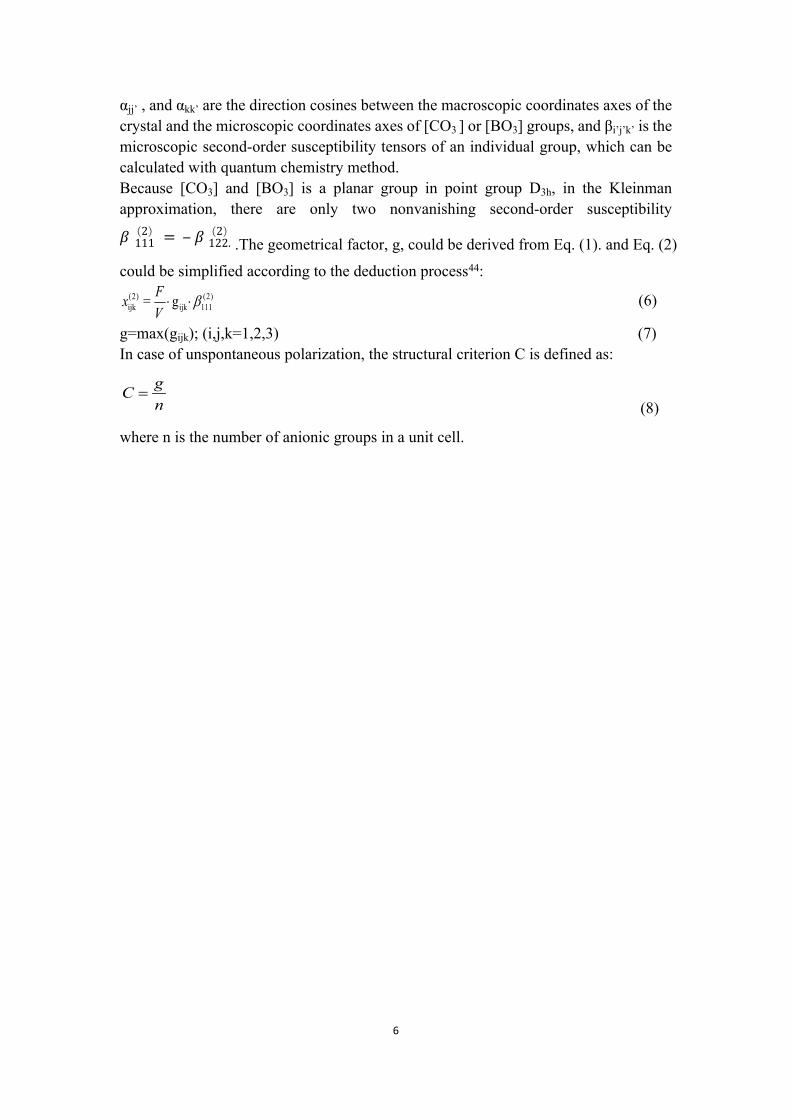
αjj’ , and αkk’ are the direction cosines between the macroscopic coordinates axes of the crystal and the microscopic coordinates axes of [CO3 ] or [BO3] groups, and βi’j’k’ is the microscopic second-order susceptibility tensors of an individual group, which can be calculated with quantum chemistry method.Because [CO3] and [BO3] is a planar group in point group D3h, in the Kleinman approximation, there are only two nonvanishing second-order susceptibility
.The geometrical factor, g, could be derived from Eq. (1). and Eq. (2) 𝛽(2)111 = ‒ 𝛽(2)122.
could be simplified according to the deduction process44:
(6)(2) (2)ijk ijk 111= gFx
V
g=max(gijk); (i,j,k=1,2,3) (7)In case of unspontaneous polarization, the structural criterion C is defined as:
(8)gCn
where n is the number of anionic groups in a unit cell.

7
8
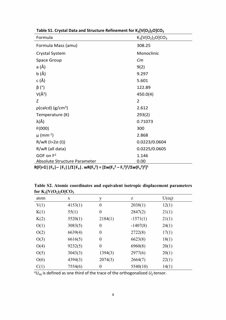
Table S1. Crystal Data and Structure Refinement for K3[V(O2)2O]CO3
Formula K3[V(O2)2O]CO3
Formula Mass (amu) 308.25
Crystal System MonoclinicSpace Group Cma (Å) 9(2)b (Å) 9.297c (Å) 5.601β (°) 122.89V(Å3) 450.0(4)Z 2ρ(calcd) (g/cm3) 2.612Temperature (K) 293(2)λ(Å) 0.71073F(000) 300μ (mm-1) 2.868 R/wR (I>2σ (I)) 0.0223/0.0604R/wR (all data) 0.0225/0.0605GOF on F2 1.146Absolute Structure Parameter 0.00
R(F)=Σ||Fo|– |Fc||/Σ|Fo|. wR(Fo2) = [Σw(Fo
2 – Fc2)2/Σw(Fo
2)2]1
Table S2. Atomic coordinates and equivalent isotropic displacement parameters for K3[V(O2)2O]CO3
atom x y z U(eq)V(1) 4153(1) 0 2038(1) 12(1)K(1) 55(1) 0 2847(2) 21(1)K(2) 5520(1) 2184(1) -1571(1) 21(1)O(1) 3083(5) 0 -1407(8) 24(1)O(2) 6639(4) 0 2722(8) 17(1)O(3) 6616(5) 0 6623(8) 18(1)O(4) 9232(5) 0 6960(8) 20(1)O(5) 3043(3) 1394(3) 2977(6) 20(1)O(6) 4394(3) 2074(3) 2664(7) 22(1)C(1) 7554(6) 0 5540(10) 14(1)
aUeq is defined as one third of the trace of the orthogonalized Uij tensor.
9
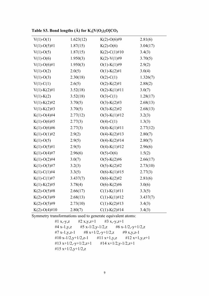
Table S3. Bond lengths (Å) for K3[V(O2)2O]CO3
V(1)-O(1) 1.623(12) K(2)-O(6)#9 2.81(6)V(1)-O(5)#1 1.87(15) K(2)-O(6) 3.04(17)V(1)-O(5) 1.87(15) K(2)-C(1)#10 3.4(3)V(1)-O(6) 1.950(3) K(2)-V(1)#9 3.70(5)V(1)-O(6)#1 1.950(3) O(1)-K(1)#9 2.9(2)V(1)-O(2) 2.0(5) O(1)-K(2)#1 3.0(4)V(1)-O(3) 2.30(18) O(2)-C(1) 1.326(7)V(1)-C(1) 2.6(5) O(2)-K(2)#1 2.88(2)V(1)-K(2)#1 3.52(18) O(2)-K(1)#11 3.0(7)V(1)-K(2) 3.52(18) O(3)-C(1) 1.28(17)V(1)-K(2)#2 3.70(5) O(3)-K(2)#3 2.68(13)V(1)-K(2)#3 3.70(5) O(3)-K(2)#2 2.68(13)K(1)-O(4)#4 2.77(12) O(3)-K(1)#12 3.2(3)K(1)-O(6)#5 2.77(3) O(4)-C(1) 1.3(3)K(1)-O(6)#6 2.77(3) O(4)-K(1)#11 2.77(12)K(1)-O(1)#2 2.9(2) O(4)-K(2)#13 2.80(7)K(1)-O(5) 2.9(5) O(4)-K(2)#14 2.80(7)K(1)-O(5)#1 2.9(5) O(4)-K(1)#12 2.96(6)K(1)-O(4)#7 2.96(6) O(5)-O(6) 1.5(2)K(1)-O(2)#4 3.0(7) O(5)-K(2)#6 2.66(17)K(1)-O(3)#7 3.2(3) O(5)-K(2)#2 2.73(10)K(1)-C(1)#4 3.3(5) O(6)-K(1)#15 2.77(3)K(1)-C(1)#7 3.437(7) O(6)-K(2)#2 2.81(6)K(1)-K(2)#5 3.78(4) O(6)-K(2)#6 3.0(6)K(2)-O(5)#8 2.66(17) C(1)-K(1)#11 3.3(5)K(2)-O(3)#9 2.68(13) C(1)-K(1)#12 3.437(7)K(2)-O(5)#9 2.73(10) C(1)-K(2)#13 3.4(3)K(2)-O(4)#10 2.80(7) C(1)-K(2)#14 3.4(3)
Symmetry transformations used to generate equivalent atoms: #1 x,-y,z #2 x,y,z+1 #3 x,-y,z+1 #4 x-1,y,z #5 x-1/2,y-1/2,z #6 x-1/2,-y+1/2,z #7 x-1,y,z-1 #8 x+1/2,-y+1/2,z #9 x,y,z-1 #10 x-1/2,y+1/2,z-1 #11 x+1,y,z #12 x+1,y,z+1 #13 x+1/2,-y+1/2,z+1 #14 x+1/2,y-1/2,z+1 #15 x+1/2,y+1/2,z
10
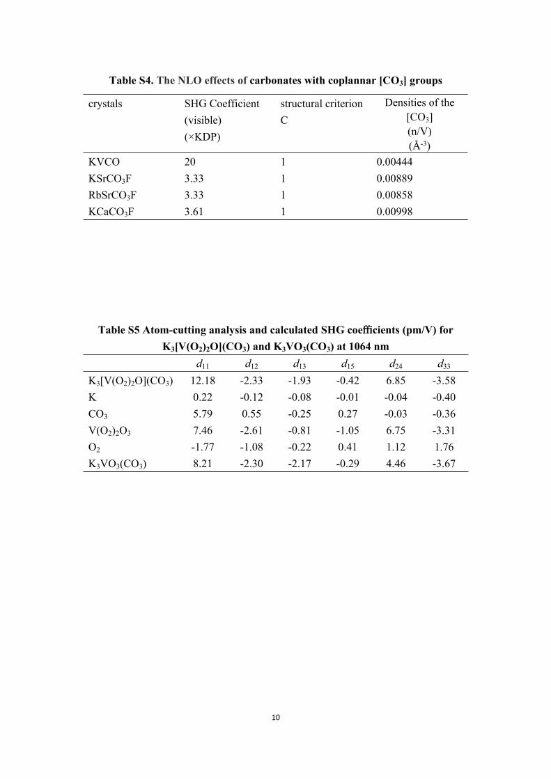
Table S4. The NLO effects of carbonates with coplannar [CO3] groups
crystals SHG Coefficient(visible)(×KDP)
structural criterionC
Densities of the [CO3](n/V)(Å-3)
KVCO 20 1 0.00444KSrCO3F 3.33 1 0.00889RbSrCO3F 3.33 1 0.00858KCaCO3F 3.61 1 0.00998
Table S5 Atom-cutting analysis and calculated SHG coefficients (pm/V) for K3[V(O2)2O](CO3) and K3VO3(CO3) at 1064 nm
d11 d12 d13 d15 d24 d33
K3[V(O2)2O](CO3) 12.18 -2.33 -1.93 -0.42 6.85 -3.58K 0.22 -0.12 -0.08 -0.01 -0.04 -0.40CO3 5.79 0.55 -0.25 0.27 -0.03 -0.36V(O2)2O3 7.46 -2.61 -0.81 -1.05 6.75 -3.31O2 -1.77 -1.08 -0.22 0.41 1.12 1.76K3VO3(CO3) 8.21 -2.30 -2.17 -0.29 4.46 -3.67
11
Figure S1. The photograph of crystals KVCO
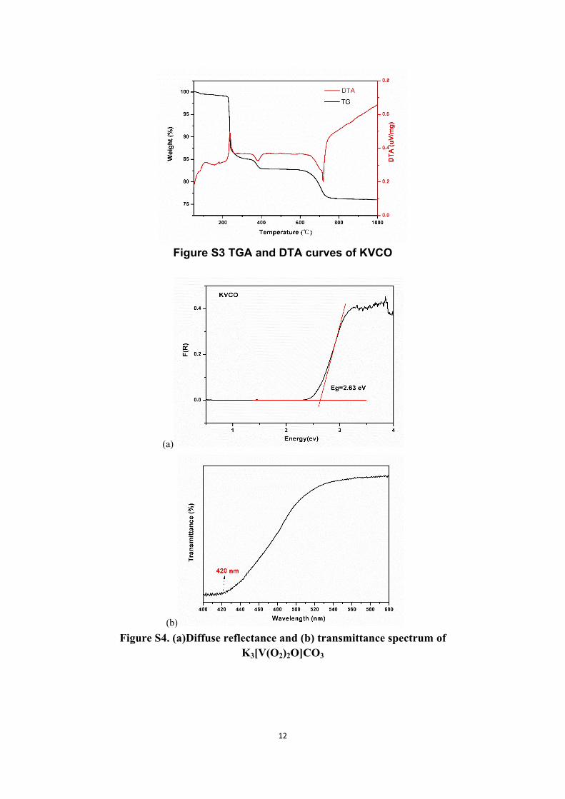
Figure S2. X-ray powder diffraction patterns of K3[V(O2)2O]CO3. (a) crystal sample and (b) simulation results
12
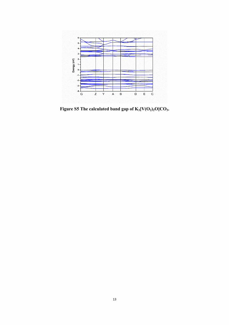
Figure S3 TGA and DTA curves of KVCO
(a)
(b)Figure S4. (a)Diffuse reflectance and (b) transmittance spectrum of
K3[V(O2)2O]CO3
13
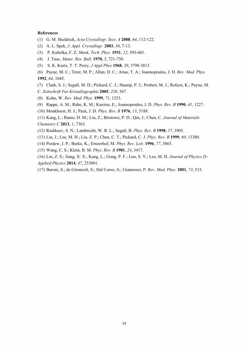
Figure S5 The calculated band gap of K3[V(O2)2O]CO3.
14
References (1) G. M. Sheldrick, Acta Crystallogr. Sect. A 2008, 64, 112-122.(2) A. L. Spek, J. Appl. Crystallogr. 2003, 36, 7-13.(3) P. Kubelka, F. Z. Munk, Tech. Phys. 1931, 12, 593-601.(4) J. Tauc, Mater. Res. Bull. 1970, 5, 721-730.(5) S. K. Kurtz, T. T. Perry, J Appl Phys 1968, 39, 3798-3813.(6) Payne, M. C.; Teter, M. P.; Allan, D. C.; Arias, T. A.; Joannopoulos, J. D. Rev. Mod. Phys. 1992, 64, 1045.(7) Clark, S. J.; Segall, M. D.; Pickard, C. J.; Hasnip, P. J.; Probert, M. J.; Refson, K.; Payne, M. C. Zeitschrift Fur Kristallographie 2005, 220, 567.(8) Kohn, W. Rev. Mod. Phys. 1999, 71, 1253.(9) Rappe, A. M.; Rabe, K. M.; Kaxiras, E.; Joannopoulos, J. D. Phys. Rev. B 1990, 41, 1227.(10) Monkhorst, H. J.; Pack, J. D. Phys. Rev. B 1976, 13, 5188.(11) Kang, L.; Ramo, D. M.; Lin, Z.; Bristowe, P. D.; Qin, J.; Chen, C. Journal of Materials Chemistry C 2013, 1, 7363.(12) Rashkeev, S. N.; Lambrecht, W. R. L.; Segall, B. Phys. Rev. B 1998, 57, 3905.(13) Lin, J.; Lee, M. H.; Liu, Z. P.; Chen, C. T.; Pickard, C. J. Phys. Rev. B 1999, 60, 13380.(14) Perdew, J. P.; Burke, K.; Ernzerhof, M. Phys. Rev. Lett. 1996, 77, 3865.(15) Wang, C. S.; Klein, B. M. Phys. Rev. B 1981, 24, 3417.(16) Lin, Z. S.; Jiang, X. X.; Kang, L.; Gong, P. F.; Luo, S. Y.; Lee, M. H. Journal of Physics D-Applied Physics 2014, 47, 253001.(17) Baroni, S.; de Gironcoli, S.; Dal Corso, A.; Giannozzi, P. Rev. Mod. Phys. 2001, 73, 515.