Αντιµ επιπλοκών αγγειοπλαστικής · Guidewire fracture (1%) ... •In...
93
X. ΓΡΑΪΔΗΣ Επεμβατικός καρδιολόγος, FSCAI Kλινική Euromedica-Κυανούς Σταυρός, Θεσσαλονίκη Αντιμετώπιση επιπλοκών αγγειοπλαστικής
Transcript of Αντιµ επιπλοκών αγγειοπλαστικής · Guidewire fracture (1%) ... •In...
X. ΓΡΑΪΔΗΣ Επεµβατικός καρδιολόγος,
FSCAI Kλινική Euromedica-Κυανούς
Σταυρός, Θεσσαλονίκη
Αντιµετώπιση επιπλοκών αγγειοπλαστικής
Prevention is better than treatment….
Complications: The never ending story ?
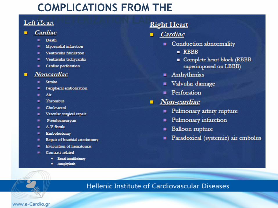
COMPLICATIONS FROM THE CATHETERIZATION LAB
PCI complications……
❖Over 2million PCI procedures
worldwide
❖Angiographic success rate of 82-98%
❖Overall major complications rates
1-5%
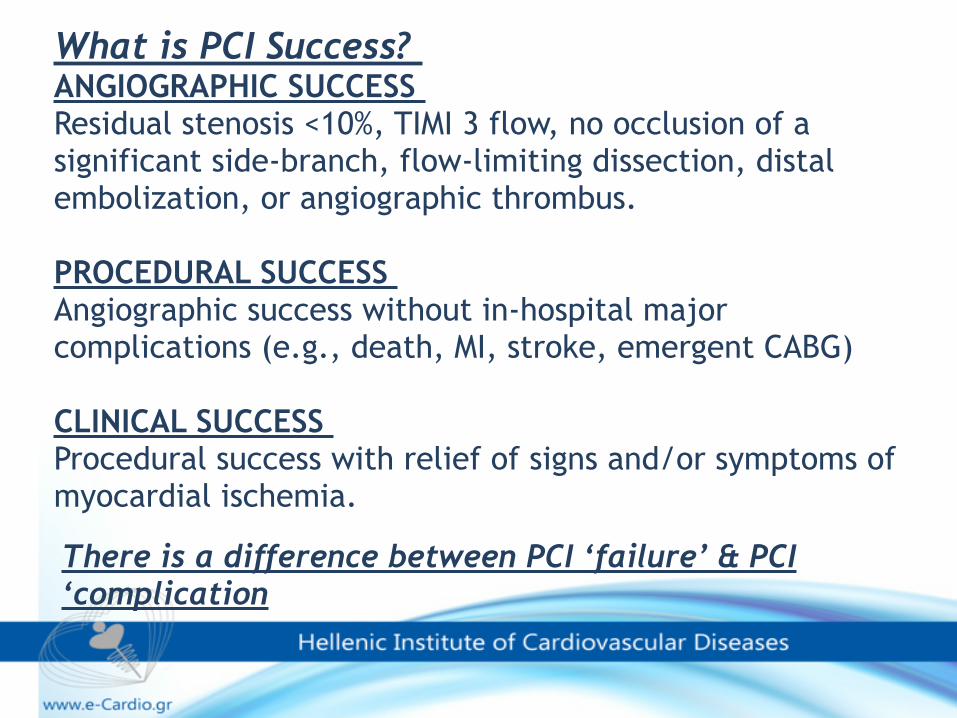
There is a difference between PCI ‘failure’ & PCI ‘complication
What is PCI Success? ANGIOGRAPHIC SUCCESS Residual stenosis <10%, TIMI 3 flow, no occlusion of a significant side-branch, flow-limiting dissection, distal embolization, or angiographic thrombus.
PROCEDURAL SUCCESS Angiographic success without in-hospital major complications (e.g., death, MI, stroke, emergent CABG)
CLINICAL SUCCESS Procedural success with relief of signs and/or symptoms of myocardial ischemia.
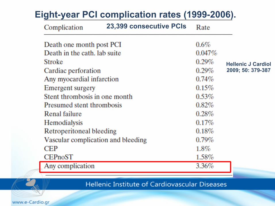
Eight-year PCI complication rates (1999-2006).23,399 consecutive PCIs
Hellenic J Cardiol 2009; 50: 379-387
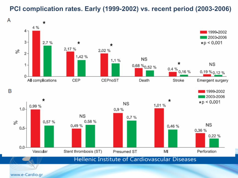
PCI complication rates. Early (1999-2002) vs. recent period (2003-2006)
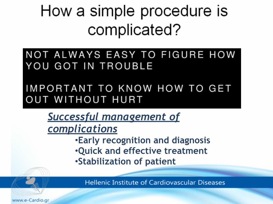
Successful management of complications
•Early recognition and diagnosis •Quick and effective treatment •Stabilization of patient
A team approach to managing complications
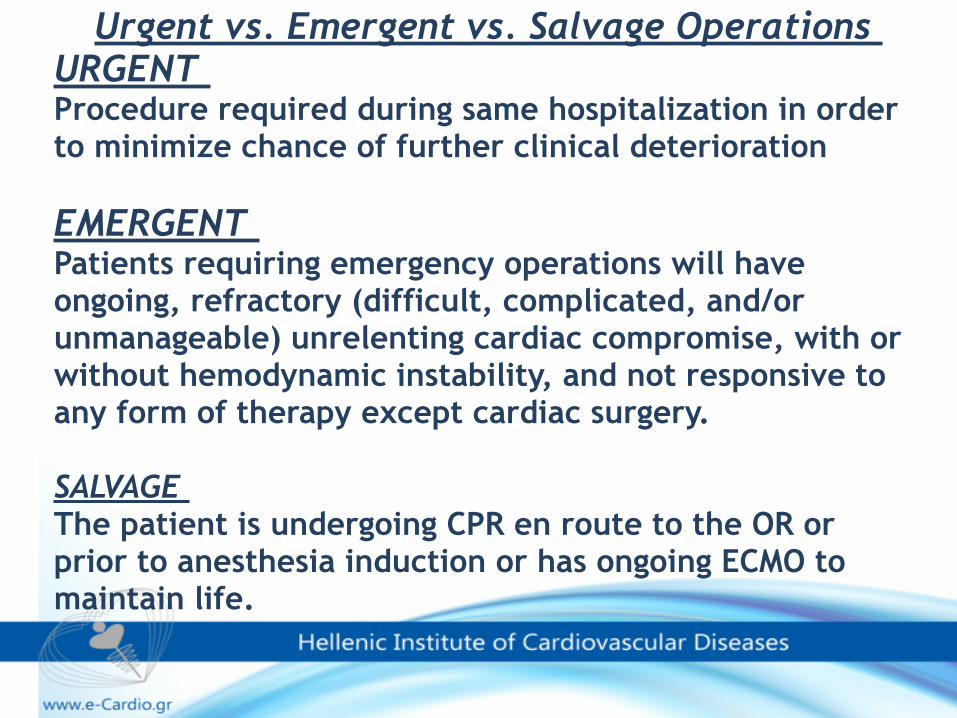
Urgent vs. Emergent vs. Salvage Operations
URGENT Procedure required during same hospitalization in order to minimize chance of further clinical deterioration
EMERGENT Patients requiring emergency operations will have ongoing, refractory (difficult, complicated, and/or unmanageable) unrelenting cardiac compromise, with or without hemodynamic instability, and not responsive to any form of therapy except cardiac surgery.
SALVAGE The patient is undergoing CPR en route to the OR or prior to anesthesia induction or has ongoing ECMO to maintain life.
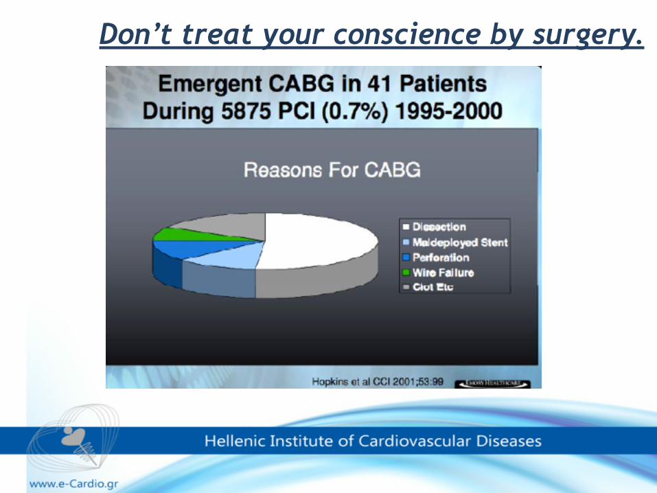
Don’t treat your conscience by surgery.
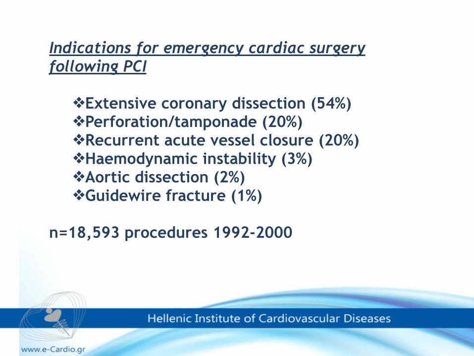
Indications for emergency cardiac surgery following PCI
❖Extensive coronary dissection (54%) ❖Perforation/tamponade (20%) ❖Recurrent acute vessel closure (20%) ❖Haemodynamic instability (3%) ❖Aortic dissection (2%) ❖Guidewire fracture (1%)
n=18,593 procedures 1992-2000
Coronary artery dissection
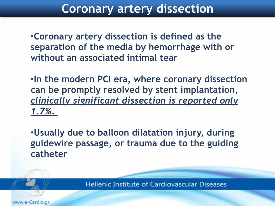
•Coronary artery dissection is defined as the separation of the media by hemorrhage with or without an associated intimal tear
•In the modern PCI era, where coronary dissection can be promptly resolved by stent implantation, clinically significant dissection is reported only 1.7%.
•Usually due to balloon dilatation injury, during guidewire passage, or trauma due to the guiding catheter
Coronary artery dissection
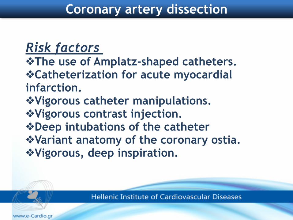
Risk factors ❖The use of Amplatz-shaped catheters.
❖Catheterization for acute myocardial infarction.
❖Vigorous catheter manipulations.
❖Vigorous contrast injection.
❖Deep intubations of the catheter
❖Variant anatomy of the coronary ostia.
❖Vigorous, deep inspiration.
Coronary artery dissection
C.GRAIDIS, D.DIMITRIADIS, A.NTATSIOS, V.KARASAVVIDIS, V.PSIFOS
EUROMEDICA – KYANOUS STAVROS HOSPITAL
Bail-Out Stenting for Left Main Coronary Artery: A Treatable Nightmare of the
Interventional Cardiologist.
Endovascular Cardiac Complications 15-17 JUNE 2011-LAUSANNE, SWITZERLAND
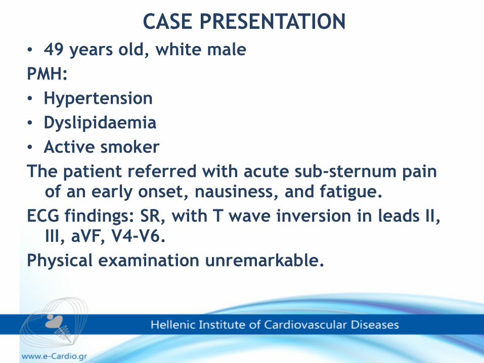
CASE PRESENTATION• 49 years old, white male PMH: • Hypertension • Dyslipidaemia • Active smoker The patient referred with acute sub-sternum pain
of an early onset, nausiness, and fatigue. ECG findings: SR, with T wave inversion in leads II,
III, aVF, V4-V6. Physical examination unremarkable.
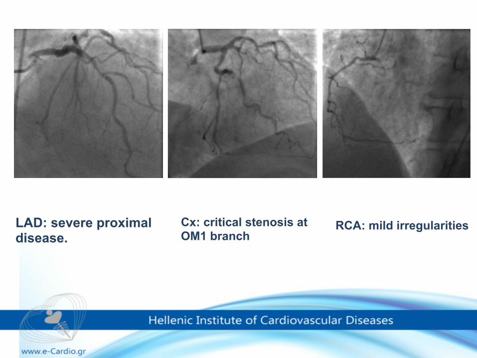
LAD: severe proximal disease.
Cx: critical stenosis at OM1 branch
RCA: mild irregularities
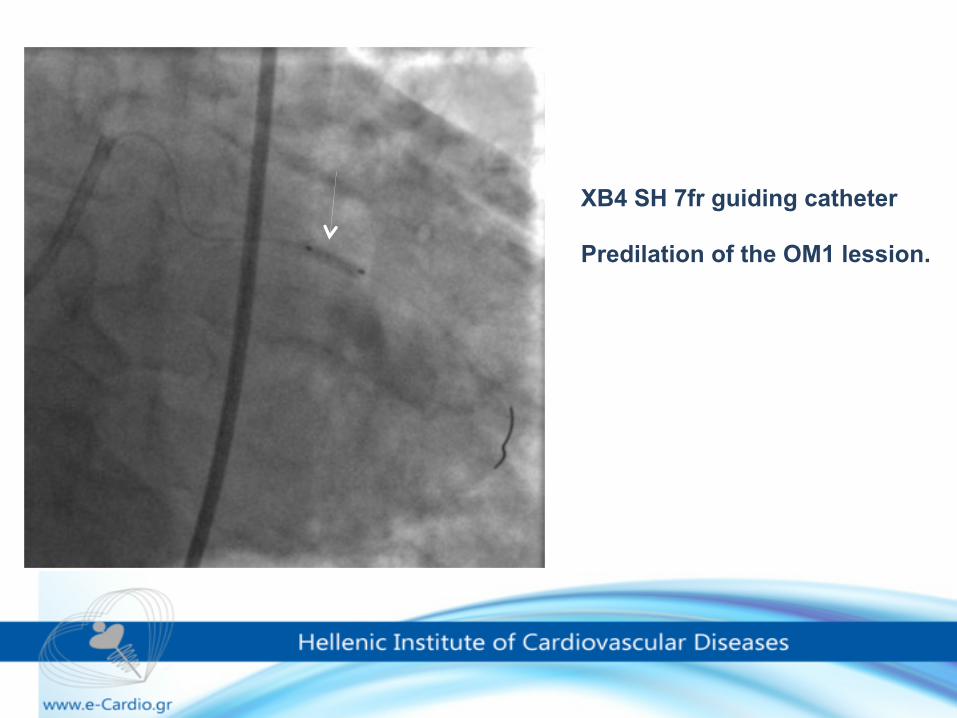
XB4 SH 7fr guiding catheter
Predilation of the OM1 lession.
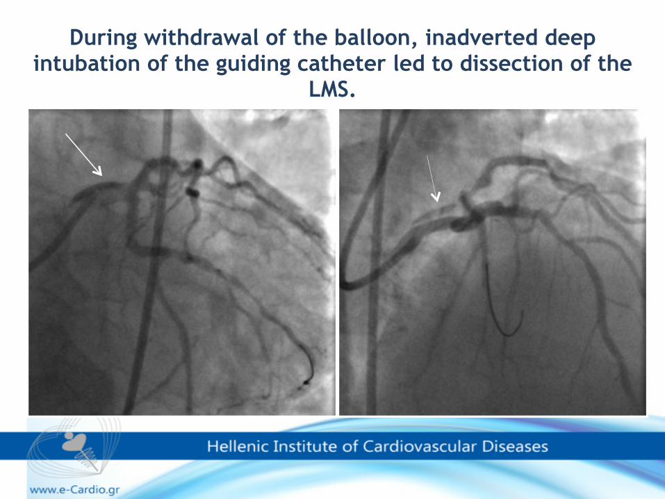
During withdrawal of the balloon, inadverted deep intubation of the guiding catheter led to dissection of the
LMS.
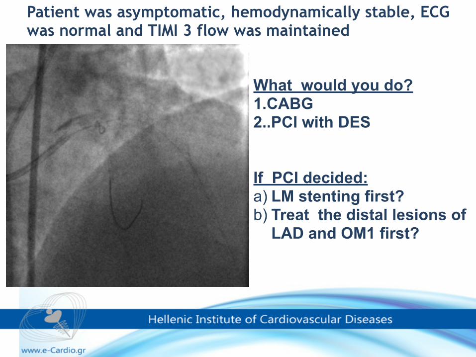
Patient was asymptomatic, hemodynamically stable, ECG was normal and TIMI 3 flow was maintained
What would you do? 1.CABG 2..PCI with DES
If PCI decided: a) LM stenting first? b) Treat the distal lesions of
LAD and OM1 first?
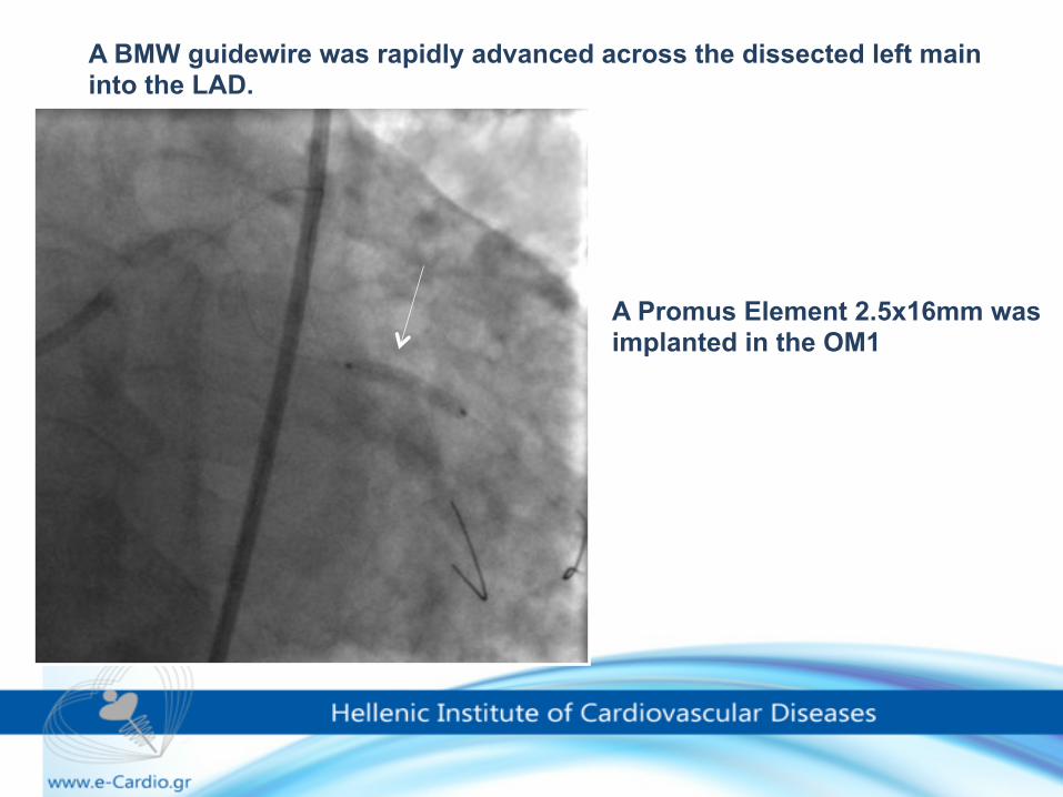
A BMW guidewire was rapidly advanced across the dissected left main into the LAD.
A Promus Element 2.5x16mm was implanted in the OM1
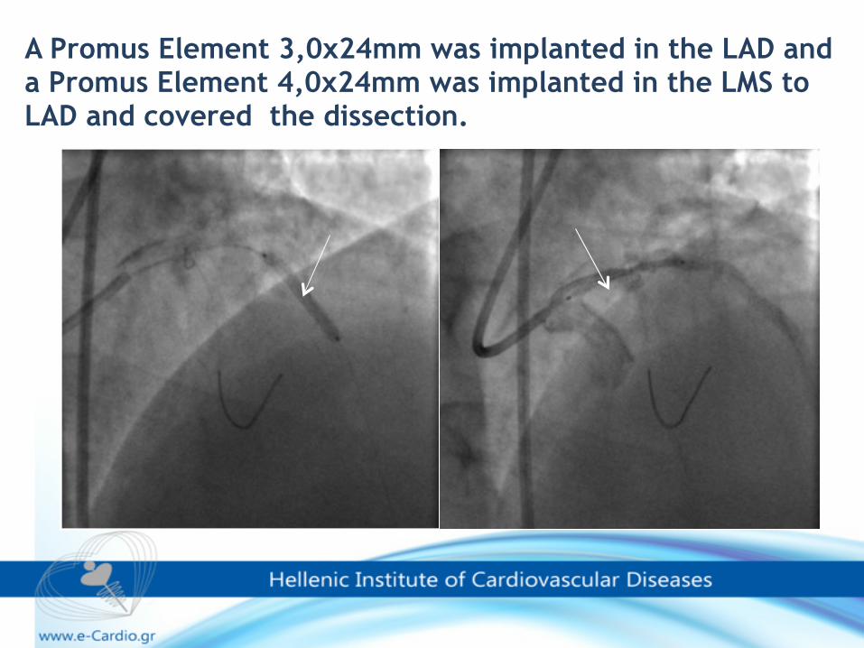
A Promus Element 3,0x24mm was implanted in the LAD and a Promus Element 4,0x24mm was implanted in the LMS to LAD and covered the dissection.
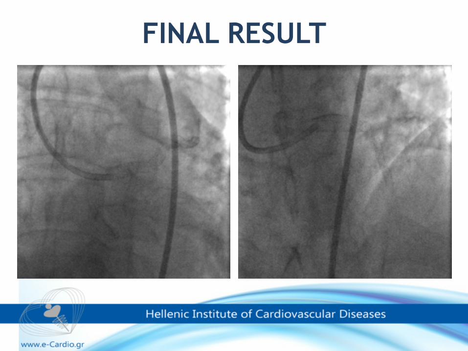
FINAL RESULT
EXTENSIVE, CATHETER-INDUCED DISSECTION OF THE RIGHT CORONARY ARTERY TREATED SUCCESSFULLY WITH FULL METAL JACKET.
C. Graidis, D. Dimitriadis, A.Ntatsios, V. Karasavvides, V. Psifos.
Cardiology Department, Euromedica – Kyanous Stavros, Thessaloniki, Greece.
Complication case ICE 2010
CASE REPORT
• 57 y.o. male with effort angina • Positive Exercise Tolerance Test • Past Medical History: Hypertension,
Dyslipidaemia • Resting 12-lead ECG, ECHO: Normal
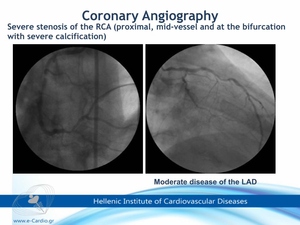
Severe stenosis of the RCA (proximal, mid-vessel and at the bifurcation with severe calcification)
Coronary Angiography
Moderate disease of the LAD
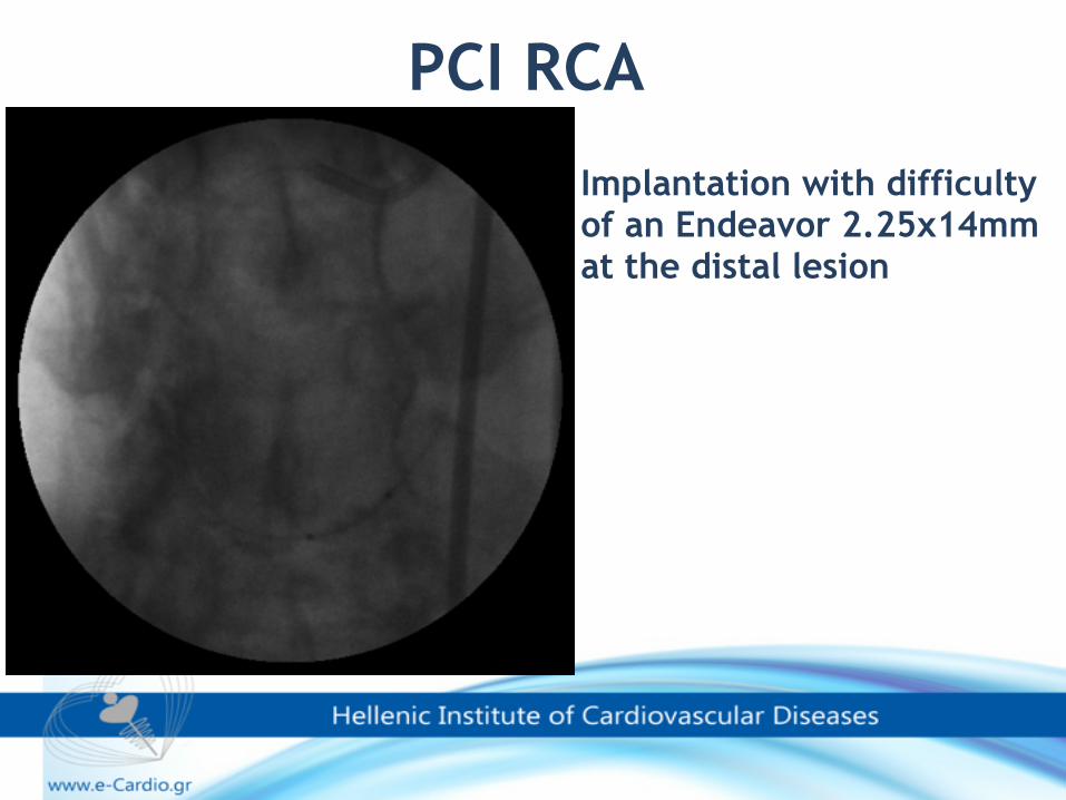
PCI RCA
Implantation with difficulty of an Endeavor 2.25x14mm at the distal lesion
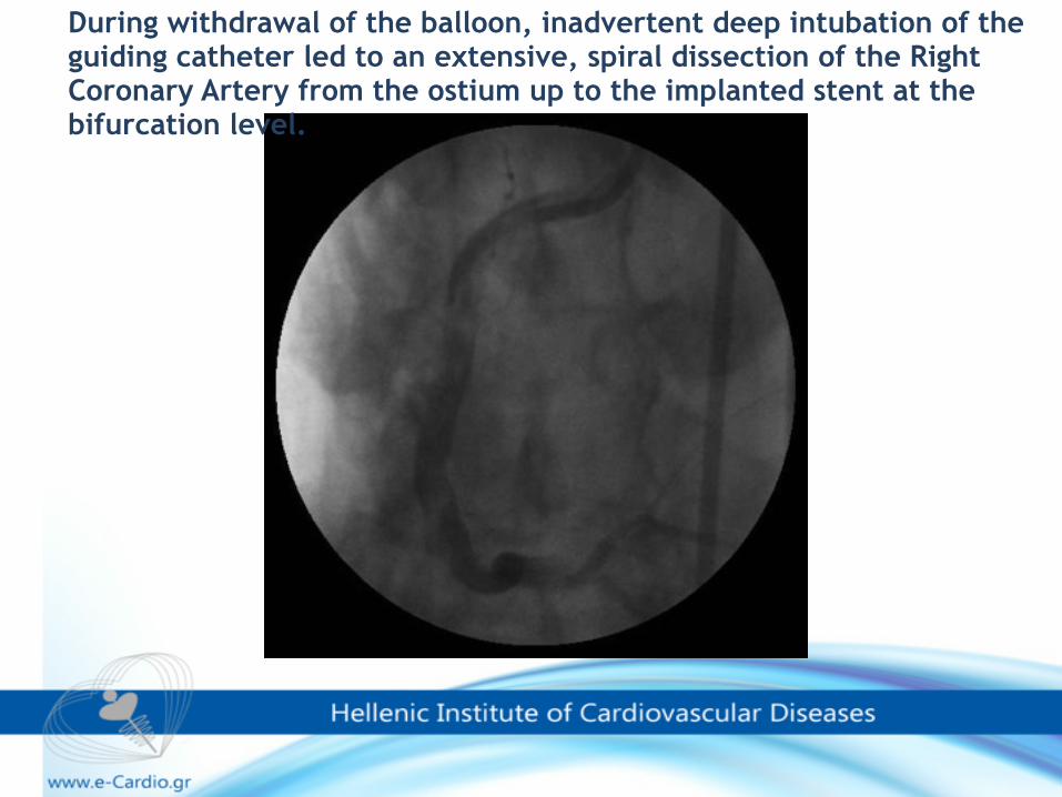
During withdrawal of the balloon, inadvertent deep intubation of the guiding catheter led to an extensive, spiral dissection of the Right Coronary Artery from the ostium up to the implanted stent at the bifurcation level.
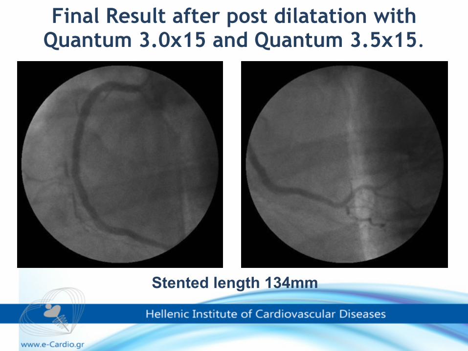
Final Result after post dilatation with Quantum 3.0x15 and Quantum 3.5x15.
Stented length 134mm
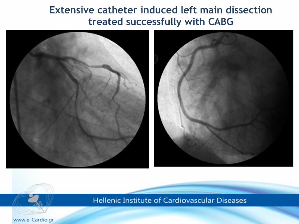
Extensive catheter induced left main dissection treated successfully with CABG
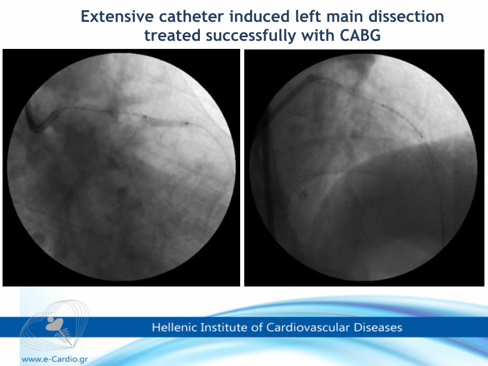
Extensive catheter induced left main dissection treated successfully with CABG
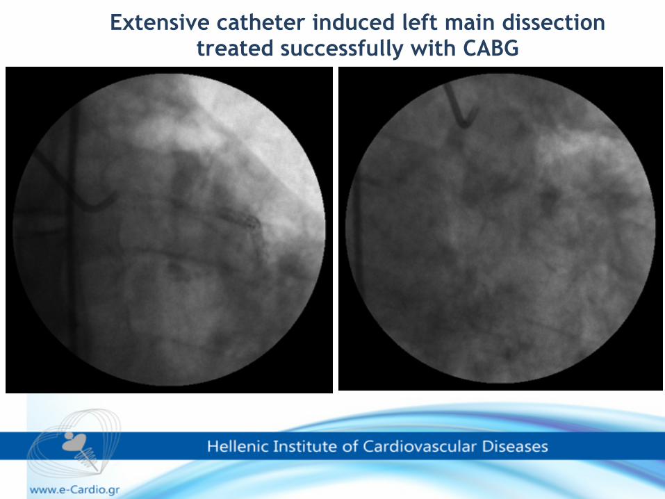
Extensive catheter induced left main dissection treated successfully with CABG
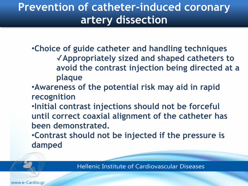
•Choice of guide catheter and handling techniques ✓Appropriately sized and shaped catheters to avoid the contrast injection being directed at a plaque
•Awareness of the potential risk may aid in rapid recognition •Initial contrast injections should not be forceful until correct coaxial alignment of the catheter has been demonstrated. •Contrast should not be injected if the pressure is damped
Prevention of catheter-induced coronary artery dissection
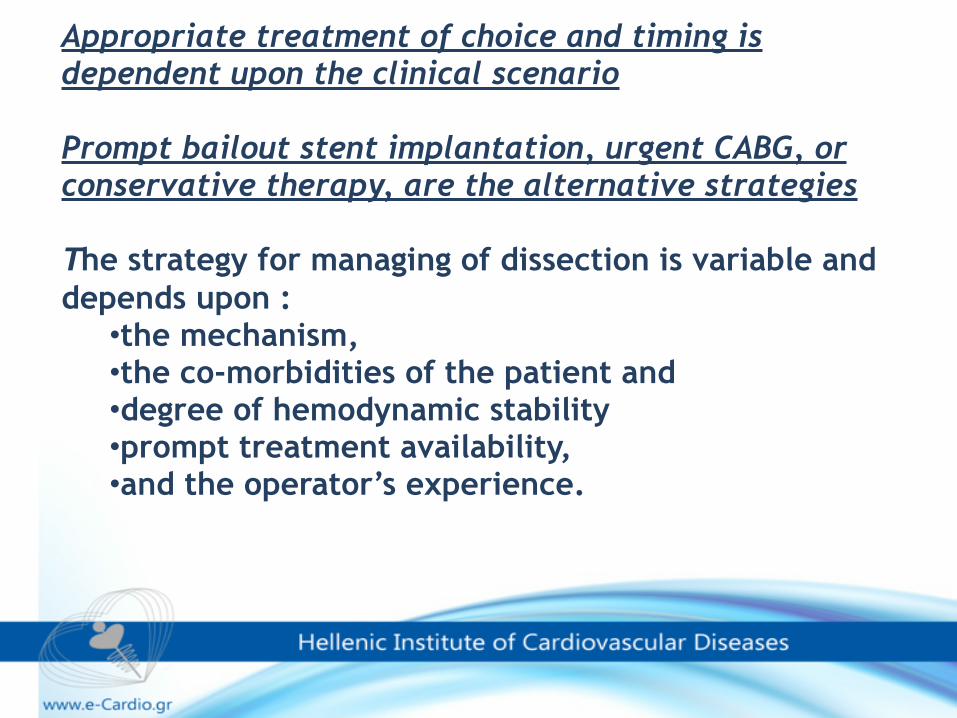
Appropriate treatment of choice and timing is dependent upon the clinical scenario
Prompt bailout stent implantation, urgent CABG, or conservative therapy, are the alternative strategies
The strategy for managing of dissection is variable and depends upon :
•the mechanism, •the co-morbidities of the patient and •degree of hemodynamic stability •prompt treatment availability, •and the operator’s experience.
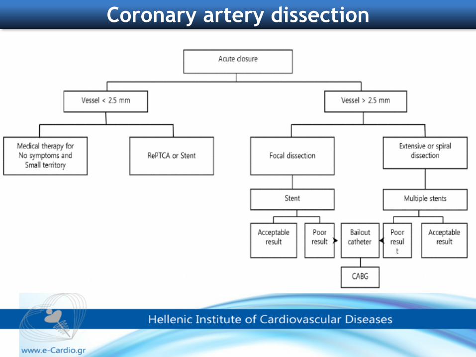
Coronary artery dissection
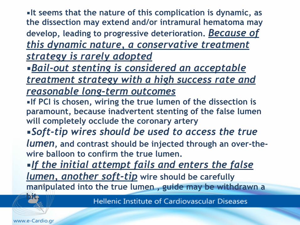
▪It seems that the nature of this complication is dynamic, as the dissection may extend and/or intramural hematoma may develop, leading to progressive deterioration. Because of this dynamic nature, a conservative treatment strategy is rarely adopted ▪Bail-out stenting is considered an acceptable treatment strategy with a high success rate and reasonable long-term outcomes ▪If PCI is chosen, wiring the true lumen of the dissection is paramount, because inadvertent stenting of the false lumen will completely occlude the coronary artery ▪Soft-tip wires should be used to access the true lumen, and contrast should be injected through an over-the-wire balloon to confirm the true lumen. ▪If the initial attempt fails and enters the false lumen, another soft-tip wire should be carefully manipulated into the true lumen , guide may be withdrawn a bit.
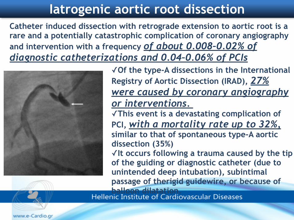
Catheter induced dissection with retrograde extension to aortic root is a rare and a potentially catastrophic complication of coronary angiography and intervention with a frequency of about 0.008–0.02% of diagnostic catheterizations and 0.04–0.06% of PCIs
Iatrogenic aortic root dissection
✓Of the type-A dissections in the International Registry of Aortic Dissection (IRAD), 27% were caused by coronary angiography or interventions. ✓This event is a devastating complication of PCI, with a mortality rate up to 32%, similar to that of spontaneous type-A aortic dissection (35%) ✓It occurs following a trauma caused by the tip of the guiding or diagnostic catheter (due to unintended deep intubation), subintimal passage of therigid guidewire, or because of balloon dilatation.
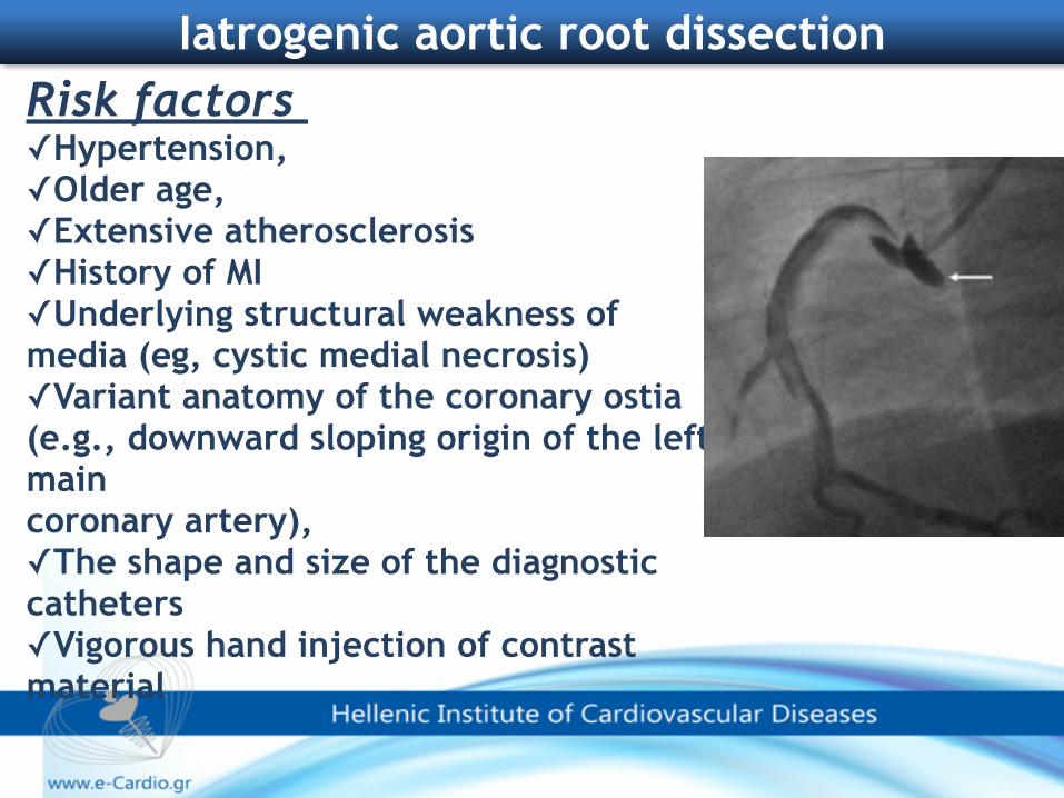
Risk factors
✓Hypertension, ✓Older age, ✓Extensive atherosclerosis ✓History of MI ✓Underlying structural weakness of media (eg, cystic medial necrosis) ✓Variant anatomy of the coronary ostia (e.g., downward sloping origin of the left main coronary artery), ✓The shape and size of the diagnostic catheters ✓Vigorous hand injection of contrast material
Iatrogenic aortic root dissection

Most described cases occurred during angiography and angioplasty of the right
coronary artery (RCA)
Iatrogenic aortic root dissection
The RCA may be more susceptible to retrograde aortocoronary dissection than the left coronary because (1) the diameter of the left coronary artery is larger than that of the RCA (2) the angle of the ascending aorta with the left coronary artery is acute, possibly providing a better approach for Catheterization and (3) the periostial wall and sinotubular junction of the left coronary artery are formed by more smooth muscle cells and by a dense matrix of collagen type-I fibers.
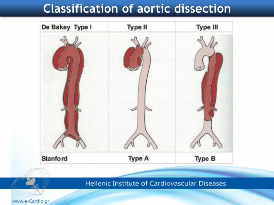
Classification of aortic dissection
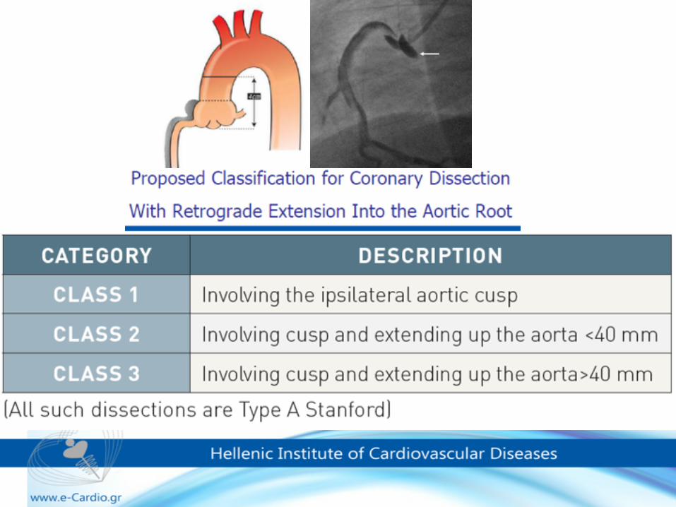
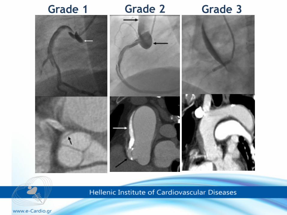
Grade 2Grade 1 Grade 3
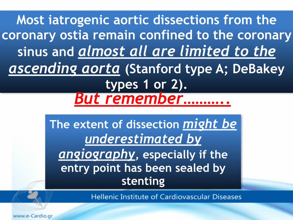
Most iatrogenic aortic dissections from the coronary ostia remain confined to the coronary
sinus and almost all are limited to the ascending aorta (Stanford type A; DeBakey
types 1 or 2).But remember………..
The extent of dissection might be underestimated by
angiography, especially if the entry point has been sealed by
stenting
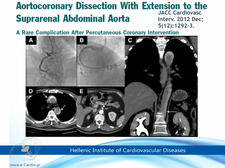
JACC Cardiovasc Interv. 2012 Dec;5(12):1292-3.
Therapeutic strategies include:
1. surgical intervention,
2. stenting the entry point of the coronary
dissection
3. conservative treatment.
The optimal management of iatrogenic aortic dissection remains controversial and is based largely on case reports and anecdotal experience.
Conservative management is a reasonable option only in hemodynamically stable patients with localized aortic dissection.
•Sinus of Valsalva dissections that remain localized during catheterization tend to resolve spontaneously in the first month •Nevertheless, conservative treatment is unsuccessful in 50% of cases due to myocardial infarction, progression of retrograde dissection or death
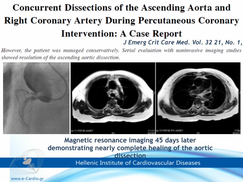
J Emerg Crit Care Med. Vol. 32 21, No. 1, 2010
Magnetic resonance imaging 45 days later demonstrating nearly complete healing of the aortic
dissection
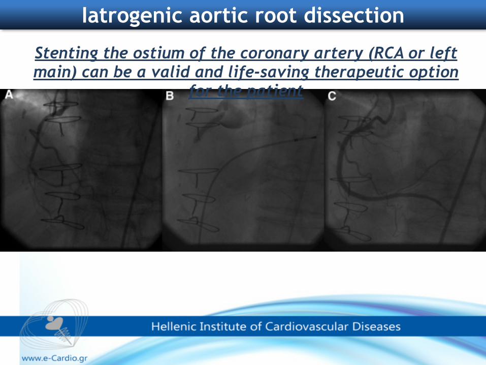
Iatrogenic aortic root dissection
Stenting the ostium of the coronary artery (RCA or left main) can be a valid and life-saving therapeutic option
for the patient
Iatrogenic aortic root dissection
Initial ostial stenting is crucial and an opportunity for conservative treatment:
•Sealing the entry point of the dissection raises the chances for and healing of the entire dissected ascending aorta
The second step is to keep the patient under close surveillance, titrate blood pressure to low normal values and perform repetitive ultrasound imaging in short intervals.
“watch and wait” strategy
The dissection it can still persist or relapse in some cases, despite ostial stenting.
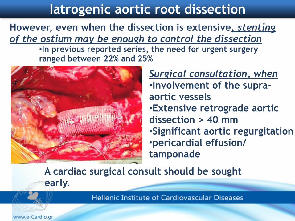
Iatrogenic aortic root dissection However, even when the dissection is extensive, stenting of the ostium may be enough to control the dissection
•In previous reported series, the need for urgent surgery ranged between 22% and 25%
Surgical consultation, when •Involvement of the supra-aortic vessels •Extensive retrograde aortic dissection > 40 mm •Significant aortic regurgitation •pericardial effusion/tamponade
A cardiac surgical consult should be sought early.
Iatrogenic Aortic Dissection Following Catheterization Without Coronary Involvement
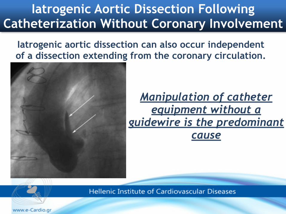
Iatrogenic aortic dissection can also occur independent of a dissection extending from the coronary circulation.
Manipulation of catheter equipment without a
guidewire is the predominant cause
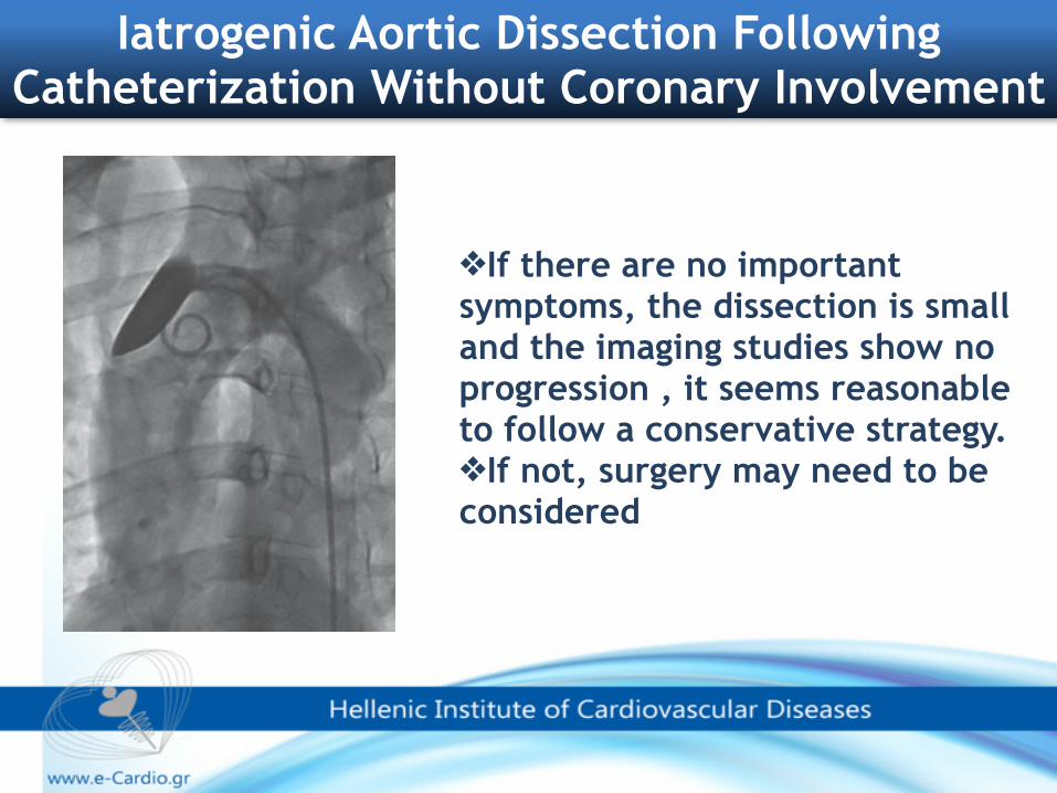
❖If there are no important symptoms, the dissection is small and the imaging studies show no progression , it seems reasonable to follow a conservative strategy. ❖If not, surgery may need to be considered
Iatrogenic Aortic Dissection Following Catheterization Without Coronary Involvement
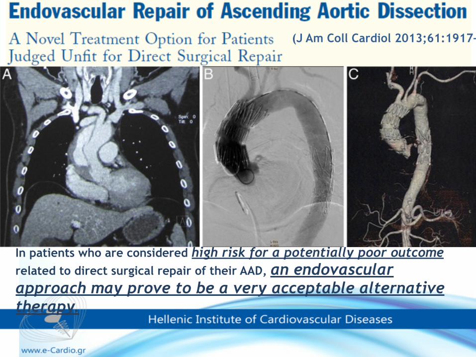
(J Am Coll Cardiol 2013;61:1917–24)
In patients who are considered high risk for a potentially poor outcome related to direct surgical repair of their AAD, an endovascular approach may prove to be a very acceptable alternative therapy.
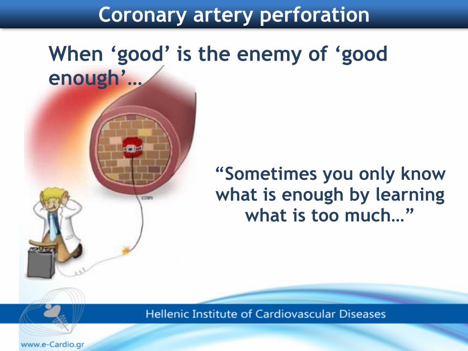
Coronary artery perforation
When ‘good’ is the enemy of ‘good enough’…
“Sometimes you only know what is enough by learning
what is too much…”
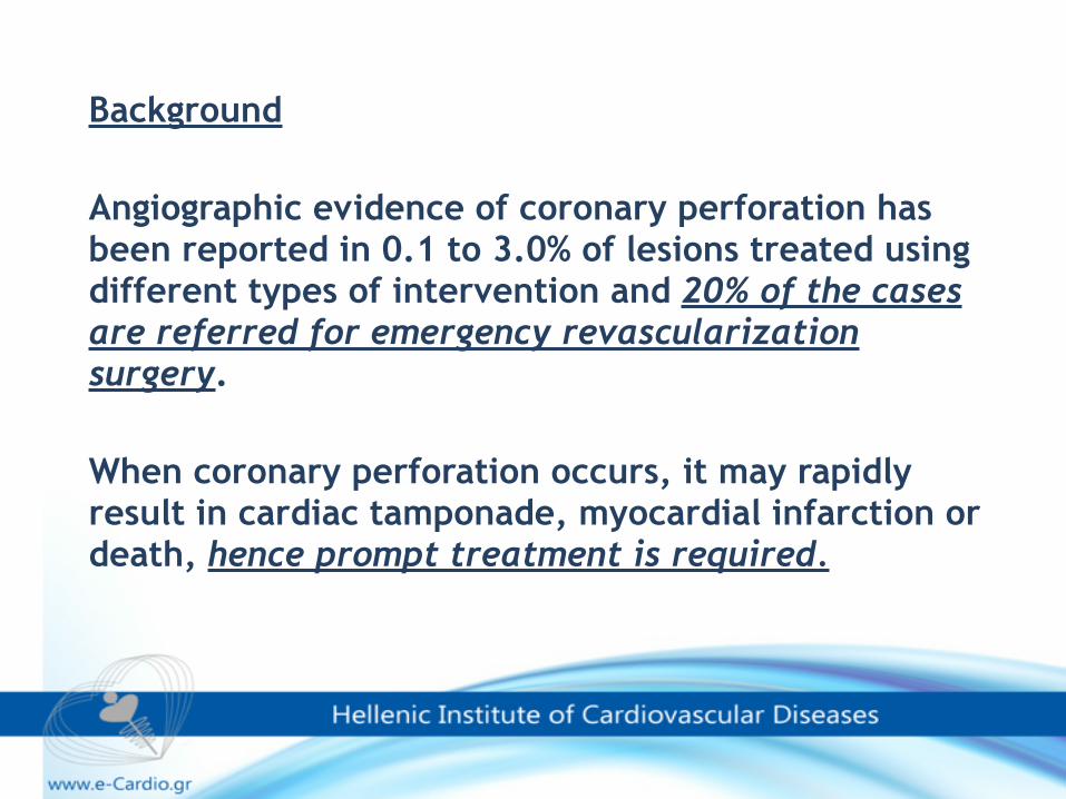
Background
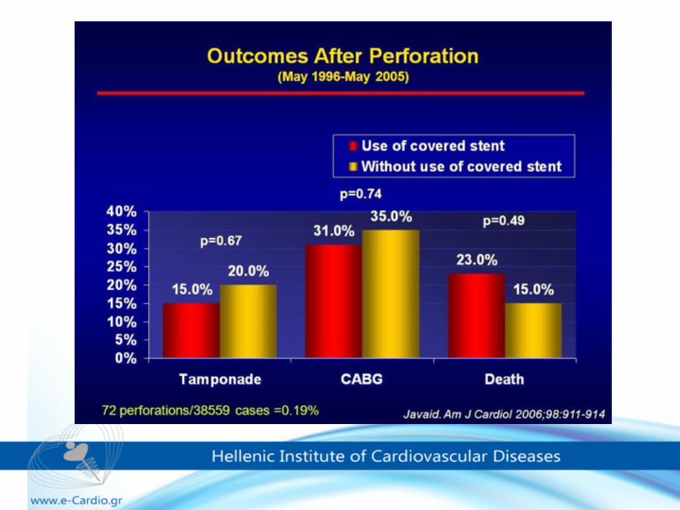
Angiographic evidence of coronary perforation has been reported in 0.1 to 3.0% of lesions treated using different types of intervention and 20% of the cases are referred for emergency revascularization surgery.
When coronary perforation occurs, it may rapidly result in cardiac tamponade, myocardial infarction or death, hence prompt treatment is required.
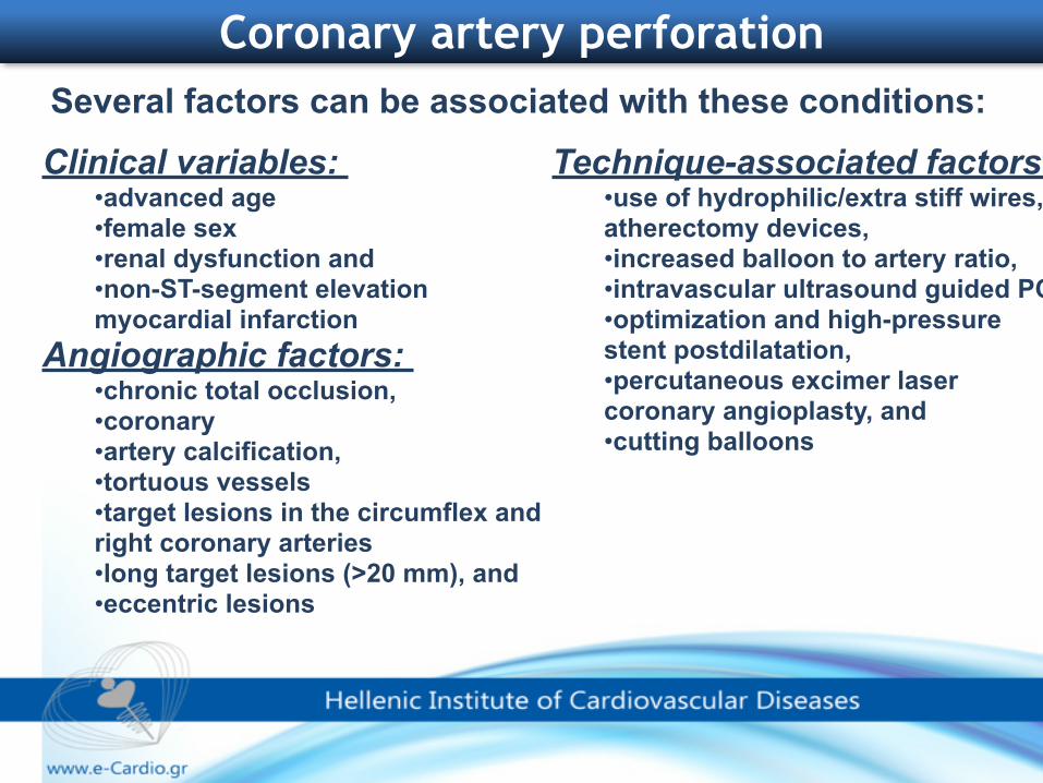
Clinical variables: •advanced age •female sex •renal dysfunction and •non-ST-segment elevation myocardial infarction
Angiographic factors: •chronic total occlusion, •coronary •artery calcification, •tortuous vessels •target lesions in the circumflex and right coronary arteries •long target lesions (>20 mm), and •eccentric lesions
Coronary artery perforation
Technique-associated factors: •use of hydrophilic/extra stiff wires, atherectomy devices, •increased balloon to artery ratio, •intravascular ultrasound guided PCI •optimization and high-pressure stent postdilatation, •percutaneous excimer laser coronary angioplasty, and •cutting balloons
Several factors can be associated with these conditions:
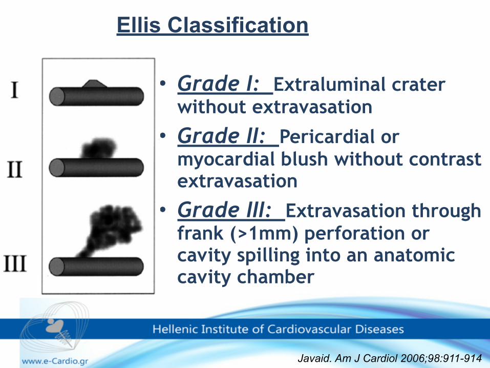
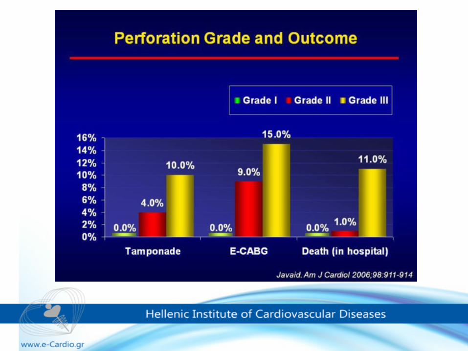
• Grade I: Extraluminal crater without extravasation
• Grade II: Pericardial or myocardial blush without contrast extravasation
• Grade III: Extravasation through frank (>1mm) perforation or cavity spilling into an anatomic cavity chamber
Javaid. Am J Cardiol 2006;98:911-914
Ellis Classification
Coronary artery rupture during Optimization of Coronary Stents Using High-Pressure-Balloon
Inflations: A treatable nightmare ?
C.GRAIDIS, D.DIMITRIADIS, V.PSIFOS, V.KARASAVVIDIS, G.TSONIS, O.HALVATZOULIS
EUROMEDICA – KYANOUS STAVROS HOSPITAL THESSALONIKI, GREECE
9o PANHELLENIC CONGRESS Of Hellenic Society of Thoracic and Cardiovascular Surgeons
Thessaloniki 23-24 November 2012
• 72 y.o female. • Risk factors for IHD: Hypertension,
Dyslipidaemia. • DHx: Aspirin, Clopidogrel, statin, β-blocker,
nitrate. • Referred for cardiac catheterisation because
of exertional angina and a strongly positive Exercise Tolerance Test.
CASE PRESENTATION
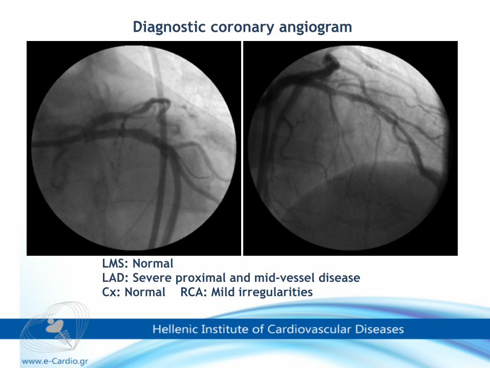
Diagnostic coronary angiogram
LMS: Normal LAD: Severe proximal and mid-vessel disease Cx: Normal RCA: Mild irregularities
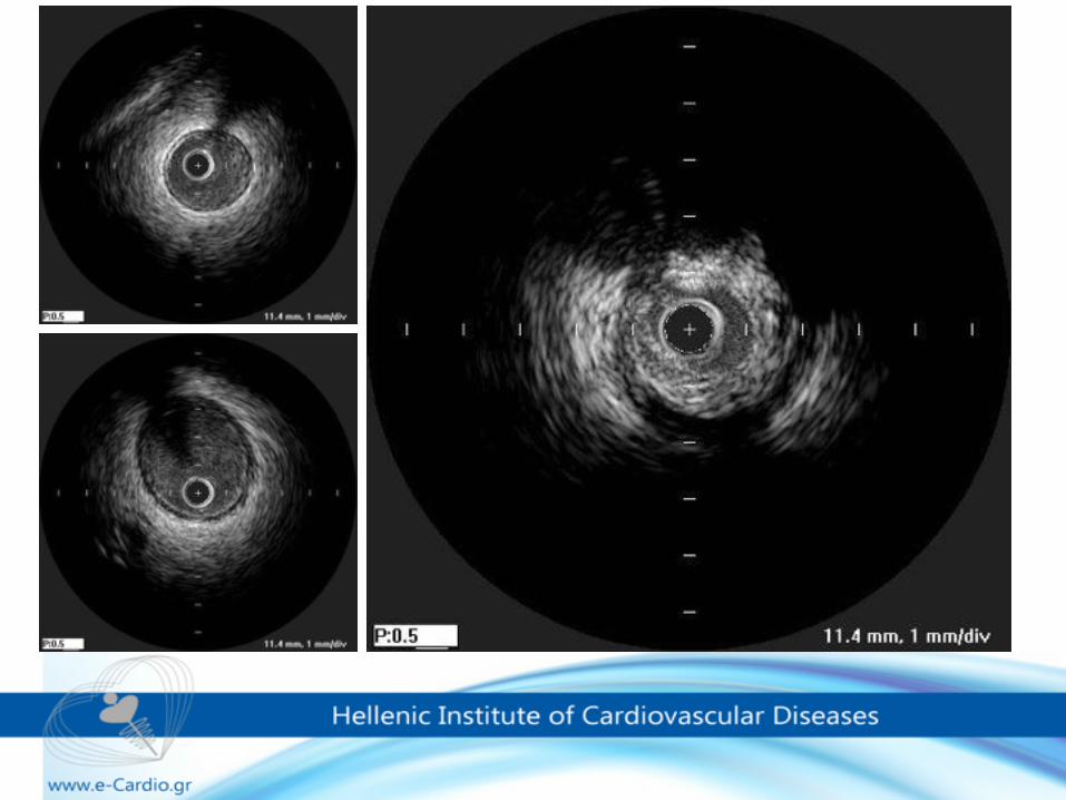
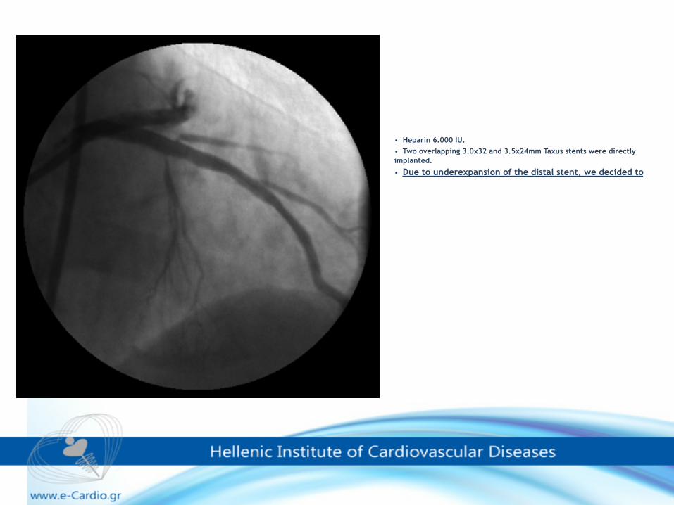
• Heparin 6.000 IU. • Two overlapping 3.0x32 and 3.5x24mm Taxus stents were directly implanted.
• Due to underexpansion of the distal stent, we decided to
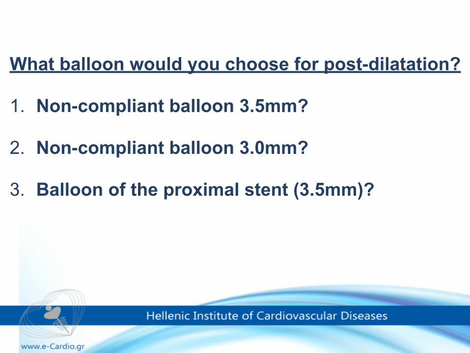
What balloon would you choose for post-dilatation?
1. Non-compliant balloon 3.5mm?
2. Non-compliant balloon 3.0mm?
3. Balloon of the proximal stent (3.5mm)?
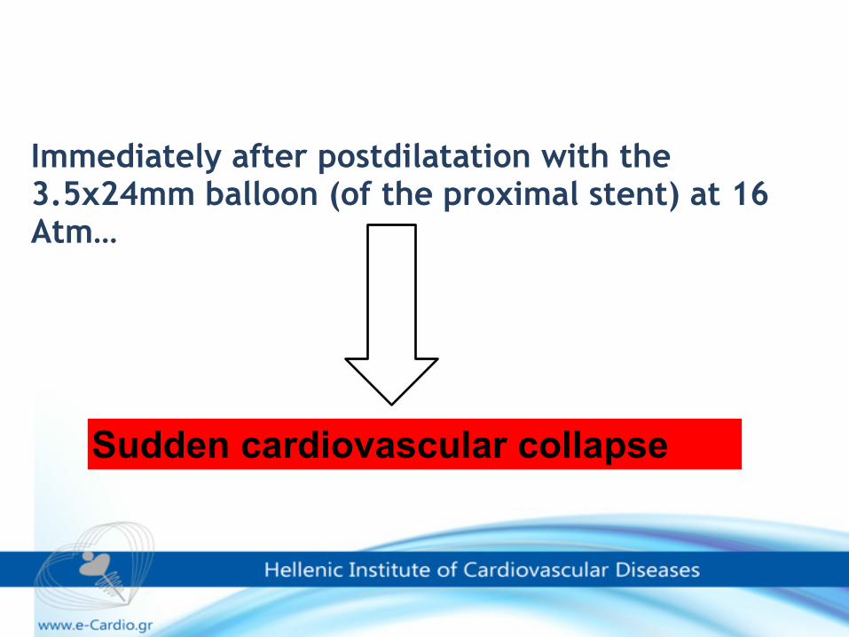
Immediately after postdilatation with the 3.5x24mm balloon (of the proximal stent) at 16 Atm…
Sudden cardiovascular collapse
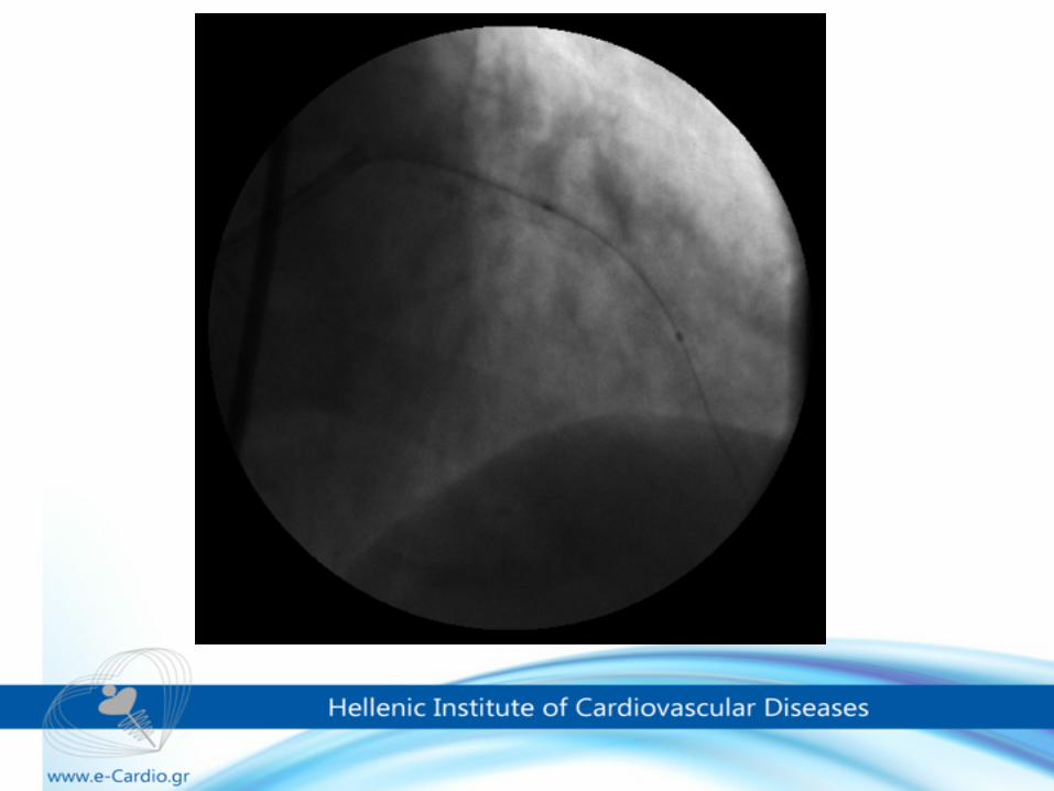
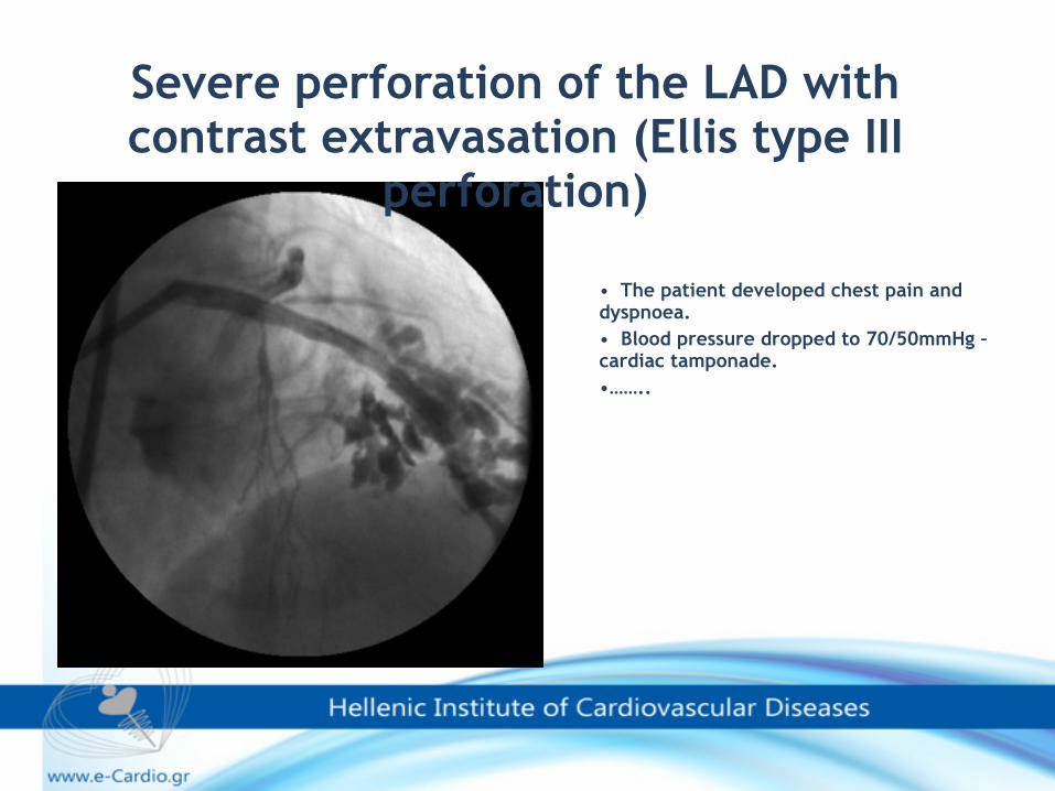
• The patient developed chest pain and dyspnoea. • Blood pressure dropped to 70/50mmHg – cardiac tamponade. •……..
Severe perforation of the LAD with contrast extravasation (Ellis type III
perforation)
How would you manage this complication?
1.Call the surgeons 2.Balloon inflation, emergent pericardiocentesis, reversal of heparine. 3.Covered stents.
OR….
Pray Hard!!!!!!
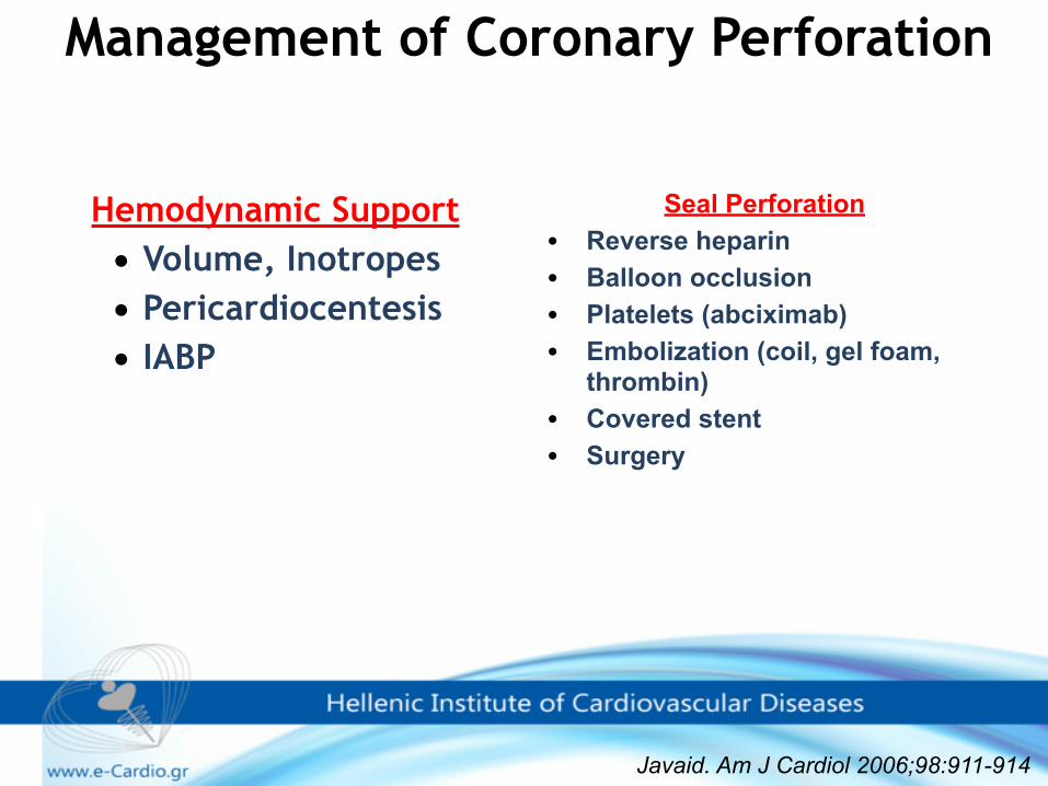
Management of Coronary Perforation
Hemodynamic Support • Volume, Inotropes • Pericardiocentesis • IABP
Seal Perforation • Reverse heparin • Balloon occlusion • Platelets (abciximab) • Embolization (coil, gel foam,
thrombin) • Covered stent • Surgery
Javaid. Am J Cardiol 2006;98:911-914
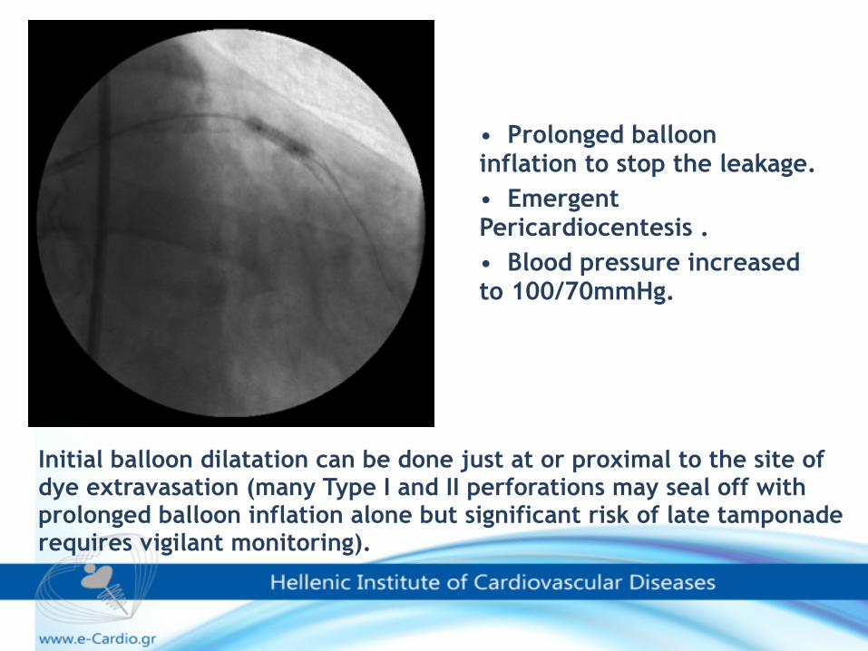
• Prolonged balloon inflation to stop the leakage. • Emergent Pericardiocentesis . • Blood pressure increased to 100/70mmHg.
Initial balloon dilatation can be done just at or proximal to the site of dye extravasation (many Type I and II perforations may seal off with prolonged balloon inflation alone but significant risk of late tamponade requires vigilant monitoring).
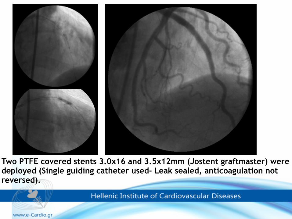
Two PTFE covered stents 3.0x16 and 3.5x12mm (Jostent graftmaster) were deployed (Single guiding catheter used- Leak sealed, anticoagulation not reversed).
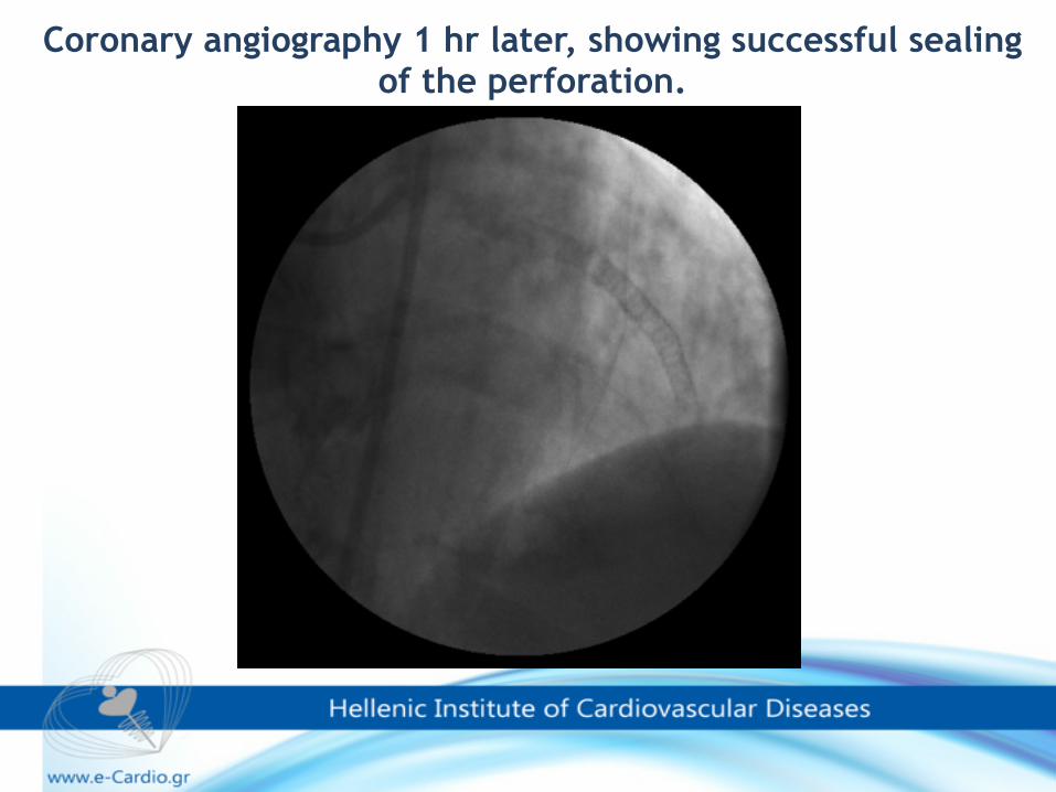
Coronary angiography 1 hr later, showing successful sealing of the perforation.
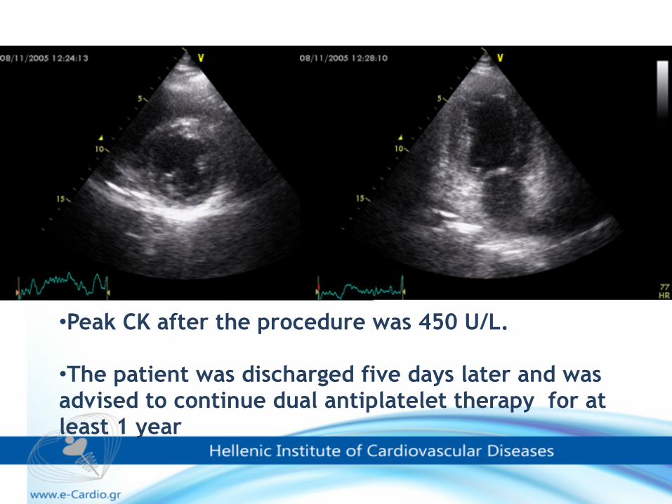
•Peak CK after the procedure was 450 U/L.
•The patient was discharged five days later and was advised to continue dual antiplatelet therapy for at least 1 year
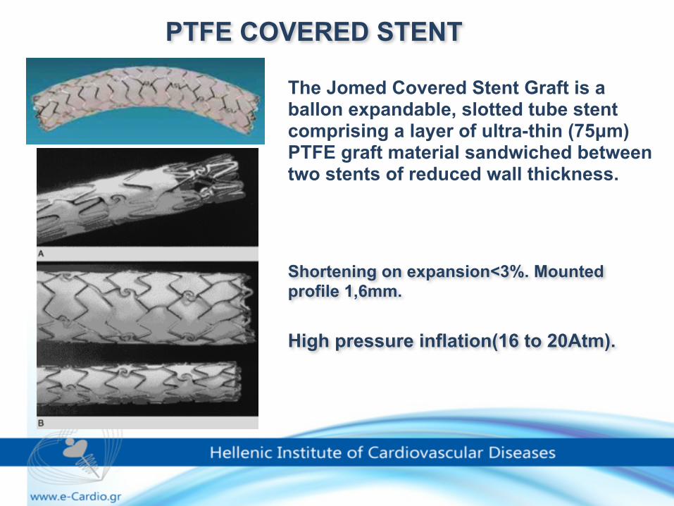
Shortening on expansion<3%. Mounted profile 1,6mm.
High pressure inflation(16 to 20Atm).
PTFE COVERED STENT
The Jomed Covered Stent Graft is a ballon expandable, slotted tube stent comprising a layer of ultra-thin (75µm) PTFE graft material sandwiched between two stents of reduced wall thickness.
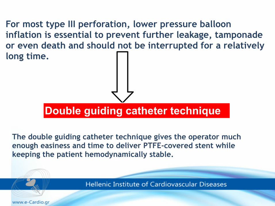
Double guiding catheter technique
For most type III perforation, lower pressure balloon inflation is essential to prevent further leakage, tamponade or even death and should not be interrupted for a relatively long time.
The double guiding catheter technique gives the operator much enough easiness and time to deliver PTFE-covered stent while keeping the patient hemodynamically stable.
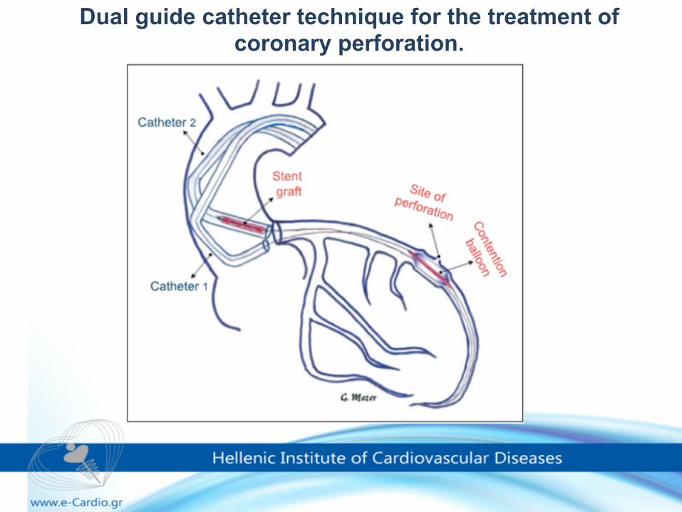
Dual guide catheter technique for the treatment of coronary perforation.
✓In coronary perforations during percutaneous interventions, today coated stent implantation seems to be the first choice in treatment of coronary perforation considering the high mortality and morbidity rate of surgery.
✓PTFE-covered stent implantation is easy, rapid and appears to offer the best treatment option for acute perforations.
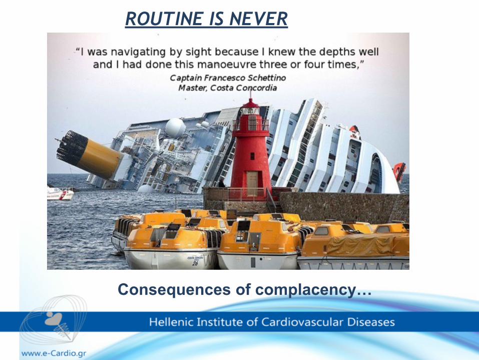
ROUTINE IS NEVER ROUTINE
Consequences of complacency…
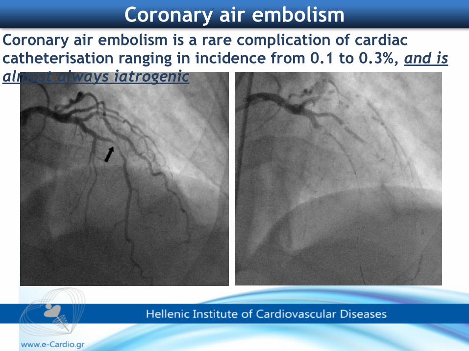
Coronary air embolism Coronary air embolism is a rare complication of cardiac catheterisation ranging in incidence from 0.1 to 0.3%, and is almost always iatrogenic
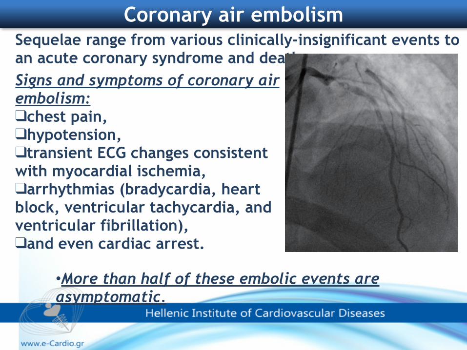
Sequelae range from various clinically-insignificant events to an acute coronary syndrome and death
Coronary air embolism
Signs and symptoms of coronary air embolism: ❑chest pain, ❑hypotension, ❑transient ECG changes consistent with myocardial ischemia, ❑arrhythmias (bradycardia, heart block, ventricular tachycardia, and ventricular fibrillation), ❑and even cardiac arrest.
•More than half of these embolic events are asymptomatic.
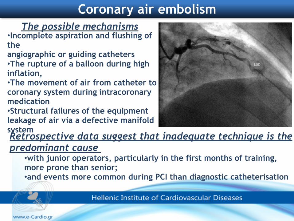
Retrospective data suggest that inadequate technique is the predominant cause
•with junior operators, particularly in the first months of training, more prone than senior; •and events more common during PCI than diagnostic catheterisation
The possible mechanisms •Incomplete aspiration and flushing of the angiographic or guiding catheters •The rupture of a balloon during high inflation, •The movement of air from catheter to coronary system during intracoronary medication •Structural failures of the equipment leakage of air via a defective manifold system
Coronary air embolism
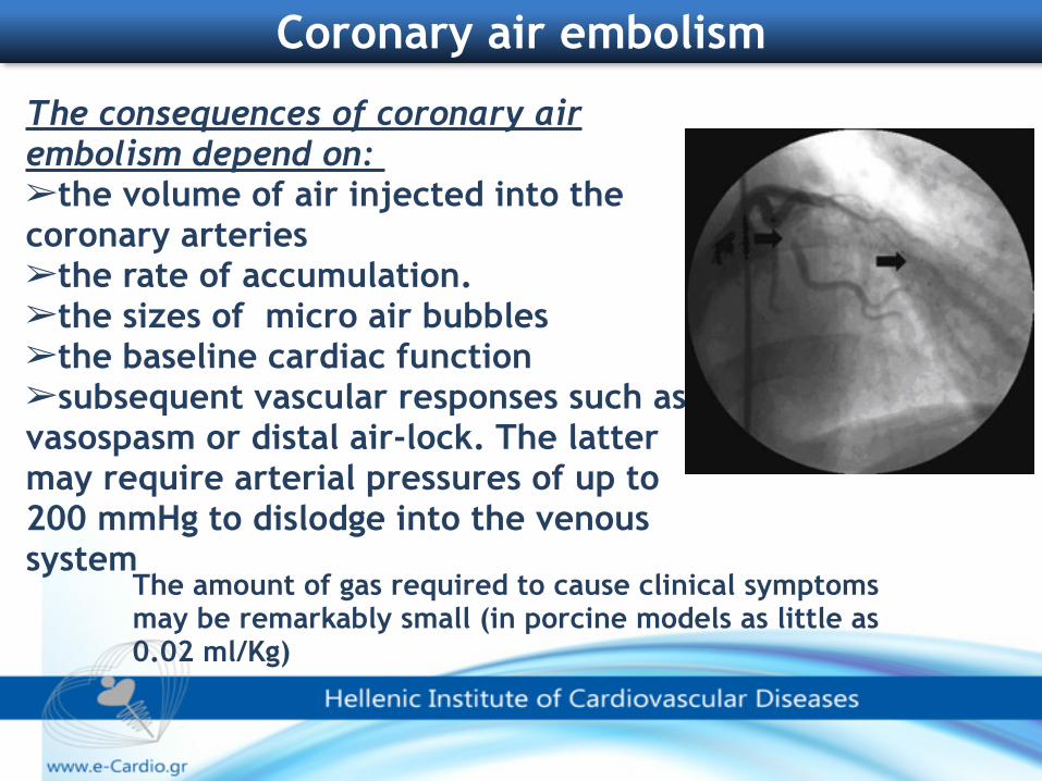
The consequences of coronary air embolism depend on: ➢the volume of air injected into the coronary arteries ➢the rate of accumulation. ➢the sizes of micro air bubbles ➢the baseline cardiac function ➢subsequent vascular responses such as vasospasm or distal air-lock. The latter may require arterial pressures of up to 200 mmHg to dislodge into the venous system
The amount of gas required to cause clinical symptoms may be remarkably small (in porcine models as little as 0.02 ml/Kg)
Coronary air embolism
Management •Aspirate immediately with manifold •100% Oxygen with non-rebreather •Vasopressors • Hemodynamic support •Give vasodilators IC •Give epinephrine IC •Increase perfusion pressure •Manual Blood perfusion •Mechanical or manual aspiration catheters •Disrupt air bubbles with guidewire
Coronary air embolism
Coronary air embolism
There are no sufficient data to guide decisions on therapyAdequate management of this complication has not been
established because these cases usually remain unreported due to the clinicians’ reluctance to discuss the complications of their work
The vast majority of observed emboli are extremely small volumes of air that do not result in symptoms or hemodynamic consequences; therefore, they require no therapy.
For the mild to moderately symptomatic episodes, routine management with supportive measures ✓100% oxygen by face mask, ✓analgesics for chest pain, ✓monitoring and treating arrhythmia for the few minutes it takes for the air to clear spontaneously.
The management of massive air embolism should be extremely quick so as to save life of the patient. Aggressive efforts to reestablish the coronary blood flow as soon as possible are essential to reduce injury to the myocardium and to speed recovery from the hemodynamic crisis. Disruption or dislodgement by the guidewire, forceful injection of saline to fragment the air embolus and allow dispersal distally.
•Such interventions may result in main vessel patency but have a potential to damage the distal microvasculature due to widespread, smaller embolizations.
Coronary air embolism
In contrast, aspiration aims at resolving the blockage by removing the air.
•Aspiration has been attempted with nondedicated devices such as over the wire (OTW) balloons or the angiography catheter itself.
•Use of aspiration catheter
Coronary air embolism
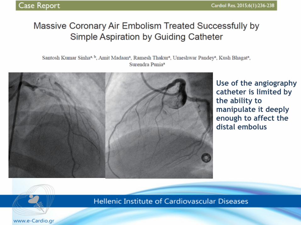
Use of the angiography catheter is limited by the ability to manipulate it deeply enough to affect the distal embolus
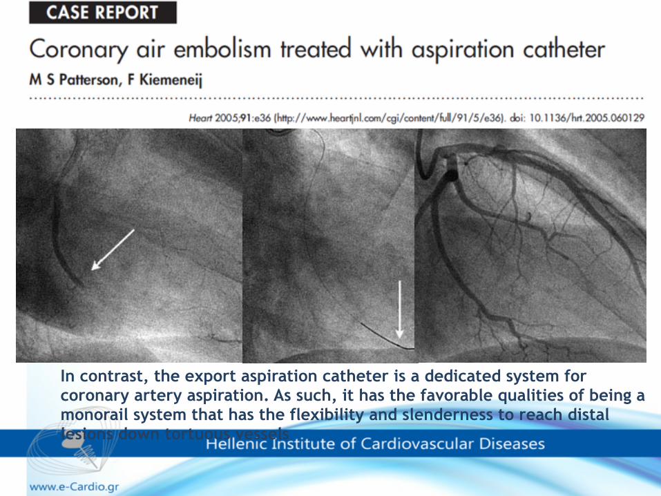
In contrast, the export aspiration catheter is a dedicated system for coronary artery aspiration. As such, it has the favorable qualities of being a monorail system that has the flexibility and slenderness to reach distal lesions down tortuous vessels


![PCI 10.6 1x1 BNC NoWindow 102 · Ø =4mm ~36 ~36 ~36 ~66 ~102 FOV [°], Ø =6mm ~36 ~36 ~36 ~88 ~124 TO39 detector package Bottom view Pin number Function 1(+), 2(-) signal 3 chassis](https://static.fdocument.org/doc/165x107/6101de4255b28b39da300aa1/pci-106-1x1-bnc-nowindow-4mm-36-36-36-66-102-fov-6mm-36-36.jpg)


![PCI σε πολυαγγειακή νόσο - Livemedia.gr · 0.1 1.0 Favorsdevice JACC meta-analysis JIC meta-analysis 0.1 1.0 10.0 1.13[0.89,1.38] 1.00[0.96,1.03] Heterogeneity test](https://static.fdocument.org/doc/165x107/5fe2317e63d82f6275457aaa/pci-f-oef-01-10-favorsdevice-jacc-meta-analysis.jpg)