Genetic dissection of α -adrenoceptor functions in...
42
MOLPHARM/2009/054544 Genetic dissection of α 2 -adrenoceptor functions in adrenergic versus non-adrenergic cells Ralf Gilsbach, Christoph Röser, Nadine Beetz, Marc Brede, Kerstin Hadamek, Miriam Haubold, Jost Leemhuis, Melanie Philipp, Johanna Schneider, Michal Urbanski, Bela Szabo, David Weinshenker, Lutz Hein Institute of Experimental and Clinical Pharmacology and Toxicology, University of Freiburg, Germany (R.G., N.B., M.H., J.L., J.S., M.U., B.S., L.H.); Centre for Biological Signaling Studies (bioss), University of Freiburg, Freiburg, Germany (N.B., L.H.); Institute of Pharmacology and Toxicology, University of Würzburg, Germany (C.R., M.B., K.H.); Institute of Cell Biology, Duke University, NC, USA (M.P.); Department of Human Genetics, Emory University, Atlanta, GA, USA (D.W.) Molecular Pharmacology Fast Forward. Published on February 27, 2009 as doi:10.1124/mol.109.054544 Copyright 2009 by the American Society for Pharmacology and Experimental Therapeutics. This article has not been copyedited and formatted. The final version may differ from this version. Molecular Pharmacology Fast Forward. Published on February 27, 2009 as DOI: 10.1124/mol.109.054544 at ASPET Journals on June 29, 2020 molpharm.aspetjournals.org Downloaded from
Transcript of Genetic dissection of α -adrenoceptor functions in...
MOLPHARM/2009/054544
Genetic dissection of α2-adrenoceptor functions
in adrenergic versus non-adrenergic cells
Ralf Gilsbach, Christoph Röser, Nadine Beetz, Marc Brede, Kerstin Hadamek,
Miriam Haubold, Jost Leemhuis, Melanie Philipp, Johanna Schneider,
Michal Urbanski, Bela Szabo, David Weinshenker, Lutz Hein
Institute of Experimental and Clinical Pharmacology and Toxicology, University of Freiburg, Germany
(R.G., N.B., M.H., J.L., J.S., M.U., B.S., L.H.); Centre for Biological Signaling Studies (bioss),
University of Freiburg, Freiburg, Germany (N.B., L.H.); Institute of Pharmacology and Toxicology,
University of Würzburg, Germany (C.R., M.B., K.H.); Institute of Cell Biology, Duke University, NC,
USA (M.P.); Department of Human Genetics, Emory University, Atlanta, GA, USA (D.W.)
Molecular Pharmacology Fast Forward. Published on February 27, 2009 as doi:10.1124/mol.109.054544
Copyright 2009 by the American Society for Pharmacology and Experimental Therapeutics.
This article has not been copyedited and formatted. The final version may differ from this version.Molecular Pharmacology Fast Forward. Published on February 27, 2009 as DOI: 10.1124/mol.109.054544
at ASPE
T Journals on June 29, 2020
molpharm
.aspetjournals.orgD
ownloaded from
MOLPHARM/2009/054544 2
Running title: Adrenergic vs. non-adrenergic cell α2-adrenoceptors
Address for correspondence:
Lutz Hein
Institute of Experimental and Clinical Pharmacology and Toxicology,
University of Freiburg, Albertstrasse 25, 79104 Freiburg, Germany,
Phone ++49-761-2035314, Fax ++49-761-2035318,
Number of text pages: 33
Number of tables: 1
Number of figures: 8
Number of references: 36
Number of words in abstract: 239
Number of words in introduction: 442
Number of words in discussion: 1329
List of abbreviations: A+/+, wild-type murine α2A-adrenoceptor gene; A-/-, null allele of the murine α2A-
adrenoceptor gene; Adra2a, Adra2b, Adra2c, murine genes encoding α2A-, α2B- or α2C-
adrenoceptors; Dbh, dopamine β-hydroxylase; PBS, phosphate buffered saline; SCG, superior
cervical ganglia; Tg, transgenic line.
This article has not been copyedited and formatted. The final version may differ from this version.Molecular Pharmacology Fast Forward. Published on February 27, 2009 as DOI: 10.1124/mol.109.054544
at ASPE
T Journals on June 29, 2020
molpharm
.aspetjournals.orgD
ownloaded from
MOLPHARM/2009/054544 3 Abstract
α2-adrenoceptors mediate diverse functions of the sympathetic system and are targets for the
treatment of cardiovascular disease, depression, pain, glaucoma, and sympathetic activation during
opioid withdrawal. To determine whether α2-adrenoceptors on adrenergic neurons or α2-
adrenoceptors on non-adrenergic neurons mediate the physiological and pharmacological responses
of α2-agonists, we used the dopamine β-hydroxylase (Dbh) promoter to drive expression of α2A-
adrenoceptors exclusively in noradrenergic and adrenergic cells of transgenic mice. Dbh-α2A
transgenic mice were crossed with double knockout mice lacking both α2A- and α2C-receptors to
generate lines with selective expression of α2A-autoreceptors in adrenergic cells. These mice were
subjected to a comprehensive phenotype analysis and compared to wild-type mice, which express
α2A- and α2C-receptors in both adrenergic and non-adrenergic cells, and α2A/α2C double knockout
mice, which do not express these receptors in any cell type. Surprisingly, only a few functions
previously ascribed to α2-adrenoceptors were mediated by receptors on adrenergic neurons,
including feedback inhibition of norepinephrine release from sympathetic nerves and spontaneous
locomotor activity. Other agonist effects including analgesia, hypothermia, sedation and anesthetic-
sparing were mediated by α2-receptors in non-adrenergic cells. In dopamine β-hydroxylase knockout
mice lacking norepinephrine, the α2-agonist medetomidine still induced a loss of the righting reflex,
confirming that the sedative effect of α2-adrenoceptor stimulation is not mediated via autoreceptor-
mediated inhibition of norepinephrine release. The present study paves the way for a revision of the
current view of the α2-adrenergic receptors and it provides important new considerations for future
drug development.
This article has not been copyedited and formatted. The final version may differ from this version.Molecular Pharmacology Fast Forward. Published on February 27, 2009 as DOI: 10.1124/mol.109.054544
at ASPE
T Journals on June 29, 2020
molpharm
.aspetjournals.orgD
ownloaded from
MOLPHARM/2009/054544 4 Introduction
Adrenergic receptors are important targets for the treatment of human diseases and conditions
including hypertension and heart failure, psychiatric and neurological diseases, asthma, and pain
(Westfall and Westfall, 2006). To date, nine different adrenergic receptor subtypes have been cloned
and grouped into three receptor groups, including α1A,B,D, α2A,B,C, β1,2,3 (Bylund et al., 1994). However,
the therapeutic potential of these subtypes has not been fully explored due to the lack of ligands with
sufficient subtype-selectivity. At present, only four out of the nine possible subtype distinctions, i.e.
α1, α2, β1 and β2, have achieved clinical relevance (Westfall and Westfall, 2006). Especially within the
α1- and α2-receptor subgroups, the physiological significance of individual receptor subtypes has
remained unclear until recently. For the α2-adrenoceptors, mouse models with targeted deletions of
the individual subtypes have greatly advanced our understanding of the physiological role and the
therapeutic potential of these receptors (for recent review, see (Gilsbach and Hein, 2008). Activation
of α2A-receptors could be linked with bradycardia and hypotension (MacMillan et al., 1996), sedation
(Lakhlani et al., 1997) and consolidation of working memory (Wang et al., 2007). In contrast, α2B-
receptors counteracted the hypotensive effect of α2A-receptors (Link et al., 1996) and were essential
for placenta vascular development (Philipp et al., 2002). α2C-receptors were identified as feedback
regulators of adrenal catecholamine release (Brede et al., 2003), an essential pathway to limit the
progression of cardiac hypertrophy and failure in experimental models (Lymperopoulos et al., 2007)
and in humans with congestive heart failure (Small et al., 2002).
α2-receptors were initially identified as presynaptic receptors inhibiting the release of
neurotransmitters in isolated tissues in vitro (Starke et al., 1975). The term ”autoreceptors” has been
introduced for those receptors which are ‘sensitive to the neuron’s own transmitter’. In contrast to
This article has not been copyedited and formatted. The final version may differ from this version.Molecular Pharmacology Fast Forward. Published on February 27, 2009 as DOI: 10.1124/mol.109.054544
at ASPE
T Journals on June 29, 2020
molpharm
.aspetjournals.orgD
ownloaded from
MOLPHARM/2009/054544 5 autoreceptors, heteroreceptors are modulated by neurotransmitters derived from neighboring
neurons (Bylund et al., 1994). Despite considerable progress in adrenergic biology, it is unknown
whether the wide array of clinical actions of α2-agonists is indeed mediated by the ”classical”
presynaptic α2-autoreceptors or whether and to what degree α2-adrenoceptors on non-adrenergic
cells are involved. In order to address this question, we crossed transgenic mice with expression of
α2A-receptors under control of the dopamine β-hydroxylase promoter to mice with constitutive
deletion of the α2A- and α2C-adrenoceptor genes, thus generating mice that exclusively express α2A-
receptors in adrenergic cells. These animals were subjected to a comprehensive phenotyping
analysis to provide a comprehensive overview of adrenergic cell vs. non-adrenergic cell functions for
α2-adrenoceptors. Surprisingly, very few α2-receptor functions were mediated by α2-adrenocceptors
in adrenergic cells; most effects of α2-agonists were mediated by α2-receptors on non-adrenergic
neurons or cells. These results underline the importance of non-adrenergic cell α2-adrenoceptors.
This article has not been copyedited and formatted. The final version may differ from this version.Molecular Pharmacology Fast Forward. Published on February 27, 2009 as DOI: 10.1124/mol.109.054544
at ASPE
T Journals on June 29, 2020
molpharm
.aspetjournals.orgD
ownloaded from
MOLPHARM/2009/054544 6 Materials and Methods
Generation of transgenic mice
A transgenic vector consisting of a 5.6-kb part of the human dopamine β-hydroxylase promoter as
described previously (promoter plasmid kindly provided by Dr. R. Palmiter) (Hoyle et al., 1994;
Mercer et al., 1991), the coding sequence of the murine α2A-adrenoceptor with an aminoterminal
epitope tag (flag epitope, DYKDDDD, (Daunt et al., 1997; Hein et al., 1999)) and the SV40 t intron
and poly A signal was constructed as depicted in Fig. 1 a. The vector was linearized, separated from
plasmid sequences, and microinjected into fertilized oocytes from superovulated FVB/N mice.
Several independent transgenic Dbh-Adra2a-Tg founder lines were obtained, two of which,
numbered A11 and A25, were investigated in detail. These transgenic mice were crossed with
congenic C57BL6/J α2A- and α2C-deficient mice (Hein et al., 1999). Genotypes were confirmed by
polymerase chain reactions (Fig. 1 b) performed with genomic DNA isolated from tail biopsies (Hein
et al., 1999). For detection of the Dbh-Adra2a-Tg, the following primers were used: forward primer 5’-
ATGTCGACGCCACCTTAGAT-3’, reverse primer 5’-AGGCAAACCAGCGTCAGTGT-3’. Dopamine β-
hydroxylase knockout (Dbh-/-) mice, maintained on a mixed C57BL/SJ and 129SvEv background,
were generated as described previously (Szot et al., 2004; Thomas et al., 1995). Littermate Dbh+/-
mice, which have normal catecholamine levels and behavior, were used as controls (Bourdelat-Parks
et al., 2005; Thomas et al., 1998). For experiments age-matched adult (3–5 months) male littermates
which were maintained in specified pathogen-free facilities were used. All animal procedures were
approved by the responsible animal care committees of the Universities of Freiburg and Würzburg,
Germany. The investigation conforms to the Guide for the Care and Use of Laboratory Animals
published by the US National Institutes of Health.
This article has not been copyedited and formatted. The final version may differ from this version.Molecular Pharmacology Fast Forward. Published on February 27, 2009 as DOI: 10.1124/mol.109.054544
at ASPE
T Journals on June 29, 2020
molpharm
.aspetjournals.orgD
ownloaded from
MOLPHARM/2009/054544 7 Quantitative real-time PCR
mRNA quantification by real-time polymerase chain reaction (qPCR) from murine tissues was
performed as described (Gilsbach et al., 2007). For qPCR, 35 μl of the amplification mixture (Qiagen,
Quantitect SYBR Green Kit) was used containing 20 ng of reverse transcribed RNA and 300 nM
primers (MWG, Ebersberg, Germany) specific for α2A-adrenoceptor (5´-
CAAGATCAACGACCAGAAGT-3´ and 5´-GTCAAGGCTGATGGCGCACAG-3´) or ribosomal protein
S29 (5'-ATGGGTCACCAGCAGCTCTA-3' and 5'-AGCCTATGTCCTTCGCGTACT-3') sequences.
Reactions were run in triplicate on a MX3000P detector (Stratagene, Amsterdam, Netherlands). The
cycling conditions were: 15 s polymerase activation at 95°C and 40 cycles at 95°C for 15 s, at 58°C
for 30 s and at 72°C for 30 s. Absolute copy numbers were determined using standard curves of
corresponding linear DNA-fragments (Gilsbach et al., 2007). Genomic DNA from tail biopsies (Hein et
al., 1999) was used to determine transgene copy numbers. The α2B-adrenoceptor gene was used as
a reference control. Reactions were carried out as described above using primers for α2A- (see
above) and α2B-adrenoceptor (5'-GCAGAGGTCTCGGAGCTAA-3' and 5'-
GCCTCTCCGACAGAAGATA-3') sequences.
Autoradiography
For receptor autoradiography, transverse brain sections (10 μm) were cut serially with a cryostat,
thaw-mounted onto slides and incubated for 60 min in 50 mM Tris-HCl (pH 7.5), 1.5 mM EDTA, 8 nM
[3H]RX821002 (Amersham, Freiburg, Germany). To determine non-specific binding, 1 μM
atipamezole was included. Following incubations, the slides were washed 2 x 5 min in cold buffer,
rinsed in distilled water, air-dried, and analyzed using a BAS5000 Fuji PhosphorImager.
Immunohistochemistry
For immunodetection of flag-tagged α2A-receptors, cryostat sections (light microscopy) or vibratome
sections (electron microscopy) from perfusion-fixed mice (4% paraformaldehyde, 0.1%
glutaraldehyde in phosphate buffered saline, PBS) were used. Sections were blocked in 1% bovine
This article has not been copyedited and formatted. The final version may differ from this version.Molecular Pharmacology Fast Forward. Published on February 27, 2009 as DOI: 10.1124/mol.109.054544
at ASPE
T Journals on June 29, 2020
molpharm
.aspetjournals.orgD
ownloaded from
MOLPHARM/2009/054544 8 serum albumin, 0.04% triton X100 in PBS and incubated for 48 h at 4°C with a rabbit polyclonal
antiserum against the epitope tag (antiserum “1809” in (Hein et al., 1994)), followed by overnight
incubation with goat anti rabbit biotinylated antibody and ABC Vectastain (Vector, Burlingame, CA,
USA) incubation. Sections were incubated with 3,3’-diaminobenzidine and hydrogen peroxide,
followed by 60 min OsO4 and embedding in araldite (Durcupan, Fluka, Germany). Ultrathin sections
which were contrast stained with 2% uranyl acetate were inspected in a LEO AB912 electron
microscope.
Locus coeruleus microdissection
For microscopical microdissection of locus coeruleus specimens, tissues were frozen in liquid
nitrogen. 15 µm cryostat sections were mounted on glas slides, dehydrated in ethanol and xylene
followed by microdissection at a Leica AM6000 inverted microscope. The area of the locus coeruleus
(Paxinos and Franklin, 2001) was microdissected using MicroChisels (Eppendorf, Hamburg,
Germany), aspirated via a micropipette, xylene was evaporated and RNA was isolated using the
RNeasy Micro-Kit (Qiagen, Hilden, Germany). The identity of the locus coeruleus was verified by
qPCR determination of tyrosine hydroxylase and dopamine β-hydroxylase mRNA expression.
Isolation of neurons from superior cervical ganglia
Superior cervical ganglia (SCG) from mice were dissociated by treatment with trypsin, collagenase
and DNase in Neurobasal-A media (Invitrogen) supplemented with 0.5 mM glutamine, 1%
penicillin/streptomycin and 10 mM Hepes at 35°C and 850 rpm. Cells were plated onto poly-D-lysine
coated coverslips in Neurobasal-A media with 0.5 mM glutamine, 2.5% fetal bovine serum, 1% B27
supplement and 1% penicillin/streptomycin conditioned on astroglial culture for 24 h before. For
immunocytochemistry, neurons were fixed in methanol, incubated overnight with anti-tyrosine
This article has not been copyedited and formatted. The final version may differ from this version.Molecular Pharmacology Fast Forward. Published on February 27, 2009 as DOI: 10.1124/mol.109.054544
at ASPE
T Journals on June 29, 2020
molpharm
.aspetjournals.orgD
ownloaded from
MOLPHARM/2009/054544 9 hydroxylase and anti-Flag M2 antiserum, followed by Alexa488 and Alexa568-coupled secondary
antibodies.
Electrophysiology
For patch-clamp recording, slides with cultured SCG neurons were fixed at the glass bottom of a
superfusion chamber and superfused with buffer at room temperature at a flow rate of 1.5 ml min-1.
The buffer was of the following composition: 126 mM NaCl, 1.2 mM NaH2PO4, 3 mM KCl, 1 mM
MgCl2, 2.5 mM CaCl2, 26 mM NaHCO3, and 10 mM glucose, pH 7.35 (after the solution was gassed
with 95% O2, 5% CO2). The superfusion buffer contained tetrodotoxin (0.3 µM) to block voltage
gated sodium channels. Neurons were visualized with infrared video-microscopy. Recordings were
obtained with an EPC-9 amplifier under the control of TIDA software (HEKA Elektronik, Lambrecht,
Germany). Series resistance compensation of 50% was usually applied. Series resistance was
measured before and after recordings and experiments with major changes in series resistance
(>20%) were discarded. Cell were patch clamped with pipettes (2-3 MΩ resistance) containing buffer
of the following composition: CsCl 100 mM, MgCl2 1mM, HEPES 10 mM, CaCl2 0.5 mM, TEA×Cl 20
mM, EGTA 10 mM, Mg-ATP 3 mM, Na-GTP 0.3 mM, (pH 7.35 adjusted with CsOH). Calcium
currents were evoked every 15 seconds by voltage ramps (from -70 mV to 50 mV, ramp speed 0.5
mV ms-1). Leak currents were determined by running hyperpolarizing ramps (from -70 mV to -94 mV,
ramp speed 0.1 mV ms-1) 5 seconds after the depolarizing ramps. The maximum leak subtracted
current measured during the ramp was used for statistical evaluation.
[3H]norepinephrine release
Electrically evoked [3H]norepinephrine (Amersham, Freiburg, Germany) release from isolated mouse
atria was determined as described (Gilsbach et al., 2007; Hein et al., 1999). In brief, freshly isolated
This article has not been copyedited and formatted. The final version may differ from this version.Molecular Pharmacology Fast Forward. Published on February 27, 2009 as DOI: 10.1124/mol.109.054544
at ASPE
T Journals on June 29, 2020
molpharm
.aspetjournals.orgD
ownloaded from
MOLPHARM/2009/054544 10 mouse atria were incubated in 2 ml medium containing 0.1 µM [3H]norepinephrine (Amersham,
Freiburg, Germany) for 45 min at 37°C. Atria were transferred to superfusion chambers. After 45 min
of superfusion successive 2-min superfusate samples were collected. The preincubation medium
consisted of (in mM): NaCl 118, KCl 4.8, CaCl2 0.2, MgSO4 1.2, NaHCO3 25, KH2PO4 1.2, glucose
11, ascorbic acid 0.57, Na2EDTA 0.03, saturated with 5% CO2 in O2. The superfusion medium was
the same but contained 2.5 mM CaCl2 and 1 µM desipramine. Six periods of electrical stimulation (20
pulses/50 Hz, 1 ms pulse width, 80 mA) were applied at 16 min intervals. At the end of the
experiments, tissues were solubilized and tritium was determined in superfusate samples and
tissues. The electrically evoked overflow of total tritium reflects exocytotic release of
[3H]norepinephrine.
Locomotor activity
Two weeks after implantation of a telemetry device (DSI, Transoma Medical, USA, TA11-PAC10),
locomotor activity was recorded in conscious unrestrained mice both during the day (7 a.m. - 7 p.m.)
and night (7 p.m. - 7 a.m.) (Gilsbach et al., 2007).
Nociception
Thermal antinociception was determined using an automated tail-flick system (Ugo Basile, Comerio,
Italy). The time to withdrawal of the tail from an infrared light source shining onto the base of the tail
was automatically recorded.
Body temperature
Body temperature (°C) was recorded with a rectal thermometer probe (TKM-0902, FMI, Seeheim-
Ober Beerbach, Germany). Saline or drug were injected (i.p.) 60 min before the test.
This article has not been copyedited and formatted. The final version may differ from this version.Molecular Pharmacology Fast Forward. Published on February 27, 2009 as DOI: 10.1124/mol.109.054544
at ASPE
T Journals on June 29, 2020
molpharm
.aspetjournals.orgD
ownloaded from
MOLPHARM/2009/054544 11
Sedation / hypnosis
Mice were injected with different doses of medetomidine (i.p.) and the time after injection at which the
righting reflex of mice was lost and the recovery time of this reflex were monitored (Lakhlani et al.,
1997). The observer was blinded with respect to the genotype of the mice. For determination of the
anesthetic sparing effect mice pretreated with sub-anesthetic drug concentrations were placed in an
air-tight plexiglas chamber and isoflurane was continuously introduced at increasing concentrations
(0-1.2 vol/vol% in O2) (Lakhlani et al., 1997). Mice were equilibrated to each concentration of
isoflurane for 5 min before the righting reflex.
Statistical analysis
Data are presented as means ± standard error of the mean (SEM) of individual data points. Data
were analyzed using 1- or 2-way ANOVA followed by Bonferroni post-hoc tests or Student t test,
respectively. Results from electrophysiological measurements were analyzed using the Mann-
Whitney test. A P value of less than 0.05 was considered as statistically significant. Graphs and
statistical analyses were generated by GraphPad Prism 4.0c (GraphPad Software, San Diego, USA).
This article has not been copyedited and formatted. The final version may differ from this version.Molecular Pharmacology Fast Forward. Published on February 27, 2009 as DOI: 10.1124/mol.109.054544
at ASPE
T Journals on June 29, 2020
molpharm
.aspetjournals.orgD
ownloaded from
MOLPHARM/2009/054544 12 Results
Transgenic mouse models to dissect α2-adrenoceptor functions in adrenergic vs. non-
adrenergic cells
In order to distinguish between functions that are mediated by α2-autoreceptors in adrenergic cells
and α2-receptors expressed in non-adrenergic neurons or cells, a transgenic mouse strain with
specific expression of α2A-adrenoceptors in adrenergic cells was generated. The transgenic construct
(Fig. 1 a) consisted of the coding sequence of an epitope-tagged murine α2A-receptor (Daunt et al.,
1997) downstream of the dopamine β-hydroxylase (Dbh) promoter sequence followed by a SV40 t-
intron and polyadenylation signal. The Dbh promoter has been successfully used to drive expression
of target genes in adrenergic neurons in vivo (Hoyle et al., 1994; Mercer et al., 1991). After
pronuclear injection of the linearized transgenic vector, several transgenic founder mice were
obtained and identified by PCR genotyping (Fig. 1 b,c). Two out of five transgenic founder lines, A11
and A25, which contained 30±4 and 17±1 transgene copies (Fig. 1b), were used for further studies.
Transgenic offspring were born at the expected Mendelian ratio and did not show any signs of
developmental or structural defects (not shown). In order to generate mouse strains with selective
expression of α2A-adrenoceptors in adrenergic cells, transgenic mice were crossed with mice lacking
α2A- and α2C-adrenoceptors (Adra2a-/- Adra2c-/-, (Hein et al., 1999)). Previous experiments have
demonstrated that α2A- and α2C-adrenoceptors are the major presynaptic feedback regulators in the
adrenergic system (Hein et al., 1999). Thus, mice with selective expression of α2A-autoreceptors in
adrenergic neurons (termed A-/-C-/-Tg; genotype Adra2a-/- Adra2c-/- Dbh-Adra2a-Tg) were compared
with wild-type mice (termed A+/+C+/+, genotype Adra2a+/+ Adra2c+/+), mice lacking α2C-adrenoceptors
(termed A+/+C-/-, genotype Adra2a+/+ Adra2c-/-), and mice lacking both α2A- and α2C-adrenoceptors (A-
/-C-/-, genotype Adra2a-/- Adra2c-/-) (Fig. 1 c).
This article has not been copyedited and formatted. The final version may differ from this version.Molecular Pharmacology Fast Forward. Published on February 27, 2009 as DOI: 10.1124/mol.109.054544
at ASPE
T Journals on June 29, 2020
molpharm
.aspetjournals.orgD
ownloaded from
MOLPHARM/2009/054544 13
Validation of α2-adrenoceptor transgene expression
Expression of the Dbh-Adra2a transgene was validated by different methods including quantitative
RT-PCR, radioligand binding, autoradiography and immunohistochemical detection methods. mRNA
for the Dbh-Adra2a transgene could be detected in peripheral and central nervous tissues, including
superior cervical ganglia and locus coeruleus, which was microdissected from mouse brains (Fig. 1
d) as well as adrenal medulla (not shown). Using primers, which recognize endogenous as well as
transgenic α2A-adrenoceptor mRNA, expression of the transgenic α2A-receptor (in A-/-C-/-Tg) was
similar to the expression level of the endogenous α2A-receptor (Fig. 1d). In non-adrenergic regions of
the central nervous system, including spinal cord, cerebellum and hypothalamus, transgenic α2A-
receptor mRNA expression was 26-169 fold lower than in locus coeruleus and sympathetic ganglia
(Fig. 1d). No specific mRNA could be detected with these RT-PCR primers in A-/-C-/- brain or
sympathetic tissue (not shown). In order to assess α2A-adrenoceptor protein expression, radioligand
binding experiments with the α2-adrenoceptor antagonist [3H]RX821002 (Fagerholm et al., 2004)
were performed with synaptosomes prepared from whole brains. α2-adrenoceptor density (Bmax) in
the transgenic line A25 was similar to the receptor level in wild-type control brain (Fig. 2 b), whereas
the amount of α2A-adrenoceptors in the A11 strain was 80% higher than the respective wild-type
value (Fid. 2 b). Both transgenic lines were phenotyped independently and yielded identical results
with respect to assignment of α2-adrenoceptor functions to adrenergic vs. non-adrenergic cells (data
not shown).
The distribution pattern of transgenic α2A-adrenoceptors in the autoradiography experiments was
identical with results obtained by immunohistochemical detection (Fig. 2 a, c). Flag-tagged transgenic
α2A-receptors were detected in brain regions with high levels of adrenergic target innervation,
This article has not been copyedited and formatted. The final version may differ from this version.Molecular Pharmacology Fast Forward. Published on February 27, 2009 as DOI: 10.1124/mol.109.054544
at ASPE
T Journals on June 29, 2020
molpharm
.aspetjournals.orgD
ownloaded from
MOLPHARM/2009/054544 14 including cortex, amygdala, and hippocampus (Fig. 2 a, c, e). Flag-tagged α2A-receptors were readily
detectable in the stratum lacunosum moleculare of the hippocampus (Fig. 2 c, e), resembling
expression of endogenous α2A-receptors (Fagerholm et al., 2004). No anti-flag staining was observed
in tissue sections from non-transgenic A+/+C+/+ mice (Fig. 2 d). Immunoelectron microcoscopy was
used to determine the subcellular localization of flag-tagged α2A-receptors in the hippocampus (Fig. 2
e). High levels of peroxidase reaction product indicating the presence of flag-tagged α2A-receptors
was observed in the hippocampus in the presynaptic plasma membrane of axon terminals (Fig. 2 e,
arrowheads) but not in neurotransmitter vesicles of axon terminals (Fig. 2 e, asterisks). No specific
immunostaining was observed in hippocampus sections from A+/+C+/+ mice (Fig. 2 f). Taken together,
these findings indicate that the Dbh-Adra2a transgene was expressed in a tissue-specific and
subcellular pattern that resembles the localization of endogenous α2A-autoreceptors.
Validation of transgenic α2A-adrenoceptor function
In order to examine whether Dbh-transgenic α2A-receptors expressed in adrenergic cells were
functional, sympathetic neurons were isolated from mouse superior cervical ganglia (SCG) and
cultivated in vitro for further studies (Fig. 3). Neurons isolated from transgenic mouse strains (A-/-C-/-
Tg) exhibited overlapping staining for tyrosine hydroxylase (TH) with the anti-flag immune serum (Fig.
3 a-d), while no anti-flag staining could be observed in TH-positive neurons from non-transgenic mice
(Fig. 3 e, f). The function of transgenic α2A-adrenoceptors in sympathetic neurons was assessed by
determining the inhibition of voltage gated Ca2+ channel currents (Fig. 4 a, b). SCG neurons were
held at -70 mV and voltage gated calcium channels were activated by ramp depolarization (Fig. 4 a).
The mean amplitude of calcium currents during the initial reference period was 0.27 ± 0.04 nA (n=19)
(Fig. 4 b). The α2-adrenoceptor agonist medetomidine (100 nM) inhibited calcium currents in wild
type neurons (A+/+C+/+) to 26 ± 21% of the prestimulation value. In neurons prepared from α2C-
This article has not been copyedited and formatted. The final version may differ from this version.Molecular Pharmacology Fast Forward. Published on February 27, 2009 as DOI: 10.1124/mol.109.054544
at ASPE
T Journals on June 29, 2020
molpharm
.aspetjournals.orgD
ownloaded from
MOLPHARM/2009/054544 15 adrenoceptor-deficient mice (A+/+C-/-) medetomidine inhibited calcium currents to 63 ± 8% of control.
In neurons prepared from α2A/C-adrenoceptor-deficient mice (A-/-C-/-), medetomidine failed to inhibit
calcium currents. However, in neurons derived from α2A/C-adrenoceptor-deficient - Dbh α2A-
adrenoceptor mice (A-/-C-/-Tg), medetomidine inhibited calcium currents to 28 ± 13% of the
prestimulation value. To test whether transgenic α2A-adrenoceptors operated as presynaptic
autoreceptors in sympathetic neurons, the inhibition of electrically evoked release of
[3H]norepinephrine by α2-agonists was tested in tissue samples with dense adrenergic innervation.
For this purpose, tissue specimens from mouse cardiac atria were incubated in [3H]norepinephrine-
containing physiological buffer in vitro and neurotransmitter release was elicited by short pulse trains
of electrical field stimulation (Hein et al., 1999) (Fig. 4 c). To inhibit [3H]norepinephrine release, the
α2A,α2C-preferring agonist UK14,304 (brimonidine) was used. UK14,304 does not efficiently activate
α2B-receptors which may be present at low levels in sympathetic postganglionic neurons
(Trendelenburg et al., 2003). In A+/+C+/+ and A+/+C-/- control atria, UK14,304 inhibited the electrically
evoked release of [3H]norepinephrine in a concentration-dependent manner. This inhibitory effect
was completely absent in specimens from A-/-C-/- mice (Hein et al., 1999). Importantly, transgenic
expression of α2A-autoreceptors (A-/-C-/-Tg) completely rescued the defect in α2-mediated inhibition in
α2A/α2C-deficient mice (A-/-C-/-, Fig. 4 c). The EC50 value as well as the degree of maximal inhibition
did not differ significantly between atria from A-/-C-/-Tg and A+/+C-/- mice (logEC50 A+/+C-/- -7.96 ± 0.07
vs. A-/-C-/-Tg -7.83 ± 0.11, n=10). However, the ability of medetomidine to inhibit norepinephrine
release from isolated wild-type sympathetic nerves was slightly but significantly more potent (logEC50
A+/+C+/+ -8.37 ± 0.07, P<0.05 vs. A+/+C-/-).
α2-adrenoceptor effects on sedation / hypnosis
This article has not been copyedited and formatted. The final version may differ from this version.Molecular Pharmacology Fast Forward. Published on February 27, 2009 as DOI: 10.1124/mol.109.054544
at ASPE
T Journals on June 29, 2020
molpharm
.aspetjournals.orgD
ownloaded from
MOLPHARM/2009/054544 16 Stimulation of central α2-adrenoceptors by α2-agonists is well documented to cause sedation and
hypnosis (Lakhlani et al., 1997; Maze et al., 2001). Sedation induced by the α2-agonist medetomidine
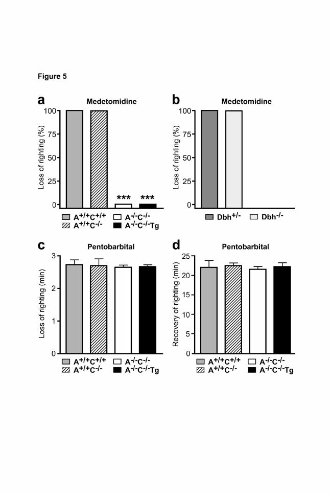
was assessed using the righting reflex (Fig. 5 a). At a dose of 1000 µg/kg, medetomidine induced a
strong sedative effect in A+/+C+/+ and A+/+C-/- mice, as all of the drug-treated mice lost their righting
reflex (LORR) (Fig. 5 a). None of the A-/-C-/- or A-/-C-/-Tg mice lost the righting reflex, indicating that
α2A-receptors in adrenergic cells were not required for the sedative effects of the α2-agonist
medetomidine. In order to confirm that α2-mediated LORR is independent of modulating
norepinephrine release, we tested dopamine β-hydroxylase deficient mice (Dbh-/-), which are unable
to synthesize norepinephrine (Thomas et al., 1995; Weinshenker et al., 2008). Medetomidine induced
LORR in all control (Dbh+/-) and Dbh-/- mice (Fig. 5 b). In order to exclude that α2-receptor deletion
and/or transgenic expression affected sensitivity to sedative stimuli non-specifically, mice received
the GABAA receptor agonist pentobarbital and the times until loss (Fig. 5 c) or recovery (Fig. 5 d) of
the righting reflex were recorded. Induction and duration of the hypnotic effect of pentobarbital did not
differ between the four α2-genotypes.
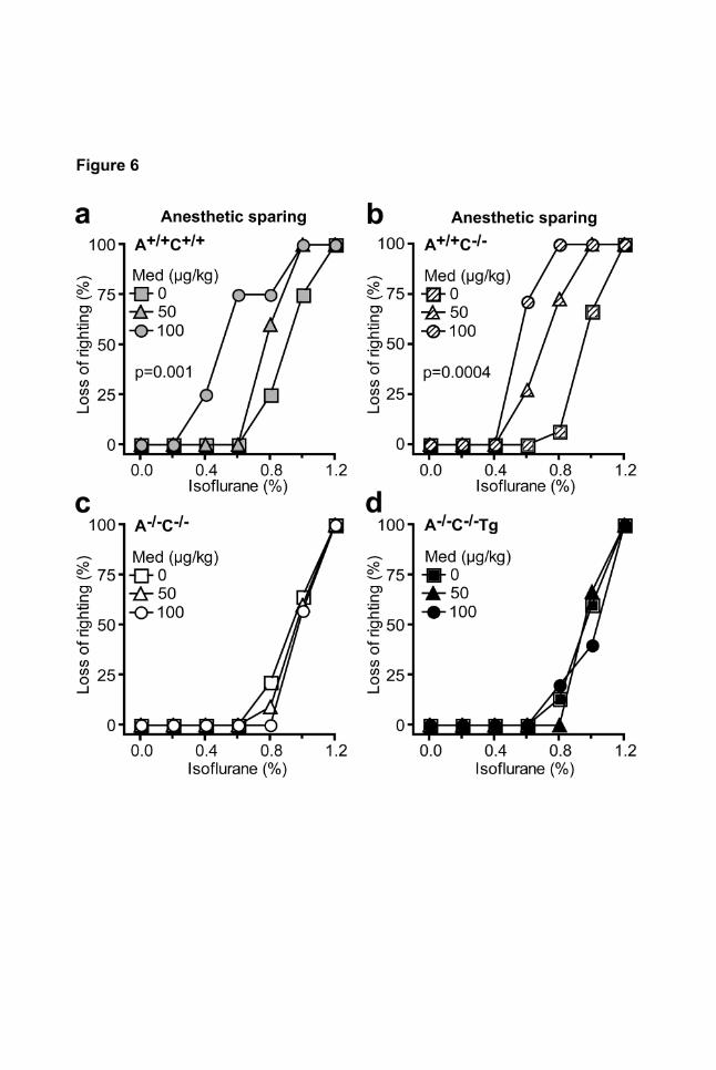
Since α2-agonists have been documented to lower the dose of inhalation anesthetics required for
anesthesia (Lakhlani et al., 1997), we evaluated the anesthetic-sparing effect of non-sedative doses
of medetomidine during isoflurane anesthesia (Fig.6). In A+/+C+/+ and A+/+C-/- mice, medetomidine
shifted the isoflurane dose response curve to the left (Fig. 6 a, b). This anesthetic-sparing effect of
medetomidine was ablated by deletion of α2A-/α2C-receptors (A-/-C-/-) and was not rescued by
transgenic expression of α2A-autoreceptors (A-/-C-/-Tg, Fig. 6 c, d).
Spontaneous locomotor activity was determined in mice two weeks after subcutaneous implantation
of a telemetric activity monitor and activity was measured during a controlled light-dark cycle in 2-
This article has not been copyedited and formatted. The final version may differ from this version.Molecular Pharmacology Fast Forward. Published on February 27, 2009 as DOI: 10.1124/mol.109.054544
at ASPE
T Journals on June 29, 2020
molpharm
.aspetjournals.orgD
ownloaded from
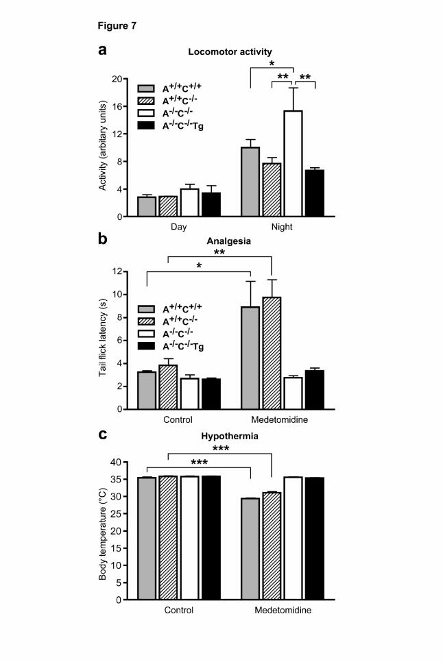
MOLPHARM/2009/054544 17 minute intervals (Fig. 7 a). During the light cycle, activity levels did not differ between genotypes (Fig.
7 a). During the dark cycle, however, locomotor activity of A-/-C-/- mice was significantly enhanced
compared with A+/+C+/+ and A+/+C-/- mice (Fig. 7 a). Transgenic expression of adrenergic cell α2A-
receptors reduced locomotor activity to the level of wild-type control mice.
Antinociceptive effects mediated by α2-adrenoceptors
α2-agonists mediate strong analgesic effects at spinal and supraspinal levels (for review, see
(Pertovaara, 2006; Sanders and Maze, 2007)). Thus, we determined the antinociceptive effect of
medetomidine in the tail-flick assay (Fig. 7 b). At baseline, the time to withdrawal of the tail from the
infrared light source did not differ significantly between genotypes. Medetomidine (250 µg/kg i.p.)
significantly prolonged the latency time of tail withdrawal in A+/+C+/+ or A+/+C-/- mice but not in A-/-C-/- or
in A-/-C-/-Tg mice (Fig. 7 b). Thus, α2A-autoreceptors are not essential for the antinociceptive effect of
medetomidine in the tail-flick assay.
α2-agonist-mediated hypothermia
At baseline, body temperature did not differ between genotypes (Fig. 7 c). When applied at a non-
sedative dose of 250 µg/kg medetomidine lowered body core temperature significantly by 5.9 ± 0.4°C
in A+/+C+/+ and A+/+C-/- mice (Fig. 7 c). However, the hypothermic effect was completely absent in A-/-
C-/- or in A-/-C-/-Tg mice, indicating that adrenergic cell α2-adrenoceptors are not required for this
function (Fig. 7 c).
This article has not been copyedited and formatted. The final version may differ from this version.Molecular Pharmacology Fast Forward. Published on February 27, 2009 as DOI: 10.1124/mol.109.054544
at ASPE
T Journals on June 29, 2020
molpharm
.aspetjournals.orgD
ownloaded from
MOLPHARM/2009/054544 18 Discussion
α2-adrenoceptors and receptors for acetylcholine and GABA were among the first receptors to be
identified as inhibitory feedback receptors which are located on their own transmitter’s nerve
terminals to inhibit the release of neurotransmitter. The discovery and investigation of these
prototypic presynaptic or “autoreceptors” has greatly advanced our understanding of the
neurobiology of transmitter release (for recent review, see (Sudhoff and Starke, 2008)). For most
transmitter systems, several receptor subtypes were identified as candidate autoreceptors by
molecular cloning. In isolated tissues from gene-targeted mice, all three cloned α2-adrenoceptors,
α2A, α2B, α2C, were shown to operate as inhibitory feedback receptors to control norepinephrine
release in vitro (Hein et al., 1999; Trendelenburg et al., 2003). However, it remained unknown, which
receptor subtype operated as an inhibitory autoreceptor in vivo and which effects of pharmacological
α2-receptor ligands are mediated via these autoreceptors on adrenergic neurons.
In order to distinguish between α2-adrenoceptor functions in adrenergic vs. non-adrenergic cells, we
have generated a transgenic model with specific expression of α2-receptors in adrenergic neurons
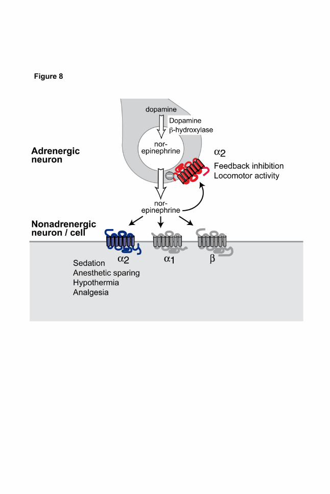
and subjected these mice to a comprehensive phenotype program. The primary finding of this study
is that the majority of α2-receptor agonist effects were mediated via α2-receptors in non-adrenergic
cells and not via adrenergic cell α2-receptors (Fig. 8, Table 1). The results of this study emphasize
the importance of non-adrenergic α2-adrenoceptors, but they do not completely exclude contribution
of α2-adrenoceptor functions in adrenergic cells.
This article has not been copyedited and formatted. The final version may differ from this version.Molecular Pharmacology Fast Forward. Published on February 27, 2009 as DOI: 10.1124/mol.109.054544
at ASPE
T Journals on June 29, 2020
molpharm
.aspetjournals.orgD
ownloaded from
MOLPHARM/2009/054544 19 Transgenic model to distinguish receptor functions in adrenergic cells from non-adrenergic
cells
Several lines of evidence indicated that the transgenic model generated to functionally separate α2-
receptors in adrenergic cells from non-adrenergic cells was successful. First, expression of α2A-
receptors under control of the dopamine β-hydroxylase promoter could be identified in adrenergic
cells and tissues, e.g. locus coeruleus and sympathetic ganglia. On the protein level, transgenic α2A-
receptors were detected by radioligand autoradiography and by immunohistochemistry. Using
immunoperoxidase labeling, transgenic α2A-receptors were identified in the presynaptic plasma
membrane but not in postsynaptic or intracellular transmitter vesicle membranes of hippocampal
neurons (Fig. 2 e). Furthermore, transgenic α2A-receptors completely restored the α2-autoreceptor
function in peripheral tissue innervated by sympathetic nerve fibers. Thus, taken together, expression
of transgenic α2A-adrenoceptors recapitulated the subcellular localization and in vitro function of α2-
adrenoceptors in adrenergic cells.
The transgenic strategy applied in the current study may have several limitations. Expression of the
Dbh-transgene may not be restricted to adrenergic cells (Hoyle et al., 1994; Mercer et al., 1991).
However, mRNA expression of the Dbh-α2A-transgene was 26-169 fold lower in non-adrenergic
regions of the CNS than in adrenergic nuclei, including locus coeruleus or sympathetic ganglia (Fig.
1d). Misexpression of α2A-receptors under control of the Dbh promoter used for the present study
may lead to false-positive assigments of α2-functions as autoreceptor, i.e. receptors in adrenergic
cells. Furthermore, higher than physiological levels of α2A-receptor expression may result in a gain of
function which is not achieved by endogenously expressed receptors. Indeed, we observed that
transgenic α2A-receptors compensated for the loss of both, α2A and α2C in sympathetic ganglia (see
This article has not been copyedited and formatted. The final version may differ from this version.Molecular Pharmacology Fast Forward. Published on February 27, 2009 as DOI: 10.1124/mol.109.054544
at ASPE
T Journals on June 29, 2020
molpharm
.aspetjournals.orgD
ownloaded from
MOLPHARM/2009/054544 20 Fig. 4b). Finally, α2A-adrenoceptors expressed under control of the Dbh promoter may alter their
expression pattern during embryonic development. While we cannot rule out alterations in neuronal
development, thorough macroscopical or microscopical investigation did not reveal differences
between transgenic and wild-type brains.
Transgenic lines were crossed with mice lacking α2A- and α2C-adrenoceptors as these two subtypes
represent the major presynaptic feedback inhibitors of norepinephrine release in vivo (Brede et al.,
2003; Hein et al., 1999). α2B-adrenoceptor-deficient mice were not included mainly for for two
reasons. First, the contribution of α2B in presynaptic feedback inhibition has been demonstrated in
isolated tissue preparations but not in vivo (Trendelenburg et al., 2003). Second, homozygous
deletion of the α2B-adrenoceptor gene was lethal during embryonic development and perinatally
(Philipp et al., 2002). However, future experiments will address whether α2-adrenoceptor subtypes
mediate specific functions in adrenergic vs. non-adrenergic cells.
α2-adrenoceptor functions in adrenergic vs. non-adrenergic cells
Using this model, only few α2-adrenoceptor functions could be ascribed to receptors expressed in
adrenergic cells: inhibition of Ca2+ currents in SCG neurons, inhibition of electrically evoked
norepinephrine from sympathetic nerves and modulation of spontaneous locomotor activity. All other
tested α2-receptors functions, including analgesia, sedation, anesthetic sparing and hypothermia,
required the presence of α2-adrenoceptors on non-adrenergic cells (Fig. 8). In addition to revising the
view of the physiology of the adrenergic system, the present results offer new insight into the
mechanisms of α2-agonist drugs. Most importantly, all CNS effects of α2-agonists on pain, sedation
and anesthetic sparing, as well as body temperature were mediated by α2-heteroreceptors. Several
This article has not been copyedited and formatted. The final version may differ from this version.Molecular Pharmacology Fast Forward. Published on February 27, 2009 as DOI: 10.1124/mol.109.054544
at ASPE
T Journals on June 29, 2020
molpharm
.aspetjournals.orgD
ownloaded from
MOLPHARM/2009/054544 21 mechanisms for the antinociceptive effects of α2-agonists have been identified on supraspinal and
spinal levels (Pertovaara, 2006; Sanders and Maze, 2007). Antinociceptive α2-receptors were
suggested to reside in spinal terminals of primary nociceptor neurons, in spinal pain relay neurons as
well as on dorsal horn excitatory interneurons. As none of these neuron groups synthesizes
(nor)epinephrine, they should be classified as non-adrenergic cell α2-adrenoceptors. The present
study demonstrates that intraperitoneal medetomidine indeed mediates its antinociceptive effect via
non-adrenergic cell α2-receptors in the tail flick assay (Fig. 7 b). This result is consistent with previous
reports from GIRK-2 mutant mice linking the analgesic effect also to postsynaptic α2-receptors
(Blednov et al., 2003).
In clinical anesthesia, α2-agonists may be used during the induction of anesthesia for their
anesthetic-sparing effect or in the perioperative period as potent sedative and analgesic drugs
(Kamibayashi and Maze, 2000). However, it has been difficult to identify whether α2-receptors
mediating sedation and hypnosis are pre- or postsynaptic receptors. According to one concept, the
locus coeruleus plays an important role in the sedative effect of α2-agonists (Mizobe et al., 1996). In
mice lacking functional α2A-adrenoceptors (α2A-D79N), the sedative and anesthetic-sparing effects of
the α2-agonist dexmedetomidine were ablated (Lakhlani et al., 1997). In brain slices from α2A-D79N
mice, clonidine failed to inhibit spontaneous firing of locus coeruleus neurons (Lakhlani et al., 1997).
Based on these and other experiments, it was hypothesized that α2-agonists lower locus coeruleus
neuron activity via presynaptic inhibitory autoreceptors (Jones, 2005). While the present results
confirm the role of the α2A-subtype for the sedative effect of α2-agonists, they also demonstrate that
α2-autoreceptors are not essential for this effect. Neither the anesthetic-sparing nor the sedative
effects of the α2-agonist medetomidine could be rescued by transgenic expression of α2-
This article has not been copyedited and formatted. The final version may differ from this version.Molecular Pharmacology Fast Forward. Published on February 27, 2009 as DOI: 10.1124/mol.109.054544
at ASPE
T Journals on June 29, 2020
molpharm
.aspetjournals.orgD
ownloaded from
MOLPHARM/2009/054544 22 adrenoceptors in adrenergic cells (Fig. 5, 6). Furthermore, the α2-agonist medetomidine still resulted
in a loss of the righting reflex in mice with genetic deficiency in dopamine β-hydroxylase, the key
enzyme required for synthesis of norepinephrine from dopamine (Thomas et al., 1995; Weinshenker
et al., 2008). In accordance with these data, earlier studies had shown that depletion of
norepinephrine from central adrenergic neurons by the neurotoxin DSP-4 or by direct injection of 6-
hydroxydopamine into the locus coeruleus of rats did not affect the sedative effects of clonidine
(Spyraki and Fibiger, 1982). Thus, the sedative effects of α2-agonists may essentially require α2-
adrenoceptors in non-adrenergic neurons in the CNS.
Norepinephrine has anticonvulsant properties in most seizure models, but the effects of α2-agonists
has been ambiguous. A previous study using dopamine β-hydroxylase knockout mice showed that
the proconvulsant effects of α2-agonists were mediated by the α2A-autoreceptor, while the
anticonvulsant effects of α2-agonists were mediated by α2A-receptors on non-adrenergic neurons
(Szot et al., 2004; Weinshenker and Szot, 2002). These results suggest that the development of
selective α2A-agonists for non-adrenergic cell receptors may also be effective anti-seizure
medications. Furthermore, a recent study has identified a neuronal pathway linking α2-adrenoceptors
in non-adrenergic brain cortex neurons via cAMP and HCN channel signalling with working memory
(Wang et al., 2007).
The present study paves the way to a search for new α2-adrenoceptor based therapeutic strategies.
The majority of pharmacological effects of α2-agonists are mediated by receptors in non-adrenergic
cells, which may greatly differ in their localization and intracellular signal transduction and effector
coupling. In conclusion, following up these pathways will result in a better understanding of receptor
This article has not been copyedited and formatted. The final version may differ from this version.Molecular Pharmacology Fast Forward. Published on February 27, 2009 as DOI: 10.1124/mol.109.054544
at ASPE
T Journals on June 29, 2020
molpharm
.aspetjournals.orgD
ownloaded from

MOLPHARM/2009/054544 23 subtype und functional diversity within the adrenergic system and may provide important new
considerations for future drug development.
This article has not been copyedited and formatted. The final version may differ from this version.Molecular Pharmacology Fast Forward. Published on February 27, 2009 as DOI: 10.1124/mol.109.054544
at ASPE
T Journals on June 29, 2020
molpharm
.aspetjournals.orgD
ownloaded from
MOLPHARM/2009/054544 24 Acknowledgments
The authors whish to thank Dr. Eva Schmitteckert, Würzburg, for assistance with the generation of
transgenic mouse lines, and Claudia Schurr, Freiburg, for assistance with genotyping. The authors
thank Dr. R. Palmiter (Seattle, USA) for providing a plasmid construct containing the DBH promoter
and Dr. M. Dziedzicka-Wasylewska (Krakow, Poland) for autoradiography.
This article has not been copyedited and formatted. The final version may differ from this version.Molecular Pharmacology Fast Forward. Published on February 27, 2009 as DOI: 10.1124/mol.109.054544
at ASPE
T Journals on June 29, 2020
molpharm
.aspetjournals.orgD
ownloaded from
MOLPHARM/2009/054544 25 References
Blednov YA, Stoffel M, Alva H and Harris RA (2003) A pervasive mechanism for analgesia: activation
of GIRK2 channels. Proc Natl Acad Sci USA 100:277-282.
Bourdelat-Parks BN, Anderson GM, Donaldson ZR, Weiss JM, Bonsall RW, Emery MS, Liles LC and
Weinshenker D (2005) Effects of dopamine β-hydroxylase genotype and disulfiram inhibition
on catecholamine homeostasis in mice. Psychopharmacol 183:72-80.
Brede M, Nagy G, Philipp M, Sorensen JB, Lohse MJ and Hein L (2003) Differential control of
adrenal and sympathetic catecholamine release by α2-adrenoceptor subtypes. Mol Endocrinol
17:1640-1646.
Bylund DB, Eikenberg DC, Hieble JP, Langer SZ, Lefkowitz RJ, Minneman KP, Molinoff PB, Ruffolo
RR, Jr. and Trendelenburg U (1994) International Union of Pharmacology nomenclature of
adrenoceptors. Pharmacol Rev 46:121-136.
Daunt DA, Hurt C, Hein L, Kallio J, Feng F and Kobilka BK (1997) Subtype-specific intracellular
trafficking of α2-adrenergic receptors. Mol Pharmacol 51:711-720.
Fagerholm V, Philipp M, Hein L and Scheinin M (2004) [Ethyl-3H]RS-79948-197 α2-adrenoceptor
autoradiography validation in α2-adrenoceptor knockout mice. Eur J Pharmacol 497:301-309.
Gilsbach R, Brede M, Beetz N, Moura E, Muthig V, Gerstner C, Barreto F, Neubauer S, Vieira-
Coelho MA and Hein L (2007) Heterozygous α2C-adrenoceptor-deficient mice develop heart
failure after transverse aortic constriction. Cardiovasc Res 75:728-737.
Gilsbach R and Hein L (2008) Presynaptic metabotropic receptors for acetylcholine and
adrenaline/noradrenaline. Handb Exp Pharmacol 184:261-288.
Hein L, Altman JD and Kobilka BK (1999) Two functionally distinct α2-adrenergic receptors regulate
sympathetic neurotransmission. Nature 402:181-184.
This article has not been copyedited and formatted. The final version may differ from this version.Molecular Pharmacology Fast Forward. Published on February 27, 2009 as DOI: 10.1124/mol.109.054544
at ASPE
T Journals on June 29, 2020
molpharm
.aspetjournals.orgD
ownloaded from
MOLPHARM/2009/054544 26 Hein L, Ishii K, Coughlin SR and Kobilka BK (1994) Intracellular targeting and trafficking of thrombin
receptors. A novel mechanism for resensitization of a G protein-coupled receptor. J Biol Chem
269:27719-27726.
Hoyle GW, Mercer EH, Palmiter RD and Brinster RL (1994) Cell-specific expression from the human
dopamine β-hydroxylase promoter in transgenic mice is controlled via a combination of
positive and negative regulatory elements. J Neurosci 14:2455-2463.
Jones BE (2005) From waking to sleeping: neuronal and chemical substrates. Trends Pharmacol Sci
26:578-586.
Kamibayashi T and Maze M (2000) Clinical uses of α2-adrenergic agonists. Anesthesiol 93:1345-
1349.
Lakhlani PP, MacMillan LB, Guo TZ, McCool BA, Lovinger DM, Maze M and Limbird LE (1997)
Substitution of a mutant α2A-adrenergic receptor via "hit and run" gene targeting reveals the
role of this subtype in sedative, analgesic, and anesthetic-sparing responses in vivo. Proc Natl
Acad Sci USA 94:9950-9955.
Link RE, Desai K, Hein L, Stevens ME, Chruscinski A, Bernstein D, Barsh GS and Kobilka BK (1996)
Cardiovascular regulation in mice lacking α2-adrenergic receptor subtypes b and c. Science
273:803-805.
Lymperopoulos A, Rengo G, Funakoshi H, Eckhart AD and Koch WJ (2007) Adrenal GRK2
upregulation mediates sympathetic overdrive in heart failure. Nat Med 13:315-323.
MacMillan LB, Hein L, Smith MS, Piascik MT and Limbird LE (1996) Central hypotensive effects of
the α2A-adrenergic receptor subtype. Science 273:801-803.
Maze M, Scarfini C and Cavaliere F (2001) New agents for sedation in the intensive care unit. Crit
Care Clin 17881-897.
This article has not been copyedited and formatted. The final version may differ from this version.Molecular Pharmacology Fast Forward. Published on February 27, 2009 as DOI: 10.1124/mol.109.054544
at ASPE
T Journals on June 29, 2020
molpharm
.aspetjournals.orgD
ownloaded from
MOLPHARM/2009/054544 27 Mercer EH, Hoyle GW, Kapur RP, Brinster RL and Palmiter RD (1991) The dopamine beta-
hydroxylase gene promoter directs expression of E. coli lacZ to sympathetic and other
neurons in adult transgenic mice. Neuron 7:703-716.
Mizobe T, Maghsoudi K, Sitwala K, Tianzhi G, Ou J and Maze M (1996) Antisense technology
reveals the α2A adrenoceptor to be the subtype mediating the hypnotic response to the highly
selective agonist, dexmedetomidine, in the locus coeruleus of the rat. J Clin Invest 98:1076-
1080.
Paxinos G and Franklin KBJ (2001) The mouse brain in stereotaxic coordinates. Academic Press.
Pertovaara A (2006) Noradrenergic pain modulation. Prog Neurobiol 80:53-83.
Philipp M, Brede ME, Hadamek K, Gessler M, Lohse MJ and Hein L (2002) Placental α2-
adrenoceptors control vascular development at the interface between mother and embryo. Nat
Genet 31:311-315.
Sanders RD and Maze M (2007) Adrenergic and cholinergic compounds. Handb Exp Pharmacol
177:251-264.
Small KM, Wagoner LE, Levin AM, Kardia SL and Liggett SB (2002) Synergistic polymorphisms of
β1- and α2C-adrenergic receptors and the risk of congestive heart failure. N Engl J Med
347:1135-1142.
Spyraki C and Fibiger HC (1982) Clonidine-induced sedation in rats: evidence for mediation by
postsynaptic α2-adrenoreceptors. J Neur Transm 54:153-163.
Starke K, Endo T and Taube HD (1975) Pre- and postsynaptic components in effect of drugs with
alpha adrenoceptor affinity. Nature 254:440-441.
Sudhoff TC and Starke K (2008) Neurotransmitter release. Springer, Heidelberg.
This article has not been copyedited and formatted. The final version may differ from this version.Molecular Pharmacology Fast Forward. Published on February 27, 2009 as DOI: 10.1124/mol.109.054544
at ASPE
T Journals on June 29, 2020
molpharm
.aspetjournals.orgD
ownloaded from
MOLPHARM/2009/054544 28 Szot P, Lester M, Laughlin ML, Palmiter RD, Liles LC and Weinshenker D (2004) The anticonvulsant
and proconvulsant effects of α2-adrenoreceptor agonists are mediated by distinct populations
of α2A-adrenoreceptors. Neuroscience 126:795-803.
Thomas SA, Marck BT, Palmiter RD and Matsumoto AM (1998) Restoration of norepinephrine and
reversal of phenotypes in mice lacking dopamine β-hydroxylase. J Neurochem 70:2468-2476.
Thomas SA, Matsumoto AM and Palmiter RD (1995) Noradrenaline is essential for mouse fetal
development. Nature 374:643-646.
Trendelenburg AU, Philipp M, Meyer A, Klebroff W, Hein L and Starke K (2003) All three α2-
adrenoceptor types serve as autoreceptors in postganglionic sympathetic neurons. Naunyn
Schmiedeberg's Arch Pharmacol 368:504-512.
Wang M, Ramos BP, Paspalas CD, Shu Y, Simen A, Duque A, Vijayraghavan S, Brennan A, Dudley
A, Nou E, Mazer JA, McCormick DA and Arnsten AF (2007) α2-adrenoceptors strengthen
working memory networks by inhibiting cAMP-HCN channel signaling in prefrontal cortex. Cell
129:397-410.
Weinshenker D, Ferrucci M, Busceti CL, Biagioni F, Lazzeri G, Liles LC, Lenzi P, Pasquali L, Murri L,
Paparelli A and Fornai F (2008) Genetic or pharmacological blockade of noradrenaline
synthesis enhances the neurochemical, behavioral, and neurotoxic effects of
methamphetamine. J Neurochem 105:471-483.
Weinshenker D and Szot P (2002) The role of catecholamines in seizure susceptibility: new results
using genetically engineered mice. Pharmacol Ther 94:213-233.
Westfall TC and Westfall DP (2006) Adrenergic agonists and antagonists, in Goodman & Gilman's
The Pharmacological Basis of Therapeutics (Brunton LL, Lazo JS and Parker KL eds) pp 237-
295, McGraw-Hill, New York.
This article has not been copyedited and formatted. The final version may differ from this version.Molecular Pharmacology Fast Forward. Published on February 27, 2009 as DOI: 10.1124/mol.109.054544
at ASPE
T Journals on June 29, 2020
molpharm
.aspetjournals.orgD
ownloaded from
MOLPHARM/2009/054544 29 Footnotes
This project was supported by the Deutsche Forschungsgemeinschaft DFG (HE 2073/3-1, in support
of L.H. and R.G.) and the DFG excellence cluster BIOSS (in support of L.H. and N.B.).
Please address reprint requests to: Lutz Hein, Institute of Experimental and Clinical Pharmacology
and Toxicology, University of Freiburg, Albertstrasse 25, 79104 Freiburg, Germany, Phone ++49-
761-2035314, Fax ++49-761-2035318, [email protected]
This article has not been copyedited and formatted. The final version may differ from this version.Molecular Pharmacology Fast Forward. Published on February 27, 2009 as DOI: 10.1124/mol.109.054544
at ASPE
T Journals on June 29, 2020
molpharm
.aspetjournals.orgD
ownloaded from
MOLPHARM/2009/054544 30 Figure Legends
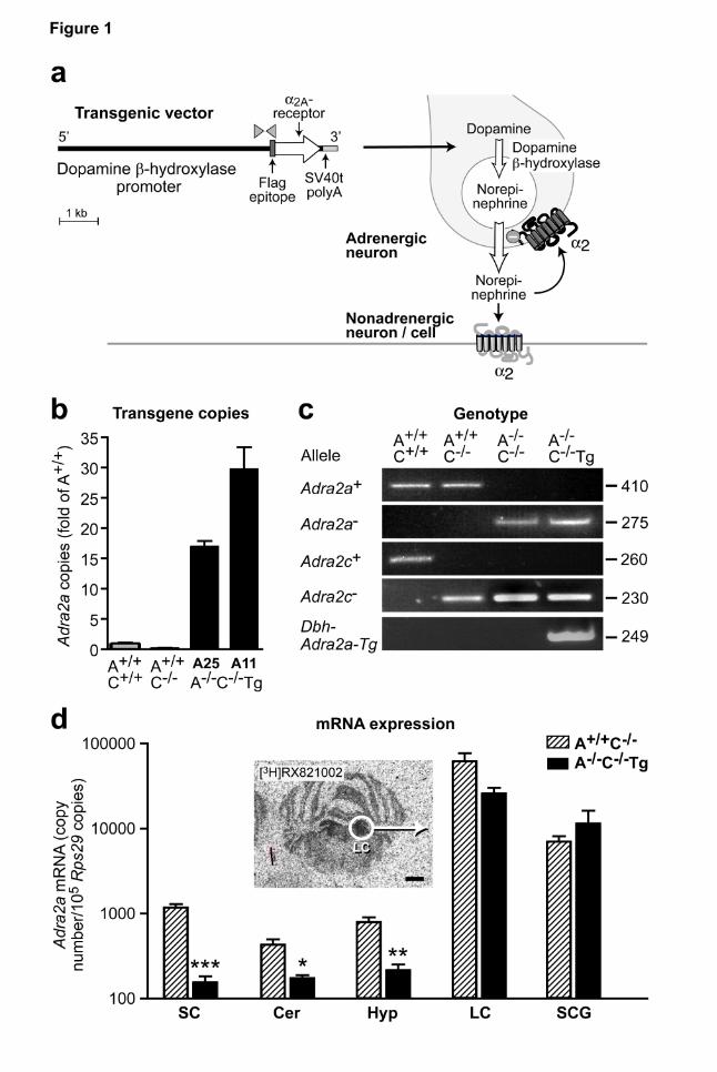
Figure 1 | Generation of transgenic mouse lines to dissect α2-adrenoceptor functions in
adrnergic vs. non-adrenergic cells. a, Schematic representation of the transgenic Dbh-Adra2a
vector to achieve seletive expression of α2A adrenoceptors in adrenergic neurons. The human
dopamine β-hydroxylase promoter was cloned upstream of the coding sequence of the murine α2A-
adrenoceptor, which was epitope-tagged at the aminoterminus for immunodetection (Flag epitope,
DYKDDDD (Daunt et al., 1997)). Dbh-Adra2a transgenic mice were backcrossed with Adra2a-/-
Adra2c-/- mice. Gray arrowheads indicate location of PCR primers used for genotyping. b, Transgene
copy number as determined by quantitative PCR on genomic DNA. c, Representative polymerase
chain reactions to identify mice with normal α2-adrenoceptor expression (”A+/+C+/+”, i.e. Adra2a+/+
Adra2c+/+, lane 1), mice deficient in α2C-receptors (“A+/+C-/-“, i.e. Adra2a+/+ Adra2c-/-, lane 2), mice
without functional α2A- and α2C-adrenoceptors (”A-/-C-/-”, i.e. Adra2a-/- Adra2c-/-, lane 3), or with
selective expression of α2A-adrenoceptors in adrenergic cells (”A-/-C-/-Tg”, i.e. Adra2a-/- Adra2c-/- Dbh-
Adra2a-Tg, lane 4). d, Expression of α2A-adrenoceptor mRNA in brain tissues as determined by
quantitative RT-PCR. Results are normalized to Rps29 expression (means ± SEM, n=4 samples per
genotype, *P<0.05, **P<0.01, ***P<0.001 vs. A+/+C-/-). No α2A mRNA expression could be detected in
specimens isolated from A-/-C-/- mice (not shown). Specimens from the locus coeruleus were
microdissected from cryostat sections. Insert: [3H]RX821002 autoradiography to detect α2-
adrenoceptors in transverse mouse brain sections with highlighted locus coeruleus (white circle
marks microdissected area for qPCR. Abbreviations: SC, spinal cord; Cer, cerebellum; Hyp,
hypothalamus; LC, locus coeruleus; SCG, superior cervical ganglia.
This article has not been copyedited and formatted. The final version may differ from this version.Molecular Pharmacology Fast Forward. Published on February 27, 2009 as DOI: 10.1124/mol.109.054544
at ASPE
T Journals on June 29, 2020
molpharm
.aspetjournals.orgD
ownloaded from
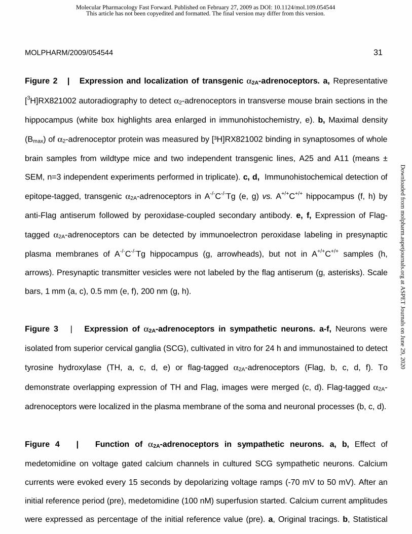
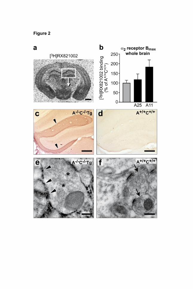
MOLPHARM/2009/054544 31 Figure 2 | Expression and localization of transgenic α2A-adrenoceptors. a, Representative
[3H]RX821002 autoradiography to detect α2-adrenoceptors in transverse mouse brain sections in the
hippocampus (white box highlights area enlarged in immunohistochemistry, e). b, Maximal density
(Bmax) of α2-adrenoceptor protein was measured by [³H]RX821002 binding in synaptosomes of whole
brain samples from wildtype mice and two independent transgenic lines, A25 and A11 (means ±
SEM, n=3 independent experiments performed in triplicate). c, d, Immunohistochemical detection of
epitope-tagged, transgenic α2A-adrenoceptors in A-/-C-/-Tg (e, g) vs. A+/+C+/+ hippocampus (f, h) by
anti-Flag antiserum followed by peroxidase-coupled secondary antibody. e, f, Expression of Flag-
tagged α2A-adrenoceptors can be detected by immunoelectron peroxidase labeling in presynaptic
plasma membranes of A-/-C-/-Tg hippocampus (g, arrowheads), but not in A+/+C+/+ samples (h,
arrows). Presynaptic transmitter vesicles were not labeled by the flag antiserum (g, asterisks). Scale
bars, 1 mm (a, c), 0.5 mm (e, f), 200 nm (g, h).
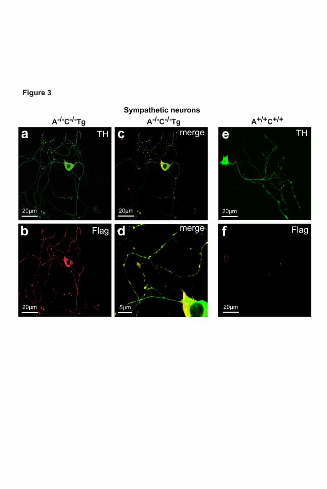
Figure 3 | Expression of α2A-adrenoceptors in sympathetic neurons. a-f, Neurons were
isolated from superior cervical ganglia (SCG), cultivated in vitro for 24 h and immunostained to detect
tyrosine hydroxylase (TH, a, c, d, e) or flag-tagged α2A-adrenoceptors (Flag, b, c, d, f). To
demonstrate overlapping expression of TH and Flag, images were merged (c, d). Flag-tagged α2A-
adrenoceptors were localized in the plasma membrane of the soma and neuronal processes (b, c, d).
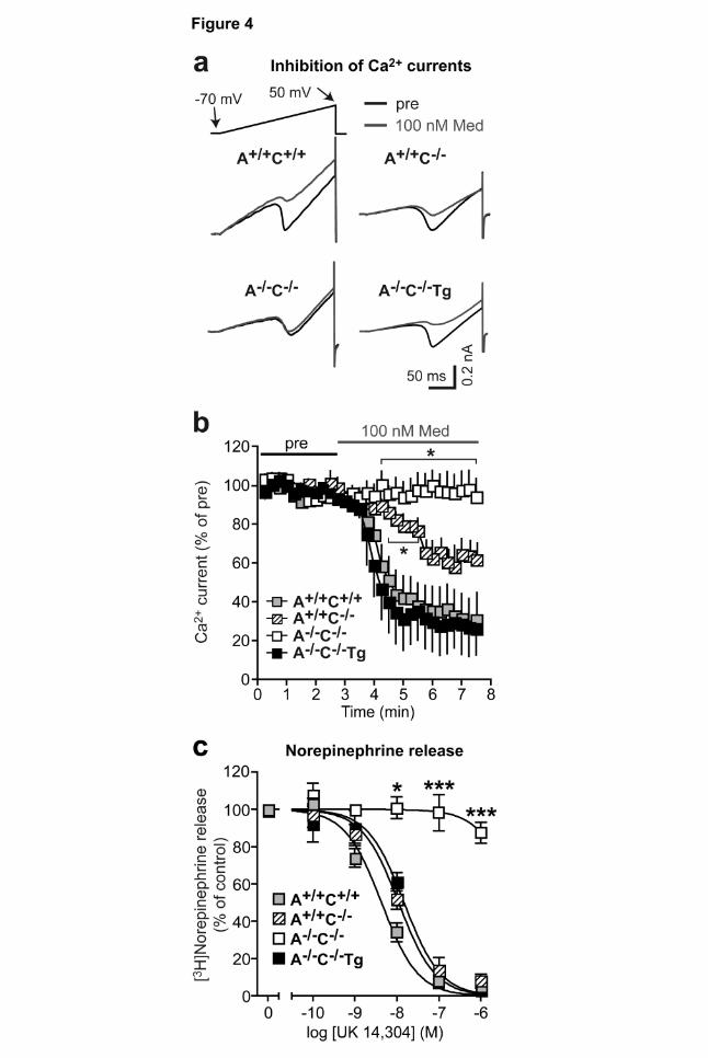
Figure 4 | Function of α2A-adrenoceptors in sympathetic neurons. a, b, Effect of
medetomidine on voltage gated calcium channels in cultured SCG sympathetic neurons. Calcium
currents were evoked every 15 seconds by depolarizing voltage ramps (-70 mV to 50 mV). After an
initial reference period (pre), medetomidine (100 nM) superfusion started. Calcium current amplitudes
were expressed as percentage of the initial reference value (pre). a, Original tracings. b, Statistical
This article has not been copyedited and formatted. The final version may differ from this version.Molecular Pharmacology Fast Forward. Published on February 27, 2009 as DOI: 10.1124/mol.109.054544
at ASPE
T Journals on June 29, 2020
molpharm
.aspetjournals.orgD
ownloaded from
MOLPHARM/2009/054544 32 evaluation: mean ± SEM of 3 (A+/+ C+/+), 6 (A+/+ C-/-), 5 (A-/- C-/-) and 5 (A-/- C-/- Tg) experiments. *
P<0.05 vs. A+/+C+/+. c, Feedback control of norepinephrine release from sympathetic nerves. The
α2A,α2C-agonist UK14,304 inhibited overflow of [3H]norepinephrine from isolated A+/+C+/+, A+/+C-/- and
A-/-C-/-Tg atria, but not from A-/-C-/- atria. Atria were stimulated by field stimulation with 20 rectangular
electrical pulses at 50 Hz (1 ms pulse width, 80 mA) applied at 16 min intervals (*P<0.05 vs. A+/+C+/+
control, n=6-10 samples per genotype).
Figure 5 | α2-agonist-mediated loss of righting reflex. a, Intraperitoneal medetomidine
(1000 µg/kg) induced a loss of the righting reflex in A+/+C+/+ and A+/+C-/- mice but not in A-/-C-/- or A-/-C-
/-Tg mice (*P<0.05, n=6-9 per genotype). b, Medetomidine (1000 µg/kg i.p.) induced a loss of the
righting reflex in dopamine β-hydroxylase-deficient mice (Dbh+/-, Dbh-/-) (n=6 per genotype). c,d,
Onset (c) and recovery time (d) of pentobarbital-induced loss of the righting reflex did not differ
between genotype groups (50 mg/kg pentobarbital i.p., n=6).
Figure 6 | Anesthetic-sparing effect of the α2-adrenoceptor agonist medetomidine.
Medetomidine was applied intraperitoneally at doses which did not cause a loss of the righting reflex
(50, 100 µg/kg) 15 min before exposure to increasing concentrations of isoflurane (in 1 L/min O2
flow). Medetomidine induced a leftward shift of the isoflurane-mediated loss of righting reflex curves
in A+/+C+/+ (a) and A+/+C-/- mice (b), but not in A-/-C-/- (c) or A-/-C-/- Tg (d) mice (n=10-15 per genotype).
Figure 7 | Locomotor, analgesic and hypothermic effects of the α2-agonist medetomidine in
α2A-transgenic mice. a, Locomotor activity was monitored two weeks after implantation of a
telemetric activity monitor. At nighttime, spontaneous locomotor activity was significantly greater in A-
/-C-/- mice as compared with A+/+C+/+, A+/+C-/- or A-/-C-/-Tg mice (n=6 per genotype). b, Medetomidine
This article has not been copyedited and formatted. The final version may differ from this version.Molecular Pharmacology Fast Forward. Published on February 27, 2009 as DOI: 10.1124/mol.109.054544
at ASPE
T Journals on June 29, 2020
molpharm
.aspetjournals.orgD
ownloaded from
MOLPHARM/2009/054544 33 (250 µg/kg i.p.) prolonged the tail flick latency in A+/+C+/+, A+/+C-/- but not in A-/-C-/- or A-/-C-/-Tg mice
(n=6-9 per genotype). c, Medetomidine (250 µg/kg i.p.) significantly lowered body temperature in
A+/+C+/+, A+/+C-/- but not in A-/-C-/- or A-/-C-/-Tg mice (n=8-12 per genotype, *P<0.05, **P<0.01,
***P<0.001).
Figure 8 | Schematic representation of α2-adrenoceptor functions mediated by receptors in
adrenergic cells vs. non-adrenergic cells.
This article has not been copyedited and formatted. The final version may differ from this version.Molecular Pharmacology Fast Forward. Published on February 27, 2009 as DOI: 10.1124/mol.109.054544
at ASPE
T Journals on June 29, 2020
molpharm
.aspetjournals.orgD
ownloaded from
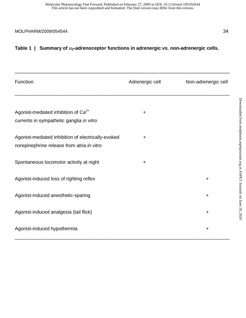
MOLPHARM/2009/054544 34 Table 1 | Summary of α2-adrenoceptor functions in adrenergic vs. non-adrenergic cells.
________________________________________________________________________________
Function Adrenergic cell Non-adrenergic cell
________________________________________________________________________________
Agonist-mediated inhibition of Ca2+ +
currents in sympathetic ganglia in vitro
Agonist-mediated inhibition of electrically-evoked +
norepinephrine release from atria in vitro
Spontaneous locomotor activity at night +
Agonist-induced loss of righting reflex +
Agonist-induced anesthetic-sparing +
Agonist-induced analgesia (tail flick) +
Agonist-induced hypothermia +
________________________________________________________________________________
This article has not been copyedited and formatted. The final version may differ from this version.Molecular Pharmacology Fast Forward. Published on February 27, 2009 as DOI: 10.1124/mol.109.054544
at ASPE
T Journals on June 29, 2020
molpharm
.aspetjournals.orgD
ownloaded from
This article has not been copyedited and formatted. The final version may differ from this version.Molecular Pharmacology Fast Forward. Published on February 27, 2009 as DOI: 10.1124/mol.109.054544
at ASPE
T Journals on June 29, 2020
molpharm
.aspetjournals.orgD
ownloaded from
This article has not been copyedited and formatted. The final version may differ from this version.Molecular Pharmacology Fast Forward. Published on February 27, 2009 as DOI: 10.1124/mol.109.054544
at ASPE
T Journals on June 29, 2020
molpharm
.aspetjournals.orgD
ownloaded from
This article has not been copyedited and formatted. The final version may differ from this version.Molecular Pharmacology Fast Forward. Published on February 27, 2009 as DOI: 10.1124/mol.109.054544
at ASPE
T Journals on June 29, 2020
molpharm
.aspetjournals.orgD
ownloaded from
This article has not been copyedited and formatted. The final version may differ from this version.Molecular Pharmacology Fast Forward. Published on February 27, 2009 as DOI: 10.1124/mol.109.054544
at ASPE
T Journals on June 29, 2020
molpharm
.aspetjournals.orgD
ownloaded from
This article has not been copyedited and formatted. The final version may differ from this version.Molecular Pharmacology Fast Forward. Published on February 27, 2009 as DOI: 10.1124/mol.109.054544
at ASPE
T Journals on June 29, 2020
molpharm
.aspetjournals.orgD
ownloaded from
This article has not been copyedited and formatted. The final version may differ from this version.Molecular Pharmacology Fast Forward. Published on February 27, 2009 as DOI: 10.1124/mol.109.054544
at ASPE
T Journals on June 29, 2020
molpharm
.aspetjournals.orgD
ownloaded from
This article has not been copyedited and formatted. The final version may differ from this version.Molecular Pharmacology Fast Forward. Published on February 27, 2009 as DOI: 10.1124/mol.109.054544
at ASPE
T Journals on June 29, 2020
molpharm
.aspetjournals.orgD
ownloaded from
This article has not been copyedited and formatted. The final version may differ from this version.Molecular Pharmacology Fast Forward. Published on February 27, 2009 as DOI: 10.1124/mol.109.054544
at ASPE
T Journals on June 29, 2020
molpharm
.aspetjournals.orgD
ownloaded from