β- Blockade in Sepsis- What is the Evidence? · Morelli 2016 Esmolol Obs 45 51% 150 Patients....
35
β- Blockade in Sepsis- What is the Evidence? Patient Engagement, Systems Science, and the Elimination of Preventable Harm Daniel Talmor, MD, MPH
Transcript of β- Blockade in Sepsis- What is the Evidence? · Morelli 2016 Esmolol Obs 45 51% 150 Patients....

β- Blockade in Sepsis-
What is the Evidence?
Patient Engagement, Systems Science, and the Elimination of Preventable Harm
Daniel Talmor, MD, MPH
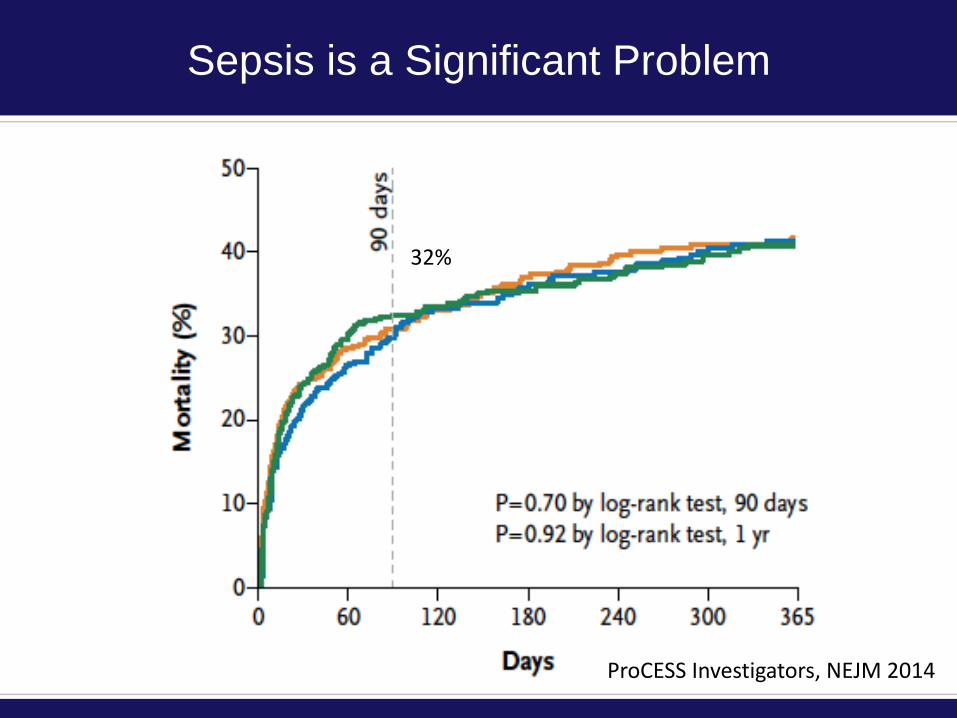
Sepsis is a Significant Problem
ProCESS Investigators, NEJM 2014
32%
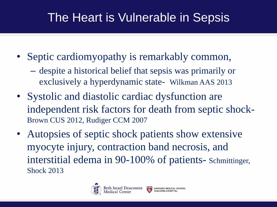
The Heart is Vulnerable in Sepsis
• Septic cardiomyopathy is remarkably common,
– despite a historical belief that sepsis was primarily or
exclusively a hyperdynamic state- Wilkman AAS 2013
• Systolic and diastolic cardiac dysfunction are
independent risk factors for death from septic shock-Brown CUS 2012, Rudiger CCM 2007
• Autopsies of septic shock patients show extensive
myocyte injury, contraction band necrosis, and
interstitial edema in 90-100% of patients- Schmittinger,
Shock 2013
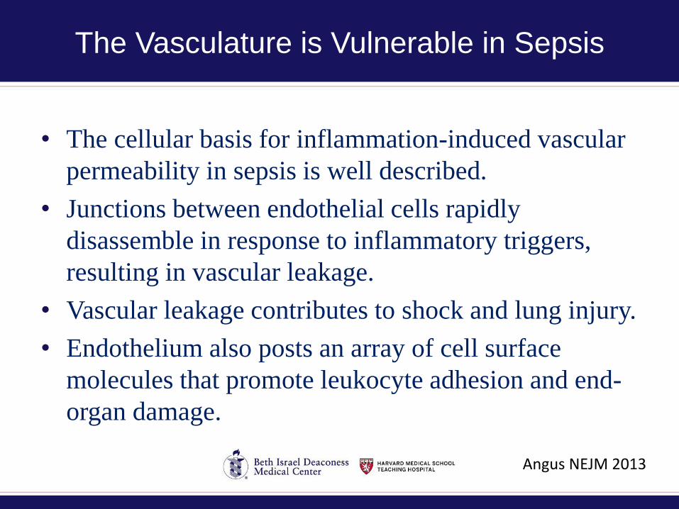
The Vasculature is Vulnerable in Sepsis
• The cellular basis for inflammation-induced vascular
permeability in sepsis is well described.
• Junctions between endothelial cells rapidly
disassemble in response to inflammatory triggers,
resulting in vascular leakage.
• Vascular leakage contributes to shock and lung injury.
• Endothelium also posts an array of cell surface
molecules that promote leukocyte adhesion and end-
organ damage.
Angus NEJM 2013
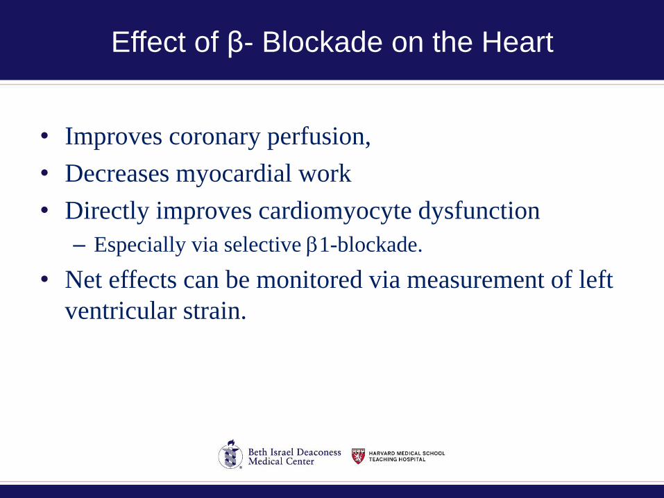
Effect of β- Blockade on the Heart
• Improves coronary perfusion,
• Decreases myocardial work
• Directly improves cardiomyocyte dysfunction
– Especially via selective 1-blockade.
• Net effects can be monitored via measurement of left
ventricular strain.
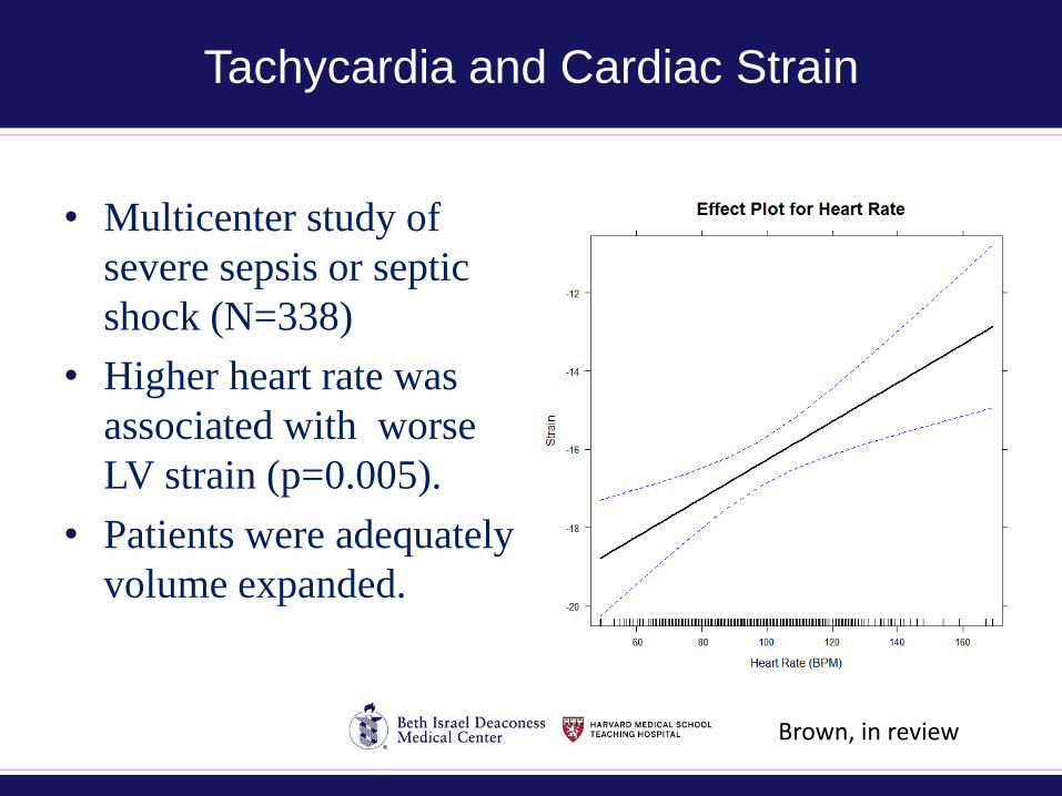
Tachycardia and Cardiac Strain
• Multicenter study of
severe sepsis or septic
shock (N=338)
• Higher heart rate was
associated with worse
LV strain (p=0.005).
• Patients were adequately
volume expanded.
Brown, in review
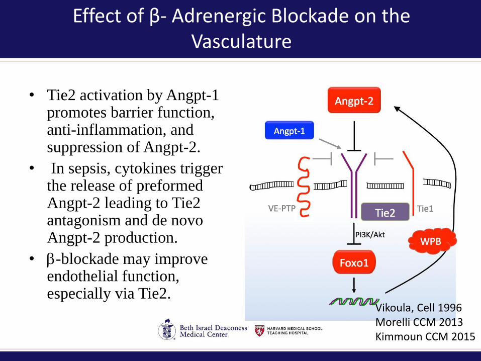
Effect of β- Adrenergic Blockade on the Vasculature
• Tie2 activation by Angpt-1 promotes barrier function, anti-inflammation, and suppression of Angpt-2.
• In sepsis, cytokines trigger the release of preformed Angpt-2 leading to Tie2 antagonism and de novo Angpt-2 production.
• -blockade may improve endothelial function, especially via Tie2.
Vikoula, Cell 1996Morelli CCM 2013Kimmoun CCM 2015
Persistent Tachycardia After Volume Expansion
Represents Adrenergic Over-Stimulation
• Early in septic shock, low cardiac filling pressures caused by capillary leak induce a reflexive tachycardia that maintains cardiac output despite lower stroke volume.
• In half or more of patients with septic shock, tachycardia persists after adequate volume expansion.
• Persistent tachycardia marks patients suffering from adrenergic over-stimulation.
• Persistent tachycardia in septic shock is associated with higher mortality.
Parker CCM 1987Kumar Crit Care 2008
Hypothesis Underlying Treatment with β-
Blockers in Septic Shock
• Cardiovascular dysfunction is central to the MODS
that precedes death.
• Usual treatment paradigm for septic shock involves
further adrenergic stimulation.
• Safe, effective, and selective adrenergic antagonists
may be beneficial in treatment of septic shock.
• Non-invasive methods can assess cardiovascular
responses to septic shock and its treatment.
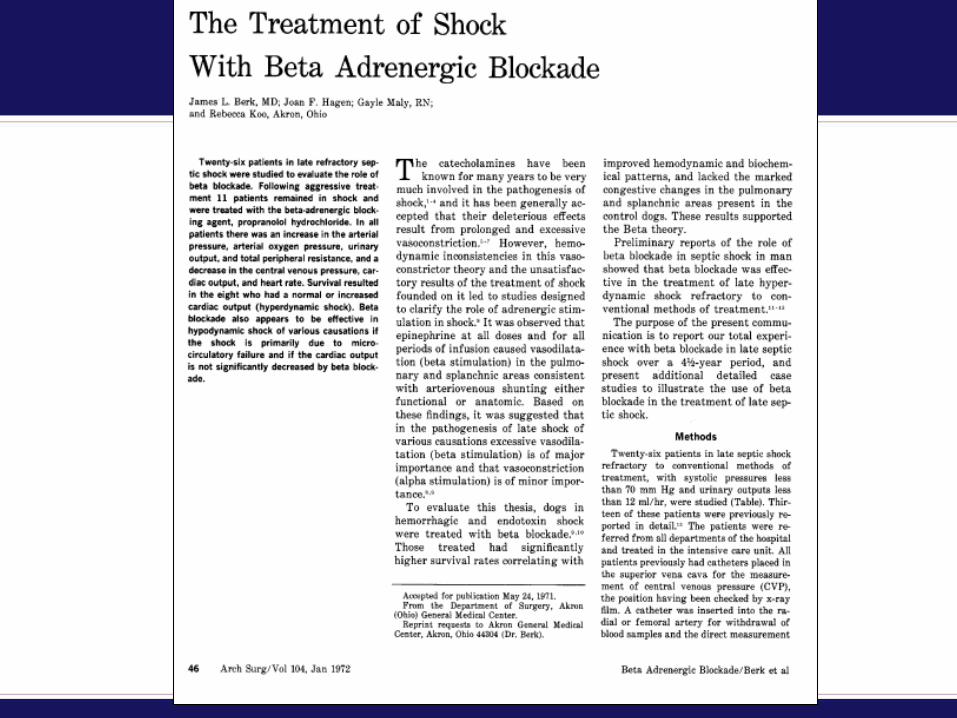
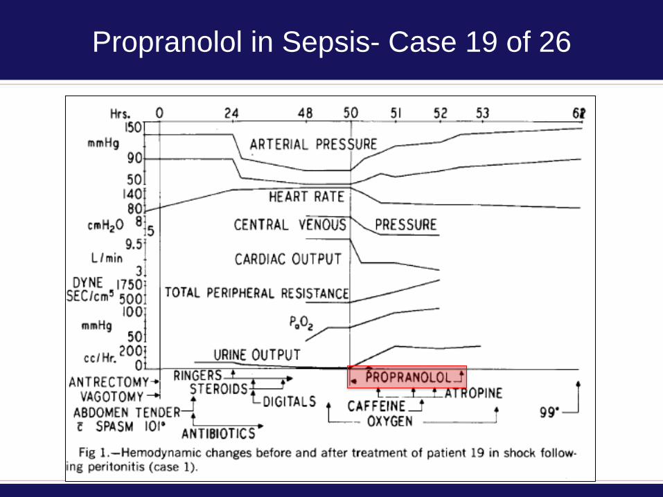
Propranolol in Sepsis- Case 19 of 26
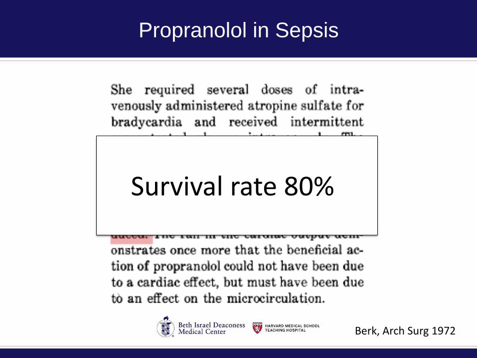
Propranolol in Sepsis
Berk, Arch Surg 1972
Survival rate 80%%
Retrospective data
AAS, 2013
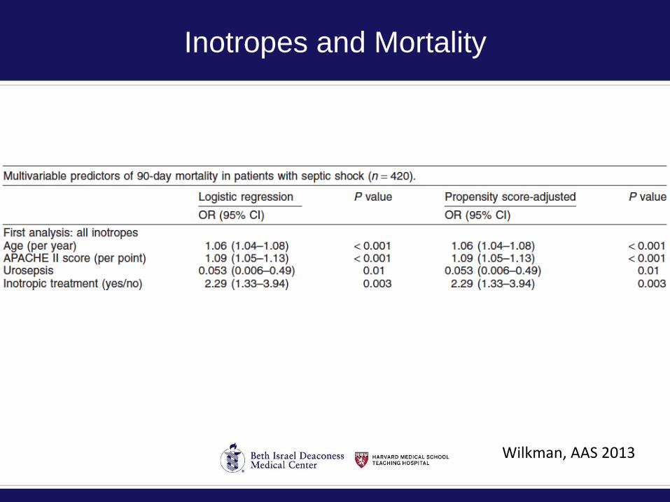
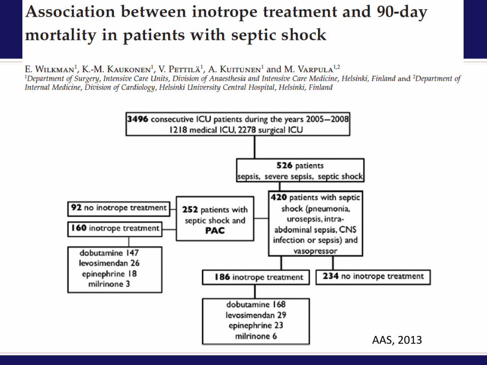
Inotropes and Mortality
Wilkman, AAS 2013
CCM 2012
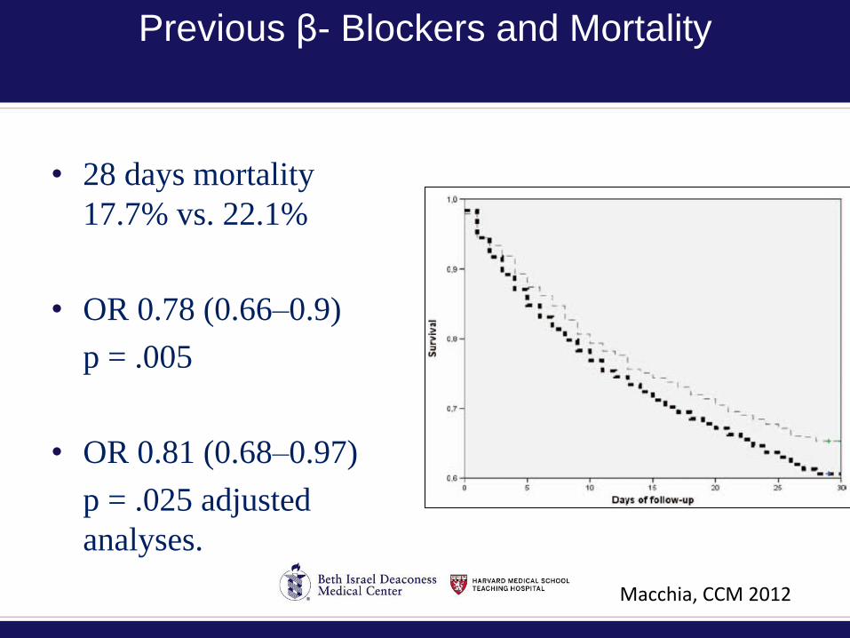
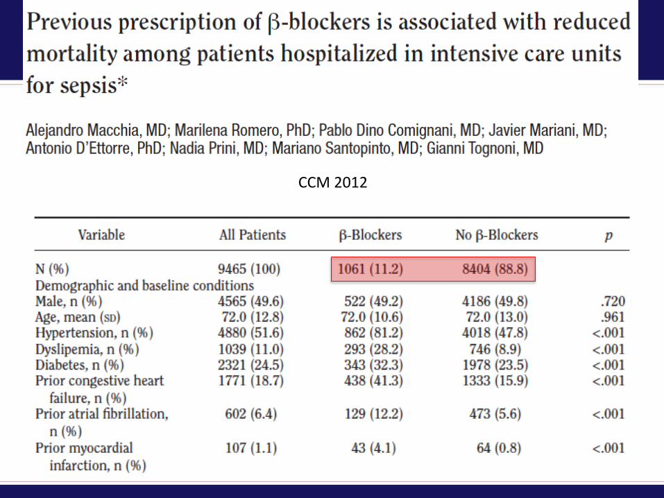
Previous β- Blockers and Mortality
• 28 days mortality
17.7% vs. 22.1%
• OR 0.78 (0.66–0.9)
p = .005
• OR 0.81 (0.68–0.97)
p = .025 adjusted
analyses.
Macchia, CCM 2012

Previous β- Blockers and Mortality
Macchia, CCM 2012
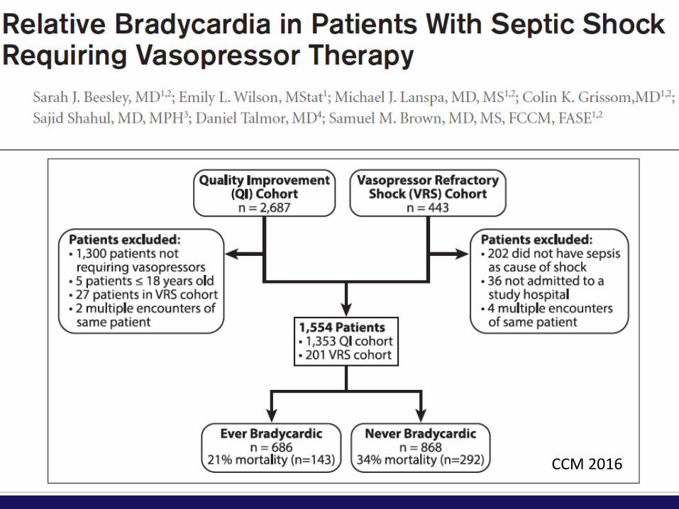
CCM 2016
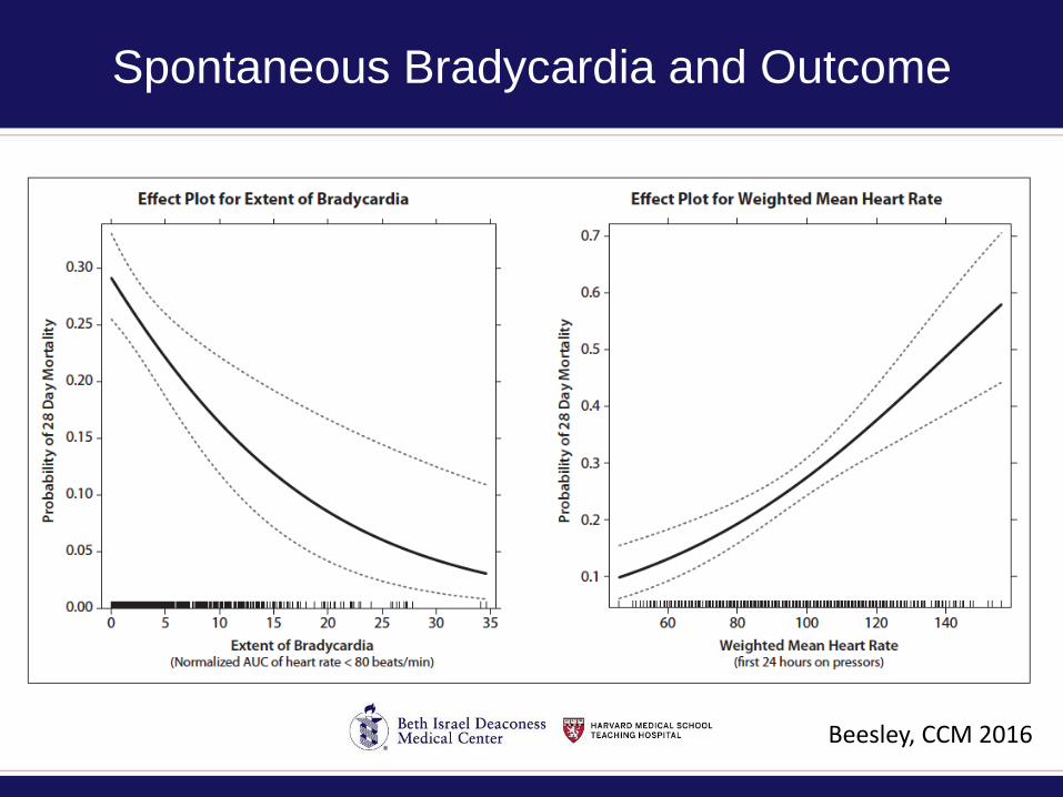
Spontaneous Bradycardia and Outcome
Beesley, CCM 2016
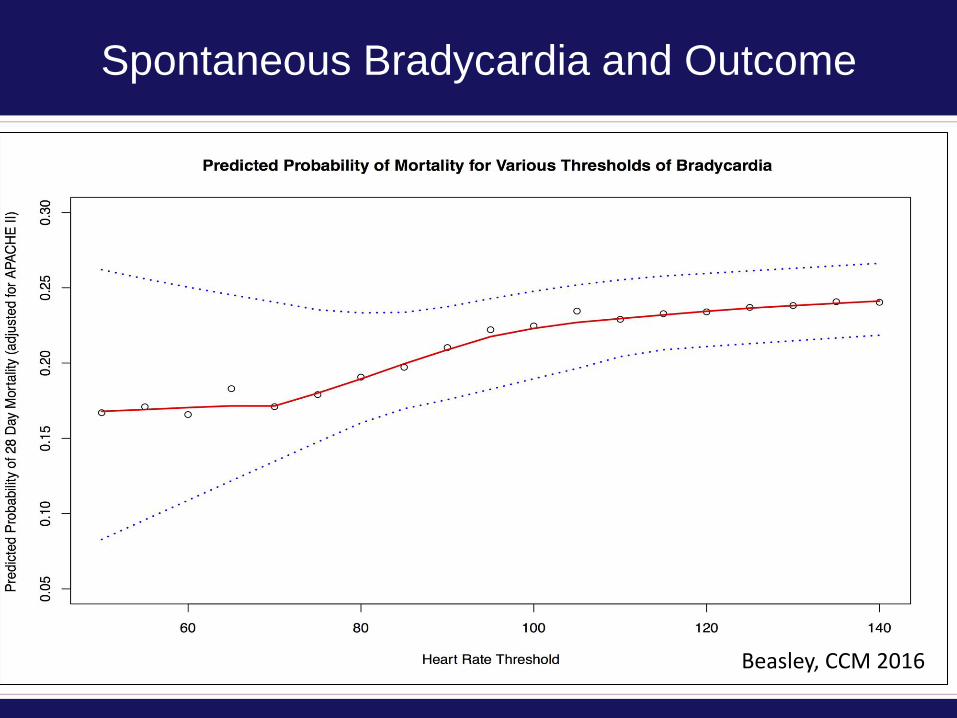
Spontaneous Bradycardia and Outcome
Beasley, CCM 2016
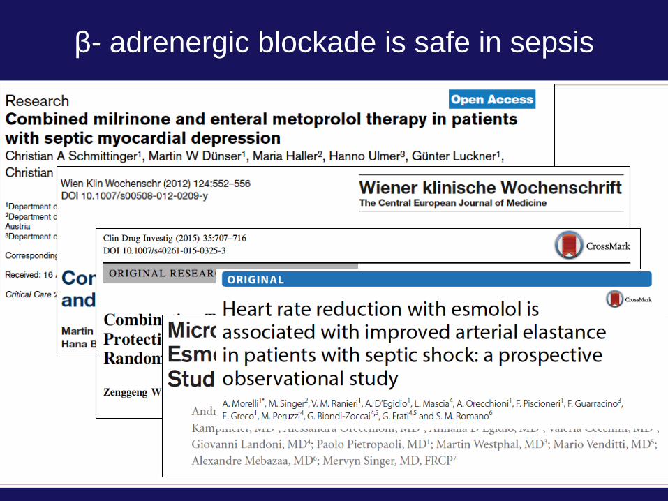
β- adrenergic blockade is safe in sepsis
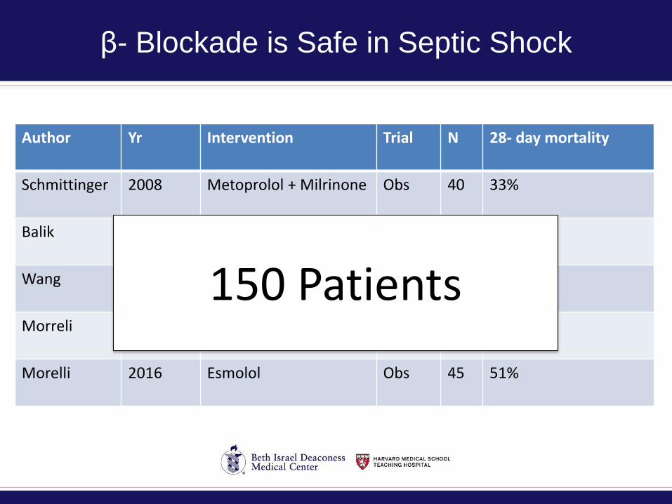
β- Blockade is Safe in Septic Shock
Author Yr Intervention Trial N 28- day mortality
Schmittinger 2008 Metoprolol + Milrinone Obs 40 33%
Balik 2012 Esmolol Obs 10
Wang 2015 control v milrinone vmil + esmolol
RCT 30 improved
Morreli 2013 Esmolol Obs 25 NR
Morelli 2016 Esmolol Obs 45 51%
150 Patients
Efficacy of β- adrenergic blockade in sepsis
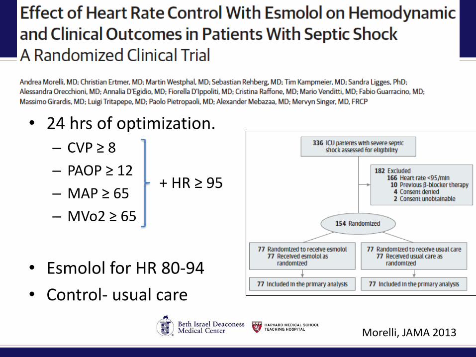
• 24 hrs of optimization.
– CVP ≥ 8
– PAOP ≥ 12
– MAP ≥ 65
– MVo2 ≥ 65
• Esmolol for HR 80-94
• Control- usual care
+ HR ≥ 95
Morelli, JAMA 2013
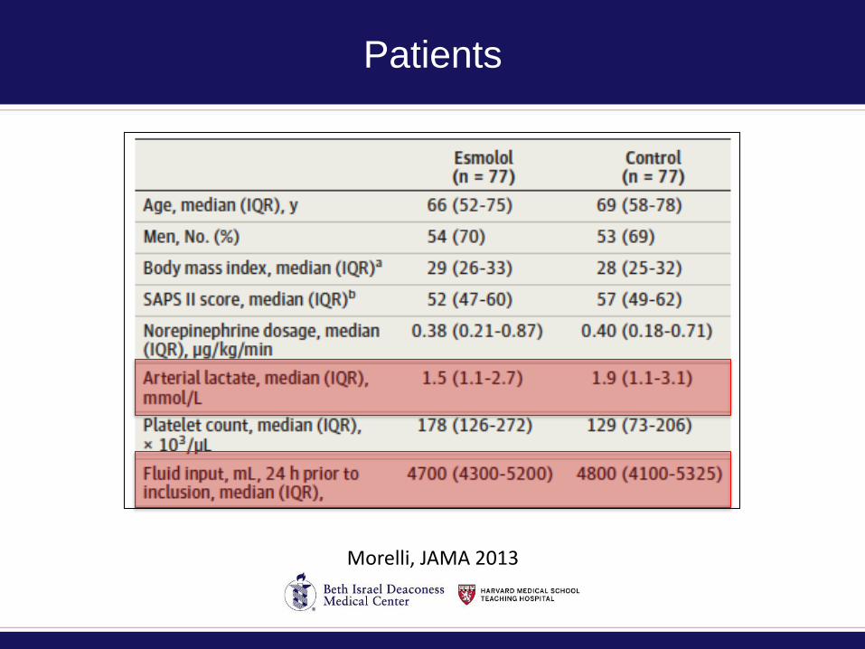
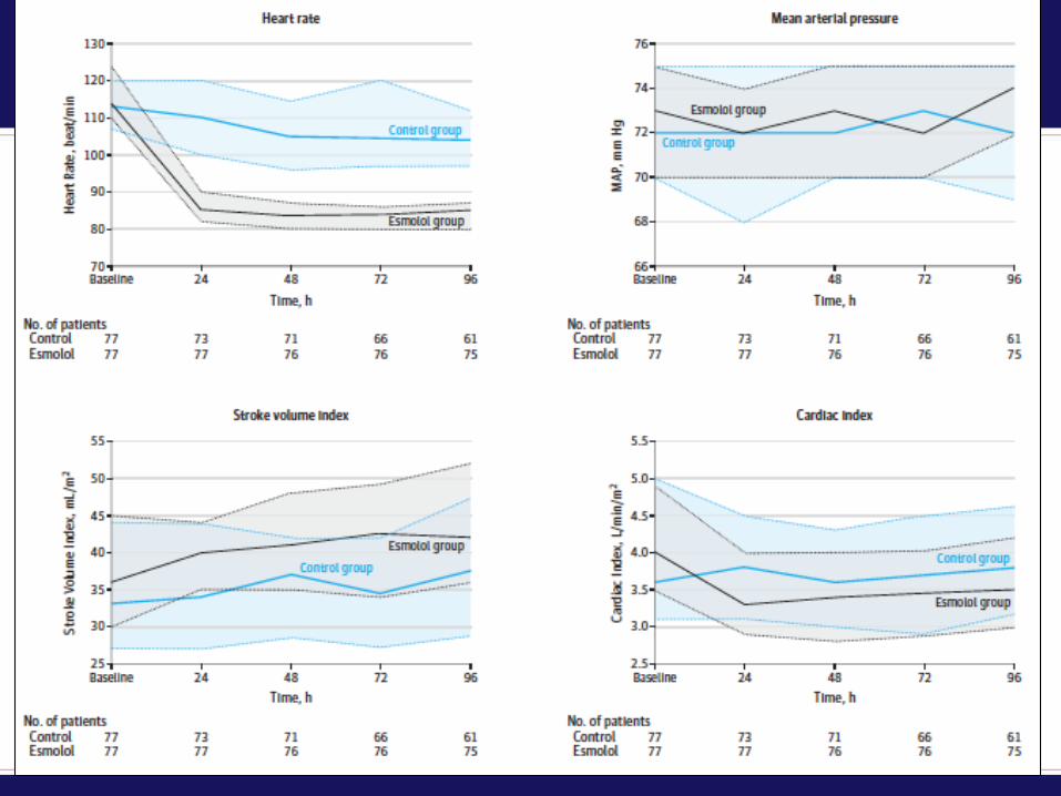
Patients
Morelli, JAMA 2013
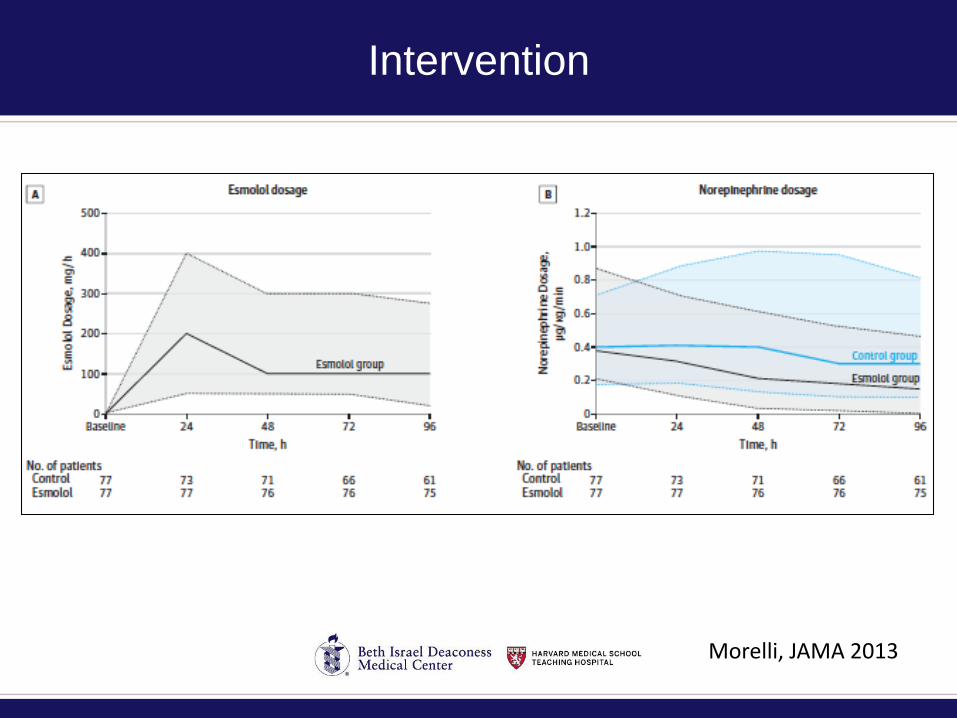
Intervention
Morelli, JAMA 2013
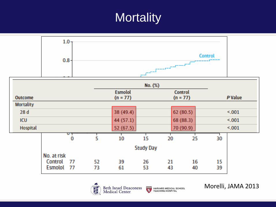
Mortality
Morelli, JAMA 2013
Caveats
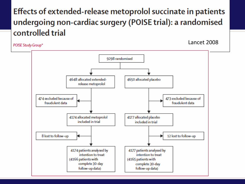
Lancet 2008
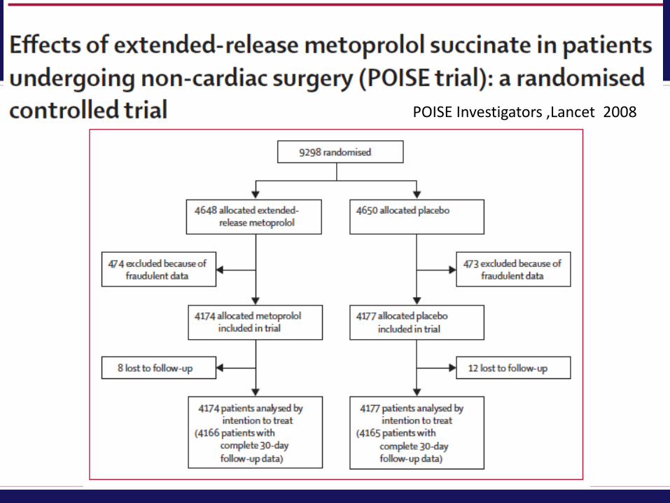
POISE Investigators ,Lancet 2008
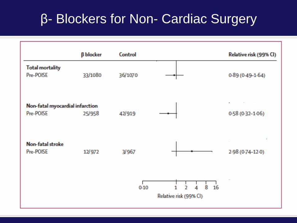
β- Blockers for Non- Cardiac Surgery
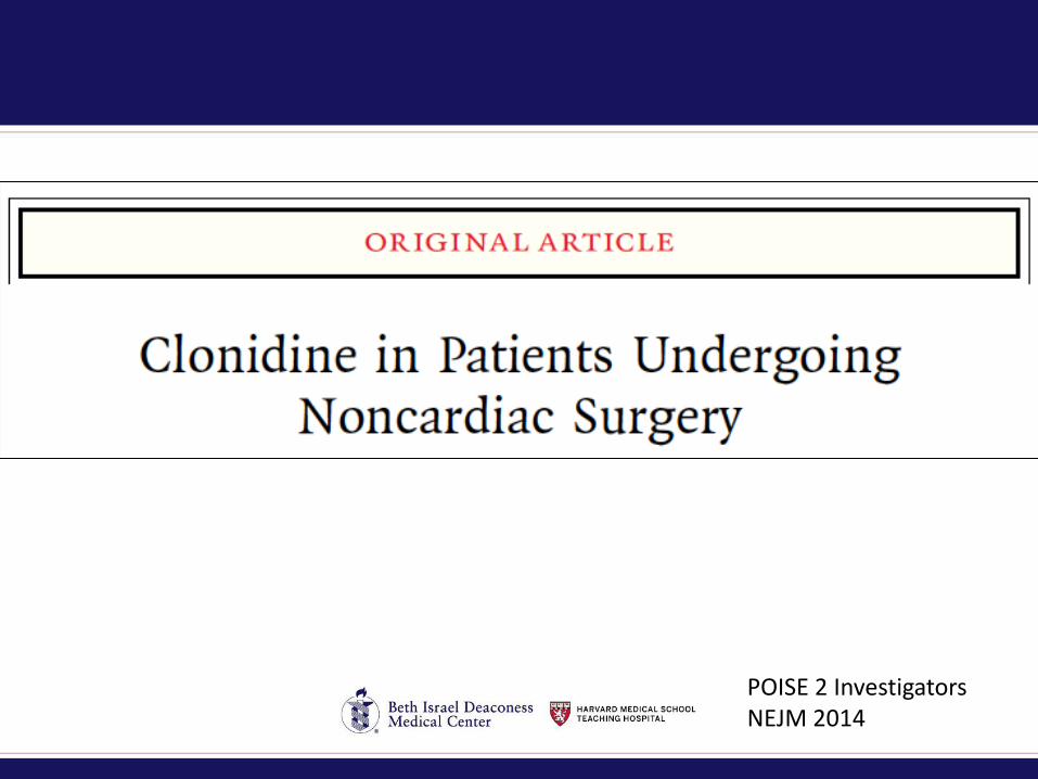
POISE 2 InvestigatorsNEJM 2014
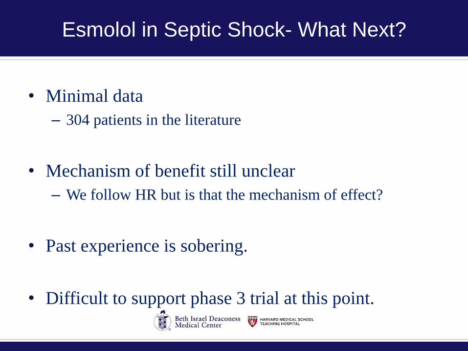
Esmolol in Septic Shock- What Next?
• Minimal data
– 304 patients in the literature
• Mechanism of benefit still unclear
– We follow HR but is that the mechanism of effect?
• Past experience is sobering.
• Difficult to support phase 3 trial at this point.
β- Blockade in Sepsis-
What is the Evidence?
Patient Engagement, Systems Science, and the Elimination of Preventable Harm
Daniel Talmor, MD, MPH