


γλώσσες
Σελίδες
Νομικός

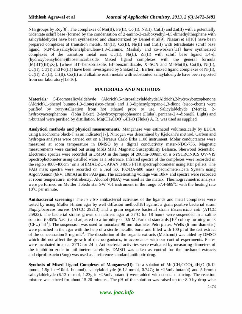
1472
Available online at www.joac.info
ISSN: 2278-1862
Journal of Applicable Chemistry 2013, 2 (6): 1472-1483
(International Peer Reviewed Journal)
Synthesis And Studies Of Mixed Ligand Complexes Of Mn(II) With
Salicylaldehyde And Substituted Salicylaldehydes,2-Hydroxyarylcarbonyl
Compounds Or β-Diketones Mithlesh Agrawal* Gurjeet Kaur and Anshu khandelwal
*Department of Chemistry, University of Rajasthan, Jaipur-302004, INDIA
Email: [email protected]
Received on 15th October and finalized on 23rd October 2013
_____________________________________________________________________________
ABSTRACT Mixed ligand complexes of Mn(II) of the type [MnLL'(H2O)2], (where HL = salicylaldehyde and HL' = 5-bromosalicylaldehyde, 5-nitrosalicylaldehyde, 2-hydroxyacetophenone, 2-hydroxypropiophenone, 2-
hydroxybenzophenone, pentane-2,4-dione, 1-phenylbutane-1,3-dione or 1,3-diphenylpropane-1,3-dione)
have been synthesized by the reactions of manganese(II) acetate with a mixture of two different ligands in
1:1:1 molar ratios. The solid complexes were separated, filtered, washed with butanol and ether successively and dried under reduced pressure. The resulting complexes have been characterized by
elemental analyses, molar conductances, magnetic moments, electronic spectra, IR spectra, FAB mass
spectra and thermo gravimetric analysis. At the same time, above mentioned complexes were studied for in vitro antimicrobial properties and found to be more potent bactericides than parent ligands. Octahedral
geometry has been proposed for the mixed ligand complexes.
Keywords: Mixed ligand complexes, FAB mass spectra, octahedral geometry, thermo gravimetric
analysis, antimicrobial properties.
______________________________________________________________________________
INTRODUCTION
Interest in the chemistry of mixed ligand complexes has increased in recent decades, because of wide applications of these coordination complexes in various fields. Mixed ligand complexes appear in
biological fluids, create specific structures and manifest themselves as enzyme-metal ion-substrate
complexes[1, 2]. These are suitable precursors for the selective CVD of high purity metals[3,4] and have been used in the analysis of semiconductor materials. Many of the inorganic medicinal compounds are
mixed ligand complexes[5].
Various oxidation states of manganese and the wide applications of its mixed ligand complexes in biological and industrial fields are gaining popularity in the chemistry nowadays. Tanaka[6] have been
synthesized Bis-[5-(p-sulfophenylazo)salicylaldehyde] metal chelates (IV) of copper, nickel, cobalt and
manganese. Rani et al.[7] prepared and characterized Mn(II), Fe(III), Co(II), Ni(II), Cu(II) and Zn(II)
complexes of a new multidentate oxygen nitrogen donor,bis(N-salicylidene)-2,3-dihydrazino-1,4-quino xaline. A new chiral amino acid Schiff base ligand (Salarg) and its metal complex (Mn-Salarg) have been
synthesized using L-Arginine, a naturally occurring chiral diamine with two kinds of asymmetric α-, ε-
Mithlesh Agrawal et al Journal of Applicable Chemistry, 2013, 2 (6):1472-1483
1473
www. joac.info
NH2 groups by Roy[8]. The complexes of Mn(II), Fe(II), Co(II), Ni(II), Cu(II) and Zn(II) with a potentially tridentate schiff base (formed by the condensation of 2-amino-3-carboxyethyl-4,5-dimethylthiophene with
salicylaldehyde) have been synthesized and characterized by Daniel et al[9]. Niasari et al[10] have been
prepared complexes of transition metals, Mn(II), Co(II), Ni(II) and Cu(II) with tetradentate schiff base ligand, N,N'-bis(salicylidene)phenulene-1,3-diamine. Mashaly and co-workers[11] have synthesized
complexes of the transition metal ions Cu(II), Ni(II), Zn(II) with schiff base ligand 1,4-di
(hydroxybenzylidene)thiosemicarbazide. Mixed ligand complexes with the general formula
[M(BT)(BI)2X2], [where BT=benzotriazole, BI=benzimidazole, X=SCN and M=Mn(II), Co(II), Ni(II), Cu(II), Cd(II) and Pd(II)] have been investigated by Shaker[12]. Earlier, mixed ligand complexes of Ni(II),
Cu(II), Zn(II), Cr(II), Co(II) and alkaline earth metals with substituted salicylaldehyde have been reported
from our laboratory[13-16].
MATERIALS AND METHODS
Materials: 5-Bromosalicylaldehyde (Aldrich),5-nitrosalicylaldehyde(Aldrich),2-hydroxybenzophenone (Aldrich),1-phenyl butane-1,3-dione(sisco-chem) and 1,3-diphenylpropane-1,3-dione (sisco-chem) were
purified by recrystallisation from hot ethanol prior to use. Salicylaldehyde (Merck), 2-
hydroxyacetophenone (John Baker), 2-hydroxypropiophenone (Fluka), pentane-2,4-dione(K. Light) and
n-butanol were purified by distillation. Mn(CH3COO)2.4H2O (Fluka) A. R. was used as supplied.
Analytical methods and physical measurements: Manganese was estimated volumetrically by EDTA
using Eriochrome black-T as an indicator[17]. Nitrogen was determined by Kjeldahl’s method. Carbon and hydrogen analyses were carried out on a Heraeus Carlo Erba 1108 instrument. Molar conductances were
measured at room temperature in DMSO by a digital conductivity meter-NDC-736. Magnetic
measurements were carried out using MSB MK1 Magnetic Susceptibility Balance, Sherwood Scientific. Electronic spectra were recorded in DMSO in the range of 200nm-800nm on a SYSTRONICS UV-VIS
Spectrophotometer using distilled water as a reference. Infrared spectra of the complexes were recorded in
the region 4000-400cm-1
on a SHIMADZU-JAPAN 8400S FTIR spectrophotometer using KBr pellets. The
FAB mass spectra were recorded on a Jeol SX 102/DA-600 mass spectrometer/Data System using Argon/Xenon (6kV, 10mA) as the FAB gas. The accelerating voltage was 10kV and spectra were recorded
at room temperature. m-Nitrobenzyl Alcohol (NBA) was used as the matrix. Thermogravimetric analysis
were performed on Mettler Toledo star SW 701 instrument in the range 57.4-689oC with the heating rate
10oC per minute.
Antibacterial screening: The in vitro antibacterial activities of the ligands and metal complexes were tested by using Muller Hinton agar by well diffusion method[18] against a gram positive bacterial strain
Staphylococcus aureus (ATCC 29213) and a gram negative bacterial strain Escherichia coli (ATCC
25922). The bacterial strains grown on nutrient agar at 37ºC for 18 hours were suspended in a saline
solution (0.85% NaCl) and adjusted to a turbidity of 0.5 McFarland standards [108 colony forming units
(CFU) ml-1
]. The suspension was used to inoculate 90 mm diameter Petri plates. Wells (6 mm diameter)
were punched in the agar with the help of a sterile metallic borer and filled with 100 µl of the test extract
of the concentration 5 mg mL-1
. The dissolution of the organic extracts (Methanol) was aided by DMSO which did not affect the growth of microorganisms, in accordance with our control experiments. Plates
were incubated in air at 37ºC for 24 h. Antibacterial activities were evaluated by measuring diameters of
the inhibition zone in millimeters carefully. DMSO was taken as control for the methanol extracts
and ciprofloxacin (5mg) was used as a reference standard antibiotic drug.
Synthesis of Mixed Ligand Complexes of Manganese(II): To a solution of Mn(CH3COO)2.4H2O (6.12
mmol, 1.5g in ~10mL butanol), salicylaldehyde (6.12 mmol, 0.747g in ~25mL butanol) and 5-bromo salicylaldehyde (6.12 m mol, 1.23g in ~25mL butanol) were added with constant stirring. The reaction
mixture was stirred for about 15-20 minutes. The pH of the solution was raised up to ~8.0 by drop wise
Mithlesh Agrawal et al Journal of Applicable Chemistry, 2013, 2 (6):1472-1483
1474
www. joac.info
addition of 5% aqueous solution of sodium hydroxide with constant stirring. The reaction mixture was stirred for 5-6 hours. The reaction mixture was kept in the refrigerator for about 30-35 days. The settled
solid was filtered, washed with butanol and later with ether to remove stickiness and dried properly under
reduced pressure. A similar method was adopted to synthesize mixed ligand complexes of Mn(II) with salicylaldehyde and 5-nitrosalicylaldehyde, 2-hydroxyacetophenone, 2-hydroxypropiophenone,2-hydroxy
benzophenone, pentane-2,4-dione, 1-phenylbutane-1,3-dione or 1,3-diphenylpropane-1,3-dione.
RESULTS AND DISCUSSION
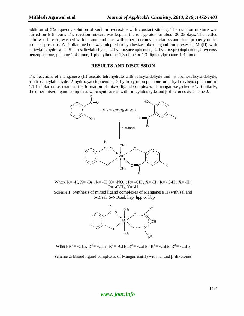
The reactions of manganese (II) acetate tetrahydrate with salicylaldehyde and 5-bromosalicylaldehyde,
5-nitrosalicylaldehyde, 2-hydroxyacetophenone, 2-hydroxypropiophenone or 2-hydroxybenzophenone in 1:1:1 molar ratios result in the formation of mixed ligand complexes of manganese ,scheme 1. Similarly,
the other mixed ligand complexes were synthesized with salicylaldehyde and β-diketones as scheme 2.
C
OH
H
O
+ Mn(CH3COO)2.4H2O +
HO
C
R
O
n-butanol
C
O
H
O O
C
R
O
Mn
OH2
OH2
X
X
Where R= -H, X= -Br ; R= -H, X= -NO2 ; R= -CH3, X= -H ; R= -C2H5, X= -H ; R= -C6H5, X= -H
Scheme 1: Synthesis of mixed ligand complexes of Manganese(II) with sal and
5-Brsal, 5-NO2sal, hap, hpp or hbp
C
O
H
OCO
C
R2
O
Mn
OH2
OH2
CH
R1
Where R1 = -CH3, R
2 = -CH3 ; R
1 = -CH3, R
2 = -C6H5 ; R
1 = -C6H5, R
2 = -C6H5
Scheme 2: Mixed ligand complexes of Manganese(II) with sal and β-diketones
Mithlesh Agrawal et al Journal of Applicable Chemistry, 2013, 2 (6):1472-1483
1475
www. joac.info
Table 1 . Characterization data for the mixed ligand complexes of Mn(II) Complex
S.
No.
Complex
Molecular Formula
Molecular Weight
Color
Yield (%)
Decompositio
n
temperature
(oC)
Found (Calculated) %
Mn
C
H
N
1. [Mn(sal)(5-Brsal)(H2O)2]
C14H13O6MnBr, 411.900
Dark-brown
(44.95)
198 13.19
(13.34)
39.66
(40.79)
2.94
(3.18)
-
2. [Mn(sal)5-NO2sal)(H2 O)2
C14H13O8MnN, 377.983
Yellow
(45.75)
260 13.64
(14.53)
43.42
(44.45)
3.21
(3.47)
3.502
(3.71)
3. [Mn(sal)(hap)(H2O)2]
C15H16O6Mn, 347.225
Light-brown
(49.24)
156 15.26
(15.82)
49.89
(51.88)
4.22
(4.64)
-
4. [Mn(sal)(hpp)(H2O)2]
C16H18O6Mn, 361.038
Brown
(31.87)
230 14.81
(15.21)
52.26
(53.18)
4.97
(5.02)
-
5. [Mn(sal)(hbp)(H2O)2]
C20H18O6Mn, 409.042
Dark-brown
(43.88)
246 13.13
(13.43)
57.91
(58.68)
4.21
(4.43)
-
6. [Mn(sal)(acac)(H2O)2]
C14H19O8Mn, 370.234
Brown
(44.79)
168 14.32
(14.73)
53.85
(54.69)
4.69
(4.86)
-
7. [Mn(sal)(bzac)(H2O)2]
C17H18O6Mn, 373.039
Dirty-yellow
(63.17)
260 12.36
(12.63)
60.25
(60.68)
4.48
(4.63)
-
8. [Mn(sal)(dbzm)(H2O)2]
C22H20O6Mn, 435.060
Brown
(65.44)
206 14.13
(14.34)
58.79
(58.97)
3.72
(3.96)
-
The resulting mixed ligand complexes are obtained in 44-65% yields as brown solid except [Mn(sal) (5-
NO2 sal)(H2O)2], which is yellow in color. The data of C, H, N and Mn analyses agree well with the calculated values corresponding to the respective complexes. The complexes decompose at high
temperature on heating. All the physical data are shown in table 1. These are insoluble in water or most of
the organic solvents like methanol, benzene and carbon tetrachloride but soluble in DMSO and DMF.
Molar conductances: Molar conductances (Λm) of 10
-3 solutions of the complexes in DMSO (table 2) lie
in very low range 4.8-18.8 Ω-1cm
2mol
-1 supporting their non-electrolytic behaviour[19]
.
Magnetic moments: The µeff values for the complexes are observed (table 2) in the range 5.60 to 6.01
B.M. as expected for five unpaired electrons. These values indicate that the complexes are high spin
paramagnetic; it lies within the octahedral range which is very close to spin value 5.90 B.M. as the ground
term is 6A1g and thus supports the octahedral geometry[20]. Siddappa et al.[13] have reported Mn(II) high
spin complex having an octahedral geometry with 5.65 B.M. magnetic moment.
Mithlesh Agrawal et al Journal of Applicable Chemistry, 2013, 2 (6):1472-1483
1476
www. joac.info
Table 2. Molar Conductance & Magnetic moments of mixed ligand complexes of Mn(II)
Complex no. Molar
Conductance
(Ω-1
cm2mol
-1)
µeff
(B.M.)
1 16.9 5.72
2 18.8 5.73
3 5.7 5.61
4 4.8 5.71
5 15.4 5.98
6 10.9 5.60
7 11.0 5.73
8 15.6 6.01
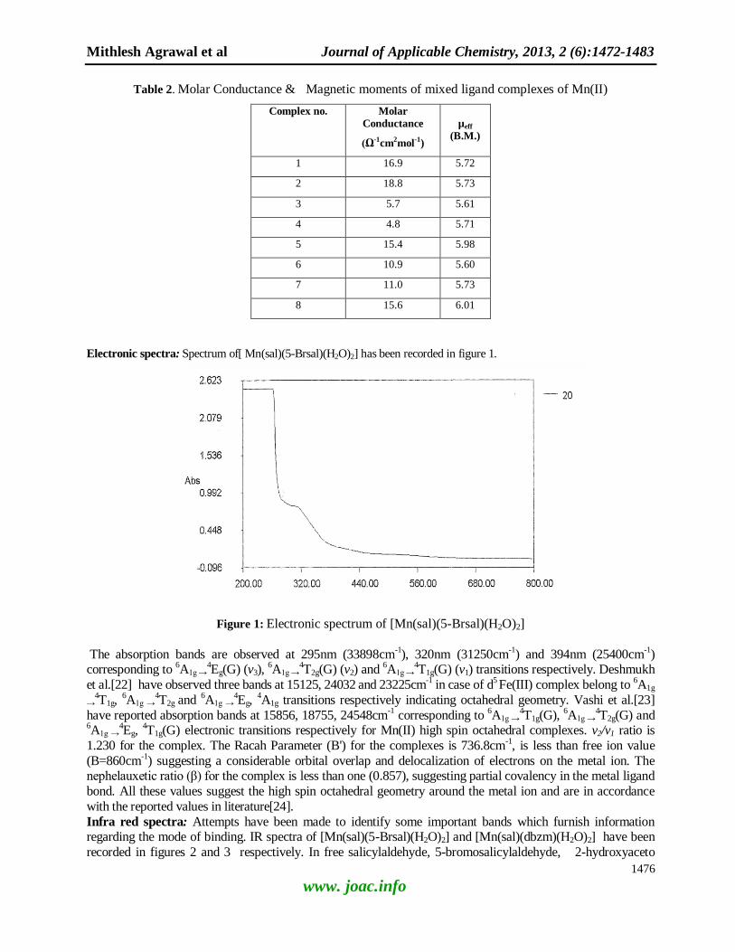
Electronic spectra: Spectrum of[ Mn(sal)(5-Brsal)(H2O)2] has been recorded in figure 1.
Figure 1: Electronic spectrum of [Mn(sal)(5-Brsal)(H2O)2]
The absorption bands are observed at 295nm (33898cm-1), 320nm (31250cm
-1) and 394nm (25400cm
-1)
corresponding to 6A1g →
4Eg(G) (v3),
6A1g →
4T2g(G) (v2) and
6A1g →
4T1g(G) (v1) transitions respectively. Deshmukh
et al.[22] have observed three bands at 15125, 24032 and 23225cm-1 in case of d
5 Fe(III) complex belong to
6A1g
→4T1g,
6A1g →
4T2g and
6A1g →
4Eg,
4A1g transitions respectively indicating octahedral geometry. Vashi et al.[23]
have reported absorption bands at 15856, 18755, 24548cm-1 corresponding to
6A1g →
4T1g(G),
6A1g →
4T2g(G) and
6A1g →
4Eg,
4T1g(G) electronic transitions respectively for Mn(II) high spin octahedral complexes. v2/v1 ratio is
1.230 for the complex. The Racah Parameter (B') for the complexes is 736.8cm-1, is less than free ion value
(B=860cm-1) suggesting a considerable orbital overlap and delocalization of electrons on the metal ion. The
nephelauxetic ratio (β) for the complex is less than one (0.857), suggesting partial covalency in the metal ligand
bond. All these values suggest the high spin octahedral geometry around the metal ion and are in accordance
with the reported values in literature[24].
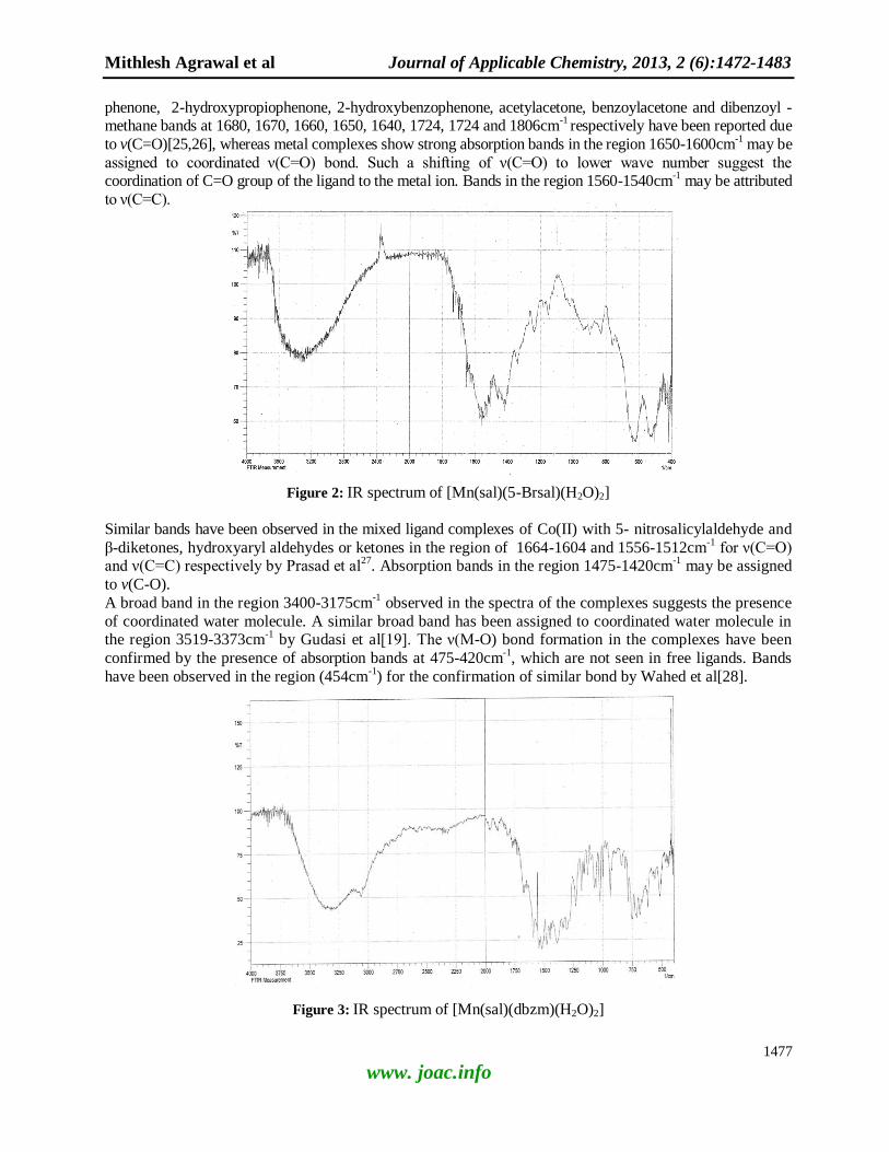
Infra red spectra: Attempts have been made to identify some important bands which furnish information regarding the mode of binding. IR spectra of [Mn(sal)(5-Brsal)(H2O)2] and [Mn(sal)(dbzm)(H2O)2] have been
recorded in figures 2 and 3 respectively. In free salicylaldehyde, 5-bromosalicylaldehyde, 2-hydroxyaceto
Mithlesh Agrawal et al Journal of Applicable Chemistry, 2013, 2 (6):1472-1483
1477
www. joac.info
phenone, 2-hydroxypropiophenone, 2-hydroxybenzophenone, acetylacetone, benzoylacetone and dibenzoyl -methane bands at 1680, 1670, 1660, 1650, 1640, 1724, 1724 and 1806cm
-1 respectively have been reported due
to v(C=O)[25,26], whereas metal complexes show strong absorption bands in the region 1650-1600cm-1 may be
assigned to coordinated ν(C=O) bond. Such a shifting of ν(C=O) to lower wave number suggest the coordination of C=O group of the ligand to the metal ion. Bands in the region 1560-1540cm
-1 may be attributed
to ν(C=C).
Figure 2: IR spectrum of [Mn(sal)(5-Brsal)(H2O)2]
Similar bands have been observed in the mixed ligand complexes of Co(II) with 5- nitrosalicylaldehyde and
β-diketones, hydroxyaryl aldehydes or ketones in the region of 1664-1604 and 1556-1512cm-1
for ν(C=O) and ν(C=C) respectively by Prasad et al
27. Absorption bands in the region 1475-1420cm
-1 may be assigned
to v(C-O).
A broad band in the region 3400-3175cm-1
observed in the spectra of the complexes suggests the presence
of coordinated water molecule. A similar broad band has been assigned to coordinated water molecule in the region 3519-3373cm
-1 by Gudasi et al[19]. The ν(M-O) bond formation in the complexes have been
confirmed by the presence of absorption bands at 475-420cm-1
, which are not seen in free ligands. Bands
have been observed in the region (454cm-1
) for the confirmation of similar bond by Wahed et al[28].
Figure 3: IR spectrum of [Mn(sal)(dbzm)(H2O)2]
Mithlesh Agrawal et al Journal of Applicable Chemistry, 2013, 2 (6):1472-1483
1478
www. joac.info
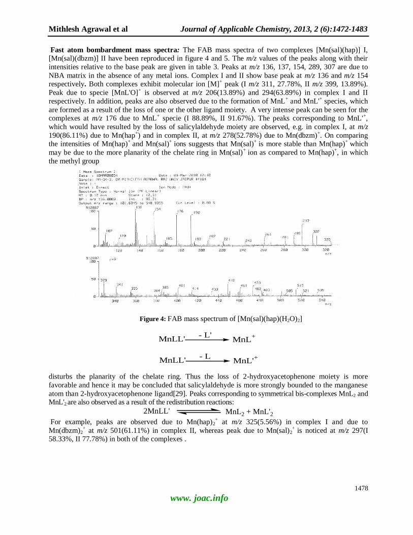
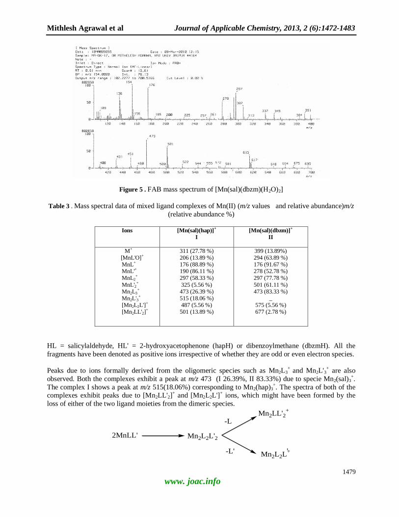
Fast atom bombardment mass spectra: The FAB mass spectra of two complexes [Mn(sal)(hap)] I, [Mn(sal)(dbzm)] II have been reproduced in figure 4 and 5. The m/z values of the peaks along with their
intensities relative to the base peak are given in table 3. Peaks at m/z 136, 137, 154, 289, 307 are due to
NBA matrix in the absence of any metal ions. Complex I and II show base peak at m/z 136 and m/z 154 respectively. Both complexes exhibit molecular ion [M]
+ peak (I m/z 311, 27.78%, II m/z 399, 13.89%).
Peak due to specie [MnL'O]+ is observed at m/z 206(13.89%) and 294(63.89%) in complex I and II
respectively. In addition, peaks are also observed due to the formation of MnL+ and MnL'
+ species, which
are formed as a result of the loss of one or the other ligand moiety. A very intense peak can be seen for the complexes at m/z 176 due to MnL
+ specie (I 88.89%, II 91.67%). The peaks corresponding to MnL'
+,
which would have resulted by the loss of salicylaldehyde moiety are observed, e.g. in complex I, at m/z
190(86.11%) due to Mn(hap+) and in complex II, at m/z 278(52.78%) due to Mn(dbzm)
+. On comparing
the intensities of Mn(hap)+
and Mn(sal)+
ions suggests that Mn(sal)+
is more stable than Mn(hap)+
which
may be due to the more planarity of the chelate ring in Mn(sal)+
ion as compared to Mn(hap)+, in which
the methyl group
Figure 4: FAB mass spectrum of [Mn(sal)(hap)(H2O)2]
MnLL' MnL+- L'
MnLL' MnL'+- L
disturbs the planarity of the chelate ring. Thus the loss of 2-hydroxyacetophenone moiety is more
favorable and hence it may be concluded that salicylaldehyde is more strongly bounded to the manganese
atom than 2-hydroxyacetophenone ligand[29]. Peaks corresponding to symmetrical bis-complexes MnL2 and MnL'2
are also observed as a result of the redistribution reactions:
2MnLL' MnL2 + MnL'2 For example, peaks are observed due to Mn(hap)2
+ at m/z 325(5.56%) in complex I and due to
Mn(dbzm)2+ at m/z 501(61.11%) in complex II, whereas peak due to Mn(sal)2
+ is noticed at m/z 297(I
58.33%, II 77.78%) in both of the complexes .
Mithlesh Agrawal et al Journal of Applicable Chemistry, 2013, 2 (6):1472-1483
1479
www. joac.info
Figure 5 . FAB mass spectrum of [Mn(sal)(dbzm)(H2O)2]
Table 3 . Mass spectral data of mixed ligand complexes of Mn(II) (m/z values and relative abundance)m/z
(relative abundance %)
Ions
[Mn(sal)(hap)]+
I
[Mn(sal)(dbzm)]+
II
M+
[MnL'O]+ MnL+ MnL'+ MnL2
+
MnL'2+
Mn2L3+
Mn2L'3+
[Mn2L2L']+ [Mn2LL'2]
+
311 (27.78 %) 206 (13.89 %) 176 (88.89 %) 190 (86.11 %) 297 (58.33 %)
325 (5.56 %) 473 (26.39 %) 515 (18.06 %) 487 (5.56 %) 501 (13.89 %)
399 (13.89%) 294 (63.89 %) 176 (91.67 %) 278 (52.78 %) 297 (77.78 %)
501 (61.11 %) 473 (83.33 %)
_ 575 (5.56 %) 677 (2.78 %)
HL = salicylaldehyde, HL' = 2-hydroxyacetophenone (hapH) or dibenzoylmethane (dbzmH). All the
fragments have been denoted as positive ions irrespective of whether they are odd or even electron species.
Peaks due to ions formally derived from the oligomeric species such as Mn2L3
+ and Mn2L'3
+ are also
observed. Both the complexes exhibit a peak at m/z 473 (I 26.39%, II 83.33%) due to specie Mn2(sal)3+.
The complex I shows a peak at m/z 515(18.06%) corresponding to Mn2(hap)3+. The spectra of both of the
complexes exhibit peaks due to [Mn2LL'2]+ and [Mn2L2L']
+ ions, which might have been formed by the
loss of either of the two ligand moieties from the dimeric species.
Mn2L2L'22MnLL'
Mn2LL'2+
Mn2L2L+-L'
-L
'
Mithlesh Agrawal et al Journal of Applicable Chemistry, 2013, 2 (6):1472-1483
1480
www. joac.info
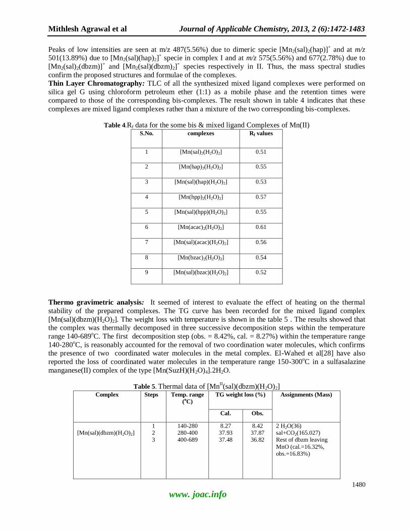
Peaks of low intensities are seen at m/z 487(5.56%) due to dimeric specie [Mn2(sal)2(hap)]+ and at m/z
501(13.89%) due to [Mn2(sal)(hap)2]+ specie in complex I and at m/z 575(5.56%) and 677(2.78%) due to
[Mn2(sal)2(dbzm)]+ and [Mn2(sal)(dbzm)2]
+ species respectively in II. Thus, the mass spectral studies
confirm the proposed structures and formulae of the complexes. Thin Layer Chromatography: TLC of all the synthesized mixed ligand complexes were performed on
silica gel G using chloroform petroleum ether (1:1) as a mobile phase and the retention times were
compared to those of the corresponding bis-complexes. The result shown in table 4 indicates that these
complexes are mixed ligand complexes rather than a mixture of the two corresponding bis-complexes.
Table 4.Rf data for the some bis & mixed ligand Complexes of Mn(II) S.No. complexes Rf values
1 [Mn(sal)2(H2O)2] 0.51
2 [Mn(hap)2(H2O)2] 0.55
3 [Mn(sal)(hap)(H2O)2] 0.53
4 [Mn(hpp)2(H2O)2] 0.57
5 [Mn(sal)(hpp)(H2O)2] 0.55
6 [Mn(acac)2(H2O)2] 0.61
7 [Mn(sal)(acac)(H2O)2] 0.56
8 [Mn(bzac)2(H2O)2] 0.54
9 [Mn(sal)(bzac)(H2O)2] 0.52
Thermo gravimetric analysis: It seemed of interest to evaluate the effect of heating on the thermal
stability of the prepared complexes. The TG curve has been recorded for the mixed ligand complex
[Mn(sal)(dbzm)(H2O)2]. The weight loss with temperature is shown in the table 5 . The results showed that the complex was thermally decomposed in three successive decomposition steps within the temperature
range 140-689oC. The first decomposition step (obs. = 8.42%, cal. = 8.27%) within the temperature range
140-280oC, is reasonably accounted for the removal of two coordination water molecules, which confirms
the presence of two coordinated water molecules in the metal complex. El-Wahed et al[28] have also
reported the loss of coordinated water molecules in the temperature range 150-300oC in a sulfasalazine
manganese(II) complex of the type [Mn(SuzH)(H2O)4].2H2O.
Table 5. Thermal data of [MnII(sal)(dbzm)(H2O)2]
Complex Steps Temp. range
(oC)
TG weight loss (%) Assignments (Mass)
Cal. Obs.
[Mn(sal)(dbzm)(H2O)2]
1 2 3
140-280 280-400 400-689
8.27 37.93 37.48
8.42 37.87 36.82
2 H2O(36) sal+CO2(165.027)
Rest of dbzm leaving
MnO (cal.=16.32%, obs.=16.83%)
-
Mithlesh Agrawal et al Journal of Applicable Chemistry, 2013, 2 (6):1472-1483
1481
www. joac.info
The second step is found between the temperature range 280-400oC (obs. = 37.87%, cal. = 37.93%), may
be attributed to the liberation of salicylaldehyde ligand moiety and one molecule of CO2, suggesting the
decomposition of the dibenzoylmethane ligand. The decomposition of the rest of the complex molecule
ended with a final oxide of MnO in the third step within the temperature range 400-689oC (obs. = 16.83%,
cal. = 16.32%).
APPLICATIONS
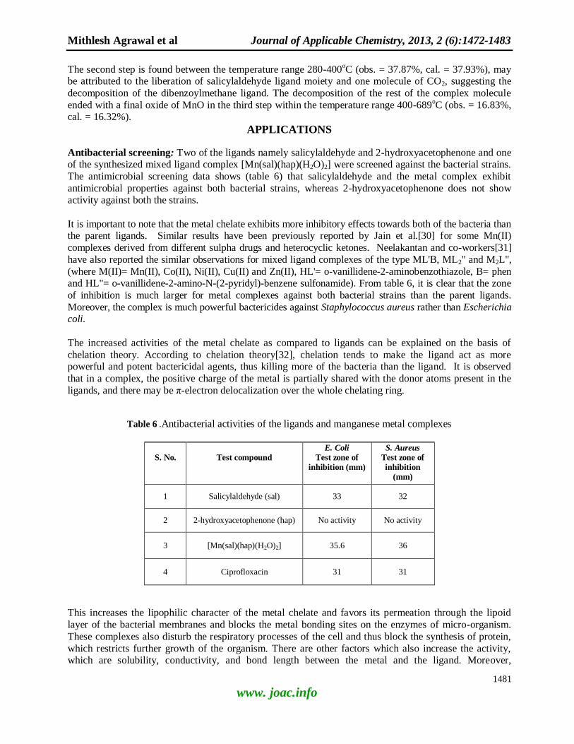
Antibacterial screening: Two of the ligands namely salicylaldehyde and 2-hydroxyacetophenone and one of the synthesized mixed ligand complex [Mn(sal)(hap)(H2O)2] were screened against the bacterial strains.
The antimicrobial screening data shows (table 6) that salicylaldehyde and the metal complex exhibit
antimicrobial properties against both bacterial strains, whereas 2-hydroxyacetophenone does not show activity against both the strains.
It is important to note that the metal chelate exhibits more inhibitory effects towards both of the bacteria than the parent ligands. Similar results have been previously reported by Jain et al.[30] for some Mn(II)
complexes derived from different sulpha drugs and heterocyclic ketones. Neelakantan and co-workers[31]
have also reported the similar observations for mixed ligand complexes of the type ML'B, ML2" and M2L",
(where M(II)= Mn(II), Co(II), Ni(II), Cu(II) and Zn(II), HL'= o-vanillidene-2-aminobenzothiazole, B= phen and HL"= o-vanillidene-2-amino-N-(2-pyridyl)-benzene sulfonamide). From table 6, it is clear that the zone
of inhibition is much larger for metal complexes against both bacterial strains than the parent ligands.
Moreover, the complex is much powerful bactericides against Staphylococcus aureus rather than Escherichia coli.
The increased activities of the metal chelate as compared to ligands can be explained on the basis of
chelation theory. According to chelation theory[32], chelation tends to make the ligand act as more powerful and potent bactericidal agents, thus killing more of the bacteria than the ligand. It is observed
that in a complex, the positive charge of the metal is partially shared with the donor atoms present in the
ligands, and there may be π-electron delocalization over the whole chelating ring.
Table 6 .Antibacterial activities of the ligands and manganese metal complexes
S. No.
Test compound
E. Coli
Test zone of
inhibition (mm)
S. Aureus
Test zone of
inhibition
(mm)
1 Salicylaldehyde (sal) 33 32
2 2-hydroxyacetophenone (hap) No activity No activity
3 [Mn(sal)(hap)(H2O)2] 35.6 36
4 Ciprofloxacin 31 31
This increases the lipophilic character of the metal chelate and favors its permeation through the lipoid
layer of the bacterial membranes and blocks the metal bonding sites on the enzymes of micro-organism.
These complexes also disturb the respiratory processes of the cell and thus block the synthesis of protein,
which restricts further growth of the organism. There are other factors which also increase the activity, which are solubility, conductivity, and bond length between the metal and the ligand. Moreover,
Mithlesh Agrawal et al Journal of Applicable Chemistry, 2013, 2 (6):1472-1483
1482
www. joac.info
Tweedy’s[33] overtone’s concept of cell permeability is also important in this contrast. According to this concept, the lipid membrane that surrounds the cell, favors the passage of only lipid-soluble material, due
to which liposolubility is also an important factor that controls the antibacterial activity of the compound.
Interestingly, it has been also observed from the study of antibacterial zone of inhibition data that the complex is much potent bactericide than the standard control ciprofloxacin against Staphylococcus aureus
and Escherichia coli. Similar results have been reported by Chordia and Chaturvedi[34] for mixed ligand
complexes of diorganotin(IV) of the type [PhCOCHCOPh] R2Sn-[SSH(S)POR'], (where R = Me, Bu, Ph;
R' = Me,Et,Pri,Bu
i, Ph).
CONCLUSIONS
In the light of the above discussion, an octahedral geometry for Mn(II) complexes are proposed. All the
complexes are non-electrolyte and high spin paramagnetic in nature. Magnetic moments and electronic
spectra confirm an octahedral geometry of the complexes. IR spectra and thermo gravimetric analysis
support the presence of coordinated water molecules in the complexes. Mass spectral study further confirms the proposed structure of the complexes. The complexes are biologically active and exhibit
enhanced antibacterial activities as compared to their parent ligands, hence further study of these
complexes could lead to interesting results.
ACKNOWLEDGEMENTS
The authors are thankful to the Head, Department of Chemistry, University of Rajasthan, Jaipur for
laboratory facilities, Department of Chemistry, Banasthali Vidyapith, Banasthali, Rajasthan for magnetic moments, the Director, CDRI, Lucknow for C, H analyses and FAB mass spectra, Ground Water
Department, Jaipur, Rajasthan for electronic spectra, DRDO, Delhi for TGA and the Principal, Dr. B. Lal
Institute of Biotechnology, Jaipur for antibacterial screening.
REFERENCES
[1] P.R. Reddy, M. Radhika, P. Manjula, J. Chem. Sci., 2005,117, 239-246
[2] R.N. Patel, N. Singh,V.L.N. Gundla, U.K. Chauhan, Spectrochim. Acta, Part A: Mol. Biomol.
Spectrosc.,2007 66(3), 726-731
[3] T. Chen, C. Xu, T.H. Baum, G.T. Stauf, J.F. Roeder, A.G. DiPasquale, A.L. Rheingold, Chem.Mater.,2010, 22, 27
[4] A. Baunemann, Y. Kim, M. Winter, R.A. Fischer, J. Chem. Soc., Dalton Trans., 2006,121
[5] T. Matsukura, H. Tanaka, Biochemistry, 2000,65, 961
[6] M. Tanaka, Bull. Chem. Soc. Jpn., 1964,37, 1210
[7] D.S. Rani, P.V.A. Lakshmi, V. Jayatyagaraju, Transition Met. Chem., 1994, 19, 75.
[8] G.B. Roy, Inorg. Chim. Acta, 2009,362, 1709.
[9] V.P. Daniel, B. Murukan, B.S. Kumari, K. Mohanan, Spectrochimica Acta Part A: Mol. Biomol. Spectrosc., 2008, 70, 403.
[10] M.S. Niasari, M. Shaterian, M.R. Ganjali, P. Norouzi, J. Mol. Catal. A: Chem., 2007, 261, 147.
[11] M.M. Mashaly, Z.H. Abu-Elwahab, A.A. Faheim, Syn. Reac. Inorg. Metal-org. Nano-Metal Chem., 2005,34, 233
[12] S.A. Shaker, E-J. Chem., 2010, 7(4), 1598.
[13] R.N. Prasad, K.M. Sharma, J. Indian Chem. Soc., 2008, 85, 26.
[14] K. Saraswat, R.N. Prasad, R. Ratnani, J.E. Drake, M.B. Hursthouse, M.E. Light, Inorg. Chim.
Acta, 2006, 359, 1291.
[15] R.N. Prasad, M. Agrawal, R. George, Syn. Reac. Inorg. Metal-org. Nano-Metal Chem., 2005, 34, 943.
Mithlesh Agrawal et al Journal of Applicable Chemistry, 2013, 2 (6):1472-1483
1483
www. joac.info
[16] R.N. Prasad, K.M. Sharma, A. Agrawal, Indian J. Chem., 2007, 46A, 600.
[17] A I. Vogel, "A Text Book of Quantitative Inorganic Analysis", Longman, London, 1996, 7th Edn.
[18] C. Parez, M. Paul, P. Bazerque, Acta Bio Med. Exp., 1990,15, 113.
[19] K.B. Gudasi, M.S. Patil, R.S. Vadavi, R.V. Shenoy, S.A. Patil, Transition Met. Chem., 2006, 31, 986.
[20] A.P. Grinsberg, R.C. Sherwood, K.E. Koube, J. Inorg. Nucl. Chem., 1967, 29, 353.
[21] K. Siddappa, K. Mallikarjun, T. Reddy, M. Mallikarjun, C.V. Reddy, M. Tambe, E-J. Chem., 2009, 6(3), 615.
[22] P.S. Deshmukh, A.R. Yaul, J.N. Bhojane, A.S. Aswar, World Journal of Chemistry, 2010, 5(1), 57.
[23] R.T. Vashi, C.D. Shelat, H. Patel, International Journal of Applied Biology and Pharmaceutical
Technology, 2010, 1(3), 883.
[24] V. Reddy, N. Patil, T. Reddy, S.D. Angadi, E-J. Chem.,2008, 5(3), 529
[25] D.P. Graddon and S.M. Mockler, Aust. J. Chem., 1967,20, 21,
[26] L.J. Bellamy and L. Beecher, J. Chem. Soc., 1954, 4487.
[27] A. Agrawal, K.M. Sharma, R.N. Prasad, Pol. J. Chem., 2007 81, 2081.
[28] M.G. Abu El-Wahed, M.S. Refat, S.M. El-Megharbel, Bull. Mater Sci., 2009, 32(2), 205.
[29] R.N. Prasad, M. Jindal, M. Jain, J. Indian Chem. Soc., 1990, 67, 874.
[30] M. Jain, S. Nehra, P.C. Trivedi, R.V. Singh, Met.-Based Drugs, 2002, 9(1-2), 53.
[31] M.A. Neelakantan, M. Esakkiammal, S.S. Mariappan, J. Dharmaraja, T. Jeyakumar, Indian J.
Pharm Sci., 2010, 72(2), 216.
[32] S.K. Sengupta, O.P. Pandey, B.K. Srivastava, V.K. Sharma, Transitions Met.Chem., 1998,23(4),
349.
[33] B.G. Tweedy, Phyto Pathology, 1964, 55, 910.
[34] L. Chordia, A. Chaturvedi, J. Chin. Chem. Soc., 2009, 56, 636.
Top Related