


γλώσσες
Σελίδες
Νομικός

ORIGINAL RESEARCH ARTICLEpublished: 31 May 2012
doi: 10.3389/fncir.2012.00032
Functional expression of the GABAA receptor α2 and α3subunits at synapses between intercalated medialparacapsular neurons of mouse amygdalaRaffaella Geracitano1, David Fischer2, Yu Kasugai2, Francesco Ferraguti 2 and Marco Capogna1*
1 Medical Research Council, Anatomical Neuropharmacology Unit, Department of Pharmacology, University of Oxford, Oxford, UK2 Department of Pharmacology, Innsbruck Medical University, Innsbruck, Austria
Edited by:
Donald A. Wilson, New YorkUniversity School of Medicine, USA
Reviewed by:
Johannes J. Letzkus, FriedrichMiescher Institute for BiomedicalResearch, SwitzerlandYuanquan Song, University ofCalifornia San Francisco, USA
*Correspondence:
Marco Capogna, Medical ResearchCouncil, AnatomicalNeuropharmacology Unit,Department of Pharmacology,University of Oxford, MansfieldRoad, Oxford, OX1 3TH, UK.e-mail: [email protected]
In the amygdala, GABAergic neurons in the intercalated medial paracapsular cluster (Imp)have been suggested to play a key role in fear learning and extinction. These neuronsproject to the central (CE) amygdaloid nucleus and to other areas within and outside theamygdala. In addition, they give rise to local collaterals that innervate other neurons in theImp. Several drugs, including benzodiazepines (BZ), are allosteric modulators of GABAAreceptors. BZ has both anxiolytic and sedative actions, which are mediated throughGABAA receptors containing α2/α3 and α1 subunits, respectively. To establish whetherα1 or α2/α3 subunits are expressed at Imp cell synapses, we used paired recordingsof anatomically identified Imp neurons and high resolution immunocytochemistry in themouse. We observed that a selective α3 subunit agonist, TP003 (100 nM), significantlyincreased the decay time constant of the unitary IPSCs. A similar effect was alsoinduced by zolpidem (10 μM) or by diazepam (1 μM). In contrast, lower doses of zolpidem(0.1–1 μM) did not significantly alter the kinetics of the unitary IPSCs. Accordingly,immunocytochemical experiments established that the α2 and α3, but not the α1 subunitsof the GABAA receptors, were present at Imp cell synapses of the mouse amygdala.These results define, for the first time, some of the functional GABAA receptor subunitsexpressed at synapses of Imp cells. The data also provide an additional rationale to promptthe search of GABAA receptor α3 selective ligands as improved anxiolytic drugs.
Keywords: intercalated cells, synaptic transmission, GABAA receptor, benzodiazepine, amygdala, anxiety
INTRODUCTIONGABAergic cells of the amygdala participate in distinct aspectsof fear learning and memory (Ehrlich et al., 2009; Herry et al.,2010; Pape and Pare, 2010). Amongst them intercalated medialparacapsular (Imp) neurons, located between the basolateral (BL)complex and the central (CE) nucleus (Millhouse, 1986; Niteckaand Ben-Ari, 1987; McDonald and Augustine, 1993; Pare andSmith, 1993), represent one of the key cell populations medi-ating fear learning and extinction (Likhtik et al., 2008; Amanoet al., 2010). The Imp neurons provide feed-forward inhibitionto neurons of the CE nucleus (Royer et al., 1999, 2000; Royerand Pare, 2002), and are under cortical control (Berretta et al.,2005; Li et al., 2011). However, their axonal fields target not onlythe CE nucleus, but also segregate within the intermediate cap-sule or project to the ansa lenticularis, resulting in three Impcell types (Busti et al., 2011). Therefore, Imp neurons are likelyto influence the activity of several cell populations. When presy-naptic Imp neurons are stimulated at physiologically relevantfrequencies (range: 0.1–10 Hz), unitary inhibitory postsynapticcurrents (uIPSCs) that display short-term facilitation, short-termdepression or that remain constant during the stimulation aredetected (Geracitano et al., 2007). Since this short-term plasticity
is not cell target-specific (Geracitano et al., 2007), it should occurin virtually all the cell types synaptically coupled to the Impcells.
The uIPSCs evoked by Imp neurons are mediated by GABAA
receptors (Geracitano et al., 2007). These are pentameric ligand-gated anion channels consisting of several subunits heteroge-neously distributed in the brain and in different subcellulardomains (Farrant and Nusser, 2005). Typically, two α subunits,two β subunits, and one γ subunit co-assemble to form a func-tional receptor. The subunit composition of GABAA receptorsaffects ligand affinity, channel gating, and modulation, therebyshaping the kinetics of the resulting inhibitory potentials andtheir functional impact on postsynaptic neurons (Capogna andPearce, 2011). Notably, GABAA receptors are modulated by clin-ically important drugs including benzodiazepines (BZ) (Farrantand Nusser, 2005). BZs increase the affinity of the receptor forGABA, and their broad spectrum effects include: anticonvul-sion, sedation, anxiolysis, myorelaxation, and anterograde amne-sia (Rudolph and Knoflach, 2011; Smith and Rudolph, 2012).GABAA receptor subtypes containing the α2 and/or α3 sub-units appear to mediate primarily the anxiolytic action of BZs(Rudolph and Mohler, 2006; Whiting, 2006; Smith and Rudolph,
Frontiers in Neural Circuits www.frontiersin.org May 2012 | Volume 6 | Article 32 | 1
NEURAL CIRCUITS
Geracitano et al. GABAA receptors of intercalated cells
2012). One of the best evidence for this involvement is theresult that a novel imidazopyridine compound, namely TP003,with full and selective agonistic activity at α3-containing recep-tors, reduces anxiety, without inducing sedation (Dias et al.,2005).
In summary, the available data suggest a prominent role of Impcells of amygdala in fear learning, and the α2 and/or α3 subunit ofGABAA receptors appears to be a novel molecular target for anx-iolytic drugs (Likhtik et al., 2008; Rudolph and Knoflach, 2011).Therefore, we aimed to define whether these subunit-containingGABAA receptors are functionally present at synapses betweenImp neurons. To address this issue, we have studied the modula-tory actions of TP003, zolpidem and diazepam (a broad spectrumBZ agonist) on synaptic inhibition mediated by Imp neurons, andtested the presence of α3, α2, and α1 subunits at these synapses.
MATERIALS AND METHODSANIMALSAcute coronal slices were prepared from 2 to 3 weeks-oldglutamate decarboxylase 65 (GAD65)-GFP transgenic mice(Lopez-Bendito et al., 2004; Geracitano et al., 2007), whereasall immunocytochemical studies were carried out using adultmale C57Bl/6N mice (25–30 g; Charles River). All proce-dures involving animals were performed according to methodsapproved by the UK Home Office or by the Austrian AnimalExperimentation Ethics Board in compliance with both theEuropean Convention for the Protection of Vertebrate Animalsused for Experimental and Other Scientific Purposes (ETS no.123) and the European Communities Council Directive of 24November 1986 (86/609/EEC). The authors further attest that allefforts were made to minimize animal suffering and the numberof animals used.
PREPARATION OF ACUTE SLICESTwo–three-weeks-old GAD65-GFP transgenic mice were deeplyanaesthetized with isoflurane in oxygenated air and then decapi-tated. The brain was rapidly removed and placed in semi-frozensucrose artificial cerebro-spinal fluid (ACSF) cutting solutioncontaining, (in mM) 75 sucrose, 87 NaCl, 2.5 KCl, 0.5 CaCl2, 7MgCl2, 1.25 NaH2PO4, 25 NaHCO3, 25 glucose, pH 7.3, andbubbled with 95%O2, 5%CO2. Slices (330 μm) were cut (LeicaVT 1000S, Leica Microsystems GmbH, Nussloch, Germany) andtransferred to a nylon mesh where they were maintained in achamber containing sucrose ACSF at 37◦C for 30 min beforereturning to room temperature (24–26◦C) for another 30 min.During this one hour time period, sucrose ACSF was substitutedwith normal ACSF at a rate of 1–2 ml/min.
ELECTROPHYSIOLOGY AND ANALYSISAcute slices were secured under a nylon mesh, submerged,and superfused (at 1–2 ml/min and at 34 ± 1◦C) with ACSF,containing (in mM) 130 NaCl, 3.5 KCl, 2.5 CaCl2, 1.5 MgSO4,1.25 NaH2PO4, 24 NaHCO3, 10 glucose (all from VWRInternational), pH 7.4 (bubbled with 95%O2, 5%CO2), in a2 ml chamber mounted on the stage of an upright microscope(Axioskop or Axioskop 2 FS, Zeiss, Jena, Germany). Sliceswere visualized with 1 0×/0.3 NA or 40×/0.8 NA (Zeiss)
water-immersion objectives coupled with infrared and dif-ferential interference contrast (DIC) optics linked to a videocamera (Newvicon C2400, Hamamatsu, Hamamatsu City,Japan), and a 100 W mercury vapour short-arc lamp (N HBO103, Zeiss) connected to an epifluorescence system to visualizethe GFP-expressing neurons. Somatic whole-cell patch clamprecordings were performed from visually identified cells usingborosilicate glass capillaries (GC120F, 1.2 mm o.d., ClarkeElectromedical Instruments, Reading, UK, 4–6 M�), pulled ona DMZ puller (Zeitz-instrumente GmbH, Munich, Germany)and filled with a filtered intracellular solution consisting of (inmM): 126 K-gluconate, 4 KCl, 4 Mg-ATP, 0.3 Na-GTP, 10 Na2-phosphocreatine, 10 Hepes, and 0.5%w/v biocytin (all fromSigma-Aldrich Co. Ltd., Poole, UK), osmolarity 270–280 mOsmwithout biocytin, pH 7.3 with KOH. Biocytin was added to allowpost-hoc visualization of the recorded neurons. Cells were onlyaccepted if the initial seal resistance was greater than 1 G�. Theseries resistance (Rs) was compensated online by 50–70% involtage clamp mode to reduce voltage errors, and cells were onlyaccepted for analysis if the initial Rs did not change by more than25% throughout the recording period. The electrophysiologicalsignals were amplified (10 mV/pA, EPC9/2 amplifier HEKAElectronik, Lambrecht, Germany, PULSE™ software), filtered at2.9 kHz, and digitized at 5 kHz. Currents/voltages were acquiredonline with Pulse software (HEKA) and analyzed offline withIGOR Pro 5 software (Wavemetrics Inc., Oregon, USA). Thepeak amplitude, latency, 20–80% rise time and decay time (fittedwith a single exponential) of unitary events were analyzed witha user-defined programme in IGOR. Data throughout the textare presented as mean ± SEM. Non-parametric two-tailedWilcoxon-signed ranks test was used in pre-drug/drug compar-ison. Non-parametric two-tailed Mann–Whitney U-test andparametric two-tailed independent sample t-test were used tocompare the effects of zolpidem (10 μM) to those of diazepam(1 μM) on uIPSCs.
HISTOLOGICAL PROCEDURESAfter electrophysiological recordings, slices were sandwichedbetween two filter papers (cellulose nitrate membrane filters,0.45 μm, Whatman International Ltd., Maidstone, UK) andimmersed in a fixative of 4% paraformaldehyde and ∼0.2% picricacid in phosphate buffer (PB, 0.1 M, pH 7.4) for at least 24 h.Then, slices were embedded in a block of gelatin and re-sectionedinto 50–60 μm slices with a Leica VT 1000 S vibratome (LeicaMicrosystems, Vienna, Austria). Sections were washed in tris-buffered saline (TBS; 0.9% NaCl, 0.05 M tris, pH 7.4) and incu-bated overnight at 4◦C in a 1:100 solution of avidin-biotinylatedhorseradish peroxidase (HRP) complex (Vector labs, Burlingame,CA, USA) in TBS + 0.1% Triton X-100 (VWR International).Sections were further washed in TBS and Tris buffer (TB, 0.05 M,pH 7.4) before incubation in 0.5 mg/ml diaminobenzidine (DAB,Sigma) in TB. Hydrogen peroxide (0.003%) was the electrondonor for the peroxidase reaction, which was carried out in TB.Sections were rinsed in TB, then PB, and subsequently mountedon gelatin-coated slides and left to air-dry overnight. Sectionswere then hydrated, counterstained with 0.5% cresyl violet acetate(Sigma), dehydrated in graded ethanol (50%, 70%, 90%, 95%,
Frontiers in Neural Circuits www.frontiersin.org May 2012 | Volume 6 | Article 32 | 2
Geracitano et al. GABAA receptors of intercalated cells
and 100% ×2), immersed in n-butyl acetate (Merck Sharp &Dohme, Hertfordshire, UK) and permanently mounted on slides.All the recorded pairs included in the study were identified asImp neurons because their somata and dendrites were located inthe intermediate capsule. Immunofluorescent experiments werecarried out as following. Briefly, free-floating sections were pre-incubated for 1 h in blocking solution, composed of 20% normalgoat serum (NGS), 0.1% Triton X-100 in TBS. The sections werethen incubated in rabbit polyclonal anti-GFP (diluted 1:2500,Molecular Probes, no. A11122), made up in TBS, 0.1% Triton X-100, and 1% NGS for approximately 48 h (4◦C). After extensivewashes in TBS, sections were incubated overnight (4◦C) with adonkey antirabbit Alexa 488 (1:1000, Molecular Probes). Biocytinwas visualized with streptavidin-Cy3 (1:1000; Vector). Sectionswere then washed and mounted onto gelatin-coated slides inVectashield (Vector). Immunofluorescence was studied using aZeiss Axioplan 2 microscope with epifluorescence illumination.Images were analyzed and displayed using the Openlab software(version 5.5.0; Improvision, Coventry, UK). Brightness and con-trast were adjusted for the whole frame and no part of a framewas modified in any way.
IMMUNOCYTOCHEMICAL PROCEDURESFor immunocytochemical light and pre-embedding immuno-electron microscopy experiments, six C57Bl/6N adult male micewere deeply anaesthetized by i.p. injection of 0.15 ml/mouse ofThiopental (50 mg/ml; Sandoz, Kundl, Austria) and perfused with0.9% saline for 30 s, followed by ice-chilled fixative made up of 4%paraformaldehyde w/v (PFA; Agar Scientific, Stansted, UK) and15% of a saturated picric acid solution v/v in phosphate buffer(PB 0.1 M, pH 7.4) for 15 min. For electron microscopy experi-ments, glutaraldehyde was added to the fixative at a final dilutionof 0.05% v/v just before the perfusion. Brains were immediatelyremoved from the skull washed in 0.1 M PB and sliced coro-nally in 40 μm (for light microscopy) or 70 μm (for electronmicroscopy) thick sections, unless otherwise specified, on a LeicaVT1000S vibratome. Sections were stored in 0.1 M PB containing0.05% NaN3 at 6◦C until immunocytochemical experiments wereperformed.
PRE-EMBEDDING IMMUNOCYTOCHEMISTRY FOR ELECTRONMICROSCOPYPre-embedding immunocytochemistry experiments were car-ried out according to previously published procedures withminor modifications (Sreepathi and Ferraguti, 2012). Briefly,free-floating sections were washed three times in 0.1 M PB, cry-oprotected in 20% sucrose made in 0.1 M PB overnight at 6◦C.After removal of the sucrose, the sections were freeze-thawedtwice to allow antibody penetration and then incubated in 20%NGS in TBS for 2 h at RT. After blocking, sections were exposedfor ∼72 h at 6◦C to primary antibodies (see Table 1) made upin a solution containing 2% NGS in TBS. After three washes inTBS, sections were incubated overnight at 6◦C with the appropri-ate secondary antibodies (Table 1). Antigen–antibody complexeswere visualized either by HRP or by nanogold-silver-enhancedreaction. After several washes with TBS, sections were incubated
with secondary antibodies either biotinylated (Vector) or cou-pled to nanogold (1.4 nm; Nanoprobes Inc., Stony Brook, NY,USA) overnight at 6◦C. Silver enhancement of the gold par-ticles was carried out using the HQ kit (Nanoprobes) for∼10–15 min. Sections were then washed extensively in milliQwater and then with TB. Sections processed for the HRP reac-tion were incubated in ABC complex (diluted 1:100; Vector)made up in TB either overnight at 6◦C or at RT for 2 h andthen washed in TB several times before the antigen/antibodycomplex was visualized by means of the DAB-H2O2 reaction.Sections were subsequently washed with 0.1 M PB and treatedwith 2% OsO4 in 0.1 M PB for 40 min at RT. After severalwashes with 0.1 M PB and then with milliQ water, sections werecontrasted with 1% uranyl-acetate in 50% ethanol for 30 minat RT, making sure they were protected from light. Sectionswere washed with milliQ water followed by graded ethanol andpropylene oxide at RT. Sections were then quickly transferredinto weighting boats containing epoxy resin (Durcupan ACM-Fluka, Sigma, Gillingham, UK) and kept overnight at RT. Thefollowing day, the sections were transferred onto siliconizedslides, coverslipped with ACLAR®–film coverslips (Ted Pella, Inc.,Redding, CA), and incubated for three days at 60◦C. Blockscontaining the Imp were cut under a stereomicroscope and re-embedded in epoxy resin. Ultrathin sections (70 nm) were cutusing a diamond knife (Diatome, Biel, Switzerland) on an ultra-microtome (EM UC7, Leica, Vienna, Austria), collected on cop-per slot grids coated with pioloform (Agar, Stansted, England)and analyzed with a transmission electron microscope (PhilipsCM120).
SDS-DIGESTED FREEZE-FRACTURE REPLICA LABELING (SDS-FRL)SDS-Freeze-Fracture Replica Labeling (FRL) was performedaccording to previously published procedures with minor modifi-cations (Kasugai et al., 2010). The brains of adult mice (C57/Bl6Nmice) were perfusion-fixed with PB (0.1 M, pH 7.4) contain-ing 1% formaldehyde, and 15% of a saturated solution of picricacid. Blocks containing the amygdala were cut into 150 μm coro-nal sections by a Leica VT1000S Vibratome and the Imp wasdissected under a stereomicroscope using an ophthalmic knife.Tissue blocks of the Imp were cryoprotected with 30% glycerol in0.1 M PB overnight at 4◦C, then frozen by use of a high-pressurefreezing machine (HPM 010; Bal-Tec, Balzers, Liechtenstein) andfractured by double replica method in a freeze-etching device(BAF 060; Bal-Tec). Fractured faces were replicated by rotarydeposition of carbon (5 nm) evaporated with an electron beamgun positioned at a 90◦ angle, shadowed unidirectionally byplatinum-carbon (2 nm) with the gun positioned at a 60◦ angle,followed by an additional carbon layer (15 nm) applied from a90◦ angle. Tissue was solubilized in a solution containing 2.5%SDS and 20% sucrose made up in 15 mM Tris buffer, pH 8.3, at80◦C on a shaking platform for 18 h. Replicas were kept in thesame solution at room temperature until processed further. Onthe day of immunolabeling, replicas were washed in 25 mM TBScontaining 0.05% BSA and incubated in a blocking solution con-taining 5% BSA in 25 mM TBS for 1 h. Subsequently, the replicaswere incubated in primary antibodies (see Table 1). After severalwashes, the replicas were reacted overnight at room temperature
Frontiers in Neural Circuits www.frontiersin.org May 2012 | Volume 6 | Article 32 | 3
Geracitano et al. GABAA receptors of intercalated cells
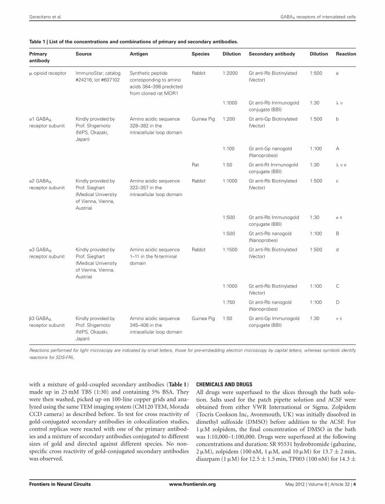
Table 1 | List of the concentrations and combinations of primary and secondary antibodies.
Primary
antibody
Source Antigen Species Dilution Secondary antibody Dilution Reaction
μ-opioid receptor ImmunoStar; catalog#24216; lot #607102
Synthetic peptidecorresponding to aminoacids 384–398 predictedfrom cloned rat MOR1
Rabbit 1:2000 Gt anti-Rb Biotinylated(Vector)
1:500 a
1:1000 Gt anti-Rb Immunogoldconjugate (BBI)
1:30 λ ν
α1 GABAA
receptor subunitKindly provided byProf. Shigemoto(NIPS, Okazaki,Japan)
Amino acidic sequence328–382 in theintracellular loop domain
Guinea Pig 1:200 Gt anti-Gp Biotinylated(Vector)
1:500 b
1:100 Gt anti-Gp nanogold(Nanoprobes)
1:100 A
Rat 1:50 Gt anti-Rt Immunogoldconjugate (BBI)
1:30 λ ν σ
α2 GABAA
receptor subunitKindly provided byProf. Sieghart(Medical Universityof Vienna, Vienna,Austria)
Amino acidic sequence322–357 in theintracellular loop domain
Rabbit 1:1000 Gt anti-Rb Biotinylated(Vector)
1:500 c
1:500 Gt anti-Rb Immunogoldconjugate (BBI)
1:30 σ τ
1:500 Gt anti-Rb nanogold(Nanoprobes)
1:100 B
α3 GABAA
receptor subunitKindly provided byProf. Sieghart(Medical Universityof Vienna, Vienna,Austria)
Amino acidic sequence1–11 in the N-terminaldomain
Rabbit 1:1500 Gt anti-Rb Biotinylated(Vector)
1:500 d
1:1000 Gt anti-Rb Biotinylated(Vector)
1:100 C
1:750 Gt anti-Rb nanogold(Nanoprobes)
1:100 D
β3 GABAA
receptor subunitKindly provided byProf. Shigemoto(NIPS, Okazaki,Japan)
Amino acidic sequence345–408 in theintracellular loop domain
Guinea Pig 1:50 Gt anti-Gp Immunogoldconjugate (BBI)
1:30 ν τ
Reactions performed for light microscopy are indicated by small letters, those for pre-embedding electron microscopy by capital letters, whereas symbols identify
reactions for SDS-FRL.
with a mixture of gold-coupled secondary antibodies (Table 1)made up in 25 mM TBS (1:30) and containing 5% BSA. Theywere then washed, picked up on 100-line copper grids and ana-lyzed using the same TEM imaging system (CM120 TEM, MoradaCCD camera) as described before. To test for cross reactivity ofgold-conjugated secondary antibodies in colocalization studies,control replicas were reacted with one of the primary antibod-ies and a mixture of secondary antibodies conjugated to differentsizes of gold and directed against different species. No non-specific cross reactivity of gold-conjugated secondary antibodieswas observed.
CHEMICALS AND DRUGSAll drugs were superfused to the slices through the bath solu-tion. Salts used for the patch pipette solution and ACSF wereobtained from either VWR International or Sigma. Zolpidem(Tocris Cookson Inc, Avonmouth, UK) was initially dissolved indimethyl sulfoxide (DMSO) before addition to the ACSF. For1 μM zolpidem, the final concentration of DMSO in the bathwas 1:10,000–1:100,000. Drugs were superfused at the followingconcentrations and duration: SR 95531 hydrobromide (gabazine,2 μM), zolpidem (100 nM, 1 μM, and 10 μM) for 13.7 ± 2 min,diazepam (1 μM) for 12.5 ± 1.5 min, TP003 (100 nM) for 14.3 ±
Frontiers in Neural Circuits www.frontiersin.org May 2012 | Volume 6 | Article 32 | 4
Geracitano et al. GABAA receptors of intercalated cells
0.7 min. The α3 subunit selective agonist TP003 was supplied byand used with the permission of Merck Research Laboratory (NJ,USA).
RESULTSACTIONS OF ZOLPIDEM AND DIAZEPAM ON uIPSCs MEDIATEDBY IMP NEURONS OF AMYGDALAWe have performed paired recordings between visually identi-fied Imp neurons in slices obtained from GAD65-GFP mice.The Imp neurons were observed as clusters of densely packedcells or as thin strands of cells present between the BL andCE nuclei. Presynaptic action potentials resulted in uIPSCs inneurons recorded at the holding potential (VH) = −50 mv, inapproximately one out of 10 recorded pairs. An action currentwas evoked in the presynaptic Imp neuron at 0.1 Hz and a uIPSCwas recorded in a postsynaptic Imp neuron. The events wereentirely mediated by GABAA receptors, since they were abol-ished by the application of 2 μM gabazine (n = 3, not shown).
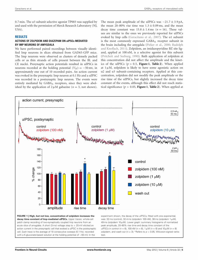
The mean peak amplitude of the uIPSCs was −21.7 ± 3.9 pA,the mean 20–80% rise time was 1.3 ± 0.09 ms, and the meandecay time constant was 15.8 ± 1.4 ms (n = 16). These val-ues are similar to the ones we previously reported for uIPSCsevoked by Imp cells (Geracitano et al., 2007). The α1 subunitis the most commonly expressed GABAA receptor subunit inthe brain including the amygdala (Pirker et al., 2000; Rudolphand Knoflach, 2011). Zolpidem, an imidazopyridine BZ site lig-and, applied at 100 nM, is a selective agonist for this subunit(Pritchett and Seeburg, 1990). Bath application of zolpidem atthis concentration did not affect the amplitude and the kinet-ics of the uIPSCs (p > 0.5, Figure 1, Table 2). When appliedat 1 μM, zolpidem is likely to have some agonistic action onα2 and α3 subunit-containing receptors. Applied at this con-centration, zolpidem did not modify the peak amplitude or therise time of the uIPSCs, but slightly increased the decay timeconstant of the events, although this effect did not reach statis-tical significance (p > 0.05, Figure 1, Table 2). When applied at
FIGURE 1 | High, but not low, concentration of zolpidem increases the
decay time constant of Imp-mediated uIPSCs. Upper traces: whole-cellpatch clamp recording of two-synaptically coupled Imp neurons from anacute slice of amygdala. A short (3 ms) voltage step to + 20 mV elicited anaction current in the presynaptic cell that evoked a uIPSC in the postsynapticcell. Each trace is the average of 10 consecutive sweeps (0.1 Hz), recordedwith a K-gluconate-based solution at the holding potential of −55 mV. In the
experiment shown, the decay of the uIPSCs, fitted with one exponential,was: 32 ms (control), 32.4 ms (zolpidem 100 nM), 38 ms (zolpidem 1 μM),49 ms (zolpidem 10 μM). Lower graph: summary histograms of normalizedpeak amplitude, 20–80% rise time and decay time constant of theuIPSCs in control (n = 6), 100 nM (n = 6), 1 μM (n = 6) and 10 μM (n = 6)zolpidem, and wash out (n = 3); ∗Refers to p < 0.05, Wilcoxon-signed rankstest.
Frontiers in Neural Circuits www.frontiersin.org May 2012 | Volume 6 | Article 32 | 5
Geracitano et al. GABAA receptors of intercalated cells
Table 2 | Effects of zolpidem (100 nM, 1 μM, and 10 μM) on uIPSCs recorded from Imp paired recordings experiments.
uIPSCs Control (n = 6) zolpidem (100 nM, n = 6) zolpidem (1 μM, n = 6) zolpidem (10 μM, n = 6) wash (n = 3)
peak amplitude (pA) −27.1± 9.5 −27.3± 9.5 −25.1 ± 7.9 −27.5± 9.0 −29.0 ± 17.4
Rise time 1.3 ± 0.2 1.3 ± 0.1 1.4 ± 0.2 1.8 ± 0.3∗ 1.1 ± 0.3
Decay time (ms) 18.6± 2.9 19.7± 2.6 21.0 ± 2.8 29.3 ± 4.4 18.2 ± 1.8
Data are expressed as mean ± SEM; n = number of recorded pairs; two-tailed Wilcoxon-signed ranks test was used for comparison between pre- and post-zolpidem
values (∗P < 0.05).
10 μM, a concentration that is likely to fully activate α2 and/or α3subunits-containing receptors, zolpidem significantly increasedthe 20–80% rise time and the decay time constant of the uIP-SCs (p < 0.05) without affecting their peak amplitude (Figure 1,Table 2). These results were confirmed by testing the non-specificBZ agonist diazepam (1 μM) in a limited number of experiments.The bath application of this drug mimicked the action of highconcentration of zolpidem (p > 0.1, independent sample t-test orp > 0.5, Mann–Whitney U-test, for the comparison of the effectson uIPSC kinetics induced by 1 μM diazepam vs. 10 μM zolpi-dem). Specifically, diazepam increased the 20–80% rise time andthe decay time constant of the uIPSCs without altering their peakamplitude (data not shown). On average, the uIPSC peak ampli-tude was −22.2 ± 6.3 and −23.2 ± 4.6 pA, the 20–80% rise timewas 1.5 ± 0.2 and 2.2 ± 0.4 ms, and the decay time constant was16.3 ± 3.9 and 24.3 ± 4.2, before and during diazepam (n = 3).
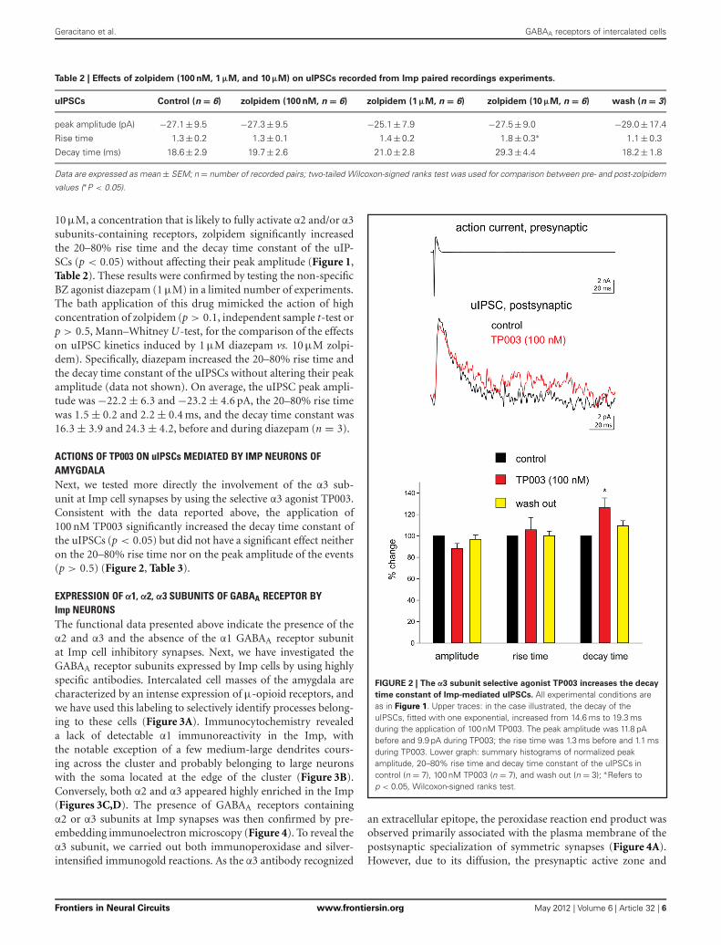
ACTIONS OF TP003 ON uIPSCs MEDIATED BY IMP NEURONS OFAMYGDALANext, we tested more directly the involvement of the α3 sub-unit at Imp cell synapses by using the selective α3 agonist TP003.Consistent with the data reported above, the application of100 nM TP003 significantly increased the decay time constant ofthe uIPSCs (p < 0.05) but did not have a significant effect neitheron the 20–80% rise time nor on the peak amplitude of the events(p > 0.5) (Figure 2, Table 3).
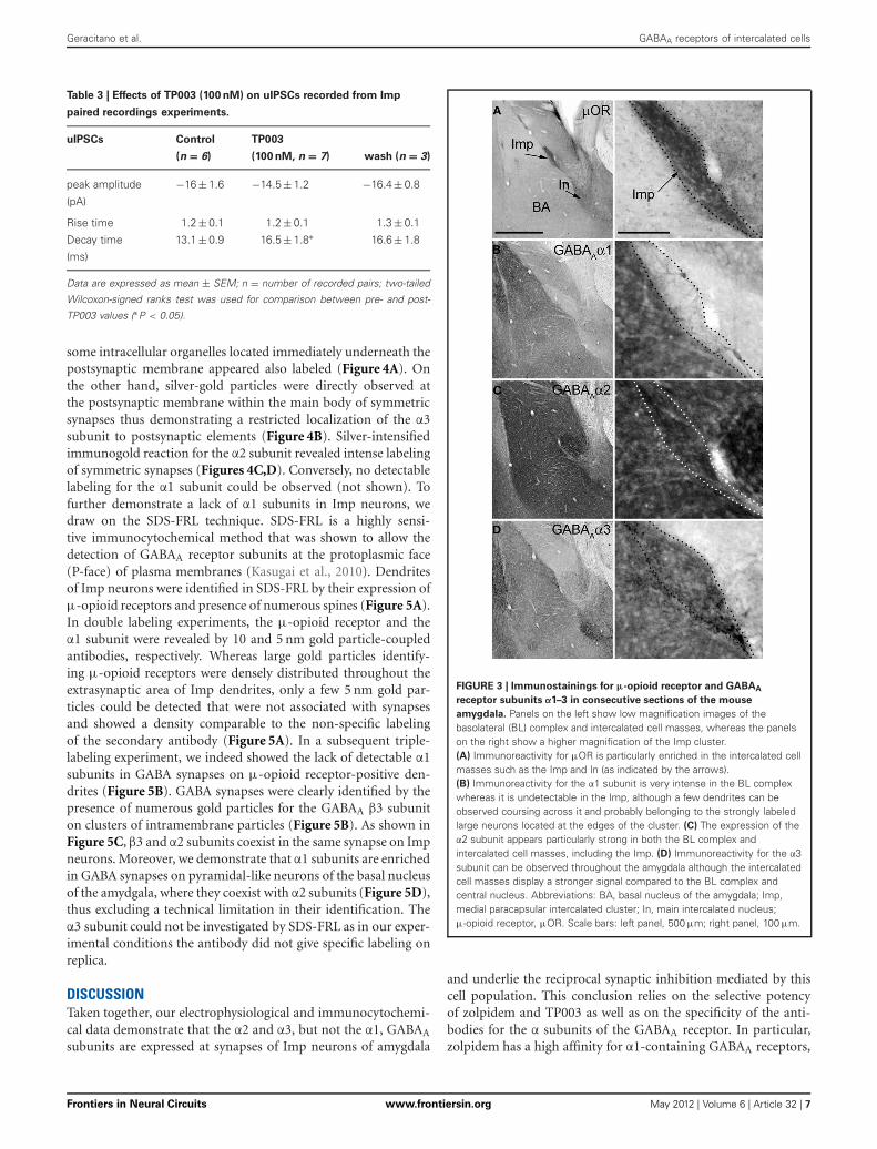
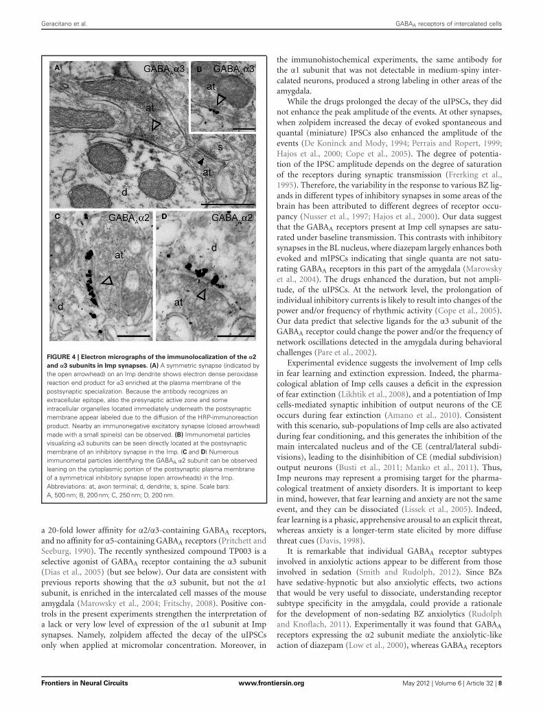
EXPRESSION OF α1, α2, α3 SUBUNITS OF GABAA RECEPTOR BYImp NEURONSThe functional data presented above indicate the presence of theα2 and α3 and the absence of the α1 GABAA receptor subunitat Imp cell inhibitory synapses. Next, we have investigated theGABAA receptor subunits expressed by Imp cells by using highlyspecific antibodies. Intercalated cell masses of the amygdala arecharacterized by an intense expression of μ-opioid receptors, andwe have used this labeling to selectively identify processes belong-ing to these cells (Figure 3A). Immunocytochemistry revealeda lack of detectable α1 immunoreactivity in the Imp, withthe notable exception of a few medium-large dendrites cours-ing across the cluster and probably belonging to large neuronswith the soma located at the edge of the cluster (Figure 3B).Conversely, both α2 and α3 appeared highly enriched in the Imp(Figures 3C,D). The presence of GABAA receptors containingα2 or α3 subunits at Imp synapses was then confirmed by pre-embedding immunoelectron microscopy (Figure 4). To reveal theα3 subunit, we carried out both immunoperoxidase and silver-intensified immunogold reactions. As the α3 antibody recognized
FIGURE 2 | The α3 subunit selective agonist TP003 increases the decay
time constant of Imp-mediated uIPSCs. All experimental conditions areas in Figure 1. Upper traces: in the case illustrated, the decay of theuIPSCs, fitted with one exponential, increased from 14.6 ms to 19.3 msduring the application of 100 nM TP003. The peak amplitude was 11.8 pAbefore and 9.9 pA during TP003; the rise time was 1.3 ms before and 1.1 msduring TP003. Lower graph: summary histograms of normalized peakamplitude, 20–80% rise time and decay time constant of the uIPSCs incontrol (n = 7), 100 nM TP003 (n = 7), and wash out (n = 3); ∗Refers top < 0.05, Wilcoxon-signed ranks test.
an extracellular epitope, the peroxidase reaction end product wasobserved primarily associated with the plasma membrane of thepostsynaptic specialization of symmetric synapses (Figure 4A).However, due to its diffusion, the presynaptic active zone and
Frontiers in Neural Circuits www.frontiersin.org May 2012 | Volume 6 | Article 32 | 6
Geracitano et al. GABAA receptors of intercalated cells
Table 3 | Effects of TP003 (100 nM) on uIPSCs recorded from Imp
paired recordings experiments.
uIPSCs Control TP003
(n = 6) (100 nM, n = 7) wash (n = 3)
peak amplitude −16 ± 1.6 −14.5± 1.2 −16.4± 0.8
(pA)
Rise time 1.2 ± 0.1 1.2 ± 0.1 1.3 ± 0.1
Decay time 13.1 ± 0.9 16.5± 1.8∗ 16.6± 1.8
(ms)
Data are expressed as mean ± SEM; n = number of recorded pairs; two-tailed
Wilcoxon-signed ranks test was used for comparison between pre- and post-
TP003 values (∗P < 0.05).
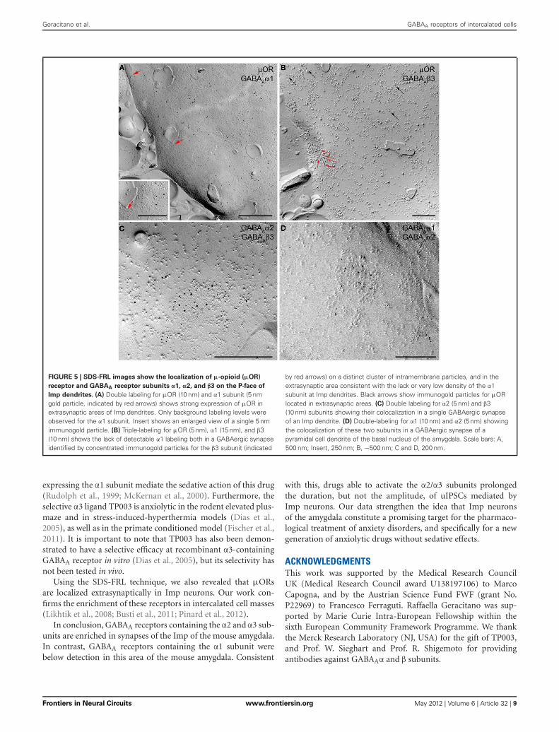
some intracellular organelles located immediately underneath thepostsynaptic membrane appeared also labeled (Figure 4A). Onthe other hand, silver-gold particles were directly observed atthe postsynaptic membrane within the main body of symmetricsynapses thus demonstrating a restricted localization of the α3subunit to postsynaptic elements (Figure 4B). Silver-intensifiedimmunogold reaction for the α2 subunit revealed intense labelingof symmetric synapses (Figures 4C,D). Conversely, no detectablelabeling for the α1 subunit could be observed (not shown). Tofurther demonstrate a lack of α1 subunits in Imp neurons, wedraw on the SDS-FRL technique. SDS-FRL is a highly sensi-tive immunocytochemical method that was shown to allow thedetection of GABAA receptor subunits at the protoplasmic face(P-face) of plasma membranes (Kasugai et al., 2010). Dendritesof Imp neurons were identified in SDS-FRL by their expression ofμ-opioid receptors and presence of numerous spines (Figure 5A).In double labeling experiments, the μ-opioid receptor and theα1 subunit were revealed by 10 and 5 nm gold particle-coupledantibodies, respectively. Whereas large gold particles identify-ing μ-opioid receptors were densely distributed throughout theextrasynaptic area of Imp dendrites, only a few 5 nm gold par-ticles could be detected that were not associated with synapsesand showed a density comparable to the non-specific labelingof the secondary antibody (Figure 5A). In a subsequent triple-labeling experiment, we indeed showed the lack of detectable α1subunits in GABA synapses on μ-opioid receptor-positive den-drites (Figure 5B). GABA synapses were clearly identified by thepresence of numerous gold particles for the GABAA β3 subuniton clusters of intramembrane particles (Figure 5B). As shown inFigure 5C, β3 and α2 subunits coexist in the same synapse on Impneurons. Moreover, we demonstrate that α1 subunits are enrichedin GABA synapses on pyramidal-like neurons of the basal nucleusof the amydgala, where they coexist with α2 subunits (Figure 5D),thus excluding a technical limitation in their identification. Theα3 subunit could not be investigated by SDS-FRL as in our exper-imental conditions the antibody did not give specific labeling onreplica.
DISCUSSIONTaken together, our electrophysiological and immunocytochemi-cal data demonstrate that the α2 and α3, but not the α1, GABAA
subunits are expressed at synapses of Imp neurons of amygdala
FIGURE 3 | Immunostainings for μ-opioid receptor and GABAA
receptor subunits α1–3 in consecutive sections of the mouse
amygdala. Panels on the left show low magnification images of thebasolateral (BL) complex and intercalated cell masses, whereas the panelson the right show a higher magnification of the Imp cluster.(A) Immunoreactivity for μOR is particularly enriched in the intercalated cellmasses such as the Imp and In (as indicated by the arrows).(B) Immunoreactivity for the α1 subunit is very intense in the BL complexwhereas it is undetectable in the Imp, although a few dendrites can beobserved coursing across it and probably belonging to the strongly labeledlarge neurons located at the edges of the cluster. (C) The expression of theα2 subunit appears particularly strong in both the BL complex andintercalated cell masses, including the Imp. (D) Immunoreactivity for the α3subunit can be observed throughout the amygdala although the intercalatedcell masses display a stronger signal compared to the BL complex andcentral nucleus. Abbreviations: BA, basal nucleus of the amygdala; Imp,medial paracapsular intercalated cluster; In, main intercalated nucleus;μ-opioid receptor, μOR. Scale bars: left panel, 500 μm; right panel, 100 μm.
and underlie the reciprocal synaptic inhibition mediated by thiscell population. This conclusion relies on the selective potencyof zolpidem and TP003 as well as on the specificity of the anti-bodies for the α subunits of the GABAA receptor. In particular,zolpidem has a high affinity for α1-containing GABAA receptors,
Frontiers in Neural Circuits www.frontiersin.org May 2012 | Volume 6 | Article 32 | 7
Geracitano et al. GABAA receptors of intercalated cells
FIGURE 4 | Electron micrographs of the immunolocalization of the α2
and α3 subunits in Imp synapses. (A) A symmetric synapse (indicated bythe open arrowhead) on an Imp dendrite shows electron dense peroxidasereaction end product for α3 enriched at the plasma membrane of thepostsynaptic specialization. Because the antibody recognizes anextracellular epitope, also the presynaptic active zone and someintracellular organelles located immediately underneath the postsynapticmembrane appear labeled due to the diffusion of the HRP-immunoreactionproduct. Nearby an immunonegative excitatory synapse (closed arrowhead)made with a small spine(s) can be observed. (B) Immunometal particlesvisualizing α3 subunits can be seen directly located at the postsynapticmembrane of an inhibitory synapse in the Imp. (C and D) Numerousimmunometal particles identifying the GABAA α2 subunit can be observedleaning on the cytoplasmic portion of the postsynaptic plasma membraneof a symmetrical inhibitory synapse (open arrowheads) in the Imp.Abbreviations: at, axon terminal; d, dendrite; s, spine. Scale bars:A, 500 nm; B, 200 nm; C, 250 nm; D, 200 nm.
a 20-fold lower affinity for α2/α3-containing GABAA receptors,and no affinity for α5-containing GABAA receptors (Pritchett andSeeburg, 1990). The recently synthesized compound TP003 is aselective agonist of GABAA receptor containing the α3 subunit(Dias et al., 2005) (but see below). Our data are consistent withprevious reports showing that the α3 subunit, but not the α1subunit, is enriched in the intercalated cell masses of the mouseamygdala (Marowsky et al., 2004; Fritschy, 2008). Positive con-trols in the present experiments strengthen the interpretation ofa lack or very low level of expression of the α1 subunit at Impsynapses. Namely, zolpidem affected the decay of the uIPSCsonly when applied at micromolar concentration. Moreover, in
the immunohistochemical experiments, the same antibody forthe α1 subunit that was not detectable in medium-spiny inter-calated neurons, produced a strong labeling in other areas of theamygdala.
While the drugs prolonged the decay of the uIPSCs, they didnot enhance the peak amplitude of the events. At other synapses,when zolpidem increased the decay of evoked spontaneous andquantal (miniature) IPSCs also enhanced the amplitude of theevents (De Koninck and Mody, 1994; Perrais and Ropert, 1999;Hajos et al., 2000; Cope et al., 2005). The degree of potentia-tion of the IPSC amplitude depends on the degree of saturationof the receptors during synaptic transmission (Frerking et al.,1995). Therefore, the variability in the response to various BZ lig-ands in different types of inhibitory synapses in some areas of thebrain has been attributed to different degrees of receptor occu-pancy (Nusser et al., 1997; Hajos et al., 2000). Our data suggestthat the GABAA receptors present at Imp cell synapses are satu-rated under baseline transmission. This contrasts with inhibitorysynapses in the BL nucleus, where diazepam largely enhances bothevoked and mIPSCs indicating that single quanta are not satu-rating GABAA receptors in this part of the amygdala (Marowskyet al., 2004). The drugs enhanced the duration, but not ampli-tude, of the uIPSCs. At the network level, the prolongation ofindividual inhibitory currents is likely to result into changes of thepower and/or frequency of rhythmic activity (Cope et al., 2005).Our data predict that selective ligands for the α3 subunit of theGABAA receptor could change the power and/or the frequency ofnetwork oscillations detected in the amygdala during behavioralchallenges (Pare et al., 2002).
Experimental evidence suggests the involvement of Imp cellsin fear learning and extinction expression. Indeed, the pharma-cological ablation of Imp cells causes a deficit in the expressionof fear extinction (Likhtik et al., 2008), and a potentiation of Impcells-mediated synaptic inhibition of output neurons of the CEoccurs during fear extinction (Amano et al., 2010). Consistentwith this scenario, sub-populations of Imp cells are also activatedduring fear conditioning, and this generates the inhibition of themain intercalated nucleus and of the CE (central/lateral subdi-visions), leading to the disinhibition of CE (medial subdivision)output neurons (Busti et al., 2011; Manko et al., 2011). Thus,Imp neurons may represent a promising target for the pharma-cological treatment of anxiety disorders. It is important to keepin mind, however, that fear learning and anxiety are not the sameevent, and they can be dissociated (Lissek et al., 2005). Indeed,fear learning is a phasic, apprehensive arousal to an explicit threat,whereas anxiety is a longer-term state elicited by more diffusethreat cues (Davis, 1998).
It is remarkable that individual GABAA receptor subtypesinvolved in anxiolytic actions appear to be different from thoseinvolved in sedation (Smith and Rudolph, 2012). Since BZshave sedative-hypnotic but also anxiolytic effects, two actionsthat would be very useful to dissociate, understanding receptorsubtype specificity in the amygdala, could provide a rationalefor the development of non-sedating BZ anxiolytics (Rudolphand Knoflach, 2011). Experimentally it was found that GABAA
receptors expressing the α2 subunit mediate the anxiolytic-likeaction of diazepam (Low et al., 2000), whereas GABAA receptors
Frontiers in Neural Circuits www.frontiersin.org May 2012 | Volume 6 | Article 32 | 8
Geracitano et al. GABAA receptors of intercalated cells
FIGURE 5 | SDS-FRL images show the localization of μ-opioid (μOR)
receptor and GABAA receptor subunits α1, α2, and β3 on the P-face of
Imp dendrites. (A) Double labeling for μOR (10 nm) and α1 subunit (5 nmgold particle, indicated by red arrows) shows strong expression of μOR inextrasynaptic areas of Imp dendrites. Only background labeling levels wereobserved for the α1 subunit. Insert shows an enlarged view of a single 5 nmimmunogold particle. (B) Triple-labeling for μOR (5 nm), α1 (15 nm), and β3(10 nm) shows the lack of detectable α1 labeling both in a GABAergic synapseidentified by concentrated immunogold particles for the β3 subunit (indicated
by red arrows) on a distinct cluster of intramembrane particles, and in theextrasynaptic area consistent with the lack or very low density of the α1subunit at Imp dendrites. Black arrows show immunogold particles for μORlocated in extrasynaptic areas. (C) Double labeling for α2 (5 nm) and β3(10 nm) subunits showing their colocalization in a single GABAergic synapseof an Imp dendrite. (D) Double-labeling for α1 (10 nm) and α2 (5 nm) showingthe colocalization of these two subunits in a GABAergic synapse of apyramidal cell dendrite of the basal nucleus of the amygdala. Scale bars: A,500 nm; Insert, 250 nm; B, −500 nm; C and D, 200 nm.
expressing the α1 subunit mediate the sedative action of this drug(Rudolph et al., 1999; McKernan et al., 2000). Furthermore, theselective α3 ligand TP003 is anxiolytic in the rodent elevated plus-maze and in stress-induced-hyperthermia models (Dias et al.,2005), as well as in the primate conditioned model (Fischer et al.,2011). It is important to note that TP003 has also been demon-strated to have a selective efficacy at recombinant α3-containingGABAA receptor in vitro (Dias et al., 2005), but its selectivity hasnot been tested in vivo.
Using the SDS-FRL technique, we also revealed that μORsare localized extrasynaptically in Imp neurons. Our work con-firms the enrichment of these receptors in intercalated cell masses(Likhtik et al., 2008; Busti et al., 2011; Pinard et al., 2012).
In conclusion, GABAA receptors containing the α2 and α3 sub-units are enriched in synapses of the Imp of the mouse amygdala.In contrast, GABAA receptors containing the α1 subunit werebelow detection in this area of the mouse amygdala. Consistent
with this, drugs able to activate the α2/α3 subunits prolongedthe duration, but not the amplitude, of uIPSCs mediated byImp neurons. Our data strengthen the idea that Imp neuronsof the amygdala constitute a promising target for the pharmaco-logical treatment of anxiety disorders, and specifically for a newgeneration of anxiolytic drugs without sedative effects.
ACKNOWLEDGMENTSThis work was supported by the Medical Research CouncilUK (Medical Research Council award U138197106) to MarcoCapogna, and by the Austrian Science Fund FWF (grant No.P22969) to Francesco Ferraguti. Raffaella Geracitano was sup-ported by Marie Curie Intra-European Fellowship within thesixth European Community Framework Programme. We thankthe Merck Research Laboratory (NJ, USA) for the gift of TP003,and Prof. W. Sieghart and Prof. R. Shigemoto for providingantibodies against GABAAα and β subunits.
Frontiers in Neural Circuits www.frontiersin.org May 2012 | Volume 6 | Article 32 | 9
Geracitano et al. GABAA receptors of intercalated cells
REFERENCESAmano, T., Unal, C. T., and Pare, D.
(2010). Synaptic correlates of fearextinction in the amygdala. Nat.Neurosci. 13, 489–494.
Berretta, S., Pantazopoulos, H.,Caldera, M., Pantazopoulos, P.,and Pare, D. (2005). Infralimbiccortex activation increases c-Fosexpression in intercalated neuronsof the amygdala. Neuroscience 132,943–953.
Busti, D., Geracitano, R., Whittle, N.,Dalezios, Y., Manko, M., Kaufmann,W., Satzler, K., Singewald, N.,Capogna, M., and Ferraguti,F. (2011). Different fear statesengage distinct networks withinthe intercalated cell clusters ofthe amygdala. J. Neurosci. 31,5131–5144.
Capogna, M., and Pearce, R. A.(2011). GABAA,slow: causes andconsequences. Trends Neurosci. 34,101–112.
Cope, D. W., Halbsguth, C.,Karayannis, T., Wulff, P., Ferraguti,F., Hoeger, H., Leppa, E., Linden,A. M., Oberto, A., Ogris, W., Korpi,E. R., Sieghart, W., Somogyi, P.,Wisden, W., and Capogna, M.(2005). Loss of zolpidem efficacyin the hippocampus of mice withthe GABAA receptor gamma2 F77Ipoint mutation. Eur. J. Neurosci. 21,3002–3016.
Davis, M. (1998). Are different parts ofthe extended amygdala involved infear versus anxiety? Biol. Psychiatry44, 1239–1247.
De Koninck, Y., and Mody, I. (1994).Noise analysis of miniature IPSCsin adult rat brain slices: propertiesand modulation of synaptic GABAA
receptor channels. J. Neurophysiol.71, 1318–1335.
Dias, R., Sheppard, W. F., Fradley, R.L., Garrett, E. M., Stanley, J. L., Tye,S. J., Goodacre, S., Lincoln, R. J.,Cook, S. M., Conley, R., Hallett,D., Humphries, A. C., Thompson,S. A., Wafford, K. A., Street, L. J.,Castro, J. L., Whiting, P. J., Rosahl,T. W., Atack, J. R., Mckernan, R.M., Dawson, G. R., and Reynolds,D. S. (2005). Evidence for a sig-nificant role of alpha 3-containingGABAA receptors in mediatingthe anxiolytic effects of ben-zodiazepines. J. Neurosci. 25,10682–10688.
Ehrlich, I., Humeau, Y., Grenier,F., Ciocchi, S., Herry, C., andLuthi, A. (2009). Amygdalainhibitory circuits and the con-trol of fear memory. Neuron 62,757–771.
Farrant, M., and Nusser, Z. (2005).Variations on an inhibitory theme:
phasic and tonic activation ofGABA(A) receptors. Nat. Rev.Neurosci. 6, 215–229.
Fischer, B. D., Atack, J. R., Platt,D. M., Reynolds, D. S., Dawson,G. R., and Rowlett, J. K. (2011).Contribution of GABA(A) receptorscontaining alpha3 subunits to thetherapeutic-related and side effectsof benzodiazepine-type drugsin monkeys. Psychopharmacology(Berl.) 215, 311–319.
Frerking, M., Borges, S., andWilson, M. (1995). Variationin GABA mini amplitude is theconsequence of variation in trans-mitter concentration. Neuron 15,885–895.
Fritschy, J. M. (2008). Is my antibody-staining specific? How to dealwith pitfalls of immunohisto-chemistry. Eur. J. Neurosci. 28,2365–2370.
Geracitano, R., Kaufmann, W. A.,Szabo, G., Ferraguti, F., andCapogna, M. (2007). Synapticheterogeneity between mouseparacapsular intercalated neuronsof the amygdala. J. Physiol. 585,117–134.
Hajos, N., Nusser, Z., Rancz, E. A.,Freund, T. F., and Mody, I. (2000).Cell type- and synapse-specific vari-ability in synaptic GABAA recep-tor occupancy. Eur. J. Neurosci. 12,810–818.
Herry, C., Ferraguti, F., Singewald,N., Letzkus, J. J., Ehrlich, I., andLuthi, A. (2010). Neuronal circuitsof fear extinction. Eur. J. Neurosci.31, 599–612.
Kasugai, Y., Swinny, J. D., Roberts, J. D.,Dalezios, Y., Fukazawa, Y., Sieghart,W., Shigemoto, R., and Somogyi, P.(2010). Quantitative localisation ofsynaptic and extrasynaptic GABAA
receptor subunits on hippocampalpyramidal cells by freeze-fracturereplica immunolabelling. Eur. J.Neurosci. 32, 1868–1888.
Li, G., Amano, T., Pare, D., andNair, S. S. (2011). Impact ofinfralimbic inputs on intercalatedamygdala neurons: a biophysicalmodeling study. Learn. Mem. 18,226–240.
Likhtik, E., Popa, D., Apergis-Schoute,J., Fidacaro, G. A., and Pare, D.(2008). Amygdala intercalated neu-rons are required for expressionof fear extinction. Nature 454,642–645.
Lissek, S., Powers, A. S., McClure, E.B., Phelps, E. A., Woldehawariat,G., Grillon, C., and Pine, D. S.(2005). Classical fear condition-ing in the anxiety disorders: ameta-analysis. Behav. Res. Ther. 43,1391–1424.
Lopez-Bendito, G., Sturgess, K.,Erdelyi, F., Szabo, G., Molnar, Z.,and Paulsen, O. (2004). Preferentialorigin and layer destination ofGAD65-GFP cortical interneurons.Cereb. Cortex 14, 1122–1133.
Low, K., Crestani, F., Keist, R., Benke,D., Brunig, I., Benson, J. A., Fritschy,J. M., Rulicke, T., Bluethmann, H.,Mohler, H., and Rudolph, U.(2000). Molecular and neuronalsubstrate for the selective atten-uation of anxiety. Science 290,131–134.
Manko, M., Geracitano, R., andCapogna, M. (2011). Functionalconnectivity of the main inter-calated nucleus of the mouseamygdala. J. Physiol. 589,1911–1925.
Marowsky, A., Fritschy, J. M., andVogt, K. E. (2004). Functional map-ping of GABAA receptor subtypes inthe amygdala. Eur. J. Neurosci. 20,1281–1289.
McDonald, A. J., and Augustine, J. R.(1993). Localization of GABA-likeimmunoreactivity in the mon-key amygdala. Neuroscience 52,281–294.
McKernan, R. M., Rosahl, T. W.,Reynolds, D. S., Sur, C., Wafford, K.A., Atack, J. R., Farrar, S., Myers,J., Cook, G., Ferris, P., Garrett, L.,Bristow, L., Marshall, G., Macaulay,A., Brown, N., Howell, O., Moore,K. W., Carling, R. W., Street, L.J., Castro, J. L., Ragan, C. I.,Dawson, G. R., and Whiting, P. J.(2000). Sedative but not anxiolyticproperties of benzodiazepines aremediated by the GABA(A) recep-tor alpha1 subtype. Nat. Neurosci. 3,587–592.
Millhouse, O. E. (1986). The interca-lated cells of the amygdala. J. Comp.Neurol. 247, 246–271.
Nitecka, L., and Ben-Ari, Y. (1987).Distribution of GABA-likeimmunoreactivity in the ratamygdaloid complex. J. Comp.Neurol. 266, 45–55.
Nusser, Z., Cull-Candy, S., and Farrant,M. (1997). Differences in synapticGABA(A) receptor number underlievariation in GABA mini amplitude.Neuron 19, 697–709.
Pape, H. C., and Pare, D. (2010). Plasticsynaptic networks of the amyg-dala for the acquisition, expression,and extinction of conditioned fear.Physiol. Rev. 90, 419–463.
Pare, D., Collins, D. R., and Pelletier,J. G. (2002). Amygdala oscilla-tions and the consolidation of emo-tional memories. Trends Cogn. Sci.6, 306–314.
Pare, D., and Smith, Y. (1993).Distribution of GABA immunore-
activity in the amygdaloid complexof the cat. Neuroscience 57,1061–1076.
Perrais, D., and Ropert, N. (1999).Effect of zolpidem on miniatureIPSCs and occupancy of post-synaptic GABAA receptors incentral synapses. J. Neurosci. 19,578–588.
Pinard, C. R., Mascagni, F., andMcDonald, A. J. (2012). Medialprefrontal cortical innervation ofthe intercalated nuclear region ofthe amygdala. Neuroscience 205,112–124.
Pirker, S., Schwarzer, C., Wieselthaler,A., Sieghart, W., and Sperk, G.(2000). GABA(A) receptors:immunocytochemical distributionof 13 subunits in the adult rat brain.Neuroscience 101, 815–850.
Pritchett, D. B., and Seeburg, P. H.(1990). Gamma-aminobutyricacidA receptor alpha 5-subunitcreates novel type II benzodi-azepine receptor pharmacology.J. Neurochem. 54, 1802–1804.
Royer, S., Martina, M., and Pare, D.(1999). An inhibitory interface gatesimpulse traffic between the inputand output stations of the amygdala.J. Neurosci. 19, 10575–10583.
Royer, S., Martina, M., and Pare, D.(2000). Polarized synaptic interac-tions between intercalated neuronsof the amygdala. J. Neurophysiol. 83,3509–3518.
Royer, S., and Pare, D. (2002).Bidirectional synaptic plasticityin intercalated amygdala neuronsand the extinction of conditionedfear responses. Neuroscience 115,455–462.
Rudolph, U., Crestani, F., Benke,D., Brunig, I., Benson, J. A.,Fritschy, J. M., Martin, J. R.,Bluethmann, H., and Mohler, H.(1999). Benzodiazepine actionsmediated by specific gamma-aminobutyric acid(A) receptorsubtypes. Nature 401, 796–800.
Rudolph, U., and Knoflach, F. (2011).Beyond classical benzodiazepines:novel therapeutic potential ofGABAA receptor subtypes. Nat. Rev.Drug Discov. 10, 685–697.
Rudolph, U., and Mohler, H.(2006). GABA-based therapeu-tic approaches: GABAA receptorsubtype functions. Curr. Opin.Pharmacol. 6, 18–23.
Smith, K. S., and Rudolph, U. (2012).Anxiety and depression: mousegenetics and pharmacologi-cal approaches to the role ofGABA(A) receptor subtypes.Neuropharmacology 62, 54–62.
Sreepathi, H. K., and Ferraguti,F. (2012). Subpopulations of
Frontiers in Neural Circuits www.frontiersin.org May 2012 | Volume 6 | Article 32 | 10

Geracitano et al. GABAA receptors of intercalated cells
neurokinin 1 receptor-expressingneurons in the rat lateral amygdaladisplay a differential pattern ofinnervation from distinct gluta-matergic afferents. Neuroscience203, 59–77.
Whiting, P. J. (2006). GABA-A recep-tors: a viable target for novelanxiolytics? Curr. Opin. Pharmacol.6, 24–29.
Conflict of Interest Statement: Theauthors declare that the researchwas conducted in the absence of anycommercial or financial relationshipsthat could be construed as a potentialconflict of interest.
Received: 02 April 2012; paper pend-ing published: 23 April 2012; accepted:
07 May 2012; published online: 31 May2012.Citation: Geracitano R, Fischer D,Kasugai Y, Ferraguti F and CapognaM (2012) Functional expression of theGABAA receptor α2 and α3 subunits atsynapses between intercalated medialparacapsular neurons of mouse amyg-dala. Front. Neural Circuits 6:32. doi:10.3389/fncir.2012.00032
Copyright © 2012 Geracitano, Fischer,Kasugai, Ferraguti and Capogna.This is an open-access article dis-tributed under the terms of theCreative Commons Attribution NonCommercial License, which permitsnon-commercial use, distribution, andreproduction in other forums, providedthe original authors and source arecredited.
Frontiers in Neural Circuits www.frontiersin.org May 2012 | Volume 6 | Article 32 | 11
Top Related