Wound healing potential of Ocimum sanctum Linn. with...
5
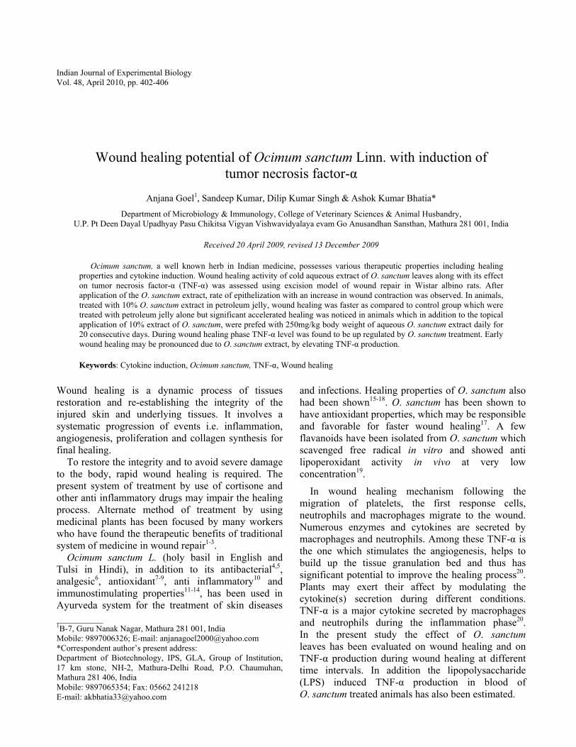
Indian Journal of Experimental Biology Vol. 48, April 2010, pp. 402-406 Wound healing potential of Ocimum sanctum Linn. with induction of tumor necrosis factor-α Anjana Goel 1 , Sandeep Kumar, Dilip Kumar Singh & Ashok Kumar Bhatia* Department of Microbiology & Immunology, College of Veterinary Sciences & Animal Husbandry, U.P. Pt Deen Dayal Upadhyay Pasu Chikitsa Vigyan Vishwavidyalaya evam Go Anusandhan Sansthan, Mathura 281 001, India Received 20 April 2009, revised 13 December 2009 Ocimum sanctum, a well known herb in Indian medicine, possesses various therapeutic properties including healing properties and cytokine induction. Wound healing activity of cold aqueous extract of O. sanctum leaves along with its effect on tumor necrosis factor-α (TNF-α) was assessed using excision model of wound repair in Wistar albino rats. After application of the O. sanctum extract, rate of epithelization with an increase in wound contraction was observed. In animals, treated with 10% O. sanctum extract in petroleum jelly, wound healing was faster as compared to control group which were treated with petroleum jelly alone but significant accelerated healing was noticed in animals which in addition to the topical application of 10% extract of O. sanctum, were prefed with 250mg/kg body weight of aqueous O. sanctum extract daily for 20 consecutive days. During wound healing phase TNF-α level was found to be up regulated by O. sanctum treatment. Early wound healing may be pronounced due to O. sanctum extract, by elevating TNF-α production. Keywords: Cytokine induction, Ocimum sanctum, TNF-α, Wound healing Wound healing is a dynamic process of tissues restoration and re-establishing the integrity of the injured skin and underlying tissues. It involves a systematic progression of events i.e. inflammation, angiogenesis, proliferation and collagen synthesis for final healing. To restore the integrity and to avoid severe damage to the body, rapid wound healing is required. The present system of treatment by use of cortisone and other anti inflammatory drugs may impair the healing process. Alternate method of treatment by using medicinal plants has been focused by many workers who have found the therapeutic benefits of traditional system of medicine in wound repair 1-3 . Ocimum sanctum L. (holy basil in English and Tulsi in Hindi), in addition to its antibacterial 4,5 , analgesic 6 , antioxidant 7-9 , anti inflammatory 10 and immunostimulating properties 11-14 , has been used in Ayurveda system for the treatment of skin diseases and infections. Healing properties of O. sanctum also had been shown 15-18 . O. sanctum has been shown to have antioxidant properties, which may be responsible and favorable for faster wound healing 17 . A few flavanoids have been isolated from O. sanctum which scavenged free radical in vitro and showed anti lipoperoxidant activity in vivo at very low concentration 19 . In wound healing mechanism following the migration of platelets, the first response cells, neutrophils and macrophages migrate to the wound. Numerous enzymes and cytokines are secreted by macrophages and neutrophils. Among these TNF-α is the one which stimulates the angiogenesis, helps to build up the tissue granulation bed and thus has significant potential to improve the healing process 20 . Plants may exert their affect by modulating the cytokine(s) secretion during different conditions. TNF-α is a major cytokine secreted by macrophages and neutrophils during the inflammation phase 20 . In the present study the effect of O. sanctum leaves has been evaluated on wound healing and on TNF-α production during wound healing at different time intervals. In addition the lipopolysaccharide (LPS) induced TNF-α production in blood of O. sanctum treated animals has also been estimated. ___________ 1 B-7, Guru Nanak Nagar, Mathura 281 001, India Mobile: 9897006326; E-mail: [email protected] *Correspondent author’s present address: Department of Biotechnology, IPS, GLA, Group of Institution, 17 km stone, NH-2, Mathura-Delhi Road, P.O. Chaumuhan, Mathura 281 406, India Mobile: 9897065354; Fax: 05662 241218 E-mail: [email protected]
Transcript of Wound healing potential of Ocimum sanctum Linn. with...
Indian Journal of Experimental Biology Vol. 48, April 2010, pp. 402-406
Wound healing potential of Ocimum sanctum Linn. with induction of tumor necrosis factor-α
Anjana Goel1, Sandeep Kumar, Dilip Kumar Singh & Ashok Kumar Bhatia*
Department of Microbiology & Immunology, College of Veterinary Sciences & Animal Husbandry, U.P. Pt Deen Dayal Upadhyay Pasu Chikitsa Vigyan Vishwavidyalaya evam Go Anusandhan Sansthan, Mathura 281 001, India
Received 20 April 2009, revised 13 December 2009
Ocimum sanctum, a well known herb in Indian medicine, possesses various therapeutic properties including healing properties and cytokine induction. Wound healing activity of cold aqueous extract of O. sanctum leaves along with its effect on tumor necrosis factor-α (TNF-α) was assessed using excision model of wound repair in Wistar albino rats. After application of the O. sanctum extract, rate of epithelization with an increase in wound contraction was observed. In animals, treated with 10% O. sanctum extract in petroleum jelly, wound healing was faster as compared to control group which were treated with petroleum jelly alone but significant accelerated healing was noticed in animals which in addition to the topical application of 10% extract of O. sanctum, were prefed with 250mg/kg body weight of aqueous O. sanctum extract daily for 20 consecutive days. During wound healing phase TNF-α level was found to be up regulated by O. sanctum treatment. Early wound healing may be pronounced due to O. sanctum extract, by elevating TNF-α production.
Keywords: Cytokine induction, Ocimum sanctum, TNF-α, Wound healing
Wound healing is a dynamic process of tissues restoration and re-establishing the integrity of the injured skin and underlying tissues. It involves a systematic progression of events i.e. inflammation, angiogenesis, proliferation and collagen synthesis for final healing. To restore the integrity and to avoid severe damage to the body, rapid wound healing is required. The present system of treatment by use of cortisone and other anti inflammatory drugs may impair the healing process. Alternate method of treatment by using medicinal plants has been focused by many workers who have found the therapeutic benefits of traditional system of medicine in wound repair1-3. Ocimum sanctum L. (holy basil in English and Tulsi in Hindi), in addition to its antibacterial4,5, analgesic6, antioxidant7-9, anti inflammatory10 and immunostimulating properties11-14, has been used in Ayurveda system for the treatment of skin diseases
and infections. Healing properties of O. sanctum also had been shown15-18. O. sanctum has been shown to have antioxidant properties, which may be responsible and favorable for faster wound healing17. A few flavanoids have been isolated from O. sanctum which scavenged free radical in vitro and showed anti lipoperoxidant activity in vivo at very low concentration19.
In wound healing mechanism following the migration of platelets, the first response cells, neutrophils and macrophages migrate to the wound. Numerous enzymes and cytokines are secreted by macrophages and neutrophils. Among these TNF-α is the one which stimulates the angiogenesis, helps to build up the tissue granulation bed and thus has significant potential to improve the healing process20. Plants may exert their affect by modulating the cytokine(s) secretion during different conditions. TNF-α is a major cytokine secreted by macrophages and neutrophils during the inflammation phase20. In the present study the effect of O. sanctum leaves has been evaluated on wound healing and on TNF-α production during wound healing at different time intervals. In addition the lipopolysaccharide (LPS) induced TNF-α production in blood of O. sanctum treated animals has also been estimated.
___________ 1B-7, Guru Nanak Nagar, Mathura 281 001, India Mobile: 9897006326; E-mail: [email protected] *Correspondent author’s present address: Department of Biotechnology, IPS, GLA, Group of Institution, 17 km stone, NH-2, Mathura-Delhi Road, P.O. Chaumuhan, Mathura 281 406, India Mobile: 9897065354; Fax: 05662 241218 E-mail: [email protected]
GOEL et al : EFFECT OF OCIMUM SANCTUM ON WOUND HEALING & TNF-α
403
Materials and Methods Chemicals and reagents—ELISA kit for TNF-α (BD.Biosciences, USA), LPS (SIGMA chemicals, USA) and white petroleum jelly (BDH) were used. Extract preparation—Ocimum sanctum leaves, collected from the garden of College of Veterinary Sciences, DUVASU, Mathura, were thoroughly washed with tap water followed by triple distilled water then rinsed with 70% ethanol. Leaves were authenticated from National Botanical Research Institute; Lucknow and voucher specimens were preserved. These leaves were dried in shade and grounded to make powder. Aqueous leaves extract was prepared by dipping the 100 g dry powder with 500 ml of triple distilled water at room temperature for 3 days with few drop of chloroform to avoid the fungal growth. The suspension was filtered through muslin cloth and then Whatman No. 1 filter paper and finally dried in lyophilizer under vacuum. The yield of the extract was approximately 12-15% in terms of dry powder. Animals—Healthy Wistar albino rats (12 weeks old) of either sex weighing between 150-180 g, obtained from the animal house, Indian Veterinary Research Institute, Izatnagar, Bareilly, India, were used. Rats were maintained under controlled condition of light (14 h) and temperature (24° ± 2°C). Food pellets (P.A. Industries Ltd, Prahlad pur, New Delhi 42) and water was provided ad libitum. Rats were divided into 3 groups of 6 rats each for the wounding experiment and into 2 groups of 5 rats each for LPS experiment. The experiments were approved by the “Institutional Animal Ethics Committee (IACE)” Experimentally induced wounds—The rats were divided into three groups of 6 each. The rats were inflicted with excision wounds as described by Morton and Malone21. An area of 2×2 cm2 on the lateral side of thigh region, previously cleaned with soapy water, was shaved and disinfected with 70% alcohol. Under local anesthesia, using 1 ml of lignocaine HCl (2%, 100 mg/5 ml) in depth of muscle, excision wound was created using sterile surgical blade. The entire wound was left open. Topical application of O. sanctum extract on wounds—Group 1 animals, served as control, were treated with petroleum jelly (vehicle) only. Animals of group 2 and 3 were topically treated with 10% O. sanctum extract in petroleum jelly vehicle. The animals used in group 3 were pre fed daily with
250 mg of O. sanctum extract/kg body weight orally for 20 days. The administration of the extract was continued throughout the experimental period. Healing of the wound was monitored visually up to 20-25 days (after wound formation), till the wounds completely healed in all the three groups. Estimation of TNF-α induction in blood—Blood samples were collected by retro orbital route from the animals of all the three groups at different time intervals i.e. 2, 12, 24 and 48 h after wound formation. TNF-α was determined by sandwich ELISA according to the protocol of manufactures. In brief, the ELISA plate was pre coated with anti rat TNF-α monoclonal antibodies overnight at 4ºC and blocked with 3% bovine serum albumin (BSA). Plate was incubated for 2 h with different dilutions of standard TNF-α and the serum samples. Anti rat TNF-α biotin conjugated monoclonal detection antibodies were added in each well and incubated for 2 h. Plate was again incubated after adding avidin –HRP conjugate for 90 min. The colour was developed by TMB and the reaction was stopped with 2N H2SO4. The readings were taken at 450-560 nm dual wavelength. The concentrations in control and O. sanctum treated animals were estimated by standard curve of TNF-α. Effect of O. sanctum extract on lipopolysaccharide (LPS) induced TNF-α production—Rats were divided into two groups of 5 animals each. Group A included O. sanctum extract fed rats as described above. Group B animals served as control. Lipopolysaccharide (1 mg/kg body weight) in 100 µl normal saline (vehicle) was injected ip. After 48 h blood was collected by retro orbital route. Collected blood was centrifuged and the serum was used for the determination of TNF-α induction by sandwich ELISA. Statistical analysis—Results are expressed as mean ±SE and analyzed using Prism -5.0 version. The statistical differences, between the groups in terms of the mean of wound healing, are calculated by using Student’s t- test. P value <0.05 is considered statistically significant. Results Effect of O. sanctum extract on the rate of healing—Wound inflicted on group-2 and group-3 were found to epithelized faster in 14 and 11 days respectively as compared to control animals where the wound was healed in 20 days. Days required for
INDIAN J EXP BIOL, APRIL 2010
404
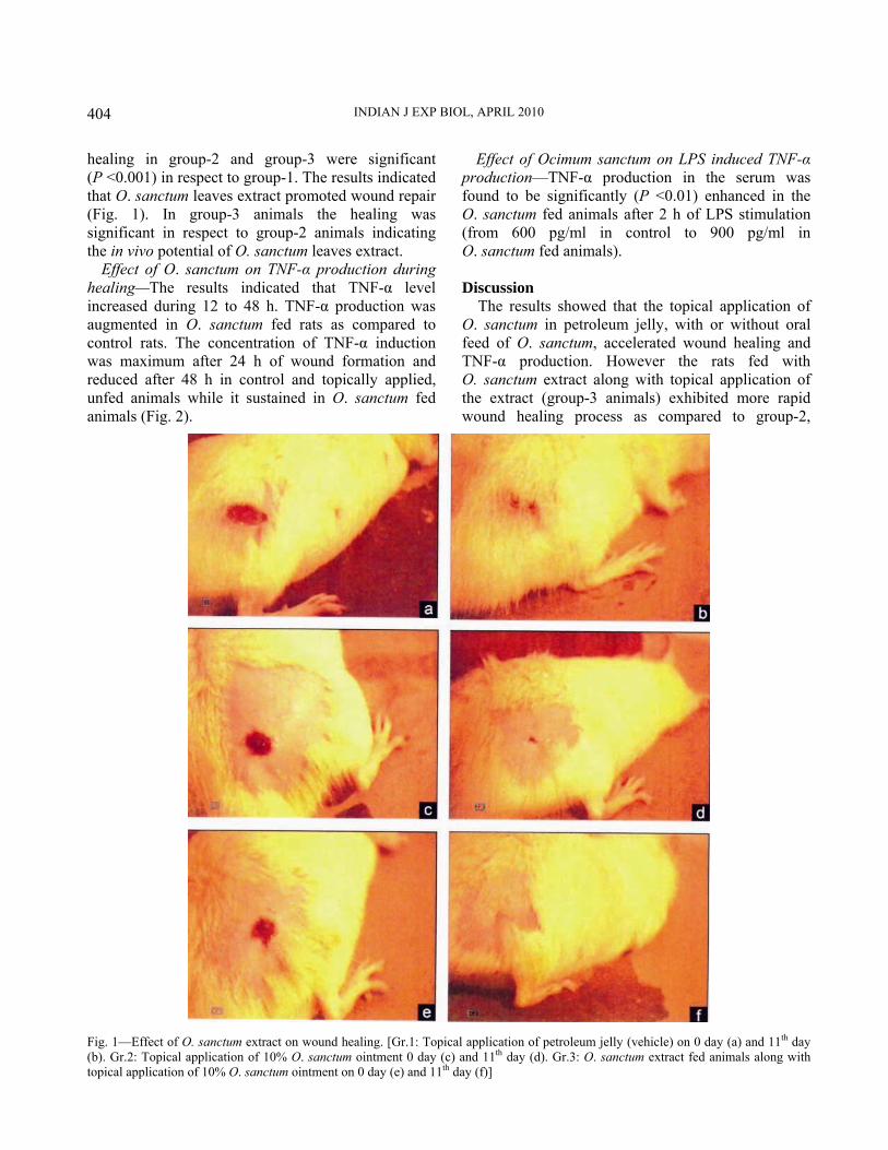
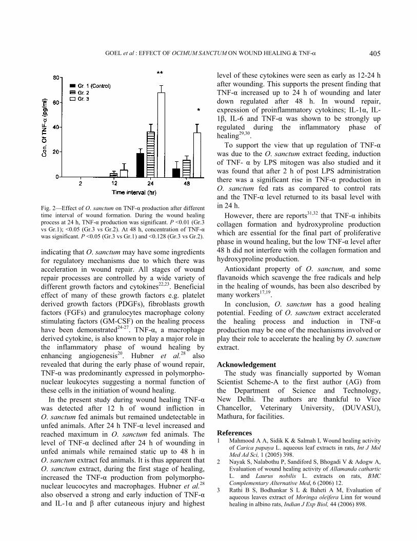
healing in group-2 and group-3 were significant (P <0.001) in respect to group-1. The results indicated that O. sanctum leaves extract promoted wound repair (Fig. 1). In group-3 animals the healing was significant in respect to group-2 animals indicating the in vivo potential of O. sanctum leaves extract. Effect of O. sanctum on TNF-α production during healing—The results indicated that TNF-α level increased during 12 to 48 h. TNF-α production was augmented in O. sanctum fed rats as compared to control rats. The concentration of TNF-α induction was maximum after 24 h of wound formation and reduced after 48 h in control and topically applied, unfed animals while it sustained in O. sanctum fed animals (Fig. 2).
Effect of Ocimum sanctum on LPS induced TNF-α production—TNF-α production in the serum was found to be significantly (P <0.01) enhanced in the O. sanctum fed animals after 2 h of LPS stimulation (from 600 pg/ml in control to 900 pg/ml in O. sanctum fed animals). Discussion The results showed that the topical application of O. sanctum in petroleum jelly, with or without oral feed of O. sanctum, accelerated wound healing and TNF-α production. However the rats fed with O. sanctum extract along with topical application of the extract (group-3 animals) exhibited more rapid wound healing process as compared to group-2,
Fig. 1—Effect of O. sanctum extract on wound healing. [Gr.1: Topical application of petroleum jelly (vehicle) on 0 day (a) and 11th day (b). Gr.2: Topical application of 10% O. sanctum ointment 0 day (c) and 11th day (d). Gr.3: O. sanctum extract fed animals along with topical application of 10% O. sanctum ointment on 0 day (e) and 11th day (f)]
GOEL et al : EFFECT OF OCIMUM SANCTUM ON WOUND HEALING & TNF-α
405
indicating that O. sanctum may have some ingredients for regulatory mechanisms due to which there was acceleration in wound repair. All stages of wound repair processes are controlled by a wide variety of different growth factors and cytokines22,23. Beneficial effect of many of these growth factors e.g. platelet derived growth factors (PDGFs), fibroblasts growth factors (FGFs) and granulocytes macrophage colony stimulating factors (GM-CSF) on the healing process have been demonstrated24-27. TNF-α, a macrophage derived cytokine, is also known to play a major role in the inflammatory phase of wound healing by enhancing angiogenesis20. Hubner et al.28 also revealed that during the early phase of wound repair, TNF-α was predominantly expressed in polymorpho-nuclear leukocytes suggesting a normal function of these cells in the initiation of wound healing. In the present study during wound healing TNF-α was detected after 12 h of wound infliction in O. sanctum fed animals but remained undetectable in unfed animals. After 24 h TNF-α level increased and reached maximum in O. sanctum fed animals. The level of TNF-α declined after 24 h of wounding in unfed animals while remained static up to 48 h in O. sanctum extract fed animals. It is thus apparent that O. sanctum extract, during the first stage of healing, increased the TNF-α production from polymorpho-nuclear leucocytes and macrophages. Hubner et al.28 also observed a strong and early induction of TNF-α and IL-1α and β after cutaneous injury and highest
level of these cytokines were seen as early as 12-24 h after wounding. This supports the present finding that TNF-α increased up to 24 h of wounding and later down regulated after 48 h. In wound repair, expression of proinflammatory cytokines; IL-1α, IL-1β, IL-6 and TNF-α was shown to be strongly up regulated during the inflammatory phase of healing29,30. To support the view that up regulation of TNF-α was due to the O. sanctum extract feeding, induction of TNF- α by LPS mitogen was also studied and it was found that after 2 h of post LPS administration there was a significant rise in TNF-α production in O. sanctum fed rats as compared to control rats and the TNF-α level returned to its basal level with in 24 h. However, there are reports31,32 that TNF-α inhibits collagen formation and hydroxyproline production which are essential for the final part of proliferative phase in wound healing, but the low TNF-α level after 48 h did not interfere with the collagen formation and hydroxyproline production. Antioxidant property of O. sanctum, and some flavanoids which scavenge the free radicals and help in the healing of wounds, has been also described by many workers17,19. In conclusion, O. sanctum has a good healing potential. Feeding of O. sanctum extract accelerated the healing process and induction in TNF-α production may be one of the mechanisms involved or play their role to accelerate the healing by O. sanctum extract. Acknowledgement The study was financially supported by Woman Scientist Scheme-A to the first author (AG) from the Department of Science and Technology, New Delhi. The authors are thankful to Vice Chancellor, Veterinary University, (DUVASU), Mathura, for facilities. References 1 Mahmood A A, Sidik K & Salmah I, Wound healing activity
of Carica papaya L. aqueous leaf extracts in rats, Int J Mol Med Ad Sci, 1 (2005) 398.
2 Nayak S, Nalabothu P, Sandiford S, Bhogadi V & Adogw A, Evaluation of wound healing activity of Allamanda cathartic L. and Laurus nobilis L. extracts on rats, BMC Complementary Alternative Med, 6 (2006) 12.
3 Rathi B S, Bodhankar S L & Baheti A M, Evaluation of aqueous leaves extract of Moringa oleifera Linn for wound healing in albino rats, Indian J Exp Biol, 44 (2006) 898.
Fig. 2—Effect of O. sanctum on TNF-α production after different time interval of wound formation. During the wound healing process at 24 h, TNF-α production was significant. P <0.01 (Gr.3 vs Gr.1); <0.05 (Gr.3 vs Gr.2). At 48 h, concentration of TNF-α was significant. P <0.05 (Gr.3 vs Gr.1) and <0.128 (Gr.3 vs Gr.2).
INDIAN J EXP BIOL, APRIL 2010
406
4 Geeta, Vasudevan D M, Kedlaya R, Deepa S & Ballal M, Activity of Ocimum sanctum (the traditional Indian medicinal plants) against the enteric pathogens, Indian J Med Sci, 55 (2001) 434.
5 Aquil F, Khan M S, Owais M & Ahmad I, Effect of certain bioactive plant extracts on clinical isolates of beta-lactamase producing methicillin resistant Staphylococcus aureus, J Basic Microbiol, 45 (2005) 106.
6 Godhwani S, Godhwani J L & Vyas D S, Ocimum sanctum: An experimental study evaluating its anti-inflammatory, analgesic and antipyretic activity in animals, J Ethnopharmacol, 21 (1987) 153.
7 Maulik G, Maulik N, Bhandari V, Kagan V E, Pakrashi S & Das D K, Evaluation of antioxidant effectiveness of a few herbal plants, Free Radic Res, 27 (1997) 221.
8 Kelm M A, Nair M G, Strasburg G M & DeWitt D L, Antioxidant and cyclooxygenase inhibitory phenolic compounds from Ocimum sanctum Linn, Phytomedicine, 7 (2000) 7.
9 Uma Devi P, Radioprotective, anticarcinogenic and antioxidant properties of the Indian holy basil, Ocimum sanctum (Tulsi), Indian J Exp Biol, 39 (2001) 185.
10 Singh S & Majumdar D K, Evaluation of anti-inflammatory activity of fatty acids of O. sanctum fixed oil, Indian J Exp Biol, 35 (1997) 80.
11 Godhwani S, Godhwani J L & Vyas D S, Ocimum sanctum—A preliminary study evaluating its immunoregulatory profile in albino rats, J Ethnopharmacol, 24 (1998) 193
12 Sadekar R D, Pimprikar N M, Bhandarkar A G & Barmase B S, Immunomodulating effect of Ocimum sanctum Linn dry leaf powder on humoral immune response in poultry naturally infected with IBD virsus, Indian Vet J, 75 (1998) 73.
13 Mediratta P K, Sharma K K & Singh S, Evaluation of immunomodulatory potential of Ocimum sanctum seed oil and its possible mechanism of action, J Ethnopharmacol, 80 (2002) 15.
14 Logambal S M, Venkatatalakshmi S & Michael R D, Immunostimulatory effect of leaf extract of Ocimum sanctum Linn in Oreochromis mossambicus (Peters), Hydrobiologia, 430 (2008) 113.
15 Salmah I, Mahmood A A & Sidik K , Synergistic effects of alcoholic extracts of Sweet Basil (Ocimum basilicum L.) leaves and honey on cutaneous wound healing in rats, Int J Mol Med Adv Sci, 1 (2005) 220.
16 Shetty S, Udupa S L, Udupa A L & Somayaji S N , Wound healing activity of Ocimum sanctum Linn with supportive role of antioxidant enzymes, Indian J Physiol Pharmacol, 50 (2006) 163.
17 Shetty S, Udupa S L & Udupa L, Evaluation of antioxidant and wound healing effects of alcholic and aqueous extract of Ocimum sanctum Linn in rats, Evid based Comp Altr Med, 5 (2008) 95.
18 Udupa S L, Somashekar S, Udupa A L & Somayaji S N, Effect of Ocimum sanctum Linn on normal and dexamethasone suppressed wound healing, Indian J Exp Biol, 44 (2006) 49.
19 Uma Devi P, Ganasoundari A, Vrinda B, Srinivasan K K & Unnikrishnan K, Radiation protection by the Ocimum flavonoids orientin and vicenin: Mechanism of action, Radiat Res, 154 (2000) 455.
20 Rosenberg L Z, Wound healing, growth factors, emedicine, 2006.
21 Morton J J & Malone M H, Evaluation of vulnerary activity by an open wound procedure in rats, Arch Inter Pharmacodyn, 176 (1972) 117.
22 Martin P, Wound healing aiming for perfect skin regeneration, Science, 276 (1997) 75.
23 Werner S & Grose R, Regulation of wound healing by growth factors and cytokines, Physiol Rev, 83 (2002) 835.
24 Abraham J A & Klagsbrun M, Modulation of wound repair by members of the fibroblast growth factor family, in The molecular and cellular biology of wound repair (2nd ed.), edited by R A F Clark, (Plenum, New York) 1996, 195.
25 Greenhalgh D G, The role of growth factors in wound healing, J Trauma, 41 (1996) 159.
26 Harding K G, Morris H L & Patel G K, Science, medicine and the future: Healing chronic wounds, Br Med J, 324 (2002) 160.
27 Nath C & Gulati S C, Role of cytokines in healing chronic skin wounds, Acta Haematol, 99 (1998) 175.
28 Hubner G, Brauchle M, Smola H, Madlener M, Fassler R & Werner S, Differential regulation of pro-inflammatory cytokines during wound healing in normal and glucocorticoid-treated mice, Cytokine, 8 (1996) 548.
29 Grellner W, Georg T & Wilske J, Quantitative analysis of proinflammatory cytokines (IL-1beta, IL-6, TNF-alpha) in human skin wounds, Forensic Sci Int, 113 (2000) 251.
30 Grose R, Werner S, Kessler D, Tuckermann J, Durka S, Huggel K, Reichardt H & Werner S, A role for endogenous glucocorticoids in wound repair, EMBO Reports, 3 (2002) 575.
31 Rapala K, The effect of tumor necrosis factor-alpha on wound healing, an experimental study, Ann Chir Gynaecol Suppl, 211 (1996) 1.
32 Buck M, Houglum K & Chojkier M, Tumor necrosis factor-alpha inhibits collagen alpha 1(I) gene expression and wound healing in a murine model of cachexia, American J Pathology, 149 (1996) 195.