Why cancer cells have a more hyperpolarised mitochondrial ...
42
1 Why cancer cells have a more hyperpolarised mitochondrial membrane potential and emergent prospects for therapy Michael D. Forrest Ph.D. Department of Computer Science, University of Warwick, Coventry, UK E-Mail: [email protected] ABSTRACT Cancer cells have a more hyperpolarised mitochondrial membrane potential (Ψ IM ) than normal cells. Ψ IM = ~-220 mV in cancer cells as compared to ~-140 mV in normal cells. Until now it has not been known why. This paper explains this disparity, in a mathematical framework, and identifies molecular targets and operations unique to cancer cells. These are thence prospective cancer drug targets. BMS-199264 is proposed as an anti-cancer drug. It inhibits the reverse, proton-pumping mode of ATP synthase, which this paper identifies as crucial to cancer cells but not to healthy, normal adult cells. In the cancer cell model, the adenine nucleotide exchanger (ANT) is inversely orientated in the mitochondrial inner membrane as compared to normal cells. This predicts it to have a different drug interaction profile, which can be leveraged for cancer therapy. Uncouplers, which dissipate the proton motive force, are proposed as anti-cancer medicines e.g. 2,4-dinitrophenol. ARTICLE During aerobic respiration, the movement of electrons along the respiratory chain pumps protons across the inner mitochondrial membrane to build a proton motive force (pmf) [1-3]. The pmf is electrochemical, consisting of a hyperpolarised transmembrane voltage (Ψ IM , negative inside) and a proton concentration gradient (δpH, alkali inside). In vivo, with high concentrations of Pi, δpH is minor as compared to Ψ IM [4]. Protons move down this electrochemical gradient, through ATP synthase, to generate ATP. Eukaryote cells must maintain a hyperpolarized voltage across their inner mitochondrial membranes. If this hyperpolarisation dissipates, the voltage-sensitive permeability transition pore (PTP) will open and release pro-apoptotic agents (e.g. cytochrome c) into the cytoplasm and drive apoptotic cell death [5]. In a normal cell, Ψ IM flickers between -108 and -159 mV: with a mean value of -139 mV [6, 7]. Thermodynamically, the optimal Ψ IM for maximal ATP production is between -130 to - 140 mV, a rule that applies for all living organisms [7]. 10% value alterations in Ψ IM , above not peer-reviewed) is the author/funder. All rights reserved. No reuse allowed without permission. The copyright holder for this preprint (which was . http://dx.doi.org/10.1101/025197 doi: bioRxiv preprint first posted online Aug. 21, 2015;
Transcript of Why cancer cells have a more hyperpolarised mitochondrial ...

1
Why cancer cells have a more hyperpolarised
mitochondrial membrane potential and emergent
prospects for therapy
Michael D. Forrest Ph.D.
Department of Computer Science, University of Warwick, Coventry, UK
E-Mail: [email protected]
ABSTRACT
Cancer cells have a more hyperpolarised mitochondrial membrane potential (ΨIM) than
normal cells. ΨIM = ~-220 mV in cancer cells as compared to ~-140 mV in normal cells. Until
now it has not been known why. This paper explains this disparity, in a mathematical
framework, and identifies molecular targets and operations unique to cancer cells. These are
thence prospective cancer drug targets. BMS-199264 is proposed as an anti-cancer drug. It
inhibits the reverse, proton-pumping mode of ATP synthase, which this paper identifies as
crucial to cancer cells but not to healthy, normal adult cells. In the cancer cell model, the
adenine nucleotide exchanger (ANT) is inversely orientated in the mitochondrial inner
membrane as compared to normal cells. This predicts it to have a different drug interaction
profile, which can be leveraged for cancer therapy. Uncouplers, which dissipate the proton
motive force, are proposed as anti-cancer medicines e.g. 2,4-dinitrophenol.
ARTICLE
During aerobic respiration, the movement of electrons along the respiratory chain pumps
protons across the inner mitochondrial membrane to build a proton motive force (pmf) [1-3].
The pmf is electrochemical, consisting of a hyperpolarised transmembrane voltage (ΨIM,
negative inside) and a proton concentration gradient (δpH, alkali inside). In vivo, with high
concentrations of Pi, δpH is minor as compared to ΨIM [4]. Protons move down this
electrochemical gradient, through ATP synthase, to generate ATP. Eukaryote cells must
maintain a hyperpolarized voltage across their inner mitochondrial membranes. If this
hyperpolarisation dissipates, the voltage-sensitive permeability transition pore (PTP) will
open and release pro-apoptotic agents (e.g. cytochrome c) into the cytoplasm and drive
apoptotic cell death [5].
In a normal cell, ΨIM flickers between -108 and -159 mV: with a mean value of -139 mV [6,
7]. Thermodynamically, the optimal ΨIM for maximal ATP production is between -130 to -
140 mV, a rule that applies for all living organisms [7]. 10% value alterations in ΨIM, above
not peer-reviewed) is the author/funder. All rights reserved. No reuse allowed without permission. The copyright holder for this preprint (which was. http://dx.doi.org/10.1101/025197doi: bioRxiv preprint first posted online Aug. 21, 2015;

2
or below its optimum, results in a ~90% decrease in ATP synthesis and a ~90% increase in
harmful reactive oxygen species (ROS) [7].
Cancer cells have a more hyperpolarised ΨIM than normal cells [8-17]. The more invasive and
dangerous the cancer, the more hyperpolarised its ΨIM is observed to be [14-16]. The
hyperpolarisation of ΨIM can be >50% greater in cancer cells than normal cells [14] e.g. ΨIM
= ~-210 mV in Neu4145 cancer cells [18]. The ΨIM hyperpolarisation in cancer cells can even
be double that of normal cells [19]. So, generally, the ΨIM of cancer cells is extremely sub-
optimal for ATP production. However, these cancer cells aren’t using aerobic respiration and
aren’t using ΨIM in the same way as normal cells.
Aerobic respiration is O2 dependent and uses glycolysis, the Krebs cycle and oxidative
phosphorylation (OXPHOS) to produce ATP [1-3]. Aerobic glycolysis is the sole use of
glycolysis to produce ATP, even in the presence of O2. Cancer cells can use aerobic
glycolysis (Warburg effect) some or all of the time [18-39]. I propose that when in this mode,
they have a more hyperpolarised ΨIM. Indeed, experimentally, when cancer cells are switched
out of aerobic glycolysis, into aerobic respiration, their ΨIM is returned to that of normal cells
[18-19].
The fact that there is a disparity in ΨIM between normal and cancer cells is well established
and has already been leveraged in human drug trials [40-41]. Delocalized lipophilic cations
(DLCs) can cross membranes and their positive charge means they are drawn to, and
accumulate in, the mitochondrial matrix (negative inside, because of hyperpolarised ΨIM).
Cancer cells have a more hyperpolarised ΨIM and so DLCs are more attracted to, and better
retained by, their mitochondria than that of normal cells [12]. Using the Nernst equation [14],
if the ΨIM of a cancer cell is 60 mV more hyperpolarised than that of a normal cell – which is
within the range of observation [14, 18-19] – then a single charged DLC will accumulate 10
times more in the mitochondrial matrix of cancer cells than normal cells (T=300 K). DLCs
with a double charge will accumulate 100 times more [42]. So, DLC poisons are more
targeted to cancer cells and this means there are likely to be doses that can kill cancer cells,
but leave normal cells unharmed. Different DLCs have been shown to accumulate in and
selectively kill cancer cells, in vitro and in vivo [11, 43-47]. This affirms that cancer cells do
have a more hyperpolarised ΨIM, although no DLC has been successful in clinical trialling to
date. For example, MKT-077 caused renal toxicity in Phase 1 trials [40-41].
It is not known why or how cancer cells have a more hyperpolarised ΨIM. I provide a
quantitative explanation, which identifies molecular targets and operations unique to cancer
not peer-reviewed) is the author/funder. All rights reserved. No reuse allowed without permission. The copyright holder for this preprint (which was. http://dx.doi.org/10.1101/025197doi: bioRxiv preprint first posted online Aug. 21, 2015;
3
cells. These can be leveraged as cancer drug targets. To understand how ΨIM generation
differs in cancer cells, we must first explain it for normal cells.
The biophysics of ΨIM in normal cells [4, 48-50]
Mitochondrial ATP synthase (F0F1-ATPase) can synthesise or hydrolyse ATP. Protons can
flow “downhill” through the ATPase, to generate ATP, or be pumped “uphill” by the
ATPase, using ATP. The mitochondrial Adenine Nucleotide Transporter (ANT) can export
ATP4-
for the import of ADP3-
, or conduct the inverse. So, both ATPase and ANT catalyse
reversible processes. Their directionality is governed by the mitochondrial membrane
potential (ΨIM) in relation to their reversal potential, Erev_ATPase and Erev_ANT respectively
(mV). Which are set by the concentrations of the participating reactants, as shown in
Equations 1-11 [4, 50].
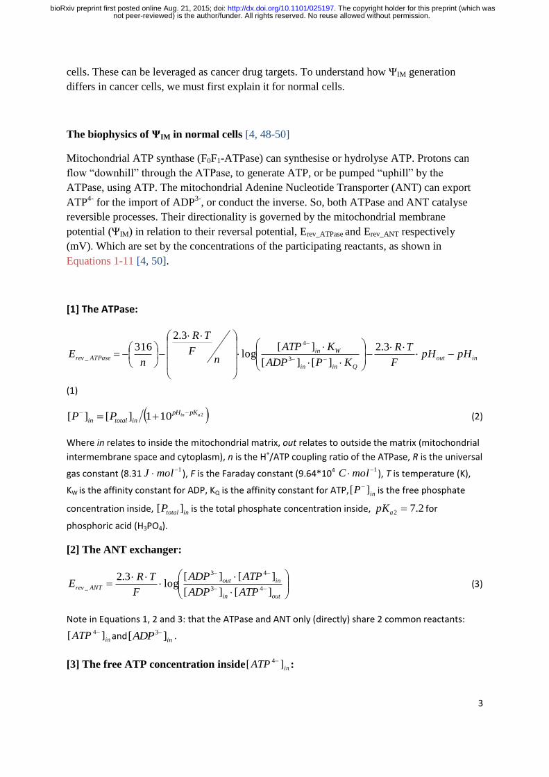
[1] The ATPase:
inout
Qinin
WinATPaserev pHpH
F
TR
KPADP
KATP
nF
TR
nE
3.2
][][
][log
3.2316
3
4
_
(1)
2101][][ ain pKpH
intotalin PP (2)
Where in relates to inside the mitochondrial matrix, out relates to outside the matrix (mitochondrial
intermembrane space and cytoplasm), n is the H+/ATP coupling ratio of the ATPase, R is the universal
gas constant (8.31 1molJ ), F is the Faraday constant (9.64*104 1molC ), T is temperature (K),
KW is the affinity constant for ADP, KQ is the affinity constant for ATP, inP ][ is the free phosphate
concentration inside, intotalP ][ is the total phosphate concentration inside, 2.72 apK for
phosphoric acid (H3PO4).
[2] The ANT exchanger:
outin
inoutANTrev
ATPADP
ATPADP
F
TRE
][][
][][log
3.243
43
_ (3)
Note in Equations 1, 2 and 3: that the ATPase and ANT only (directly) share 2 common reactants:
inATP ][ 4and
inADP ][ 3 .
[3] The free ATP concentration inside inATP ][ 4:
not peer-reviewed) is the author/funder. All rights reserved. No reuse allowed without permission. The copyright holder for this preprint (which was. http://dx.doi.org/10.1101/025197doi: bioRxiv preprint first posted online Aug. 21, 2015;
4
H
pH
Lfreeinin
inK
KMgLATP
in /101
/][1/][][
_
2
4
(4)
ininininin MgHATPMgATPHATPATPL ][][][][][ 234 (5)
LK is the dissociation constant for the reaction: 242 MgATPMgATPLK
(6)
HK is the dissociation constant for the reaction: HATPHATPHK
43 (7)
[4] The free ADP concentration insideinADP ][ 3 :
J
pH
Rfreeinin
inK
KMgYADP
in /101
/][1/][][
_
2
3
(8)
ininininin MgHATPMgADPHADPADPY ][][][][][ 23 (9)
RK is the dissociation constant for the reaction: 23 MgADPMgADPRK
(10)
JK is the dissociation constant for the reaction: HADPHADPJK
32 (11)
[5] The free ADP and ATP concentrations outside - outADP ][ 3 and outATP ][ 4 respectively - are
calculated by equations of the same form as those for inside; but are not shown here for brevity.
The dissociation constants for outADP ][ 3 are KZ and KX. The dissociation constants for
outATP ][ 4 are KO and KU.
Figure 1 shows computational estimations of Erev_ATPase and Erev_ANT (mV) at different
[ATP]in/[ADP]in ratios [4, 48, 50]. This graph was made by computing Equations 1-11 with
parameters from [4]: [ATP]out =1.2 mM, [ADP]out = 10 µM, [P]in = 0.01 M, n = 3.7 (2.7 for
the ATP synthase plus 1 for the electrogenic ATP4-
/ADP3-
exchange of the ANT and the
nonelectrogenic symport of phosphate and a proton by the phosphate carrier [51]), pHin =
7.38, pHout = 7.25, T = 310 K, [Mg2+
]in_free=0.5 mM, KW= 10-3.198
, KQ= 10-4.06
, KL= 0.114
mM, KH= 10-7.06
M, KR= 0.906 mM, KJ= 10-6.8
M, KZ= 0.906 mM, KX= 10-6.8
M, KO= 0.114
mM, KU= 10-7.06
M. Traces were computed by Erev estimator software [4, 48, 50], which can
be downloaded at: http://www.tinyurl.com/Erev-estimator.
During OXPHOS, protons are pumped by the complexes of the respiratory chain out of the
mitochondrial matrix and into the mitochondrial intermembrane space. This hyperpolarises
ΨIM and makes it more negative than Erev_ATPase and Erev_ANT (the green coloured “A-space” of
Figure 1). With ΨIM hyperpolarised to Erev_ATPase, ATPase works in its “forward” mode and
synthesises ATP. With ΨIM hyperpolarised to Erev_ANT, ANT works in its “forward” mode and
not peer-reviewed) is the author/funder. All rights reserved. No reuse allowed without permission. The copyright holder for this preprint (which was. http://dx.doi.org/10.1101/025197doi: bioRxiv preprint first posted online Aug. 21, 2015;
5
exports mitochondrial matrix ATP for the import of cytoplasmic ADP. So, the mitochondrion
produces and exports ATP. The “forward” operation of ANT and ATPase is a depolarising
force to ΨIM. ATP4-
export for ADP3-
import is depolarising and so are protons flowing
“downhill” through ATPase. However, ΨIM doesn’t depolarise because at the same time
protons are being continually pumped “uphill” by the respiratory chain complexes, which is a
hyperpolarising drive to ΨIM. Actually, ΨIM doesn’t remain constant during OXPHOS – but
“flickers” (as much as >100 mV) [48-50] as these depolarising and hyperpolarising forces
wrestle back and forth for a temporary net dominance.
If ΨIM is more positive (depolarised) than Erev_ATPase and Erev_ANT, they both work in their
“reverse” mode (the grey coloured “C-space” of Figure 1). ATPase hydrolyses ATP and ANT
imports cytoplasmic ATP for the exchange of mitochondrial matrix ADP. So, the
mitochondrion imports and consumes ATP. In this state, ATPase pumps protons into the
intermembrane space which hyperpolarises ΨIM. In addition, ANT imports ATP4-
and exports
ADP3-
, so a negative charge is gained on the matrix side which hyperpolarises ΨIM.
During OXPHOS, ΨIM is more negative than Erev_ATPase and Erev_ANT. If aerobic respiration is
switched off, for example if the cell switches into aerobic glycolysis, then there will no
longer be the hyperpolarising offset to the depolarising, “forward” action of ANT and
ATPase: ΨIM will depolarise. Erev_ATPase is more negative than Erev_ANT and so ΨIM will
depolarise past this reversal potential first. In this case, ATPase will switch into its “reverse”
mode and ANT will remain in its “forward” mode (the orange coloured “B-space” of Figure
1). So, the ANT action will remain depolarising but the ATPase action will switch to being
hyperpolarising – pumping protons out rather passing protons in. However, this reverse
ATPase action requires ATP and with ANT pumping ATP out, there is little to be had.
Furthermore, near the reversal potential of ATPase there is little driving force for an ATPase
action. Hence, depolarising forces dominate and ΨIM depolarises further. When ΨIM is equal
to Erev_ANT then ANT does no “forward” or “reverse” ATP/ADP exchange and its effect on
ΨIM is lost. At this point: With no ATP coming into the matrix, ATPase can no longer
hydrolyse ATP to pump protons and its hyperpolarising action is also lost. In the absence of
these forces, ensuing proton leak will depolarise the membrane potential. This will then make
ΨIM more depolarised than Erev_ANT and permit ANT to conduct a hyperpolarising exchange
of ATP/ADP. The more depolarised it is past Erev_ANT, the more drive there is for this
hyperpolarising exchange and the more that will occur. The resultant ATP entry will permit
ATPase to conduct a hyperpolarising pumping of protons. So, there are hyperpolarising
forces, conducted by ATPase and ANT, that come into play to prevent further depolarisation
past Erev_ANT. They cannot hyperpolarise ΨIM to be more negative than Erev_ANT because these
forces are largely lost at this point, but they can prevent further depolarisation past this point.
The result is that ΨIM will oscillate around Erev_ANT. So, at the loss aerobic respiration ΨIM
will converge to Erev_ANT. Hence, at the loss of aerobic respiration there is a “safety net” of
not peer-reviewed) is the author/funder. All rights reserved. No reuse allowed without permission. The copyright holder for this preprint (which was. http://dx.doi.org/10.1101/025197doi: bioRxiv preprint first posted online Aug. 21, 2015;

6
mechanisms to prevent the collapse of ΨIM and the mass consumption of cytoplasmic ATP
through mitochondrial proton pumping by ATPase.
At certain mitochondrial matrix [ATP]in/[ADP]in ratios, it is possible for ATPase to be in
“forward” operation and ANT to be in “reverse” operation (the whine coloured “D-space” of
Figure 1). The former is depolarising, the latter hyperpolarising. However, it may be unlikely
for mitochondria to have such a hyperpolarised ΨIM and a low matrix [ATP]in/[ADP]in ratio.
So, it may be that this part of the graph has no biological representation and can be
discounted [4, 50]. The ΨIM in the “D-space” prompts ATPase to create ATP and ANT to
import ATP. The ensuing rise in the [ATP]in/[ADP]in ratio would push the system out of the
“D- space” and into another area of the graph (Figure 1).
If proton pumping by the respiratory chain is stopped, but the Krebs cycle still persists, ΨIM
will depolarise past Erev_ATP but not all the way to Erev_ANT. The Krebs cycle can produce ATP
(or GTP) in its succinyl CoA to succinate step. Once ΨIM is less negative than Erev_ATP, the
ATP produced by the Krebs cycle may support the “reverse” ATP hydrolysing, proton
pumping, hyperpolarising action of ATPase [4, 48-50]. This action will “hold” ΨIM in this
range between Erev_ATP and Erev_ANT. However, during aerobic glycolysis: both OXPHOS and
the Krebs cycle are shunted. So, this situation will not apply in this case. As aforementioned,
ΨIM should converge upon and oscillate around Erev_ANT.
IF-1 is a physiological protein, expressed by some tissues of some organisms, that inhibits the
consumption of ATP by the F0F1-ATPase [4]. So, it prevents the “reversal” of the ATPase
upon depolarisation of ΨIM past Erev_ATP. Its blockage isn’t complete, but increases with
matrix [ATP], decreased matrix pH (acidification) and dissipated ΨIM. With F0F1-ATPases
unable to “reverse”, to confer a hyperpolarising pump of protons, they offer little resistance to
an external, imposed depolarisation. So, this imposed depolarisation can converge relatively
unopposed to Erev_ANT. Depolarisation past this point switches the ANT into producing a
hyperpolarising exchange and this tries to “hold” ΨIM at Erev_ANT, as described earlier. The
result is that ΨIM will oscillate around Erev_ANT. Unless the imposed depolarisation is strong
enough to overcome this resistance, in which case the continued depolarisation will
eventually open the voltage-dependent PTP and apoptosis is then all but assured.
Why cancer cells have a more hyperpolarised ΨIM is mysterious
As aforementioned, I suggest that cancer cells have a more hyperpolarised ΨIM when they are
utilising aerobic glycolysis. I suggest that this mode is a function of cancer proliferation and
so the more aggressive and dangerous the cancer, the more time they spend in this operating
not peer-reviewed) is the author/funder. All rights reserved. No reuse allowed without permission. The copyright holder for this preprint (which was. http://dx.doi.org/10.1101/025197doi: bioRxiv preprint first posted online Aug. 21, 2015;

7
state. During aerobic glycolysis, the Krebs cycle and OXPHOS are shunted and aren’t used.
By the reasoning of the previous section, ΨIM should thus converge upon – and oscillate
around - Erev_ANT. However, there is a problem. Refer to Figure 1 and note that Erev_ANT is in a
range around ~-120 mV (-115 mV at matrix [ATP]in/[ADP]in ratio = 1.5). But the ΨIM of
cancer cells is much more hyperpolarised: e.g. the ΨIM of Neu4145 cancer cells is ~-210 mV
[18].
Cancer cells use a different ANT isoform
The ANT referred to thus far in this manuscript is the ANT1 isoform. There are 4 human
ANT isoforms (gene names in brackets): ANT1 (SLC25A4), ANT2 (SLC25A5), ANT3
(SLC25A6) and ANT4 (SLC25A31) [52]. They have ~90% homology, except ANT4 which
has ~70% homology to the others. A comparative study in yeast with the heterologous
expression of ANT1, ANT2 and ANT3 (underneath the same promotor region) showed them
all to have similar adenine nucleotide exchange characteristics [52]. ANT1 is expressed in
differentiated adult cells. ANT2 is expressed in rapidly proliferating cells (embryonic stem
(ES) cells, cancer cells). ANT3 is expressed at a low level ubiquitously. ANT4 is highly
expressed in male gametes.
In differentiated adult cells, ANT1 is expressed highly and ANT2 expression is minimal [53].
I surmise that as a differentiated cell turns cancerous, and as it switches from aerobic
respiration to aerobic glycolysis, it downregulates ANT1 and upregulates ANT2 expression.
Indeed, in the majority of cancer cell lines, ANT1 expression is very low, whereas the
expression of ANT2 is very high [53]. ANT3 expression is low across the board.
ANT1 and ANT3 are associated with aerobic respiration. They export the ATP produced by
OXPHOS from the mitochondria into the cytosol, while importing ADP [54]. ANT2, by
contrast, is associated with aerobic glycolysis [54]. When a cell is rapidly proliferating (e.g.
cancerous) it switches off OXPHOS and ANT2 may import glycolytically produced ATP into
mitochondria, while exporting ADP [54]. “Reverse” ATP synthase action hydrolyses this
ATP to pump protons and maintain ΨIM and so prevent apoptosis [54]. ANT2 expression is a
marker for rapid proliferation and/or cancer. Hence, specific inhibition of ANT2 is a
prospective anti-cancer strategy.
But ANT2 may not import ATP into mitochondria
ANT2 import of ATP into mitochondria [54] is a hypothesis, although it is stated as an
established fact in some of the literature, and there is a problem with this account [55-56].
not peer-reviewed) is the author/funder. All rights reserved. No reuse allowed without permission. The copyright holder for this preprint (which was. http://dx.doi.org/10.1101/025197doi: bioRxiv preprint first posted online Aug. 21, 2015;
8
Inhibition of OXPHOS alone (e.g. using myxothiazol, a Complex III respiratory inhibitor)
cannot collapse the ΨIM of a cell using aerobic respiration. In addition, ATP synthase must be
inhibited (e.g. using oligomycin) or ANT must be inhibited (e.g. using carboxyatractyloside
or bongkrekic acid). Inhibiting ATP synthase or ANT inhibits the “reverse” action of ATP
synthase, which can maintain ΨIM in the absence of OXPHOS. ANT inhibition services this
requirement by cutting off ATP delivery to ATP synthase. However, this is all the case for
cells using aerobic respiration. It is not the case for cancer cells. In A549 human lung cancer
cells, myxothiazol slightly decreases ΨIM and subsequent oligomycin collapses ΨIM.
However, carboxyatractyloside and bongkrekic acid fail to collapse ΨIM. Nonetheless,
subsequent oligomycin does lead to full depolarization. Similarly, 2-deoxyglucose, a
glycolytic inhibitor, can collapse ΨIM. These results show that the cancer cells are using
mitochondrial ATP synthase to hydrolyse glycolytic ATP, to maintain ΨIM. But that in cancer
cells, entry of glycolytic ATP into mitochondria can occur by a pathway other than ANT
[55]. This pathway is unknown. What could it be?
It could be through the electroneutral ATP-Mg/Pi carrier (APC) [55]. APC exchanges ATP
(Mg-ATP2-
) for Pi (HPO42-
) [57-58]. APC can alternatively exchange ADP (HADP2-
) for Pi,
if Mg2+
is absent. So, it could perform some “fudged” electroneutral ATP-ADP exchange.
Some intracellular parasites express ATP transporters on their plasma membrane when inside
a host cell, to steal ATP from the host’s cytoplasmic pool. There is a diversity of such
parasites – e.g. chlamydia and rickettsiae bacteria [59-61], Lawsonia intracellularis [62] and
the eukaryote: Encephalitozoon cuniculi [63]. PamNTT1, the ATP/ADP transporter from the
amoeba symbiont Protochlamydia amoebophila (a chlamydia-related bacterium) [59] has an
electroneutral action independent of the membrane potential. It exchanges [ATP4-
] in for
[ADP3-
and Pi-] out [63]. So, it is very distinct from ANT. Furthermore, it likely has 11-12
transmembrane domains; which is a significant difference from the 6 transmembrane helices
of ANT and other members of the mitochondrial carrier family (MCF). I used BLAST [64] at
http://blast.ncbi.nlm.nih.gov, with its default search parameters for “somewhat similar
sequences” (blastn algorithm), to search the human genome for a PamNTT1 (NCBI accession
number: AJ582021) homologue. With the idea that maybe such a homologue could be how
rapidly proliferating/cancer cells import ATP into their mitochondria. However, none was
found (data not shown). I then used BLAST again, with the same search parameters, to search
the human genome for homologues of the ATP/ADP transporters from the eukaryote E.
cuniculi [65]: EcNNT1–4 (GenBank accession numbers: EU040266–EU040270). However,
none was found (data not shown).
Members of the mitochondrial carrier family (MCF) have a tripartite structure. That consists
of three homologous sequence repeats of about 100 amino acid residues. Which each have a
not peer-reviewed) is the author/funder. All rights reserved. No reuse allowed without permission. The copyright holder for this preprint (which was. http://dx.doi.org/10.1101/025197doi: bioRxiv preprint first posted online Aug. 21, 2015;

9
signature motif: P-X-[D/E]-X-X-[R/K] [66] (Prosite: PS50920). ANT and APC are members.
The human genome likely encodes 48 different mitochondrial carriers [66]. A sizable
proportion of these have not yet had their function assigned. It could be that one or more of
these “orphan” transports facilitate ATP entry into mitochondria.
ANT2 is essential to cancer cells; ANT1 and ANT3 kill cancer cells
It might still be that ANT2 transports glycolytic ATP into mitochondria in cancer cells, but it
isn’t the only pathway for this. There may be a redundancy in this cancer system. Or ANT2
may have some other role. ANT2 does seem crucial to cancer cells.
I propose that cancer cell metabolism is similar to that of embryonic stem (ES) cells. Indeed,
they share genetic expression fingerprints [67-68] and ES cells have a hyperpolarised ΨIM
also [69]. They both employ aerobic glycolysis some or all of the time [18-39, 70], are
immortal (divide forever without limit) [71-72] (as a function of using aerobic glycolysis
[73]), respond to ROS damage by apoptosis rather than repair [19, 74] and can proliferate
rapidly. So, with caution, we can learn more about cancer from ES cells and vice versa. In
mice, ANT2 deficiency is embryonically lethal [75]. ANT2 is crucial to ES cells and we
extrapolate from this to suggest that it is crucial to cancer cells. Indeed, ANT2 knockdown
(RNA interference, shRNA) represses cancer proliferation and induces apoptotic death to
cancer cells in vitro and in vivo [76]. Although others have reported ANT2 knockdown to
have no such effect [77]; but this earlier, alternative report can be considered an inferior study
because it used siRNA rather than shRNA; shRNA produces a more complete, robust, long
lasting, long term knockdown [76].
In cancer cells, whereas ANT2 is anti-apoptotic [76], ANT1 is pro-apoptotic [78]. Over-
expression of ANT1 induces apoptosis in cultured cancer cells by collapsing ΨIM and opening
the voltage-dependent PTP [78]. Indeed, ANT1 transfection significantly suppresses tumor
growth in vivo [78]. ANT3 is pro-apoptotic also [79]. Interestingly, over-expression of ANT1
is lethal to embryo cells [80], like it is to cancer cells.
I predict that ANT2 is inserted in the inner mitochondrial membrane in the opposite
orientation to ANT1 and ANT3
I suggest that ANT2 does import ATP into mitochondria in cancer cells (and ES cells). I
predict that ANT2 is orientated in the inner mitochondrial membrane in the opposite
orientation than ANT1 and ANT3 (Figure 2). Furthermore, that this makes ANT2 resistant to
inhibition by carboxyatractyloside (CAT) and bongkrekic acid (BKA), which are both
not peer-reviewed) is the author/funder. All rights reserved. No reuse allowed without permission. The copyright holder for this preprint (which was. http://dx.doi.org/10.1101/025197doi: bioRxiv preprint first posted online Aug. 21, 2015;
10
inhibitors of ANT. This accounts for why these drugs cannot block ATP entry into the
mitochondria of cancer cells, as mentioned earlier and described in [55-56].
BKA can be lipid soluble and can cross the inner mitochondrial membrane. CAT isn’t and
can’t [81]. BKA crosses membranes as the electroneutral BKAH3. But it is its anionic form,
BKA3-
, which inhibits ANT [81]. CAT binds the intermembranous (“cytoplasmic”) side of
ANT. BKA binds the matrix side of ANT [81]. If ANT2 is inserted in the inner mitochondrial
membrane in the opposite orientation than ANT1/ANT3 - if its “m-side” is instead facing the
cytoplasm (intermembranous space) and its “c-side” is instead facing the mitochondrial
matrix – I anticipate that it will be resistant to inhibition by CAT or BKA. CAT won’t be able
to cross the inner mitochondrial membrane to access its binding site on the “c-side” of ANT,
which is now in the mitochondrial matrix. BKA will be able to access its binding site on the
“m-side” of ANT. But now this site is located in the acidic, proton rich environment of the
intermembrane space. So, here, BKA3-
will quickly pick up protons and be converted to
BKAH3. This form cannot inhibit ANT.
If ANT2 is orientated oppositely to ANT1/ANT3, it could confer it very different adenine
nucleotide exchange kinetics; which are more suitable for cancer cells. Indeed, it may favour
the import, rather than the export, of ATP. Without this re-interpretation, it is hard to see why
ANT2 is so vital to cancer cells [76], and ANT1 and 3 so harmful [78-79], while they all have
high sequence homology (~90%). And similar adenine nucleotide transport capabilities when
expressed in a heterologous yeast system [52, 82]. I surmise that this heterologous system
doesn’t have the infrastructure to orientate ANT2 oppositely. So, ANT1, ANT2 and ANT3
are all orientated in the same way and, in this scenario, they do all have similar exchange
characteristics. A further distinction is that an opposite orientation of ANT2 may make it less
predisposed to joining in to make the permeability transition pore (PTP) in apoptotic
scenarios (ANT can be a PTP component [83]).
An opposite orientation of ANT2 opens prospects for selective drugs. A drug that binds the
“m-side” of ANT molecules, but that can’t cross the inner mitochondrial membrane, may
inhibit ANT2 (which has its “m-side” facing the cytoplasm) but not ANT 1 or 3 (which have
their “m-side” in the mitochondrial matrix). So, this would be a selective anti-cancer drug.
Alternatively, a drug (lipid or non-lipid soluble) that only binds the “m-side” of ANT in an
acidic environment might selectively inhibit ANT2. It has its “m-side” in the acidic
intermembranous space (IMS) rather than the alkali mitochondrial matrix. A delocalised
lipophilic cation (DLC), that binds the “c-side” of ANT, would be preferentially located to
the mitochondrial matrix by ΨIM where it could inhibit ANT2 and not ANT1/3. Alternatively,
a drug that binds the “c-side” of ANT, tethered to a lipophilic cation to convey it passage
through the inner mitochondrial membrane, and a positive charge(s) for targeting to the
not peer-reviewed) is the author/funder. All rights reserved. No reuse allowed without permission. The copyright holder for this preprint (which was. http://dx.doi.org/10.1101/025197doi: bioRxiv preprint first posted online Aug. 21, 2015;
11
mitochondrial matrix, could inhibit ANT2 and not ANT1/3. A lipophilic anion (or a molecule
tethered to one), that binds the “m-side” of ANT, would be preferentially located to the
mitochondrial IMS by ΨIM where it could inhibit ANT2 and not ANT1/3. Antibodies against
the “m-side” or “c-side” of ANT may yield novel inhibitors. That could then be used in
cancer therapy, to leverage the orientation disparity between ANT2 in cancer cells and
ANT1/3 in normal cells. We have an ANT crystal structure [84]. The “m-side” and “c-side”
of ANT are now conceivable cancer drug targets and should be targeted via in silico virtual
screening, structure based drug design and high-throughput in vitro screening e.g. using chip
technologies [85].
The Erev_ANT of ANT1 and ANT3 is dangerous for cancer cells and this is why they
downregulate them
ANT3 is pro-apoptotic to cancer cells [79], ANT1 also [78]. Over-expression of ANT1
induces apoptosis in cultured cancer cells by collapsing ΨIM and opening the voltage-
dependent PTP [78]. For normal adult cells, we previously discussed how the Erev_ANT of
ANT1 protects ΨIM from depolarising to a voltage that opens the PTP and causes apoptosis.
But here, in cancer cells, we see that ANT1 is pro-apoptotic [78] rather than anti-apoptotic as
it is in normal cells [4, 48-50]. I suggest that this is because the Erev_ANT of ANT1 is more
depolarised in cancer cells. If ΨIM converges to, and is held at this new Erev_ANT potential, far
from being protective – this takes ΨIM to a depolarised potential that opens the PTP and
brings apoptosis. It is for this reason that cancer cells heavily downregulate the expression of
ANT1 (and ANT3); such that it has a minimal effect on cancer metabolism unless it is
upregulated by an artificial/experimental/therapeutic intervention. Why is the Erev_ANT of
ANT1 (and ANT3) more depolarised in cancer cells?
A high cytosolic ATP/ADP ratio inhibits glycolysis, through allosteric feedback of ATP on
key glycolytic enzymes [1]. In aerobic respiration, much of this ATP will come from
mitochondrial export. During aerobic glycolysis, ATP is imported into, rather than mass-
exported out of, mitochondria and the ATP/ADP ratio is much lower in the cytoplasm [55]. A
low cytoplasmic ATP/ADP ratio favours a high glycolytic rate, which is needed for rapidly
proliferating cells. Indeed, one of the postulated reasons as to why rapidly proliferating cells
favour aerobic glycolysis (with its higher glycolytic rate) some or all of the time is because
glycolytic intermediates are needed in large quantities for macromolecular biosynthesis [86-
87]. A lower cytosolic ATP/ADP ratio shifts the Erev_ANT of ANT1 (and ANT3) to more
depolarised potentials. Figure 3(B) shows the shift in Erev_ANT when cytoplasmic [ATP] is
reduced fivefold from 1.2 to 0.24 mM and cytoplasmic [ADP] is increased fivefold from 10
to 50 µM (so the cytoplasmic ATP/ADP ratio decreases 25-fold). The midpoint on the
Erev_ANT curve, taken where the mitochondrial matrix ATP/ADP ratio on the y-axis=1.5, shifts
from -115 to -30 mV. Incidentally, these changes do not alter the Erev_ATP curve.
not peer-reviewed) is the author/funder. All rights reserved. No reuse allowed without permission. The copyright holder for this preprint (which was. http://dx.doi.org/10.1101/025197doi: bioRxiv preprint first posted online Aug. 21, 2015;

12
If there is a lower cytosolic ATP/ADP ratio we might surmise that there is a higher
mitochondrial matrix ATP/ADP ratio. In Equations 1-11, a higher matrix ATP/ADP ratio
shifts the Erev_ANT of ANT1 (and ANT3) to more depolarised potentials. Looking at the y-axis
of Figure 3(B), if the ATP/ADP matrix ratio=1.5: Erev_ANT is -30 mV. If the matrix ratio=3,
Erev_ANT is -12 mV. Increasing the matrix ATP/ADP depolarises Erev_ANT but it hyperpolarises
Erev_ATP. At matrix ratio = 1.5, Erev_ATP is -125 mV. At matrix ratio=3, Erev_ATP is -130 mV.
The Erev_ATP of ATP synthase is more hyperpolarised in cancer cells
The coupling ratio of ATP synthase, n, is how many protons need to flow “downhill” for it to
synthesise one ATP molecule from ADP and Pi. 2.7 protons need to flow through ATP
synthase itself but the final value is one proton more than this: 3.7 [51]. This is because a
proton is needed by the mitochondrial phosphate carrier to symport one Pi molecule into the
mitochondrial matrix. Equation 1 employs this n parameter and n=3.7 in Figure 1. The
coupling ratio is widely believed to be equal for the “forward” and “reverse” modes of ATP
synthase. That is, in the “reverse” mode, the hydrolysis of one ATP molecule can pump the
same number of protons that are required to synthesise one ATP molecule in the “forward”
mode. I suggest that this isn’t true. The 2.7 value ascribed to ATP synthase itself likely holds.
However, in the “reverse” mode there is no longer the need for Pi import and its associated
proton cost. So, this renders n=2.7. Actually, Pi needs to be exported and this can be done by
the ATP-Mg/Pi carrier (APC) and/or the phosphate carrier, which has an alternative mode
that can perform an electroneutral exchange of a Pi molecule for a hydroxyl ion (OH-). The
latter could be suspiciously stretched to be considered proton import if one considers a
hydroxyl ion, with its proton component, equivalent to a proton flow. However, the former
definitely cannot be. Figure 3(C) shows the more hyperpolarised Erev_ATP plot when n=2.7
instead of 3.7. The midpoint of the Erev_ATP curve (where matrix ATP/ADP ratio = 1.5) shifts
from -125 to -174 mV. If n=2.2, the midpoint shifts to -215 mV (not shown). Incidentally, the
Erev_ANT plot is unchanged.
ANT2 expression is under the control of the glycolysis regulated box (GRBOX), which
regulates expression of machinery for the aerobic glycolysis operating state [53-54]. Also
under this control is a gene for a β subunit of ATP synthase [53]. The F1F0 ATP synthase
consists of two domains: a transmembrane proton translocating F0 domain (with subunits
ab2c12) and a cytoplasmic ATP catalytic domain (with subunits α3β3γδε) [88]. The two
domains are attached by a “stalk” of subunits γ and ε. This new β subunit in the catalytic
domain of ATP synthase may convey a different affinity constant, Km, for ADP and/or ATP
(Kw and Kq in Equation 1 respectively). Increasing Kw and/or decreasing Kq hyperpolarises
Erev_ATP. Increasing Kw by 50% (from 10-3.198
to 10-1.599
) and decreasing Kq by 50% (from 10-
4.06 to 10
-6.09) shifts the midpoint of the Erev_ATP curve from -125 to -185 mV, Figure 3(D).
not peer-reviewed) is the author/funder. All rights reserved. No reuse allowed without permission. The copyright holder for this preprint (which was. http://dx.doi.org/10.1101/025197doi: bioRxiv preprint first posted online Aug. 21, 2015;
13
As aforementioned, mitochondria in cancer cells export Pi and no longer have its import,
driven by the pmf. So, the Pi level in the mitochondrial matrix is likely to be lower than in
normal cells. If [P]in is reduced hundred-fold from 10 to 0.1 mM, as in Figure 3(E), then the
midpoint of the Erev_ATP curve shifts from -125 to -158 mV. The Erev_ANT plot is unchanged.
Erev_ANT (ANT1/ANT3) and Erev_ATP in cancer cells
Figure 4 shows these values modelled for cancer (panel B) and normal cells (panel A) using
Equations 1-11. As compared to the normal cell parameters, the cancer equations have a
fivefold reduction in the cytosolic ATP/ADP ratio ([ATP]out is maintained at 1.2 mM,
[ADP]out is increased fivefold from 10 to 50 µM), the H+/ATP coupling ratio: n = 2.7 instead
of 3.7, matrix Pi is reduced twentyfold from 10 to 0.5 mM and the matrix ATP/ADP ratio is
doubled (if one reads the value on the x-axis at y-axis values: y = 3 for cancer panel and y
=1.5 for normal panel). These cancer values are set by the rationale presented thus far and
were chosen to present how the same equations used for normal cells can replicate the ΨIM of
cancer cells, all be it with different parameter values. In Figure 4, Erev_ANT = -115 mV,
Erev_ATP = -124 mV for normal (A); Erev_ANT = -55 mV, Erev_ATP = -211 mV for cancer (B). So,
the cancer system has a more depolarised Erev_ANT (for ANT1/ANT3) and a more
hyperpolarised Erev_ATP. ANT2 is discussed in the next section.
What is the Erev_ANT of ANT2?
Firstly, note that ADP/ATP exchange by any ANT isoform is not energy dependent. It
proceeds with high activity when the mitochondria are completely depolarised by uncouplers;
in which case ADP and ATP are transported in both directions at nearly equal rates [81].
With the orientation of ANT1 and ANT3, a hyperpolarised ΨIM (negative inside) favours the
export of ATP4-
and import of ADP3-
. How energisation of the membrane affects the
exchange by ANT2, in its postulated inverse orientation, is not known. The exchange of
ANT1 in an inverse orientation has been studied in sub-mitochondrial vesicles produced by
sonication. The vesicles typically form “inside-out” with their mitochondrial matrix face
exposed on the outside of the vesicle [89]. In this inverted orientation, the exchange seems to
occur without influence by the membrane potential (isn’t altered by uncoupling chemicals).
But then there is the complication that how much membrane potential do these vesicles
actually retain or sustain?
I think it is fair to suggest, in the absence of direct data, that at membrane potentials that
ANT1 and ANT3 – in their “conventional” orientation – are exporting ATP: ANT2, in its
“inverted” orientation, is importing ATP.
not peer-reviewed) is the author/funder. All rights reserved. No reuse allowed without permission. The copyright holder for this preprint (which was. http://dx.doi.org/10.1101/025197doi: bioRxiv preprint first posted online Aug. 21, 2015;
14
The biophysics of ΨIM in cancer cells
In cancer cells ΨIM is hyperpolarised at ~-220 mV. I suggest because Erev_ATP is ~-220 mV in
cancer cells. And that ΨIM oscillates around this point. When more depolarised, ATPase is in
it “reverse” mode and pumping protons at the expense of ATP hydrolysis to ADP and Pi.
ANT2 imports ATP to service this hydrolysis and exports the ensuing ADP. The ATP-Mg/Pi
carrier (APC) imports further ATP and exports Pi. Glycolysis in the cytoplasm synthesises
ATP from this ADP and Pi. APC is electroneutral. The “reverse” mode of ATPase is
hyperpolarising as is the ATP4-
import and ADP3-
export by ANT2. These hyperpolarising
forces drive ΨIM to Erev_ATP and then past it at which point ATPase switches into its
depolarising “forward” mode. This then depolarises ΨIM back towards Erev_ATP. So ΨIM
oscillates around Erev_ATP (~-220 mV). At Erev_ATP precisely there is no drive for proton
conductance or pumping through ATPase, so oscillating around this point ensures little ATP
generation but not much ATP hydrolysis either. It is a “cheap” way to hold ΨIM at a
hyperpolarised potential, safely well away from “dangerous” depolarised potentials that could
open PTP and drive apoptosis. In cancer cells, ANT1 and ANT3 are expressed at low levels
and so are irrelevant. However, if by an experimental intervention they are expressed at
significant levels, they can kill the cancer cell. In cancer cells, the Erev_ANT of ANT1 and
ANT3 are abnormally depolarised. This means that their depolarising “forward” mode of
operation - ATP4-
export, ADP3-
import – depolarises ΨIM towards their “dangerously”
depolarised Erev_ANT value. What is more, their export of ATP undermines the import of ATP
by ANT2 and denies it to ATPase. Hence the hyperpolarising “reverse” mode of ATPase,
wherein it needs ATP to pump protons, is compromised. Thus, it isn’t able to combat the
depolarisation conveyed by ANT1 and/or ANT3. They have such depolarised Erev_ANT values
that the driving force for their depolarising exchange is immense at even rather modestly
hyperpolarised potentials e.g. -100 mV.
An alternative biophysics of ΨIM in cancer cells
The prior account assumes that cancer mitochondria are looking to maintain ΨIM by the
lowest, “cheapest” ATP spend. However, an alternative view is that during aerobic glycolysis
mitochondria are tasked with burning through ATP as a specific aim in itself [90]. To lower
[ATP] in the cytoplasm, release key glycolytic enzymes from negative feedback by ATP [1,
90]), and permit high glycolytic rates. In proliferating cells, glycolysis isn’t just tasked with
ATP production. It has important roles in shuttling its metabolic intermediates into
macromolecular biosynthesis pathways [86-87]. The rapidly proliferating cell may have to
forsake ATP to facilitate this [90]. Low cytosolic and high mitochondrial ATP/ADP ratios
produce a hyperpolarised Erev_ATP and depolarised Erev_ANT for ANT1 and ANT3 (which are
expressed at low levels). Proton pumping by ATP synthase (hyperpolarising), fuelled by ATP
entry through ANT2 (hyperpolarising) and the ATP-Mg/Pi carrier (electroneutral), may be
balanced electrically by ATP export/ADP import (depolarising) and/or proton leak
not peer-reviewed) is the author/funder. All rights reserved. No reuse allowed without permission. The copyright holder for this preprint (which was. http://dx.doi.org/10.1101/025197doi: bioRxiv preprint first posted online Aug. 21, 2015;

15
(depolarising) by ANT1 and/or ANT3. ANT1 seems to convey a basal proton leak that ANT2
doesn’t [91-93]. This electrical balance (at ~-220 mV) may keep ΨIM consistently and
sufficiently depolarised to Erev_ATP (Erev_ATP < -220mV) such that ATP hydrolysis occurs at
high rates. The amount of ANT1 and ANT3 is low but at this point of balance, ΨIM is
sufficiently hyperpolarised to their Erev_ANT that they produce crucial activity, despite being
expressed at low levels. This is all a balance and will naturally have its fluctuations but if
ANT1 and/or ANT3 are overexpressed: it depolarises ΨIM out of this balance and into a spiral
of depolarisation, PTP opening and apoptosis. But then similarly if ANT1 (particularly) is
underexpressed it will disturb the balance and ΨIM will hyperpolarise and oscillate around
Erev_ATP (the first scenario) and there won’t be as much ATP hydrolysis. So, this second
account details a role for ANT1 and/or ANT3 in aerobic glycolysis where the first one
rendered them redundant. It suggests that not just overexpression of ANT1 will perturb
function, but under-expression of ANT1 will disturb function also. Indeed, ANT1 over-
expression is pro-apoptotic [78] and ANT1 under-expression can kill cancer cells also [94].
In the latter case, cancer cells appear to die because of oxidative damage [94]. I postulate that
ANT1 loss renders a lower rate of ATP hydrolysis in mitochondria and thence the glycolytic
rate in the cytoplasm is lower, because of ATP feedback. A postulated role of enhanced
glycolysis is protection from oxidative damage [73]. Indeed it conveys so much protection
that it can gift cancer cells immortality [73]. A lower glycolytic rate conveys less protection
and death. A high glycolytic flux permits a high flux into the pentose phosphate pathway
(PPP) that branches from glycolysis. It produces NADPH from NADP+, which is needed for
glutathione (GSH)-dependent anti-oxidant mechanisms. To protect, GSH needs to be in its
reduced form and NADPH puts it into this reduced form (as it is converted to NADP+). GSH
is needed by glutathione peroxidase (GP), which converts hydrogen peroxide (a ROS) into
water. Upstream, superoxide dismutase (SOD) converts superoxide (O2•−, a ROS) into
hydrogen peroxide. Increased GP activity will pull through greater SOD activity. So, less
ANT1 produces less NADPH, less oxidative protection and death (by paraptosis). This is a
different interpretation than that in the paper itself [94]. So, an imposed hyperpolarisation
(e.g. by ANT1 knockdown) of the ΨIM in cancer cells may be able to kill them, in addition to
an imposed depolarisation (e.g. by ANT1 overexpression).
Re-interpreting the data of [94]
We both agree death is by oxidative damage. [94] suggests that lower ANT1 generates more
ROS. I suggest, instead, that it compromises the enhanced ROS mitigation apparatus of
cancer cells, by reducing their spurious ATP hydrolysis, thence their glycolytic rate, PPP rate
and NADPH production. [94] talk of electron fumbles by the respiratory chain generating
ROS, but during aerobic glycolysis electrons don’t passage along this chain. The higher
ANT2 level in these cancer cells suggests they are utilising aerobic glycolysis; as does the
lack of response to atractyloside (ATR) or bongkrekic acid (BKA). These drugs would kill a
cell using aerobic respiration. They inhibit ATP/ADP exchange by ANT1 (but not ANT2
because of its inverted orientation; a prediction of this manuscript). They observe siRNA
not peer-reviewed) is the author/funder. All rights reserved. No reuse allowed without permission. The copyright holder for this preprint (which was. http://dx.doi.org/10.1101/025197doi: bioRxiv preprint first posted online Aug. 21, 2015;
16
knockdown of ANT2 to have no effect but then as aforementioned, a working knockdown of
ANT2 can require shRNA [76].
They observe enhanced glucose consumption, and presumably glycolysis, 72 hours after
ANT1 has been reduced (by siRNA interference). But this could be an after-effect once the
increase in un-sequestered ROS has committed the cell to death. It could be due to the active
processes of paraptosis (a form of programmed cell death; an active process) consuming
ATP, lowering ATP levels and reducing its allosteric feedback on key glycolytic enzymes
and thus permitting a higher glycolytic rate.
The basal proton leak function of ANT1 can work even when its nucleotide exchange
function is blocked by carboxyatractylate (CAT) [93] (and so maybe also ATR or BKA). It is
likely to be this proton-leak function, and its loss, that is important in this case. So,
uncoupling may be a physiological feature not just of aerobic respiration (“uncoupling to
survive theory” [95]), but aerobic glycolysis also.
However, in contrast to [94], ANT1 knockdown by siRNA did not cause cancer cell death in
a different study by a different group in a different cancer cell line [77]. Furthermore, ANT1
genetic knockout animals can live to adulthood [92]. So, this suggests that it isn’t vital to
aerobic glycolysis (in the embryonic stage) or aerobic respiration in adult cells. However,
there may be developmental adaptations in these knockout animals that make them non-
typical e.g. an up-regulation of ANT3. Also, the knockout may not be complete.
It is unclear whether ANT1 associated proton leak is by a specific pathway within the carrier.
Or, since the proton conductance is not dependent on the function or turnover of ANT, it may
occur at the protein–phospholipid interface. It is still unclear to what extent this particular
“function” is tractable to pharmacology. It is interesting that ANT1 conveys a proton leak
that ANT2 doesn’t [91-92], which could be related to the postulated inverse orientation of
ANT2.
Cancer cells reduce ROS at source and sink
I propose that during aerobic glycolysis, ROS are confronted at source and sink. OXPHOS is
shunted leading to less ROS generation and the mitigation system is upregulated leading to
more ROS mitigation. ROS generation is determined by the redox state of NAD+, while the
NADP+ redox state is pivotal to antioxidant defence. As compared to normal cells, cancer
cells decrease NADH and increase NADPH levels. The latter may carry over to confer
not peer-reviewed) is the author/funder. All rights reserved. No reuse allowed without permission. The copyright holder for this preprint (which was. http://dx.doi.org/10.1101/025197doi: bioRxiv preprint first posted online Aug. 21, 2015;
17
greater protection if the cancer cell periodically switches into aerobic respiration.
Investigators have reported cancer cells to have higher NADH levels than normal cells [97].
But their spectroscopy can’t discriminate between NADH and NADPH and I suggest they are
actually observing higher NADPH levels in cancer cells. Indeed, later studies with a
spectroscopy that can distinguish between these two species reports higher NADPH, rather
than NADH, in cancer cells [98]. Cancer may be combatted by increasing NADH [34] and/or
lowering NADPH. This could be achieved by transfecting cancer cells with a mutant lactate
dehydrogenase (LDH) that uses NADPH rather than NADH. Such a form has been
engineered for a prokaryote LDH [99]. In a prior paper, I propose exogenous NADH as a
cancer medicine [34].
UCP2 depolarises ΨIM past Erev_ATP and primes greater ATP hydrolysis by ATP
synthase
Uncoupling proteins (UCPs) passage protons through the inner mitochondrial membrane,
dissipating their electrochemical potential as heat [3, 100]. UCP1 is implicated in
thermogenesis. The role of its homologues - UCP2, UCP3, UCP4 and UCP5 - is less clear. If
ΨIM becomes too great during OXPHOS, there is greater ROS production [95]. A postulated
role of UCP2 is to reduce ΨIM in this scenario and decrease ROS production [101]. UCP2 can
be activated by reactive oxygen species (ROS), perhaps forming part of a negative feedback
mechanism that shunts excessive ROS production [96]. ROS can produce mutations, which
can lead to cancer [100]. A lower UCP2 activity can confer a greater risk of cancer [101].
However, once cancer develops – and it is utilising aerobic glycolysis - there is evidence that
it upregulates UCP2 activity above that of normal cells [101]. Indeed, it is overexpressed in
many cancers [101-102]. I reason that UCP2 conveys depolarisation that keeps ΨIM
depolarised to Erev_ATP, such that significant ATP hydrolysis can occur, the cytoplasmic
ATP/ADP ratio is kept low, glycolytic rate is high, PPP rate is high, NADPH levels are high
and ROS are significantly sequestered. Indeed, UCP2 silenced cancer cells show a more
hyperpolarised ΨIM, a greater cytoplasmic ATP/ADP ratio and less lactate release than
control cancer cells [103]. I suggest a more hyperpolarised ΨIM (closer to Erev_ATP) means that
ATP synthase cannot hydrolyse as much ATP: this is why the cytoplasmic ATP level is
higher, the glycolytic rate is lower and less lactate is released. I would argue that these cells
have a lower ROS mitigation capability. Indeed, suppression of UCP2 (siRNA) results in
greater ROS levels and the induction of apoptosis in cancer cells (during hypoxia, so any
OXPHOS effects are redundant) [104]. Furthermore, overexpression of UCP2 is anti-
apoptotic for cancer cells (during hypoxia, so any OXPHOS effects are redundant) [104].
Many anti-cancer drugs exert their effect by generating ROS. UCP2 inhibition potentiates
their effect [105], perhaps by undermining the cancer cell’s ROS mitigation apparatus. For
example, genipin is a UCP2 inhibitor. It sensitizes cancer cells to cytotoxic agents by
decreased proton leak and increased ROS levels [106]. Genipin has been reported to induce
apoptotic cell death in cancer cells via increased ROS [107-109]. UCP2 expression is higher
in a modified cancer cell line lacking mitochondrial DNA, which cannot use OXPHOS and
not peer-reviewed) is the author/funder. All rights reserved. No reuse allowed without permission. The copyright holder for this preprint (which was. http://dx.doi.org/10.1101/025197doi: bioRxiv preprint first posted online Aug. 21, 2015;

18
respires fully by aerobic glycolysis; UCP2 inhibition by genipin impairs the tumorigenicity of
this cell line [102]. When UCP2 is overexpressed in the parent cancer cell line, that still has
mitochondrial DNA, it decreases its ΨIM and increases its tumorigenicity [102]. UCP2, in
addition to conferring a proton leak, is involved in the switch into aerobic glycolysis [103,
109-110]. siRNA-mediated UCP2 knockdown leads to reversal of the glycolytic phenotype in
some cancer cells [111]. So, we see a link between aerobic glycolysis and resistance to
chemotherapy. Cisplatin, one of the most important chemotherapeutics found thus far, acts by
inhibiting UCP2 (amongst other targets) [112].
UCP2 exports pyruvate, oxaloacetate and related C4 compounds from mitochondria, denying
them to aerobic respiration and helping the switch to aerobic glycolysis [102, 103, 109].
Indeed, in quiescent human pluripotent stem cells, high levels of UCP2 expression prevent
mitochondrial glucose oxidation, favouring aerobic glycolysis, whereas during cell
differentiation, UCP2 is repressed and glucose metabolism is shifted toward mitochondrial
oxidation [110]. The former can be applied to cancer cells with our construct of equivalence -
in the use of aerobic glycolysis - between cancer and ES cells. Furthermore, ROS are
substantially increased in UCP2 shRNA knockdown pluripotent stem cells and result in
elevated apoptosis [110].
This UCP2 role in cancer cells gives two contrasting points of attack. One can reduce UCP2
activity to hyperpolarise ΨIM, reduce ATP hydrolysis, increase unmitigated ROS levels and
bring cancer cell death. An alternative tact is to increase UCP2 expression/activity – to
increase its depolarising proton flow – to collapse the mitochondrial ΨIM in cancer cells and
bring apoptosis. Low doses of the uncoupler, FCCP, can replicate the lower ROS and anti-
apoptotic effect of UCP2 overexpression in cancer cells; higher doses of FCCP produce cell
death [105]. This FCCP experiment yields insight into the two disparate paths to killing a
cancer cell via modulation of UCP2 proton leak.
A drug that hyperpolarises ΨIM will convey a specific anti-cancer action e.g. nigericin
The antibiotic nigericin is an ionophore that performs K+/H
+ exchange [3]. In normal cells,
nigericin decreases the δpH component of the pmf, which prompts a compensatory increase
in the hyperpolarisation of ΨIM (~30 mV) to maintain the pmf [113]. If it hyperpolarises ΨIM
in cancer cells, and it does in HeLa cells [114], this may be a basis to its observed anti-cancer
action in vitro and in vivo [115]. Ionomycin can also cause ΨIM hyperpolarisation (~10 mV)
[113] and this may be a basis to its observed anti-cancer action in vitro and in vivo [116]. I
suggest that an ionophore species that hyperpolarises ΨIM will convey an anti-cancer action.
not peer-reviewed) is the author/funder. All rights reserved. No reuse allowed without permission. The copyright holder for this preprint (which was. http://dx.doi.org/10.1101/025197doi: bioRxiv preprint first posted online Aug. 21, 2015;
19
Figure 3(F) shows how nigericin might cause a hyperpolarisation in ΨIM in cancer cells.
Nigericin acidifies the matrix and correspondingly increases the pH of the intermembrane
space. Figure 3(F) shows matrix pH = 6.38, instead of 7.38, and intermembrane space pH =
8.25, instead of 7.25. Given the pH parameters of Equation 1, this moves Erev_ATP to a more
hyperpolarised potential. This increases the drive for, and action of, the hyperpolarising,
proton-pumping “reverse” action of ATPase. At the same time, depolarising forces are
diminished because Erev_ANT also moves to a more hyperpolarised potential. Hence ΨIM
hyperpolarises; it moves closer to Erev_ATP than previously, so reducing the amount of ATP
hydrolysis, and it may even converge upon the new hyperpolarised Erev_ATP value, which
would dramatically reduce the amount of ATP hydrolysis. This reduces the glycolytic rate
which decreases the NADPH mediated ROS mitigation apparatus of cancer cells which
promotes ROS mediated apoptosis. Furthermore, it sensitizes the cancer to other drugs or
means (e.g. radiation) that increase ROS levels.
Uncoupling Cancer: a drug that can depolarise ΨIM will convey a specific anti-cancer
action
Exogenous uncouplers transport protons across the inner mitochondrial membrane and
dissipate the pmf as heat [3]. Eukaryotes must maintain a hyperpolarised ΨIM or they will
undergo apoptosis [5]. Aerobic respiration hyperpolarises ΨIM as it produces ATP; Aerobic
glycolysis consumes ATP to produce a hyperpolarised ΨIM. Under the challenge of an
uncoupler drug, the former is more sustainable than the latter. Cancer cells use aerobic
glycolysis, some or all of the time, and so will be exquisitely sensitive to uncoupling drugs.
Furthermore, we have shown that cancer cells may have a very delicately balanced ΨIM value,
which if mildly depolarised can tip into a runaway depolarisation because of the excessively
depolarised Erev_ANT values (for ANT1 and ANT3) in cancer cells. By contrast, in normal
cells, the Erev_ANT values are at more hyperpolarised values that are protective and act against
excessive depolarisation.
Of course, a cancer cell could switch out of aerobic glycolysis and start using aerobic
respiration to counter the uncoupling threat. But no, they can’t. Not long term. There is
growing evidence that cancer cells must use aerobic glycolysis for at least a component of
their proliferation cycle: constitutively activating OXPHOS kills cancer cells [19, 34, 37,] or
halts their proliferation [18, 38-39] via ROS production [19, 34].
Additionally, because cancer cells have a more hyperpolarised ΨIM than normal cells,
uncouplers will be selectively targeted to, and accumulated by, cancer cells. Uncouplers are
lipophilic and can be cations or anions [117]. They will be more targeted to the mitochondrial
matrix and intermembrane space of cancer cells respectively. At these locations they are
not peer-reviewed) is the author/funder. All rights reserved. No reuse allowed without permission. The copyright holder for this preprint (which was. http://dx.doi.org/10.1101/025197doi: bioRxiv preprint first posted online Aug. 21, 2015;

20
primed to uncouple. If we consider, for example, that ΨIM = ~-220 mV in cancer and ΨIM =
~-140 mV in normal cells. This is an 80 mV difference which, using the Nernst equation (T =
300 k), suggests that lipophilic uncouplers will be accumulated and retained by cancer cells
~20 times more if they are single charged and ~500 times more if double charged. These
differentials are significant. They mean that cancer cells will be selectively targeted and that,
crucially, they will sequester the poison from normal cells. The more aggressive the cancer,
the more hyperpolarised its ΨIM [14-16] and the more it will be targeted.
A major problem in cancer therapy is that cancer cells can accrue DNA mutations which
confer drug resistance. This is a problem for drugs that target or rely (e.g. for transport) on
DNA encoded proteins. The cancer cell develops a DNA mutation that confers a change in
the protein structure which means the drug can no longer interact with it in the same way.
Protonophores do not interact with or rely on proteins to collapse ΨIM and kill cancer cells.
The cytoplasm is, and needs to be, neutral in normal and cancer cells [118-120]. Tumours are
acidic, normal tissue is neutral [120-122]. This is likely because cancer cells, unlike normal
cells, are using aerobic glycolysis and excreting lactate and protons through the
monocarboxylate symporter (a promising cancer drug target). The more aggressive the cancer
is, the more acidic its tumour [123]. So, cancer cells, unlike normal cells, must maintain their
intracellular pH above their extracellular acidity and protonophores will shuttle protons,
undermine this homeostasis and kill cancer cells; a prediction.
Uncouplers can kill cancer cells
2,4-dinitrophenol (DNP) and FCCP are uncouplers. In cancer cells, they cause cell cycle
arrest at low doses and apoptosis at higher doses, via depolarising ΨIM which opens PTP
[124-125]. The uncouplers: moronone [126], CCCP [127], clusianone [128] and hyperforin
[129] also kill cancer cells. So, uncouplers can kill cancer cells. However, the crux issue is:
can they kill cancer cells whilst leaving normal cells unharmed?
In vitro, F16 - a lipophilic cation uncoupler (a DLC) - kills cancer cells but not normal cells
[11]. In vitro, nemorosone - a lipophilic anion uncoupler - kills HepG2 cancer cells (~75%
cell death) but not non-cancer human embryonic kidney HEK293T cells to the same degree
(~10% cell death) [130]. So, there is a possible selectivity of action; which might be
enhanced if cancer and normal cells were to be tested side-by-side in the same in vitro assay.
Because cancer cells, with their greater affinity for a charged lipophilic protonophore (as
previously discussed), may accumulate and sequester it from the normal cells. A DLC
not peer-reviewed) is the author/funder. All rights reserved. No reuse allowed without permission. The copyright holder for this preprint (which was. http://dx.doi.org/10.1101/025197doi: bioRxiv preprint first posted online Aug. 21, 2015;

21
derivative of gallic acid, TPP+C10, can uncouple and selectively kill cancer cells in vitro and
in a singenic mouse model [131].
Valinomycin depolarises ΨIM; nigericin hyperpolarises ΨIM [3]. As aforementioned,
depolarisation or hyperpolarisation of ΨIM may kill cancer cells and these drugs both
demonstrate an anti-cancer activity [115, 132]. Together, valinomycin and nigericin can
uncouple H+, while K
+ cycles around the membrane [3]. This combined uncoupling activity
should be tested against cancer cells. Coumarins comprise a structurally diverse group of
natural compounds found in a variety of plant sources [133-135]. Some coumarin molecules
(mammea A/BA, mammea A/BB) can reduce tumour weight by 83% in test animals by
halting the cell cycle and inducing apoptosis selectively in cancer cells, by an unknown
mechanism [133]. Possibly by uncoupling ΨIM: mammea A/BB collapses the ΨIM of the
Leishmania amazonensis parasite [134]. A different coumarin molecule (mammea E/BB),
with an anti-cancer action, has been shown to be an anionic protonophore with an uncoupling
potency equivalent to that of FCCP [135].
Uncoupling chemicals can shuttle protons alone (e.g. DNP) or in interaction with a
transmembrane protein in the inner mitochondrial membrane e.g. ANT [136]. There are
other, further conceivable mechanisms to uncouple e.g. neutralising a negative molecular
species residing in the mitochondrial matrix, shuttling a negative species out of the matrix,
stimulating UCP activity or the intrinsic, basal uncoupling activity of other inner
mitochondrial membrane proteins e.g. ANT.
Phenols, benzimidazoles, N-phenylanthranilates, salicylanilides, phenylhydrazones, salicylic
acids, acyldithiocarbazates, cumarines, and aromatic amines can induce uncoupling [137]. I
anticipate that these will have an anti-cancer activity and merit investigation.
2,4-dinitrophenol as an anti-cancer drug
2,4-dinitrophenol (DNP) is a lipophilic anionic (-1) protonophore. It can cross membranes
protonated, then lose the proton and return as the anion, then reprotonate and repeat the cycle.
It was legally (1933-38, USA), and is now illegally, used as dieting drug. It can be bought
over the internet and does cause weight loss [138]. However, the therapeutic margin between
a slimming and poisonous dose, which can cause death by overheating [139], is close (3-10
fold) [136]. There were reports of cataracts as a side effect in the 1930s but this relation has
been later queried [140]. In the correct doses, the deleterious effects of DNP are few [141]. It
has been taken by hundreds of thousands of people, often without proper medical
consultation, and typically without ill effect [141]. From 1900-2011 (>100 years), there were
not peer-reviewed) is the author/funder. All rights reserved. No reuse allowed without permission. The copyright holder for this preprint (which was. http://dx.doi.org/10.1101/025197doi: bioRxiv preprint first posted online Aug. 21, 2015;

22
62 deaths in the medical literature attributed to DNP [139]; but some of these were
intentional, suicide acts. Although not appropriate as a weight loss pharmaceutical, DNP may
be useful as a cancer drug. The risk-reward axis is different for terminal patients - with just
days, weeks or months to live – than for healthy adults chasing aesthetic goals. Moreover, a
further distinction is that the cancer, with its hyperpolarised ΨIM, will accumulate and
sequester the drug from normal cells. Incidentally, low doses of DNP increase lifespan in
healthy mice [142].
DLCs can enhance uncoupling by anionic uncouplers e.g. DNP
The negative DLC and anionic uncoupler will be more targeted to cancer cells because of
their more hyperpolarised ΨIM. Thence this enhancement effect, by the DLC upon uncoupling
activity [143], will be more targeted to cancer cells. This may be especially pronounced if a
DLC with two positive charges is used e.g. dequalinium. If the disparity in ΨIM is ~80 mV
between normal and cancer cells, for example, dequalinium will localise ~500 times more to
the mitochondria of cancer cells. So, the enhancement effect will be ~500 times more
pronounced in cancer cells. It will permit lower concentrations of uncoupler be used.
Relevantly, dequalinium has an anti-cancer activity of its own in vitro and in vivo [44].
Drugs that selectively inhibit the reverse, and not the forward, mode of ATP synthase
will selectively kill cancer cells
The ΨIM of cancer cells could be collapsed by a drug that inhibits the reverse, ATP
hydrolysing, but not the forward, ATP synthesising, operation of ATP synthase. There are a
number of such drugs, based around five different molecular scaffolds (Figure 5), and listed
in Table 1 [144]. These drugs may also kill cancer cells by disrupting their pH homeostasis.
Unlike normal cells that sit in neutral tissues, cancer cells reside in acidic tumours [120-122]
and express ATP synthase at their plasma membrane [145]. They may be using its “reverse”
mode to excrete protons. The drugs of Table 1 should be harmless to normal cells as they
aren’t using this “reverse” mode of ATP synthase. I urge their trialling on cancer cells and in
xenograft mouse models [146]. Especially BMS-199264 [147-149]; it is already being
investigated as a protective agent during ischemia [147-148] and can cross plasma and
mitochondrial membranes [148].
Promoted expression or increased activity of endogenous IF-1 also blocks the “reverse” mode
of ATPase and is another direction for anti-cancer drug development. BTB06584 [150] and
diazoxide [144] can block this mode through IF-1 and should be tested for anti-cancer
activity. Analogues of IF-1, and antibodies of its binding site on ATP synthase, should be
explored. Melittin and the 25-residue mitochondrial import pre-sequence of yeast cytochrome
oxidase subunit IV (and its synthetic derivatives: Syn-A2, Syn-C, Δ11,12) can act upon ATP
not peer-reviewed) is the author/funder. All rights reserved. No reuse allowed without permission. The copyright holder for this preprint (which was. http://dx.doi.org/10.1101/025197doi: bioRxiv preprint first posted online Aug. 21, 2015;
23
synthase like IF-1 [144] and should be tested for anti-cancer activity. Indeed, melittin has
anti-cancer activity in vitro and in vivo [151].
The ΨIM of cancer cells could also be collapsed by a drug or DNA/RNA that promotes the
expression/activity of ANT1, ANT3, UCP2 [78-79]; or a drug/DNA/RNA that decreases the
expression/activity of ANT2 [76]. As aforementioned, the (postulated) inverse orientation of
ANT2 in the membrane opens many prospects for drug design, wherein the drug is specific to
ANT2 (cancer cells) rather than ANT1/ANT3 (normal, adult cells). Drugs may attack the
trafficking apparatus that inverts ANT2; if not inverted it would have incorrect activity
(prediction). ANT2 expression is under the control of the glycolysis regulated box (GRBOX),
which regulates expression of machinery for the aerobic glycolysis operating state [53-54].
This is a possible cancer drug target. These therapies can be utilised in combinations amongst
themselves or with other treatments.
These candidate drugs, or DNA/RNA, can be targeted to the negative mitochondrial matrix,
and thence disproportionally to cancer cells because their matrix is more negative, by
attachment to a DLC. For example, they can be attached to a lipophilic
triphenylphosphonium (TPP+) cation(s) [152-153], or to its methylated form (TPMP
+) or to a
N-arylpyridinium ion [154]. The attachment can be via esterase labile ester bonds that are
hydrolysed by esterase enzymes in the mitochondrial matrix, releasing the payload [152]. Or
a linkage that is broken by hydrogen peroxide, a ROS present in the matrix [155]. Or it may
not be necessary for the linkage to be broken if it doesn’t prevent required interactions. Using
dequalinium, or P2C5 or P2C10, as the localising DLC has the advantage that they each have
two positive charges and so will be more localised than mono-cations [156-157]. It may be
that multiple DLC molecules can be joined together to produce even stronger targeting
frames. The more DLC molecules conjoined, the higher the charge of the conjugate and the
greater it’s targeting to cancer cells, if it can retain lipid solubility. The lipophilic character
may be diminished but this will be equivalent for cancer and normal cells and won’t affect
targeting. Multiple TPP+ cations have been used relatively unsuccessfully [158] and
successfully [158-159] to convey increased targeting.
Atractyloside (ATR), carboxyatractyloside (CAT), epiatractylate (epi-ATR), coffee-ATR,
wedeloside, AppCCl2p (adenosine-5′-[β,γ-dichloromethylene]triphosphate; a metabolite of
clodronate) or ApppI (triphosphoric acid 1-adenosin-5′-yl ester 3-(3-methylbut-3-enyl) ester)
are inhibitors of ANT, acting on its “c-side” [81, 160-161]. Antibodies can be produced
against elements on the “c-side” and these may act as further ANT inhibitors. These drugs
and antibodies aren’t lipid soluble and so may not inhibit ANT2, which I predict is inverted
and has its “c-side” in the mitochondrial matrix. However, they may be able to permeate this
membrane if they are attached to delocalised lipophillic cations (DLC), conferring them
not peer-reviewed) is the author/funder. All rights reserved. No reuse allowed without permission. The copyright holder for this preprint (which was. http://dx.doi.org/10.1101/025197doi: bioRxiv preprint first posted online Aug. 21, 2015;
24
lipophilic character and a net positive charge(s). Perhaps the attachment(s) can be via an ester
bond, hydrolysed by esterases in the matrix [152]. Their targeting to the mitochondrial matrix
would make them harmless to normal cells, with no relevant vulnerability there, but deadly to
cancer cells using an inverted ANT2. If ΨIM = ~-140 mV, the net positive complex will
accumulate ~225 times more in the matrix than in the cytoplasm and if ΨIM = ~-220 mV, as
in cancer cells, it will be ~5000 times more; ~25 million times more if the complex is net
double positive (Nernst equation, T = 300 K).
Alternatively these drugs, or others, could be delivered to the mitochondrial matrix by a
tiered liposome delivery system. The larger, outer liposome confers passage into the cell of
the secondary, smaller, constituent liposome(s) which themselves confer passage past the
outer mitochondrial membrane (OMM) for the tertiary, smaller constituent liposome(s) which
passage the drug past the inner mitochondrial membrane (IMM) and into the matrix. Only
two liposome tiers, rather than three, will be needed if the secondary liposomes can passage
the OMM through VDAC channels. The number of required tiers will be cut if the initial,
primary liposome is internalized by endocytosis (and so acquires a further bilayer upon
cellular entry). Positive molecules can be embedded into the membrane of liposomes to assist
in targeting to the negative mitochondrial matrix or a transmembrane potential (negative
inside) can be attributed to the liposomes. Targeting molecules can be embedded in
liposomes to piggyback upon physiological trafficking processes. Some alternative, tested,
and already successful, liposome delivery systems, to mitochondria, are described in [154].
Alternatively, or in addition, these drugs or others could be delivered by attachment to
membrane permeant peptides e.g. penetratin or the herpes virus protein VP22 [154, 162].
Antibodies can be produced against elements on the “m-side” of the ANT and these may act
as further ANT inhibitors. They likely won’t be lipid soluble and so won’t be able to access
and inhibit ANT1 and ANT3, with their “m-side” in the mitochondrial matrix. However,
assuming they can passage into the cell (e.g. via liposome entry), they may access the “m-
side” of ANT2, which faces the cytoplasm (prediction). So, they would be ANT2 specific
inhibitors. They could be attached to a lipophilic anion, which would target them to the
mitochondrial intermembrane space. Eosin maleimide (EMA) is membrane impermeable and
can only bind the “m-side” of ANT [81]. It could be used in an experiment to test if ANT2 is
inverted in mitochondria isolated from cancer cells.
A drug that inhibits ATP hydrolysis, and not synthesis, by ATP synthase (e.g. BMS-
199264) in combination therapy with an uncoupling drug (e.g. DNP)
An uncoupling drug applied with a drug that inhibits the “reverse” mode of ATPase but not
its “forward” mode - e.g. BMS-199264 – could bring devastation to cancer cells and leave
not peer-reviewed) is the author/funder. All rights reserved. No reuse allowed without permission. The copyright holder for this preprint (which was. http://dx.doi.org/10.1101/025197doi: bioRxiv preprint first posted online Aug. 21, 2015;
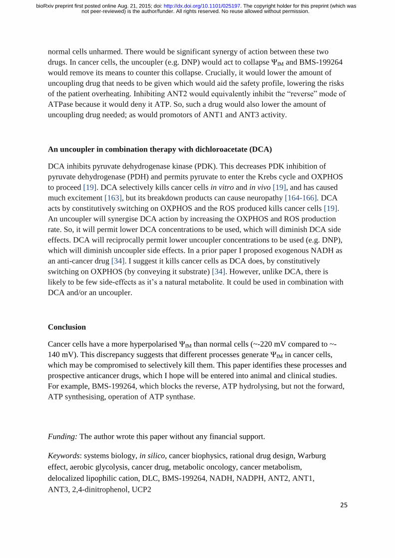
25
normal cells unharmed. There would be significant synergy of action between these two
drugs. In cancer cells, the uncoupler (e.g. DNP) would act to collapse ΨIM and BMS-199264
would remove its means to counter this collapse. Crucially, it would lower the amount of
uncoupling drug that needs to be given which would aid the safety profile, lowering the risks
of the patient overheating. Inhibiting ANT2 would equivalently inhibit the “reverse” mode of
ATPase because it would deny it ATP. So, such a drug would also lower the amount of
uncoupling drug needed; as would promotors of ANT1 and ANT3 activity.
An uncoupler in combination therapy with dichloroacetate (DCA)
DCA inhibits pyruvate dehydrogenase kinase (PDK). This decreases PDK inhibition of
pyruvate dehydrogenase (PDH) and permits pyruvate to enter the Krebs cycle and OXPHOS
to proceed [19]. DCA selectively kills cancer cells in vitro and in vivo [19], and has caused
much excitement [163], but its breakdown products can cause neuropathy [164-166]. DCA
acts by constitutively switching on OXPHOS and the ROS produced kills cancer cells [19].
An uncoupler will synergise DCA action by increasing the OXPHOS and ROS production
rate. So, it will permit lower DCA concentrations to be used, which will diminish DCA side
effects. DCA will reciprocally permit lower uncoupler concentrations to be used (e.g. DNP),
which will diminish uncoupler side effects. In a prior paper I proposed exogenous NADH as
an anti-cancer drug [34]. I suggest it kills cancer cells as DCA does, by constitutively
switching on OXPHOS (by conveying it substrate) [34]. However, unlike DCA, there is
likely to be few side-effects as it’s a natural metabolite. It could be used in combination with
DCA and/or an uncoupler.
Conclusion
Cancer cells have a more hyperpolarised ΨIM than normal cells (~-220 mV compared to ~-
140 mV). This discrepancy suggests that different processes generate ΨIM in cancer cells,
which may be compromised to selectively kill them. This paper identifies these processes and
prospective anticancer drugs, which I hope will be entered into animal and clinical studies.
For example, BMS-199264, which blocks the reverse, ATP hydrolysing, but not the forward,
ATP synthesising, operation of ATP synthase.
Funding: The author wrote this paper without any financial support.
Keywords: systems biology, in silico, cancer biophysics, rational drug design, Warburg
effect, aerobic glycolysis, cancer drug, metabolic oncology, cancer metabolism,
delocalized lipophilic cation, DLC, BMS-199264, NADH, NADPH, ANT2, ANT1,
ANT3, 2,4-dinitrophenol, UCP2
not peer-reviewed) is the author/funder. All rights reserved. No reuse allowed without permission. The copyright holder for this preprint (which was. http://dx.doi.org/10.1101/025197doi: bioRxiv preprint first posted online Aug. 21, 2015;
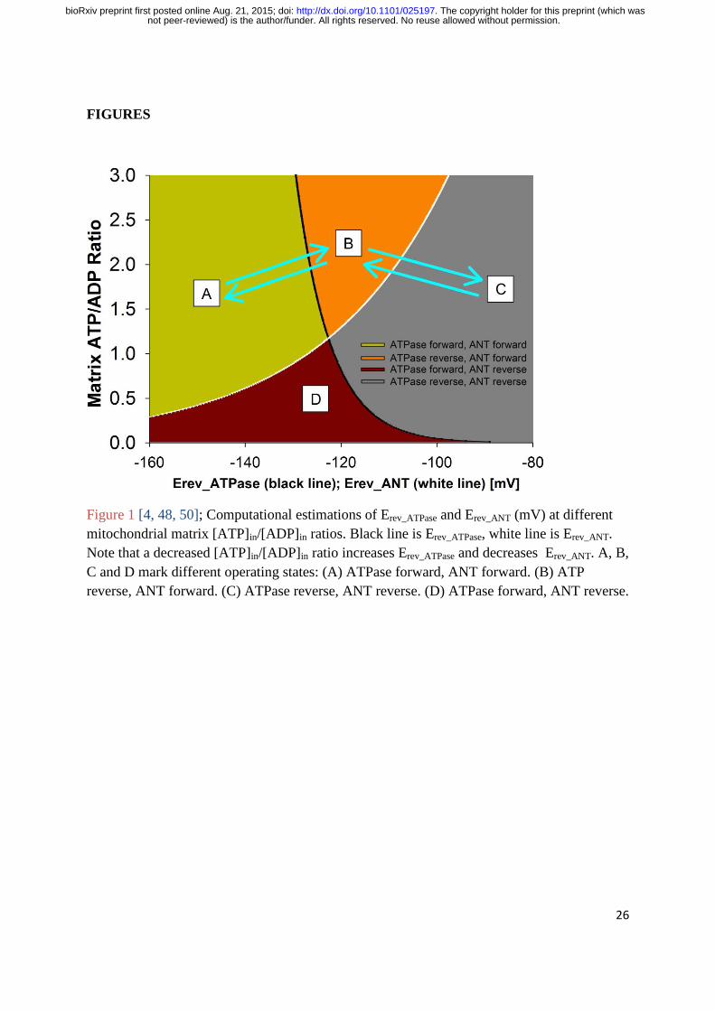
26
FIGURES
Figure 1 [4, 48, 50]; Computational estimations of Erev_ATPase and Erev_ANT (mV) at different
mitochondrial matrix [ATP]in/[ADP]in ratios. Black line is Erev_ATPase, white line is Erev_ANT.
Note that a decreased [ATP]in/[ADP]in ratio increases Erev_ATPase and decreases Erev_ANT. A, B,
C and D mark different operating states: (A) ATPase forward, ANT forward. (B) ATP
reverse, ANT forward. (C) ATPase reverse, ANT reverse. (D) ATPase forward, ANT reverse.
not peer-reviewed) is the author/funder. All rights reserved. No reuse allowed without permission. The copyright holder for this preprint (which was. http://dx.doi.org/10.1101/025197doi: bioRxiv preprint first posted online Aug. 21, 2015;
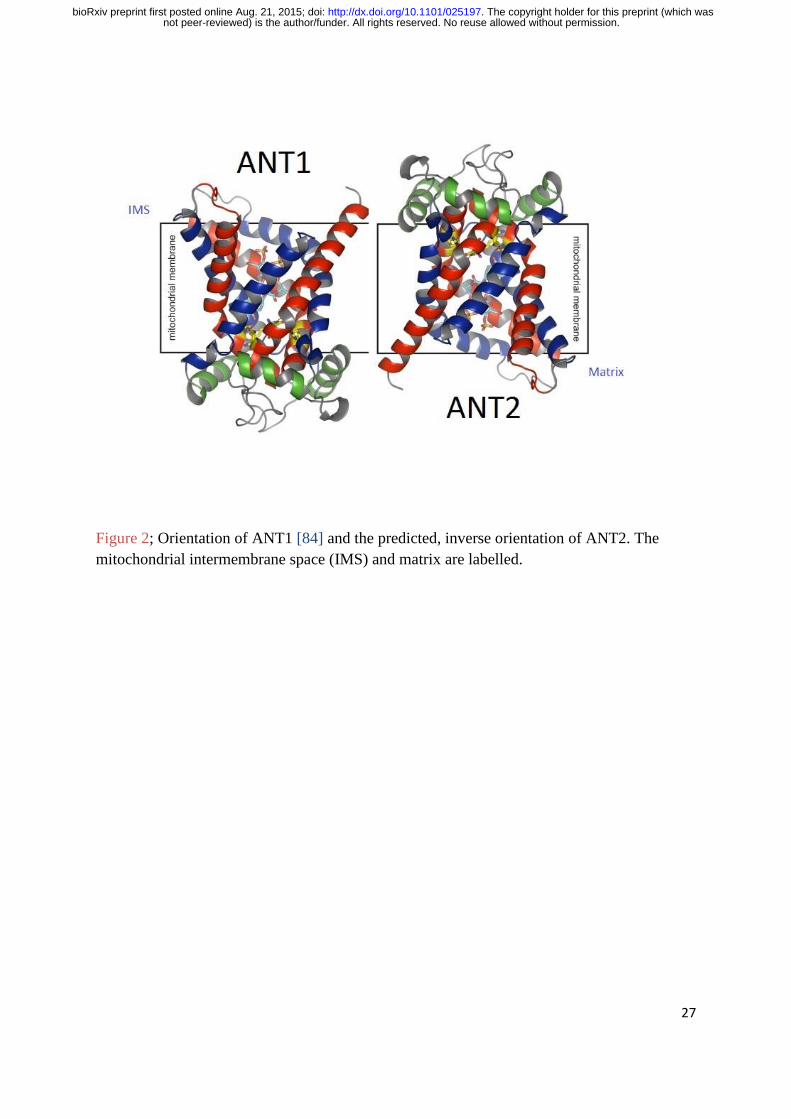
27
Figure 2; Orientation of ANT1 [84] and the predicted, inverse orientation of ANT2. The
mitochondrial intermembrane space (IMS) and matrix are labelled.
not peer-reviewed) is the author/funder. All rights reserved. No reuse allowed without permission. The copyright holder for this preprint (which was. http://dx.doi.org/10.1101/025197doi: bioRxiv preprint first posted online Aug. 21, 2015;
28
Figure 3; Computational estimations of Erev_ATPase (black line) and Erev_ANT (white line),
which show their dependence on certain key parameters. (A) Same plots, using the same
parameters, as Figure 1 (but shown with a different x-axis range). (B) Cytoplasmic
[ATP/ADP] ratio changed from 120 to 4.8. (C) ATPase coupling ratio, n, changed from 3.7 to
2.7. (D) ATPase affinity for ADP and ATP modified: Kw changed from 10-3.198
to 10-1.599
, Kq
from 10-4.06
to 10-6.09
. (E) Matrix Pi level reduced from 10 to 0.01 mM. (F) Matrix pH
decreased from 7.38 to 6.38, intermembrane space pH increased from 7.25 to 8.25. This
represents the action of the nigericin ionophore.
not peer-reviewed) is the author/funder. All rights reserved. No reuse allowed without permission. The copyright holder for this preprint (which was. http://dx.doi.org/10.1101/025197doi: bioRxiv preprint first posted online Aug. 21, 2015;
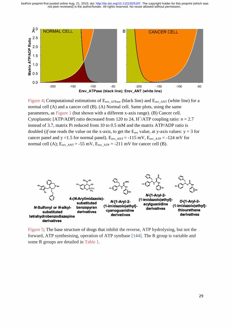
29
Figure 4; Computational estimations of Erev_ATPase (black line) and Erev_ANT (white line) for a
normal cell (A) and a cancer cell (B). (A) Normal cell. Same plots, using the same
parameters, as Figure 1 (but shown with a different x-axis range). (B) Cancer cell.
Cytoplasmic [ATP/ADP] ratio decreased from 120 to 24, H+/ATP coupling ratio: n = 2.7
instead of 3.7, matrix Pi reduced from 10 to 0.5 mM and the matrix ATP/ADP ratio is
doubled (if one reads the value on the x-axis, to get the Erev value, at y-axis values: y = 3 for
cancer panel and y =1.5 for normal panel). Erev_ANT = -115 mV, Erev_ATP = -124 mV for
normal cell (A); Erev_ANT = -55 mV, Erev_ATP = -211 mV for cancer cell (B).
Figure 5; The base structure of drugs that inhibit the reverse, ATP hydrolysing, but not the
forward, ATP synthesising, operation of ATP synthase [144]. The R group is variable and
some R groups are detailed in Table 1.
not peer-reviewed) is the author/funder. All rights reserved. No reuse allowed without permission. The copyright holder for this preprint (which was. http://dx.doi.org/10.1101/025197doi: bioRxiv preprint first posted online Aug. 21, 2015;
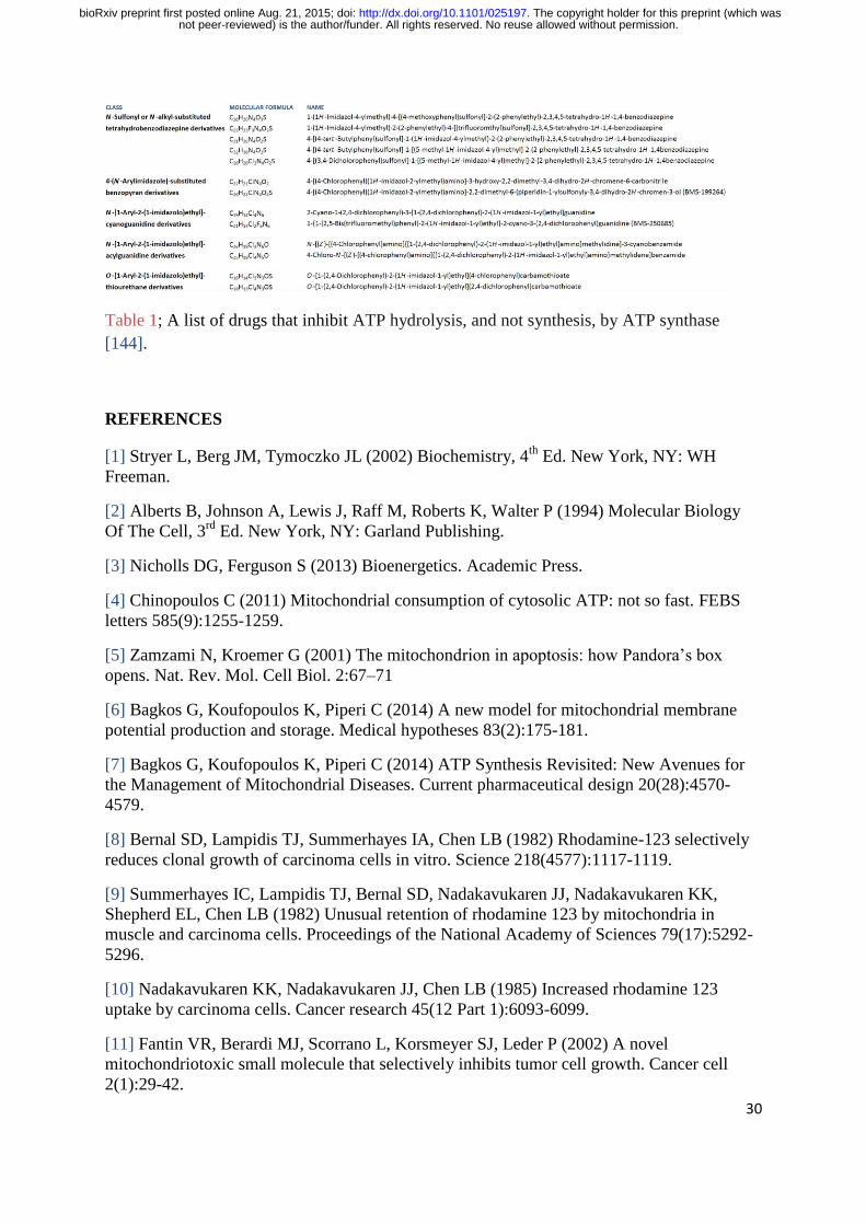
30
Table 1; A list of drugs that inhibit ATP hydrolysis, and not synthesis, by ATP synthase
[144].
REFERENCES
[1] Stryer L, Berg JM, Tymoczko JL (2002) Biochemistry, 4th
Ed. New York, NY: WH
Freeman.
[2] Alberts B, Johnson A, Lewis J, Raff M, Roberts K, Walter P (1994) Molecular Biology
Of The Cell, 3rd
Ed. New York, NY: Garland Publishing.
[3] Nicholls DG, Ferguson S (2013) Bioenergetics. Academic Press.
[4] Chinopoulos C (2011) Mitochondrial consumption of cytosolic ATP: not so fast. FEBS
letters 585(9):1255-1259.
[5] Zamzami N, Kroemer G (2001) The mitochondrion in apoptosis: how Pandora’s box
opens. Nat. Rev. Mol. Cell Biol. 2:67–71
[6] Bagkos G, Koufopoulos K, Piperi C (2014) A new model for mitochondrial membrane
potential production and storage. Medical hypotheses 83(2):175-181.
[7] Bagkos G, Koufopoulos K, Piperi C (2014) ATP Synthesis Revisited: New Avenues for
the Management of Mitochondrial Diseases. Current pharmaceutical design 20(28):4570-
4579.
[8] Bernal SD, Lampidis TJ, Summerhayes IA, Chen LB (1982) Rhodamine-123 selectively
reduces clonal growth of carcinoma cells in vitro. Science 218(4577):1117-1119.
[9] Summerhayes IC, Lampidis TJ, Bernal SD, Nadakavukaren JJ, Nadakavukaren KK,
Shepherd EL, Chen LB (1982) Unusual retention of rhodamine 123 by mitochondria in
muscle and carcinoma cells. Proceedings of the National Academy of Sciences 79(17):5292-
5296.
[10] Nadakavukaren KK, Nadakavukaren JJ, Chen LB (1985) Increased rhodamine 123
uptake by carcinoma cells. Cancer research 45(12 Part 1):6093-6099.
[11] Fantin VR, Berardi MJ, Scorrano L, Korsmeyer SJ, Leder P (2002) A novel
mitochondriotoxic small molecule that selectively inhibits tumor cell growth. Cancer cell
2(1):29-42.
not peer-reviewed) is the author/funder. All rights reserved. No reuse allowed without permission. The copyright holder for this preprint (which was. http://dx.doi.org/10.1101/025197doi: bioRxiv preprint first posted online Aug. 21, 2015;

31
[12] Modica-Napolitano JS, Aprille JR (2001) Delocalized lipophilic cations selectively
target the mitochondria of carcinoma cells. Advanced drug delivery reviews 49(1):63-70.
[13] Davis S, Weiss MJ, Wong JR (1985) Mitochondrial and plasma membrane potentials
cause unusual accumulation and retention of rhodamine 123 by human breast
adenocarcinoma-derived MCF-7 cells. Journal of Biological Chemistry 260(25):13844–
13850.
[14] Houston MA, Augenlicht LH, Heerdt BG (2011) Stable differences in intrinsic
mitochondrial membrane potential of tumor cell subpopulations reflect phenotypic
heterogeneity. International journal of cell biology 2011.
[15] Heerdt BG, Houston MA, Augenlicht LH (2005) The intrinsic mitochondrial membrane
potential of colonic carcinoma cells is linked to the probability of tumor progression. Cancer
Res 65:9861–9867.
[16] Heerdt BG, Houston MA, Augenlicht LH. Growth properties of colonic tumor cells are a
function of the intrinsic mitochondrial membrane potential. Cancer Res 2006;66(3):1591–6.
[17] Chen LB (1988) Mitochondrial membrane potential in living cells. Annual review of cell
biology 4(1):155-181.
[18] Fantin VR, St-Pierre J, Leder P (2006) Attenuation of LDH-A expression uncovers a
link between glycolysis, mitochondrial physiology, and tumor maintenance. Cancer cell
9(6):425-434.
[19] Bonnet S, Archer SL, Allalunis-Turner J, Haromy A, Beaulieu C et al. (2007) A
mitochondria-K+ channel axis is suppressed in cancer and its normalization promotes
apoptosis and inhibits cancer growth. Cancer cell 11(1):37-51.
[20] Warburg O (1956) On the origin of cancer cells. Science 123(3191):309-314.
[21] Ferreira LM (2010) Cancer metabolism: the Warburg effect today. Exp Mol Pathol
89(3):372-380.
[22] Kim JW, Dang CV (2006) Cancer's molecular sweet tooth and the Warburg effect.
Cancer Res 66(18):8927-8930.
[23] Lopez-Lazaro M (2008) The warburg effect: why and how do cancer cells activate
glycolysis in the presence of oxygen? Anti-Cancer Agent Me 8(3):305-312.
[24] Elliott RL, Jiang XP, Head JF (2014) Want to Cure Cancer? Then Revisit the Past;
“Warburg Was Correct”, Cancer Is a Metabolic Disease. Journal of Cancer Therapy
[25] Gatenby RA, Gillies RJ (2004) Why do cancers have high aerobic glycolysis? Nat Rev
Cancer 4(11):891-899.
[26] Bui T, Thompson CB (2006) Cancer's sweet tooth. Cancer cell 9(6):419-420.
[27] Nakashima RA, Paggi MG, Pedersen PL (1984) Contributions of glycolysis and
oxidative phosphorylation to adenosine 5′-triphosphate production in AS-30D hepatoma
cells. Cancer Res 44(12 Part 1):5702-5706.
not peer-reviewed) is the author/funder. All rights reserved. No reuse allowed without permission. The copyright holder for this preprint (which was. http://dx.doi.org/10.1101/025197doi: bioRxiv preprint first posted online Aug. 21, 2015;
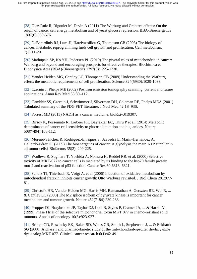
32
[28] Diaz-Ruiz R, Rigoulet M, Devin A (2011) The Warburg and Crabtree effects: On the
origin of cancer cell energy metabolism and of yeast glucose repression. BBA-Bioenergetics
1807(6):568-576.
[29] DeBerardinis RJ, Lum JJ, Hatzivassiliou G, Thompson CB (2008) The biology of
cancer: metabolic reprogramming fuels cell growth and proliferation. Cell metabolism,
7(1):11-20.
[30] Mathupala SP, Ko YH, Pedersen PL (2010) The pivotal roles of mitochondria in cancer:
Warburg and beyond and encouraging prospects for effective therapies. Biochimica et
Biophysica Acta (BBA)-Bioenergetics 1797(6):1225-1230.
[31] Vander Heiden MG, Cantley LC, Thompson CB (2009) Understanding the Warburg
effect: the metabolic requirements of cell proliferation. Science 324(5930):1029-1033.
[32] Czernin J, Phelps ME (2002) Positron emission tomography scanning: current and future
applications. Annu Rev Med 53:89–112.
[33] Gambhir SS, Czernin J, Schwimmer J, Silverman DH, Coleman RE, Phelps MEA (2001)
Tabulated summary of the FDG PET literature. J Nucl Med 42:1S–93S.
[34] Forrest MD (2015) NADH as a cancer medicine. bioRxiv:019307.
[35] Birsoy K, Possemato R, Lorbeer FK, Bayraktar EC, Thiru P et al. (2014) Metabolic
determinants of cancer cell sensitivity to glucose limitation and biguanides. Nature
508(7494):108-112.
[36] Moreno‐Sánchez R, Rodríguez‐Enríquez S, Saavedra E, Marín‐Hernández A,
Gallardo‐Pérez JC (2009) The bioenergetics of cancer: is glycolysis the main ATP supplier in
all tumor cells? Biofactors 35(2): 209-225.
[37] Wadhwa R, Sugihara T, Yoshida A, Nomura H, Reddel RR, et al. (2000) Selective
toxicity of MKT-077 to cancer cells is mediated by its binding to the hsp70 family protein
mot-2 and reactivation of p53 function. Cancer Res 60:6818–6821.
[38] Schulz TJ, Thierbach R, Voigt A, et al (2006) Induction of oxidative metabolism by
mitochondrial frataxin inhibits cancer growth: Otto Warburg revisited. J Biol Chem 281:977-
81.
[39] Christofk HR, Vander Heiden MG, Harris MH, Ramanathan A, Gerszten RE, Wei R, ...
& Cantley LC (2008) The M2 splice isoform of pyruvate kinase is important for cancer
metabolism and tumour growth. Nature 452(7184):230-233.
[40] Propper DJ, Braybrooke JP, Taylor DJ, Lodi R, Styles P, Cramer JA, ... & Harris AL
(1999) Phase I trial of the selective mitochondrial toxin MKT 077 in chemo-resistant solid
tumours. Annals of oncology 10(8):923-927.
[41] Britten CD, Rowinsky EK, Baker SD, Weiss GR, Smith L, Stephenson J, ... & Eckhardt
SG (2000) A phase I and pharmacokinetic study of the mitochondrial-specific rhodacyanine
dye analog MKT 077. Clinical cancer research 6(1):42-49.
not peer-reviewed) is the author/funder. All rights reserved. No reuse allowed without permission. The copyright holder for this preprint (which was. http://dx.doi.org/10.1101/025197doi: bioRxiv preprint first posted online Aug. 21, 2015;
33
[42] Ross MF, Da Ros T, Blaikie FH, Prime TA, Porteous CM, Severina II, Skulachev VP,
Kjaergaard HG, Smith RA, Murphy MP (2006) Accumulation of lipophilic dications by
mitochondria and cells. Biochem. J. 400:199–208.
[43] Koya K, Li Y, Wang H, Ukai T, Tatsuta N, Kawakami M, ... Chen LB (1996) MKT-077,
a novel rhodacyanine dye in clinical trials, exhibits anticarcinoma activity in preclinical
studies based on selective mitochondrial accumulation. Cancer research 56(3):538-543.
[44] Weiss MJ, Wong JR, Ha CS, Bleday R, Salem RR, Steele GD, Chen LB (1987)
Dequalinium, a topical antimicrobial agent, displays anticarcinoma activity based on selective
mitochondrial accumulation. Proceedings of the National Academy of Sciences 84(15):5444-
5448.
[45] Sun X, Wong JR, Song K, Hu J, Garlid KD, Chen LB (1994) AA1, a newly synthesized
monovalent lipophilic cation, expresses potent in vivo antitumor activity. Cancer
research 54(6):1465-1471.
[46] Modica-Napolitano JS, Weiss MJ, Chen LB, Aprille JR (1984) Rhodamine 123 inhibits
bioenergetic function in isolated rat liver mitochondria. Biochemical and biophysical research
communications 118(3):717-723.
[47] Chiba Y, Kubota T, Watanabe M, Matsuzaki SW, Otani Y, Teramoto T, ... & Kitajima
M (1997) MKT-077, localized lipophilic cation: antitumor activity against human tumor
xenografts serially transplanted into nude mice. Anticancer research 18(2A):1047-1052.
[48] Chinopoulos C, Gerencser AA, Mandi M, Mathe K, Torocsik B, Doczi J, Turiak L, Kiss
G, Konrad C, Vajda S, Vereczki V, Oh RJ, Adam-Vizi V (2010) Forward operation of
adenine nucleotide translocase during F0F1-ATPase reversal: critical role of matrix substrate-
level phosphorylation. FASEB J 24:2405–2416.
[49] Chinopoulos C, Adam-Vizi V (2010) Mitochondria as ATP consumers in cellular
pathology. Biochim Biophys Acta 1802:221–227.
[50] Chinopoulos C (2011) The ‘‘B Space’’ of mitochondrial phosphorylation. Journal of
Neuroscience Research 89:1897–1904.
[51] Watt IN, Montgomery MG, Runswick MJ, Leslie AG, Walker JE (2010) Bioenergetic
cost of making an adenosine triphosphate molecule in animal mitochondria. Proceedings of
the National Academy of Sciences 107(39):16823-16827.
[52] Clémençon B, Babot M, Trézéguet V (2013) The mitochondrial ADP/ATP carrier
(SLC25 family): Pathological implications of its dysfunction. Molecular aspects of
medicine 34(2):485-493.
[53] Chevrollier A, Loiseau D, Chabi B, Renier G, Douay O, Malthièry Y, Stepien G (2005)
ANT2 isoform required for cancer cell glycolysis. Journal of bioenergetics and
biomembranes 37(5):307-317.
[54] Chevrollier A, Loiseau D, Reynier P, Stepien G (2011) Adenine nucleotide translocase 2
is a key mitochondrial protein in cancer metabolism. Biochimica et Biophysica Acta (BBA)-
Bioenergetics 1807(6):562-567.
not peer-reviewed) is the author/funder. All rights reserved. No reuse allowed without permission. The copyright holder for this preprint (which was. http://dx.doi.org/10.1101/025197doi: bioRxiv preprint first posted online Aug. 21, 2015;
34
[55] Maldonado EN, Lemasters JJ (2014) ATP/ADP ratio, the missed connection between
mitochondria and the Warburg effect. Mitochondrion 19:78-84.
[56] Maldonado EN, Vuicich J, DeHart DN, Rodebaugh HS, Lemasters JJ (2013)
Translocation of glycolytic ATP into mitochondria of cancer cells does not utilize the adenine
nucleotide transporter. Biophys J 104:303a–304a.
[57] Tewari SG, Dash RK, Beard DA, Bazil JN (2012) A Biophysical Model of the
Mitochondrial ATP-Mg/Pi Carrier. Biophysical journal 103(7):1616-1625.
[58] TTraba J, Froschauer EM, Wiesenberger G, Satrústegui J, Del Arco A (2008) Yeast
mitochondria import ATP through the calcium‐dependent ATP‐Mg/Pi carrier Sal1p, and are
ATP consumers during aerobic growth in glucose. Molecular microbiology 69(3):570-585.
[59] Schmitz-Esser S, Linka N, Collingro A, Beier CL, Neuhaus HE, Wagner M, Horn M
(2004) ATP/ADP translocases: a common feature of obligate intracellular amoebal symbionts
related to Chlamydiae and Rickettsiae. J Bacteriol 186(3):683-691.
[60] Audia JP, Winkler HH (2006) Study of the five Rickettsia prowazekii proteins annotated
as ATP/ADP translocases (Tlc): only Tlc1 transports ATP/ADP, while Tlc4 and Tlc5
transport other ribonucleotides. J Bacteriol 188(17):6261-6268.
[61] Trentmann O, Horn M, van Scheltinga ACT, Neuhaus HE, Haferkamp I (2007)
Enlightening Energy Parasitism by Analysis of an ATP/ADP Transporter from Chlamydiae.
PLoS Biol 5(9):e231. doi:10.1371/journal.pbio.0050231.
[62] Schmitz-Esser S, Haferkamp I, Knab S, Penz T, Ast M, Kohl C, et al. (2008) Lawsonia
intracellularis contains a gene encoding a functional rickettsia-like ATP/ADP translocase for
host exploitation. J Bacteriol 190(17):5746-5752.
[63] Trentmann O, Jung B, Neuhaus HE, Haferkamp I (2008) Nonmitochondrial ATP/ADP
transporters accept phosphate as third substrate. J Biol Chem 283(52):36486-36493.
[64] Altschul SF, Gish W, Miller W, Myers EW, Lipman DJ (1990) Basic local alignment
search tool. Journal of molecular biology 215(3):403-410.
[65] Tsaousis AD, Kunji ER, Goldberg AV, Lucocq JM, Hirt RP, Embley TM (2008) A
novel route for ATP acquisition by the remnant mitochondria of Encephalitozoon cuniculi.
Nature 453(7194):553-556.
[66] Kunji ER (2004) The role and structure of mitochondrial carriers. FEBS
letters 564(3):239-244.
[67] Alves S (2008) Two of a kind. Nature Reports Stem Cells.
[68] Baker M (2008) Cancer and embryonic stem cells share genetic fingerprints. Nature
Reports Stem Cells.
[69] Chung S, Dzeja PP, Faustino RS, Perez-Terzic C, Behfar A, Terzic A (2007)
Mitochondrial oxidative metabolism is required for the cardiac differentiation of stem cells.
Nat Clin Pract Cardiovasc Med 4: S60 – S67.
not peer-reviewed) is the author/funder. All rights reserved. No reuse allowed without permission. The copyright holder for this preprint (which was. http://dx.doi.org/10.1101/025197doi: bioRxiv preprint first posted online Aug. 21, 2015;

35
[70] Teslaa T, Teitell MA (2014) Pluripotent stem cell energy metabolism: an update. The
EMBO journal e201490446.
[71] Zalzman M, Falco G, Sharova LV, Nishiyama A, Thomas M, Lee SL, ... & Ko MS
(2010) Zscan4 regulates telomere elongation and genomic stability in ES cells. Nature
464(7290):858-863.
[72] Hayflick L (1997) Mortality and immortality at the cellular level. A
review.Biochemistry-New York-English Translation of Biokhimiya 62(11):1180-1190.
[73] Kondoh H, Lleonart ME, Bernard D, Gil J (2007) Protection from oxidative stress by
enhanced glycolysis; a possible mechanism of cellular immortalization. Histology and
histopathology 22(1): 85-90.
[74] Hong Y, Stambrook PJ (2004) Restoration of an absent G1 arrest and protection from
apoptosis in embryonic stem cells after ionizing radiation. PNAS 101(40):14443-14448.
[75] Kokoszka JE, Waymire KG, Levy SE, Sligh JE, Cai J, Jones DP, MacGregor GR,
Wallace DC (2004) The ADP/ATP translocator is not essential for the mitochondrial
permeability transition pore. Nature 427:461–465.
[76] Jang JY, Choi Y, Jeon YK, Kim CW (2008) Suppression of adenine nucleotide
translocase-2 by vector-based siRNA in human breast cancer cells induces apoptosis and
inhibits tumor growth in vitro and in vivo. Breast Cancer Res 10(1):R11.
[77] Le Bras M, Borgne-Sanchez A, Touat Z, El Dein OS, Deniaud A, Maillier E, Lecellier
G, Rebouillat D, Lemair C, Kroemer G, Jacotot E, Brenner C (2006) Chemosensitization by
knockdown of adenine nucleotide translocase-2. Cancer Res 66:9143-9152.
[78] Jang JY, Choi Y, Jeon YK, Aung KC, Kim CW (2008) Over-expression of adenine
nucleotide translocase 1 (ANT1) induces apoptosis and tumor regression in vivo. BMC
cancer 8(1):160.
[79] Zamora M, Granell M, Mampel T, Vinas O (2004) Adenine nucleotide translocase 3
(ANT3) overexpression induces apoptosis in cultured cells. FEBS Lett 563:155-160.
[80] Bauer MK, Schubert A, Rocks O, Grimm S (1999) Adenine nucleotide translocase-1, a
component of the permeability transition pore, can dominantly induce apoptosis. The Journal
of cell biology 147(7):1493-1502.
[81] Klingenberg M (2008) The ADP and ATP transport in mitochondria and its
carrier. Biochimica et Biophysica Acta (BBA)-Biomembranes 1778(10):1978-2021.
[82] De Marcos Lousa C, Trézéguet V, Dianoux AC, Brandolin G, Lauquin GJM (2002) The
human mitochondrial ADP/ATP carriers: kinetic properties and biogenesis of wild-type and
mutant proteins in yeast S. cerevisiae. Biochemistry 41:14412–14420.
[83] Dahout-Gonzalez C, Nury H, Trezeguet V, Lauquin GJ, Pebay-Peyroula E, Brandolin G
(2006) Molecular, functional, and pathological aspects of the mitochondrial ADP/ATP
carrier. Physiology (Bethesda) 21:242–249.
not peer-reviewed) is the author/funder. All rights reserved. No reuse allowed without permission. The copyright holder for this preprint (which was. http://dx.doi.org/10.1101/025197doi: bioRxiv preprint first posted online Aug. 21, 2015;
36
[84] Pebay-Peyroula E, Dahout-Gonzalez C, Kahn R, Trezeguet V, Lauquin GJ, Brandolin G
(2003) Structure of mitochondrial ADP/ATP carrier in complex with carboxyatractyloside.
Nature 426:39-44.
[85] Dong GC, Chuang PH, Forrest MD, Lin YC, Chen HM (2006) Immuno-suppressive
effect of blocking the CD28 signaling pathway in T-cells by an active component of
Echinacea found by a novel pharmaceutical screening method. Journal of medicinal
chemistry 49(6):1845-1854.
[86] Lunt SY, Vander Heiden MG (2011) Aerobic glycolysis: meeting the metabolic
requirements of cell proliferation. Annu Rev Cell Dev Bi 27:441-464.
[87] Ganapathy-Kanniappan S, Geschwind JF (2013) Tumor glycolysis as a target for cancer
therapy: progress and prospects. Mol Cancer 12(152):4598-12.
[88] Vinogradov AD (2000) Steady-state and pre-steady state kinetics of mitochondrial F1F0
ATPase: is ATP synthase a reversible molecular machine. Journal of Experimental Biology
203:41–9.
[89] Klingenberg M (1977) The Adenine‐Nucleotide Exchange in Submitochondrial (Sonic)
Particles. European Journal of Biochemistry 76.2:553-565.
[90] Israelsen WJ, Vander Heiden MG (2010) ATP consumption promotes cancer
metabolism. Cell 143(5):669-671.
[91] Shabalina IG, Kramarova TV, Nedergaard J, Cannon B (2006) Carboxyatractyloside
effects on brown-fat mitochondria imply that the adenine nucleotide translocator isoforms
ANT1 and ANT2 may be responsible for basal and fatty-acid-induced uncoupling
respectively. Biochem J 399:405–414.
[92] Brand MD, Pakay JL, Ocloo A, Kokoszka J, Wallace DC, Brookes PS, Cornwall EJ
(2005) The basal proton conductance of mitochondria depends on adenine nucleotide
translocase content. Biochem J 392:353–362
[93] Brand MD, Pakay JL, Ocloo A, Kokozka J, Wallace DC, Brookes PS, Cornwall EJ
(2005) The basal proton conductance of mitochondria depends on adenine nucleotide
translocase content. Biochemical Journal 392(2):353-362.
[94] Lena A, Rechichi M, Salvetti A, Vecchio D, Evangelista M, Rainaldi G, Gremigni V,
Rossi L (2010) The silencing of adenine nucleotide translocase isoform 1 induces oxidative
stress and programmed cell death in ADF human glioblastoma cells. FEBS journal
277(13):2853-2867.
[95] Brand MD (2000) Uncoupling to survive? The role of mitochondrial inefficiency in
ageing. Exp Gerontol 35:811–820.
[96] Echtay KS, Roussel D, St-Pierre J, Jekabsons MB, Cadenas S, Stuart JA, Brand MD
(2002) Superoxide activates mitochondrial uncoupling proteins. Nature 415(6867):96-99.
[97] Yu Q, Heikal AA (2009) Two-photon autofluorescence dynamics imaging reveals
sensitivity of intracellular NADH concentration and conformation to cell physiology at the
single-cell level. Journal of Photochemistry and Photobiology B: Biology 95(1):46-57.
not peer-reviewed) is the author/funder. All rights reserved. No reuse allowed without permission. The copyright holder for this preprint (which was. http://dx.doi.org/10.1101/025197doi: bioRxiv preprint first posted online Aug. 21, 2015;
37
[98] Blacker TS, Mann ZF, Gale JE, Ziegler M, Bain AJ, Szabadkai G, Duchen MR (2014)
Separating NADH and NADPH fluorescence in live cells and tissues using FLIM. Nature
communications 5.
[99] Feeney R, Clarke AR, Holbrook JJ (1990) A single amino acid substitution in lactate
dehydrogenase improves the catalytic efficiency with an alternative coenzyme. Biochemical
and biophysical research communications 166(2):667-672.
[100] Valle A, Oliver J, Roca P (2010) Role of uncoupling proteins in cancer. Cancers
2(2):567-591.
[101] Donadelli M, Dando I, Dalla Pozza E, Palmieri M (2015) Mitochondrial uncoupling
protein 2 and pancreatic cancer: A new potential target therapy. World journal of
gastroenterology: WJG 21(11):3232.
[102] Ayyasamy V, Owens KM, Desouki MM, Liang P, Bakin A, Thangaraj K, et al. (2011)
Cellular Model of Warburg Effect Identifies Tumor Promoting Function of UCP2 in Breast
Cancer and Its Suppression by Genipin. PLoS ONE 6(9):e24792.
doi:10.1371/journal.pone.0024792.
[103] Vozza A, Parisi G, De Leonardis F, Lasorsa FM, Castegna A, Amorese D, Marmo R,
Calcagnile VM, Palmieri L, Ricquier D, Paradies E, Scarcia P, Palmieri F, Bouillaud F,
Fiermonte G (2014) UCP2 transports C4 metabolites out of mitochondria, regulating glucose
and glutamine oxidation. Proc Natl Acad Sci USA 111:960-965.
[104] Deng S, Yang Y, Han Y, Li X, Wang X, Li X, et al. (2012) UCP2 Inhibits ROS-
Mediated Apoptosis in A549 under Hypoxic Conditions. PLoS ONE 7(1): e30714.
[105] Derdak Z, Mark NM, Beldi G, Robson SC, Wands JR, Baffy G (2008) The
mitochondrial uncoupling protein-2 promotes chemoresistance in cancer cells. Cancer
research 68(8):2813-2819
[106] Mailloux RJ, Adjeitey CN-K, Harper M-E (2010) Genipin-Induced Inhibition of
Uncoupling Protein-2 Sensitizes Drug-Resistant Cancer Cells to Cytotoxic Agents. PLoS
ONE 5(10):e13289
[107] Hong HY, Kim BC (2007) Mixed lineage kinase 3 connects reactive oxygen species to
c-Jun NH2-terminal kinase-induced mitochondrial apoptosis in genipin-treated PC3 human
prostate cancer cells. Biochem. Biophys. Res. Commun. 362:307–312.
[108] Kim BC, Kim HG, Lee SA, Lim S, Park EH, Kim SJ, Lim CJ (2005) Genipin-induced
apoptosis in hepatoma cells is mediated by reactive oxygen species/c-Jun NH2-terminal
kinasedependent activation of mitochondrial pathway. Biochem. Pharmacol. 70:1398–1407.
[109] Baffy G (2010) Uncoupling protein-2 and cancer. Mitochondrion 10:243-252
[110] Zhang J, et al. (2011) UCP2 regulates energy metabolism and differentiation potential
of human pluripotent stem cells. EMBO J 30(24):4860–4873.
[111] Samudio I, Fiegl M, McQueen T, Clise-Dwyer K, Andreeff M (2008) The warburg
effect in leukemia-stroma cocultures is mediated by mitochondrial uncoupling associated
with uncoupling protein 2 activation. Cancer Res 68:5198–5205.
not peer-reviewed) is the author/funder. All rights reserved. No reuse allowed without permission. The copyright holder for this preprint (which was. http://dx.doi.org/10.1101/025197doi: bioRxiv preprint first posted online Aug. 21, 2015;
38
[112] Santandreu FM, Roca P, Oliver J (2010) Uncoupling protein-2 knockdown mediates
the cytotoxic effects of cisplatin. Free Radical Biology and Medicine 49(4):658-666.
[113] Nicholls DG (2006) Simultaneous monitoring of ionophore-and inhibitor-mediated
plasma and mitochondrial membrane potential changes in cultured neurons. Journal of
Biological Chemistry 281(21):14864-14874.
[114] Akhmedov D, Braun M, Mataki C, Park KS, Pozzan T, Schoonjans K, ... & Wiederkehr
A (2010) Mitochondrial matrix pH controls oxidative phosphorylation and metabolism-
secretion coupling in INS-1E clonal β cells. The FASEB Journal 24(11):4613-4626.
[115] Deng CC, Liang Y, Wu MS, Feng FT, Hu WR, Chen LZ, ... & Zeng YX (2013)
Nigericin selectively targets cancer stem cells in nasopharyngeal carcinoma. The
international journal of biochemistry & cell biology 45(9):1997-2006.
[116] Miyake H, Hara I, Yamanaka K, Arakawa S, Kamidono S (1999) Calcium ionophore,
ionomycin inhibits growth of human bladder cancer cells both in vitro and in vivo with
alteration of Bcl-2 and Bax expression levels. The Journal of urology 162(3):916-921.
[117] Rokitskaya TI, Ilyasova TM, Severina II, Antonenko YN, Skulachev VP (2013)
Electrogenic proton transport across lipid bilayer membranes mediated by cationic
derivatives of rhodamine 19: comparison with anionic protonophores. European Biophysics
Journal 42(6):477-485.
[118] Casey JR, Grinstein S, Orlowski J (2010) Sensors and regulators of intracellular pH.
Nature reviews Molecular cell biology 11(1):50-61.
[119] Davies KM, Strauss M, Daum B, Kief JH, Osiewacz HD, Rycovska A, ... Kühlbrandt
W (2011) Macromolecular organization of ATP synthase and complex I in whole
mitochondria. Proceedings of the National Academy of Sciences 108(34):14121-14126.
[120] Damaghi M, Wojtkowiak JW, Gillies RJ (2013) pH sensing and regulation in cancer.
Frontiers in physiology 4.
[121] Gerweck LE, Seetharaman K (1996) Cellular pH gradient in tumor versus normal
tissue: potential exploitation for the treatment of cancer. Cancer Res. 56:119–1198.
[122] Gerweck LE, Vijayappa S, Kozin S (2006) Tumor pH controls the in vivo efficacy of
weak acid and base chemotherapeutics. Mol. Cancer Ther. 5:1275–1279.
[123] McCarty MF, Whitaker J (2010) Manipulating tumor acidification as a cancer
treatment strategy. Altern Med Rev 15(3):264-72.
[124] Han YH, Kim SW, Kim SH, Kim SZ, Park WH (2008) 2,4-Dinitrophenol induces G1
phase arrest and apoptosis in human pulmonary adenocarcinoma Calu-6 cells. Toxicology in
Vitro 22(3):659-670.
[125] Han YH, Yang YM, Park WH (2010) Carbonyl Cyanide P-(Trifluoromethoxy)
Phenylhydroazone Induces Caspase-independent Apoptosis in As4. 1 Juxtaglomerular Cells.
Anticancer research 30(7):2863-2868.
not peer-reviewed) is the author/funder. All rights reserved. No reuse allowed without permission. The copyright holder for this preprint (which was. http://dx.doi.org/10.1101/025197doi: bioRxiv preprint first posted online Aug. 21, 2015;

39
[126] Datta S, Li J, Mahdi F, Jekabsons MB, Nagle DG, Zhou YD (2012) Glycolysis
inhibitor screening identifies the bis-geranylacylphloroglucinol protonophore moronone from
Moronobea coccinea. Journal of natural products 75(12):2216-2222.
[127] Newell KJ, Tannock IF (1989) Reduction of intracellular pH as a possible mechanism
for killing cells in acidic regions of solid tumors: effects of carbonylcyanide-
3chlorophenylhydrazone. Cancer Res. 49:4477–4482.
[128] Reis FH, Pardo-Andreu GL, Nuñez-Figueredo Y, Cuesta-Rubio O, Marín-Prida J,
Uyemura SA, ... Alberici LC (2014) Clusianone, a naturally occurring nemorosone
regioisomer, uncouples rat liver mitochondria and induces HepG2 cell death. Chemico-
biological interactions 212:20-29.
[129] Hostanska K, Reichling J, Bommer S, Weber M, Saller R (2003) Hyperforin a
constituent of St John's wort (Hypericum perforatum L.) extract induces apoptosis by
triggering activation of caspases and with hypericin synergistically exerts cytotoxicity
towards human malignant cell lines. Eur. J. Pharm. Biopharm. 56:121–132.
[130] Pardo-Andreu GL, Nuñez-Figueredo Y, Tudella VG, Cuesta-Rubio O, Rodrigues FP,
Pestana CR, ... Curti C (2011) The anti-cancer agent nemorosone is a new potent
protonophoric mitochondrial uncoupler. Mitochondrion 11(2):255-263.
[131] Jara JA, Castro-Castillo V, Saavedra-Olavarría J, Peredo L, Pavanni M, Jana F, ... &
Ferreira J (2014) Antiproliferative and uncoupling effects of delocalized, lipophilic, cationic
gallic acid derivatives on cancer cell lines. Validation in vivo in singenic mice. Journal of
medicinal chemistry 57(6):2440-2454.
[132] Ryoo IJ, Park HR, Choo SJ, Hwang JH, Park YM, Bae KH, ... Yoo ID (2006) Selective
cytotoxic activity of valinomycin against HT-29 human colon carcinoma cells via down-
regulation of GRP78. Biological and Pharmaceutical Bulletin 29(4):817-820.
[133] Ruiz-Marcial C, Chilpa RR, Estrada E, Reyes-Esparza J, Fariña GG, Rodriguez-
Fragoso L (2007) Antiproliferative, cytotoxic and antitumour activity of coumarins isolated
from Calophyllum brasiliense. Journal of pharmacy and pharmacology 59(5):719-726.
[134] Brenzan MA, Santos AO, Nakamura CV, Dias Filho BP, Ueda-Nakamura T, Young
MCM, Correa G, Junior JA, Morgado-Diaz A, Cortez DAG (2012) Effects of (−) mammea
A/BB isolated from Calophyllum brasiliense leaves and derivatives on mitochondrial
membrane of Leishmania amazonensis. Phytomedicine 19(3):223-230.
[135] Du L, Mahdi F, Jekabsons MB, Nagle DG, Zhou YD (2010) Mammea E/BB, an
isoprenylated dihydroxycoumarin protonophore that potently uncouples mitochondrial
electron transport, disrupts hypoxic signaling in tumor cells. Journal of natural products
73(11):1868-1872.
[136] Lou P, Hansen B, Olsen P, Tullin S, Murphy M, Brand M (2007) Mitochondrial
uncouplers with an extraordinary dynamic range. Biochem. J 407:129-140.
[137] Terada H (1990) Uncouplers of oxidative phosphorylation. Environmental Health
Perspectives 87:213.
not peer-reviewed) is the author/funder. All rights reserved. No reuse allowed without permission. The copyright holder for this preprint (which was. http://dx.doi.org/10.1101/025197doi: bioRxiv preprint first posted online Aug. 21, 2015;

40
[138] Goldgof M, Xiao C, Chanturiya T, Jou W, Gavrilova O, Reitman ML (2014) The
chemical uncoupler 2, 4-dinitrophenol (DNP) protects against diet-induced obesity and
improves energy homeostasis in mice at thermoneutrality. Journal of Biological Chemistry
289(28):19341-19350.
[139] Grundlingh J, Dargan PI, El-Zanfaly M, Wood DM (2011) 2, 4-dinitrophenol (DNP): a
weight loss agent with significant acute toxicity and risk of death. Journal of Medical
Toxicology 7(3):205-212.
[140] Horner WD (1941) A study of dinitrophenol and its relation to cataract formation.
Transactions of the American Ophthalmological Society 39:405.
[141] Harper JA, Dickinson K, Brand MD (2001) Mitochondrial uncoupling as a target for
drug development for the treatment of obesity. Obesity Reviews 2(4):255-265.
[142] Cerqueira FM, Laurindo FRM, Kowaltowski AJ (2011) Mild Mitochondrial
Uncoupling and Calorie Restriction Increase Fasting eNOS, Akt and Mitochondrial
Biogenesis. PLoS ONE 6(3):e18433
[143] Antonenko YN, Khailova LS, Knorre DA, Markova OV, Rokitskaya TI, Ilyasova TM,
et al. (2013) Penetrating Cations Enhance Uncoupling Activity of Anionic Protonophores in
Mitochondria. PLoS ONE 8(4):e61902.
[144] Hong S, Pedersen PL (2008) ATP synthase and the actions of inhibitors utilized to
study its roles in human health, disease, and other scientific areas. Microbiology and
Molecular Biology Reviews 72(4):590-641.
[145] Ma Z, Cao M, Liu Y, He Y, Wang Y, Yang C, ... & Gao F (2010) Mitochondrial F1Fo-
ATP synthase translocates to cell surface in hepatocytes and has high activity in tumor-like
acidic and hypoxic environment. Acta biochimica et biophysica Sinica 42(8):530-537.
[146] Huang CY, Huang HY, Forrest MD, Pan YR, Wu WJ, Chen HM (2014) Inhibition
Effect of a Custom Peptide on Lung Tumors. PloS ONE 9(10):e109174.
[147] Grover GJ, Marone PA, Koetzner L, Seto-Young D (2008) Energetic signalling in the
control of mitochondrial F 1 F 0 ATP synthase activity in health and disease. The
international journal of biochemistry & cell biology 40(12):2698-2701.
[148] Grover GJ, Malm J (2008) Pharmacological Profile of the Selective Mitochondrial
F1F0 ATP Hydrolase Inhibitor BMS‐199264 in Myocardial Ischemia. Cardiovascular
therapeutics 26(4):287-296.
[149] Grover GJ, Atwal KS, Sleph PG, Wang FL, Monshizadegan H, Monticello T, Green
DW (2004) Excessive ATP hydrolysis in ischemic myocardium by mitochondrial F1F0-
ATPase: effect of selective pharmacological inhibition of mitochondrial ATPase hydrolase
activity. American Journal of Physiology-Heart and Circulatory Physiology 287(4):H1747-
H1755.
[150] Ivanes F, Faccenda D, Gatliff J, Ahmed AA, Cocco S, Cheng CHK, ... & Campanella
M (2014) The compound BTB06584 is an IF1‐dependent selective inhibitor of the
mitochondrial F1Fo‐ATPase. British journal of pharmacology 171(18):4193-4206.
not peer-reviewed) is the author/funder. All rights reserved. No reuse allowed without permission. The copyright holder for this preprint (which was. http://dx.doi.org/10.1101/025197doi: bioRxiv preprint first posted online Aug. 21, 2015;

41
[151] Song CC, Lu X, Cheng BB, Du J, Li B, Ling CQ (2007) Effects of melittin on growth
and angiogenesis of human hepatocellular carcinoma BEL-7402 cell xenografts in nude mice.
Chinese journal of cancer 26(12):1315-1322.
[152] Pathak RK, Marrache S, Harn DA, Dhar S (2014) Mito-DCA: a mitochondria targeted
molecular scaffold for efficacious delivery of metabolic modulator dichloroacetate. ACS
chemical biology 9(5):1178-1187.
[153] Blaikie F, Brown S, Samuelsson L, Brand M, Smith R, Murphy M (2006) Targeting
dinitrophenol to mitochondria: limitations to the development of a self-limiting mitochondrial
protonophore. Bioscience reports 26:231-243.
[154] Apostolova N, Victor VM (2015) Molecular Strategies for Targeting Antioxidants to
Mitochondria: Therapeutic Implications. Antioxidants & Redox Signaling. 22(8):686-729.
[155] McQuaker SJ, Quinlan CL, Caldwell ST, Brand MD, Hartley RC (2013) A Prototypical
Small‐Molecule Modulator Uncouples Mitochondria in Response to Endogenous Hydrogen
Peroxide Production. Chembiochem 14(8):993-1000.
[156] Weissig V, Torchilin VP (2001) Cationic bolasomes with delocalized charge centers as
mitochondria-specific DNA delivery systems. Advanced drug delivery reviews 49(1):127-
149.
[157] Severina II, Vyssokikh MY, Pustovidko AV, Simonyan RA, Rokitskaya TI, Skulachev
VP (2007) Effects of lipophilic dications on planar bilayer phospholipid membrane and
mitochondria. Biochimica et Biophysica Acta (BBA)-Bioenergetics 1767(9):1164-1168.
[158] Ross M, Da Ros T, Blaikie F, Prime T, Porteous C, Severina I, ... & Murphy M (2006)
Accumulation of lipophilic dications by mitochondria and cells. Biochem J 400:199-208.
[159] Abu-Gosh SE, Kolvazon N, Tirosh B, Ringel I, Yavin E (2009) Multiple
triphenylphosphonium cations shuttle a hydrophilic peptide into mitochondria. Molecular
pharmaceutics 6(4):1138-1144.
[160] Lehenkari PP, Kellinsalmi M, Näpänkangas JP, Ylitalo KV, Mönkkönen J, Rogers MJ,
... Hassinen IE (2002) Further insight into mechanism of action of clodronate: inhibition of
mitochondrial ADP/ATP translocase by a nonhydrolyzable, adenine-containing
metabolite. Molecular pharmacology 61(5):1255-1262.
[161] Mönkkönen H, Auriola S, Lehenkari P, Kellinsalmi M, Hassinen IE, Vepsäläinen J,
Mönkkönen J (2006) A new endogenous ATP analog (ApppI) inhibits the mitochondrial
adenine nucleotide translocase (ANT) and is responsible for the apoptosis induced by
nitrogen‐containing bisphosphonates. British journal of pharmacology 147(4):437-445.
[162] Murphy MP, Smith RA (2000) Drug delivery to mitochondria: the key to mitochondrial
medicine. Advanced drug delivery reviews 41(2):235-250.
[163] Pearson H (2007) Cancer patients opt for unapproved drug. Nature 446(7135):474-475.
[164] Chu QSC, Sangha R, Spratlin J, Vos LJ, Mackey JR, McEwan AJ, ... & Michelakis ED
(2015) A phase I open-labeled, single-arm, dose-escalation, study of dichloroacetate (DCA)
in patients with advanced solid tumors. Investigational new drugs:1-8.
not peer-reviewed) is the author/funder. All rights reserved. No reuse allowed without permission. The copyright holder for this preprint (which was. http://dx.doi.org/10.1101/025197doi: bioRxiv preprint first posted online Aug. 21, 2015;
42
[165] Michelakis ED, Sutendra G, Dromparis P, Webster L, Haromy A, Niven E, ... & Petruk
KC (2010) Metabolic modulation of glioblastoma with dichloroacetate. Science translational
medicine 2(31):31ra34-31ra34.
[166] Calcutt NA, Lopez VL, Bautista AD, Mizisin LM, Torres BR, Shroads AL, ... &
Stacpoole PW (2009) Peripheral neuropathy in rats exposed to dichloroacetate. Journal of
neuropathology and experimental neurology 68(9):985.
not peer-reviewed) is the author/funder. All rights reserved. No reuse allowed without permission. The copyright holder for this preprint (which was. http://dx.doi.org/10.1101/025197doi: bioRxiv preprint first posted online Aug. 21, 2015;