Assay Kit ab113850 –Potential Membrane JC1 - Mitochondrial · Discover more at 2 INTRODUCTION 1....
24
Version 3 Last Updated 05/06/2017 Instructions for Use For the measurement of mitochondrial membrane potential by fluorescence plate reader This product is for research use only and is not intended for diagnostic use. ab113850 – JC1 - Mitochondrial Membrane Potential Assay Kit
Transcript of Assay Kit ab113850 –Potential Membrane JC1 - Mitochondrial · Discover more at 2 INTRODUCTION 1....

Version 3 Last Updated 05/06/2017
Instructions for Use
For the measurement of mitochondrial membrane potential by fluorescence plate reader
This product is for research use only and is not intended for diagnostic use.
ab113850 – JC1 - Mitochondrial Membrane Potential Assay Kit
Discover more at www.abcam.com 1
Table of ContentsINTRODUCTION1. BACKGROUND 22. SUSPENSION CELL ASSAY SUMMARY – MICROPLATE 43. ADHERENT CELL ASSAY SUMMARY – MICROPLATE 5
GENERAL INFORMATION4. PRECAUTIONS 65. STORAGE AND STABILITY 66. MATERIALS SUPPLIED 67. MATERIALS REQUIRED, NOT SUPPLIED 78. LIMITATIONS 89. TECHNICAL HINTS 8
ASSAY PREPARATION10. REAGENT PREPARATION 9
ASSAY PROCEDURE11. ASSAY PROCEDURE 11
DATA ANALYSIS12. CALCULATIONS 1413. TYPICAL DATA 15
RESOURCES14. FREQUENTLY ASKED QUESTIONS 1915. NOTES 22
Discover more at www.abcam.com 2
INTRODUCTION
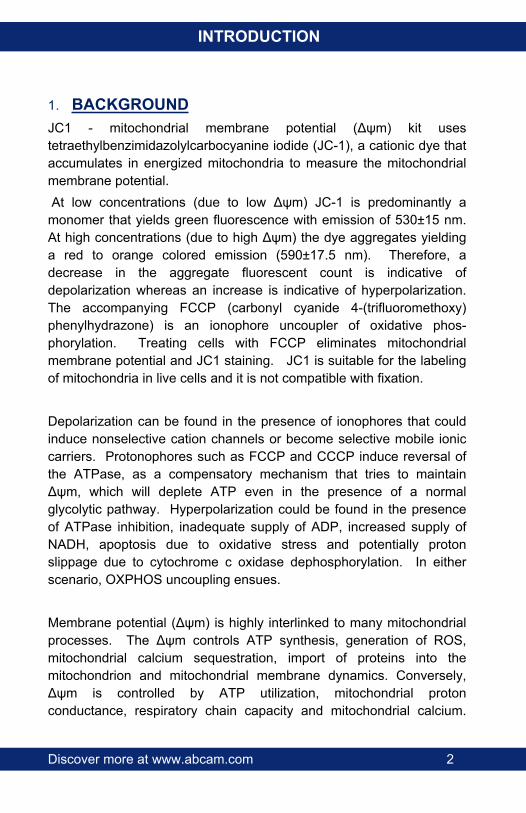
1. BACKGROUNDJC1 - mitochondrial membrane potential (Δψm) kit uses tetraethylbenzimidazolylcarbocyanine iodide (JC-1), a cationic dye that accumulates in energized mitochondria to measure the mitochondrial membrane potential. At low concentrations (due to low Δψm) JC-1 is predominantly a monomer that yields green fluorescence with emission of 530±15 nm. At high concentrations (due to high Δψm) the dye aggregates yielding a red to orange colored emission (590±17.5 nm). Therefore, a decrease in the aggregate fluorescent count is indicative of depolarization whereas an increase is indicative of hyperpolarization. The accompanying FCCP (carbonyl cyanide 4-(trifluoromethoxy) phenylhydrazone) is an ionophore uncoupler of oxidative phos-phorylation. Treating cells with FCCP eliminates mitochondrial membrane potential and JC1 staining. JC1 is suitable for the labeling of mitochondria in live cells and it is not compatible with fixation.
Depolarization can be found in the presence of ionophores that could induce nonselective cation channels or become selective mobile ionic carriers. Protonophores such as FCCP and CCCP induce reversal of the ATPase, as a compensatory mechanism that tries to maintain Δψm, which will deplete ATP even in the presence of a normal glycolytic pathway. Hyperpolarization could be found in the presence of ATPase inhibition, inadequate supply of ADP, increased supply of NADH, apoptosis due to oxidative stress and potentially proton slippage due to cytochrome c oxidase dephosphorylation. In either scenario, OXPHOS uncoupling ensues.
Membrane potential (Δψm) is highly interlinked to many mitochondrial processes. The Δψm controls ATP synthesis, generation of ROS, mitochondrial calcium sequestration, import of proteins into the mitochondrion and mitochondrial membrane dynamics. Conversely, Δψm is controlled by ATP utilization, mitochondrial proton conductance, respiratory chain capacity and mitochondrial calcium.

Discover more at www.abcam.com 3
INTRODUCTION
Hence pharmacological changes in Δψm can be associated with a multitude of other mitochondrial pathological parameters which may require further independent evaluation.
Discover more at www.abcam.com 4
INTRODUCTION
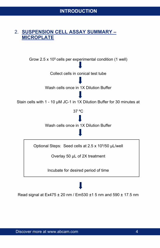
2. SUSPENSION CELL ASSAY SUMMARY – MICROPLATE
Grow 2.5 x 10⁵ cells per experimental condition (1 well)
Collect cells in conical test tube
Wash cells once in 1X Dilution Buffer
Stain cells with 1 - 10 µM JC-1 in 1X Dilution Buffer for 30 minutes at
37 ºC
Wash cells once in 1X Dilution Buffer
Optional Steps: Seed cells at 2.5 x 10⁵/50 µL/well
Overlay 50 µL of 2X treatment
Incubate for desired period of time
Read signal at Ex475 ± 20 nm / Em530 ±1 5 nm and 590 ± 17.5 nm

Discover more at www.abcam.com 5
INTRODUCTION
3. ADHERENT CELL ASSAY SUMMARY – MICROPLATE
Harvest 3-4x10⁶ cells
Seed cells at 1.5x10⁴ cells/well on a 96 well plate
Allow cells to attach overnight
Wash cells once in 1X Dilution Buffer
Stain cells with 20 µM JC-1 in 1X Dilution Buffer for 10 minutes at 37ºC
Wash cells twice in 1X Dilution Buffer
Optional Steps: Add 100 µL/well of treatment
Incubate for desired period of time
Read signal at Ex475±20 nm / Em530 ± 15 nm and 590 ± 17.5 nm
(Buffer or compound must be present in the wells during the reading of the signal. Do not allow wells to dry out)
Discover more at www.abcam.com 6
GENERAL INFORMATION
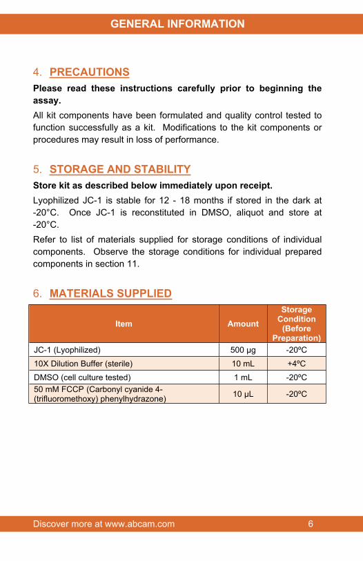
4. PRECAUTIONSPlease read these instructions carefully prior to beginning the assay.All kit components have been formulated and quality control tested to function successfully as a kit. Modifications to the kit components or procedures may result in loss of performance.
5. STORAGE AND STABILITYStore kit as described below immediately upon receipt. Lyophilized JC-1 is stable for 12 - 18 months if stored in the dark at -20°C. Once JC-1 is reconstituted in DMSO, aliquot and store at -20°C. Refer to list of materials supplied for storage conditions of individual components. Observe the storage conditions for individual prepared components in section 11.
6. MATERIALS SUPPLIED
Item AmountStorage
Condition(Before
Preparation)JC-1 (Lyophilized) 500 µg -20ºC
10X Dilution Buffer (sterile) 10 mL +4ºC
DMSO (cell culture tested) 1 mL -20ºC50 mM FCCP (Carbonyl cyanide 4-(trifluoromethoxy) phenylhydrazone) 10 µL -20ºC
Discover more at www.abcam.com 7
GENERAL INFORMATION
7. MATERIALS REQUIRED, NOT SUPPLIEDThese materials are not included in the kit, but will be required to successfully utilize this assay:
Fluorescence plate reader or Flow cytometer. JC-1 may also be detected with similar settings to those used to detect rhodamine (excitation/emission wavelengths: 540/570 nm) or texas red (excitation/emission wavelengths: 590/610 nm)
General tissue culture supplies
PBS (1.4 mM KH2PO4, 8 mM Na2HPO4, 140 mM NaCl, 2.7 mM KCl, pH 7.4)
Fetal Bovine Serum (FBS)
Sterile, tissue culture treated, clear bottom, dark sided 96-well microplates.
Multichannel pipette (50 – 300 μL)
Optional:
Test compounds/diluents of interest
Uncouplers include CCCP (carbonyl cyanide 3-chlorophenylhydrazone), 2’, 4’ Dinitrophenol
96-Well deep sided, clear bottom, dark sided microplates with lids

Discover more at www.abcam.com 8
GENERAL INFORMATION
8. LIMITATIONS Assay kit intended for research use only. Not for use in diagnostic
procedures
Do not mix or substitute reagents or materials from other kit lots or vendors. Kits are QC tested as a set of components and performance cannot be guaranteed if utilized separately or substituted
Any variation in operator, pipetting technique, washing technique, incubation time or temperature, and kit age can cause variation in the results
9. TECHNICAL HINTS Clear bottom, dark sided microplates are recommended with this
assay. Clear sided microplates have not been tested with this kit
It is essential to warm the 1X dilution buffer to 37°C prior to making the working JC-1 solution. If the JC-1 dye is added to a cold buffer, precipitates will form.
Washing of suspension or adherent cells may also be done with standard sterile PBS.
While working with JC-1 inside the biological safety cabinet, turn the fluorescent lights off to prevent quenching of the dye during handling.
This kit is sold based on number of tests. A ‘test’ simply refers to a single assay well. The number of wells that contain sample, control or standard will vary by product. Review the protocol completely to confirm this kit meets your requirements. Please contact our Technical Support staff with any questions
Discover more at www.abcam.com 9
ASSAY PREPARATION
10.REAGENT PREPARATIONEquilibrate all reagents to room temperature (18-25°C) prior to use.The sample volumes below are sufficient for 96 100 µL tests; adjust volumes as needed for the number of wells used in your experiment.
11.1 1X Dilution BufferPrepare 1X Dilution Buffer by adding 10 mL 10X Dilution Buffer to 90 mL deionized water. Mix gently and thoroughly.
11.2 1X Supplemented Dilution BufferPrepare 1X Supplemented Dilution Buffer by adding 2 mL FBS to 18 mL 1X Dilution Buffer.
11.3 1 mM JC-1 Stock SolutionAllow the lyophilized vial JC-1 and the DMSO to warm to room temperature. Reconstitute the JC-1 by adding 766.8 µL DMSO to the 500 µg vial of JC-1. Any unused 1 mM JC-1 solution should be aliquoted and stored at -20 ºC.
11.4 Working JC-1 Solution To prepare the Working JC-1 Solution, add the appropriate volume of 1 mM JC-1 Solution to previously warmed 1X Dilution Buffer. As an example, to generate a 1 µM JC-1 Solution, mix 10 µL 1 mM JC-1 with 10 mL of 1X Dilution Buffer. Mix thoroughly and gently. If necessary, centrifuge the 1 µM solution at 13,000 x g for 3 minutes to sediment non-soluble particles.
Discover more at www.abcam.com 10
ASSAY PREPARATION
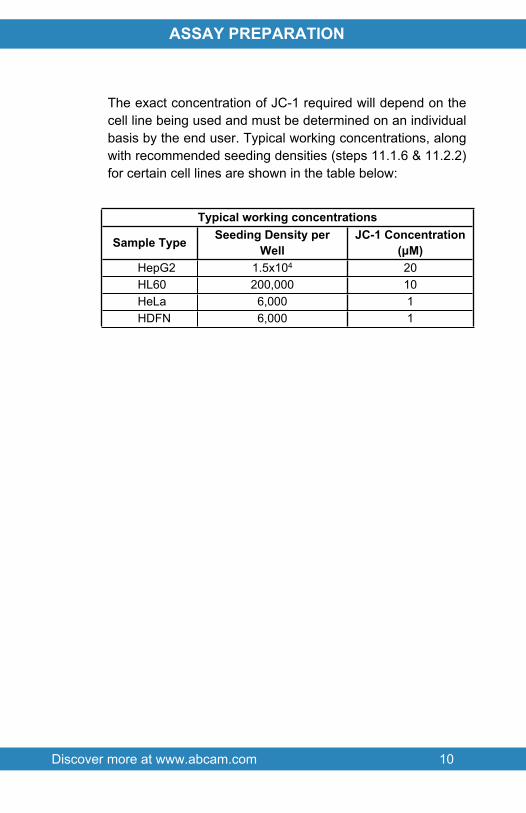
The exact concentration of JC-1 required will depend on the cell line being used and must be determined on an individual basis by the end user. Typical working concentrations, along with recommended seeding densities (steps 11.1.6 & 11.2.2) for certain cell lines are shown in the table below:
Typical working concentrations
Sample Type Seeding Density per Well
JC-1 Concentration (μM)
HepG2 1.5x10⁴ 20HL60 200,000 10HeLa 6,000 1HDFN 6,000 1
Discover more at www.abcam.com 11
ASSAY PROCEDURE
11.ASSAY PROCEDURE Equilibrate all materials and prepared reagents to 37°C prior
to use. It is recommended to assay all standards, controls and
samples in duplicate.
11.1. Fluorescent Microplate Measurement (Suspension Cells, e.g. HL60 cells)11.1.1. Grow HL60 cells in glucose based media so that
approximately 2.5x10⁷ cells are available on the day of the experiment per plate
11.1.2. If performing toxicity assays, dilute compounds of interest in 1X supplemented dilution buffer to 2X of final desired concentration for the experiment. A 96-well deep well microplate may be use in this step. Compounds may also be diluted in complete media with 10% FBS without phenol red. Include a depolarization control (100 μM FCCP) and a normal control (vehicle or diluent of choice).
11.1.3. Collect cells and wash by centrifugation once in 1X Dilution Buffer or 1X PBS.
11.1.4. Resuspend cells in 10 mL of the Working JC-1 Solution and incubate at 37°C for 30 minutes in the dark.
11.1.5. Wash cells by centrifugation with 10 mL of 1X Dilution Buffer.
11.1.6. Resuspend 2x10⁷ cells in 5 mL of 1X Supplemented Dilution Buffer.
11.1.7. Seed a 96-well dark plate as follows: 200,000 stained cells/50 μL/well. Include blank wells (with non-stained cells).

Discover more at www.abcam.com 12
ASSAY PROCEDURE
11.1.8. If performing toxicity assays, add to each well 50 μL of previously diluted 2X compounds and treat for desired period of time
11.1.9. Read plate end point in the presence of compounds, media or buffer on a fluorescent plate reader. Set excitation wavelength at 535 ± 17.5 (aggregate excitation only) or 475 ± 20 nm (for simultaneous aggregate and monomer excitation). Set emission wavelength at 590 ± 17.5 nm (aggregate emission only). If reading of the monomer species is also desired, set a second emission reading at 530 ± 15 nm.FCCP 100 µM treatment for 4 hours should decrease the JC-1 aggregate signal to at least 25-30% from control levels.
11.2. Fluorescent Microplate Measurement (Adherent Cells, e.g. HepG2 cells)11.2.1. Grow HepG2 cells in standard media so that 3x 10⁶
to 4x 10⁶ cells are obtained the day before the experiment per plate.
11.2.2. Harvest cells the day before the experiment and seed a dark 96-well microplate with 1.5x10⁴ cells per well in standard growing culture media. Allowed to attach overnight.
11.2.3. If performing toxicity assays, dilute compounds of interest to the final desired concentration in 1X Supplemented Dilution Buffer solution. A 96-well deep well microplate may be use in this step. Compounds may also be diluted in complete media with 10% FBS without phenol red. Include positive (100 μM FCCP) and negative controls (vehicle of choice)

Discover more at www.abcam.com 13
ASSAY PROCEDURE
11.2.4. Wash the HepG2 cells seeded on the 96-well plate with 100 μL/well of 1X Dilution Buffer or 1X PBS once.
11.2.5. Add 100 μL/well of the Working JC-1 Solution and incubate for 10 minutes at 37°C in the dark. Include blank wells (with non-stained cells).
11.2.6. Wash the plate twice with 1X Dilution Buffer solution.11.2.7. If performing cytotoxicity assays, add compounds of
interest and treat for desired period of time.11.2.8. Read plate end point in the presence of compounds,
media or buffer on a fluorescent plate reader. Set excitation wavelength at 535 ± 17.5 nm (aggregate excitation only) or 475 ± 20 nm (for simultaneous aggregate and monomer excitation). Set emission wavelength at 590 ± 17.5 nm (aggregate emission only). If reading of the monomer species is also desired, set a second emission reading at 530 ± 15 nm.
11.2.9. FCCP 100 µM treatment for 4 hours should decrease the JC-1 aggregate signal to at least 25-30% from control levels.Note: Buffer or compound must be present in the wells during the reading of the signal. Do not allow wells to dry out
Discover more at www.abcam.com 14
DATA ANALYSIS
12.CALCULATIONSSubtract background (A590 of non-stained cells) from test signal and express signal as percentage from control (untreated healthy cells). If both monomer and aggregate forms are measured, a ratio between the two measurements may be obtained and plotted. Data obtained with the JC-1 assay gives a relative measure of mitochondrial membrane potential as a percentage of control and cannot be used for absolute measurements of membrane potential in millivolts. Decrease in JC-1 signal may indicate either mitochondrial depolarization or cell death and must be interpreted in parallel with a cytotoxicity assay (such as the ATP detection kit ab113849). The data in Figure 1 below shows the uncoupling effect of FCCP acute treatment on HL60 cells as measured with the JC-1 stain and read on a fluorescent plate reader.
Discover more at www.abcam.com 15
DATA ANALYSIS
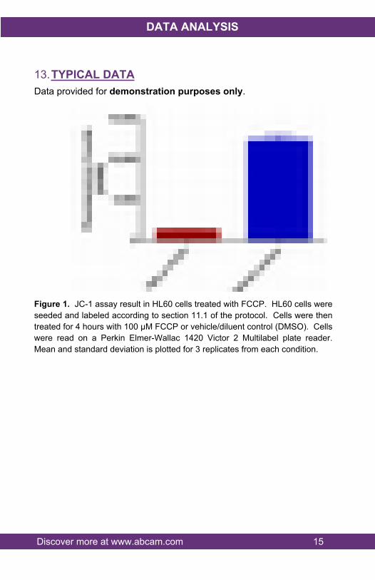
13.TYPICAL DATAData provided for demonstration purposes only.
Figure 1. JC-1 assay result in HL60 cells treated with FCCP. HL60 cells were seeded and labeled according to section 11.1 of the protocol. Cells were then treated for 4 hours with 100 µM FCCP or vehicle/diluent control (DMSO). Cells were read on a Perkin Elmer-Wallac 1420 Victor 2 Multilabel plate reader. Mean and standard deviation is plotted for 3 replicates from each condition.

Discover more at www.abcam.com 16
DATA ANALYSIS
Figure 2. JC-1 assay result in HepG2 cells treated with CCCP. HepG2 cells were seeded and labeled according to section 11.2 of the protocol. Cells were then treated for 4 hours with a titration series of CCCP (carbonyl cyanide 3-chlorophenylhydrazone) and both monomer and aggregate forms were read on a Perkin Elmer-Wallac 1420 Victor 2 Multilabel plate reader. Mean and standard deviation of aggregate/monomer ratios is plotted for 12 replicates for each concentration. IC50 of CCCP in HepG2 cells was calculated at 8.7 µM
Discover more at www.abcam.com 17
DATA ANALYSIS
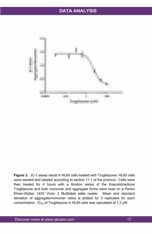
Figure 3. JC-1 assay result in HL60 cells treated with Troglitazone. HL60 cells were seeded and labeled according to section 11.1 of the protocol. Cells were then treated for 4 hours with a titration series of the thiazolidinedione Troglitazone and both monomer and aggregate forms were read on a Perkin Elmer-Wallac 1420 Victor 2 Multilabel plate reader. Mean and standard deviation of aggregate/monomer ratios is plotted for 3 replicates for each concentration. IC50 of Troglitazone in HL60 cells was calculated at 1.2 µM
Discover more at www.abcam.com 18
DATA ANALYSIS
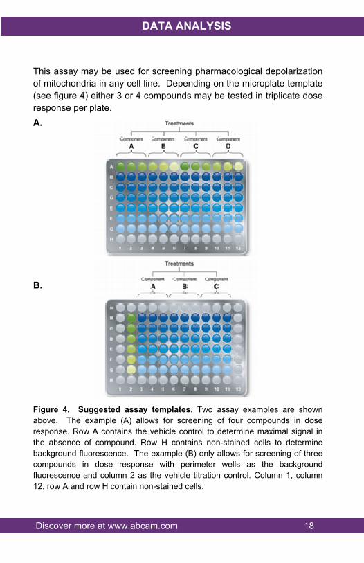
This assay may be used for screening pharmacological depolarization of mitochondria in any cell line. Depending on the microplate template (see figure 4) either 3 or 4 compounds may be tested in triplicate dose response per plate.A.
B.
Figure 4. Suggested assay templates. Two assay examples are shown above. The example (A) allows for screening of four compounds in dose response. Row A contains the vehicle control to determine maximal signal in the absence of compound. Row H contains non-stained cells to determine background fluorescence. The example (B) only allows for screening of three compounds in dose response with perimeter wells as the background fluorescence and column 2 as the vehicle titration control. Column 1, column 12, row A and row H contain non-stained cells.

Discover more at www.abcam.com 19
RESOURCES
14.FREQUENTLY ASKED QUESTIONSQ. Is this kit designed to do a cell treatment before or after JC-1
incubation?A. It is essential to read the JC-1 signal in the presence of compound
(or treatment) because changes in membrane potential can be short lived and reversible as soon as the treatment is removed from the cells
Q. I am already growing some cells on coverslips, will I be able to use these cells with the assay?
A. Any mammalian live cell may be stained with JC-1 provided that the staining concentration has been optimized. When cells are seeded on coverslips, two major changes must be done to the protocol in order to obtained the correct results: 1. Volumes will have to be adjusted from 100µL per well to 500 -
100 µL per well depending on whether the cells are seeded on a 6-well plate or on a 24-well plate. Due to the change in volume, only 10 to 20 tests can be run using this kit under these conditions.
2. Coverslips must be ideally imaged on an inverted fluorescence microscope or a microscope with a lens that can be immersed in water, using a Texas Red filter. In the absence of these instruments, the coverslip could be mounted with the buffer provided on top of an imaging chamber gasket. In this later case, imaging must be prompt to prevent bleaching of the dye.
Q. I am using primary cells and I’m afraid that 10 minutes without media is too much. Can I mix the JC-1 with the growing meda rather than the supplied buffer?
A. Yes, the JC-1 may be mixed with the growing media. However caution must be taken as the phenol red present in most media formulations may cause increased background. Furthermore, we have also observed higher background on RPMI media in comparison to DMEM media.
Discover more at www.abcam.com 20
RESOURCES
Q. Is fixation of the cells with paraformaldehyde in the wells possible, after live imaging, for storage purposes?
A. Fixation of the cells will interfere with the dye signal. Once the signal has been obtained by live imaging or fluorescent read out, cells cannot be stored for future use.
Q. I am planning a drug treatment for 18 hours; can I run FCCP in parallel for 18 hours? Will the signal dissipate after 4 hours?
A. We do not recommend treating cells with FCCP for more than 4 hours as this will generate excessive toxicity and results will represent depolarization but rather general cell toxicity. Furthermore incubations for longer than 4 hours after JC-1 staining may lead to dissipation of the signal. In this case, we suggest to reverse the protocol and treat first prior to staining. The following protocol should be follow in this scenario:3. Dilute compounds of interest in complete media without
phenol red. Make four times the volume required.4. Treat suspension or adherent cells for the desired period of
time. If treating cells for microplate measurements, treat with 100 µL per well.
5. Include blank wells with no cells but with compound at the same concentration used for treatment.
6. Include at least 2 depolarized control wells, to be reserved for FCCP treatment, containing cells but none of the test compounds.
7. 4 hours prior to completion of treatment, dilute FCCP to 10X of final concentration (1 mM) and spiked 10X FCCP into the reserved depolarized control wells by adding 11 µL per well.
8. 1 hour prior to completion of the treatment, dilute JC-1 at 2X of the final concentration desired in the same media used for treatment (containing experimental compounds) and warm at 37°C.
Discover more at www.abcam.com 21
RESOURCES
9. 10 – 30 minutes prior to completion of the treatment, overlay 2X JC-1 dilution on top of the treated cells. If treating cells for microplate measurements, overlay 100 µL of 2X JC-1 dilution per well.
10. Incubate JC-1 and compounds for the desired period of time (10 – 30 minutes).
11. Once incubation is completed, wash the wells twice with 100 µL per well of 1X Dilution buffer containing compounds. Leave last wash in the wells.
12. Transfer the plate to the microplate reader and read according to the protocol.

Discover more at www.abcam.com 22
RESOURCES
15.NOTES

RESOURCES 23
UK, EU and ROWEmail: [email protected] | Tel: +44-(0)1223-696000
AustriaEmail: [email protected] | Tel: 019-288-259
FranceEmail: [email protected] | Tel: 01-46-94-62-96 GermanyEmail: [email protected] | Tel: 030-896-779-154 SpainEmail: [email protected] | Tel: 911-146-554 SwitzerlandEmail: [email protected] Tel (Deutsch): 0435-016-424 | Tel (Français): 0615-000-530
US and Latin AmericaEmail: [email protected] | Tel: 888-77-ABCAM (22226)
CanadaEmail: [email protected] | Tel: 877-749-8807
China and Asia Pacific Email: [email protected] | Tel: 108008523689 (中國聯通) JapanEmail: [email protected] | Tel: +81-(0)3-6231-0940
www.abcam.com | www.abcam.cn | www.abcam.co.jp
Copyright © 2017 Abcam, All Rights Reserved. The Abcam logo is a registered trademark.
All information / detail is correct at time of going to print.