Validated High-Performance Thin-Layer …...2019/10/20 · Jyotsana Dwivedi, Abhishek Gupta,...
5
377 Summary A high-performance thin-layer chromatography (HPTLC) method for the simultaneous quantitative determination of ursolic acid and β-sitosterol in the methanolic fraction of Paederia foetida L. leaves was developed for the first time. For achieving good separation, a mobile phase of toluene‒ethyl acetate‒formic acid (8:2:0.1, v/v) was used. The densitometric determination was carried out at 550 and 522 nm in reflection/absorption mode for ursolic acid and β-sito- sterol. The calibration curves were linear in the range of 100-600 ng per spot for ursolic acid and β-sitosterol. During the analysis, the methanolic fraction of P. foetida L. leaves showed the pres- ence of ursolic acid (0.12 ± 0.05%) and β-sitosterol (0.08 ± 0.12%). The proposed method is simple, precise, specific, accurate, less time-consuming, and cost-effective. The statistical analysis of the data obtained proves that the method is reproducible and selective and can be used for the routine analysis of the reported phenolic compounds in crude drug and extracts. The simultaneous quanti- fication of these compounds has not yet been reported in P. foetida L. leaves which may be utilized for the proper standardization of the plant. 1 Introduction The genus Paederia contains 20–30 species worldwide, gen- erally distributed in Asia, and is commonly known as skunk vine, which is a shrub or a perennial climbing herb in India. It is found in the Himalayas from Dehradun eastwards up to an altitude of 1800 meters as well as in Assam, Bihar, Bengal, J. Dwivedi, M. Dwivedi, and A.K.S. Rawat, Pharmacognosy & Ethnopharma- cology Division, CSIR‒National Botanical Research Institute, Lucknow, India; A. Gupta, Pranveer Singh Institute of Technology, Department of Pharmacy, Kanpur, India; S. Verma, Amity Institute of Pharmacy, Amity University, Luck now, India; and S. Paliwal, Department of Pharmacy, Banasthali Vidyapeeth, Banasthali, India. *Email: pharmacognosy1@rediffmail.com Orissa and Andhra Pradesh. Paederia foetida L., a member of the Rubiaceae family, is an extensive climber [1, 2]. It is known as Chinese flower in English, Gandha prasarini in Hindi, and Prasarani in Sanskrit [3]. P. foetida is native to both temperate and tropical Asia, from India to Japan and South East Asia. It has a bitter taste and foul smell [4]. Phytochemical investigations reported that P. foetida contains paederolone, paederone, βsitosterol, paederoside, asperulo- side, and their related glucosides [5–7]. The leaves of the plant are also rich in carotene, vitamin C, keto-alcohol, and alka- loid [8]. Asperuloside, βsitosterol, and lupeol were reported in leaves [9]. P. foetida also contains friedelin, campesterol, ursolic acid, hentriacontane, hentriacontanol, ceryl alcohol, palmitic acid, and methyl mercaptan [3] as well as ellagic acids, volatile oil [10], epifriedelinol, terpenoids, and alkaloids [11]. Four iridoid glucosides and seven sulfur-containing iridoid glucosides were isolated from the methanol extract of the roots of the Vietnamese P. scandens (Lour.) Merrill, together with the five known glucosides, namely, paederoside, asperuloside, paederosidic acid, asperulosidic acid, and geniposide [12]. Ethyl p-methoxy-trans-cinnamate was also isolated from the methanol extract of P. foetida [13]. It is a well-known medicinal plant for its anthelmintic [14], antihyperglycemic, antioxidant [15], antiinflammatory [16, 17], antiulcer [18], antinociceptive [19], and antitussive activities [20]. High-performance thin- layer chromatography (HPTLC) of βsitosterol [21], quercetin [22], ascorbic acid [15], gallic acid, and rutin were performed earlier [23]. HPTLC analysis of lupeol and ursolic acid was ear - lier performed on different species of Bauhinia [24]. The simul- taneous quantification with method validation of ursolic acid and βsitosterol has not yet been reported in P. foetida, which may be utilized for the proper standardization of this species. βSitosterol is usually used for the treatment of heart disease and hypercholesterolemia, modulation of the immune sys- tem, and prevention of cancer as well as for curing rheumatoid arthritis, tuberculosis, cervical cancer, hair loss, and benign prostatic hyperplasia [25]. Ursolic acid has been confirmed to have several biological and pharmacological effects, such as antiinflammatory, antitumor, antiplatelet aggregation, antiHIV, and anti-Mycobacterium tuberculosis effects [26]. Validated High-Performance Thin-Layer Chromatographic Analysis of Ursolic Acid and β-Sitosterol in the Methanolic Fraction of Paederia foetida L. Leaves Jyotsana Dwivedi, Abhishek Gupta, Shikhar Verma, Monika Dwivedi, Sarvesh Paliwal, and Ajay Kumar Singh Rawat* Key Words: Paederia foetida L. High-performance thin-layer chromatography β-Sitosterol Ursolic acid Journal of Planar Chromatography 31 (2018) 5, 377–381 DOI: 10.1556/1006.2018.31.5.5 0933-4173 © Akadémiai Kiadó, Budapest
Transcript of Validated High-Performance Thin-Layer …...2019/10/20 · Jyotsana Dwivedi, Abhishek Gupta,...
Journal of Planar Chromatography 29 (2016) 3 377
Summary
A high-performance thin-layer chromatography (HPTLC) method for the simultaneous quantitative determination of ursolic acid and β-sitosterol in the methanolic fraction of Paederia foetida L. leaves was developed for the first time. For achieving good separation, a mobile phase of toluene‒ethyl acetate‒formic acid (8:2:0.1, v/v) was used. The densitometric determination was carried out at 550 and 522 nm in reflection/absorption mode for ursolic acid and β-sito-sterol. The calibration curves were linear in the range of 100-600 ng per spot for ursolic acid and β-sitosterol. During the analysis, the methanolic fraction of P. foetida L. leaves showed the pres-ence of ursolic acid (0.12 ± 0.05%) and β-sitosterol (0.08 ± 0.12%). The proposed method is simple, precise, specific, accurate, less time-consuming, and cost-effective. The statistical analysis of the data obtained proves that the method is reproducible and selective and can be used for the routine analysis of the reported phenolic compounds in crude drug and extracts. The simultaneous quanti-fication of these compounds has not yet been reported in P. foetida L. leaves which may be utilized for the proper standardization of the plant.
1 Introduction
The genus Paederia contains 20–30 species worldwide, gen-erally distributed in Asia, and is commonly known as skunk vine, which is a shrub or a perennial climbing herb in India. It is found in the Himalayas from Dehradun eastwards up to an altitude of 1800 meters as well as in Assam, Bihar, Bengal,
J. Dwivedi, M. Dwivedi, and A.K.S. Rawat, Pharmacognosy & Ethnopharma-cology Division, CSIR‒National Botanical Research Institute, Lucknow, India; A. Gupta, Pranveer Singh Institute of Technology, Department of Pharmacy, Kanpur, India; S. Verma, Amity Institute of Pharmacy, Amity University, Lucknow, India; and S. Paliwal, Department of Pharmacy, Banasthali Vidyapeeth, Banasthali, India.*Email: [email protected]
Orissa and Andhra Pradesh. Paederia foetida L., a member of the Rubiaceae family, is an extensive climber [1, 2]. It is known as Chinese flower in English, Gandha prasarini in Hindi, and Prasarani in Sanskrit [3]. P. foetida is native to both temperate and tropical Asia, from India to Japan and South East Asia. It has a bitter taste and foul smell [4].
Phytochemical investigations reported that P. foetida contains paederolone, paederone, βsitosterol, paederoside, asperulo-side, and their related glucosides [5–7]. The leaves of the plant are also rich in carotene, vitamin C, keto-alcohol, and alka-loid [8]. Asperuloside, βsitosterol, and lupeol were reported in leaves [9]. P. foetida also contains friedelin, campesterol, ursolic acid, hentriacontane, hentriacontanol, ceryl alcohol, palmitic acid, and methyl mercaptan [3] as well as ellagic acids, volatile oil [10], epifriedelinol, terpenoids, and alkaloids [11]. Four iridoid glucosides and seven sulfur-containing iridoid glucosides were isolated from the methanol extract of the roots of the Vietnamese P. scandens (Lour.) Merrill, together with the five known glucosides, namely, paederoside, asperuloside, paederosidic acid, asperulosidic acid, and geniposide [12]. Ethyl p-methoxy-trans-cinnamate was also isolated from the methanol extract of P. foetida [13]. It is a well-known medicinal plant for its anthelmintic [14], antihyperglycemic, antioxidant [15], antiinflammatory [16, 17], antiulcer [18], antinociceptive [19], and antitussive activities [20]. High-performance thin-layer chromatography (HPTLC) of βsitosterol [21], quercetin [22], ascorbic acid [15], gallic acid, and rutin were performed earlier [23]. HPTLC analysis of lupeol and ursolic acid was ear-lier performed on different species of Bauhinia [24]. The simul-taneous quantification with method validation of ursolic acid and βsitosterol has not yet been reported in P. foetida, which may be utilized for the proper standardization of this species.
βSitosterol is usually used for the treatment of heart disease and hypercholesterolemia, modulation of the immune sys-tem, and prevention of cancer as well as for curing rheumatoid arthritis, tuberculosis, cervical cancer, hair loss, and benign prostatic hyperplasia [25]. Ursolic acid has been confirmed to have several biological and pharmacological effects, such as antiinflammatory, antitumor, antiplatelet aggregation, antiHIV, and anti-Mycobacterium tuberculosis effects [26].
Validated High-Performance Thin-Layer Chromatographic Analysis of Ursolic Acid and β-Sitosterol in the Methanolic Fraction of Paederia foetida L. Leaves
Jyotsana Dwivedi, Abhishek Gupta, Shikhar Verma, Monika Dwivedi, Sarvesh Paliwal, and Ajay Kumar Singh Rawat*
Key Words:Paederia foetida L.High-performance thin-layer chromatographyβ-SitosterolUrsolic acid
Journal of Planar Chromatography 31 (2018) 5, 377–381 DOI: 10.1556/1006.2018.31.5.50933-4173 © Akadémiai Kiadó, Budapest
HPTLC Analysis of Paederia foetida Leaves
378 Journal of Planar Chromatography 31 (2018) 5
2 Experimental
2.1 Chemicals and Reagents
HPTLC analyses were performed on 20 cm × 10 cm HPTLC silica gel 60 F254 (0.25 mm) plates (Merck, Darmstadt, Ger-many). Ursolic acid and βsitosterol were supplied by Sig-maAldrich (Taufkirchen, Germany). All the reagents used in the experiment were of analytical grade and were supplied by Merck.
2.2 Preparation of Standard Solutions
Stock solutions ursolic acid and βsitosterol were prepared sep-arately by dissolving those in 0.1 mg mL−1 methanol.
2.3 Plant Material
The plant material, i.e., leaves of P. foetida, was collected from the Council for Scientific and Industrial Research‒National Botanical Research Institute (CSIR‒NBRI), Lucknow, India. The plant was identified and authenticated by Dr. A.K.S. Rawat, and the voucher specimens were submitted in the LWG herbarium (Figure 1).
Figure 1
Aerial parts of Paederia foetida.
2.4 Sample Preparation
The fresh leaves of P. foetida were collected and thoroughly washed with water to remove all debris. The plant materials were shade-dried and powdered by using an electric grinder at
60 mesh size. Extraction was performed by soxhlation method. Firstly, the powdered plant material was defatted under Sox-hlet assembly using 250 mL of 98% petroleum ether for 6 h. This was followed by a 9-h soxhlation of the defatted powder by using 250 mL chloroform, followed by methanol. The final methanolic fraction obtained was passed through Whatman No. 1 filter paper. The filtrate obtained was concentrated under vacuum in a rotary evaporator at 40°C and stored at 4°C for further use. The dried extracts were dissolved in 98% methanol to obtain a stock solution of 10 mg mL−1, which was used for the application of spots on HPTLC plates.
2.5 Development of HPTLC Fingerprinting of Ursolic Acid and β-Sitosterol
2.5.1 Instrumentation and Chromatographic Conditions
The following were the instruments and chromatographic conditions used. Spotting device: Linomat V automatic sam-ple applicator (CAMAG, Muttenz, Switzerland). Syringe: 100 μL Hamilton (Bonaduz, Switzerland). HPTLC chamber: glass twintrough chamber (20 cm × 10 cm × 4 cm) (CAMAG). Densitometer: HPTLC Scanner 3 linked to winCATS software V.4.06 (CAMAG). HPTLC plates: 20 × 10 cm, 0.2 mm thick-ness, precoated with silica gel 60 F254 (E. Merck, Darmstadt, Germany). Experimental conditions: temperature, 25 ± 2°C; relative humidity, 40%. Solvent system: toluene–ethyl ace-tate–formic acid (95%) in the ratio of 8:2:0.1 v/v. Detection wavelength: 550 nm for ursolic acid and 522 nm for βsitos-terol. Visualization agent: anisaldehyde–sulfuric acid rea-gent. Heating temperature: 110°C for 1 min. Slit dimension: 5.00 mm × 0.45 mm. Scanning speed: 10 mm s−1. Source of radiation: deuterium lamp.
2.5.2 Calibration Curve of Ursolic Acid and β-Sitosterol
Stock solutions of ursolic acid and βsitosterol (100 μg mL−1) were prepared in HPLCgrade methanol. Different volumes of stock solution were spotted on the HPTLC plate to obtain con-centrations of 100–600 ng per band of ursolic acid and βsi-tosterol, respectively. The data of peak areas plotted against the corresponding concentrations were treated by least squares regression analysis method validation.
2.6 Method Validation
The method was validated according to the International Con-ference on Harmonization (ICH) guidelines [27, 28], and the statistical analysis was done using Excel 2000 (MS Office®).
2.6.1 Precision
Repeatability of the sample application and measurement of the peak area were carried out using nine determinants (3 concen-trations per 3 replicates) covering the specified range for the procedure (200, 400, and 600 ng per band of ursolic acid and βsitosterol) and it was expressed in terms of relative stand-ard deviation (RSD). The intra and interday variation for the determination of ursolic acid and βsitosterol were carried out in three different concentration levels of 200, 400, and 600 ng per band. The acceptance criteria for a procedure’s repeatabil-ity or intermediate precision are based on the intended use of the analytical method.
HPTLC Analysis of Paederia foetida Leaves
Journal of Planar Chromatography 31 (2018) 5 379
2.6.2 Robustness of the Method
By introducing small changes in the mobile phase composition, mobile phase volume, duration of mobile phase saturation, and activation of prewashed HPTLC plates with methanol, the effects on the results were examined. The robustness of the method was determined in triplicate at a concentration level of 200 ng per band for ursolic acid and βsitosterol, and the RSD and SD of the peak areas were calculated.
2.6.3 Limit of Detection and Limit of Quantification
In order to estimate the limit of detection (LOD) and limit of quantification (LOQ), blank methanol was spotted six times, and the signaltonoise ratio was determined. LOD was con-sidered to be 3:1, and LOQ was considered to be 10:1. LOD and LOQ were experimentally verified by diluting the known concentrations of ursolic acid and βsitosterol until the average responses were approximately 3 or 10 times the responses for six replicate determinations.
2.6.4 Recovery
The pre-analyzed samples were spiked with extra 50, 100 and 150% of the standard ursolic acid and βsitosterol and the mix-tures were reanalyzed by the proposed method. The experiment was conducted six times. This was done to check the recovery of the targeted analytes at different levels in the formulations.
2.6.5 Ruggedness
Ursolic acid and βsitosterol solutions of concentration 200 ng per band were prepared and analyzed on day 0 and after 6, 12, 24, 48, and 72 h. Data were treated for %RSD to assess the rug-gedness of the method.
2.6.6 Specificity
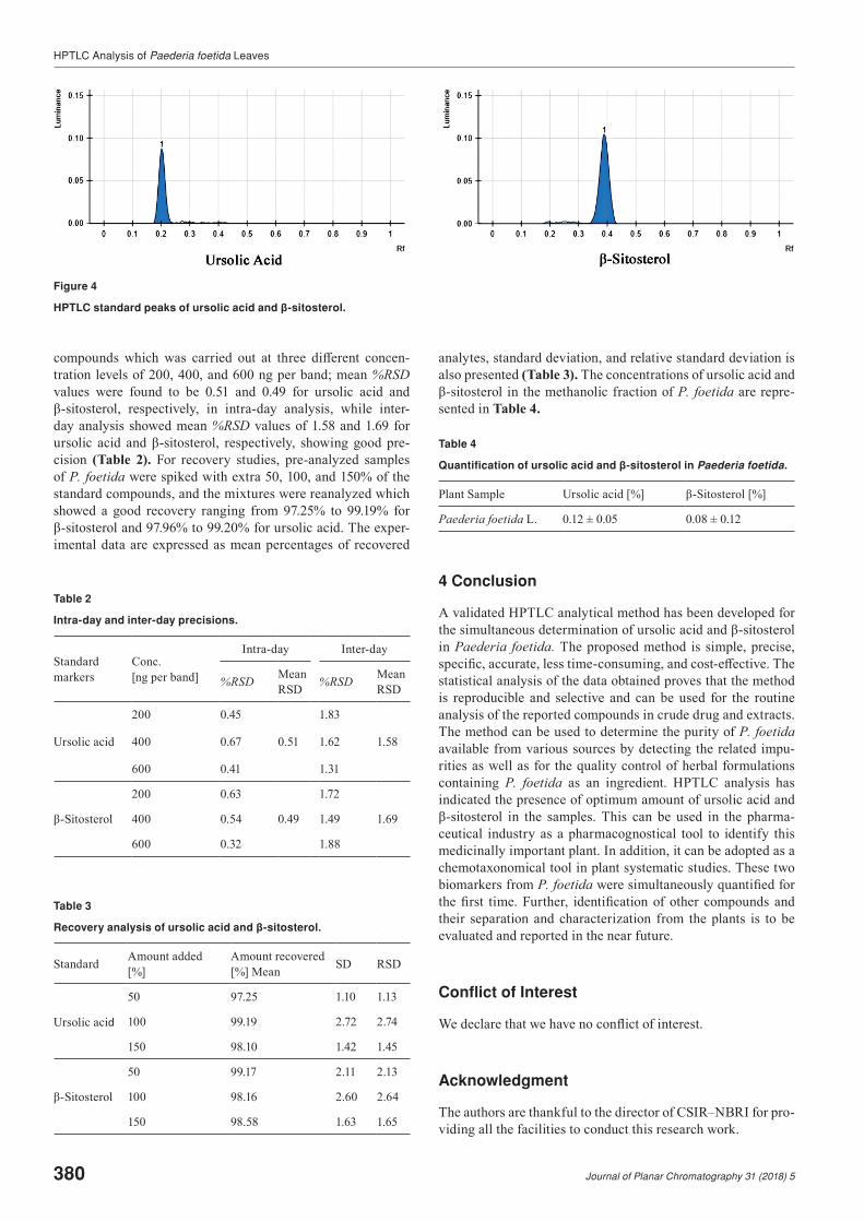
The specificity of the method was confirmed by analyzing the standard drugs and the extract. The band for ursolic acid and βsitosterol in the sample was confirmed by comparing the retention factor (RF) values and spectra of the band with those of the standard. The peak purity of the ursolic acid and βsitosterol was assessed by comparing the spectra at three different levels, viz., peak start (S), peak apex (M), and peak end (E) positions of the band.
3 Results and Discussion
In this study, several solvent systems used for the individual estimation of these compounds were investigated to evaluate the combinatorial separation of these compounds in a sin-gle solvent system and between different components of the extract. Among the different solvents systems investigated, the mobile phase consisting of toluene–ethyl acetate–formic acid in the ratio of 8:2:0.1, v/v demonstrated good resolution between other peaks of the extract.
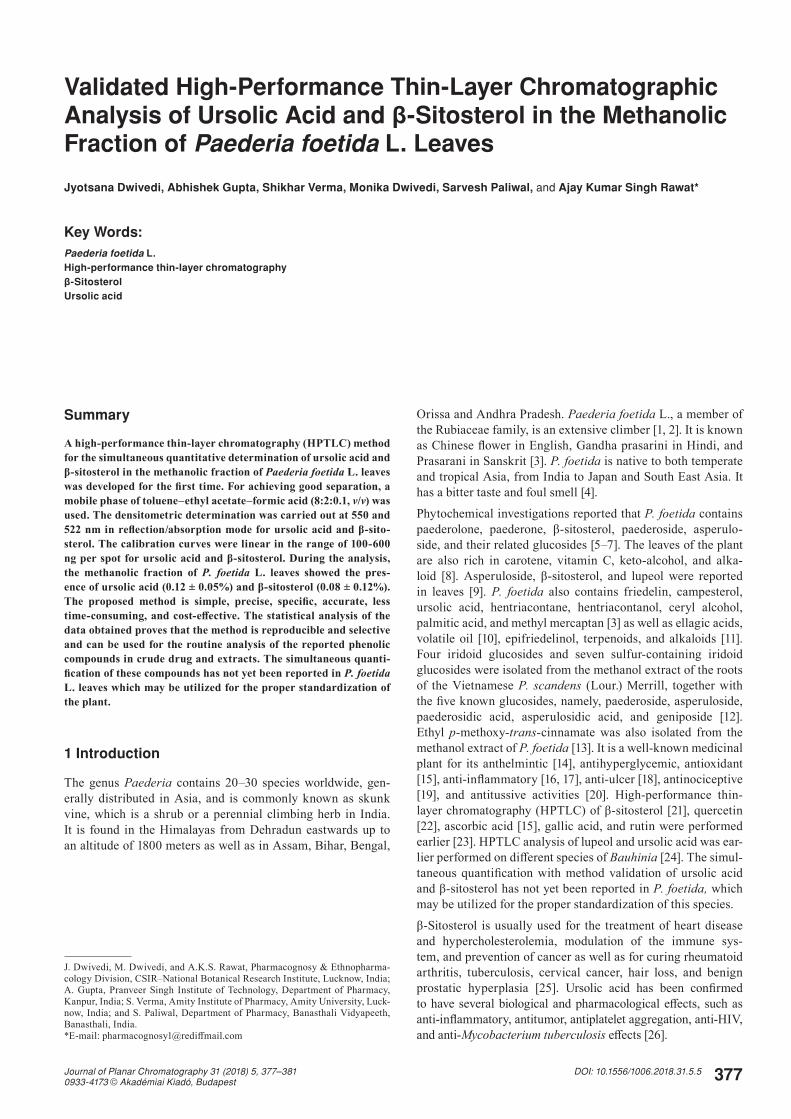
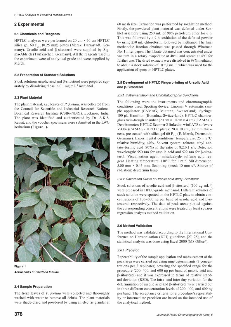
The procedure for the separation and determination of differ-ent compounds in the methanolic fraction of P. foetida using HPTLC–densitometry is reported at sixpoint calibration curve, in which ursolic acid and βsitosterol were observed and quantified with method validation (Table 1). HPTLC profile with chromatograms was obtained for standard compounds and plant extracts, and both targeted compounds were identified by
RF, peak purity, and overlaid ultraviolet (UV) spectra (Figures 2‒4). Precision studies have been performed by analyzing intra- and inter-day variations for the determination of these
Figure 2
HPTLC profiling of Paederia foetida methanolic fraction with ursolic acid and β-sitosterol.
Figure 3
HPTLC chromatograms of Paederia foetida methanolic fraction with ursolic acid and β-sitosterol.
Table 1
Summary of validation parameters.
Parameters Ursolic acid βSitosterol
RF 0.22 ± 0.00 0.38 ± 0.00
Linearity range [ng] 100–500 100–500
Regression via area y = 8.2848x + 464.86 y = 3.4676x + 32.534
r 0.996 0.998
Slope 8.2848 3.4676
Intercept 464.86 32.534
LOD [ng] 40 50
LOQ [ng] 121.20 151.50
Scanning [nm] 550 522
HPTLC Analysis of Paederia foetida Leaves
380 Journal of Planar Chromatography 31 (2018) 5
compounds which was carried out at three different concen-tration levels of 200, 400, and 600 ng per band; mean %RSD values were found to be 0.51 and 0.49 for ursolic acid and βsitosterol, respectively, in intraday analysis, while interday analysis showed mean %RSD values of 1.58 and 1.69 for ursolic acid and βsitosterol, respectively, showing good pre-cision (Table 2). For recovery studies, pre-analyzed samples of P. foetida were spiked with extra 50, 100, and 150% of the standard compounds, and the mixtures were reanalyzed which showed a good recovery ranging from 97.25% to 99.19% for βsitosterol and 97.96% to 99.20% for ursolic acid. The exper-imental data are expressed as mean percentages of recovered
analytes, standard deviation, and relative standard deviation is also presented (Table 3). The concentrations of ursolic acid and βsitosterol in the methanolic fraction of P. foetida are repre-sented in Table 4.
Table 4
Quantification of ursolic acid and β-sitosterol in Paederia foetida.
Plant Sample Ursolic acid [%] βSitosterol [%]
Paederia foetida L. 0.12 ± 0.05 0.08 ± 0.12
4 Conclusion
A validated HPTLC analytical method has been developed for the simultaneous determination of ursolic acid and βsitosterol in Paederia foetida. The proposed method is simple, precise, specific, accurate, less timeconsuming, and costeffective. The statistical analysis of the data obtained proves that the method is reproducible and selective and can be used for the routine analysis of the reported compounds in crude drug and extracts. The method can be used to determine the purity of P. foetida available from various sources by detecting the related impu-rities as well as for the quality control of herbal formulations containing P. foetida as an ingredient. HPTLC analysis has indicated the presence of optimum amount of ursolic acid and βsitosterol in the samples. This can be used in the pharma-ceutical industry as a pharmacognostical tool to identify this medicinally important plant. In addition, it can be adopted as a chemotaxonomical tool in plant systematic studies. These two biomarkers from P. foetida were simultaneously quantified for the first time. Further, identification of other compounds and their separation and characterization from the plants is to be evaluated and reported in the near future.
Conflict of Interest
We declare that we have no conflict of interest.
Acknowledgment
The authors are thankful to the director of CSIR‒NBRI for pro-viding all the facilities to conduct this research work.
Figure 4
HPTLC standard peaks of ursolic acid and β-sitosterol.
Table 2
Intra-day and inter-day precisions.
Standard markers
Conc. [ng per band]
Intra-day Inter-day
%RSD Mean RSD %RSD Mean
RSD
Ursolic acid
200 0.45 1.83
400 0.67 0.51 1.62 1.58
600 0.41 1.31
βSitosterol
200 0.63 1.72
400 0.54 0.49 1.49 1.69
600 0.32 1.88
Table 3
Recovery analysis of ursolic acid and β-sitosterol.
Standard Amount added [%]
Amount recovered [%] Mean SD RSD
Ursolic acid
50 97.25 1.10 1.13
100 99.19 2.72 2.74
150 98.10 1.42 1.45
βSitosterol
50 99.17 2.11 2.13
100 98.16 2.60 2.64
150 98.58 1.63 1.65
HPTLC Analysis of Paederia foetida Leaves
Journal of Planar Chromatography 31 (2018) 5 381
Funding
This work was supported by the Department of Science & Technology, India, under the INSPIRE Fellowship Program.
References
[1] The Ayurvedic Pharmacopoeia of India, Part I (2), Ministry of Health and Family Welfare, Department and Indian System of Medicine and Homeopathy, New Delhi, 1999, 137–140.
[2] E. Blatter, J.F, Caius, K.S. Mhaskar, Indian Med. Plants 2 (1981) 1297–1299.
[3] C.P. Khare (ed.), Indian medicinal plants, SpringerVerlag, Hei-delberg, 2007, 459.
[4] B.N. Sastri, The wealth of India, A dictionary of Indian raw ma-terials and industrial products; Raw materials, vol. 7, CSIR, New Delhi, 1962.
[5] Y.N. Shukla, H.A. Lloyd, J.F. Mortons, G.J. Kapadia, Phytochem-istry 15 (1976) 1989–1990.
[6] H. Inouye, S. Inouye, N. Shimokawa, M. Okigawa, Chem. Pharm. Bull. 17 (1969) 1942–1948.
[7] M.U. Ahmad, M.R. Islam, E. Huo, M.W. Khan, S. Gupta, J. Bang-ladesh Acad. Sci. 15 (1991) 19–22.
[8] A. Ghani, Medicinal plants of Bangladesh; Chemical constituents and uses, 2nd edn., Asiatic Society of Bangladesh, Dhaka, 2003, 331–332.
[9] C.S. Shreedhara, N. Udupa, S. Shetty, Book of Abstracts, Inter-national Symposium for HighPerformance ThinLayer Chroma-tography (HPTLC), Basel, 6‒8 July 2011, p-10u, 230.
[10] M. Thirupathi, D. Srinivas, K. Rajendar, D. Raju, R.K. Jaganmo-han, J. Atoms Mol. 3 (2013) 17–22.
[11] K.C. Wong, G.L. Tan, Flavour Fragr. J. 9 (1994) 25–28.
[12] D.N. Quang, T. Hashimoto, M. Tanaka, N.X. Dung, Y Asakawa, Phytochemistry 60 (2002) 505–514.
[13] N. Uddin, M.K. Hossain, M.R. Haque, C.M. Hasan, Asian J. Chem. 25 (2013) 1163–1164.
[14] B. Uddin, T. Nahar, K.M. Ibrahim, S. Hossain, Bangladesh J. Life Sci. 19 (2007) 141–143.
[15] V. Kumar, F. Anwar, A. Ahmed, A. Verma, A. Ahmed, Z.A. Dam-anhouri, V. Mishra, P.W. Ramteke, P.C. Bhatt, M. Mujeeb, BMC Complement. Altern. Med. 14 (2014) 76.
[16] S. De, B. Ravishankar, J. Ethnopharmacol. 43 (1994) 31–38.
[17] M.C. Srivastava, J.P. Tewari, V. Kant, Indian J. Med. Sci. 27 (1973) 231–234.
[18] K. Srinivasreddy, S.A. Kumar, S. Ganapathy, Int. J. Res. Ayur. Pharm. 2 (2011) 1556–1559.
[19] B.A. Whittle, Br. J. Pharmacol. Chemother. 22 (1964) 246–253.
[20] G. Nosalova, J. Mokry, A. Ather, M.T.H. Khan, Acta Vet. Brno 76 (2007) 27–33.
[21] S. Chanda, L. Deb, R.K. Tiwari, K. Singh, S. Ahmad, BMC Com-plement. Altern. Med. 15 (2015) 304.
[22] V. Kumar, F.A. Al-Abbasi, D. Ahmed, A. Verma, M. Mujeeb, F. Anwar, Food Funct. 6 (2015) 1652–1666.
[23] M.R. Senapati, P.C. Behera, P.C. Bisoi, A. Maity, S.C. Parija, Bioscan 8 (2013) 603–609.
[24] A. Gupta, S. Verma, H. Dwivedi, A.K.S. Rawat, J. Planar Chro-matogr. 29 (2016) 423–428.
[25] S. Saeidnia, A. Manayi, A.R. Gohari, M. Abdollahi, Eur. J. Med. Plants 4 (2014) 590–609.
[26] S.Y. Lee, Y.J. Kim, S.O. Chung, S.U. Park, EXCLI J. 15 (2016) 221–228.
[27] ICHQ2A, Text on Validation of Analytical Procedures, Har-monized Tripartite Guideline prepared within the International Conference on Harmonization of Technical Requirements for the Registration of Pharmaceuticals for Human Use, Geneva, 1994.
[28] ICHQ2B, Validation of Analytical Procedures: Methodology, Harmonized Tripartite Guideline prepared within the International Conference on Harmonization of Technical Requirements for the Registration of Pharmaceuticals for Human Use, Geneva, 1996.
Ms received: May 28, 2018Accepted: July 4, 2018