TNF Receptor Family Member BCMA (B Cell Maturation) Associates
10
of April 10, 2019. This information is current as Mitogen-Activated Protein Kinase Elk-1, c-Jun N-Terminal Kinase, and p38 B, κ TRAF2, and TRAF3 and Activates NF- Receptor-Associated Factor (TRAF) 1, Cell Maturation) Associates with TNF TNF Receptor Family Member BCMA (B Odile Devergne and Andreas Tsapis Bourgeade, Edith Rogier, Christine Madry, Junichiro Inoue, Anastassia Hatzoglou, Jérôme Roussel, Marie-Françoise http://www.jimmunol.org/content/165/3/1322 doi: 10.4049/jimmunol.165.3.1322 2000; 165:1322-1330; ; J Immunol References http://www.jimmunol.org/content/165/3/1322.full#ref-list-1 , 18 of which you can access for free at: cites 37 articles This article average * 4 weeks from acceptance to publication Fast Publication! • Every submission reviewed by practicing scientists No Triage! • from submission to initial decision Rapid Reviews! 30 days* • Submit online. ? The JI Why Subscription http://jimmunol.org/subscription is online at: The Journal of Immunology Information about subscribing to Permissions http://www.aai.org/About/Publications/JI/copyright.html Submit copyright permission requests at: Email Alerts http://jimmunol.org/alerts Receive free email-alerts when new articles cite this article. Sign up at: Print ISSN: 0022-1767 Online ISSN: 1550-6606. Immunologists All rights reserved. Copyright © 2000 by The American Association of 1451 Rockville Pike, Suite 650, Rockville, MD 20852 The American Association of Immunologists, Inc., is published twice each month by The Journal of Immunology by guest on April 10, 2019 http://www.jimmunol.org/ Downloaded from by guest on April 10, 2019 http://www.jimmunol.org/ Downloaded from
Transcript of TNF Receptor Family Member BCMA (B Cell Maturation) Associates
of April 10, 2019.This information is current as
Mitogen-Activated Protein KinaseElk-1, c-Jun N-Terminal Kinase, and p38
B,κTRAF2, and TRAF3 and Activates NF-Receptor-Associated Factor (TRAF) 1,Cell Maturation) Associates with TNF TNF Receptor Family Member BCMA (B
Odile Devergne and Andreas TsapisBourgeade, Edith Rogier, Christine Madry, Junichiro Inoue, Anastassia Hatzoglou, Jérôme Roussel, Marie-Françoise
http://www.jimmunol.org/content/165/3/1322doi: 10.4049/jimmunol.165.3.1322
2000; 165:1322-1330; ;J Immunol
Referenceshttp://www.jimmunol.org/content/165/3/1322.full#ref-list-1
, 18 of which you can access for free at: cites 37 articlesThis article
average*
4 weeks from acceptance to publicationFast Publication! •
Every submission reviewed by practicing scientistsNo Triage! •
from submission to initial decisionRapid Reviews! 30 days* •
Submit online. ?The JIWhy
Subscriptionhttp://jimmunol.org/subscription
is online at: The Journal of ImmunologyInformation about subscribing to
Permissionshttp://www.aai.org/About/Publications/JI/copyright.htmlSubmit copyright permission requests at:
Email Alertshttp://jimmunol.org/alertsReceive free email-alerts when new articles cite this article. Sign up at:
Print ISSN: 0022-1767 Online ISSN: 1550-6606. Immunologists All rights reserved.Copyright © 2000 by The American Association of1451 Rockville Pike, Suite 650, Rockville, MD 20852The American Association of Immunologists, Inc.,
is published twice each month byThe Journal of Immunology
by guest on April 10, 2019
http://ww
w.jim
munol.org/
Dow
nloaded from
by guest on April 10, 2019
http://ww
w.jim
munol.org/
Dow
nloaded from
TNF Receptor Family Member BCMA (B Cell Maturation)Associates with TNF Receptor-Associated Factor (TRAF) 1,TRAF2, and TRAF3 and Activates NF-kB, Elk-1, c-JunN-Terminal Kinase, and p38 Mitogen-Activated ProteinKinase1
Anastassia Hatzoglou,2* Jerome Roussel,2† Marie-Francoise Bourgeade,2† Edith Rogier,†
Christine Madry, † Junichiro Inoue,‡ Odile Devergne,† and Andreas Tsapis3†
BCMA (B cell maturation) is a nonglycosylated integral membrane type I protein that is preferentially expressed in mature Blymphocytes. Previously, we reported in a human malignant myeloma cell line that BCMA is not primarily present on the cellsurface but lies in a perinuclear structure that partially overlaps the Golgi apparatus. We now show that in transiently or stablytransfected cells, BCMA is located on the cell surface, as well as in a perinulear Golgi-like structure. We also show that overex-pression of BCMA in 293 cells activates NF-kB, Elk-1, the c-Jun N-terminal kinase, and the p38 mitogen-activated protein kinase.Coimmunoprecipitation experiments performed in transfected cells showed that BCMA associates with TNFR-associated factor(TRAF) 1, TRAF2, and TRAF3 adaptor proteins. Analysis of deletion mutants of the intracytoplasmic tail of BCMA showed thatthe 25-aa protein segment, from position 119 to 143, conserved between mouse and human BCMA, is essential for its associationwith the TRAFs and the activation of NF-kB, Elk-1, and c-Jun N-terminal kinase. BCMA belongs structurally to the TNFR family.Its unique TNFR motif corresponds to a variant motif present in the fourth repeat of the TNFRI molecule. This study confirmsthat BCMA is a functional member of the TNFR superfamily. Furthermore, as BCMA is lacking a “death domain” and itsoverexpression activates NF-kB and c-Jun N-terminal kinase, we can reasonably hypothesize that upon binding of its correspond-ing ligand BCMA transduces signals for cell survival and proliferation. The Journal of Immunology,2000, 165: 1322–1330.
T he TNF-related cytokines are a large family of pleiotropicmediators of host defense and immune system regulators.Those that are integral membrane proteins act locally
through cell-to-cell contact, and those that are secreted proteins acton distant target cells. The TNFR are a heterologous family, ofwhich 18 members are known. They mediate the action of TNF-related cytokines leading to cell death or cell proliferation anddifferentiation (1, 2). Most members of the TNFR family are typeI transmembrane proteins with an extracellular ligand-binding do-main, a single membrane-spanning region, and a cytoplasmic re-gion that activates cell functions (3). The common characteristic ofall TNFR family members is the repetition of a six-cysteine motifin the extracellular N-terminal part of the molecule. In contrast tothe extracellular parts of the receptors, the sequences of the cyto-plasmic tails are generally dissimilar, and none possess sequences
suggestive of catalytic activity. However, several motifs in theC-terminal part of TNFR have been shown to bind protein factorstransducing the signal initiated by ligand binding and receptor tri-merization. One of these motifs, the “death domain,” is present inTNFRI, Fas, DR3, DR4, and DR5 and is responsible for the ca-pacity of these receptors to induce apoptosis (4, 5). A second groupof motifs binds signal transducers, TNFR-associated factors(TRAFs).4 TRAFs interact directly with several TNFRs, likeTNFRII, CD40, CD30, and lymphotoxinb receptor (6–9) andwith the EBV oncogene LMP1 (10). TRAF2, TRAF5, and TRAF6mediate the activation of the transcriptional factor NF-kB (11–13)and activate the c-Jun N-terminal protein kinase (JNK) (14).TRAF6 also mediates the activation of extracellular signal-regu-lated kinase (ERK) (15).
Recently, we identified of a novel TNFR (16, 17) through themolecular analysis of a t(4;16) translocation (16, 18), characteristicof a malignant human T cell lymphoma. The gene product is se-lectively expressed in mature B lymphocytes (19) and was there-fore named BCMA for B cell maturation protein. The BCMA genecodes for a nonglycosylated integral membrane type I protein. TheN-terminal part of both mouse and human proteins contains a con-served six-cysteine motif (17). A sensitive method of sequenceanalysis, hydrophobic cluster analysis (20), indicated that this con-served motif is similar to the six-cysteine repeat motif found in the
*Laboratory of Experimental Endocrinology, Faculty of Medicine, University ofCrete, Heraklion, Greece;†Institut National de la Sante et de la Recherche Medicale,Unite 131, Institut Paris-Sud sur les Cytokines, Clamart, France; and‡Department ofOncology, Institute of Medical Science, University of Tokyo, Tokyo, Japan
Received for publication January 10, 2000. Accepted for publication May 18, 2000.
The costs of publication of this article were defrayed in part by the payment of pagecharges. This article must therefore be hereby markedadvertisementin accordancewith 18 U.S.C. Section 1734 solely to indicate this fact.1 This work was supported in part by a grant from the Comite Departmental des Hautsde Seine de la Ligue Nationale contre le Cancer (to A.T.) and by a grant from theAssociation de Recherche contre le Cancer (Grant 9907 to A.T.).2 A.H., J.R., and M.-F.B. contributed equally to this work.3 Address correspondence and reprint requests to Dr. Andreas Tsapis, Institut Na-tional de la Sante et de la Recherche Medicale Unite 131, Institut Paris-Sud sur lesCytokines, 32, rue des Carnets, 92140 Clamart, France. E-mail address:[email protected]
4 Abbreviations used in this paper: TRAF, TNFR-associated factor; JNK, c-JunN-terminal kinase; ERK, extracellular signal-related kinase; BCMA, B cell matura-tion; HA, hemagglutinin; h, human; m, mouse; ATF, activating transcription factor;GFP, green fluorescence protein; MEK, MAPK/ERK kinase; MEKK, MEK kinase;PI3-K, phosphatidylinositol 3-kinase.
Copyright © 2000 by The American Association of Immunologists 0022-1767/00/$02.00
by guest on April 10, 2019
http://ww
w.jim
munol.org/
Dow
nloaded from
extracellular part of TNFRs. There are two notable differences be-tween the BCMA protein and other members of the TNFR family.The first is that BCMA contains only one six-cysteine-rich motif,whereas the members of the TNFR family contain more than onecopy. The second is that the six-cysteine motif of BCMA is not thecanonical motif of TNFRs but corresponds to a variant motifpresent in the fourth repeat of the TNFRI molecule. The full namefor BCMA in the new TNF nomenclature scheme is TNFRSF17.The human BCMA gene is the first TNFR gene that has beenimplicated in chromosome translocation.
We report a study of the cellular localization of BCMA in tran-siently and stably transfected cells. We show that overexpressionof BCMA activates the NF-kB, Elk-1, p38, and JNK. We alsostudied the association of the six known TRAFs with BCMA anddefined the region of the BCMA protein responsible for thisassociation.
Materials and MethodsAbs and reagents
The rabbit polyclonal anti-TRAF1 (H-132), anti-TRAF2 (C-20), anti-TRAF3 (H-122), anti-TRAF5 (H-257), anti-JNK1 (sc-474), and goat poly-clonal anti-TRAF4 (N-16) and anti-goat HRP-conjugated Abs were pur-chased from Santa Cruz Biotechnology (Santa Cruz, CA). M2 anti-FLAGmAb, M2 mAb bound to agarose beads, and FLAG peptide and proteaseinhibitor mixture were purchased from Sigma-Aldrich (St. Louis, MO).Anti-F mAb was a generous gift from M. C. Rio (Institut National de laSante et de la Recherche Medicale, Unite 184, Strasbourg, France). 12CA5anti-hemagglutinin (anti-HA) mAb was purchased from Roche Diagnostics(Somerville, NJ). PE-, FITC-, and HRP-conjugated goat anti-mouse IgGpolyclonal Abs and HRP-conjugated donkey anti-rabbit IgG polyclonalAbs were purchased from Immunotech (Marseille, France). Rabbit poly-clonal anti-phosphatidylinositol 3 kinase (PI3-K) p85 Abs were obtainedfrom Upstate Biotechnology (Lake Placid, NY). RPMI 1640, DMEM,FCS, additional reagents for cell culture, optimem, and lipofectamine werepurchased from Life Technologies (Grand Island, NY).
Primers
The following primers were used in this study: BCMA59ATG (59-AAGCTTATGTTGCAGATGGCTGGGCA-39), BCMA39TAA (59-GGATCCTTACCTAGCAGAAATTGATTTC-39), 37 (59-CCCAAGCTTATGGCTGGGCAGTGCTCC-39), 43 (59-CGCGGATCCTTATGGTTCAGAGCTTATCTTCCT-39), AH7 (CGCGGATCCTTACCTAGCAGAAATTGATTTCT-39), AH9 (59-CCGCTCGAGGGCGCAACAGTGTTTCCACA-39), AH10(GGAAGATCTCTAACGACATCTAAAACACCAG-39), BFL1 (59-AACTGCAGCTGGGCAGTGCTCCCAAAA-39), BFL2 (59-CGGGATCCTTAATAGTCATTCGTTTTCGTGGTG-39), BFL3 (59-CGGGATCCTTAGCAATGGTCATAGTCGACCT-39), BFL4 (59-CGGGATCCTTAGCCTCTCGGAAGAATAATTTC-39), and BFL5 (59-CGGGATCCTTAGTTTTTAAACTCGTCCTTTAATG-39). All primers used in this study were purchased fromGenset (Paris, France).
Expression vectors
A full-length human BCMA (h184) was amplified by PCR from humancDNA using the BCMA59ATG and BCMA39TAA primers; a fragmentencoding the N-terminal and transmembrane parts of hBCMA (h84) wasamplified by PCR using primers 37 and 43. The PCR fragments weredigested withBamHI andHindIII restriction enzymes and ligated into theBamHI andHindIII sites of the vector pcDNA3 (Invitrogen, Groningen,The Netherlands).
A full-length mouse BCMA was amplified by PCR from a mouse cDNAlibrary with the primers AH9 and AH10. The PCR fragment was digestedwith XhoI andBglII and ligated into theXhoI andBglII sites of the vectorpDEB (21), giving rise to a fusion encoding a N-terminal HA-taggedmouse BCMA (HAm185). The HA-tagged mBCMA was digested withEcoRI andNotI and ligated into theEcoRI andNotI sites of the vectorpcDNA3.
N-terminal FLAG-tagged hBCMA deletion mutants were constructedby PCR amplification using the following pairs of primers: BFL1 and AH7for FLAG-hBCMA without deletion (Fh184), BFL1 and BFL2 for FLAG-hBCMAD165–184 (Fh164), BFL1 and BFL3 for FLAG-hBCMAD144–184 (Fh143), BFL1 and BFL4 for FLAG-hBCMAD119–184 (Fh118), andBFL1 and BFL5 for FLAG-hBCMAD92–184 (Fh91). All PCR productswere digested withPstI and BamHI and ligated between thePstI and
BamHI sites of the vector pSG5-FLAG (22). All expression vectors wereconstructed by standard recombinant DNA procedures. The sequence ofthe plasmids constructed by PCR amplification were subsequently verifiedby dideoxy sequencing.
The vectors pSG5hTRAF1 (10), pSG5hTRAF2, pSG5FLAGhTRAF2(23), pSG5FLAGhTRAF1, pSG5hTRAF3, pSG5FLAGhTRAF3 (24),pMEFLAGmTRAF5 (13), pMEFLAGmTRAF6 (25), pEBBhTRAF5 (26),pcLMP1 (27), pcDNA3TRAF2.DN (TRAF2D6–86) (23), pGEX-Jun(1–79), pcDNA3-HA-JNK (28), and theb-galactosidase expression vector(pGK-bgal), in which expression is driven by the phosphoglucokinase pro-moter (22), have been already described. pAT3FhTRAF4 encoding humanFLAG-tagged TRAF4 was a generous gift from Dr. Catherine Regnier(Strasbourg, France).
Cell lines and transfections
Human embryonic kidney 293, 293T, and 293EBNA and simian kidneyCOS7 cells were maintained in high-glucose DMEM supplemented with10% heat-inactivated FCS, 2 mM glutamine, 100 U/ml of penicillin, and100 mg/ml of streptomycin and were grown at 37°C in 5% CO2. The293EBNA cell line was purchased from Invitrogen and maintained in cul-ture according to the supplier’s instructions. The BJAB cell line is anEBV-negative Burkitt lymphoma cell line (29) and was cultured in RPMI1640 supplemented with 10% heat-inactivated FCS, 2 mM glutamine, 100U/ml of penicillin, and 100mg/ml of streptomycin and grown at 37°C in5% CO2. Adherent cells were seeded in six-well plates (53 105 cells perwell) in 2 ml of complete medium, incubated at 37°C in 5% CO2 for 20–24h, and transfected with lipofectamine according to the manufacturer’s in-structions, using 1mg of total plasmid DNA, for 6 h. BJAB cells weretransfected by electroporation (960mF, 210 V) in 400ml optimem mediumusing a Bio-Rad Gene Pulser apparatus (Bio-Rad, Richmond, CA). Cellextracts were tested for gene expression 24–48 h after transfection. Toestablish cells stably expressing BCMA, 293 cells were transfected withHAm185-expressing vector and were selected in high-glucose DMEM,10% FCS, in presence of 400mg/ml geneticin. Geneticin-resistant cloneswere screened by immunoblotting for BCMA expression.
Luciferase reporter system for NF-kB, Elk-1, and JNK
The NF-kB, Elk-1, and JNK activation assays were performed using thecorresponding luciferase reporter PathDetect Reporting systems purchasedfrom Stratagene (La Jolla, CA).
Luciferase andb-galactosidase assays
Transfected cells were washed twice with PBS and lysed with reporter lysisbuffer (Promega, Madison, WI). The luciferase activity was measured us-ing the reporter assay system (Promega).b-galactosidase activity was mea-sured using the luminescentb-galactosidase reporter system (Clontech,Palo Alto, CA) in a Packard luminometer analyzer (Packard, Meriden, CT).Measurements of luciferase were normalized tob-galactosidase activityand are expressed as a ratio to values obtained from cells treated withvector alone. The relative luciferase activities given are representative oftriplicate assays in three independent experiments.
Determination of JNK activity
JNK activity was determined as described previously (28) with minor mod-ifications. Transfected cells were lysed in 10 mM Tris, pH 7.4, 150 mMNaCl, 1 mM EDTA, 1 mM EGTA, 1% Triton X-100, 0.5% Nonidet P-40,0.5 mM sodium vanadate, 0.2 mM PMSF, and 10% glycerol. Lysates wereclarified by centrifugation, and HA-tagged JNK was immunoprecipitatedusing anti-HA mAb 12CA5. Immune complexes were collected on pro-tein-G Agarose beads, washed three times in lysis buffer, once in kinasereaction buffer (12.5 mM MOPS, pH 7.5, 12.5 mMb-glycerophoshate, 7.5mM MgCl2, 0.5 mM EGTA, 0.5 mM NaF, 0.5 mM sodium vanadate), andresuspended in 30ml of the same buffer containing 2mg of GST-Jun, 20mM unlabeled ATP, and 5mCi [g-32P]ATP. After incubation at 20°C for30 min, kinase reaction products were analyzed by SDS-PAGE and auto-radiography. Part of the immunoprecipitated material was resolved bySDS-PAGE, transferred to a polyvinylidene difluoride membrane, and im-munoblotted with anti-JNK polyclonal Abs to check that the same amountof HA-JNK was used in each case.
Determination of p38 and ERK activity
The activity of these two kinases was assayed using the correspondingassay kit purchased from New England Biolabs (Beverly, MA). Briefly,transfected cells were lyzed, and the active phosphorylated kinase wasimmunoprecipitated using specific mAbs. The immunoprecipitated proteinwas assayed for its ability to phosphorylate activating transcription factor
1323The Journal of Immunology
by guest on April 10, 2019
http://ww
w.jim
munol.org/
Dow
nloaded from
(ATF) 2 (p38) or Elk-1 (ERK) substrates. Analysis of phosphorylated sub-strates was performed by Western blotting using specific polyclonalphosphoantibodies.
Coimmunoprecipitation experiments
COS7 cells were cotransfected with one vector encoding a TRAF and onevector encoding one of the various FLAG-tagged BCMA constructs. Eigh-teen to 24 h after transfection, cells were lysed in lysis buffer (50 mM Tris,pH 7.4, 150 mM NaCl, 1.5 mM EDTA, 10% glycerol, 0.1% Nonidet P-40,and a protease inhibitor mixture) by incubation for 1 h at 4°C, and thesupernatant was then clarified by centrifugation. One-fortieth of this lysate(input) was conserved to test the efficiency of transfection, and the rest wasincubated for 2 h, at 4°C, with M2 monoclonal anti-FLAG Ab, covalentlybound to agarose beads. The beads were washed three times with the lysisbuffer, and the bound proteins were eluted twice by addition of 250mMFLAG peptide diluted in PBS. The eluate and the input were analyzed byPAGE and transferred onto a polyvinylidene difluoride membrane (Hy-bond-P; Amersham, Little Chalfont, U.K.). The presence of BCMA and ofthe various TRAFs was tested by immunoblotting using M2 Ab to evidenceFLAG-tagged BCMA constructs and the corresponding anti-TRAF Ab foreach TRAF. HRP-conjugated anti-rabbit, anti-goat, and anti-mouse IgGand SuperSignal Chemiluminescent substrate (Pierce, Rockford, IL) wereused to reveal the blots.
Immunofluorescence staining and FACS analysis
For immunofluorescence observation, transfected ells were stained withM2 mAb then incubated with fluorescein-conjugated goat anti-mouse Abbefore analysis under a Leica DM microscope (Leica, Deerfield, IL) aspreviously described (30). For FACS analysis,;5 3 105 cells per condi-tion were stained with saturating concentrations of Ab, then incubated withPE-conjugated goat anti-mouse Ab before analysis in a FACScan flowcytometer (Becton Dickinson, San Diego, CA), as previously described(31). A minimum of 10,000 events per sample was analyzed. CellQuestsoftware (Becton Dickinson) was used for data analysis.
ResultsBCMA is present on both the surface of cells and in anintracellular perinuclear structure
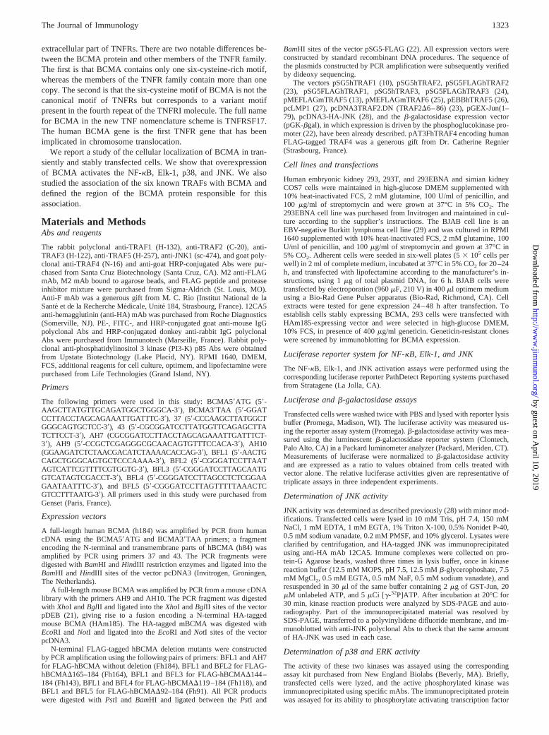
In a previous study of BCMA localization, in the human myelomacell line U266, we found most BCMA in a perinuclear Golgi-likestructure (30). We further analyzed the localization of BCMA bytransfection experiments in two cell lines: the human B lympho-cyte BJAB cell line and the monkey kidney COS7 cell line. BJABcell line has been chosen because it expresses detectable amount ofBCMA mRNA, has nondetectable amounts of BCMA protein(data not shown), and can be transiently transfected with high ef-ficiency (50–60%). On the contrary, COS7 cell line does not ex-press BCMA. The cell lines were transfected with two vectors, onecoding for a FLAG-tagged full-length hBCMA (Fh184) and a sec-ond one coding for a FLAG-tagged hBCMA construct lacking theentire intracellular cytoplasmic tail (Fh91), together with a greenfluorescence protein (GFP) expression vector. Eighteen hours aftertransfection, cells were stained with M2 anti-FLAG Ab and a sec-ondary PE-conjugated anti-mouse IgG. The GFP-expressing cellpopulation was gated, and the presence of FLAG-tagged proteinson the surface and intracellularly was determined by two-colorcytofluorometry (Fig. 1A). Full-length hBCMA and the mutant hB-CMA missing its cytoplasmic tail were similarly distributed inboth BJAB and COS7 cell lines. Both proteins displayed an intra-cytoplasmic localization, but were also present on the cell surface.The localization of full-length hBCMA was further examined byfluorescence microscopy. Our results (Fig. 1B) confirmed thatBCMA was present on the cell surface. As previously observed inthe myeloma U266 cell line, intracellular BCMA was detected ina perinuclear Golgi-like structure in both transfected BJAB andCOS7 cell lines.
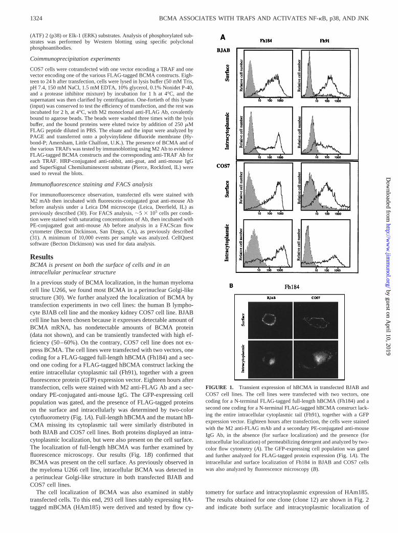
The cell localization of BCMA was also examined in stablytransfected cells. To this end, 293 cell lines stably expressing HA-tagged mBCMA (HAm185) were derived and tested by flow cy-
tometry for surface and intracytoplasmic expression of HAm185.The results obtained for one clone (clone 12) are shown in Fig. 2and indicate both surface and intracytoplasmic localization of
FIGURE 1. Transient expression of hBCMA in transfected BJAB andCOS7 cell lines. The cell lines were transfected with two vectors, onecoding for a N-terminal FLAG-tagged full-length hBCMA (Fh184) and asecond one coding for a N-terminal FLAG-tagged hBCMA construct lack-ing the entire intracellular cytoplasmic tail (Fh91), together with a GFPexpression vector. Eighteen hours after transfection, the cells were stainedwith the M2 anti-FLAG mAb and a secondary PE-conjugated anti-mouseIgG Ab, in the absence (for surface localization) and the presence (forintracellular localization) of permeabilizing detergent and analyzed by two-color flow cytometry (A). The GFP-expressing cell population was gatedand further analyzed for FLAG-tagged protein expression (Fig. 1A). Theintracellular and surface localization of Fh184 in BJAB and COS7 cellswas also analyzed by fluorescence microscopy (B).
1324 BCMA ASSOCIATES WITH TRAFS AND ACTIVATES NF-kB, p38, AND JNK
by guest on April 10, 2019
http://ww
w.jim
munol.org/
Dow
nloaded from
HAm185. Similar results have been obtained in two other clonestested (data not shown).
BCMA-mediated NF-kB activation
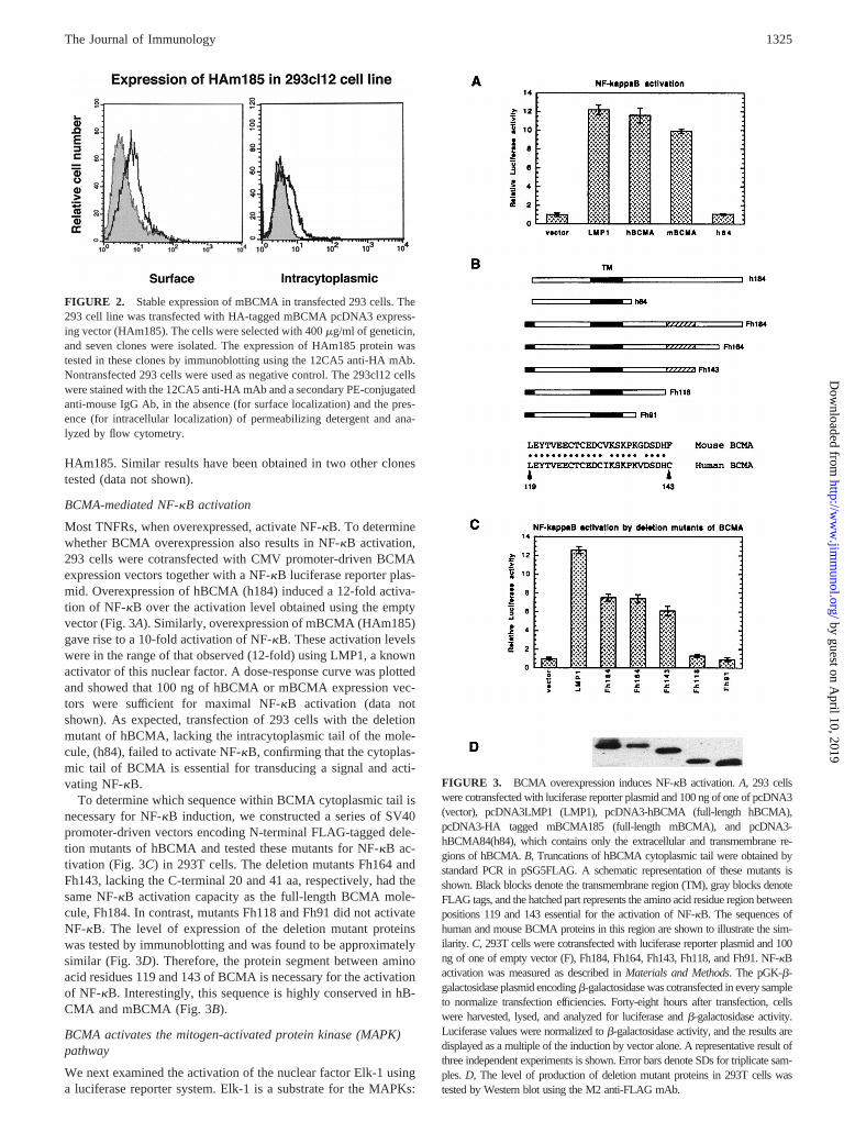
Most TNFRs, when overexpressed, activate NF-kB. To determinewhether BCMA overexpression also results in NF-kB activation,293 cells were cotransfected with CMV promoter-driven BCMAexpression vectors together with a NF-kB luciferase reporter plas-mid. Overexpression of hBCMA (h184) induced a 12-fold activa-tion of NF-kB over the activation level obtained using the emptyvector (Fig. 3A). Similarly, overexpression of mBCMA (HAm185)gave rise to a 10-fold activation of NF-kB. These activation levelswere in the range of that observed (12-fold) using LMP1, a knownactivator of this nuclear factor. A dose-response curve was plottedand showed that 100 ng of hBCMA or mBCMA expression vec-tors were sufficient for maximal NF-kB activation (data notshown). As expected, transfection of 293 cells with the deletionmutant of hBCMA, lacking the intracytoplasmic tail of the mole-cule, (h84), failed to activate NF-kB, confirming that the cytoplas-mic tail of BCMA is essential for transducing a signal and acti-vating NF-kB.
To determine which sequence within BCMA cytoplasmic tail isnecessary for NF-kB induction, we constructed a series of SV40promoter-driven vectors encoding N-terminal FLAG-tagged dele-tion mutants of hBCMA and tested these mutants for NF-kB ac-tivation (Fig. 3C) in 293T cells. The deletion mutants Fh164 andFh143, lacking the C-terminal 20 and 41 aa, respectively, had thesame NF-kB activation capacity as the full-length BCMA mole-cule, Fh184. In contrast, mutants Fh118 and Fh91 did not activateNF-kB. The level of expression of the deletion mutant proteinswas tested by immunoblotting and was found to be approximatelysimilar (Fig. 3D). Therefore, the protein segment between aminoacid residues 119 and 143 of BCMA is necessary for the activationof NF-kB. Interestingly, this sequence is highly conserved in hB-CMA and mBCMA (Fig. 3B).
BCMA activates the mitogen-activated protein kinase (MAPK)pathway
We next examined the activation of the nuclear factor Elk-1 usinga luciferase reporter system. Elk-1 is a substrate for the MAPKs:
FIGURE 2. Stable expression of mBCMA in transfected 293 cells. The293 cell line was transfected with HA-tagged mBCMA pcDNA3 express-ing vector (HAm185). The cells were selected with 400mg/ml of geneticin,and seven clones were isolated. The expression of HAm185 protein wastested in these clones by immunoblotting using the 12CA5 anti-HA mAb.Nontransfected 293 cells were used as negative control. The 293cl12 cellswere stained with the 12CA5 anti-HA mAb and a secondary PE-conjugatedanti-mouse IgG Ab, in the absence (for surface localization) and the pres-ence (for intracellular localization) of permeabilizing detergent and ana-lyzed by flow cytometry.
FIGURE 3. BCMA overexpression induces NF-kB activation.A, 293 cellswere cotransfected with luciferase reporter plasmid and 100 ng of one of pcDNA3(vector), pcDNA3LMP1 (LMP1), pcDNA3-hBCMA (full-length hBCMA),pcDNA3-HA tagged mBCMA185 (full-length mBCMA), and pcDNA3-hBCMA84(h84), which contains only the extracellular and transmembrane re-gions of hBCMA.B, Truncations of hBCMA cytoplasmic tail were obtained bystandard PCR in pSG5FLAG. A schematic representation of these mutants isshown. Black blocks denote the transmembrane region (TM), gray blocks denoteFLAG tags, and the hatched part represents the amino acid residue region betweenpositions 119 and 143 essential for the activation of NF-kB. The sequences ofhuman and mouse BCMA proteins in this region are shown to illustrate the sim-ilarity. C, 293T cells were cotransfected with luciferase reporter plasmid and 100ng of one of empty vector (F), Fh184, Fh164, Fh143, Fh118, and Fh91. NF-kBactivation was measured as described inMaterials and Methods. The pGK-b-galactosidase plasmid encodingb-galactosidase was cotransfected in every sampleto normalize transfection efficiencies. Forty-eight hours after transfection, cellswere harvested, lysed, and analyzed for luciferase andb-galactosidase activity.Luciferase values were normalized tob-galactosidase activity, and the results aredisplayed as a multiple of the induction by vector alone. A representative result ofthree independent experiments is shown. Error bars denote SDs for triplicate sam-ples.D, The level of production of deletion mutant proteins in 293T cells wastested by Western blot using the M2 anti-FLAG mAb.
1325The Journal of Immunology
by guest on April 10, 2019
http://ww
w.jim
munol.org/
Dow
nloaded from
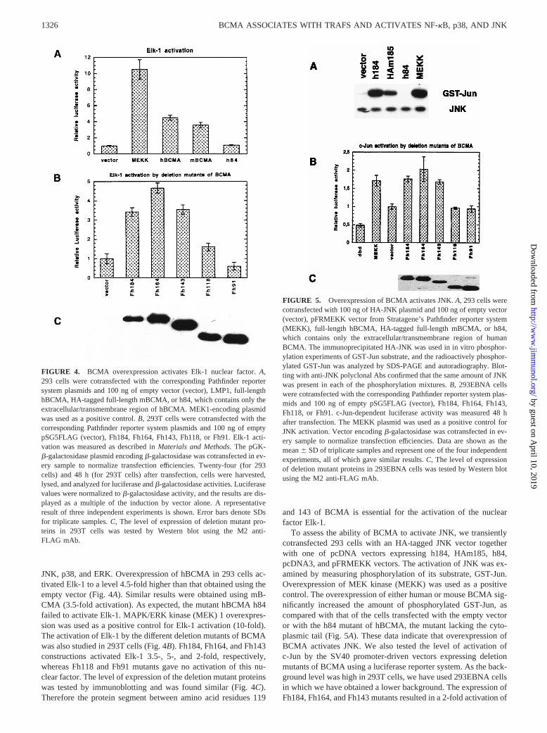
JNK, p38, and ERK. Overexpression of hBCMA in 293 cells ac-tivated Elk-1 to a level 4.5-fold higher than that obtained using theempty vector (Fig. 4A). Similar results were obtained using mB-CMA (3.5-fold activation). As expected, the mutant hBCMA h84failed to activate Elk-1. MAPK/ERK kinase (MEK) 1 overexpres-sion was used as a positive control for Elk-1 activation (10-fold).The activation of Elk-1 by the different deletion mutants of BCMAwas also studied in 293T cells (Fig. 4B). Fh184, Fh164, and Fh143constructions activated Elk-1 3.5-, 5-, and 2-fold, respectively,whereas Fh118 and Fh91 mutants gave no activation of this nu-clear factor. The level of expression of the deletion mutant proteinswas tested by immunoblotting and was found similar (Fig. 4C).Therefore the protein segment between amino acid residues 119
and 143 of BCMA is essential for the activation of the nuclearfactor Elk-1.
To assess the ability of BCMA to activate JNK, we transientlycotransfected 293 cells with an HA-tagged JNK vector togetherwith one of pcDNA vectors expressing h184, HAm185, h84,pcDNA3, and pFRMEKK vectors. The activation of JNK was ex-amined by measuring phosphorylation of its substrate, GST-Jun.Overexpression of MEK kinase (MEKK) was used as a positivecontrol. The overexpression of either human or mouse BCMA sig-nificantly increased the amount of phosphorylated GST-Jun, ascompared with that of the cells transfected with the empty vectoror with the h84 mutant of hBCMA, the mutant lacking the cyto-plasmic tail (Fig. 5A). These data indicate that overexpression ofBCMA activates JNK. We also tested the level of activation ofc-Jun by the SV40 promoter-driven vectors expressing deletionmutants of BCMA using a luciferase reporter system. As the back-ground level was high in 293T cells, we have used 293EBNA cellsin which we have obtained a lower background. The expression ofFh184, Fh164, and Fh143 mutants resulted in a 2-fold activation of
FIGURE 4. BCMA overexpression activates Elk-1 nuclear factor.A,293 cells were cotransfected with the corresponding Pathfinder reportersystem plasmids and 100 ng of empty vector (vector), LMP1, full-lengthhBCMA, HA-tagged full-length mBCMA, or h84, which contains only theextracellular/transmembrane region of hBCMA. MEK1-encoding plasmidwas used as a positive control.B, 293T cells were cotransfected with thecorresponding Pathfinder reporter system plasmids and 100 ng of emptypSG5FLAG (vector), Fh184, Fh164, Fh143, Fh118, or Fh91. Elk-1 acti-vation was measured as described inMaterials and Methods. The pGK-b-galactosidase plasmid encodingb-galactosidase was cotransfected in ev-ery sample to normalize transfection efficiencies. Twenty-four (for 293cells) and 48 h (for 293T cells) after transfection, cells were harvested,lysed, and analyzed for luciferase andb-galactosidase activities. Luciferasevalues were normalized tob-galactosidase activity, and the results are dis-played as a multiple of the induction by vector alone. A representativeresult of three independent experiments is shown. Error bars denote SDsfor triplicate samples.C, The level of expression of deletion mutant pro-teins in 293T cells was tested by Western blot using the M2 anti-FLAG mAb.
FIGURE 5. Overexpression of BCMA activates JNK.A, 293 cells werecotransfected with 100 ng of HA-JNK plasmid and 100 ng of empty vector(vector), pFRMEKK vector from Stratagene’s Pathfinder reporter system(MEKK), full-length hBCMA, HA-tagged full-length mBCMA, or h84,which contains only the extracellular/transmembrane region of humanBCMA. The immunoprecipitated HA-JNK was used in in vitro phosphor-ylation experiments of GST-Jun substrate, and the radioactively phosphor-ylated GST-Jun was analyzed by SDS-PAGE and autoradiography. Blot-ting with anti-JNK polyclonal Abs confirmed that the same amount of JNKwas present in each of the phosphorylation mixtures.B, 293EBNA cellswere cotransfected with the corresponding Pathfinder reporter system plas-mids and 100 ng of empty pSG5FLAG (vector), Fh184, Fh164, Fh143,Fh118, or Fh91. c-Jun-dependent luciferase activity was measured 48 hafter transfection. The MEKK plasmid was used as a positive control forJNK activation. Vector encodingb-galactosidase was cotransfected in ev-ery sample to normalize transfection efficiencies. Data are shown as themean6 SD of triplicate samples and represent one of the four independentexperiments, all of which gave similar results.C, The level of expressionof deletion mutant proteins in 293EBNA cells was tested by Western blotusing the M2 anti-FLAG mAb.
1326 BCMA ASSOCIATES WITH TRAFS AND ACTIVATES NF-kB, p38, AND JNK
by guest on April 10, 2019
http://ww
w.jim
munol.org/
Dow
nloaded from
c-Jun phosphorylation, whereas the Fh119 and Fh91 mutants gavelower induction levels than the empty vector (Fig. 5B). The levelof expression of the deletion mutant proteins was tested by West-ern immunoblotting and was found similar (Fig. 5C).
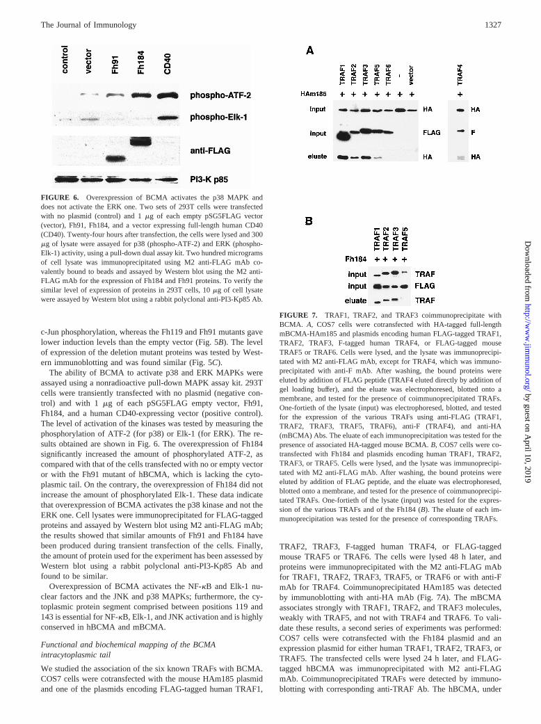
The ability of BCMA to activate p38 and ERK MAPKs wereassayed using a nonradioactive pull-down MAPK assay kit. 293Tcells were transiently transfected with no plasmid (negative con-trol) and with 1 mg of each pSG5FLAG empty vector, Fh91,Fh184, and a human CD40-expressing vector (positive control).The level of activation of the kinases was tested by measuring thephosphorylation of ATF-2 (for p38) or Elk-1 (for ERK). The re-sults obtained are shown in Fig. 6. The overexpression of Fh184significantly increased the amount of phosphorylated ATF-2, ascompared with that of the cells transfected with no or empty vectoror with the Fh91 mutant of hBCMA, which is lacking the cyto-plasmic tail. On the contrary, the overexpression of Fh184 did notincrease the amount of phosphorylated Elk-1. These data indicatethat overexpression of BCMA activates the p38 kinase and not theERK one. Cell lysates were immunoprecipitated for FLAG-taggedproteins and assayed by Western blot using M2 anti-FLAG mAb;the results showed that similar amounts of Fh91 and Fh184 havebeen produced during transient transfection of the cells. Finally,the amount of protein used for the experiment has been assessed byWestern blot using a rabbit polyclonal anti-PI3-Kp85 Ab andfound to be similar.
Overexpression of BCMA activates the NF-kB and Elk-1 nu-clear factors and the JNK and p38 MAPKs; furthermore, the cy-toplasmic protein segment comprised between positions 119 and143 is essential for NF-kB, Elk-1, and JNK activation and is highlyconserved in hBCMA and mBCMA.
Functional and biochemical mapping of the BCMAintracytoplasmic tail
We studied the association of the six known TRAFs with BCMA.COS7 cells were cotransfected with the mouse HAm185 plasmidand one of the plasmids encoding FLAG-tagged human TRAF1,
TRAF2, TRAF3, F-tagged human TRAF4, or FLAG-taggedmouse TRAF5 or TRAF6. The cells were lysed 48 h later, andproteins were immunoprecipitated with the M2 anti-FLAG mAbfor TRAF1, TRAF2, TRAF3, TRAF5, or TRAF6 or with anti-FmAb for TRAF4. Coimmunoprecipitated HAm185 was detectedby immunoblotting with anti-HA mAb (Fig. 7A). The mBCMAassociates strongly with TRAF1, TRAF2, and TRAF3 molecules,weakly with TRAF5, and not with TRAF4 and TRAF6. To vali-date these results, a second series of experiments was performed:COS7 cells were cotransfected with the Fh184 plasmid and anexpression plasmid for either human TRAF1, TRAF2, TRAF3, orTRAF5. The transfected cells were lysed 24 h later, and FLAG-tagged hBCMA was immunoprecipitated with M2 anti-FLAGmAb. Coimmunoprecipitated TRAFs were detected by immuno-blotting with corresponding anti-TRAF Ab. The hBCMA, under
FIGURE 6. Overexpression of BCMA activates the p38 MAPK anddoes not activate the ERK one. Two sets of 293T cells were transfectedwith no plasmid (control) and 1mg of each empty pSG5FLAG vector(vector), Fh91, Fh184, and a vector expressing full-length human CD40(CD40). Twenty-four hours after transfection, the cells were lysed and 300mg of lysate were assayed for p38 (phospho-ATF-2) and ERK (phospho-Elk-1) activity, using a pull-down dual assay kit. Two hundred microgramsof cell lysate was immunoprecipitated using M2 anti-FLAG mAb co-valently bound to beads and assayed by Western blot using the M2 anti-FLAG mAb for the expression of Fh184 and Fh91 proteins. To verify thesimilar level of expression of proteins in 293T cells, 10mg of cell lysatewere assayed by Western blot using a rabbit polyclonal anti-PI3-Kp85 Ab.
FIGURE 7. TRAF1, TRAF2, and TRAF3 coimmunoprecipitate withBCMA. A, COS7 cells were cotransfected with HA-tagged full-lengthmBCMA-HAm185 and plasmids encoding human FLAG-tagged TRAF1,TRAF2, TRAF3, F-tagged human TRAF4, or FLAG-tagged mouseTRAF5 or TRAF6. Cells were lysed, and the lysate was immunoprecipi-tated with M2 anti-FLAG mAb, except for TRAF4, which was immuno-precipitated with anti-F mAb. After washing, the bound proteins wereeluted by addition of FLAG peptide (TRAF4 eluted directly by addition ofgel loading buffer), and the eluate was electrophoresed, blotted onto amembrane, and tested for the presence of coimmunoprecipitated TRAFs.One-fortieth of the lysate (input) was electrophoresed, blotted, and testedfor the expression of the various TRAFs using anti-FLAG (TRAF1,TRAF2, TRAF3, TRAF5, TRAF6), anti-F (TRAF4), and anti-HA(mBCMA) Abs. The eluate of each immunoprecipitation was tested for thepresence of associated HA-tagged mouse BCMA.B, COS7 cells were co-transfected with Fh184 and plasmids encoding human TRAF1, TRAF2,TRAF3, or TRAF5. Cells were lysed, and the lysate was immunoprecipi-tated with M2 anti-FLAG mAb. After washing, the bound proteins wereeluted by addition of FLAG peptide, and the eluate was electrophoresed,blotted onto a membrane, and tested for the presence of coimmunoprecipi-tated TRAFs. One-fortieth of the lysate (input) was tested for the expres-sion of the various TRAFs and of the Fh184 (B). The eluate of each im-munoprecipitation was tested for the presence of corresponding TRAFs.
1327The Journal of Immunology
by guest on April 10, 2019
http://ww
w.jim
munol.org/
Dow
nloaded from
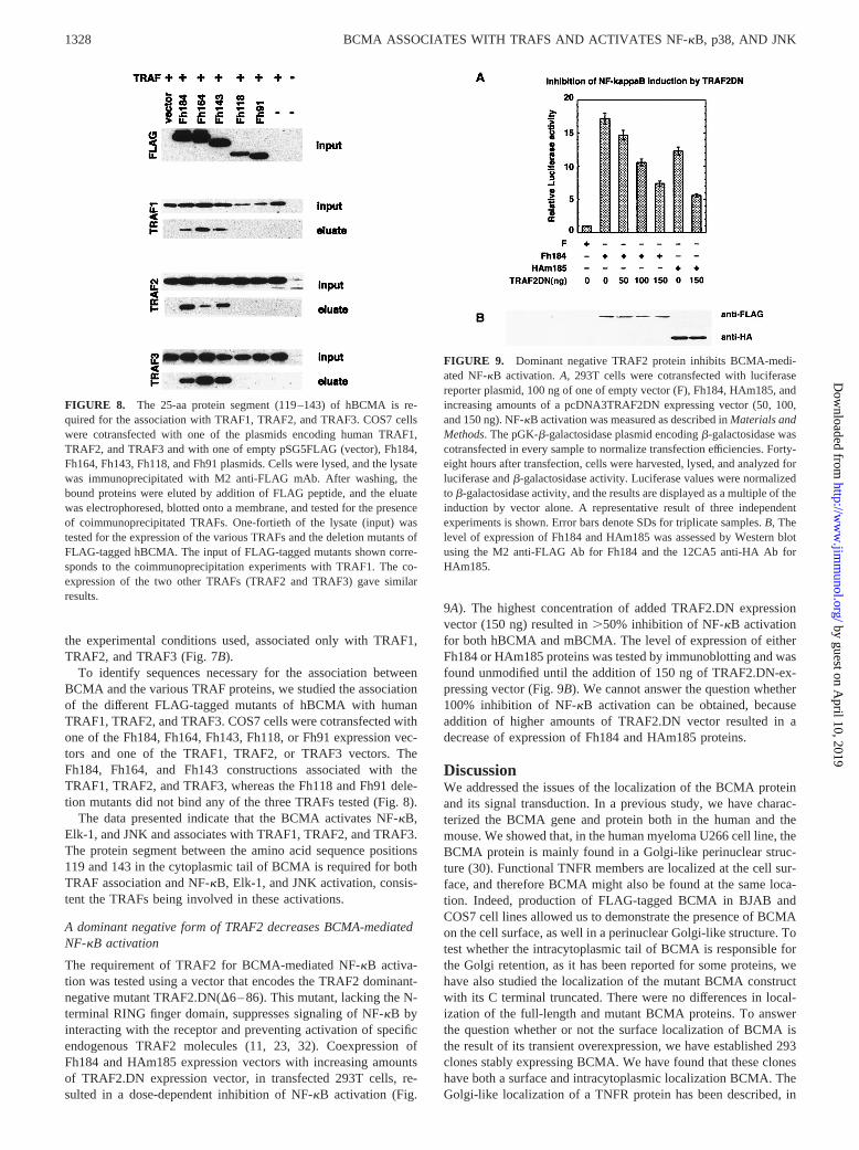
the experimental conditions used, associated only with TRAF1,TRAF2, and TRAF3 (Fig. 7B).
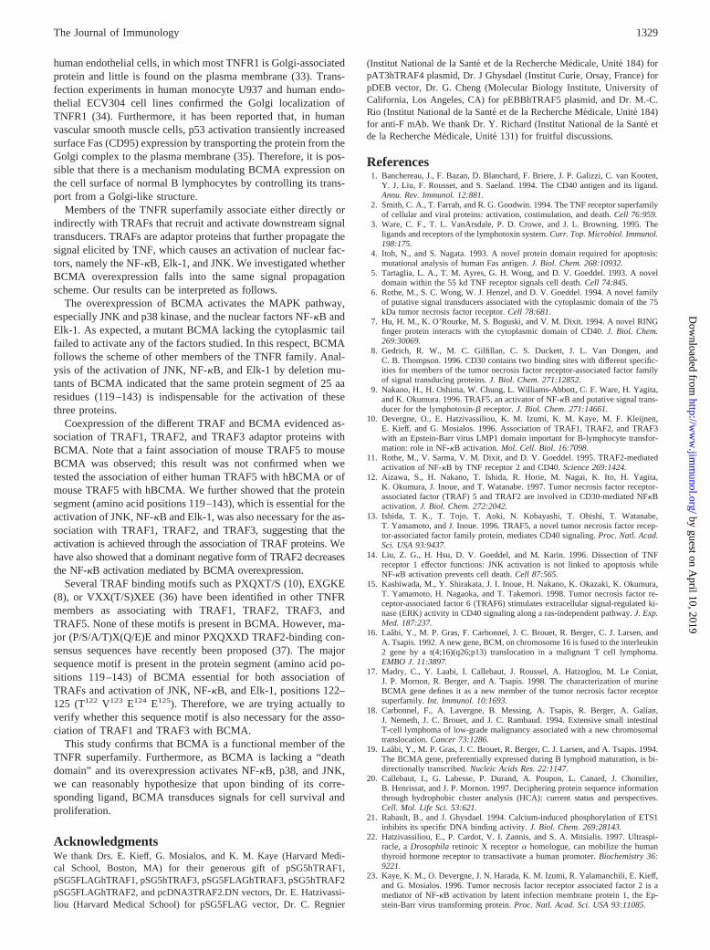
To identify sequences necessary for the association betweenBCMA and the various TRAF proteins, we studied the associationof the different FLAG-tagged mutants of hBCMA with humanTRAF1, TRAF2, and TRAF3. COS7 cells were cotransfected withone of the Fh184, Fh164, Fh143, Fh118, or Fh91 expression vec-tors and one of the TRAF1, TRAF2, or TRAF3 vectors. TheFh184, Fh164, and Fh143 constructions associated with theTRAF1, TRAF2, and TRAF3, whereas the Fh118 and Fh91 dele-tion mutants did not bind any of the three TRAFs tested (Fig. 8).
The data presented indicate that the BCMA activates NF-kB,Elk-1, and JNK and associates with TRAF1, TRAF2, and TRAF3.The protein segment between the amino acid sequence positions119 and 143 in the cytoplasmic tail of BCMA is required for bothTRAF association and NF-kB, Elk-1, and JNK activation, consis-tent the TRAFs being involved in these activations.
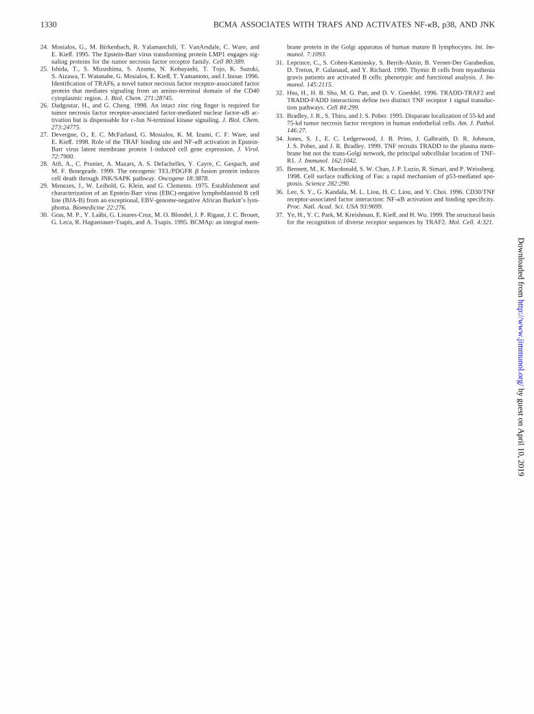
A dominant negative form of TRAF2 decreases BCMA-mediatedNF-kB activation
The requirement of TRAF2 for BCMA-mediated NF-kB activa-tion was tested using a vector that encodes the TRAF2 dominant-negative mutant TRAF2.DN(D6–86). This mutant, lacking the N-terminal RING finger domain, suppresses signaling of NF-kB byinteracting with the receptor and preventing activation of specificendogenous TRAF2 molecules (11, 23, 32). Coexpression ofFh184 and HAm185 expression vectors with increasing amountsof TRAF2.DN expression vector, in transfected 293T cells, re-sulted in a dose-dependent inhibition of NF-kB activation (Fig.
9A). The highest concentration of added TRAF2.DN expressionvector (150 ng) resulted in.50% inhibition of NF-kB activationfor both hBCMA and mBCMA. The level of expression of eitherFh184 or HAm185 proteins was tested by immunoblotting and wasfound unmodified until the addition of 150 ng of TRAF2.DN-ex-pressing vector (Fig. 9B). We cannot answer the question whether100% inhibition of NF-kB activation can be obtained, becauseaddition of higher amounts of TRAF2.DN vector resulted in adecrease of expression of Fh184 and HAm185 proteins.
DiscussionWe addressed the issues of the localization of the BCMA proteinand its signal transduction. In a previous study, we have charac-terized the BCMA gene and protein both in the human and themouse. We showed that, in the human myeloma U266 cell line, theBCMA protein is mainly found in a Golgi-like perinuclear struc-ture (30). Functional TNFR members are localized at the cell sur-face, and therefore BCMA might also be found at the same loca-tion. Indeed, production of FLAG-tagged BCMA in BJAB andCOS7 cell lines allowed us to demonstrate the presence of BCMAon the cell surface, as well in a perinuclear Golgi-like structure. Totest whether the intracytoplasmic tail of BCMA is responsible forthe Golgi retention, as it has been reported for some proteins, wehave also studied the localization of the mutant BCMA constructwith its C terminal truncated. There were no differences in local-ization of the full-length and mutant BCMA proteins. To answerthe question whether or not the surface localization of BCMA isthe result of its transient overexpression, we have established 293clones stably expressing BCMA. We have found that these cloneshave both a surface and intracytoplasmic localization BCMA. TheGolgi-like localization of a TNFR protein has been described, in
FIGURE 8. The 25-aa protein segment (119–143) of hBCMA is re-quired for the association with TRAF1, TRAF2, and TRAF3. COS7 cellswere cotransfected with one of the plasmids encoding human TRAF1,TRAF2, and TRAF3 and with one of empty pSG5FLAG (vector), Fh184,Fh164, Fh143, Fh118, and Fh91 plasmids. Cells were lysed, and the lysatewas immunoprecipitated with M2 anti-FLAG mAb. After washing, thebound proteins were eluted by addition of FLAG peptide, and the eluatewas electrophoresed, blotted onto a membrane, and tested for the presenceof coimmunoprecipitated TRAFs. One-fortieth of the lysate (input) wastested for the expression of the various TRAFs and the deletion mutants ofFLAG-tagged hBCMA. The input of FLAG-tagged mutants shown corre-sponds to the coimmunoprecipitation experiments with TRAF1. The co-expression of the two other TRAFs (TRAF2 and TRAF3) gave similarresults.
FIGURE 9. Dominant negative TRAF2 protein inhibits BCMA-medi-ated NF-kB activation.A, 293T cells were cotransfected with luciferasereporter plasmid, 100 ng of one of empty vector (F), Fh184, HAm185, andincreasing amounts of a pcDNA3TRAF2DN expressing vector (50, 100,and 150 ng). NF-kB activation was measured as described inMaterials andMethods. The pGK-b-galactosidase plasmid encodingb-galactosidase wascotransfected in every sample to normalize transfection efficiencies. Forty-eight hours after transfection, cells were harvested, lysed, and analyzed forluciferase andb-galactosidase activity. Luciferase values were normalizedto b-galactosidase activity, and the results are displayed as a multiple of theinduction by vector alone. A representative result of three independentexperiments is shown. Error bars denote SDs for triplicate samples.B, Thelevel of expression of Fh184 and HAm185 was assessed by Western blotusing the M2 anti-FLAG Ab for Fh184 and the 12CA5 anti-HA Ab forHAm185.
1328 BCMA ASSOCIATES WITH TRAFS AND ACTIVATES NF-kB, p38, AND JNK
by guest on April 10, 2019
http://ww
w.jim
munol.org/
Dow
nloaded from
human endothelial cells, in which most TNFR1 is Golgi-associatedprotein and little is found on the plasma membrane (33). Trans-fection experiments in human monocyte U937 and human endo-thelial ECV304 cell lines confirmed the Golgi localization ofTNFR1 (34). Furthermore, it has been reported that, in humanvascular smooth muscle cells, p53 activation transiently increasedsurface Fas (CD95) expression by transporting the protein from theGolgi complex to the plasma membrane (35). Therefore, it is pos-sible that there is a mechanism modulating BCMA expression onthe cell surface of normal B lymphocytes by controlling its trans-port from a Golgi-like structure.
Members of the TNFR superfamily associate either directly orindirectly with TRAFs that recruit and activate downstream signaltransducers. TRAFs are adaptor proteins that further propagate thesignal elicited by TNF, which causes an activation of nuclear fac-tors, namely the NF-kB, Elk-1, and JNK. We investigated whetherBCMA overexpression falls into the same signal propagationscheme. Our results can be interpreted as follows.
The overexpression of BCMA activates the MAPK pathway,especially JNK and p38 kinase, and the nuclear factors NF-kB andElk-1. As expected, a mutant BCMA lacking the cytoplasmic tailfailed to activate any of the factors studied. In this respect, BCMAfollows the scheme of other members of the TNFR family. Anal-ysis of the activation of JNK, NF-kB, and Elk-1 by deletion mu-tants of BCMA indicated that the same protein segment of 25 aaresidues (119–143) is indispensable for the activation of thesethree proteins.
Coexpression of the different TRAF and BCMA evidenced as-sociation of TRAF1, TRAF2, and TRAF3 adaptor proteins withBCMA. Note that a faint association of mouse TRAF5 to mouseBCMA was observed; this result was not confirmed when wetested the association of either human TRAF5 with hBCMA or ofmouse TRAF5 with hBCMA. We further showed that the proteinsegment (amino acid positions 119–143), which is essential for theactivation of JNK, NF-kB and Elk-1, was also necessary for the as-sociation with TRAF1, TRAF2, andTRAF3, suggesting that theactivation is achieved through the association of TRAF proteins. Wehave also showed that a dominant negative form of TRAF2 decreasesthe NF-kB activation mediated by BCMA overexpression.
Several TRAF binding motifs such as PXQXT/S (10), EXGKE(8), or VXX(T/S)XEE (36) have been identified in other TNFRmembers as associating with TRAF1, TRAF2, TRAF3, andTRAF5. None of these motifs is present in BCMA. However, ma-jor (P/S/A/T)X(Q/E)E and minor PXQXXD TRAF2-binding con-sensus sequences have recently been proposed (37). The majorsequence motif is present in the protein segment (amino acid po-sitions 119–143) of BCMA essential for both association ofTRAFs and activation of JNK, NF-kB, and Elk-1, positions 122–125 (T122 V123 E124 E125). Therefore, we are trying actually toverify whether this sequence motif is also necessary for the asso-ciation of TRAF1 and TRAF3 with BCMA.
This study confirms that BCMA is a functional member of theTNFR superfamily. Furthermore, as BCMA is lacking a “deathdomain” and its overexpression activates NF-kB, p38, and JNK,we can reasonably hypothesize that upon binding of its corre-sponding ligand, BCMA transduces signals for cell survival andproliferation.
AcknowledgmentsWe thank Drs. E. Kieff, G. Mosialos, and K. M. Kaye (Harvard Medi-cal School, Boston, MA) for their generous gift of pSG5hTRAF1,pSG5FLAGhTRAF1, pSG5hTRAF3, pSG5FLAGhTRAF3, pSG5hTRAF2pSG5FLAGhTRAF2, and pcDNA3TRAF2.DN vectors, Dr. E. Hatzivassi-liou (Harvard Medical School) for pSG5FLAG vector, Dr. C. Regnier
(Institut National de la Sante et de la Recherche Medicale, Unite 184) forpAT3hTRAF4 plasmid, Dr. J Ghysdael (Institut Curie, Orsay, France) forpDEB vector, Dr. G. Cheng (Molecular Biology Institute, University ofCalifornia, Los Angeles, CA) for pEBBhTRAF5 plasmid, and Dr. M.-C.Rio (Institut National de la Sante et de la Recherche Medicale, Unite 184)for anti-F mAb. We thank Dr. Y. Richard (Institut National de la Sante´ etde la Recherche Medicale, Unite 131) for fruitful discussions.
References1. Banchereau, J., F. Bazan, D. Blanchard, F. Briere, J. P. Galizzi, C. van Kooten,
Y. J. Liu, F. Rousset, and S. Saeland. 1994. The CD40 antigen and its ligand.Annu. Rev. Immunol. 12:881.
2. Smith, C. A., T. Farrah, and R. G. Goodwin. 1994. The TNF receptor superfamilyof cellular and viral proteins: activation, costimulation, and death.Cell 76:959.
3. Ware, C. F., T. L. VanArsdale, P. D. Crowe, and J. L. Browning. 1995. Theligands and receptors of the lymphotoxin system.Curr. Top. Microbiol. Immunol.198:175.
4. Itoh, N., and S. Nagata. 1993. A novel protein domain required for apoptosis:mutational analysis of human Fas antigen.J. Biol. Chem. 268:10932.
5. Tartaglia, L. A., T. M. Ayres, G. H. Wong, and D. V. Goeddel. 1993. A noveldomain within the 55 kd TNF receptor signals cell death.Cell 74:845.
6. Rothe, M., S. C. Wong, W. J. Henzel, and D. V. Goeddel. 1994. A novel familyof putative signal transducers associated with the cytoplasmic domain of the 75kDa tumor necrosis factor receptor.Cell 78:681.
7. Hu, H. M., K. O’Rourke, M. S. Boguski, and V. M. Dixit. 1994. A novel RINGfinger protein interacts with the cytoplasmic domain of CD40.J. Biol. Chem.269:30069.
8. Gedrich, R. W., M. C. Gilfillan, C. S. Duckett, J. L. Van Dongen, andC. B. Thompson. 1996. CD30 contains two binding sites with different specific-ities for members of the tumor necrosis factor receptor-associated factor familyof signal transducing proteins.J. Biol. Chem. 271:12852.
9. Nakano, H., H. Oshima, W. Chung, L. Williams-Abbott, C. F. Ware, H. Yagita,and K. Okumura. 1996. TRAF5, an activator of NF-kB and putative signal trans-ducer for the lymphotoxin-b receptor.J. Biol. Chem. 271:14661.
10. Devergne, O., E. Hatzivassiliou, K. M. Izumi, K. M. Kaye, M. F. Kleijnen,E. Kieff, and G. Mosialos. 1996. Association of TRAF1, TRAF2, and TRAF3with an Epstein-Barr virus LMP1 domain important for B-lymphocyte transfor-mation: role in NF-kB activation.Mol. Cell. Biol. 16:7098.
11. Rothe, M., V. Sarma, V. M. Dixit, and D. V. Goeddel. 1995. TRAF2-mediatedactivation of NF-kB by TNF receptor 2 and CD40.Science 269:1424.
12. Aizawa, S., H. Nakano, T. Ishida, R. Horie, M. Nagai, K. Ito, H. Yagita,K. Okumura, J. Inoue, and T. Watanabe. 1997. Tumor necrosis factor receptor-associated factor (TRAF) 5 and TRAF2 are involved in CD30-mediated NFkBactivation.J. Biol. Chem. 272:2042.
13. Ishida, T. K., T. Tojo, T. Aoki, N. Kobayashi, T. Ohishi, T. Watanabe,T. Yamamoto, and J. Inoue. 1996. TRAF5, a novel tumor necrosis factor recep-tor-associated factor family protein, mediates CD40 signaling.Proc. Natl. Acad.Sci. USA 93:9437.
14. Liu, Z. G., H. Hsu, D. V. Goeddel, and M. Karin. 1996. Dissection of TNFreceptor 1 effector functions: JNK activation is not linked to apoptosis whileNF-kB activation prevents cell death.Cell 87:565.
15. Kashiwada, M., Y. Shirakata, J. I. Inoue, H. Nakano, K. Okazaki, K. Okumura,T. Yamamoto, H. Nagaoka, and T. Takemori. 1998. Tumor necrosis factor re-ceptor-associated factor 6 (TRAF6) stimulates extracellular signal-regulated ki-nase (ERK) activity in CD40 signaling along a ras-independent pathway.J. Exp.Med. 187:237.
16. Laabi, Y., M. P. Gras, F. Carbonnel, J. C. Brouet, R. Berger, C. J. Larsen, andA. Tsapis. 1992. A new gene, BCM, on chromosome 16 is fused to the interleukin2 gene by a t(4;16)(q26;p13) translocation in a malignant T cell lymphoma.EMBO J. 11:3897.
17. Madry, C., Y. Laabi, I. Callebaut, J. Roussel, A. Hatzoglou, M. Le Coniat,J. P. Mornon, R. Berger, and A. Tsapis. 1998. The characterization of murineBCMA gene defines it as a new member of the tumor necrosis factor receptorsuperfamily.Int. Immunol. 10:1693.
18. Carbonnel, F., A. Lavergne, B. Messing, A. Tsapis, R. Berger, A. Galian,J. Nemeth, J. C. Brouet, and J. C. Rambaud. 1994. Extensive small intestinalT-cell lymphoma of low-grade malignancy associated with a new chromosomaltranslocation.Cancer 73:1286.
19. Laabi, Y., M. P. Gras, J. C. Brouet, R. Berger, C. J. Larsen, and A. Tsapis. 1994.The BCMA gene, preferentially expressed during B lymphoid maturation, is bi-directionally transcribed.Nucleic Acids Res. 22:1147.
20. Callebaut, I., G. Labesse, P. Durand, A. Poupon, L. Canard, J. Chomilier,B. Henrissat, and J. P. Mornon. 1997. Deciphering protein sequence informationthrough hydrophobic cluster analysis (HCA): current status and perspectives.Cell. Mol. Life Sci. 53:621.
21. Rabault, B., and J. Ghysdael. 1994. Calcium-induced phosphorylation of ETS1inhibits its specific DNA binding activity.J. Biol. Chem. 269:28143.
22. Hatzivassiliou, E., P. Cardot, V. I. Zannis, and S. A. Mitsialis. 1997. Ultraspi-racle, aDrosophila retinoic X receptora homologue, can mobilize the humanthyroid hormone receptor to transactivate a human promoter.Biochemistry 36:9221.
23. Kaye, K. M., O. Devergne, J. N. Harada, K. M. Izumi, R. Yalamanchili, E. Kieff,and G. Mosialos. 1996. Tumor necrosis factor receptor associated factor 2 is amediator of NF-kB activation by latent infection membrane protein 1, the Ep-stein-Barr virus transforming protein.Proc. Natl. Acad. Sci. USA 93:11085.
1329The Journal of Immunology
by guest on April 10, 2019
http://ww
w.jim
munol.org/
Dow
nloaded from
24. Mosialos, G., M. Birkenbach, R. Yalamanchili, T. VanArsdale, C. Ware, andE. Kieff. 1995. The Epstein-Barr virus transforming protein LMP1 engages sig-naling proteins for the tumor necrosis factor receptor family.Cell 80:389.
25. Ishida, T., S. Mizushima, S. Azuma, N. Kobayashi, T. Tojo, K. Suzuki,S. Aizawa, T. Watanabe, G. Mosialos, E. Kieff, T. Yamamoto, and J. Inoue. 1996.Identification of TRAF6, a novel tumor necrosis factor receptor-associated factorprotein that mediates signaling from an amino-terminal domain of the CD40cytoplasmic region.J. Biol. Chem. 271:28745.
26. Dadgostar, H., and G. Cheng. 1998. An intact zinc ring finger is required fortumor necrosis factor receptor-associated factor-mediated nuclear factor-kB ac-tivation but is dispensable for c-Jun N-terminal kinase signaling.J. Biol. Chem.273:24775.
27. Devergne, O., E. C. McFarland, G. Mosialos, K. M. Izumi, C. F. Ware, andE. Kieff. 1998. Role of the TRAF binding site and NF-kB activation in Epstein-Barr virus latent membrane protein 1-induced cell gene expression.J. Virol.72:7900.
28. Atfi, A., C. Prunier, A. Mazars, A. S. Defachelles, Y. Cayre, C. Gespach, andM. F. Bourgeade. 1999. The oncogenic TEL/PDGFRb fusion protein inducescell death through JNK/SAPK pathway.Oncogene 18:3878.
29. Menezes, J., W. Leibold, G. Klein, and G. Clements. 1975. Establishment andcharacterization of an Epstein-Barr virus (EBC)-negative lymphoblastoid B cellline (BJA-B) from an exceptional, EBV-genome-negative African Burkitt’s lym-phoma.Biomedicine 22:276.
30. Gras, M. P., Y. Laabi, G. Linares-Cruz, M. O. Blondel, J. P. Rigaut, J. C. Brouet,G. Leca, R. Haguenauer-Tsapis, and A. Tsapis. 1995. BCMAp: an integral mem-
brane protein in the Golgi apparatus of human mature B lymphocytes.Int. Im-munol. 7:1093.
31. Leprince, C., S. Cohen-Kaminsky, S. Berrih-Aknin, B. Vernet-Der Garabedian,D. Treton, P. Galanaud, and Y. Richard. 1990. Thymic B cells from myastheniagravis patients are activated B cells: phenotypic and functional analysis.J. Im-munol. 145:2115.
32. Hsu, H., H. B. Shu, M. G. Pan, and D. V. Goeddel. 1996. TRADD-TRAF2 andTRADD-FADD interactions define two distinct TNF receptor 1 signal transduc-tion pathways.Cell 84:299.
33. Bradley, J. R., S. Thiru, and J. S. Pober. 1995. Disparate localization of 55-kd and75-kd tumor necrosis factor receptors in human endothelial cells.Am. J. Pathol.146:27.
34. Jones, S. J., E. C. Ledgerwood, J. B. Prins, J. Galbraith, D. R. Johnson,J. S. Pober, and J. R. Bradley. 1999. TNF recruits TRADD to the plasma mem-brane but not the trans-Golgi network, the principal subcellular location of TNF-R1. J. Immunol. 162:1042.
35. Bennett, M., K. Macdonald, S. W. Chan, J. P. Luzio, R. Simari, and P. Weissberg.1998. Cell surface trafficking of Fas: a rapid mechanism of p53-mediated apo-ptosis.Science 282:290.
36. Lee, S. Y., G. Kandala, M. L. Liou, H. C. Liou, and Y. Choi. 1996. CD30/TNFreceptor-associated factor interaction: NF-kB activation and binding specificity.Proc. Natl. Acad. Sci. USA 93:9699.
37. Ye, H., Y. C. Park, M. Kreishman, E. Kieff, and H. Wu. 1999. The structural basisfor the recognition of diverse receptor sequences by TRAF2.Mol. Cell. 4:321.
1330 BCMA ASSOCIATES WITH TRAFS AND ACTIVATES NF-kB, p38, AND JNK
by guest on April 10, 2019
http://ww
w.jim
munol.org/
Dow
nloaded from