Timing and completion of puberty in female mice depend on … › content › pnas › 107 › 52...
6
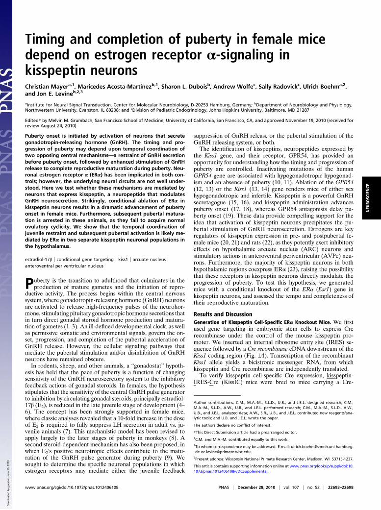
Timing and completion of puberty in female mice depend on estrogen receptor α-signaling in kisspeptin neurons Christian Mayer a,1 , Maricedes Acosta-Martinez b,1 , Sharon L. Dubois b , Andrew Wolfe c , Sally Radovick c , Ulrich Boehm a,2 , and Jon E. Levine b,2,3 a Institute for Neural Signal Transduction, Center for Molecular Neurobiology, D-20253 Hamburg, Germany; b Department of Neurobiology and Physiology, Northwestern University, Evanston, IL 60208; and c Division of Pediatric Endocrinology, Johns Hopkins University, Baltimore, MD 21287 Edited* by Melvin M. Grumbach, San Francisco School of Medicine, University of California, San Francisco, CA, and approved November 19, 2010 (received for review August 24, 2010) Puberty onset is initiated by activation of neurons that secrete gonadotropin-releasing hormone (GnRH). The timing and pro- gression of puberty may depend upon temporal coordination of two opposing central mechanisms—a restraint of GnRH secretion before puberty onset, followed by enhanced stimulation of GnRH release to complete reproductive maturation during puberty. Neu- ronal estrogen receptor α (ERα) has been implicated in both con- trols; however, the underlying neural circuits are not well under- stood. Here we test whether these mechanisms are mediated by neurons that express kisspeptin, a neuropeptide that modulates GnRH neurosecretion. Strikingly, conditional ablation of ERα in kisspeptin neurons results in a dramatic advancement of puberty onset in female mice. Furthermore, subsequent pubertal matura- tion is arrested in these animals, as they fail to acquire normal ovulatory cyclicity. We show that the temporal coordination of juvenile restraint and subsequent pubertal activation is likely me- diated by ERα in two separate kisspeptin neuronal populations in the hypothalamus. estradiol-17β | conditional gene targeting | kiss1 | arcuate nucleus | anteroventral periventricular nucleus P uberty is the transition to adulthood that culminates in the production of mature gametes and the initiation of repro- ductive activity. The process begins within the central nervous system, where gonadotropin-releasing hormone (GnRH) neurons are activated to release high-frequency pulses of the neurohor- mone, stimulating pituitary gonadotropic hormone secretions that in turn direct gonadal steroid hormone production and matura- tion of gametes (1–3). An ill-defined developmental clock, as well as permissive somatic and environmental signals, govern the on- set, progression, and completion of the pubertal acceleration of GnRH release. However, the cellular signaling pathways that mediate the pubertal stimulation and/or disinhibition of GnRH neurons have remained obscure. In rodents, sheep, and other animals, a “gonadostat” hypoth- esis has held that the pace of puberty is a function of changing sensitivity of the GnRH neurosecretory system to the inhibitory feedback actions of gonadal steroids. In females, the hypothesis stipulates that the sensitivity of the central GnRH pulse generator to inhibition by circulating gonadal steroids, principally estradiol- 17β (E 2 ), is reduced in the late juvenile stage of development (4– 6). The concept has been strongly supported in female mice, where classic analyses revealed that a 10-fold increase in the dose of E 2 is required to fully suppress LH secretion in adult vs. ju- venile animals (7). This mechanistic model has been revised to apply largely to the later stages of puberty in monkeys (8). A second steroid-dependent mechanism has also been proposed, in which E 2 ’s positive neurotropic effects contribute to the matu- ration of the GnRH pulse generator during puberty (9). We sought to determine the specific neuronal populations in which estrogen receptors may mediate either the juvenile feedback suppression of GnRH release or the pubertal stimulation of the GnRH releasing system, or both. The identification of kisspeptins, neuropeptides expressed by the Kiss1 gene, and their receptor, GPR54, has provided an opportunity for understanding how the timing and progression of puberty are controlled. Inactivating mutations of the human GPR54 gene are associated with hypogonadotropic hypogonad- ism and an absence of puberty (10, 11). Ablation of the GPR54 (12, 13) or the Kiss1 (13, 14) gene renders mice of either sex hypogonadotropic and infertile. Kisspeptin is a powerful GnRH secretagogue (15, 16), and kisspeptin administration advances puberty onset (17, 18), whereas GPR54 antagonists delay pu- berty onset (19). These data provide compelling support for the idea that activation of kisspeptin neurons precipitates the pu- bertal stimulation of GnRH neurosecretion. Estrogens are key regulators of kisspeptin expression in pre- and postpubertal fe- male mice (20, 21) and rats (22), as they potently exert inhibitory effects on hypothalamic arcuate nucleus (ARC) neurons and stimulatory actions in anteroventral periventricular (AVPe) neu- rons. Furthermore, the majority of kisspeptin neurons in both hypothalamic regions coexpress ERα (23), raising the possibility that these receptors in kisspeptin neurons directly modulate the progression of puberty. To test this hypothesis, we generated mice with a conditional knockout of the ERα (Esr1) gene in kisspeptin neurons, and assessed the tempo and completeness of their reproductive maturation. Results and Discussion Generation of Kisspeptin Cell-Specific ERα Knockout Mice. We first used gene targeting in embryonic stem cells to express Cre recombinase under the control of the mouse kisspeptin pro- moter. We inserted an internal ribosome entry site (IRES) se- quence followed by a Cre recombinase cDNA downstream of the Kiss1 coding region (Fig. 1A). Transcription of the recombinant Kiss1 allele yields a bicistronic messenger RNA, from which kisspeptin and Cre recombinase are independently translated. To verify kisspeptin cell-specific Cre expression, kisspeptin- IRES- Cre (KissIC) mice were bred to mice carrying a Cre- Author contributions: C.M., M.A.-M., S.L.D., U.B., and J.E.L. designed research; C.M., M.A.-M., S.L.D., A.W., U.B., and J.E.L. performed research; C.M., M.A.-M., S.L.D., A.W., U.B., and J.E.L. analyzed data; A.W., S.R., U.B., and J.E.L. contributed new reagents/ana- lytic tools; and U.B. and J.E.L. wrote the paper. The authors declare no conflict of interest. *This Direct Submission article had a prearranged editor. 1 C.M. and M.A.-M. contributed equally to this work. 2 To whom correspondence may be addressed. E-mail: [email protected]. de or [email protected]. 3 Present address: Wisconsin National Primate Research Center, Madison, WI 53715-1237. This article contains supporting information online at www.pnas.org/lookup/suppl/doi:10. 1073/pnas.1012406108/-/DCSupplemental. www.pnas.org/cgi/doi/10.1073/pnas.1012406108 PNAS | December 28, 2010 | vol. 107 | no. 52 | 22693–22698 NEUROSCIENCE Downloaded by guest on June 13, 2020
Transcript of Timing and completion of puberty in female mice depend on … › content › pnas › 107 › 52...
Timing and completion of puberty in female micedepend on estrogen receptor α-signaling inkisspeptin neuronsChristian Mayera,1, Maricedes Acosta-Martinezb,1, Sharon L. Duboisb, Andrew Wolfec, Sally Radovickc, Ulrich Boehma,2,and Jon E. Levineb,2,3
aInstitute for Neural Signal Transduction, Center for Molecular Neurobiology, D-20253 Hamburg, Germany; bDepartment of Neurobiology and Physiology,Northwestern University, Evanston, IL 60208; and cDivision of Pediatric Endocrinology, Johns Hopkins University, Baltimore, MD 21287
Edited* by Melvin M. Grumbach, San Francisco School of Medicine, University of California, San Francisco, CA, and approved November 19, 2010 (received forreview August 24, 2010)
Puberty onset is initiated by activation of neurons that secretegonadotropin-releasing hormone (GnRH). The timing and pro-gression of puberty may depend upon temporal coordination oftwo opposing central mechanisms—a restraint of GnRH secretionbefore puberty onset, followed by enhanced stimulation of GnRHrelease to complete reproductive maturation during puberty. Neu-ronal estrogen receptor α (ERα) has been implicated in both con-trols; however, the underlying neural circuits are not well under-stood. Here we test whether these mechanisms are mediated byneurons that express kisspeptin, a neuropeptide that modulatesGnRH neurosecretion. Strikingly, conditional ablation of ERα inkisspeptin neurons results in a dramatic advancement of pubertyonset in female mice. Furthermore, subsequent pubertal matura-tion is arrested in these animals, as they fail to acquire normalovulatory cyclicity. We show that the temporal coordination ofjuvenile restraint and subsequent pubertal activation is likely me-diated by ERα in two separate kisspeptin neuronal populations inthe hypothalamus.
estradiol-17β | conditional gene targeting | kiss1 | arcuate nucleus |anteroventral periventricular nucleus
Puberty is the transition to adulthood that culminates in theproduction of mature gametes and the initiation of repro-
ductive activity. The process begins within the central nervoussystem, where gonadotropin-releasing hormone (GnRH) neuronsare activated to release high-frequency pulses of the neurohor-mone, stimulating pituitary gonadotropic hormone secretions thatin turn direct gonadal steroid hormone production and matura-tion of gametes (1–3). An ill-defined developmental clock, as wellas permissive somatic and environmental signals, govern the on-set, progression, and completion of the pubertal acceleration ofGnRH release. However, the cellular signaling pathways thatmediate the pubertal stimulation and/or disinhibition of GnRHneurons have remained obscure.In rodents, sheep, and other animals, a “gonadostat” hypoth-
esis has held that the pace of puberty is a function of changingsensitivity of the GnRH neurosecretory system to the inhibitoryfeedback actions of gonadal steroids. In females, the hypothesisstipulates that the sensitivity of the central GnRH pulse generatorto inhibition by circulating gonadal steroids, principally estradiol-17β (E2), is reduced in the late juvenile stage of development (4–6). The concept has been strongly supported in female mice,where classic analyses revealed that a 10-fold increase in the doseof E2 is required to fully suppress LH secretion in adult vs. ju-venile animals (7). This mechanistic model has been revised toapply largely to the later stages of puberty in monkeys (8). Asecond steroid-dependent mechanism has also been proposed, inwhich E2’s positive neurotropic effects contribute to the matu-ration of the GnRH pulse generator during puberty (9). Wesought to determine the specific neuronal populations in whichestrogen receptors may mediate either the juvenile feedback
suppression of GnRH release or the pubertal stimulation of theGnRH releasing system, or both.The identification of kisspeptins, neuropeptides expressed by
the Kiss1 gene, and their receptor, GPR54, has provided anopportunity for understanding how the timing and progression ofpuberty are controlled. Inactivating mutations of the humanGPR54 gene are associated with hypogonadotropic hypogonad-ism and an absence of puberty (10, 11). Ablation of the GPR54(12, 13) or the Kiss1 (13, 14) gene renders mice of either sexhypogonadotropic and infertile. Kisspeptin is a powerful GnRHsecretagogue (15, 16), and kisspeptin administration advancespuberty onset (17, 18), whereas GPR54 antagonists delay pu-berty onset (19). These data provide compelling support for theidea that activation of kisspeptin neurons precipitates the pu-bertal stimulation of GnRH neurosecretion. Estrogens are keyregulators of kisspeptin expression in pre- and postpubertal fe-male mice (20, 21) and rats (22), as they potently exert inhibitoryeffects on hypothalamic arcuate nucleus (ARC) neurons andstimulatory actions in anteroventral periventricular (AVPe) neu-rons. Furthermore, the majority of kisspeptin neurons in bothhypothalamic regions coexpress ERα (23), raising the possibilitythat these receptors in kisspeptin neurons directly modulate theprogression of puberty. To test this hypothesis, we generatedmice with a conditional knockout of the ERα (Esr1) gene inkisspeptin neurons, and assessed the tempo and completeness oftheir reproductive maturation.
Results and DiscussionGeneration of Kisspeptin Cell-Specific ERα Knockout Mice. We firstused gene targeting in embryonic stem cells to express Crerecombinase under the control of the mouse kisspeptin pro-moter. We inserted an internal ribosome entry site (IRES) se-quence followed by a Cre recombinase cDNA downstream of theKiss1 coding region (Fig. 1A). Transcription of the recombinantKiss1 allele yields a bicistronic messenger RNA, from whichkisspeptin and Cre recombinase are independently translated.To verify kisspeptin cell-specific Cre expression, kisspeptin-
IRES-Cre (KissIC) mice were bred to mice carrying a Cre-
Author contributions: C.M., M.A.-M., S.L.D., U.B., and J.E.L. designed research; C.M.,M.A.-M., S.L.D., A.W., U.B., and J.E.L. performed research; C.M., M.A.-M., S.L.D., A.W.,U.B., and J.E.L. analyzed data; A.W., S.R., U.B., and J.E.L. contributed new reagents/ana-lytic tools; and U.B. and J.E.L. wrote the paper.
The authors declare no conflict of interest.
*This Direct Submission article had a prearranged editor.1C.M. and M.A.-M. contributed equally to this work.2To whom correspondence may be addressed. E-mail: [email protected] or [email protected].
3Present address: Wisconsin National Primate Research Center, Madison, WI 53715-1237.
This article contains supporting information online at www.pnas.org/lookup/suppl/doi:10.1073/pnas.1012406108/-/DCSupplemental.
www.pnas.org/cgi/doi/10.1073/pnas.1012406108 PNAS | December 28, 2010 | vol. 107 | no. 52 | 22693–22698
NEU
ROSC
IENCE
Dow
nloa
ded
by g
uest
on
June
13,
202
0
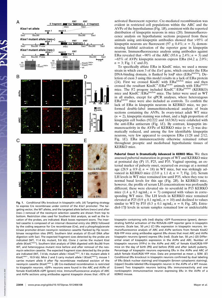
activated fluorescent reporter. Cre-mediated recombination wasevident in restricted cell populations within the ARC and theAVPe of the hypothalamus (Fig. 1B), consistent with the reporteddistribution of kisspeptin neurons in mice (20). Immunofluores-cence analysis on hypothalamic sections prepared from theseanimals using anti-kisspeptin antibodies showed that >95% ofkisspeptin neurons are fluorescent (97 ± 0.4%; n = 3), demon-strating faithful activation of the reporter gene in kisspeptinneurons. Immunofluorescence analysis using antibodies againstERα revealed that ∼90% of the ARC (93.6 ± 2.4%; n = 3) and∼65% of AVPe kisspeptin neurons express ERα (64.2 ± 2.0%;n = 3; Fig. 1 C and D).To specifically ablate ERα in KissIC mice, we used a mouse
strain in which exon 3 of the Esr1 gene, which encodes the ERαDNA-binding domain, is flanked by loxP sites (ERαlox/lox). De-letion of exon 3 using this model results in a lack of ERα protein(24). First we crossed KissIC with ERαlox/lox mice and thencrossed the resultant KissIC+/ERαlox/wt animals with ERαlox/loxmice. The F2 progeny included KissIC+/ERαlox/lox (KERKO)mice and KissIC−/ERαlox/lox mice. The latter were used as WTin all studies, except for qPCR analyses, where heterozygousERαlox/wt mice were also included as controls. To confirm thelack of ERα in kisspeptin neurons in KERKO mice, we per-formed double-label immunohistochemical analysis of brainsections containing the AVPe. In ovary-intact adult WT mice(n = 2), kisspeptin staining was robust, and a high proportion ofkisspeptin cell bodies (92/222 and 163/363) were colabeled withthe anti-ERα antiserum (Fig. 1E). By contrast, kisspeptin im-munoreactivity in the AVPe of KERKO mice (n = 2) was dra-matically reduced, and among the few identifiable kisspeptinneurons, very few appeared to coexpress ERα (1/28 and 2/12;Fig. 1E). ERα immunoreactivity otherwise remained robustthroughout preoptic and mediobasal hypothalamic tissues ofKERKO mice.
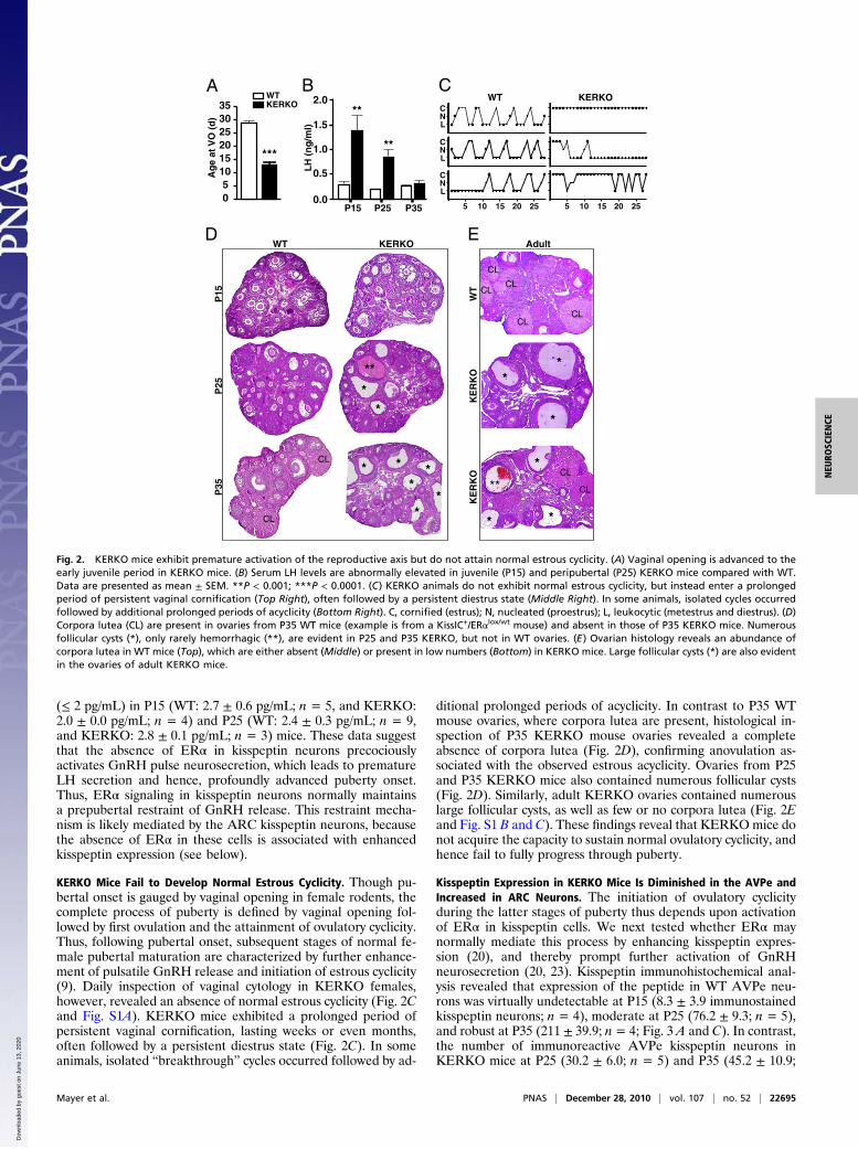
Pubertal Onset Is Dramatically Advanced in KERKO Mice. We thenassessed pubertal maturation in groups of WT and KERKOmiceat postnatal day (P) 15, P25, and P35. Vaginal opening, an ex-ternal marker of puberty onset, occurred on average at a normalage (28.9 ± 0.9 d; n = 18) in WT mice, but was strikingly ad-vanced in KERKO mice (13.0 ± 1.1 d; n = 7; Fig. 2A). SerumLH levels in WT mice remained low until P35, when they rose tonormal basal levels for this age (Fig. 2B). In KERKO mice,however, the profile of serum LH concentrations was profoundlydifferent; these were elevated six- to sevenfold in P15 KERKOmice (1.4 ± 0.3 ng/mL; n = 7) compared with values in corre-sponding WT mice. The LH levels in KERKO mice remainedelevated at P25 (0.9 ± 0.1 ng/mL; n = 10) and declined to valuessimilar to WT by P35 (0.3 ± 0.1 ng/mL; n = 8; Fig. 2B). Estra-diol-17β levels in serum samples remained low or undetectable
10.99.17.4 wt
KissICneo+ KissICneo- 11.4
9.6 KissICneo+ wt
5' fragmentIRES
CreFRT
pgk neo
exon 2exon 1
SacI
Bsu36I
exon 2exon 1
exon 2
Probe
Probe
exon 1
1 2 1 2 3 4
3' fragmentFRT
IRESCre
FRT
SacI
FRTKiss1 mutant allele (neo+)
Kiss1 mutant allele (neo-)
Kiss1 wt allele
SacISacI
SacI SacI
SacIBsu36IBsu36I
Bsu36I
Bsu36I Bsu36I
SacI
FRT
IRESCre
1 kb
pgk neo
kbkb
YFP ERα Merge
YFP ERα Merge
τGFP Kiss Merge
τGFP Kiss Merge
50µm
50µm
AV
Pe
AR
CA
VP
eA
RC
A
B
C
D
Kis
spep
tin
neu
ron
nu
mb
er
% K
issp
epti
n n
euro
ns
wit
h E
Rα
E
1000
2000
3000
AVPeARC
0P20 adult
25
50
75
100
0
WT
KERKO
Kiss & ERα
Kiss & ERα
P0
Fig. 1. Conditional ERα knockout in kisspeptin cells. (A) Targeting strategyto express Cre recombinase under control of the Kiss1 promoter. The tar-geting vector, the WT alleles, and the targeted allele before (neo+) and after(neo−) removal of the neomycin selection cassette are shown from top tobottom. Restriction sites used for Southern blot analysis, as well as the lo-cation of the probes, are indicated. Black boxes represent exons. The inser-ted cassette is composed of an internal ribosomal entry site (IRES) followedby the coding sequence for Cre recombinase (Cre), and a phosphoglyceratekinase promoter-driven neomycin resistance cassette flanked by Flp recom-binase recognition sites (FRT). Southern blot analysis of ES-cell DNA afterdigestion with SacI. The expected fragment sizes detected by the probe areindicated (WT, 11.4 kb; mutant, 9.6 kb). Clone 2 carries the mutant KissIallele (KissICneo+). Southern blot analysis of DNA digested with Bsu36I fromWT, and heterozygous mutant mice before and after removal of the neo-mycin selection cassette. The expected fragment sizes detected by the probeare indicated (WT, 7.4 kb; mutant allele I KissICneo+, 9.1 kb; mutant allele IIKissICneo−, 10.9 kb). Mice 2 and 3 carry mutant allele I (KissICneo+), mouse 1carries mutant allele II after Flp recombinase mediated excision of theneomycin cassette (KissICneo−). (B) Cre-mediated recombination is restrictedto kisspeptin neurons. τGFP+ neurons were found in the ARC and AVPe offemale KissIC/eR26-τGPF (green) mice. Immunofluorescence analysis of ARCand AVPe sections using antibodies against kisspeptin shows that >95% of
kisspeptin containing cells (red) display τGFP fluorescence (green), demon-strating faithful activation of the ROSA26-τGFP reporter gene in kisspeptinneurons. (Scale bar, 50 μm.) (C) ERα expression in kisspeptin neurons. Im-munofluorescence analysis of ARC and AVPe sections from female KissIC/R26-YFP mice using antibodies against ERα shows that most ARC and AVPekisspeptin neurons (green) express ERα (red). (Scale bar, 50 μm.) (D) Differ-ential onset of kisspeptin expression in the AVPe and ARC. Number ofkisspeptin neurons (YFP+) in the AVPe and ARC of female KissIC/R26-YFPmice on the day of birth (P0) and before (P20) and after (adult) puberty.Percentage of kisspeptin neurons expressing ERα in the AVPe and ARC ofadult female KissIC/R26-YFP mice. Data are presented as mean ± SEM. (E)Conditional ERα knockout in kisspeptin neurons confirmed by dual labelingof ERα (black nuclear staining) and kisspeptin (brown cytoplasmic staining).(Upper) Double-labeled ERα-kisspeptin neuron in the AVPe of a WT mouse.(Lower) Two kisspeptin neurons lacking ERα immunoreactivity and onenonkisspeptin immunoreactive neuron expressing ERα in the AVPe of aKERKO mouse.
22694 | www.pnas.org/cgi/doi/10.1073/pnas.1012406108 Mayer et al.
Dow
nloa
ded
by g
uest
on
June
13,
202
0
(≤ 2 pg/mL) in P15 (WT: 2.7 ± 0.6 pg/mL; n = 5, and KERKO:2.0 ± 0.0 pg/mL; n = 4) and P25 (WT: 2.4 ± 0.3 pg/mL; n = 9,and KERKO: 2.8 ± 0.1 pg/mL; n = 3) mice. These data suggestthat the absence of ERα in kisspeptin neurons precociouslyactivates GnRH pulse neurosecretion, which leads to prematureLH secretion and hence, profoundly advanced puberty onset.Thus, ERα signaling in kisspeptin neurons normally maintainsa prepubertal restraint of GnRH release. This restraint mecha-nism is likely mediated by the ARC kisspeptin neurons, becausethe absence of ERα in these cells is associated with enhancedkisspeptin expression (see below).
KERKO Mice Fail to Develop Normal Estrous Cyclicity. Though pu-bertal onset is gauged by vaginal opening in female rodents, thecomplete process of puberty is defined by vaginal opening fol-lowed by first ovulation and the attainment of ovulatory cyclicity.Thus, following pubertal onset, subsequent stages of normal fe-male pubertal maturation are characterized by further enhance-ment of pulsatile GnRH release and initiation of estrous cyclicity(9). Daily inspection of vaginal cytology in KERKO females,however, revealed an absence of normal estrous cyclicity (Fig. 2Cand Fig. S1A). KERKO mice exhibited a prolonged period ofpersistent vaginal cornification, lasting weeks or even months,often followed by a persistent diestrus state (Fig. 2C). In someanimals, isolated “breakthrough” cycles occurred followed by ad-
ditional prolonged periods of acyclicity. In contrast to P35 WTmouse ovaries, where corpora lutea are present, histological in-spection of P35 KERKO mouse ovaries revealed a completeabsence of corpora lutea (Fig. 2D), confirming anovulation as-sociated with the observed estrous acyclicity. Ovaries from P25and P35 KERKO mice also contained numerous follicular cysts(Fig. 2D). Similarly, adult KERKO ovaries contained numerouslarge follicular cysts, as well as few or no corpora lutea (Fig. 2Eand Fig. S1 B and C). These findings reveal that KERKOmice donot acquire the capacity to sustain normal ovulatory cyclicity, andhence fail to fully progress through puberty.
Kisspeptin Expression in KERKO Mice Is Diminished in the AVPe andIncreased in ARC Neurons. The initiation of ovulatory cyclicityduring the latter stages of puberty thus depends upon activationof ERα in kisspeptin cells. We next tested whether ERα maynormally mediate this process by enhancing kisspeptin expres-sion (20), and thereby prompt further activation of GnRHneurosecretion (20, 23). Kisspeptin immunohistochemical anal-ysis revealed that expression of the peptide in WT AVPe neu-rons was virtually undetectable at P15 (8.3 ± 3.9 immunostainedkisspeptin neurons; n = 4), moderate at P25 (76.2 ± 9.3; n = 5),and robust at P35 (211 ± 39.9; n = 4; Fig. 3 A and C). In contrast,the number of immunoreactive AVPe kisspeptin neurons inKERKO mice at P25 (30.2 ± 6.0; n = 5) and P35 (45.2 ± 10.9;
A
D
CB
0.5
1.0
1.5
0.0P15 P25 P35
2.0**
**
E
CL
CL
CL
CL
CL
CL
CL
CL
*
*
*
*
*
*
**
*
**
CL *
*
*
**
**
5101520
0
253035
WTKERKO
LH
(n
g/m
l)
P15
P25
P35
WT
KE
RK
OK
ER
KO
WT KERKO Adult
Ag
e at
VO
(d
)
***
CNL
CNL
CNL
5 10 15 20 25 5 10 15 20 25
WT KERKO
Fig. 2. KERKO mice exhibit premature activation of the reproductive axis but do not attain normal estrous cyclicity. (A) Vaginal opening is advanced to theearly juvenile period in KERKO mice. (B) Serum LH levels are abnormally elevated in juvenile (P15) and peripubertal (P25) KERKO mice compared with WT.Data are presented as mean ± SEM. **P < 0.001; ***P < 0.0001. (C) KERKO animals do not exhibit normal estrous cyclicity, but instead enter a prolongedperiod of persistent vaginal cornification (Top Right), often followed by a persistent diestrus state (Middle Right). In some animals, isolated cycles occurredfollowed by additional prolonged periods of acyclicity (Bottom Right). C, cornified (estrus); N, nucleated (proestrus); L, leukocytic (metestrus and diestrus). (D)Corpora lutea (CL) are present in ovaries from P35 WT mice (example is from a KissIC+/ERαlox/wt mouse) and absent in those of P35 KERKO mice. Numerousfollicular cysts (*), only rarely hemorrhagic (**), are evident in P25 and P35 KERKO, but not in WT ovaries. (E) Ovarian histology reveals an abundance ofcorpora lutea in WT mice (Top), which are either absent (Middle) or present in low numbers (Bottom) in KERKO mice. Large follicular cysts (*) are also evidentin the ovaries of adult KERKO mice.
Mayer et al. PNAS | December 28, 2010 | vol. 107 | no. 52 | 22695
NEU
ROSC
IENCE
Dow
nloa
ded
by g
uest
on
June
13,
202
0
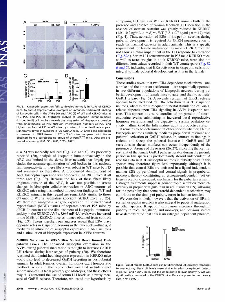
n = 5) was markedly reduced (Fig. 3 A and C). As previouslyreported (20), analysis of kisspeptin immunoreactivity in theARC was limited to the dense fiber network that largely pre-cludes the accurate quantitation of cell bodies in this nucleus.Immunoreactivity in these fibers was robust in WT mice by P15and remained so thereafter. A pronounced diminishment ofARC kisspeptin expression was observed in KERKO mice at allthree ages (Fig. 3B). Because the bulk of these fibers likelyoriginate outside of the ARC, it was not possible to assesschanges in kisspeptin cellular expression in ARC neurons ofKERKO mice using this method. Indeed, our findings in WT andKERKO animals in this regard are remarkably similar to thoseobtained in WT vs. aromatase knockout (ArKO) mice (20, 25).We therefore analyzed Kiss1 gene expression in the mediobasalhypothalamic (MBH) tissues of separate sets of P25 mice byqPCR. In contrast to the diminishment of kisspeptin immunore-activity in the KERKOAVPe, Kiss1mRNA levels were increasedin the MBH of KERKO mice vs. tissues obtained from controls(Fig. 3D). Taken together, our analyses reveal that ERα playsopposite roles in kisspeptin neurons in the two nuclei—that is, itmediates an inhibition of kisspeptin expression in ARC neuronsand a stimulation of kisspeptin expression in AVPe neurons.
GnRH/LH Secretions in KERKO Mice Do Not Reach Normal Post-pubertal Levels. The enhanced kisspeptin expression in theAVPe during pubertal maturation is thought to increase GnRH/LH release during later stages of puberty (20). We thereforereasoned that diminished kisspeptin expression in KERKO micewould also lead to decreased GnRH secretion in postpubertalanimals. In adult females, ovarian hormones exert homeostaticfeedback actions in the reproductive axis that include directsuppression of LH from pituitary gonadotropes, and these effectsmay thus confound the use of serum LH levels as a proxy mea-sure of GnRH release. Therefore, we tested our hypothesis by
comparing LH levels in WT vs. KERKO animals both in thepresence and absence of ovarian feedback. LH secretion in theabsence of ovarian restraint was greatly reduced in KERKO(1.0 ± 0.2 ng/mL; n = 8) vs. WT (3.8 ± 0.7 ng/mL; n = 17) mice(Fig. 4). Thus, activation of ERα in kisspeptin neurons duringpubertal development is required for GnRH neurosecretion toreach its maximal capacity in adult animals. This is a specificrequirement for female maturation, as male KERKO mice didnot show a similar impairment in the LH response to castration(Fig. S2A). Serum LH concentrations in P35 male KERKOmice,as well as testes weights in adult KERKO mice, were also notdifferent from values recorded in their WT counterparts (Fig. S2B and C), indicating that ERα activation in kisspeptin cells is notintegral to male pubertal development as it is in the female.
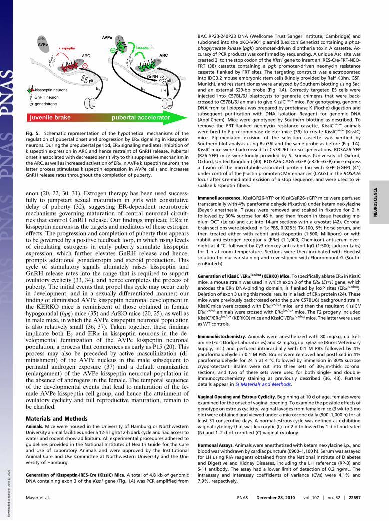
ConclusionsThese studies reveal that two ERα-dependent mechanisms—onea brake and the other an accelerator— are sequentially operatedin two different populations of kisspeptin neurons during pu-bertal development of female mice to gate, and then to activate,GnRH release (Fig. 5). A juvenile restraint of GnRH releaseappears to be mediated by ERα activation in ARC kisspeptinneurons, whereas the subsequent pubertal stimulation of GnRHrelease depends upon ERα signaling in AVPe kisspeptin neu-rons. This appears to ensure coordinated progression of neuro-endocrine events culminating in increased basal reproductivehormone secretions and the capacity to sustain ovulatory cy-clicity, hallmarks of the fully mature adult reproductive state.It remains to be determined in other species whether ERα in
kisspeptin neurons similarly mediates prepubertal restraint andpubertal activation of GnRH release. In contrast to findings inrodents and sheep, the pubertal increase in GnRH and LHsecretions in rhesus monkeys can occur independently of thepresence or absence of the ovaries (26, 27), indicating that centralrestraint of the female GnRH pulse generator during the juvenileperiod in this species is predominantly steroid independent. Arole for ERα in ARC kisspeptin neurons in puberty onset in thisspecies may therefore figure less importantly, although it ispossible that central ERs are activated in a ligand-independentmanner (28) by peripheral and central signals in prepubertalmonkeys, thereby constituting an estrogen-independent, yet es-trogen receptor-dependent, mechanism for the pubertal restraint.Estrogen treatments suppress gonadotropin secretion more ef-fectively in prepubertal girls than in adult women (29), allowingfor the possibility that some steroid-dependent mechanism maycontribute to the timing of puberty onset in human females.We consider it likely, however, that the activation of ERα in
rostral kisspeptin neurons is also integral to pubertal maturationin other species. Kisspeptin expression increases throughoutpuberty in mice, rat, sheep, and monkeys, and previous studieshave demonstrated that this is an estrogen-dependent phenom-
P15
P25
P35
WT KERKOWT KERKOAVPe ARC
C
A B
D
Rel
ativ
e K
iss1
mR
NA
/ G
AP
DH
mR
NA
(M
BH
)
2.5
2.0
1.5
1.0
0.5
0.0P25
*
Kis
spep
tin
Cel
l B
od
ies
(AV
Pe)
0P15 P25 P35
250
200
150
100
50 **
WTKERKO
Fig. 3. Kisspeptin expression fails to develop normally in AVPe of KERKOmice. (A and B) Representative examples of immunohistochemical labelingof kisspeptin cells in the AVPe (A) and ARC (B) of WT and KERKO mice atP15, P25, and P35. (C) Statistical analysis of kisspeptin immunoreactive(kisspeptin-IR) cell numbers reveals the progression of kisspeptin expressionfrom undetectable at P15, through intermediate numbers at P25, andhighest numbers at P35 in WT mice; by contrast, kisspeptin-IR cells appearsignificantly lower in numbers in P35 KERKO mice. (D) Kiss1 gene expressionis increased in MBH tissues of P25 KERKO mice, compared with tissuesobtained from a corresponding group of WT/ERαlox/wt mice. Data are pre-sented as mean ± SEM. *P < 0.01; **P < 0.001.
1
2
3
0Intact OVX
4
5
**LH
(n
g/m
l)
WTKERKO
Fig. 4. Adult female KERKO mice exhibit diminished LH secretory responsesto ovariectomy. Basal LH levels were similar in adult ovary-intact (Intact)mice, WT, and KERKO mice, but the LH response to ovariectomy (OVX) wassignificantly attenuated in the KERKO mice. Data are presented as mean ±SEM. **P < 0.001.
22696 | www.pnas.org/cgi/doi/10.1073/pnas.1012406108 Mayer et al.
Dow
nloa
ded
by g
uest
on
June
13,
202
0
enon (20, 22, 30, 31). Estrogen therapy has been used success-fully to jumpstart sexual maturation in girls with constitutivedelay of puberty (32), suggesting ER-dependent neurotropicmechanisms governing maturation of central neuronal circuit-ries that control GnRH release. Our findings implicate ERα inkisspeptin neurons as the targets and mediators of these estrogeneffects. The progression and completion of puberty thus appearsto be governed by a positive feedback loop, in which rising levelsof circulating estrogens in early puberty stimulate kisspeptinexpression, which further elevates GnRH release and hence,prompts additional gonadotropin and steroid production. Thiscycle of stimulatory signals ultimately raises kisspeptin andGnRH release rates into the range that is required to supportovulatory cyclicity (33, 34), and hence completes the process ofpuberty. The initial events that propel this cycle may occur earlyin development, and in a sexually differentiated manner; ourfinding of diminished AVPe kisspeptin neuronal development inthe KERKO mice is reminiscent of those obtained in femalehypogonadal (hpg) mice (35) and ArKO mice (20, 25), as well asin male mice, in which the AVPe kisspeptin neuronal populationis also relatively small (36, 37). Taken together, these findingsimplicate both E2 and ERα in kisspeptin neurons in the de-velopmental feminization of the AVPe kisspeptin neuronalpopulation, a process that commences as early as P15 (20). Thisprocess may also be preceded by active masculinization (di-minishment) of the AVPe nucleus in the male subsequent toperinatal androgen exposure (37) and a default organization(enlargement) of the AVPe kisspeptin neuronal population inthe absence of androgens in the female. The temporal sequenceof the developmental events that lead to maturation of the fe-male AVPe kisspeptin cell group, and hence the attainment ofovulatory cyclicity and full reproductive maturation, remain tobe clarified.
Materials and MethodsAnimals. Mice were housed in the University of Hamburg or NorthwesternUniversity animal facilities under a 12-h light/12-h dark cycle and had access towater and rodent chow ad libitum. All experimental procedures adhered toguidelines provided in the National Institutes of Health Guide for the Careand Use of Laboratory Animals and were approved by the InstitutionalAnimal Care and Use Committee at Northwestern University and the Uni-versity of Hamburg.
Generation of Kisspeptin-IRES-Cre (KissIC) Mice. A total of 4.8 kb of genomicDNA containing exon 3 of the Kiss1 gene (Fig. 1A) was PCR amplified from
BAC RP23-240P23 DNA (Wellcome Trust Sanger Institute, Cambridge) andsubcloned into the pKO-V901 plasmid (Lexicon Genetics) containing a phos-phoglycerate kinase (pgk) promoter-driven diphtheria toxin A cassette. Ac-curacy of PCR products was confirmed by sequencing. A unique AscI site wascreated 3′ to the stop codon of the Kiss1 gene to insert an IRES-Cre-FRT-NEO-FRT (38) cassette containing a pgk promoter-driven neomycin resistancecassette flanked by FRT sites. The targeting construct was electroporatedinto IDG3.2 mouse embryonic stem cells (kindly provided by Ralf Kühn, GSF,Munich), and resistant clones were analyzed by Southern blotting using SacIand an external 629-bp probe (Fig. 1A). Correctly targeted ES cells wereinjected into C57BL/6J blastocysts to generate chimeras that were back-crossed to C57BL/6J animals to give KissICneo+ mice. For genotyping, genomicDNA from tail biopsies was prepared by proteinase K (Roche) digestion andsubsequent purification with DNA Isolation Reagent for genomic DNA(AppliChem). Mice were genotyped by Southern blotting as described. Toremove the FRT-flanked neomycin resistance cassette, KissICneo+ animalswere bred to Flp recombinase deleter mice (39) to create KissICneo- (KissIC)mice. Flp-mediated excision of the selection cassette was verified bySouthern blot analysis using Bsu36I and the same probe as before (Fig. 1A).KissIC mice were backcrossed to C57BL/6J for six generations. ROSA26-YFP(R26-YFP) mice were kindly provided by S. Srinivas (University of Oxford,Oxford, United Kingdom) (40). ROSA26-CAGS-τGFP (eR26-τGFP) mice expressa fusion of the microtubule-associated protein tau with GFP (τGFP) (41)under control of the β-actin promoter/CMV enhancer (CAGS) in the ROSA26locus after Cre-mediated excision of a stop sequence, and were used to vi-sualize kisspeptin fibers.
Immunofluorescence. KissIC/R26-YFP or KissIC/eR26-τGFP mice were perfusedtranscardially with 4% paraformaldehyde (fixative) under ketamine/xylazine(Bayer) anesthesia. Tissues were removed and soaked in fixative for 2 h,followed by 30% sucrose for 48 h, and then frozen in tissue freezing me-dium OCT (Leica) and cut into 14-μm sections with a cryostat (42). Coronalbrain sections were blocked in 1× PBS, 0.025% TX-100, 5% horse serum, andthen treated either with rabbit anti-kisspeptin (1:500; Millipore) or withrabbit anti-estrogen receptor α (ERα) (1:1,000; Chemicon) antiserum over-night at 4 °C, followed by Cy3-donkey anti-rabbit IgG (1:500; Jackson Labs)for 1 h at room temperature. Sections were then incubated with Hoechstsolution for nuclear staining and coverslipped with Fluoromount-G (South-ernBiotech).
Generation of KissIC+/ERαlox/lox (KERKO) Mice. Tospecifically ablateERα inKissICmice, a mouse strain was used in which exon 3 of the ERα (Esr1) gene, whichencodes the ERα DNA-binding domain, is flanked by loxP sites (ERαlox/lox).Deletion of exon 3 using this model results in a lack of ERα protein (24). Thesemice were previously backcrossed onto the pure C57BL/6J background strain.KissIC mice were crossed with ERαlox/lox mice, and then the resultant KissIC+/ERαlox/wt animals were crossed with ERαlox/lox mice. The F2 progeny includedKissIC+/ERαlox/lox (KERKO)mice andKissIC−/ERαlox/loxmice. The latterwere usedas WT controls.
Immunohistochemistry. Animals were anesthetized with 80 mg/kg, i.p. ket-amine (Fort Dodge Laboratories) and 32mg/kg, i.p. xylazine (Burns VeterinarySupply, Inc.) and perfused intracardially with 0.1 M PBS followed by 4%paraformaldehyde in 0.1 M PBS. Brains were removed and postfixed in 4%paraformaldehyde for 24 h at 4 °C followed by immersion in 30% sucrosecryoprotectant. Brains were cut into three sets of 30-μm-thick coronalsections, and two of these sets were used for both single- and double-immunocytochemistry staining as previously described (36, 43). Furtherdetails appear in SI Materials and Methods.
Vaginal Opening and Estrous Cyclicity. Beginning at 10 d of age, females wereexamined for the onset of vaginal opening. To examine the possible effects ofgenotype on estrous cyclicity, vaginal lavages from female mice (3 wk to 3moold) were obtained and viewed under a microscope daily (900–1,000 h) for atleast 31 consecutive days. A normal estrous cycle was defined as exhibitingvaginal cytology that was leukocytic (L) for 2 d followed by 1 d of nucleated(N) and 1–2 d of cornified (C) vaginal cytology.
Hormonal Assays.Animals were anesthetized with ketamine/xylazine i.p., andblood was withdrawn by cardiac puncture (0900–1,100 h). Serumwas assayedfor LH using RIA reagents obtained from the National Institute of Diabetesand Digestive and Kidney Diseases, including the LH reference (RP-3) andS-11 antibody. The assay had a lower limit of detection of 0.2 ng/mL. Theintraassay and interassay coefficients of variance (CVs) were 4.1% and7.9%, respectively.
Fig. 5. Schematic representation of the hypothetical mechanisms of theregulation of pubertal onset and progression by ERα signaling in kisspeptinneurons. During the prepubertal period, ERα signaling mediates inhibition ofkisspeptin expression in ARC and hence restraint of GnRH release. Pubertalonset is associated with decreased sensitivity to this suppressive mechanism inthe ARC, as well as increased activation of ERα in AVPe kisspeptin neurons; thelatter process stimulates kisspeptin expression in AVPe cells and increasesGnRH release rates throughout the completion of puberty.
Mayer et al. PNAS | December 28, 2010 | vol. 107 | no. 52 | 22697
NEU
ROSC
IENCE
Dow
nloa
ded
by g
uest
on
June
13,
202
0
Ovarian Histology. Ovaries were dissected, immersed in 4% paraformal-dehyde overnight at 4 °C, and then washed with ethanol, processed, andembedded in paraffin wax. Paraffin-embedded ovaries were mounted ona microtome, cut into 5-μm-thick slices, and then stained with H&E.
Quantitative Real-Time PCR. Total RNA was extracted from MBH tissue usingTRIzol reagent (Invitrogen) according to the manufacturer’s protocol. RNAwas reverse transcribed using the iScript cDNA synthesis kit (BioRad). PCRreactions were performed with IQ SYBR Green Supermix (BioRad), andfluorescence was measured using the MyiQ quantitative real-time thermo-cycler (BioRad). The following primer sets were used: Kiss1 (sense 5′-AGCTGCTGCTTCTCCTCTGT-3′ and antisense 5′-GCATACCGCGATTCCTTTT-3′)(44). TaqMan was used to measure GAPDH reference gene levels, and weremeasured on an IQ5 thermocylcer (BioRad); sense 5′-GGGCATCTTGGGCTA-CACT-3′, antisense 5′-GGCATCGAAGGTGGAAGAGT-3′, probe 5′-Cal fluorored-610-AGGACCAGGTTGTCTCCTGCGA-BHQ2 3′. Each reaction was run intriplicate, and for each assay a standard curve was created using 10-fold
serial dilutions of cDNA. All experiments had efficiencies between 95% and105%, and Kiss1 measures displayed normal melt curves. Fold changes inrelative gene expression were calculated by 2−Δ(ΔCt), where ΔCt = Ct (Kiss1) −Ct(GAPDH) and Δ(ΔCt) = ΔCt (KERKO) − mean ΔCt (WT). Results areexpressed as fold differences in relative gene expression with respect to WT.
Statistical Analysis. Data are presented as the mean ± SEM. Two-tailed, un-paired t tests with Welch’s correction were used to determine statisticalsignificance for age at vaginal opening and Kiss1 expression in the MBH.Two-way ANOVA followed by Bonferroni post hoc tests were used to de-termine statistical significance in the remaining experiments. Differenceswere considered significant when P < 0.05.
ACKNOWLEDGMENTS. We gratefully acknowledge the technical assis-tance of Brigitte Mann. Support for this work was provided by DeutscheForschungsgemeinschaft Grant DFG BO1743/2 and National Institutes ofHealth Grants K99 HD55446, P01 HD21921, T32 HD07068, and P50 HD44405.
1. Watanabe G, Terasawa E (1989) In vivo release of luteinizing hormone releasinghormone increases with puberty in the female rhesus monkey. Endocrinology 125:92–99.
2. Sisk CL, Richardson HN, Chappell PE, Levine JE (2001) In vivo gonadotropin-releasinghormone secretion in female rats during peripubertal development and on proestrus.Endocrinology 142:2929–2936.
3. Harris GC, Levine JE (2003) Pubertal acceleration of pulsatile gonadotropin-releasinghormone release in male rats as revealed by microdialysis. Endocrinology 144:163–171.
4. Kulin HE, Grumbach MM, Kaplan SL (1969) Changing sensitivity of the pubertalgonadal hypothalamic feedback mechanism in man. Science 166:1012–1013.
5. Steele RE, Weisz J (1974) Changes in sensitivity of the estradiol-LH feedback systemwith puberty in the female rat. Endocrinology 95:513–520.
6. Foster DL, Ryan KD (1979) Endocrine mechanisms governing transition intoadulthood: A marked decrease in inhibitory feedback action of estradiol on tonicsecretion of luteinizing hormone in the lamb during puberty. Endocrinology 105:896–904.
7. Bronson FH (1981) The regulation of luteinizing hormone secretion by estrogen:Relationships among negative feedback, surge potential, and male stimulation injuvenile, peripubertal, and adult female mice. Endocrinology 108:506–516.
8. Rapisarda JJ, Bergman KS, Steiner RA, Foster DL (1983) Response to estradiolinhibition of tonic luteinizing hormone secretion decreases during the final stage ofpuberty in the rhesus monkey. Endocrinology 112:1172–1179.
9. Ojeda SR, Skinner MK (2006) Puberty in the rat. Physiology of Reproduction, edNeill JD (Elsevier, St. Louis), 3rd Ed, pp 2061–2126.
10. Seminara SB, et al. (2003) The GPR54 gene as a regulator of puberty. N Engl J Med349:1614–1627.
11. de Roux N, et al. (2003) Hypogonadotropic hypogonadism due to loss of function ofthe KiSS1-derived peptide receptor GPR54. Proc Natl Acad Sci USA 100:10972–10976.
12. Funes S, et al. (2003) The KiSS-1 receptor GPR54 is essential for the development ofthe murine reproductive system. Biochem Biophys Res Commun 312:1357–1363.
13. Lapatto R, et al. (2007) Kiss1−/− mice exhibit more variable hypogonadism thanGpr54−/− mice. Endocrinology 148:4927–4936.
14. d’Anglemont de Tassigny X, et al. (2007) Hypogonadotropic hypogonadism in micelacking a functional Kiss1 gene. Proc Natl Acad Sci USA 104:10714–10719.
15. Irwig MS, et al. (2004) Kisspeptin activation of gonadotropin releasing hormoneneurons and regulation of KiSS-1 mRNA in the male rat. Neuroendocrinology 80:264–272.
16. Messager S, et al. (2005) Kisspeptin directly stimulates gonadotropin-releasinghormone release via G protein-coupled receptor 54. Proc Natl Acad Sci USA 102:1761–1766.
17. Navarro VM, et al. (2004) Advanced vaginal opening and precocious activation of thereproductive axis by KiSS-1 peptide, the endogenous ligand of GPR54. J Physiol 561:379–386.
18. Matsui H, Takatsu Y, Kumano S, Matsumoto H, Ohtaki T (2004) Peripheral admin-istration of metastin induces marked gonadotropin release and ovulation in the rat.Biochem Biophys Res Commun 320:383–388.
19. Pineda R, et al. (2010) Critical roles of kisspeptins in female puberty and preovulatorygonadotropin surges as revealed by a novel antagonist. Endocrinology 151:722–730.
20. Clarkson J, Boon WC, Simpson ER, Herbison AE (2009) Postnatal development of anestradiol-kisspeptin positive feedback mechanism implicated in puberty onset.Endocrinology 150:3214–3220.
21. Gottsch ML, et al. (2009) Regulation of Kiss1 and dynorphin gene expression in themurine brain by classical and nonclassical estrogen receptor pathways. J Neurosci 29:9390–9395.
22. Takase K, et al. (2009) Possible role of oestrogen in pubertal increase of Kiss1/kisspeptinexpression in discrete hypothalamic areas of female rats. J Neuroendocrinol 21:527–537.
23. Smith JT, Popa SM, Clifton DK, Hoffman GE, Steiner RA (2006) Kiss1 neurons in theforebrain as central processors for generating the preovulatory luteinizing hormonesurge. J Neurosci 26:6687–6694.
24. Singh SP, et al. (2009) Impaired estrogen feedback and infertility in female mice withpituitary-specific deletion of estrogen receptor alpha (ESR1). Biol Reprod 81:488–496.
25. Bakker J, Pierman S, González-Martínez D (2010) Effects of aromatase mutation(ArKO) on the sexual differentiation of kisspeptin neuronal numbers and theiractivation by same versus opposite sex urinary pheromones. Horm Behav 57:390–395.
26. Chongthammakun S, Claypool LE, Terasawa E (1993) Ovariectomy increases in vivoluteinizing hormone-releasing hormone release in pubertal, but not prepubertal,female rhesus monkeys. J Neuroendocrinol 5:41–50.
27. Chongthammakun S, Terasawa E (1993) Negative feedback effects of estrogen onluteinizing hormone-releasing hormone release occur in pubertal, but not prepu-bertal, ovariectomized female rhesus monkeys. Endocrinology 132:735–743.
28. Olesen KM, Jessen HM, Auger CJ, Auger AP (2005) Dopaminergic activation ofestrogen receptors in neonatal brain alters progestin receptor expression and juvenilesocial play behavior. Endocrinology 146:3705–3712.
29. Kelch RP, Kaplan SL, GhumbachMM (1973) Suppression of urinary and plasma follicle-stimulating hormone by exogenous estrogens in prepubertal and pubertal children. JClin Invest 52:1122–1128.
30. Smith JT, Li Q, Pereira A, Clarke IJ (2009) Kisspeptin neurons in the ovine arcuatenucleus and preoptic area are involved in the preovulatory luteinizing hormonesurge. Endocrinology 150:5530–5538.
31. Terasawa E, et al. (2010) Recent discoveries on the control of gonadotrophin-releasing hormone neurones in nonhuman primates. J Neuroendocrinol 22:630–638.
32. Kulin HE (1996) Delayed puberty. J Clin Endocrinol Metab 81:3460–3464.33. Terasawa E (1985) Developmental changes in the positive feedback effect of estrogen
on luteinizing hormone release in ovariectomized female rhesus monkeys. Endo-crinology 117:2490–2497.
34. Foster DL, Karsch FJ (1975) Development of the mechanism regulating thepreovulatory surge of luteinizing hormone in sheep. Endocrinology 97:1205–1209.
35. Gill JC, et al. (2010) Reproductive hormone-dependent and -independent con-tributions to developmental changes in kisspeptin in GnRH-deficient hypogonadalmice. PLoS ONE 5:e11911.
36. Clarkson J, Herbison AE (2006) Postnatal development of kisspeptin neurons in mousehypothalamus; sexual dimorphism and projections to gonadotropin-releasing hor-mone neurons. Endocrinology 147:5817–5825.
37. Kauffman AS, et al. (2007) Sexual differentiation of Kiss1 gene expression in the brainof the rat. Endocrinology 148:1774–1783.
38. Eggan K, et al. (2004) Mice cloned from olfactory sensory neurons. Nature 428:44–49.39. Rodríguez CI, et al. (2000) High-efficiency deleter mice show that FLPe is an
alternative to Cre-loxP. Nat Genet 25:139–140.40. Srinivas S, et al. (2001) Cre reporter strains produced by targeted insertion of EYFP
and ECFP into the ROSA26 locus. BMC Dev Biol 1:4.41. Rodriguez I, Feinstein P, Mombaerts P (1999) Variable patterns of axonal projections
of sensory neurons in the mouse vomeronasal system. Cell 1:199–208.42. Boehm U, Zou Z, Buck LB (2005) Feedback loops link odor and pheromone signaling
with reproduction. Cell 123:683–695.43. Clarkson J, d’Anglemont de Tassigny X, Moreno AS, Colledge WH, Herbison AE (2008)
Kisspeptin-GPR54 signaling is essential for preovulatory gonadotropin-releasinghormone neuron activation and the luteinizing hormone surge. J Neurosci 28:8691–8697.
44. Luque RM, Kineman RD, Tena-Sempere M (2007) Regulation of hypothalamicexpression of KiSS-1 and GPR54 genes by metabolic factors: Analyses using mousemodels and a cell line. Endocrinology 148:4601–4611.
22698 | www.pnas.org/cgi/doi/10.1073/pnas.1012406108 Mayer et al.
Dow
nloa
ded
by g
uest
on
June
13,
202
0