Thin Solid Films - SKKUhome.skku.edu/~jhboo/publications/paper/2011/2011-11.pdf · sol–gel method...
7
Comparison of hydrophilic properties of TiO 2 thin films prepared by sol–gel method and reactive magnetron sputtering system S.-H. Nam a, ⁎, S.-J. Cho a , C.-K. Jung a , J.-H. Boo a, ⁎, J. Šícha b , D. Heřman b , J. Musil b , J. Vlček b a Department of Chemistry, Sungkyunkwan University, Suwon 440-746, South Korea b Department of Physics, University of West Bohemia, Univerzitní 22, 306 14 Plzeň, Czech Republic abstract article info Available online 30 April 2011 Keywords: TiO 2 films Sol–gel Reactive magnetron sputtering Photocatalytic property Oxygen plasma treatment This article reports on preparation, characterization and comparison of TiO 2 films prepared by sol–gel method using the titanium isopropoxide sol (TiO 2 coating sol 3%) as solvent precursor and reactive magnetron sputtering from substoichiometric TiO 2 −x targets of 50 mm in diameter. Dual magnetron supplied by dc bipolar pulsed power source was used for reactive magnetron sputtering. Depositions were performed on unheated glass substrates. Comparison of photocatalytic properties was based on measurements of hydrophilicity, i.e. evaluation of water contact angle on the film surface after UV irradiation. It is shown, that TiO 2 films prepared by the sol–gel method exhibited higher hydrophilicity in the as-deposited state but has significant deterioration of hydrophilicity during aging, compared to TiO 2 films prepared by magnetron sputtering. To explain this effect AFM, SEM and high resolution XPS measurements were performed. It is shown that the deterioration of hydrophilicity of sol–gel TiO 2 films can be suppressed if as-deposited films are exposed to the plasma of microwave oxygen discharge. © 2011 Elsevier B.V. All rights reserved. 1. Introduction Today the photocatalytic technology is becoming more and more attractive to industry, because a global environmental pollution has recognized to be a serious problem that needs to be addressed immediately. TiO 2 is an inexpensive, non-toxic, and biocompatible material which exhibits also a high photocatalytic activity. As a result, TiO 2 -based photocatalytic process proved to be very effective in removing many air and water pollutants [1]. Also, TiO 2 is a good optical material with high transmittance from the ultraviolet (UV) to visible (vis) light. These properties can be used in many applications, for example, the dissolution of hazardous volatile organic compounds (VOC), the removal of endocrine disrupters, the recovery of heavy metal, the anti-fogging, decontamination and self-cleaning, etc. [2]. At present, the TiO 2 photocatalytic thin films can be prepared by a number of methods such as dip-coating, sol–gel method, thermal oxidation of metal or reactive magnetron sputtering [3–9]. Sol–gel process is one of the most common methods used for producing photocatalytic TiO 2 material in the form of powder or coatings. Recently, a special attention is focused on a plasma treatment for thin TiO 2 films with the aim to improve photocatalytic activity for the decomposition of organic and inorganic pollutants under UV light [10]. Besides, the plasma treatment is also used for the creation of hydrophobic or hydrophilic surfaces on metals, plastics or polymers [11]. The photocatalytic efficiency of TiO 2 photocatalytic films depends on many parameters. For instance, the TiO 2 film should exhibit crystalline anatase structure [12–14]. The aim of this article is to compare properties of photocatalytic TiO 2 films prepared by the sol– gel method and the reactive magnetron sputtering. An enhancement of the photocatalytic activity of TiO 2 films prepared by the sol–gel method with the microwave plasma treatment is also discussed. 2. Experimental details 2.1. Sol–gel deposition of TiO 2 films The deposition of thin TiO 2 films was carried out with a dip-coating method using the titanium isopropoxide sol (TiO 2 coating sol 3%) used as a solvent precursor. The isopropoxide sol for TiO 2 -based photo- catalysts was prepared from titanium tetraisopropoxide, HNO 3 acid and water. The titanium tetraisopropoxide was slowly dropped into the 0.4% nitric acid solution which was stirred vigorously for 2 h at room temperature (RT), and then heated at 80 °C for 24 h. During reaction the isopropanol was removed by distillation and the originally coarse-milky solution was gradually changing to a blue fine-milky solution. Prior to the deposition of TiO 2 photocatalysts, the glass substrates were at first degreased, thoroughly cleaned and dried. Then the substrate was dipped into the viscous TiO 2 -precursor sol and several times pulled out at a uniform pulling rate of 5 mm/s (2, 4, 6 dip-coatings were formed) and dried at room temperature (RT) for 24 h. Thin Solid Films 519 (2011) 6944–6950 ⁎ Corresponding authors. Tel.: +82 31 290 7072; fax: +82 31 290 7075. E-mail addresses: [email protected] (S.-H. Nam), [email protected] (J.-H. Boo). 0040-6090/$ – see front matter © 2011 Elsevier B.V. All rights reserved. doi:10.1016/j.tsf.2011.04.144 Contents lists available at ScienceDirect Thin Solid Films journal homepage: www.elsevier.com/locate/tsf
Transcript of Thin Solid Films - SKKUhome.skku.edu/~jhboo/publications/paper/2011/2011-11.pdf · sol–gel method...

Thin Solid Films 519 (2011) 6944–6950
Contents lists available at ScienceDirect
Thin Solid Films
j ourna l homepage: www.e lsev ie r.com/ locate / ts f
Comparison of hydrophilic properties of TiO2 thin films prepared by sol–gel methodand reactive magnetron sputtering system
S.-H. Nam a,⁎, S.-J. Cho a, C.-K. Jung a, J.-H. Boo a,⁎, J. Šícha b, D. Heřman b, J. Musil b, J. Vlček b
a Department of Chemistry, Sungkyunkwan University, Suwon 440-746, South Koreab Department of Physics, University of West Bohemia, Univerzitní 22, 306 14 Plzeň, Czech Republic
⁎ Corresponding authors. Tel.: +82 31 290 7072; faxE-mail addresses: [email protected] (S.-H. Nam), j
0040-6090/$ – see front matter © 2011 Elsevier B.V. Adoi:10.1016/j.tsf.2011.04.144
a b s t r a c t
a r t i c l e i n f oAvailable online 30 April 2011
Keywords:TiO2 filmsSol–gelReactive magnetron sputteringPhotocatalytic propertyOxygen plasma treatment
This article reports on preparation, characterization and comparison of TiO2 films prepared by sol–gel methodusing the titanium isopropoxide sol (TiO2 coating sol 3%) as solvent precursor and reactive magnetronsputtering from substoichiometric TiO2−x targets of 50 mm in diameter. Dual magnetron supplied by dcbipolar pulsed power source was used for reactive magnetron sputtering. Depositions were performed onunheated glass substrates. Comparison of photocatalytic properties was based on measurements ofhydrophilicity, i.e. evaluation of water contact angle on the film surface after UV irradiation. It is shown,that TiO2 films prepared by the sol–gel method exhibited higher hydrophilicity in the as-deposited state buthas significant deterioration of hydrophilicity during aging, compared to TiO2 films prepared by magnetronsputtering. To explain this effect AFM, SEM and high resolution XPS measurements were performed. It isshown that the deterioration of hydrophilicity of sol–gel TiO2 films can be suppressed if as-deposited films areexposed to the plasma of microwave oxygen discharge.
: +82 31 290 [email protected] (J.-H. Boo).
ll rights reserved.
© 2011 Elsevier B.V. All rights reserved.
1. Introduction
Today the photocatalytic technology is becoming more and moreattractive to industry, because a global environmental pollution hasrecognized to be a serious problem that needs to be addressedimmediately. TiO2 is an inexpensive, non-toxic, and biocompatiblematerial which exhibits also a high photocatalytic activity. As a result,TiO2-based photocatalytic process proved to be very effective inremoving many air and water pollutants [1]. Also, TiO2 is a goodoptical material with high transmittance from the ultraviolet (UV) tovisible (vis) light. These properties can be used in many applications,for example, the dissolution of hazardous volatile organic compounds(VOC), the removal of endocrine disrupters, the recovery of heavymetal, the anti-fogging, decontamination and self-cleaning, etc. [2].
At present, the TiO2 photocatalytic thin films can be prepared by anumber of methods such as dip-coating, sol–gel method, thermaloxidation of metal or reactive magnetron sputtering [3–9]. Sol–gelprocess is one of the most common methods used for producingphotocatalytic TiO2material in the formof powder or coatings. Recently,a special attention is focused on a plasma treatment for thin TiO2 filmswith the aim to improve photocatalytic activity for the decomposition oforganic and inorganic pollutants under UV light [10]. Besides, theplasma treatment is also used for the creation of hydrophobic orhydrophilic surfaces on metals, plastics or polymers [11].
The photocatalytic efficiency of TiO2 photocatalytic films dependson many parameters. For instance, the TiO2 film should exhibitcrystalline anatase structure [12–14]. The aim of this article is tocompare properties of photocatalytic TiO2 films prepared by the sol–gel method and the reactive magnetron sputtering. An enhancementof the photocatalytic activity of TiO2 films prepared by the sol–gelmethod with the microwave plasma treatment is also discussed.
2. Experimental details
2.1. Sol–gel deposition of TiO2 films
The deposition of thin TiO2 filmswas carried out with a dip-coatingmethod using the titanium isopropoxide sol (TiO2 coating sol 3%) usedas a solvent precursor. The isopropoxide sol for TiO2-based photo-catalysts was prepared from titanium tetraisopropoxide, HNO3 acidand water. The titanium tetraisopropoxide was slowly dropped intothe 0.4% nitric acid solution which was stirred vigorously for 2 h atroom temperature (RT), and then heated at 80 °C for 24 h. Duringreaction the isopropanol was removed by distillation and theoriginally coarse-milky solution was gradually changing to a bluefine-milky solution. Prior to the deposition of TiO2 photocatalysts, theglass substrates were at first degreased, thoroughly cleaned and dried.Then the substrate was dipped into the viscous TiO2-precursor sol andseveral times pulled out at a uniform pulling rate of 5 mm/s (2, 4, 6dip-coatings were formed) and dried at room temperature (RT) for24 h.
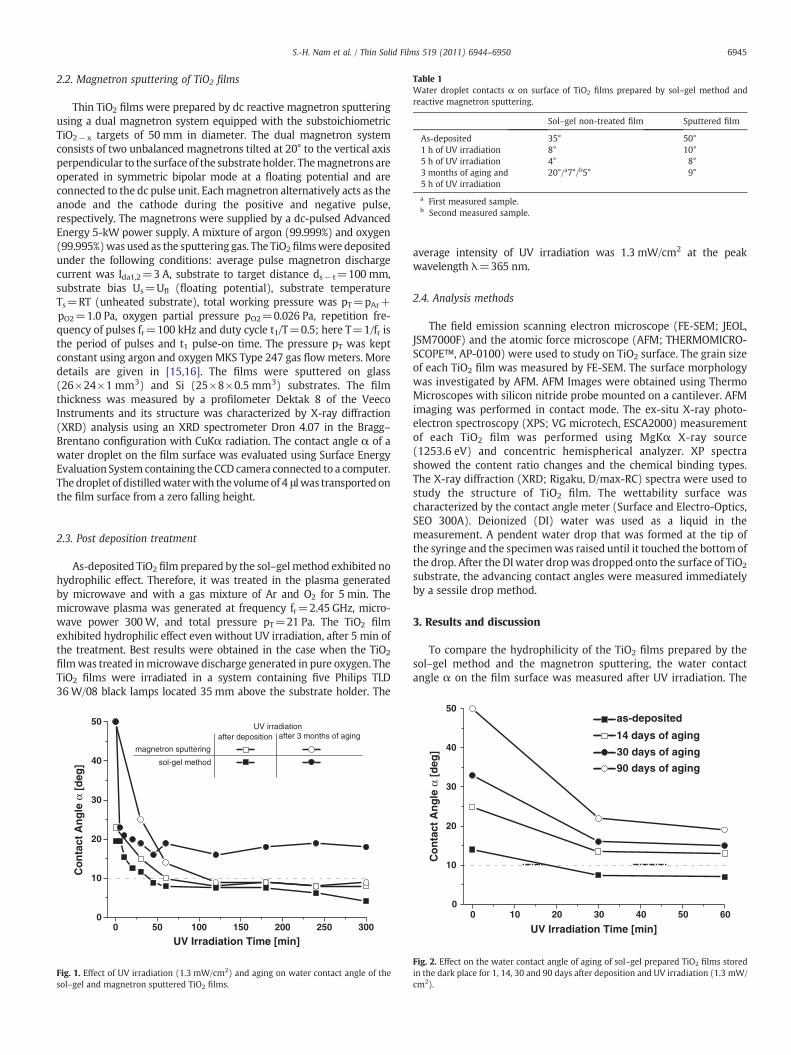
Table 1Water droplet contacts α on surface of TiO2 films prepared by sol–gel method andreactive magnetron sputtering.
Sol–gel non-treated film Sputtered film
As-deposited 35° 50°1 h of UV irradiation 8° 10°5 h of UV irradiation 4° 8°3 months of aging and5 h of UV irradiation
20°/a7°/b5° 9°
a First measured sample.b Second measured sample.
6945S.-H. Nam et al. / Thin Solid Films 519 (2011) 6944–6950
2.2. Magnetron sputtering of TiO2 films
Thin TiO2 films were prepared by dc reactive magnetron sputteringusing a dual magnetron system equipped with the substoichiometricTiO2−x targets of 50 mm in diameter. The dual magnetron systemconsists of two unbalanced magnetrons tilted at 20° to the vertical axisperpendicular to the surface of the substrate holder. Themagnetrons areoperated in symmetric bipolar mode at a floating potential and areconnected to the dc pulse unit. Eachmagnetron alternatively acts as theanode and the cathode during the positive and negative pulse,respectively. The magnetrons were supplied by a dc-pulsed AdvancedEnergy 5-kW power supply. A mixture of argon (99.999%) and oxygen(99.995%)was used as the sputtering gas. The TiO2filmswere depositedunder the following conditions: average pulse magnetron dischargecurrent was Ida1,2=3 A, substrate to target distance ds− t=100 mm,substrate bias Us=Ufl (floating potential), substrate temperatureTs=RT (unheated substrate), total working pressure was pT=pAr+pO2=1.0 Pa, oxygen partial pressure pO2=0.026 Pa, repetition fre-quency of pulses fr=100 kHz and duty cycle t1/T=0.5; here T=1/fr isthe period of pulses and t1 pulse-on time. The pressure pT was keptconstant using argon and oxygen MKS Type 247 gas flowmeters. Moredetails are given in [15,16]. The films were sputtered on glass(26×24×1 mm3) and Si (25×8×0.5 mm3) substrates. The filmthickness was measured by a profilometer Dektak 8 of the VeecoInstruments and its structure was characterized by X-ray diffraction(XRD) analysis using an XRD spectrometer Dron 4.07 in the Bragg–Brentano configuration with CuKα radiation. The contact angle α of awater droplet on the film surface was evaluated using Surface EnergyEvaluation System containing the CCD camera connected to a computer.The droplet of distilledwaterwith thevolumeof 4 μlwas transported onthe film surface from a zero falling height.
2.3. Post deposition treatment
As-deposited TiO2 film prepared by the sol–gel method exhibited nohydrophilic effect. Therefore, it was treated in the plasma generatedby microwave and with a gas mixture of Ar and O2 for 5 min. Themicrowave plasma was generated at frequency fr=2.45 GHz, micro-wave power 300W, and total pressure pT=21 Pa. The TiO2 filmexhibited hydrophilic effect even without UV irradiation, after 5 min ofthe treatment. Best results were obtained in the case when the TiO2
filmwas treated inmicrowave discharge generated in pure oxygen. TheTiO2 films were irradiated in a system containing five Philips TLD36W/08 black lamps located 35mm above the substrate holder. The
00
10
20
30
40
50
magnetron sputtering
sol-gel method
UV irradiationafter deposition after 3 months of aging
Co
nta
ct A
ng
le α
[d
eg]
UV Irradiation Time [min]30025020015010050
Fig. 1. Effect of UV irradiation (1.3 mW/cm2) and aging on water contact angle of thesol–gel and magnetron sputtered TiO2 films.
average intensity of UV irradiation was 1.3 mW/cm2 at the peakwavelength λ=365 nm.
2.4. Analysis methods
The field emission scanning electron microscope (FE-SEM; JEOL,JSM7000F) and the atomic force microscope (AFM; THERMOMICRO-SCOPE™, AP-0100) were used to study on TiO2 surface. The grain sizeof each TiO2 film was measured by FE-SEM. The surface morphologywas investigated by AFM. AFM Images were obtained using ThermoMicroscopes with silicon nitride probe mounted on a cantilever. AFMimaging was performed in contact mode. The ex-situ X-ray photo-electron spectroscopy (XPS; VG microtech, ESCA2000) measurementof each TiO2 film was performed using MgKα X-ray source(1253.6 eV) and concentric hemispherical analyzer. XP spectrashowed the content ratio changes and the chemical binding types.The X-ray diffraction (XRD; Rigaku, D/max-RC) spectra were used tostudy the structure of TiO2 film. The wettability surface wascharacterized by the contact angle meter (Surface and Electro-Optics,SEO 300A). Deionized (DI) water was used as a liquid in themeasurement. A pendent water drop that was formed at the tip ofthe syringe and the specimenwas raised until it touched the bottom ofthe drop. After the DI water dropwas dropped onto the surface of TiO2
substrate, the advancing contact angles were measured immediatelyby a sessile drop method.
3. Results and discussion
To compare the hydrophilicity of the TiO2 films prepared by thesol–gel method and the magnetron sputtering, the water contactangle α on the film surface was measured after UV irradiation. The
00
10
20
30
40
50
Co
nta
ct A
ng
le α
[d
eg]
UV Irradiation Time [min]
as-deposited
14 days of aging
30 days of aging
90 days of aging
605040302010
Fig. 2. Effect on the water contact angle of aging of sol–gel prepared TiO2 films storedin the dark place for 1, 14, 30 and 90 days after deposition and UV irradiation (1.3 mW/cm2).
6946 S.-H. Nam et al. / Thin Solid Films 519 (2011) 6944–6950
effect of aging of the TiO2 films was also evaluated. The aging effectwas tested for the TiO2 films stored in a dark box for 3 months.Obtained results are summarized in Fig. 1. From this figure, thefollowing issues can be drawn:
1. TiO2 films prepared by sol–gel method.a) The contact angle α decreases with increasing time of UV
irradiation and achieves approximately 9° after 40 min ofirradiation (tUV=40′), remains constant up to tUV=180′ anddecreases again for tUVN180′ with α=4° at tUV=300′
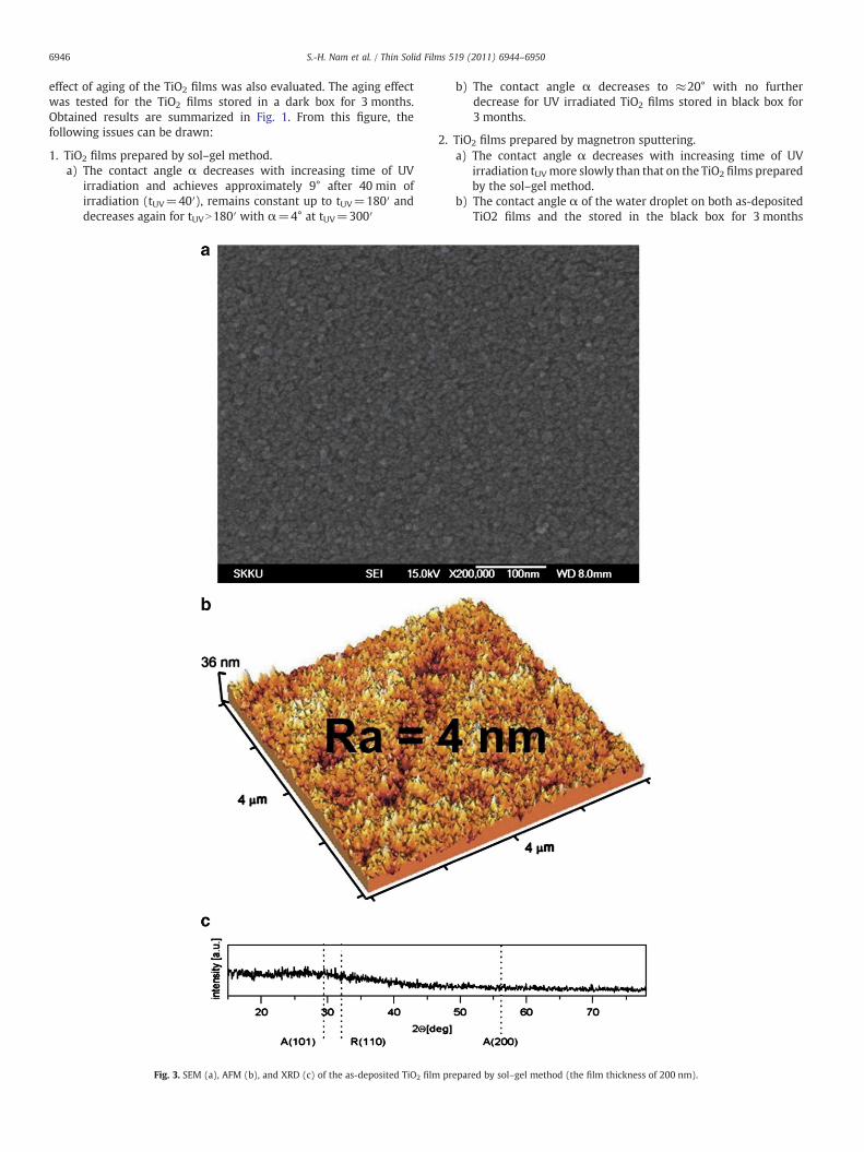
Fig. 3. SEM (a), AFM (b), and XRD (c) of the as-deposited TiO2 film
b) The contact angle α decreases to ≈20° with no furtherdecrease for UV irradiated TiO2 films stored in black box for3 months.
2. TiO2 films prepared by magnetron sputtering.a) The contact angle α decreases with increasing time of UV
irradiation tUVmore slowly than that on the TiO2 films preparedby the sol–gel method.
b) The contact angle α of the water droplet on both as-depositedTiO2 films and the stored in the black box for 3 months
prepared by sol–gel method (the film thickness of 200 nm).
6947S.-H. Nam et al. / Thin Solid Films 519 (2011) 6944–6950
decreases to approximately the same value α≈8° after theirUV irradiation for tUV≥120′.
All obtained results are summarized in Table 1.
3.1. Effect of aging on hydrophilicity of TiO2 films prepared by the sol–gelmethod
The hydrophilicity of films was characterized by the water dropletcontact angle α. The contact angle α was measured for the as-deposited film and films stored in the dark box for 14, 30 and 90 days,see Fig. 2. For all TiO2 films prepared by the sol–gel method the watercontact angle α decreases with increasing tUV approximately to30 min and saturates at a constant value α S at tUV≥30′. The value α S
increases with increasing storage time tS and achieves ≈20° afterstorage for tS=90 days and 60 min of UV irradiation.
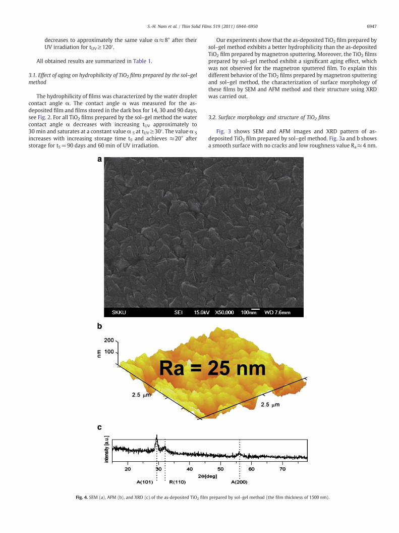
Fig. 4. SEM (a), AFM (b), and XRD (c) of the as-deposited TiO2 film
Our experiments show that the as-deposited TiO2 film prepared bysol–gel method exhibits a better hydrophilicity than the as-depositedTiO2 film prepared by magnetron sputtering. Moreover, the TiO2 filmsprepared by sol–gel method exhibit a significant aging effect, whichwas not observed for the magnetron sputtered film. To explain thisdifferent behavior of the TiO2 films prepared bymagnetron sputteringand sol–gel method, the characterization of surface morphology ofthese films by SEM and AFM method and their structure using XRDwas carried out.
3.2. Surface morphology and structure of TiO2 films
Fig. 3 shows SEM and AFM images and XRD pattern of as-deposited TiO2 film prepared by sol–gel method. Fig. 3a and b showsa smooth surface with no cracks and low roughness value Ra≈4 nm.
prepared by sol–gel method (the film thickness of 1500 nm).
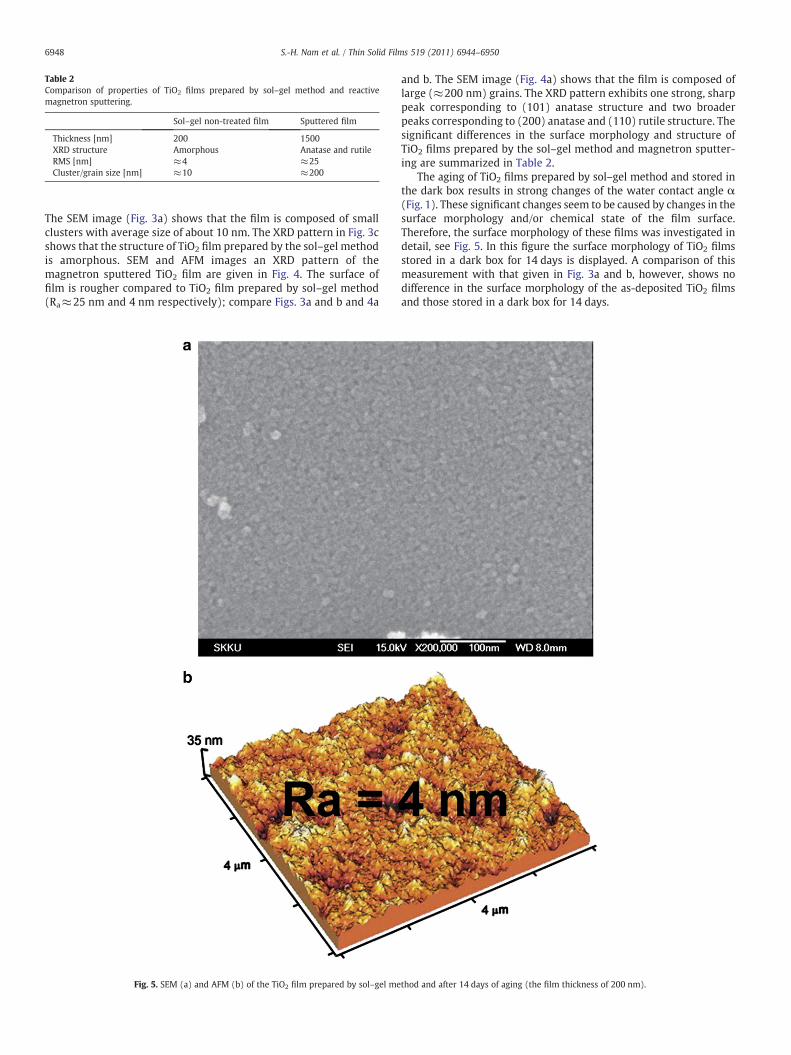
Table 2Comparison of properties of TiO2 films prepared by sol–gel method and reactivemagnetron sputtering.
Sol–gel non-treated film Sputtered film
Thickness [nm] 200 1500XRD structure Amorphous Anatase and rutileRMS [nm] ≈4 ≈25Cluster/grain size [nm] ≈10 ≈200
6948 S.-H. Nam et al. / Thin Solid Films 519 (2011) 6944–6950
The SEM image (Fig. 3a) shows that the film is composed of smallclusters with average size of about 10 nm. The XRD pattern in Fig. 3cshows that the structure of TiO2 film prepared by the sol–gel methodis amorphous. SEM and AFM images an XRD pattern of themagnetron sputtered TiO2 film are given in Fig. 4. The surface offilm is rougher compared to TiO2 film prepared by sol–gel method(Ra≈25 nm and 4 nm respectively); compare Figs. 3a and b and 4a
Fig. 5. SEM (a) and AFM (b) of the TiO2 film prepared by sol–gel me
and b. The SEM image (Fig. 4a) shows that the film is composed oflarge (≈200 nm) grains. The XRD pattern exhibits one strong, sharppeak corresponding to (101) anatase structure and two broaderpeaks corresponding to (200) anatase and (110) rutile structure. Thesignificant differences in the surface morphology and structure ofTiO2 films prepared by the sol–gel method and magnetron sputter-ing are summarized in Table 2.
The aging of TiO2 films prepared by sol–gel method and stored inthe dark box results in strong changes of the water contact angle α(Fig. 1). These significant changes seem to be caused by changes in thesurface morphology and/or chemical state of the film surface.Therefore, the surface morphology of these films was investigated indetail, see Fig. 5. In this figure the surface morphology of TiO2 filmsstored in a dark box for 14 days is displayed. A comparison of thismeasurement with that given in Fig. 3a and b, however, shows nodifference in the surface morphology of the as-deposited TiO2 filmsand those stored in a dark box for 14 days.
thod and after 14 days of aging (the film thickness of 200 nm).
4560
20
40
60
80
100
120
Ti2p
inte
nsi
ty [
a.u
.]
binding energy [eV]
after 14 days of aging
a
5260
20
40
60
80
100
120
O1s
inte
nsi
ty [
a.u
.]
binding energy [eV]
as-deposited
as-deposited
after 14 days of aging
b
468466464462460458
538536534532530528
Fig. 6. XPS Ti2p (a) and O1s (b) spectra of the as-deposited sol–gel TiO2 films before(solid curve) and after 14 days aging (dashed curve).
Table 3Plasma conditions for treating TiO2 films prepared by sol–gel method.
Ar plasma treatment O2 plasma treatment
MW power [W] 300 300Ar flow rate [sccm] 100 0O2 flow rate [sccm] 0 100Exposure time [min] 5 5Discharge pressure [Pa] 21 21
6949S.-H. Nam et al. / Thin Solid Films 519 (2011) 6944–6950
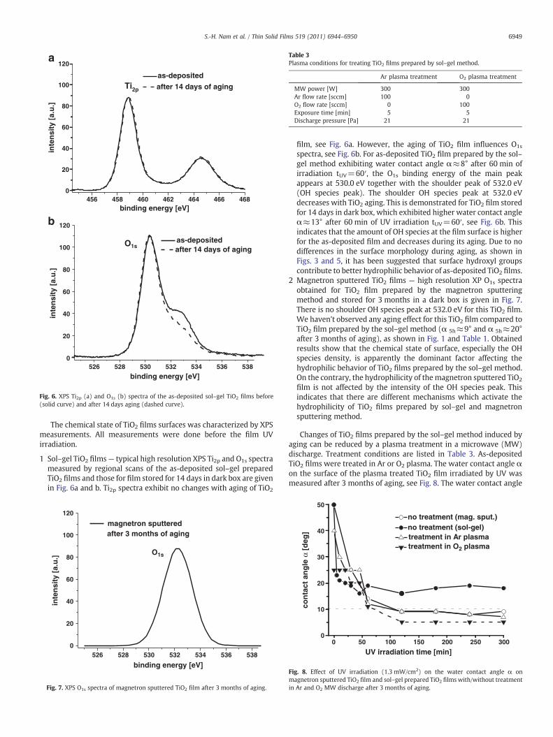
The chemical state of TiO2 films surfaces was characterized by XPSmeasurements. All measurements were done before the film UVirradiation.
1 Sol–gel TiO2 films— typical high resolution XPS Ti2p and O1s spectrameasured by regional scans of the as-deposited sol–gel preparedTiO2 films and those for film stored for 14 days in dark box are givenin Fig. 6a and b. Ti2p spectra exhibit no changes with aging of TiO2
5260
20
40
60
80
100
120
O1s
inte
nsi
ty [
a.u
.]
binding energy [eV]
magnetron sputtered after 3 months of aging
538536534532530528
Fig. 7. XPS O1s spectra of magnetron sputtered TiO2 film after 3 months of aging.
film, see Fig. 6a. However, the aging of TiO2 film influences O1s
spectra, see Fig. 6b. For as-deposited TiO2 film prepared by the sol–gel method exhibiting water contact angle α≈8° after 60 min ofirradiation tUV=60′, the O1s binding energy of the main peakappears at 530.0 eV together with the shoulder peak of 532.0 eV(OH species peak). The shoulder OH species peak at 532.0 eVdecreases with TiO2 aging. This is demonstrated for TiO2 film storedfor 14 days in dark box, which exhibited higher water contact angleα≈13° after 60 min of UV irradiation tUV=60′, see Fig. 6b. Thisindicates that the amount of OH species at the film surface is higherfor the as-deposited film and decreases during its aging. Due to nodifferences in the surface morphology during aging, as shown inFigs. 3 and 5, it has been suggested that surface hydroxyl groupscontribute to better hydrophilic behavior of as-deposited TiO2 films.
2 Magnetron sputtered TiO2 films — high resolution XP O1s spectraobtained for TiO2 film prepared by the magnetron sputteringmethod and stored for 3 months in a dark box is given in Fig. 7.There is no shoulder OH species peak at 532.0 eV for this TiO2 film.We haven't observed any aging effect for this TiO2 film compared toTiO2 film prepared by the sol–gel method (α 5h≈9° and α 5h≈20°after 3 months of aging), as shown in Fig. 1 and Table 1. Obtainedresults show that the chemical state of surface, especially the OHspecies density, is apparently the dominant factor affecting thehydrophilic behavior of TiO2 films prepared by the sol–gel method.On the contrary, the hydrophilicity of themagnetron sputtered TiO2
film is not affected by the intensity of the OH species peak. Thisindicates that there are different mechanisms which activate thehydrophilicity of TiO2 films prepared by sol–gel and magnetronsputtering method.
Changes of TiO2 films prepared by the sol–gel method induced byaging can be reduced by a plasma treatment in a microwave (MW)discharge. Treatment conditions are listed in Table 3. As-depositedTiO2 films were treated in Ar or O2 plasma. The water contact angle αon the surface of the plasma treated TiO2 film irradiated by UV wasmeasured after 3 months of aging, see Fig. 8. The water contact angle
00
10
20
30
40
50
no treatment (mag. sput.)no treatment (sol-gel) treatment in Ar plasmatreatment in O2 plasma
con
tact
an
gle
α [
deg
]
UV irradiation time [min]30025020015010050
Fig. 8. Effect of UV irradiation (1.3 mW/cm2) on the water contact angle α onmagnetron sputtered TiO2 film and sol–gel prepared TiO2 films with/without treatmentin Ar and O2 MW discharge after 3 months of aging.
6950 S.-H. Nam et al. / Thin Solid Films 519 (2011) 6944–6950
α measured on TiO2 film treated in Ar plasma is α 2h≈9° and that onTiO2 film treated in O2 plasma only 5° after irradiation of these filmsby UV for 2 h. Longer UV irradiation time resulted in further reductionof α. Experiment shows that this new approach using the MW plasmatreatment in Ar and O2 plasma enhances the hydrophilic properties ofTiO2 films.
4. Conclusions
The article presents a critical comparison of the properties andhydrophilicity of TiO2 films prepared by sol–gel method and reactivemagnetron sputtering. Obtained results can be summarized as follows:
The TiO2 films prepared by the sol–gel method are amorphous andthe water contact angle α decreases with increasing time of UVirradiation more rapidly than that on the anatase TiO2 film preparedby reactive magnetron sputtering. The water contact angle αmeasured after 5 h of UV irradiation on the sol–gel prepared TiO2
film is also lower (4°) than that (8°) on magnetron sputtered TiO2
film. The aging has no effect on the hydrophilicity of TiO2 filmsprepared by magnetron sputtering. The same water droplet contactangle α≈9° is measured for both the as-deposited TiO2 film and TiO2
film stored for 3 months in the dark box and UV irradiated for 5 h. Onthe contrary, the angle α on the surface of sol–gel TiO2 film increaseswith storage time and for 3 months storage achieves ≈20° after UVirradiation for 5 h. This increase inα is connected with decrease of OHspecies density (binding energy 532 eV in XPS O1s spectra) when thestorage time (aging) increases. The surface roughness of the TiO2 filmprepared by magnetron sputtering is higher (25 nm) than thatprepared by the sol–gel method (4 nm). Treatment of as-deposited
sol–gel TiO2 films in oxygen microwave discharge enhances theirhydrophilicity, especially the aging effect.
Acknowledgments
This research was supported by grants NRF-20100025481 (BasicScience Research Program), NRF-20100029699 (Priority ResearchCenters Program), and NRF-20100029417 (Plasma Bioscience Re-search Center, SRC Program).
References
[1] S. Gelover, P. Mondragon, A. Jimenez, J. Photochem. Photobiol., A Chemistry 165(2004) 241.
[2] R. Fretwell, P. Douglas, J. Photochem. Photobiol. A Chemistry 143 (2001) 229.[3] E. Sanchez, T. Lopez, Mater. Lett. 25 (1995) 271.[4] B.D. Fabes, D.P. Birine, B.J.J. Zelinski, Thin Solid Films 254 (1995) 175.[5] H. Ohsaki, Y. Tachibana, A. Mitsui, T. Kamiyama, Y. Hayashi, Thin Solid Films 392
(2001) 169.[6] C.-K. Jung, S.-H. Cho, S.-B. Lee, T.-K. Kim, M.-N. Lee, J.-H. Boo, Surf. Rev. Lett. 10
(2003) 635.[7] P. Zeman, S. Takabayashi, Thin Solid Films 433 (2003) 57.[8] P. Frach, D. Gloss, K. Goedicke, M. Fahland, W.-M. Gnehr, Thin Solid Films (2003)
251.[9] M.C. Barnes, S. Kumar, L. Green, N.-M. Hwang, A.R. Gerson, Surf. Coat. Technol. 190
(2005) 321.[10] J.M. Lee, M.S. Kim, B.W. Kim, Water Res. 38 (2004) 3605.[11] W. Zhang, W. Liu, C. Wang, Wear 253 (2002) 377.[12] S.K. Zheng, T.M. Wang, G. Xiang, C. Wang, Vacuum 62 (2001) 361.[13] S. Takeda, S. Suzuki, H. Odaka, H. Hosono, Thin Solid Films 392 (2001) 338.[14] S. Ohno, D. Sato, M. Kon, P.K. Song, M. Yoshikawa, K. Suzuki, P. Frach, Y. Shigesato,
Thin Solid FIlms 445 (2003) 207.[15] P. Baroch, J. Musil, J. Vlcek, K.H. Nam, J.G. Han, Surf. Coat. Technol. 193 (2005) 107.[16] J. Musil, P. Baroch, IEEE Trans. Plasma Sci. 33 (2005) 338.


![[PPT]No Slide Title - Prof. Stephen J. Pearton's Research Grouppearton.mse.ufl.edu/research/FTFTs/MRS_fall_2007-2.ppt · Web viewIndium Zinc Oxide Thin Films Deposited by Sputtering](https://static.fdocument.org/doc/165x107/5aa9b1b37f8b9a90188d2f55/pptno-slide-title-prof-stephen-j-peartons-research-viewindium-zinc-oxide.jpg)