The Principle of Maximum Chiral Discrimination: Chiral Recognition in Permethyl-β-cyclodextrin
14
The Principle of Maximum Chiral Discrimination: Chiral Recognition in Permethyl--cyclodextrin Kenny B. Lipkowitz,* Robert Coner, ² Michael A. Peterson, ‡ Antonio Morreale, and Jason Shackelford Department of Chemistry, Indiana University-Purdue University at Indianapolis (IUPUI), 402 North Blackford Street, Indianapolis, Indiana 46202 Received September 12, 1997 Five guest molecules, isomenthone, pulegone, 1-fluoro-1-phenylethane, 1-phenylethanol, and 2-meth- ylbutanoic acid, binding to permethyl--cyclodextrin, a chiral host molecule, have been simulated by molecular dynamics techniques. From the simulations we find the preferred binding site to be the interior of the macrocyclic cavity. A new technique was used for locating the host’s most enantiodiscriminating domain, which was also found to be inside the macrocyclic cavity. It is concluded that this particular host molecule displays its enhanced chiral discriminating capacity because of this spatial coincidence. Also evaluated in this paper are the types and magnitudes of intermolecular forces responsible for diastereomeric complexation and chiral discrimination; in both cases the short-range dispersion forces dominate. This study illustrates the “principle of maximum chiral recognition”, the idea that maximum chiral recognition can be achieved by maintaining a spatial congruence between the host’s domain of greatest enantiodifferentiation with the guest’s preferred binding site. Introduction There has been a remarkable infusion of cyclodextrin- assisted studies into the chemical sciences during the last three decades. 1 Concurrent with this has been the use of cyclodextrins (CD) in technology. 2 One especially im- portant application of cyclodextrin technology, for ex- ample, has been in the area of separation science. 3 These host molecules have been extensively used for separations because they offer the possibility of coordinating guests of differing size (depending on which CD macrocycle is used), because of their ability to recognize different guest functional groups (depending on which CD derivative is employed), and most significantly, since they are derived from glucose units, these macrocycles are inherently dis- symmetric and are thus able to distinguish between ster- eochemical isomers including enantiomers. The versatil- ity of these macrocycles is remarkable, and because of their price-to-performance ratio they are extensively used in chiral chromatography. 4 Both native and derivatized cyclodextrins have been used to separate enantiomers in planar chromatography (TLC), 5 high-performance liquid chromatography (HPLC), 6 in super- and subcritical fluid- phase chromatographies, and more recently, as additives that enantioselectively bind and control the migratory aptitudes of analytes in capillary electrophoresis. 7 An- other area of chromatographic application of cyclodex- trins is in gas-liquid-phase chromatography (GLC). 8 Here, analyses of volatile natural and nonnatural prod- ucts have been undertaken in disciplines as disparate as * To whom correspondence should be addressed. E-mail: lipkowitz@ chem.iupui.edu. ² Permanent address: Lilly Research Laboratories, Lilly Corporate Center, Drop 0540, Indianapolis, IN 46285. ‡ Permanent address: Department of Chemistry, University of Florida, Box 117200, Gainesville, FL 32611-7200. (1) (a) Bender, M. L.; Komiyama, M. Cyclodextrin Chemistry, Reactivity and Structure, Concepts in Organic Chemistry, 6; Springer- Verlag: New York, 1978. (b) Saenger, W. Angew. Chem., Int. Ed. Engl., 1980, 19, 344. (c) Szejtli, J. Cyclodextrins and Their Inclusion Complexes; Akade ´miai Kaido ´: Budapest, 1982. (2) (a) Duche ˆne, D., Ed. Cyclodextrins and Their Industrial Uses; Editions de Sante: Paris, 1987. (b) Szejtli, J. Cyclodextrin Technology; Kluwer Academic: Dordrecht, 1988. (3) Snopek, J.; Smolkova ´ -Keulemansova ´ , E.; Cserha ´ ti, T.; Gahm, K. H.; Stalcup, A. In Comprehensive Supramolecular Chemistry; Szejtli, J., Osa, T., Eds.; Pergamon Press: Oxford, 1996; pp 515-571. (4) (a) Souter, R. W. Chromatographic Separations of Stereoisomers; CRC Press: Boca Raton, 1985. (b) Chromatographic Chiral Separa- tions; Zeif, M., Crane, L., Eds.; Chromatographic Science Series, Vol. 40; Marcel Dekker: New York, 1987. (c) Ko ¨nig, W. A. The Practice of Enantiomer Separation by Capillary Gas Chromatography; Hu ¨ thig: Heidelberg, 1987. (d) Ordered Media in Chemical Separations; Hinze, W. L., Armstrong, D. W., Eds.; ACS Symposium Series 342; American Chemical Society: Washington, DC, 1987. (e) Allenmark, S. G. Chromatographic Enantioseparation. Methods and Application; Ellis Horwood: Chichester, 1988. (f) Chiral Separations; Stevenson, D., Wilson, I. D., Eds.; Plenum: New York, 1988. (g) Chiral Liquid Chromatography; Lough, W. J., Ed.; Blackie: London, 1989. (h) Recent Advances in Chiral Separations; Stevenson, D., Wilson, I. D., Eds.; Plenum: New York, 1990. (i) Chiral Separations by Liquid Chroma- tography; Ahuja, S., Ed.; ACS Symposium Series 471; American Chemical Society: Washington, DC, 1991. (j) Chiral Separations by Liquid Chromatography; Subramanian, G., Ed.; VCH: Weinheim, 1994. (k) Chiral Separations. Applications and Technology; Ahuja, S., Ed.; American Chemical Society: Washington, DC, 1997. (5) For reviews see: (a) Ward, T. J.; Armstrong, D. W. J. Liq. Chromatogr. 1986, 9, 407. (b) Martens, J.; Bhushan, R. Chem. Ztg. 1988, 112, 367. (c) Martens, J.; Bhushan, R. Int. J. Peptide Protein Res. 1989, 34, 433. (d) Martens, J.; Bhushan, R. J. Pharm. Biomed. Anal. 1990, 8, 259. (e) Bhushan, R.; Joshi, S. Biomed. Chromatogr. 1993, 7, 235. Table 1. MD Results (kJ/mol) for Isomenthone/CD total stretch bend torsion vdW elect RR1 2031.99 387.65 604.42 525.93 38.03 475.97 RR2 2053.27 389.62 613.65 509.29 65.62 475.09 RR3 2042.79 388.26 608.65 523.18 47.45 475.25 RR4 2069.09 388.14 608.19 526.81 68.59 477.36 RR5 2029.95 387.84 602.46 526.24 36.94 476.47 avg E RR 2045.42 388.30 607.47 522.29 51.32 476.03 SS1 2061.01 388.36 608.17 524.21 62.46 477.81 SS2 2038.96 388.43 608.18 521.60 47.31 473.44 SS3 2035.29 387.71 603.75 530.23 38.50 475.10 SS4 2059.38 388.48 605.41 523.73 64.42 477.34 SS5 2029.23 387.55 603.88 525.15 36.72 475.93 avg E SS 2044.77 388.11 605.88 524.98 49.88 475.92 ΔΔE R-S 0.64 0.20 1.60 -2.69 1.44 0.10 732 J. Org. Chem. 1998, 63, 732-745 S0022-3263(97)01709-X CCC: $15.00 © 1998 American Chemical Society Published on Web 01/22/1998
Transcript of The Principle of Maximum Chiral Discrimination: Chiral Recognition in Permethyl-β-cyclodextrin
The Principle of Maximum Chiral Discrimination: ChiralRecognition in Permethyl-â-cyclodextrin
Kenny B. Lipkowitz,* Robert Coner,† Michael A. Peterson,‡ Antonio Morreale, andJason Shackelford
Department of Chemistry, Indiana University-Purdue University at Indianapolis (IUPUI),402 North Blackford Street, Indianapolis, Indiana 46202
Received September 12, 1997
Five guest molecules, isomenthone, pulegone, 1-fluoro-1-phenylethane, 1-phenylethanol, and 2-meth-ylbutanoic acid, binding to permethyl-â-cyclodextrin, a chiral host molecule, have been simulatedby molecular dynamics techniques. From the simulations we find the preferred binding site to bethe interior of the macrocyclic cavity. A new technique was used for locating the host’s mostenantiodiscriminating domain, which was also found to be inside the macrocyclic cavity. It isconcluded that this particular host molecule displays its enhanced chiral discriminating capacitybecause of this spatial coincidence. Also evaluated in this paper are the types and magnitudes ofintermolecular forces responsible for diastereomeric complexation and chiral discrimination; in bothcases the short-range dispersion forces dominate. This study illustrates the “principle of maximumchiral recognition”, the idea that maximum chiral recognition can be achieved by maintaining aspatial congruence between the host’s domain of greatest enantiodifferentiation with the guest’spreferred binding site.
Introduction
There has been a remarkable infusion of cyclodextrin-assisted studies into the chemical sciences during the lastthree decades.1 Concurrent with this has been the useof cyclodextrins (CD) in technology.2 One especially im-portant application of cyclodextrin technology, for ex-ample, has been in the area of separation science.3 Thesehost molecules have been extensively used for separationsbecause they offer the possibility of coordinating guestsof differing size (depending on which CD macrocycle isused), because of their ability to recognize different guestfunctional groups (depending on which CD derivative isemployed), and most significantly, since they are derivedfrom glucose units, these macrocycles are inherently dis-symmetric and are thus able to distinguish between ster-eochemical isomers including enantiomers. The versatil-ity of these macrocycles is remarkable, and because oftheir price-to-performance ratio they are extensively usedin chiral chromatography.4 Both native and derivatizedcyclodextrins have been used to separate enantiomers inplanar chromatography (TLC),5 high-performance liquidchromatography (HPLC),6 in super- and subcritical fluid-phase chromatographies, and more recently, as additivesthat enantioselectively bind and control the migratoryaptitudes of analytes in capillary electrophoresis.7 An-
other area of chromatographic application of cyclodex-trins is in gas-liquid-phase chromatography (GLC).8Here, analyses of volatile natural and nonnatural prod-ucts have been undertaken in disciplines as disparate as
* To whom correspondence should be addressed. E-mail: [email protected].
† Permanent address: Lilly Research Laboratories, Lilly CorporateCenter, Drop 0540, Indianapolis, IN 46285.
‡ Permanent address: Department of Chemistry, University ofFlorida, Box 117200, Gainesville, FL 32611-7200.
(1) (a) Bender, M. L.; Komiyama, M. Cyclodextrin Chemistry,Reactivity and Structure, Concepts in Organic Chemistry, 6; Springer-Verlag: New York, 1978. (b) Saenger, W. Angew. Chem., Int. Ed. Engl.,1980, 19, 344. (c) Szejtli, J. Cyclodextrins and Their InclusionComplexes; Akademiai Kaido: Budapest, 1982.
(2) (a) Duchene, D., Ed. Cyclodextrins and Their Industrial Uses;Editions de Sante: Paris, 1987. (b) Szejtli, J. Cyclodextrin Technology;Kluwer Academic: Dordrecht, 1988.
(3) Snopek, J.; Smolkova-Keulemansova, E.; Cserhati, T.; Gahm, K.H.; Stalcup, A. In Comprehensive Supramolecular Chemistry; Szejtli,J., Osa, T., Eds.; Pergamon Press: Oxford, 1996; pp 515-571.
(4) (a) Souter, R. W. Chromatographic Separations of Stereoisomers;CRC Press: Boca Raton, 1985. (b) Chromatographic Chiral Separa-tions; Zeif, M., Crane, L., Eds.; Chromatographic Science Series, Vol.40; Marcel Dekker: New York, 1987. (c) Konig, W. A. The Practice ofEnantiomer Separation by Capillary Gas Chromatography; Huthig:Heidelberg, 1987. (d) Ordered Media in Chemical Separations; Hinze,W. L., Armstrong, D. W., Eds.; ACS Symposium Series 342; AmericanChemical Society: Washington, DC, 1987. (e) Allenmark, S. G.Chromatographic Enantioseparation. Methods and Application; EllisHorwood: Chichester, 1988. (f) Chiral Separations; Stevenson, D.,Wilson, I. D., Eds.; Plenum: New York, 1988. (g) Chiral LiquidChromatography; Lough, W. J., Ed.; Blackie: London, 1989. (h) RecentAdvances in Chiral Separations; Stevenson, D., Wilson, I. D., Eds.;Plenum: New York, 1990. (i) Chiral Separations by Liquid Chroma-tography; Ahuja, S., Ed.; ACS Symposium Series 471; AmericanChemical Society: Washington, DC, 1991. (j) Chiral Separations byLiquid Chromatography; Subramanian, G., Ed.; VCH: Weinheim,1994. (k) Chiral Separations. Applications and Technology; Ahuja, S.,Ed.; American Chemical Society: Washington, DC, 1997.
(5) For reviews see: (a) Ward, T. J.; Armstrong, D. W. J. Liq.Chromatogr. 1986, 9, 407. (b) Martens, J.; Bhushan, R. Chem. Ztg.1988, 112, 367. (c) Martens, J.; Bhushan, R. Int. J. Peptide ProteinRes. 1989, 34, 433. (d) Martens, J.; Bhushan, R. J. Pharm. Biomed.Anal. 1990, 8, 259. (e) Bhushan, R.; Joshi, S. Biomed. Chromatogr.1993, 7, 235.
Table 1. MD Results (kJ/mol) for Isomenthone/CD
total stretch bend torsion vdW elect
RR1 2031.99 387.65 604.42 525.93 38.03 475.97RR2 2053.27 389.62 613.65 509.29 65.62 475.09RR3 2042.79 388.26 608.65 523.18 47.45 475.25RR4 2069.09 388.14 608.19 526.81 68.59 477.36RR5 2029.95 387.84 602.46 526.24 36.94 476.47avg ERR 2045.42 388.30 607.47 522.29 51.32 476.03
SS1 2061.01 388.36 608.17 524.21 62.46 477.81SS2 2038.96 388.43 608.18 521.60 47.31 473.44SS3 2035.29 387.71 603.75 530.23 38.50 475.10SS4 2059.38 388.48 605.41 523.73 64.42 477.34SS5 2029.23 387.55 603.88 525.15 36.72 475.93avg ESS 2044.77 388.11 605.88 524.98 49.88 475.92
∆∆ER-S0.64 0.20 1.60 -2.69 1.44 0.10
732 J. Org. Chem. 1998, 63, 732-745
S0022-3263(97)01709-X CCC: $15.00 © 1998 American Chemical SocietyPublished on Web 01/22/1998
physical organic chemistry,9 geochemistry,10 and phero-mone research11 as well as in the aromas,12 fragrances,13
and food additives business.14 Cyclodextrins, both intheir native and derivatized forms, have thus clearlyestablished themselves as a mainstay for chiral resolu-tions and are prototypical host-guest complexing agentsthat have been used by many researchers whose studieshave focused on aspects of molecular recognition.Despite these well-established technological applica-
tions and exhaustive scientific studies, one of the ques-tions many scientists still pose is: “How do they work?”In this paper, we address that general question bydecomposing it into the following four key questions, eachof which will be answered through molecular simulation.(1) Where in or around the macrocycle do analytes tend
to bind? This is a particularly important question to ad-
(6) Pertinent discussions can be found throughout ref 4; for ad-ditional reviews concerning applications of cyclodextrins see: (a)Armstrong, D. W.; Alak, A.; Bui, K.; DeMond, W.; Ward, T.; Riehl, T.E.; Hinze, W. L. J. Inclusion Phenom. 1984, 2, 533. (b) Armstrong, D.W. J. Liq. Chromatogr. 1984, 7, 353. (c) Beesley, T. E. Am. Lab., 1985,May, 78. (d) Ward, T. J.; Armstrong, D. W. J. Liq. Chromatogr. 1986,9, 407. (e) Armstrong, D. W.; Han, S. CRC Crit. Rev. Anal. Chem.,1988, 19, 175. (f) Johns, D. Am. Lab. 1987, January, 72. (g) Armstrong,D. W. Anal. Chem. 1987, 59, 84. (h) Pettersson, C. Trends Anal. Chem.,1988, 7, 209. (i) Armstrong, D. W.; Hilton, M.; Coffin, L. LC-GC 1991,September, 646. (j) Husain, N.; Warner, I. M. Am. Lab. 1993, October,80.
(7) For reviews see: (a) Vespalec, R.; Bocek, P. Electrophoresis 1994,15, 755. (b) Nishi, H.; Terabe, S. J. Chromatogr. A 1995, 694, 246. (c)Guttman, A.; Brunet, S.; Cooke, N. LC-GC 1996, January, 14, 32.
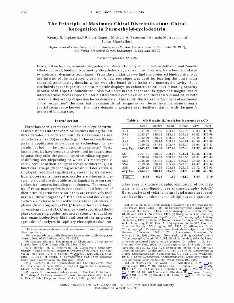
Figure 1. (Top) view looking into the permethyl-â-cyclodex-trin chiral cavity. (Bottom) side view illustrating the typicalconical shape of these molecules. The more open end on top isdesignated the secondary rim and the narrower end on thebottom is the primary rim because it once had primaryhydroxyl groups before alkylation. These views are what onewould expect to see using a slow spectral technique like NMRspectroscopy. These molecules tend to undergo wide amplitudefluctuations and at any given time are distorted from 7-foldsymmetry. Dark gray tones represent oxygen atoms and lightgray tones are carbons. Hydrogen atoms omitted for clarity.
Table 2. MD Results (kJ/mol) for Pulegone/CD
total stretch bend torsion vdW elect
R1 2118.64 416.85 644.53 544.26 65.36 447.64R2 2112.79 416.27 641.31 547.01 61.40 446.79R3 2126.26 417.33 651.10 540.74 77.40 439.69R4 2118.25 417.68 652.61 532.53 73.03 442.40R5 2118.13 417.80 652.55 527.37 75.06 445.34avg ER 2118.81 417.19 648.42 538.38 70.45 444.37
S1 2114.78 417.23 648.23 533.96 72.90 442.46S2 2113.55 416.68 645.76 542.67 62.09 446.36S3 2119.81 417.55 650.97 529.47 76.52 445.30S4 2113.30 417.17 647.21 536.51 67.16 445.25S5 2119.57 416.27 644.29 546.84 67.03 445.14avg ES 2116.20 416.98 647.29 537.89 69.14 444.90
∆∆ER-S2.61 0.21 1.13 0.49 1.31 -0.53
Table 3. MD Results (kJ/mol) for1-Fluorophenylethane/CD
total stretch bend torsion vdW elect
R1 1904.63 369.67 566.61 510.56 31.84 425.95R2 1903.98 369.75 566.04 511.17 21.98 427.04R3 1918.67 370.76 572.73 500.32 50.83 424.03R4 1924.43 370.17 569.47 509.52 48.81 426.46R5 1920.68 369.70 568.05 510.07 46.48 426.38avg ER 1912.93 370.01 568.58 508.33 41.59 425.97
S1 1904.18 369.49 565.24 511.46 30.79 427.70S2 1917.63 370.65 571.97 500.75 50.52 423.79S3 1905.39 369.76 567.58 510.69 31.69 425.67S4 1926.71 370.23 569.74 509.44 48.82 428.48S5 1906.38 369.06 564.39 512.20 33.08 427.65avg ES 1913.49 369.84 567.78 508.91 38.98 426.56
∆∆ER-S-0.56 0.17 0.80 -0.58 2.61 -0.59
Table 4. MD Results (kJ/mol) for 1-Phenylethanol/CD
total stretch bend torsion vdW elect
R1 1972.82 396.15 602.55 516.44 42.36 415.32R2 1985.65 397.42 612.16 501.76 62.97 411.34R3 1983.59 397.13 611.06 505.41 57.99 412.00R4 1975.31 396.55 606.22 516.50 42.41 413.63R5 1965.03 396.18 605.01 517.21 34.60 412.03avg ER 1976.42 396.69 607.40 511.46 48.07 412.86
S1 1979.34 396.55 604.90 517.42 46.96 413.51S2 1968.16 396.43 606.33 516.84 36.68 411.88S3 1973.52 397.35 608.55 505.56 48.28 413.78S4 1992.69 397.54 611.03 505.51 65.86 412.75S5 1963.82 396.16 603.08 518.47 33.47 412.64avg ES 1975.51 396.81 606.78 512.76 46.25 412.91
∆∆ER-S0.97 -0.12 0.62 -1.30 1.82 0.05
Table 5. MD Results (kJ/mol) for 2-MethylbutanoicAcid/CD
total stretch bend torsion vdW elect
R1 1922.13 393.74 617.95 498.10 51.24 361.10R2 1926.97 393.53 614.31 506.94 46.09 366.10R3 1927.44 392.73 612.95 503.83 44.27 373.66R4 1917.53 392.15 606.54 511.03 33.73 374.08R5 1924.47 393.12 614.21 497.73 49.46 369.95avg ER 1923.71 393.05 613.19 503.53 44.96 368.98
S1 1920.88 393.19 611.32 502.34 43.00 371.03S2 1919.82 393.09 608.54 508.01 38.41 371.77S3 1924.52 393.32 613.91 499.62 47.32 370.35S4 1929.35 392.56 611.05 507.48 46.18 372.08S5 1928.14 393.28 614.78 498.89 49.14 372.05avg ES 1924.54 393.09 611.92 503.27 44.81 371.46
∆∆ER-S-0.83 -0.03 1.27 0.26 0.15 -2.48
Chiral Recognition in Permethyl-â-cyclodextrin J. Org. Chem., Vol. 63, No. 3, 1998 733
dress because most host-guest studies of cyclodextrinshave been carried out in very polar media like water. Inthose cases, the hydrophobic effect is a dominant drivingforce for inclusion complexation. While the cyclodextrin-based CSPs used in gas chromatography are often dis-solved in a moderately polar medium, there is no hydro-phobic force per se and it is not clear if analytes preferto bind to the interior of the macrocycle or to the exteriorof the macrocycle. This controversial issue has been
brought to light experimentally by Berthod, Li, andArmstrong,15 who provided compelling evidence for bothinterior and exterior binding based on an extrathermo-dynamic study of a large and diverse set of analytes.Related to this question is whether the analyte prefersto bind to the wider secondary rim, as found in manyexamples of aqueous guest binding from NMR measure-ments,16 at the narrower primary rim, or perhaps some-where in between.(2) What are the intermolecular forces holding the com-
plexes together? Are these forces predominantly weakvan der Waals forces or more powerful electrostaticforces? Moreover, what percent of the interaction energycan be attributed to each?(3) What intermolecular forces are most responsible for
chiral recognition? Are these the same forces that holdthe complexes together or are they different? Whatpercent of the total enantiodifferentiation can be at-tributed to the short-range dispersion forces and whatpercent come from the long-range electrical interactions?
(8) Pertinent discussions can be found in ref 4; for additional reviewsconcerning applications of cyclodextrins in gas chromatography see:(a) Armstrong, D. W.; Han, S. CRC Crit. Rev. Anal. Chem. 1988, 19,175. (b) Konig, W. A. In Drug Stereochemistry, Analytical Methods andPharmacology; Wainer, I. W., Drayer, D. E., Eds.; Marcel Dekker: NewYork, 1988; pp 113-145. (c) Schurig, V.; Nowotny, H.-P. Angew. Chem.,Int. Ed. Engl. 1990, 29, 939. (d) Jung, M.; Mayer, S.; Schurig, V. LC--GC 1994, June, 458. (e) Mani, V.; Wooley, C. Ibid. 1995, September,734.
(9) (a) Enders, D.; Gatzweiler, W.; Dederichs, E. Tetrahedron 1990,46, 4757. (b) Baldwin, J. E.; Bonacorsi, S., Jr. J. Am. Chem. Soc. 1993,115, 10621. (c) Schurig, V.; Glausch, A.; Fluck, M. Tetrahedron:Asymmetry 1995, 6, 2161. (d) Asuncion, L.; Baldwin, J. E. J. Org. Chem.1995, 60, 5778.
(10) Armstrong, D. W.; Tang, Y.; Zukowski, J. Anal. Chem. 1991,63, 2858.
(11) (a) Fletcher, M. T.; Jacobs, M. F.; Kitching, W.; Krohn, S.; Drew,R. A. I.; Haniotakis, G. E.; Francke, W. J. Chem. Soc., Chem. Commun.1992, 1457. (b) Konig, W. A.; Gehrcke, B.; Peter, M. G.; Prestwich, G.D. Tetrahedron: Asymmetry 1993, 4, 165.
(12) (a) Bruche, G.; Dietrich, A.; Mosandl, A. J. High Resolut.Chromatogr. 1993, 16, 101. (b) Sybilska, D.; Asztemborska, M.;Kowalczyk, J.; Ochocka, R. J.; Ossicini, L.; Perez, G. J. Chromatogr.A 1994, 659, 389. (c) Reinhardt, R.; Steinborn, A.; Engewald, W.;Anhalt, K.; Schulze, K. Ibid. 1995, 697, 475. (d) Steinborn, A.;Reinhardt, R.; Engewald, W.; Wyssuwa, K.; Schulze, K. Ibid. 1995,697, 485.
(13) (a) Marner, F.-J.; Runge, T.; Konig, W. A. Helv. Chim. Acta1990, 73, 2165. (b) Askari, C.; Hener, U.; Schmarr, H.-G.; Rapp, A.;Mosandl, A. Fresenius J. Anal. Chem. 1991, 340, 768. (c) Kopke, T.;Mosandl, A. Z. Lebensm. Unters. Forsch. 1992, 194, 372.
(14) (a) Mosandl, A.; Hollnagel, A. Chirality 1989, 1, 293. (b) Konig,W. A.; Evers, P.; Krebber, R.; Schulz, S.; Fehr, C.; Ohloff, G.Tetrahedron 1989, 45, 7006. (c) Mosandl, A.; Rettinger, K.; Fischer,K.; Schubert, V.; Schmarr, H.-G.; Maas, B. J. High Resolut. Chro-matogr. 1990, 13, 382. (d) Mosandl, A.; Bruche, G.; Askari, C.; Schmarr,H.-G. Ibid, 1990, 13, 660. (e) Mosandl, A.; Fischer, K.; Hener, U.; Kreis,P.; Rettinger, K.; Schubert, V.; Schmarr, H.-G. J. Agric. Food Chem.1991, 39, 1131. (f) Askari, C.; Mosandl, A. Phytochem. Anal. 1991, 2,211. (g) Kopke, T.; Schmarr, H.-G.; Mosandl, A. Flavor Fragr. J., 1992,7, 205. (h) Bruche, G.; Mosandl, A.; Kinkel, J. N. J. High Resolut.Chromatogr. 1993, 16, 254. (i) Karl, V.; Gutser, J.; Dietrich, A.; Maas,B.; Mosandl, A. Chirality 1994, 6, 427.
(15) Berthod, A.; Li, W.; Armstrong, D. W. Anal. Chem. 1992, 64,873.
(16) Schneider, H.-J.; Ikeda, H. Chem. Rev. 1997, in press.
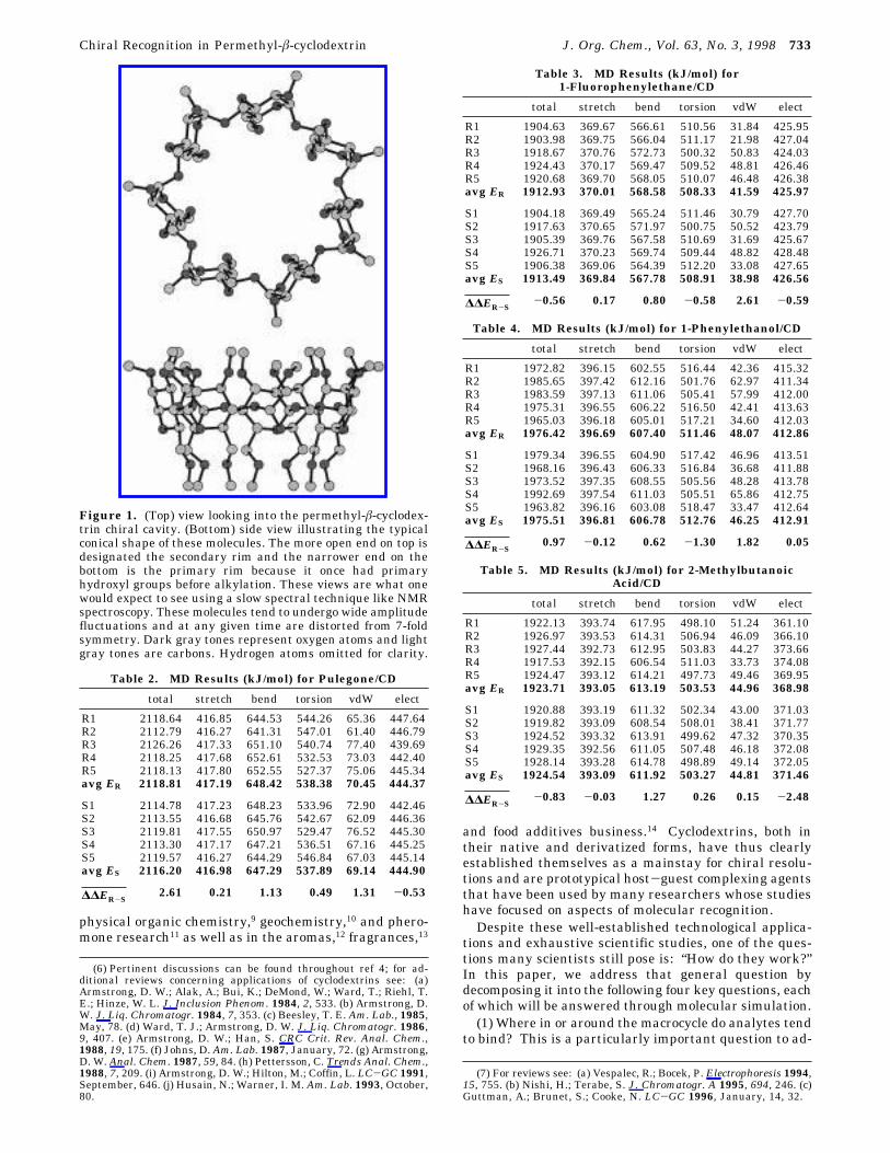
Figure 2. “Dot plot” illustrating the center of mass of guest 2, relative to the cyclodextrin, over the combined 25 ns simulation.Top: end-on and side views of the RR enantiomer. Bottom: end-on and side views of the SS enantiomer. Original diagrams arealso color coded to indicate the intermolecular energy at each point. Lowest energies are at the interior.
734 J. Org. Chem., Vol. 63, No. 3, 1998 Lipkowitz et al.
(4) What regions around the CSP are inherently mostenantiodiscriminating? Although the entire cyclodextrinis chiral, there probably exist regions in or around thecyclodextrin that better discriminate between enanti-omers than do other regions. This is an especiallyimportant but overlooked aspect of chiral discrimination.If, for example, the preferred binding site of a cyclodex-trin is the interior of the cavity, but the most discrimi-nating region is on the exterior of the cyclodextrin, onemay see a diminished ability in chiral discrimination.Related to this, then, is the question: Are the guestbinding domains and the regions of maximum enantio-discrimination spatially coincident?Although the focus of these questions is on permethy-
lated â-cyclodextrin (Figure 1) because of its popularityin gas chromatography, the underlying physical prin-ciples, in addition to the new concepts being developedhere, are directly applicable to any area of organicchemistry where chiral recognition is important.Below we present the results from molecular dynamics
simulations of five pairs of enantiomers binding to per-methyl-â-cyclodextrin, 1. These guest molecules includethe following: isomenthone,17 2, pulegone,18 3, 1-fluo-rophenylethane,19 4, 1-phenylethanol,19 5, and 2-meth-ylbutanoic acid,20 6, each of which have been resolved by
1 that was used as a chiral stationary phase (CSP) inthe above-cited gas chromatographic resolutions. Theseguest molecules contain low to moderately polar func-tional groups (compounds 2 and 3), weakly polar (com-pound 4), and polar protic functional groups (5 and 6),providing a diverse but representative set of analytes ty-pically separated on this popular CSP. Of particularinterest are the structurally related phenylethanes 4 and5. While the S enantiomer of 5 is retained longer on 1,the order is reversed for 4 where the R enantiomer is re-tained longer. This is not an artifact of changing Cahn-Ingold-Prelog nomenclature, but rather is based ondifferences in intermolecular associations between guestand host that we can reproduce by simulation (vide infra).
Simulation Strategy
Under laboratory conditions, analyte molecules areswept through the chromatographic column and interact
(17) Askari, C.; Kreis, P.; Mosandl, A.; Schmarr, H. Arch. Pharm.(Weinheim) 1992, 325, 35.
(18) Kopke, T.; Mosandl, A. Z. Lebensm. Unters. Forsch. 1992, 194,372.
(19) Engwald, W. Chromatographia 1994, 39.(20) Mosandl, A.; Rettinger, K.; Fischer, K.; Schubert, V.; Schmarr,
H.; Maas, B. J. High Resol. Chromatogr. 1990, 13, 382.
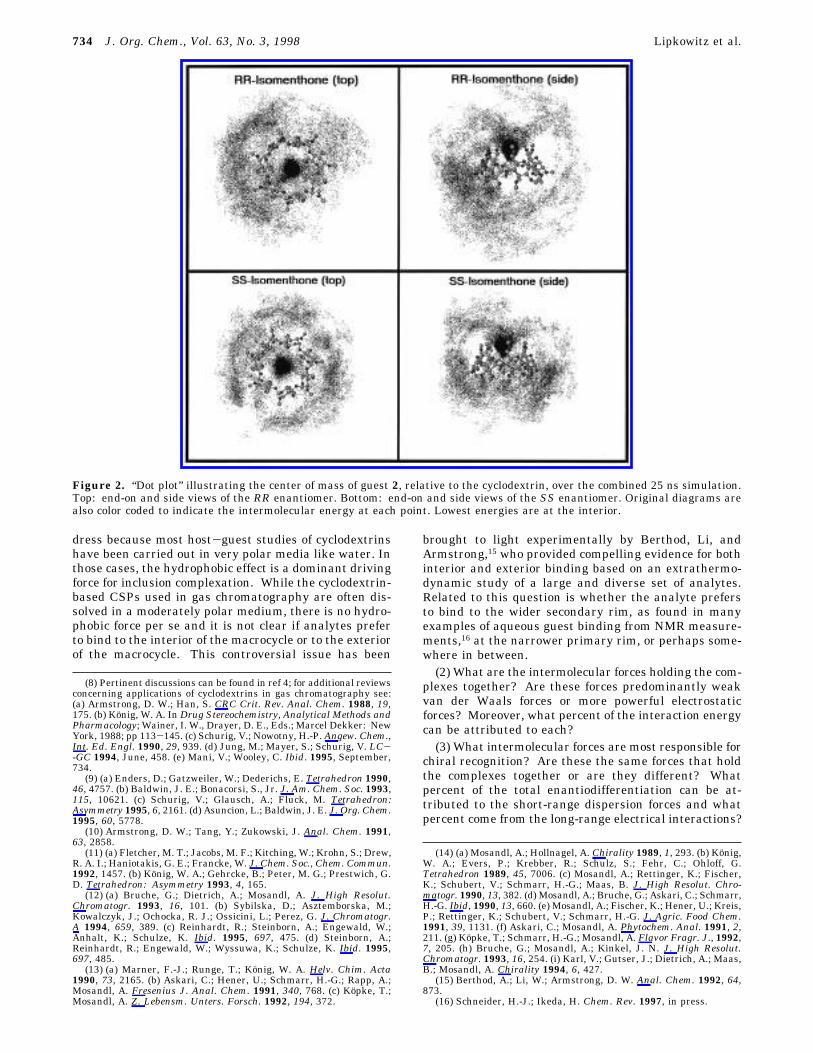
Figure 3. “Dot plot” illustrating the center of mass of guest 3 relative to the cyclodextrin, over the combined 25 ns simulation.Top: end-on and side views of the R enantiomer. Bottom: end-on and side views of the S enantiomer. Original diagrams are alsocolor coded to indicate the intermolecular energy at each point. Lowest energies are at the interior.
Chiral Recognition in Permethyl-â-cyclodextrin J. Org. Chem., Vol. 63, No. 3, 1998 735
in a random way with cyclodextrin molecules they en-counter during their transit. It would be appropriate tomodel this situation in its entirety, but this is not possiblewith existing computing machinery. Instead, we stripthe simulation down to its most relevant parts: a hostand a guest interacting in a dynamical way. The ap-proach we take is to use a single CSP molecule with asingle analyte molecule and carry out stochastic molec-ular dynamics simulations21 on those systems. To effectthis simulation with a single selector complexing a singleselectand, we allow the molecules to bind, dissociate, andrebind multiple times by placing a reflective wall aroundeach diastereomeric complex. To accomplish this, we usea flat-bottom potential having no restraining forces untilthe analyte molecule moves 20 Å from the cyclodextrin.At that point, a 100 kJ/mol/Å restraining force is appliedthat pulls the analyte back toward the CSP where it canfurther associate. Effectively, then, what we’ve ac-complished this way is to allow the analyte to randomlycollide with the CSP in all possible conformations,orientations, and positions as it would in the real system.To probe the system fully we need to sample as large
a volume of phase space for each diastereomeric complexas possible. Moreover, to ensure no computationalartifacts are introduced into the system, e.g., starting theR enantiomer’s trajectory on one rim of the cavity andthe S enantiomer’s on the other rim or one enantiomerinside the cavity with the other on the outside, weinitially superimpose selected atoms of each enantiomerto generate a racemic “supermolecule” that is then placed
in specified starting positions in and around the cyclo-dextrin. Once both enantiomers have been docked in thesame place and with the same orientations/conforma-tions, one of the enantiomers is removed, leaving behinda well-defined binary, diastereomeric complex with thedesired stereochemistry.Five trajectories are run for each enantiomer beginning
from five arbitrarily selected initial positions. For dock-ing purposes the cyclodextrin used is a 7-fold symmetricstructure that allows us to place the analytes at precisepositions parallel or perpendicular to the 7-fold axis. Thereader is to note that cyclodextrins are not symmetric atany given time. They are inherently flexible and undergowide-amplitude flexing motions.22 Over a long enoughsimulation time, however, the time-averaged CD struc-ture is expected, and found, to be nearly symmetric. Thesymmetric structure is used only for docking purposes.The upshot of our simulation strategy is to sample a
wide volume of phase space beginning from differentinitial conditions corresponding to different locations onthe complexes’ potential hypersurface. We then run verylong simulations (5 ns for each trajectory), generating anensemble of trajectories whose energies are then aver-aged. This computational protocol has been describedin detail in a paper comparing and contrasting rigid-bodyMonte Carlo simulations with flexible molecular dynam-ics simulations.23 The MD simulations were shown inthat paper to fully reproduce host-guest binding energiesand is de facto the computational protocol used in thisstudy.
(21) van Gunsteren, W. F.; Berendsen, H. J. C. Mol. Sim. 1988, 1,173.
(22) Lipkowitz, K. B. J. Org. Chem. 1991, 56, 6357.(23) Lipkowitz, K. B.; Pearl, G.; Coner, B.; Peterson. M. A. J. Am.
Chem. Soc. 1997, 119, 600.
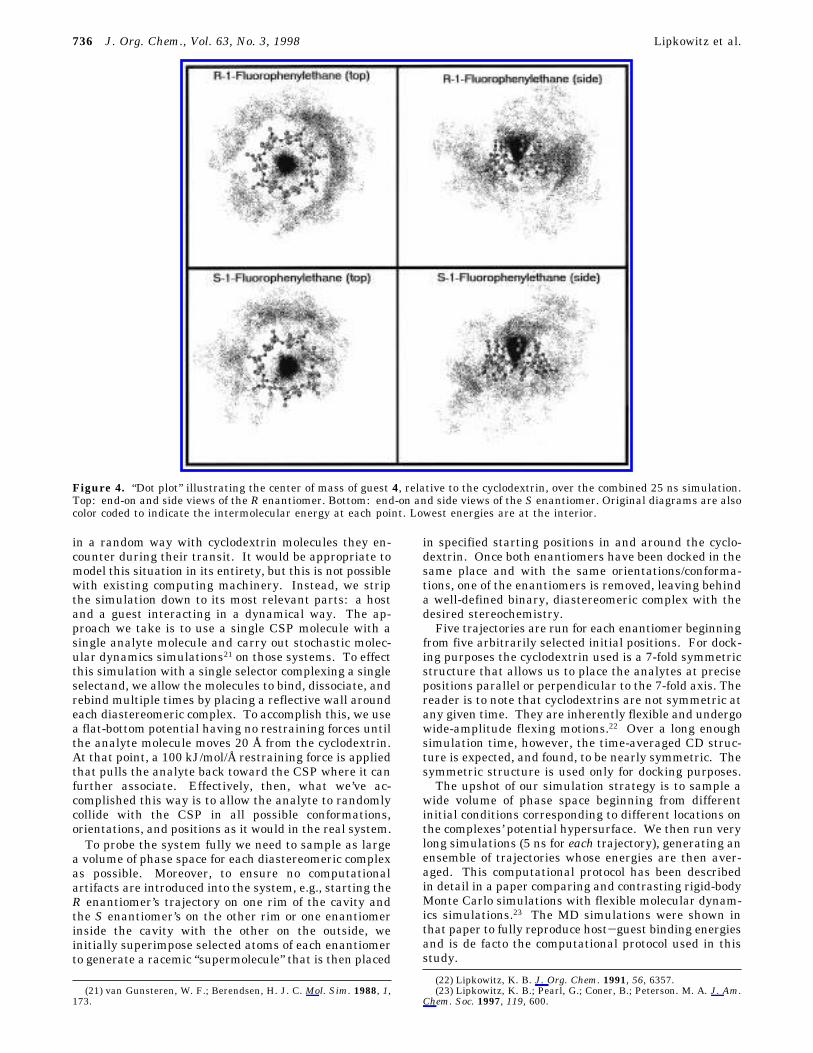
Figure 4. “Dot plot” illustrating the center of mass of guest 4, relative to the cyclodextrin, over the combined 25 ns simulation.Top: end-on and side views of the R enantiomer. Bottom: end-on and side views of the S enantiomer. Original diagrams are alsocolor coded to indicate the intermolecular energy at each point. Lowest energies are at the interior.
736 J. Org. Chem., Vol. 63, No. 3, 1998 Lipkowitz et al.
Computational Tools
All molecular mechanics and molecular dynamics calcula-tions were carried out with the AMBER* force field as foundin Macromodel 5.5.24 The PR conjugate gradient minimizerwas used to minimize the energies, and convergence wasobtained when the gradient root mean square was below10-3 kJ/mol/Å. Throughout this paper, all force field calcula-tions assume a dielectric of 1.0, and no cutoffs of any kind wereused.Stochastic dynamics (SD) simulations were carried out
beginning from the fully optimized lowest-energy molecularmechanics structure. The guest/CD complexes were warmedto the simulation temperature over a period of 5 ps and thenequilibrated for 25 ps. During the production simulations of5000 ps each, structures were saved to disk every 0.5 ps,resulting in 10 000 saved structures from each trajectory. TheSD simulation each had a time step of 0.5 fs with equilibrationand production run temperatures of 353 K (analyte 2), 383 K(analyte 3), 353 K (analyte 4), 353 K (analyte 5), and 423 K
(analyte 6) to be consistent with experimental gas chroma-tography conditions. Translational and rotational momentumswere removed every 100 time steps. To keep the CD/guestcomplexes together, flat-bottom restraints were used betweenthe stereogenic center of the guest molecule and the linkingacetal oxygens of the CD. Using these restraints, if the gueststrayed more than 20 Å away from any linking acetal oxygen,it was gently pushed back toward the CD. These restraintswere used in the heating, equilibration, and production por-tions of the simulations.Post-simulation analysis of the SD trajectories was per-
formed with an in-house program called anout25 that computes,among other things, intermolecular energies (using the AM-BER* force field in this case) and the center of mass positionsof a molecule relative to another. In this work, the guest’spositions were calculated relative to the centroid of the best-fit plane through the acetal linking oxygens of the CD. Fortrajectories being averaged, these guest occurrences were
(24) Mohamadi, F.; Richards, N. G. J.; Guida, W. C.; Liskamp, R.;Caufield, C.; Chang, G.; Hendrickson, T.; Still, W. C. J. Comput. Chem.1990, 11, 440.
(25) ANOUT: written by M.A.P. to analyze MacroModel moleculardynamics trajectories. This software is available from Dr. Michael A.Peterson, Department of Chemistry, University of Florida, Gainesville,FL 32611.
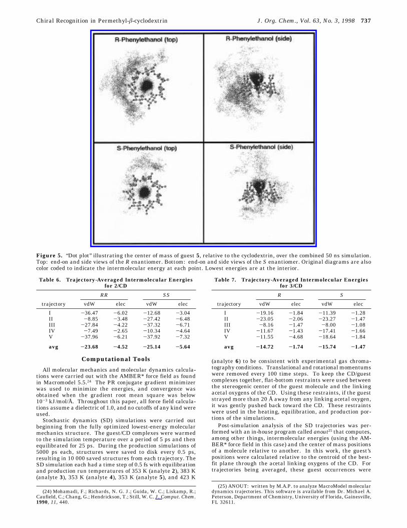
Figure 5. “Dot plot” illustrating the center of mass of guest 5, relative to the cyclodextrin, over the combined 50 ns simulation.Top: end-on and side views of the R enantiomer. Bottom: end-on and side views of the S enantiomer. Original diagrams are alsocolor coded to indicate the intermolecular energy at each point. Lowest energies are at the interior.
Table 6. Trajectory-Averaged Intermolecular Energiesfor 2/CD
RR SS
trajectory vdW elec vdW elec
I -36.47 -6.02 -12.68 -3.04II -8.85 -3.48 -27.42 -6.48III -27.84 -4.22 -37.32 -6.71IV -7.49 -2.65 -10.34 -4.64V -37.96 -6.21 -37.92 -7.32
avg -23.68 -4.52 -25.14 -5.64
Table 7. Trajectory-Averaged Intermolecular Energiesfor 3/CD
R S
trajectory vdW elec vdW elec
I -19.16 -1.84 -11.39 -1.28II -23.05 -2.06 -23.27 -1.47III -8.16 -1.47 -8.00 -1.08IV -11.67 -1.43 -17.41 -1.66V -11.55 -4.68 -18.64 -1.84
avg -14.72 -1.74 -15.74 -1.47
Chiral Recognition in Permethyl-â-cyclodextrin J. Org. Chem., Vol. 63, No. 3, 1998 737
combined and placed on a three-dimensional grid. The sidesconnecting eight adjacent grid points define a volume element.The number of guest positions in each volume element istallied, and the resulting number densities are output in aform suitable for visualization with IRIS Explorer.26 Thisallows us to identify where the guests prefer to bind to theCD (see below).To determine regions of maximum enantiodifferentiation
(see discussion later) we used an in-house program called
mmodgrid 27 running on an SGI computer. It is written in Clanguage, parallelized, and available from one of the authors.27Among other features, this program allows one to carry outgrid scans using different force fields. The AMBER* force fieldwas used with an effective dielectric set to unity and withoutany cutoffs of any kind. Because of the nature of thesystematic grid search being done, the software has beenparallelized, and in this study we used simultaneously eightprocessors of a Cray J90 in addition to 15 available processorson various types of SGI workstations. The dimensions of thegrid surrounding the cyclodextrin are 27 Å × 23 Å × 26 Å.We have selected grid spacings of 0.25 Å and 45° rotationsper axis. Hence, at each grid point we sample 512 uniqueorientations of guest relative to host. The total number of gridpoints for the R and also for the S guest is approximately269 000 each. Visualization of the results was done using IRISExplorer as above.
(26) IRIS Explorer Center (North America), Downers Grove, IL60551-5702 or via <URL http://www.nag.co.uk/1h/Welcome IEC>.
(27) MMODGRID: written by M.A.P. to calculate inter-molecular interaction energies along a grid using the AMBER* andMM3* force fields. This program is available from B. Coner:coner bob@lilly. com. A parallel version, which uses the PVMlibrary (PVM: Parallel Virtual Machine, MIT Press: Cambridge, 1994)is also available.
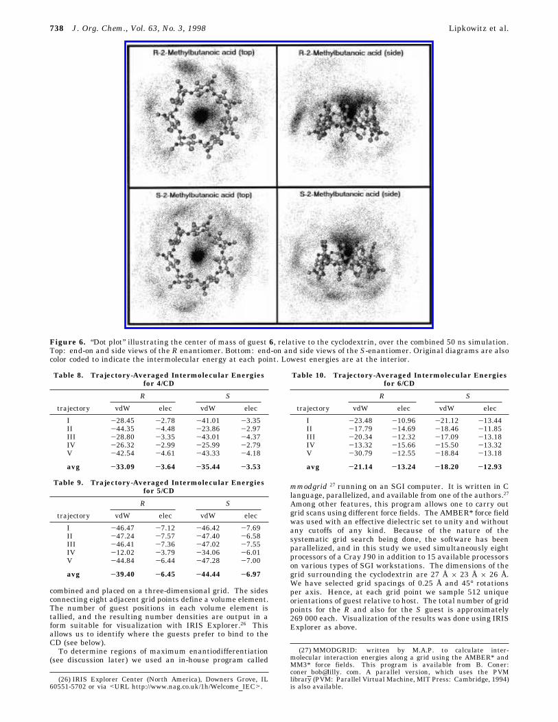
Figure 6. “Dot plot” illustrating the center of mass of guest 6, relative to the cyclodextrin, over the combined 50 ns simulation.Top: end-on and side views of the R enantiomer. Bottom: end-on and side views of the S-enantiomer. Original diagrams are alsocolor coded to indicate the intermolecular energy at each point. Lowest energies are at the interior.
Table 8. Trajectory-Averaged Intermolecular Energiesfor 4/CD
R S
trajectory vdW elec vdW elec
I -28.45 -2.78 -41.01 -3.35II -44.35 -4.48 -23.86 -2.97III -28.80 -3.35 -43.01 -4.37IV -26.32 -2.99 -25.99 -2.79V -42.54 -4.61 -43.33 -4.18
avg -33.09 -3.64 -35.44 -3.53
Table 9. Trajectory-Averaged Intermolecular Energiesfor 5/CD
R S
trajectory vdW elec vdW elec
I -46.47 -7.12 -46.42 -7.69II -47.24 -7.57 -47.40 -6.58III -46.41 -7.36 -47.02 -7.55IV -12.02 -3.79 -34.06 -6.01V -44.84 -6.44 -47.28 -7.00
avg -39.40 -6.45 -44.44 -6.97
Table 10. Trajectory-Averaged Intermolecular Energiesfor 6/CD
R S
trajectory vdW elec vdW elec
I -23.48 -10.96 -21.12 -13.44II -17.79 -14.69 -18.46 -11.85III -20.34 -12.32 -17.09 -13.18IV -13.32 -15.66 -15.50 -13.32V -30.79 -12.55 -18.84 -13.18
avg -21.14 -13.24 -18.20 -12.93
738 J. Org. Chem., Vol. 63, No. 3, 1998 Lipkowitz et al.
Results
The results of our molecular simulations are compiledin Tables 1-5. In these tables, the column heading“total” refers to the sum of the component force fieldenergies computed by the AMBER* force field. Theheading “stretch” refers to the compression/elongationenergies for all bonds in the system averaged over aparticular trajectory. Similarly the column heading“bend” refers to the bending energies of all bond anglesin the system averaged over a given trajectory, and theterm “torsion” refers to the corresponding dihedral anglesaround all bonds in the system. The “vdW” and “elec-trostatic” column headings refer to the short-rangedispersion (van der Waals) and long-range electrostatic(Coulombic) components of the total energy, respectively.The row entries are labeled as R1, R2, etc., indicatingtrajectory no. 1 for the R enantiomer, trajectory no. 2 forthe R enantiomer, and so on. Likewise, S1, S2, etc. refer
to the corresponding trajectories for the S enantiomer.Note that in all cases each trajectory begins from thesame initial location and with the same orientation asits mirror image. Also note that each trajectory is 5 nsin duration (25 ns total) with the exception of those forthe phenylethanol and butanoic acid, which are 10 nseach (50 ns total). The reason for extending the simula-tion times for these analytes is that they contain hydroxyland carboxylic acid moieties that were found to be more“sticky” than the others. By “sticky” we mean that eachenantiomer tended to bind more tightly and for longerperiods of time to a single oxygen atom on the host thandid the other molecules, giving rise to a very nonuniformdistribution of binding domains (after 5 ns). By remain-ing in one region around the cyclodextrin for extendedtime periods we were worried that these guests wouldnot be sampling a large enough volume of phase spaceto provide thermodynamically meaningful results. To
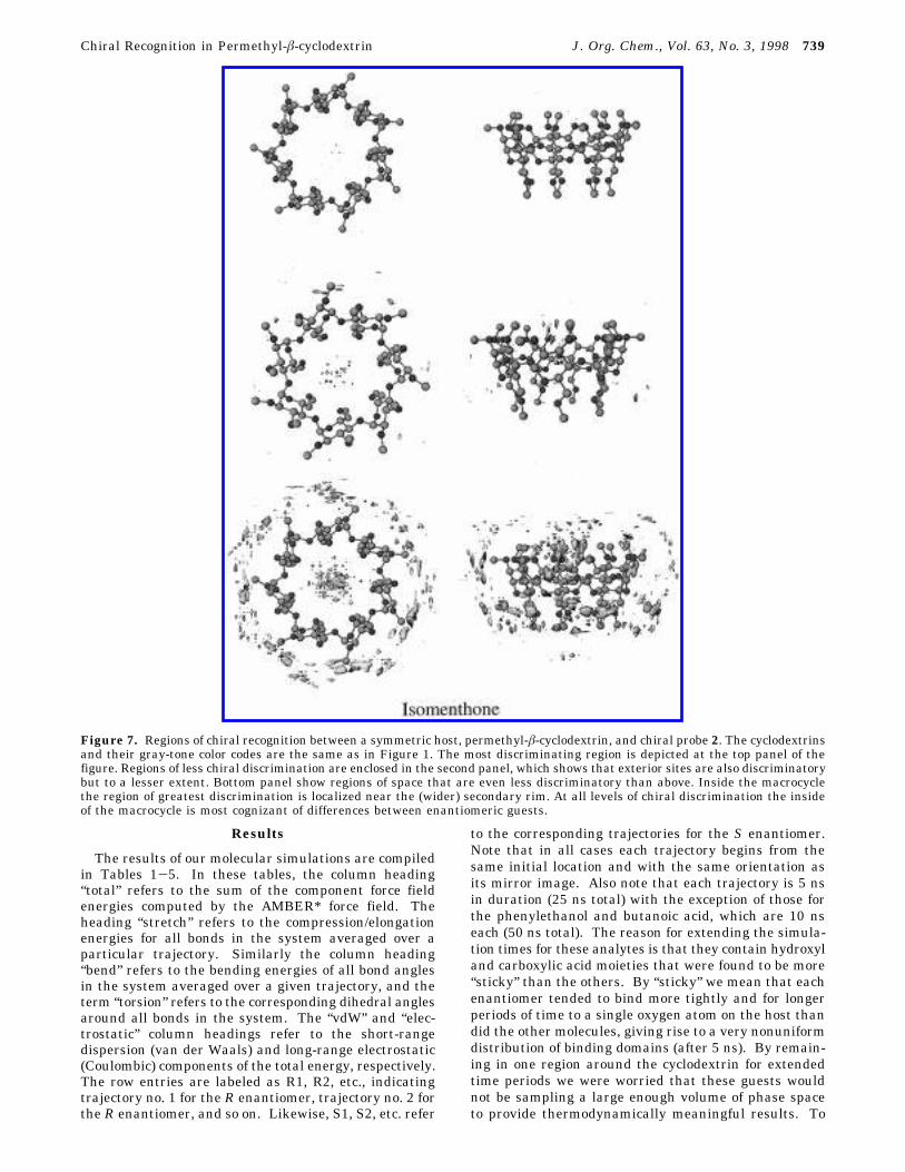
Figure 7. Regions of chiral recognition between a symmetric host, permethyl-â-cyclodextrin, and chiral probe 2. The cyclodextrinsand their gray-tone color codes are the same as in Figure 1. The most discriminating region is depicted at the top panel of thefigure. Regions of less chiral discrimination are enclosed in the second panel, which shows that exterior sites are also discriminatorybut to a lesser extent. Bottom panel show regions of space that are even less discriminatory than above. Inside the macrocyclethe region of greatest discrimination is localized near the (wider) secondary rim. At all levels of chiral discrimination the insideof the macrocycle is most cognizant of differences between enantiomeric guests.
Chiral Recognition in Permethyl-â-cyclodextrin J. Org. Chem., Vol. 63, No. 3, 1998 739
overcome this stickiness problem we simply ran thesimulation for a longer time period, allowing the guestto better explore all binding regions in and around theCSP.The results in these tables are consistent with the
corresponding retention orders found experimentally,17-20
and they are also consistent with the small enantiose-lective energy differences found experimentally. Accord-ingly, we feel confident in using the data from thesesimulations to begin answering the four questions posedabove.
Discussion
In this section we begin extracting information fromthe simulations with the intention of answering thosequestions. The following subsections are presented in theorder of those four questions to accomplish this goal.(a) Guest Binding Sites. To answer the question of
where the guest is most likely to be found, we placed thetime-averaged CD’s center of mass at the origin of a
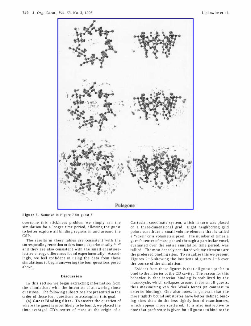
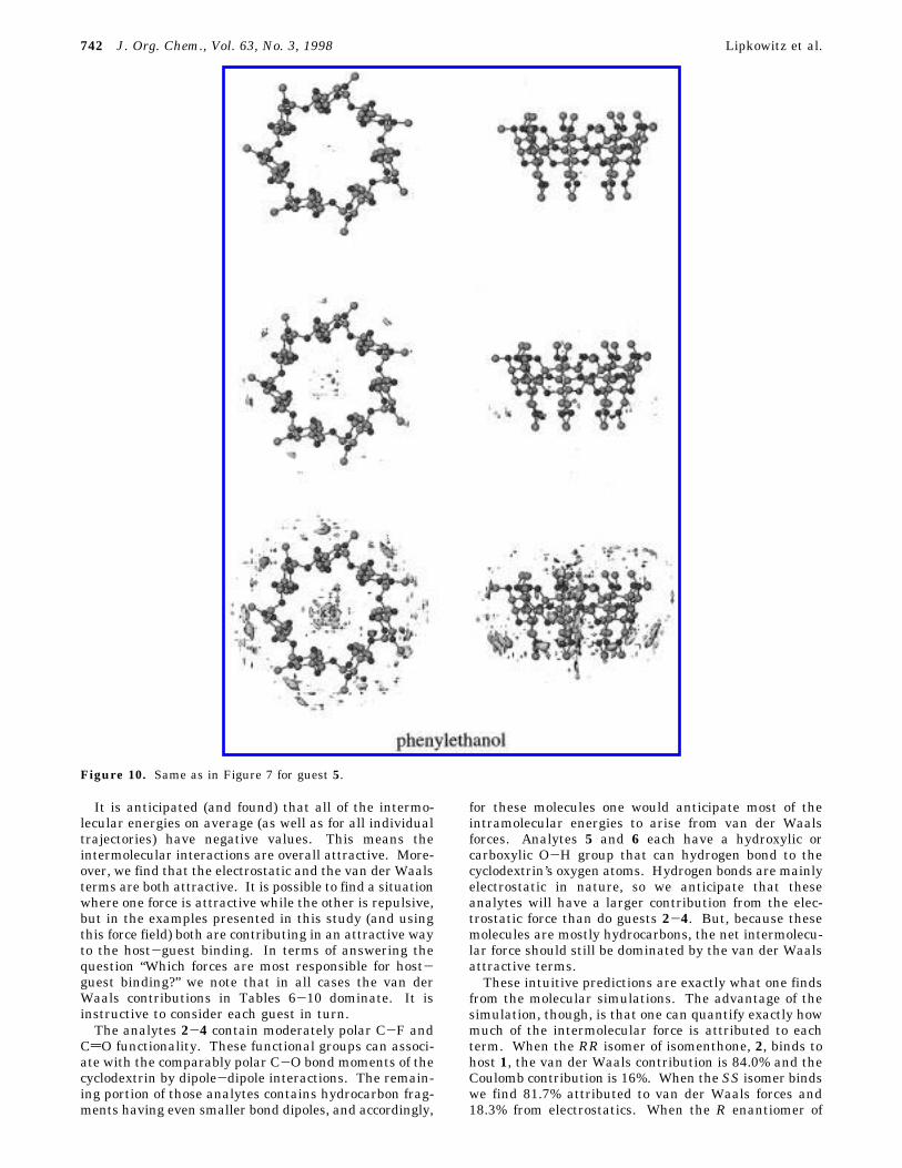
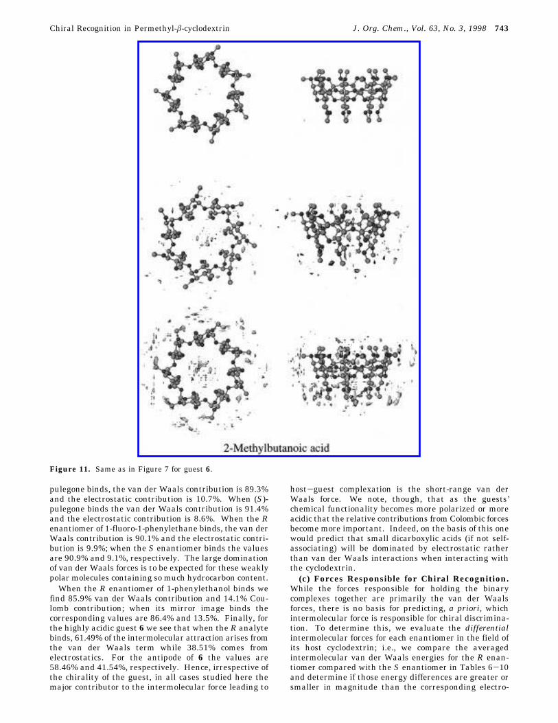
Cartesian coordinate system, which in turn was placedon a three-dimensional grid. Eight neighboring gridpoints constitute a small volume element that is calleda “voxel” or a volumetric pixel. The number of times aguest’s center of mass passed through a particular voxel,evaluated over the entire simulation time period, wastallied. The most densely populated volume elements arethe preferred binding sites. To visualize this we presentFigures 2-6 showing the locations of guests 2-6 overthe course of the simulation.Evident from these figures is that all guests prefer to
bind to the interior of the CD cavity. The reason for thisbehavior is that interior binding is stabilized by themacrocycle, which collapses around these small guests,thus maximizing van der Waals forces (in contrast toexterior binding). One also notes, in general, that themore tightly bound substrates have better defined bind-ing sites than do the less tightly bound enantiomers,which appear more scattered. It is also instructive tonote that preference is given for all guests to bind to the
Figure 8. Same as in Figure 7 for guest 3.
740 J. Org. Chem., Vol. 63, No. 3, 1998 Lipkowitz et al.
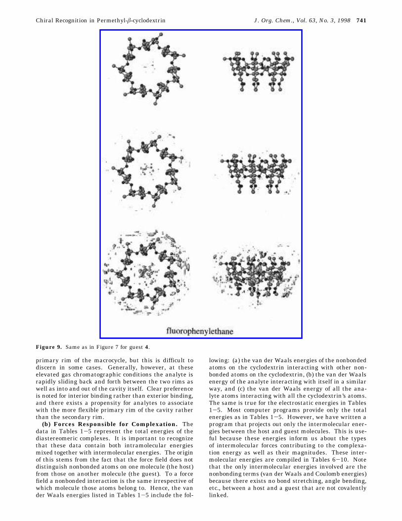
primary rim of the macrocycle, but this is difficult todiscern in some cases. Generally, however, at theseelevated gas chromatographic conditions the analyte israpidly sliding back and forth between the two rims aswell as into and out of the cavity itself. Clear preferenceis noted for interior binding rather than exterior binding,and there exists a propensity for analytes to associatewith the more flexible primary rim of the cavity ratherthan the secondary rim.(b) Forces Responsible for Complexation. The
data in Tables 1-5 represent the total energies of thediastereomeric complexes. It is important to recognizethat these data contain both intramolecular energiesmixed together with intermolecular energies. The originof this stems from the fact that the force field does notdistinguish nonbonded atoms on one molecule (the host)from those on another molecule (the guest). To a forcefield a nonbonded interaction is the same irrespective ofwhich molecule those atoms belong to. Hence, the vander Waals energies listed in Tables 1-5 include the fol-
lowing: (a) the van der Waals energies of the nonbondedatoms on the cyclodextrin interacting with other non-bonded atoms on the cyclodextrin, (b) the van der Waalsenergy of the analyte interacting with itself in a similarway, and (c) the van der Waals energy of all the ana-lyte atoms interacting with all the cyclodextrin’s atoms.The same is true for the electrostatic energies in Tables1-5. Most computer programs provide only the totalenergies as in Tables 1-5. However, we have written aprogram that projects out only the intermolecular ener-gies between the host and guest molecules. This is use-ful because these energies inform us about the typesof intermolecular forces contributing to the complexa-tion energy as well as their magnitudes. These inter-molecular energies are compiled in Tables 6-10. Notethat the only intermolecular energies involved are thenonbonding terms (van der Waals and Coulomb energies)because there exists no bond stretching, angle bending,etc., between a host and a guest that are not covalentlylinked.
Figure 9. Same as in Figure 7 for guest 4.
Chiral Recognition in Permethyl-â-cyclodextrin J. Org. Chem., Vol. 63, No. 3, 1998 741
It is anticipated (and found) that all of the intermo-lecular energies on average (as well as for all individualtrajectories) have negative values. This means theintermolecular interactions are overall attractive. More-over, we find that the electrostatic and the van der Waalsterms are both attractive. It is possible to find a situationwhere one force is attractive while the other is repulsive,but in the examples presented in this study (and usingthis force field) both are contributing in an attractive wayto the host-guest binding. In terms of answering thequestion “Which forces are most responsible for host-guest binding?” we note that in all cases the van derWaals contributions in Tables 6-10 dominate. It isinstructive to consider each guest in turn.The analytes 2-4 contain moderately polar C-F and
CdO functionality. These functional groups can associ-ate with the comparably polar C-O bond moments of thecyclodextrin by dipole-dipole interactions. The remain-ing portion of those analytes contains hydrocarbon frag-ments having even smaller bond dipoles, and accordingly,
for these molecules one would anticipate most of theintramolecular energies to arise from van der Waalsforces. Analytes 5 and 6 each have a hydroxylic orcarboxylic O-H group that can hydrogen bond to thecyclodextrin’s oxygen atoms. Hydrogen bonds are mainlyelectrostatic in nature, so we anticipate that theseanalytes will have a larger contribution from the elec-trostatic force than do guests 2-4. But, because thesemolecules are mostly hydrocarbons, the net intermolecu-lar force should still be dominated by the van der Waalsattractive terms.These intuitive predictions are exactly what one finds
from the molecular simulations. The advantage of thesimulation, though, is that one can quantify exactly howmuch of the intermolecular force is attributed to eachterm. When the RR isomer of isomenthone, 2, binds tohost 1, the van der Waals contribution is 84.0% and theCoulomb contribution is 16%. When the SS isomer bindswe find 81.7% attributed to van der Waals forces and18.3% from electrostatics. When the R enantiomer of
Figure 10. Same as in Figure 7 for guest 5.
742 J. Org. Chem., Vol. 63, No. 3, 1998 Lipkowitz et al.
pulegone binds, the van der Waals contribution is 89.3%and the electrostatic contribution is 10.7%. When (S)-pulegone binds the van der Waals contribution is 91.4%and the electrostatic contribution is 8.6%. When the Renantiomer of 1-fluoro-1-phenylethane binds, the van derWaals contribution is 90.1% and the electrostatic contri-bution is 9.9%; when the S enantiomer binds the valuesare 90.9% and 9.1%, respectively. The large dominationof van der Waals forces is to be expected for these weaklypolar molecules containing so much hydrocarbon content.When the R enantiomer of 1-phenylethanol binds we
find 85.9% van der Waals contribution and 14.1% Cou-lomb contribution; when its mirror image binds thecorresponding values are 86.4% and 13.5%. Finally, forthe highly acidic guest 6 we see that when the R analytebinds, 61.49% of the intermolecular attraction arises fromthe van der Waals term while 38.51% comes fromelectrostatics. For the antipode of 6 the values are58.46% and 41.54%, respectively. Hence, irrespective ofthe chirality of the guest, in all cases studied here themajor contributor to the intermolecular force leading to
host-guest complexation is the short-range van derWaals force. We note, though, that as the guests’chemical functionality becomes more polarized or moreacidic that the relative contributions from Colombic forcesbecome more important. Indeed, on the basis of this onewould predict that small dicarboxylic acids (if not self-associating) will be dominated by electrostatic ratherthan van der Waals interactions when interacting withthe cyclodextrin.(c) Forces Responsible for Chiral Recognition.
While the forces responsible for holding the binarycomplexes together are primarily the van der Waalsforces, there is no basis for predicting, a priori, whichintermolecular force is responsible for chiral discrimina-tion. To determine this, we evaluate the differentialintermolecular forces for each enantiomer in the field ofits host cyclodextrin; i.e., we compare the averagedintermolecular van der Waals energies for the R enan-tiomer compared with the S enantiomer in Tables 6-10and determine if those energy differences are greater orsmaller in magnitude than the corresponding electro-
Figure 11. Same as in Figure 7 for guest 6.
Chiral Recognition in Permethyl-â-cyclodextrin J. Org. Chem., Vol. 63, No. 3, 1998 743
static values. For example, in Table 6 the averageintermolecular van der Waals energy difference, ∆∆Evdw,is 1.46 kJ/mol (-25.14 vs -23.68) and the differentialelectrostatic value, ∆∆Eelec, is 1.12 kJ/mol (-5.64 vs 4.52).Likewise for analyte 3 we find ∆∆Evdw ) 1.02 kJ/mol and∆∆Eelec ) 0.27 kJ/mol. For analyte 4 we find ∆∆Evdw )2.35 kJ/mol and ∆∆Eelec ) 0.11 kJ/mol. For analyte 5we find ∆∆Evdw ) 5.04 kJ/mol and ∆∆Eelec ) 0.52 kJ/mol. Finally, for analyte 6 we find ∆∆Evdw ) 2.94 kJ/mol and ∆∆Eelec ) 0.31 kJ/mol. In all cases, the largerdifferences exist in the van der Waals forces so weconclude that the most enantiodifferentiating forces arethe van der Waals forces. Hence, the same forcesresponsible for host-guest complexation are also mostresponsible for chiral discrimination.(d) Regions of Greatest Chiral Discrimination.
Knowing where guests tend to bind in or around acyclodextrin is important, but this aspect of molecularrecognition constitutes only part of what leads to effectivechiral recognition. Another aspect that is critical foreffective resolutions is knowing which region of the CSPis most discriminating. This is especially importantbecause if the preferred binding site differs from the sitethat is most highly discriminating one is relegated to aninferior region of chiral selection leading to loss ofdiscriminatory power or even no recognition at all. Thisaspect of chiral recognition is what we will refer to as“the principle of maximum chiral recognition”, whichstates that maximum chiral recognition is assured whenthe guest’s binding site and the host’s most enantiodis-criminating region are spatially coincident. This is anintuitive and seemingly trivial concept in some ways, butin other ways it is a profound concept that appears notto have been discussed in the literature. Having a prioriknowledge of which region of a host is most discrimina-tory is especially useful because one could then devisetechniques to force the substrate to bind to a thermody-namically less-favored but more discriminatory site. Interms of chiral chromatography with, say, cyclodextrins,this could be as simple as adding to the mobile phase acompeting substrate that binds exclusively to the interiorof the cyclodextrin, thus forcing the guest to the lessfavored and (possibly) more discriminating exterior of thecyclodextrin. This would first reduce the retention timeof the analytes on the column and second enhance theresolution, both of which are desirable traits in chroma-tography, but the concept can be generalized to any othersituation where chiral recognition is important.Although we have located the preferred binding site
(inside the cyclodextrin cavity) and we were able to definethe intermolecular forces responsible for both complex-ation and molecular recognition (dispersion forces) wehave not identified the most discriminating regions of thehost molecule. To do this, we adopt the followingprocedure. The time-averaged, 7-fold symmetric cyclo-dextrin’s center of mass is placed at the origin of aCartesian coordinate system and a uniform grid is placedover that coordinate system. At each grid point theguest’s center of mass is positioned and then systemati-cally rotated about a local coordinate system along threeorthogonal axes. The number of grid points and thenumber of rotations per axis is arbitrary, but we use 512unique orientations per guest at each grid point (seecomputational tools section). Rather than saving onlythe lowest energy orientation at each grid point weBoltzmann average the energies at each grid point.
Because we are using a deterministic grid search meth-odology we note that whatever is being done to the Renantiomer is being done equivalently to the S enanti-omer; this way sampling artifacts are removed. Thedifferences at each grid point, between R vs S guests,indicate discriminatory regions. Those grid points withzero or small energy differences between mirror imageisomers are not discriminating, while those grid pointswith the largest differences are most discriminating.The results of our calculations show that the greatest
difference in interaction energy for R vs S probes existsin the interior of the macrocycle as illustrated in Figures7-11. In these figures are plotted isoenergy contoursurfaces of differences in Boltzmann-weighted energiesbetween R and S probe molecules at each grid point. Atthe top of each figure is the region of greatest energydifference and is thus the most enantiodiscriminatingpart of the CSP. In the next two panels of each figureare regions containing smaller and smaller differentialenergies, which in turn are encompassing volumes ofspace having less and less enantiodiscrimination. Ofcourse, the regions not being rendered are very weaklydiscriminating or have no chiral recognition at all (atleast for that particular probe).What we find, then, is that the most enantiodifferen-
tiating region of the macrocycle (the interior) is alsowhere the analytes prefer to bind. So, in this case ofmolecular recognition Nature places the analytes in thevicinity of highest chiral discrimination. On the basisof the spatial congruence of the analyte binding site withthe host’s regions of maximum enantiodifferentiation wecan now see why permethyated â-cyclodextrin is such auseful chiral stationary phase.
Summary
To help answer the question “How does permethyl-â-cyclodextrin work as a chiral stationary phase in gaschromatography?” we posed and answered four importantquestions. By addressing those questions we concludethe following: (1) Analyte molecules tend to bind to theinterior of the cyclodextrin’s cavity with a slight prefer-ence for the primary rim. Binding to the interior leadsto a stabilization of intermolecular attraction by way ofcyclodextrin collapse around the analyte. Nonetheless,the guest molecules can quickly escape from the interiorof this cavity, especially at such elevated temperatures.(2) The dominant intermolecular force responsible forhost-guest coordination is the short-range dispersionforce rather than the long-range electrostatic force. Asthe polarity of functional groups on the guest increaseone expects and finds more of a contribution from theelectrostatic part of the intermolecular force. Usually,however, this electrostatic force is overshadowed by thedispersion forces arising from the remaining hydrocarbonpart of the guest. (3) The intermolecular force mostresponsible for enantiodifferentiation is also the short-range dispersion force. (4) The region of maximumenantiodifferentiation is found, for the molecules studiedin this paper using the AMBER* empirical force field, tobe the interior of the cyclodextrin. Although enantiose-lective regions exist both outside and inside of themacrocycle, the interior is more discriminatory.In summary, the molecular simulations carried out in
this study have illuminated not only how permethyl-â-cyclodextrins work, but also where they work most
744 J. Org. Chem., Vol. 63, No. 3, 1998 Lipkowitz et al.
effectively. From these results we can also begin toappreciate why this particular cyclodextrin is such a goodCSP for chromatographic resolutions: essentially, Natureplaces the analyte in the region of the cyclodextrin thatis most enantiodifferentiating. The concept of maintain-ing spatial coincidence between the host’s region ofmaximum enantiodifferentiation and the preferred siteof guest binding was presented here. This concept isseemingly trivial but is profoundly significant when one
wants to maximize chiral recognition and we urge otherscientists to consider this aspect of molecular recognitionin their studies.
Acknowledgment. This work was sponsored by agrant from the National Science Foundation (CHE94-12512).
JO9717090
Chiral Recognition in Permethyl-â-cyclodextrin J. Org. Chem., Vol. 63, No. 3, 1998 745