The Methionine Transamination Pathway Controls Hepatic ... · homeostasis. In mammals, in response...
21
Methionine regulates PGC-1α activity 1 The Methionine Transamination Pathway Controls Hepatic Glucose Metabolism through Regulation of the GCN5 Acetyl Transferase and the PGC-1α Transcriptional Coactivator Clint D. J. Tavares 1,2,† , Kfir Sharabi 1,2,† , John E. Dominy 1,2 , Yoonjin Lee 1,2 , Marta Isasa 2 , Jose M. Orozco 1,2 , Mark P. Jedrychowski 2 , Theodore M. Kamenecka 3 , Patrick R. Griffin 3 , Steven P. Gygi 2 and Pere Puigserver 1,2,* 1 Department of Cancer Biology, Dana-Farber Cancer Institute and 2 Department of Cell Biology, Harvard Medical School, Boston, MA 02115, USA. 3 Department of Molecular Therapeutics, The Scripps Research Institute, Jupiter, FL 33458, USA. Running Title: Methionine regulates PGC-1α activity † These authors contributed equally to this work * To whom correspondence should be addressed: Pere Puigserver, PhD, Dana-Farber Cancer Institute, 450 Brookline Ave, CLSB-11144, Boston, MA 02215; E-mail: [email protected]; Phone: +1-617-582-7977; Fax: +1-617-632-5363 Keywords: transcription coactivator, peroxisome proliferator‐activated receptor gamma coactivator 1‐alpha (PGC‐1α) (Ppargc1a), gluconeogenesis, acetyltransferase, methionine, methylthiopropionic acid (MTP), methionine transamination, acetylation, phosphoenolpyruvate carboxykinase ( Pck1), glucose-6- phosphatase (G6pc). ABSTRACT Methionine is an essential sulfur amino acid that is engaged in key cellular functions such as protein synthesis, and is a precursor for critical metabolites involved in maintaining cellular homeostasis. In mammals, in response to nutrient conditions, the liver plays a significant role in regulating methionine concentrations by altering its flux through the transmethylation, transsulfuration and transamination metabolic pathways. A comprehensive understanding of how hepatic methionine metabolism intersects with other regulatory nutrient signaling and transcriptional events is however lacking. Here, we show that methionine and derived- sulfur metabolites in the transamination pathway activate the GCN5 acetyl transferase promoting acetylation of the transcriptional coactivator PGC-1α to control hepatic gluconeogenesis. Methionine was the only essential amino acid that rapidly induced PGC- 1α acetylation through activating the GCN5 acetyl transferase. Experiments employing metabolic pathway intermediates revealed that methionine transamination, and not the transmethylation or transsulfuration pathways, contributed to methionine-induced PGC-1α acetylation. Moreover, aminooxyacetic acid, a transaminase inhibitor, was able to potently suppress PGC-1α acetylation stimulated by methionine, which was accompanied by predicted alterations in PGC-1α-mediated gluconeogenic gene expression and glucose production in primary murine hepatocytes. Methionine administration in mice likewise induced hepatic PGC-1α acetylation, suppressed the gluconeogenic gene program and lowered glycemia, indicating that a similar phenomenon occurs in vivo. These results highlight a communication between methionine metabolism and PGC-1α-mediated hepatic gluconeogenesis, suggesting that influencing methionine metabolic flux has the potential to be therapeutically exploited for diabetes treatment. Amino acid sensing is a critical mechanism that cells employ to maintain homeostasis and enable survival upon fluctuations in the nutritional environment (1,2). Despite variability in side chain chemical structure, cells are able to http://www.jbc.org/cgi/doi/10.1074/jbc.M115.706200 The latest version is at JBC Papers in Press. Published on March 28, 2016 as Manuscript M115.706200 Copyright 2016 by The American Society for Biochemistry and Molecular Biology, Inc. by guest on March 7, 2019 http://www.jbc.org/ Downloaded from
-
Upload
duongkhanh -
Category
Documents
-
view
221 -
download
0
Transcript of The Methionine Transamination Pathway Controls Hepatic ... · homeostasis. In mammals, in response...
Methionine regulates PGC-1α activity
1
The Methionine Transamination Pathway Controls Hepatic Glucose Metabolism through
Regulation of the GCN5 Acetyl Transferase and the PGC-1α Transcriptional Coactivator
Clint D. J. Tavares1,2,†
, Kfir Sharabi1,2,†
, John E. Dominy1,2
, Yoonjin Lee1,2
, Marta Isasa2, Jose M.
Orozco1,2
, Mark P. Jedrychowski2, Theodore M. Kamenecka
3, Patrick R. Griffin
3, Steven P. Gygi
2 and
Pere Puigserver1,2,*
1Department of Cancer Biology, Dana-Farber Cancer Institute and
2Department of Cell Biology, Harvard
Medical School, Boston, MA 02115, USA. 3Department of Molecular Therapeutics, The Scripps
Research Institute, Jupiter, FL 33458, USA.
Running Title: Methionine regulates PGC-1α activity
† These authors contributed equally to this work
* To whom correspondence should be addressed:
Pere Puigserver, PhD, Dana-Farber Cancer Institute, 450 Brookline Ave, CLSB-11144, Boston, MA
02215; E-mail: [email protected]; Phone: +1-617-582-7977; Fax: +1-617-632-5363
Keywords: transcription coactivator, peroxisome proliferator‐activated receptor gamma coactivator
1‐alpha (PGC‐1α) (Ppargc1a), gluconeogenesis, acetyltransferase, methionine, methylthiopropionic acid
(MTP), methionine transamination, acetylation, phosphoenolpyruvate carboxykinase (Pck1), glucose-6-
phosphatase (G6pc).
ABSTRACT
Methionine is an essential sulfur amino acid
that is engaged in key cellular functions such as
protein synthesis, and is a precursor for critical
metabolites involved in maintaining cellular
homeostasis. In mammals, in response to
nutrient conditions, the liver plays a significant
role in regulating methionine concentrations by
altering its flux through the transmethylation,
transsulfuration and transamination metabolic
pathways. A comprehensive understanding of
how hepatic methionine metabolism intersects
with other regulatory nutrient signaling and
transcriptional events is however lacking.
Here, we show that methionine and derived-
sulfur metabolites in the transamination
pathway activate the GCN5 acetyl transferase
promoting acetylation of the transcriptional
coactivator PGC-1α to control hepatic
gluconeogenesis. Methionine was the only
essential amino acid that rapidly induced PGC-
1α acetylation through activating the GCN5
acetyl transferase. Experiments employing
metabolic pathway intermediates revealed that
methionine transamination, and not the
transmethylation or transsulfuration pathways,
contributed to methionine-induced PGC-1α
acetylation. Moreover, aminooxyacetic acid, a
transaminase inhibitor, was able to potently
suppress PGC-1α acetylation stimulated by
methionine, which was accompanied by
predicted alterations in PGC-1α-mediated
gluconeogenic gene expression and glucose
production in primary murine hepatocytes.
Methionine administration in mice likewise
induced hepatic PGC-1α acetylation,
suppressed the gluconeogenic gene program
and lowered glycemia, indicating that a similar
phenomenon occurs in vivo. These results
highlight a communication between methionine
metabolism and PGC-1α-mediated hepatic
gluconeogenesis, suggesting that influencing
methionine metabolic flux has the potential to
be therapeutically exploited for diabetes
treatment.
Amino acid sensing is a critical mechanism
that cells employ to maintain homeostasis and
enable survival upon fluctuations in the nutritional
environment (1,2). Despite variability in side
chain chemical structure, cells are able to
http://www.jbc.org/cgi/doi/10.1074/jbc.M115.706200The latest version is at JBC Papers in Press. Published on March 28, 2016 as Manuscript M115.706200
Copyright 2016 by The American Society for Biochemistry and Molecular Biology, Inc.
by guest on March 7, 2019
http://ww
w.jbc.org/
Dow
nloaded from
Methionine regulates PGC-1α activity
2
distinguish the distinct amino acids through the
use of specific tRNAs. The amino acid protein
sensor GCN2 detects deficiencies in amino acids
in a tRNA-dependent manner and reprograms
cellular metabolism to modulate energy
consumption and maintain homeostasis (3,4).
While mTORC1 is not considered to be a direct
sensor of amino acid levels, it functions similarly
to GCN2 to alter metabolic pathways in response
to amino acid deprivation (5). Methionine, a
dietary essential amino acid, is among the more
toxic of the L-amino acids, and hence its
abundance needs to be precisely regulated (6,7).
The liver is the primary organ concerned with the
detoxification of methionine, and hence its ability
to sense alterations in concentrations of this amino
acid is vital (8). The existence of any crosstalk
between hepatic methionine metabolism and other
nutrient-regulated metabolic processes in the liver,
such as glucose production, requires further
investigation.
Intracellularly, apart from being utilized for
the synthesis of proteins, methionine acts as a
precursor for various metabolites that are
associated with the anabolism of biological
macromolecules, including nucleic acids (9,10).
The two main routes for methionine catabolism
include the transmethylation and transsulfuration
pathways. The initial step in transmethylation
involves conversion of methionine to S-
adenosylmethionine (SAM). SAM plays a
significant role as a donor of methyl groups, and is
also involved in formation of polyamines such as
spermidine (11). As a consequence of methyl
transfer, SAM is metabolized to homocysteine via
the S-adenosyl homocysteine (SAH) intermediate
(9,10). Homocysteine can then either be recycled
back to methionine in a folate/choline-dependent
manner, or it can be shunted down the
transsulfuration pathway. This transsulfuration
process is comprised of the condensation of
homocysteine with serine succeeded by a
hydrolysis reaction, and results in the formation of
cysteine via cystathionine. The cysteine produced
can then be consumed in protein translation or
synthesis of the antioxidant glutathione and the
osmolyte taurine (9,10).
In addition to the transmethylation and
transsulfuration pathways, methionine is also
shunted down the transamination pathway (12,13).
Flux of methionine down this pathway involves an
initial transaminase-mediated conversion to α-
keto-γ-methylthiobutyrate (KMTB) (14). KMTB
is then oxidatively decarboxylated to 3-
methylthiopropionic acid (MTP) through an
irreversible reaction catalyzed by the branched
chain 2-oxo acid dehydrogenase complex
(BCKDC) (15). 3-MTP is further metabolized to
the gaseous methanethiol (16), which has been
reported to then be broken down into simpler
molecules, including hydrogen sulfide, dimethyl
sulfide and formic acid, with the end products
being carbon dioxide and sulfur dioxide (17,18).
While initial reports on methionine transamination
suggested that it provides a route for the
degradation and clearance of methionine (19),
more recently researchers have claimed that
transsulfuration serves as the major pathway for
the detoxification of excessive methionine (20).
Early studies in rats also indicate that activity of
the methionine transamination pathway is altered
during fasting; however, no clear functional
significance has been attributed to this pathway.
Despite methionine transamination being
suggested to occur in various rodent tissues
including the liver, the fraction of methionine that
is shunted through this pathway as well as its
necessity in humans, remains to be accurately
determined.
One of the mechanisms that functions to
maintain blood glucose levels within a narrow
range involves glucose production by the liver,
which is differentially regulated during the fasting
and fed states (21). Hepatic gluconeogenesis is
induced upon fasting, and is extensively controlled
by PGC-1α (22). This transcriptional coactivator
interacts with hepatocyte nuclear factor 4α
(HNF4α) and forkhead box protein O1 (FOXO1)
to promote transcription of phosphoenolpyruvate
carboxykinase (Pck1) and glucose-6-phosphatase
(G6pc) genes, which encode two enzymes that
significantly moderate gluconeogenesis (23,24).
The activity, localization and stability of PGC-1α
are regulated by various post-translational
modifications, including acetylation which is
known to impede its transcriptional co-activating
ability and leads to the suppression of
gluconeogenesis (25-27). The acetylation status of
PGC-1α is primarily determined by opposing
activities of the general control nonrepressed
protein 5 (GCN5) acetyl transferase and the
NAD+-dependent silent mating type information
by guest on March 7, 2019
http://ww
w.jbc.org/
Dow
nloaded from
Methionine regulates PGC-1α activity
3
regulation 2 homolog 1 (Sirt1) deacetylase
(25,26). Various signaling cascades converge on
GCN5 and Sirt1 to regulate their activities,
including those stimulated by insulin and glucagon
(28,29). These peptide hormones play a major
role in defining the necessity for glucose
production by the liver during fasting and fed
states (30), and operate, at least in part, through
regulating PGC-1α.
Identifying metabolites that link host
nutritional status to hepatic glucose output is
important for the development of novel
therapeutics targeting diabetes. Here we report a
potential role for methionine and its metabolic
regulation, in modulating PGC-1α-mediated
hepatic gluconeogenesis and consequently blood
glucose levels. Methionine flux down the
transamination pathway leads to activation of the
GCN5 acetyl transferase. This promotes PGC-1α
acetylation and inhibition, leading to the
suppression of hepatic glucose production. These
results hint at the existence of cellular mechanisms
that keep blood glucose levels in check through
shunting metabolites down specific pathways in
the liver to control gluconeogenesis.
EXPERIMENTAL PROCEDURES
Cell lines and primary hepatocytes cultures
HEK293A and HepG2 cell lines were
purchased from ATCC (CRL-1573 and HB-8065),
and cultured in DMEM supplemented with 10%
FBS. Cell cultures were maintained at 37 ˚C in a
humidified incubator containing 5% CO2. Primary
hepatocytes were isolated from 7- to 10-week-old
male C57BL/6 mice by perfusion with liver digest
medium (Life Technologies, 17703-034), followed
by 70 μm mesh filtration. Primary hepatocytes
were isolated from debris and other cell types by
Percoll (Sigma, P7828) gradient centrifugation.
Isolated hepatocytes were seeded in six-well plates
(4 x 10⁵ cells per well) in plating medium (DMEM
supplemented with 10% FBS, 2 mM sodium
pyruvate, 1 μM dexamethasone, 100 nM insulin
and 1% penicillin/streptomycin). The medium
was changed to maintenance medium (DMEM
supplemented with 0.2% BSA, 2 mM sodium
pyruvate, 0.1 μM dexamethasone, 1 nM insulin
and 1% penicillin/streptomycin), 3 h post-seeding.
Medium was replaced daily with fresh
maintenance medium for the duration of the
experiment.
Adenoviruses and infections FLAG-HA-PGC-1α (mouse) and FLAG-
GCN5 (mouse) adenoviruses were prepared as
previously described (25,26). Adenoviruses were
generated using the pAdTrack/pAdEasy system
and amplified in HEK293A cells. Adenoviruses
were purified by cesium chloride gradient
centrifugation and dialyzed in Buffer A (10 mM
Tris-HCl (pH 8.0), 2 mM MgCl2 and 4% glycerol
(v/v)). Cells were infected with the adenovirus (3
x 106 to 8 x 10
6 infectious particles per well) for
4 h, following which media was changed. Cells
were incubated for a further 48 h to allow for
expression, and then either lysed or treated with
various stimuli before being lysed. Controls
included infection with the adenovirus generated
with the empty GFP backbone vector. Medium
was replaced daily with fresh maintenance
medium for the duration of the experiment, as well
as 3 h before harvesting.
Treatment of cells with stimuli
For treatment of primary hepatocytes with
essential amino acids (EAAs), cells were
incubated overnight with amino-acid-free medium
(Earle’s Balanced Salt Solution (EBSS)
supplemented with 0.2% fatty-acid-free BSA,
25 mM glucose, BME vitamin mix, 2 mM sodium
pyruvate, 0.1 μM dexamethasone, 1 nM insulin
and 1% penicillin/streptomycin), to deplete all
amino acids in the medium. For treatment of
hepatocytes with methionine or its
metabolites/analogs, prior to treatment cells were
incubated for 3 h (unless indicated otherwise) in
medium lacking methionine (DMEM (no
methionine, no cystine) supplemented with
0.2 mM cystine, 0.2% fatty-acid-free BSA, 2 mM
sodium pyruvate, 0.1 μM dexamethasone, 1 nM
insulin and 1% penicillin/streptomycin). For
experiments monitoring the ectopically expressed
PGC-1α acetylation status, primary hepatocytes
were incubated in appropriate starvation medium
for the indicated times. Cells were then treated
with EAAs, methionine or methionine
metabolites/analogs for 2 h, unless indicated
otherwise, following which they were harvested.
For gene expression analysis in primary
hepatocytes, starvation of cells was performed for
by guest on March 7, 2019
http://ww
w.jbc.org/
Dow
nloaded from
Methionine regulates PGC-1α activity
4
3 h as mentioned above; however, after
methionine (or its metabolites/analogs)
stimulation, cells were incubated for 4 h prior to
harvesting. For gene expression analysis in
HepG2 cells, cells were treated with DMSO or
forskolin (30 μM) overnight, following which
HEPES or 1 mM methionine was added to the
cells for 3 h prior to harvesting. Inhibition of
deacetylases was accomplished with trichostatin A
(TSA) or EX-527 (1 μM), which were added 4 h
before methionine treatment. For studies utilizing
rapamycin, the mTOR inhibitor (20 nM) was
added 4 h prior to stimulation with methionine. In
experiments involving aminooxyacetic acid
(AOAA), the inhibitor was added 30 min prior to
stimulation with methionine (or its
metabolites/analogs).
Cell lysis, immunoprecipitation and Western
blotting
Cell lysis. Following treatments, cells were
washed twice in ice-cold PBS (pH 7.4) (Life
Technologies), and lysed in ice-cold buffer. For
whole cell extracts, cells were lysed in Buffer B
(1X PBS (pH 7.4), 0.5% SDC (w/v), 0.1% SDS
(w/v), 1 mM EDTA, 1 mM DTT, 1% IGEPAL
(v/v), 5 mM NaF, 5 mM β-glycerophosphate, 5
mM sodium butyrate and 20 mM nicotinamide),
supplemented with cOmplete EDTA-free Protease
Inhibitor Cocktail (Roche Diagnostics). Lysates
were then clarified by centrifugation at 15,000 x g
for 15 min. To obtain nuclear extracts, cells were
harvested in Buffer C (10 mM HEPES-KOH (pH
7.9), 10 mM KCl, 1.5 mM MgCl2, 0.5 mM DTT,
0.25% IGEPAL (v/v), 5 mM NaF, 5 mM β-
glycerophosphate, 5 mM sodium butyrate and
20 mM nicotinamide), supplemented with Protease
Inhibitor Cocktail. Once cytoplasmic fractions
were separated, nuclear pellets were lysed in
Buffer D (20 mM HEPES-KOH (pH 7.9), 125 mM
NaCl, 1 mM EDTA, 1 mM DTT, 1% IGEPAL
(v/v), 10% glycerol (v/v), 5 mM NaF, 5 mM β-
glycerophosphate, 5 mM sodium butyrate and
20 mM nicotinamide), supplemented with Protease
Inhibitor Cocktail.
Immunoprecipitation. Immunoprecipitation
for ectopically expressed FLAG-HA-PGC-1α (for
detection of PGC-1α acetylation) and FLAG-
GCN5 (for GCN5 acetyltransferase activity
assays) was performed with FLAG-beads (Sigma-
Aldrich A2220), according to the manufacturer’s
protocol. Endogenous PGC-1α was
immunoprecipitated with anti-PGC-1α antibody
coupled to Dynabeads Protein A (Life
Technologies 10001D). The lysates were pre-
cleared with Protein A Sepharose CL-4B (GE
Healthcare Life Sciences 17-0780-01). All
immunoprecipitated samples were washed at least
four times with lysis buffer. For all endogenous
immunoprecipitation experiments, anti-Rabbit IgG
antibody was used as a negative control.
Western blot analysis. Samples were resolved
by SDS-PAGE and then transferred to PVDF
membranes (EMD Millipore). Membranes were
blocked with 5% BSA or non-fat dry milk in Tris-
buffered saline/Tween 20 (TBST) for 1 h, and then
incubated with primary antibodies at 4 ˚C
overnight, according to the manufacturer’s
protocol. The membranes were washed with
TBST and incubated with the appropriate HRP-
conjugated secondary antibody at room
temperature for 1 h. After washing with TBST,
chemiluminescence detection was performed with
ECL Western Blotting Detection Reagents
(Thermo Fisher Scientific).
Commercial antibodies. The following
antibodies were purchased from: a. Cell Signaling
Technology: Anti-Acetylated-Lysine (#9441);
Anti-GCN5 (#3305); Anti-S6 (#2217); Anti-
Phospho-S6 (Ser235/236) (#4856). b. Santa Cruz
Biotechnology: Anti-PGC-1α (#sc-13067). c.
Abcam: Anti-Lamin B1 (#ab16048); Anti-Rabbit
IgG (#ab37415). d. Millipore: Anti-β-Tubulin
(#05-661). e. Jackson ImmunoResearch: Anti-
Rabbit IgG secondary (#711-035-152); Anti-
Mouse IgG secondary (#715-035-150).
Gene expression analysis
For gene expression analysis, following
treatments, total RNA was isolated from cells or
homogenized liver using TRIzol (Life
Technologies) according to the manufacturer’s
protocol. cDNA was synthesized from 2 µg RNA
using random primers and a High Capacity cDNA
Reverse Transcription Kit (Applied Biosystems).
A CFX384 Real-TimePCRSystem (Bio-Rad) and
PowerSYBRGreenPCRMaster Mix (Applied
Biosystems) were used to perform qRT-PCR gene
expression analysis. Gene expression data were
corrected against 36B4 control gene expression,
and normalized using the ΔΔCT method. All
primers are available upon request.
by guest on March 7, 2019
http://ww
w.jbc.org/
Dow
nloaded from
Methionine regulates PGC-1α activity
5
Glucose production assays
Primary hepatocytes were infected with either
GFP or FLAG-PGC-1α adenovirus. Two days
post-infection, glucose production capacity of
primary hepatocytes was measured by incubating
the cells for 3 h in glucose-free medium (phenol-
red/glucose free DMEM, 0.2% BSA, ± 2 mM
sodium pyruvate and ± 20 mM sodium lactate).
Methionine (0.1, 3, 4 mM), MTP (4 mM) and
MTPA (4 mM) were added to the cells 3 h prior to
collection of the medium samples. Medium
aliquots were collected and glucose levels
quantified using an enzyme-based glucose assay,
according to the manufacturer’s protocol (Eton
Bioscience). The glucose concentration was
calculated based on the difference between
pyruvate/lactate-free medium and
pyruvate/lactate-supplemented medium.
In vitro acetyltransferase activity assays
Acetyltransferase assays were conducted with
FLAG-tagged GCN5 immunoprecipitated from the
nuclear fraction of hepatocytes infected with
GCN5 adenovirus and treated with HEPES or 2
mM methionine as described earlier. Controls
included immunoprecipitation from hepatocytes
infected with the GFP adenovirus. FLAG-tagged
GCN5 was eluted from FLAG antibody beads at 4
˚C for 2 h with 3X FLAG peptide, and used in a
fluorometric acetyltransferase assay according to
the manufacturer’s protocol (Active Motif).
Animal Experiments
For mouse experiments, 7-week-old male
C57BL/6 mice were purchased from Taconic
Farms. After a one week acclimation period, mice
were handled for three days and then fasted
overnight for 16 h. Through intraperitoneal
injections, mice were administered PBS, EAAs
(100 mg/kg, dissolved in PBS), or methionine
(100 mg/kg, 300 mg/kg, dissolved in PBS). All
mice were sacrificed 2 h post-injection, and livers
were removed and snap-frozen in liquid nitrogen
until processed further for gene expression
analysis or immunoprecipitation of PGC-1α. Prior
to sacrifice, glycemia was measured by tail-
bleeding using an Ascensia Elite Blood Glucose
Meter (Bayer). The generation of GCN5 flox
allele was previously described (31). To generate
liver-specific deletion of GCN5, GCN5flox/flox
mice
were crossed with GCN5flox/+
; Albumin-Cre+/-
mice. Homozygous GCN5flox/flox
animals were
identified using primers described in (31). Animal
experiments were conducted in accordance with
the DFCI’s Institutional Animal Care and Use
Committee.
Statistics
All data are presented as means ± SEM. One-
way analysis of variance (ANOVA) tests and t-
tests were conducted, along with corresponding
post-tests, as indicated; p < 0.05 was considered
significant. *p < 0.05, **p < 0.01, ***p < 0.001.
RESULTS
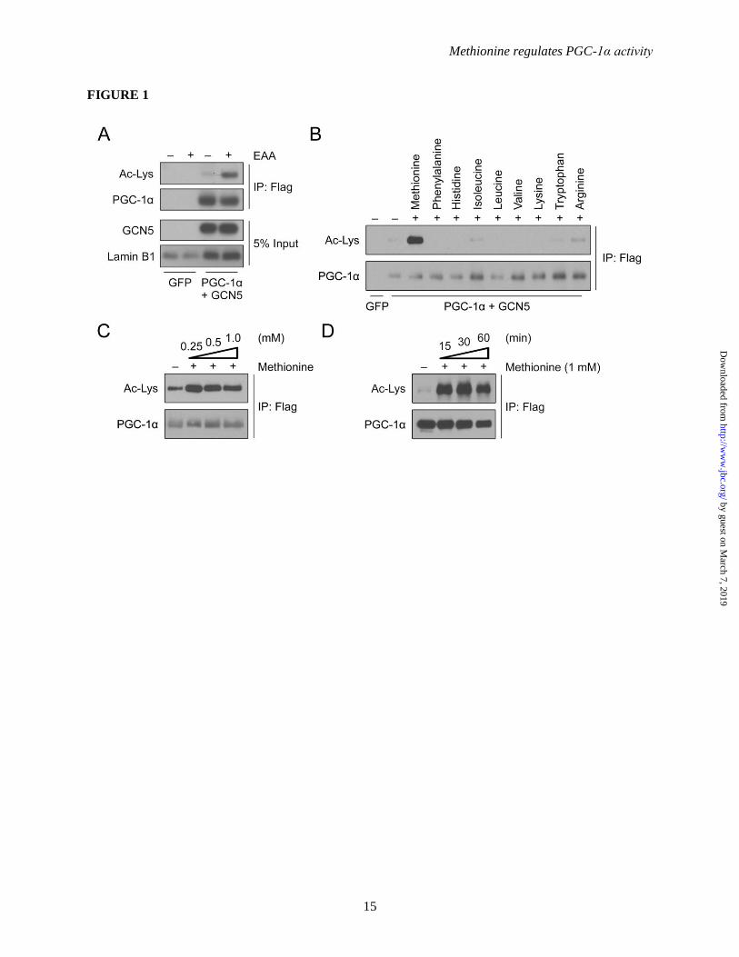
Methionine induces PGC-1α acetylation in
primary hepatocytes – PGC-1α responds to
nutrient and hormonal cues and plays a critical
role in the regulation of energy metabolism in
various tissues. Fasting induces PGC-1α activity
in the liver to promote the gluconeogenic program.
During fasting the availability of amino acids is
also altered, and moreover, dietary amino acids are
shown to contribute to the fed response along with
insulin signaling (29). This suggests a possible
role for the regulation of PGC-1α-mediated
gluconeogenesis through amino acid signaling. To
examine whether essential amino acids (EAAs) are
able to regulate PGC-1α activity, we tested their
ability to induce its acetylation in mouse primary
hepatocytes, as this post-translational modification
is known to inhibit the transcriptional coactivator
gluconeogenic function. Interestingly,
immunoprecipitation of ectopically expressed
PGC-1α indicates an increase in its acetylation in
hepatocytes treated with EAAs (Figure 1A). To
identify which of the EAAs contributed to the
induction of PGC-1α acetylation, we incubated
hepatocytes with individual EAAs (1 mM).
Methionine promotes a marked increase in the
acetylation of PGC-1α in contrast to the other
EAAs tested (Figure 1B). Methionine
concentrations in murine hepatocytes are reported
to range between 40 and 400 µM (32). In humans,
plasma concentrations of methionine drop to
around 30 µM during fasting, while after an oral
methionine load they can reach around 1 mM and
remain considerably elevated for a few hours (33).
Dose- and time-dependence assays in primary
hepatocytes indicate that 250 µM methionine
administered for 4 h was sufficient to induce
by guest on March 7, 2019
http://ww
w.jbc.org/
Dow
nloaded from
Methionine regulates PGC-1α activity
6
significant PGC-1α acetylation (Figure 1C), with
effects occurring relatively rapidly, within 15 min,
when tested at 1 mM (Figure 1D). These results
suggest that the observed in vitro effects of
methionine on PGC-1α acetylation occur at
physiologically relevant methionine
concentrations and durations of exposure.
PGC-1α acetylation is induced by methionine
through modulating GCN5 activity – Regulation
of PGC-1α acetylation levels is accomplished by
the delicate interplay between acetyltransferases
and histone deacetylases. To examine whether
methionine-induced PGC-1α acetylation was a
consequence of blunted histone deacetylase action,
Class I/II histone deacetylases and SIRT1 were
pharmacologically inhibited with trichostatin A
(TSA) and EX-527 respectively. As expected,
both TSA and EX-527 promote PGC-1α
acetylation, but the inhibitors failed to suppress a
further increase of PGC-1α acetylation induced by
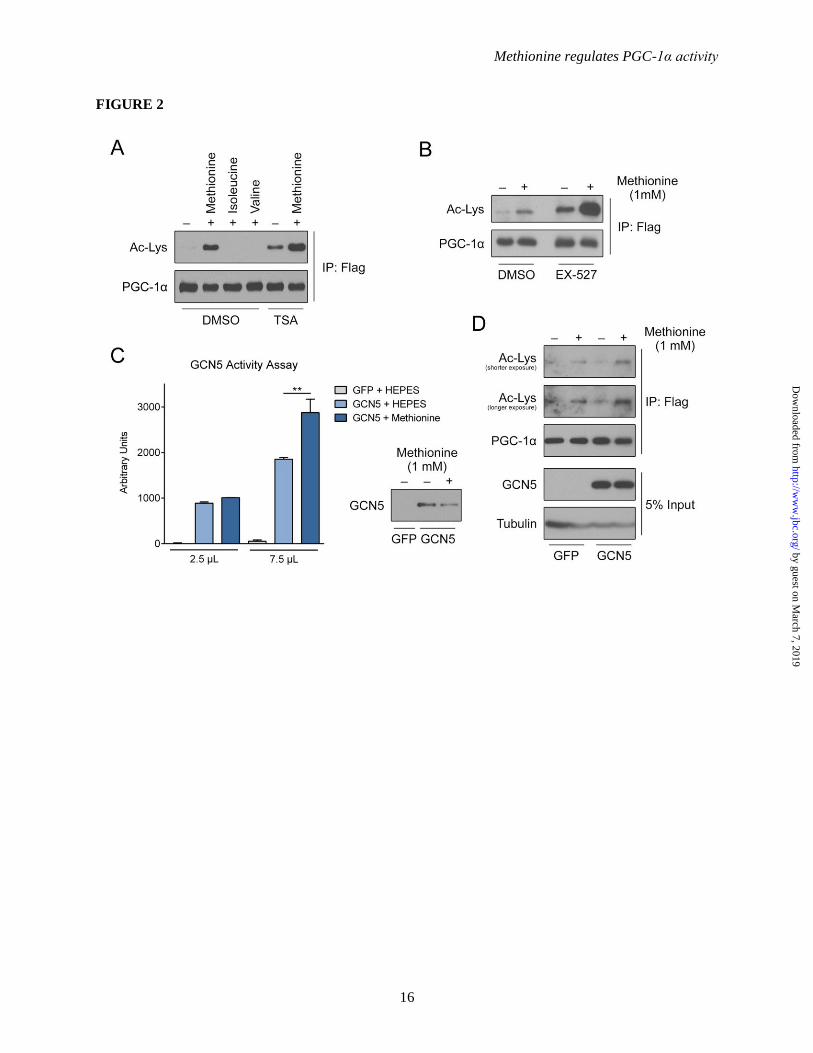
methionine (Figure 2A and 2B). As these results
suggest the involvement of an acetyltransferase,
we then tested whether methionine regulates PGC-
1α acetylation through GCN5, a key PGC-1α
acetyltransferase. Indeed, GCN5
immunoprecipitated from hepatocytes treated with
methionine displayed an increase in catalytic
activity in vitro, relative to vehicle control (Figure
2C). To further examine the involvement of
GCN5 in methionine-induced PGC-1α acetylation,
primary hepatocytes from liver-specific GCN5–/–
(KO) mice were employed. Ectopic expression of
GCN5 in these GCN5–/–
hepatocytes enhances the
effect of methionine on inducing PGC-1α
acetylation (Figure 2D). As methionine is also
able to induce PGC-1α acetylation in the GCN5–/–
background, it implies that GCN5 may not account
for the entire observed effect of methionine on
PGC-1α acetylation, however, our results suggest
that it a significant contributor. These results
taken together suggest that methionine promotes
PGC-1α acetylation, at least in part, through
activation of the GCN5 acetyltransferase.
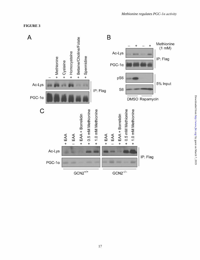
Classical amino acid signaling pathways do
not contribute to PGC-1α acetylation induced by
methionine – Metabolism of methionine primarily
involves the transmethylation (TM) and
transsulfuration (TS) pathways. To examine
whether the flux of methionine through these
pathways was responsible for the observed effects
on PGC-1α acetylation, hepatocytes were treated
with various pathway metabolites.
Betaine/choline/folate, cysteine and spermidine (1
mM each) were unable to induce PGC-1α
acetylation (Figure 3A). Interestingly,
homocysteine was able to mimic the effects of
methionine on the PGC-1α acetylation status
(Figure 3A); however, these effects are likely due
to the reconversion of homocysteine to
methionine. These results suggest that the
methionine TM and TS pathways are not
significantly responsible for methionine-induced
PGC-1α acetylation. Alternatively, amino acids
can be sensed by mTORC1 and GCN2 to trigger
downstream signaling cascades. To investigate
whether the mTOR pathway was responsible for
mediating the observed effects of methionine on
PGC-1α, methionine treatments were performed in
the presence or absence of rapamycin, an
mTORC1 allosteric inhibitor. As indicated in
Figure 3B, rapamycin did not block methionine-
induced PGC-1α acetylation. Levels of
phosphorylated S6 were measured to determine
the efficacy of rapamycin treatment. Additionally,
to evaluate the involvement of GCN2, primary
hepatocytes with the kinase knocked-out (GCN2-/-
)
were treated with methionine. Methionine-
induced PGC-1α acetylation was unaffected in the
knock-out cells (Figure 3C), suggesting that this
amino acid sensor does not contribute to the
process. Western blotting also indicated that there
is an increased level of ectopically expressed
GCN5 in GCN2-/-
hepatocytes (data not shown).
This could account for the increased PGC-1α
acetylation observed in GCN2-/-
hepatocytes
(Figure 3C). Collectively, these results suggest
that methionine does not promote PGC-1α
acetylation via the conventional amino acid
signaling pathways.
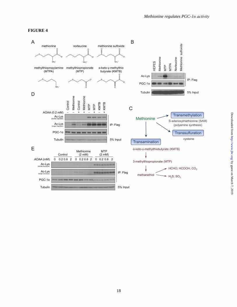
Flux of methionine through the methionine
transamination pathway increases the acetylation
of PGC-1α – To further investigate the mechanism
through which methionine induces PGC-1α
acetylation, various methionine analogs (Figure
4A) were tested for their ability to mimic the
effects of the amino acid. Despite significant
structural similarity with methionine, 3-
methylthiopropylamine (MTPA), norleucine and
methionine sulfoxide were unable to induce PGC-
1α acetylation; surprisingly however, 3-
methylthiopropionate (MTP) was strikingly more
potent than methionine (Figure 4B). This result
by guest on March 7, 2019
http://ww
w.jbc.org/
Dow
nloaded from
Methionine regulates PGC-1α activity
7
suggests a couple of things: First, since MTP
should not be able to charge tRNAMet
, it is unlikely
that a mechanism involving tRNAMet
contributes
to the methionine-induced effects on PGC-1α;
Secondly, MTP is an intermediary metabolite that
is generated through shunting of methionine down
the transamination pathway (Figure 4C), which
raises the possibility that PGC-1α acetylation is a
consequence of methionine transamination. To
test this, α-keto-γ-methylthiobutyrate (KMTB), the
intermediate metabolite between methionine and
MTP, was tested for its ability to induce PGC-1α
acetylation. Similar to MTP, KMTB is able to
promote PGC-1α acetylation (Figure 4D).
Furthermore, aminooxyacetic acid (AOAA), an
aminotransferase inhibitor that blocks conversion
of methionine to KMTB, is able to blunt PGC-1α
acetylation induced by methionine but not that by
KMTB and MTP (Figure 4D). An AOAA dose-
response assay indicates that the inhibitor is able
to suppress methionine-induced PGC-1α
acetylation to levels comparable to control
treatment, while being ineffective against MTP
stimulation (Figure 4E). In combination, these
data indicate that methionine-induced PGC-1α
acetylation primarily occurs via flux of the amino
acid through the methionine transamination
pathway.
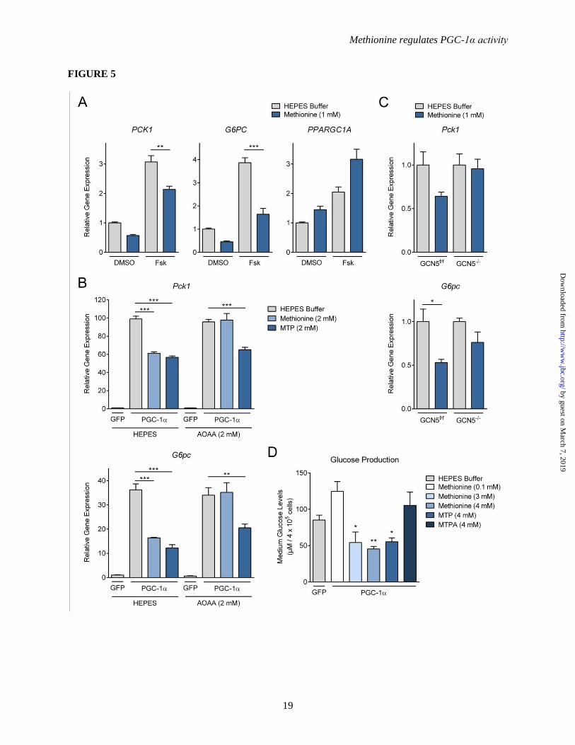
Methionine suppresses gluconeogenesis in
primary hepatocytes – As acetylation of PGC-1α
restricts its ability to stimulate gluconeogenesis
and promote hepatic glucose production, we
evaluated the physiological effects of methionine
on the gluconeogenic program in liver cells in
culture. We assessed the influence of methionine
on two key enzymes in the gluconeogenic
pathway, phosphoenolpyruvate carboxykinase
(Pck1) and glucose-6-phosphatase (G6pc), which
are well-characterized targets of PGC-1α
coactivation. In HepG2 human liver carcinoma
cells we used forskolin treatment to activate the
CREB pathway, which induces endogenous PGC-
1α expression and activity. In these cells,
methionine was potently able to suppress
forskolin-induced expression of Pck1 and G6pc
(Figure 5A). In mouse primary hepatocytes with
ectopically expressed PGC-1α, methionine and
MTP also caused a significant suppression of the
PGC-1α-induced Pck1 and G6pc expression
(Figure 5B). Moreover, in accordance with earlier
data, AOAA rescued methionine-induced, but not
MTP-induced, repression of Pck1 and G6pc
expression (Figure 5B). To test the contribution of
GCN5 to this suppression, we used the GCN5–/–
hepatocytes. Figure 5C indicates that loss of
GCN5 blunts the effects of methionine on Pck1
and G6pc gene suppression, suggesting that GCN5
at least partially contributes to the observed effect,
which supports our earlier data on PGC-1α
acetylation. Concomitant with the methionine-
mediated reduction in gluconeogenic gene
expression, we observed a decrease in ability of
hepatocytes to produce glucose; methionine and
MTP, but not MTPA, were able to suppress PGC-
1α-mediated glucose production in mouse primary
hepatocytes (Figure 5D). These data suggest that
methionine, through its metabolism via the
transamination pathway, is able to potently
suppress the PGC-1α-mediated hepatic
gluconeogenic program in vitro.
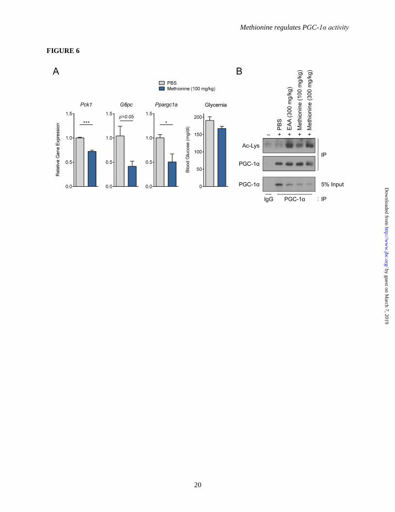
Gluconeogenesis is repressed by methionine
in fasted mice – Fasting is known to induce
hepatic PGC-1α to promote gluconeogenesis. To
evaluate if methionine has a physiological effect
on PGC-1α in vivo, 8-week old male C57BL/6
mice were fasted overnight (16 h), and then
administered methionine (100 mg/kg)
intraperitoneally. Analysis of hepatic gene
expression 2 h post-injection indicates that
compared to vehicle control, methionine
administration significantly repressed Pck1 and
G6pc expression in vivo, which was accompanied
by a reduction in blood glucose levels (Figure 6A).
Methionine administration in vivo decreases
Ppargc1a expression (Figure 6A), which contrasts
the effects observed in HepG2 cells (Figure 5A).
One possibility that could account for this
difference is that HepG2 being a cancer cell line is
proliferative in nature, which could potentially
result in metabolic variations when compared to
normal hepatocytes. To verify that methionine
induces PGC-1α acetylation in vivo, endogenous
PGC-1α immunoprecipitated from liver nuclear
extracts of fasted mice was assessed. Both, EAAs
(300 mg/kg) and methionine (100 and 300 mg/kg)
administration, notably increased PGC-1α
acetylation compared to vehicle control (Figure
6B), supporting data obtained from cultured
mouse primary hepatocytes.
These results highlight a potential role for
amino acids, in particular methionine and its
metabolic regulation, in modulating PGC-1α-
by guest on March 7, 2019
http://ww
w.jbc.org/
Dow
nloaded from
Methionine regulates PGC-1α activity
8
mediated hepatic gluconeogenesis and
consequently blood glucose levels.
DISCUSSION
The liver plays a major role in maintaining
normal blood glucose homeostasis by increasing
endogenous glucose production during periods of
low nutrient availability. Upon refeeding, when
nutrients become available, hormonal and
nutritional cues signal the liver to suppress glucose
production and switch to an energy storage mode.
In type 2 diabetes, hepatic glucose production
becomes refractory to these cues and glucose
production remains elevated even in the refed
state, where it significantly contributes to fasting
hyperglycemia observed in these patients (34).
While the hormonal signaling associated with the
refed state, specifically insulin, has been
extensively studied, the role of specific nutrients in
the hepatic response to the refed state is largely
unknown. Here, we identify the essential amino
acid methionine as the only amino acid that is able
to increase the acetylation state of PGC-1α. As a
result, methionine leads to a suppression of hepatic
glucose production in isolated mouse primary
hepatocytes and to a reduction in fasting blood
glucose in vivo.
Acute administration of methionine to isolated
hepatocytes potently increases the acetylation
status of PGC-1α by augmenting the catalytic
activity of GCN5, the major acetyl transferase
toward PGC-1α (26). Interestingly, the effect of
methionine on PGC-1α acetylation does not
require an active mTORC1 since in the presence
of rapamycin, methionine can still increase PGC-
1α acetylation. In addition, in the absence of
GCN2, the only known amino acid sensor that is
activated during amino acid deficiency (35),
methionine is still able to induce PGC-1α
acetylation. These observations suggest that
methionine, and maybe other amino acids as well,
can induce specific cellular responses
independently of these two pathways and
modulate the response of the liver to the refed
state. Specifically, we have found that flux of
methionine through the transamination pathway is
responsible for the methionine-induced PGC-1α
acetylation.
We recently reported that dietary
supplementation of essential amino acids to mice
can contribute to the refed response by increasing
hepatic cyclin D1 expression levels (29). As a
result, the cyclin D1-Cdk4 complex is activated,
leading to an increase in GCN5 activity and
suppression of PGC-1α during refeeding.
Although the methionine-induced PGC-1α
acetylation does not seem to be mediated by this
pathway (data not shown), our study highlights the
importance of GCN5 as a critical component of
the refed response that can potentially be
manipulated to control glucose homeostasis in the
diabetic state. The molecular mechanisms
underlying the methionine-mediated increase of
GCN5 activity are currently unknown and future
studies will hopefully shed more light on this
matter. It is possible that alteration in the post-
translational modifications on GCN5, such as
phosphorylation (28,36) or acetylation (28), could
be responsible for the increased activity observed.
GCN5 is part of the SAGA (Spt-Ada-Gcn5
acetyltransferase) and ATAC (Ada2a-containing)
coactivator complexes (37). Thus, there is the
alternative possibility that other proteins in the
acetyl transferase complexes could undergo post-
translational modification to alter the complex
composition and increase GCN5 activity, as has
been described earlier (38). GCN5 is highly
conserved across species (39), and was originally
identified in yeast as an essential component for
proper activation of the transcriptional response to
amino acid starvation mediated by GCN4 (40). It
was recently shown in lower organisms that amino
acids can activate acetyl transferase activity of
GCN5-related N-acetyl transferase (GNAT)
protein (41). Whether GCN5 is a conserved
component that can be activated in response to
amino acids availability is of particular interest.
Methionine can be further metabolized in the
liver through several different biochemical
pathways. Of particular metabolic significance is
the transmethylation pathway which involves the
transfer of a methyl group from S-
adenosylmethionine, the major methyl donor in
the cell. Methionine degradation includes the
transsulfuration and transamination pathways.
Whether methionine degradation through
transamination is quantitatively important is not
clear. While some studies suggest that this
pathway might be important in rodents (9), other
studies found that methionine is mostly degraded
via transsulfuration (42). Studies in humans show
that transamination exists but its importance is not
by guest on March 7, 2019
http://ww
w.jbc.org/
Dow
nloaded from
Methionine regulates PGC-1α activity
9
clear (13). Our study implies that under some
conditions of methionine load, degradation
through the transamination pathway can contribute
to glucose homeostasis in the refed state.
Several studies have demonstrated the benefits
of methionine restriction (MR), which include
prevention of insulin resistance and obesity (43),
increase in energy expenditure (44), and life span
extension (45). Interestingly, MR has also been
reported to improve hepatic insulin sensitivity
(46), which promotes glucose homeostasis. Here,
we demonstrate that methionine administration
upon fasting is able to suppress the hepatic
gluconeogenic program. MR studies are usually
conducted over relatively longer durations, which
differ from our experiments which involve acute
administration of methionine in the fasted state.
An insight into any crosstalk between metabolic
pathways stimulated under MR and methionine
administration would be critical for developing
dietary regimens employed for treating metabolic
disorders.
Collectively, we have identified a novel role
for the amino acid methionine in the adaptive
response of the liver to the refed state. Our study
highlights the importance of specific metabolites
in the regulation of molecular components of
insulin signaling, specifically the acetylation state
of PGC-1α, and suggests that manipulation of
these metabolites’ flux can be potentially utilized
to improve diabetic symptoms.
ACKNOWLEDGEMENTS
We thank members of the Puigserver laboratory for discussions on this project. We also thank
Malcolm Whitman for helpful discussions and GCN2 -/- hepatocytes. CDT was supported by a mentor-
based American Diabetes Association postdoctoral fellowship and also by a postdoctoral fellowship from
the American Diabetes Association (Grant #1-16-PDF-111), KS by a postdoctoral fellowship from the
American Heart Association, and JED by a National Research Service Award Kirschstein fellowship from
the NIH. This work was supported by NIH/NIDDK funding RO11069966 (PP) and R24DK080261-06
(PP, PRG).
CONFLICT OF INTEREST
The authors declare that they have no conflicts of interest with the contents of this article. The
content is solely the responsibility of the authors and does not necessarily represent the official views of
the National Institutes of Health.
AUTHOR CONTRIBUTIONS
JED and PP conceived the project. CDT, KS, JED, YL and PP designed the research strategy and
experimental approach. CDT, KS, JED and YL conducted the experiments. CDT, KS, JED, YL and PP
analyzed the data. MI, JMO, MPJ and TMK assisted in performing the experiments. CDT and KS wrote
the manuscript with editing from JED and PP, and PRG, SPG and PP supervised the studies.
REFERENCES
1. Gietzen, D. W., and Rogers, Q. R. (2006) Nutritional homeostasis and indispensable amino acid
sensing: a new solution to an old puzzle. Trends in neurosciences 29, 91-99
2. Gallinetti, J., Harputlugil, E., and Mitchell, J. R. (2013) Amino acid sensing in dietary-restriction-
mediated longevity: roles of signal-transducing kinases GCN2 and TOR. The Biochemical
journal 449, 1-10
3. Wek, R. C., Jackson, B. M., and Hinnebusch, A. G. (1989) Juxtaposition of domains homologous
to protein kinases and histidyl-tRNA synthetases in GCN2 protein suggests a mechanism for
coupling GCN4 expression to amino acid availability. Proceedings of the National Academy of
Sciences of the United States of America 86, 4579-4583
4. Dong, J., Qiu, H., Garcia-Barrio, M., Anderson, J., and Hinnebusch, A. G. (2000) Uncharged
tRNA activates GCN2 by displacing the protein kinase moiety from a bipartite tRNA-binding
domain. Molecular cell 6, 269-279
by guest on March 7, 2019
http://ww
w.jbc.org/
Dow
nloaded from
Methionine regulates PGC-1α activity
10
5. Sancak, Y., Peterson, T. R., Shaul, Y. D., Lindquist, R. A., Thoreen, C. C., Bar-Peled, L., and
Sabatini, D. M. (2008) The Rag GTPases bind raptor and mediate amino acid signaling to
mTORC1. Science (New York, N.Y.) 320, 1496-1501
6. Harper, A. E., Benevenga, N. J., and Wohlhueter, R. M. (1970) Effects of ingestion of
disproportionate amounts of amino acids. Physiological reviews 50, 428-558
7. Benevenga, N. J. (1974) Toxicities of methionine and other amino acids. Journal of agricultural
and food chemistry 22, 2-9
8. Marchesini, G., Bugianesi, E., Bianchi, G., Fabbri, A., Marchi, E., Zoli, M., and Pisi, E. (1992)
Defective methionine metabolism in cirrhosis: relation to severity of liver disease. Hepatology
(Baltimore, Md.) 16, 149-155
9. Griffith, O. W. (1987) Mammalian sulfur amino acid metabolism: an overview. Methods in
enzymology 143, 366-376
10. Stipanuk, M. H. (2004) Sulfur amino acid metabolism: pathways for production and removal of
homocysteine and cysteine. Annual review of nutrition 24, 539-577
11. Fontecave, M., Atta, M., and Mulliez, E. (2004) S-adenosylmethionine: nothing goes to waste.
Trends in biochemical sciences 29, 243-249
12. Livesey, G., and Lund, P. (1980) Methionine metabolism via the transamination pathway in rat
liver. Biochemical Society transactions 8, 540-541
13. Blom, H. J., Boers, G. H., van den Elzen, J. P., Gahl, W. A., and Tangerman, A. (1989)
Transamination of methionine in humans. Clin Sci (Lond) 76, 43-49
14. Scislowski, P. W., and Pickard, K. (1993) Methionine transamination--metabolic function and
subcellular compartmentation. Molecular and cellular biochemistry 129, 39-45
15. Steele, R. D., and Benevenga, N. J. (1978) Identification of 3-methylthiopropionic acid as an
intermediate in mammalian methionine metabolism in vitro. The Journal of biological chemistry
253, 7844-7850
16. Scislowski, P. W., Bremer, J., van Thienen, W. I., and Davis, E. J. (1989) Heart mitochondria
metabolize 3-methylthiopropionate to CO2 and methanethiol. Archives of biochemistry and
biophysics 273, 602-605
17. Toue, S., Kodama, R., Amao, M., Kawamata, Y., Kimura, T., and Sakai, R. (2006) Screening of
toxicity biomarkers for methionine excess in rats. The Journal of nutrition 136, 1716S-1721S
18. Dever, J. T., and Elfarra, A. A. (2008) L-methionine toxicity in freshly isolated mouse
hepatocytes is gender-dependent and mediated in part by transamination. The Journal of
pharmacology and experimental therapeutics 326, 809-817
19. Mitchell, A. D., and Benevenga, N. J. (1978) The role of transamination in methionine oxidation
in the rat. The Journal of nutrition 108, 67-78
20. Yamada, H., Akahoshi, N., Kamata, S., Hagiya, Y., Hishiki, T., Nagahata, Y., Matsuura, T.,
Takano, N., Mori, M., Ishizaki, Y., Izumi, T., Kumagai, Y., Kasahara, T., Suematsu, M., and
Ishii, I. (2012) Methionine excess in diet induces acute lethal hepatitis in mice lacking
cystathionine gamma-lyase, an animal model of cystathioninuria. Free radical biology &
medicine 52, 1716-1726
21. Cahill, G. F., Jr. (1970) Starvation in man. The New England journal of medicine 282, 668-675
22. Yoon, J. C., Puigserver, P., Chen, G., Donovan, J., Wu, Z., Rhee, J., Adelmant, G., Stafford, J.,
Kahn, C. R., Granner, D. K., Newgard, C. B., and Spiegelman, B. M. (2001) Control of hepatic
gluconeogenesis through the transcriptional coactivator PGC-1. Nature 413, 131-138
23. Puigserver, P., Rhee, J., Donovan, J., Walkey, C. J., Yoon, J. C., Oriente, F., Kitamura, Y.,
Altomonte, J., Dong, H., Accili, D., and Spiegelman, B. M. (2003) Insulin-regulated hepatic
gluconeogenesis through FOXO1-PGC-1alpha interaction. Nature 423, 550-555
24. Rhee, J., Inoue, Y., Yoon, J. C., Puigserver, P., Fan, M., Gonzalez, F. J., and Spiegelman, B. M.
(2003) Regulation of hepatic fasting response by PPARgamma coactivator-1alpha (PGC-1):
requirement for hepatocyte nuclear factor 4alpha in gluconeogenesis. Proceedings of the National
Academy of Sciences of the United States of America 100, 4012-4017
by guest on March 7, 2019
http://ww
w.jbc.org/
Dow
nloaded from
Methionine regulates PGC-1α activity
11
25. Rodgers, J. T., Lerin, C., Haas, W., Gygi, S. P., Spiegelman, B. M., and Puigserver, P. (2005)
Nutrient control of glucose homeostasis through a complex of PGC-1alpha and SIRT1. Nature
434, 113-118
26. Lerin, C., Rodgers, J. T., Kalume, D. E., Kim, S. H., Pandey, A., and Puigserver, P. (2006) GCN5
acetyltransferase complex controls glucose metabolism through transcriptional repression of
PGC-1alpha. Cell metabolism 3, 429-438
27. Patten, I. S., and Arany, Z. (2012) PGC-1 coactivators in the cardiovascular system. Trends in
endocrinology and metabolism: TEM 23, 90-97
28. Dominy, J. E., Jr., Lee, Y., Jedrychowski, M. P., Chim, H., Jurczak, M. J., Camporez, J. P., Ruan,
H. B., Feldman, J., Pierce, K., Mostoslavsky, R., Denu, J. M., Clish, C. B., Yang, X., Shulman,
G. I., Gygi, S. P., and Puigserver, P. (2012) The deacetylase Sirt6 activates the acetyltransferase
GCN5 and suppresses hepatic gluconeogenesis. Molecular cell 48, 900-913
29. Lee, Y., Dominy, J. E., Choi, Y. J., Jurczak, M., Tolliday, N., Camporez, J. P., Chim, H., Lim, J.
H., Ruan, H. B., Yang, X., Vazquez, F., Sicinski, P., Shulman, G. I., and Puigserver, P. (2014)
Cyclin D1-Cdk4 controls glucose metabolism independently of cell cycle progression. Nature
510, 547-551
30. Cherrington, A. D., Stevenson, R. W., Steiner, K. E., Davis, M. A., Myers, S. R., Adkins, B. A.,
Abumrad, N. N., and Williams, P. E. (1987) Insulin, glucagon, and glucose as regulators of
hepatic glucose uptake and production in vivo. Diabetes/metabolism reviews 3, 307-332
31. Lin, W., Zhang, Z., Srajer, G., Chen, Y. C., Huang, M., Phan, H. M., and Dent, S. Y. (2008)
Proper expression of the Gcn5 histone acetyltransferase is required for neural tube closure in
mouse embryos. Developmental dynamics : an official publication of the American Association of
Anatomists 237, 928-940
32. Korendyaseva, T. K., Kuvatov, D. N., Volkov, V. A., Martinov, M. V., Vitvitsky, V. M.,
Banerjee, R., and Ataullakhanov, F. I. (2008) An Allosteric Mechanism for Switching between
Parallel Tracks in Mammalian Sulfur Metabolism. PLoS Computational Biology 4, e1000076
33. Fukagawa, N. K., Martin, J. M., Wurthmann, A., Prue, A. H., Ebenstein, D., and O'Rourke, B.
(2000) Sex-related differences in methionine metabolism and plasma homocysteine
concentrations. The American Journal of Clinical Nutrition 72, 22-29
34. Lin, H. V., and Accili, D. (2011) Hormonal regulation of hepatic glucose production in health and
disease. Cell metabolism 14, 9-19
35. Dever, T. E. (1999) Translation initiation: adept at adapting. Trends in biochemical sciences 24,
398-403
36. Barlev, N. A., Poltoratsky, V., Owen-Hughes, T., Ying, C., Liu, L., Workman, J. L., and Berger,
S. L. (1998) Repression of GCN5 histone acetyltransferase activity via bromodomain-mediated
binding and phosphorylation by the Ku-DNA-dependent protein kinase complex. Molecular and
cellular biology 18, 1349-1358
37. Wang, L., and Dent, S. Y. (2014) Functions of SAGA in development and disease. Epigenomics
6, 329-339
38. Riss, A., Scheer, E., Joint, M., Trowitzsch, S., Berger, I., and Tora, L. (2015) Subunits of ADA-
two-A-containing (ATAC) or Spt-Ada-Gcn5-acetyltrasferase (SAGA) Coactivator Complexes
Enhance the Acetyltransferase Activity of GCN5. The Journal of biological chemistry 290,
28997-29009
39. Sternglanz, R., and Schindelin, H. (1999) Structure and mechanism of action of the histone
acetyltransferase Gcn5 and similarity to other N-acetyltransferases. Proceedings of the National
Academy of Sciences of the United States of America 96, 8807-8808
40. Georgakopoulos, T., and Thireos, G. (1992) Two distinct yeast transcriptional activators require
the function of the GCN5 protein to promote normal levels of transcription. The EMBO journal
11, 4145-4152
by guest on March 7, 2019
http://ww
w.jbc.org/
Dow
nloaded from
Methionine regulates PGC-1α activity
12
41. Xu, J. Y., You, D., Leng, P. Q., and Ye, B. C. (2014) Allosteric regulation of a protein
acetyltransferase in Micromonospora aurantiaca by the amino acids cysteine and arginine. The
Journal of biological chemistry 289, 27034-27045
42. Finkelstein, J. D., and Martin, J. J. (1986) Methionine metabolism in mammals. Adaptation to
methionine excess. The Journal of biological chemistry 261, 1582-1587
43. Ables, G. P., Perrone, C. E., Orentreich, D., and Orentreich, N. (2012) Methionine-restricted
C57BL/6J mice are resistant to diet-induced obesity and insulin resistance but have low bone
density. PloS one 7, e51357
44. Hasek, B. E., Stewart, L. K., Henagan, T. M., Boudreau, A., Lenard, N. R., Black, C., Shin, J.,
Huypens, P., Malloy, V. L., Plaisance, E. P., Krajcik, R. A., Orentreich, N., and Gettys, T. W.
(2010) Dietary methionine restriction enhances metabolic flexibility and increases uncoupled
respiration in both fed and fasted states. American journal of physiology. Regulatory, integrative
and comparative physiology 299, R728-739
45. Lee, B. C., Kaya, A., and Gladyshev, V. N. (2015) Methionine restriction and life-span control.
Annals of the New York Academy of Sciences
46. Stone, K. P., Wanders, D., Orgeron, M., Cortez, C. C., and Gettys, T. W. (2014) Mechanisms of
increased in vivo insulin sensitivity by dietary methionine restriction in mice. Diabetes 63, 3721-
3733
FIGURE LEGENDS
Figure 1. Methionine increases the acetylation of PGC-1α in primary hepatocytes. (A) Western-blot
analysis of PGC-1α acetylation status upon essential amino acids (EAAs) treatment. Primary hepatocytes
were infected with FLAG-PGC-1α and GCN5 adenovirus. The day after infection, cells were treated
overnight with EBSS to deplete all amino acids in the medium. EAAs were added 4 h prior to harvesting.
All cells were harvested within 48 h post-infection. FLAG-PGC-1α was immunoprecipitated with
FLAG-antibody conjugated beads in order to analyze the acetylation status. (B) Western-blot analysis of
PGC-1α acetylation status upon the addition of individual amino acids. The experiment follows the same
procedure described in A, except the cells were treated with individual amino acids (1 mM) for 4 h prior
to harvesting. (C) Western-blot analysis of PGC-1α acetylation status upon the addition of various
concentrations of methionine. The experiment follows the same procedure as described in A. (D)
Western-blot analysis of PGC-1α acetylation status upon the addition of methionine for various durations.
The experiment follows the same procedure as described in A.
Figure 2. Methionine induces the acetylation of PGC-1α via modulating GCN5 activity. (A) Western-blot analysis of PGC-1α acetylation status upon methionine treatment with a Class I/II HDAC
inhibitor, trichostatin A (TSA). The experiment follows the same procedure as described in Figure 1A,
except the cells were treated with TSA (1 μM) for 6 h prior to harvesting. Amino acids (1 mM) were
added 2 h prior to harvesting. (B) Western-blot analysis of PGC-1α acetylation status upon methionine
treatment with a SIRT1 inhibitor. The experiment follows the same procedure as described in A, except
cells were treated with EX-527 (1 μM) for 6 h prior to harvesting. (C) GCN5 in vitro acetyl-transferase
activity assay (n=2). Primary hepatocytes were infected with either GFP or FLAG-GCN5 adenovirus.
The cells were treated overnight with EBSS medium, and methionine (1 mM) was added 2 h prior to
harvesting. All cells were harvested within 48 h post-infection. FLAG-GCN5 was immnunoprecipitated
from nuclear extracts of cells that were treated with HEPES buffer or methionine. The activity assay was
carried out according to the manufacturer’s protocol. The corresponding amounts of GCN5 protein in the
assay were analyzed via western-blot. (D) Western-blot analysis of PGC-1α acetylation status upon
methionine treatment in GCN5-/-
(KO) primary hepatocytes. Primary hepatocytes were isolated from
liver-specific GCN5-/-
(KO) mice. Primary hepatocytes were infected with FLAG-PGC-1α and either
GFP or GCN5 adenovirus. Two days post-infection, cells were incubated in DMEM maintenance
medium lacking methionine for 5 h prior to harvesting. Methionine (1 mM) was added 2 h prior to
by guest on March 7, 2019
http://ww
w.jbc.org/
Dow
nloaded from
Methionine regulates PGC-1α activity
13
harvesting. For all multiple comparisons, One-Way ANOVA with post-hoc Tukey’s test was used. **-
p<0.01.
Figure 3. Methionine does not induce the acetylation of PGC-1α via classical amino acid signaling
pathways. (A) Western-blot analysis of PGC-1α acetylation status upon methionine metabolites
treatment. The experiment follows the same procedure as described in Figure 1A, except the cells were
treated with various metabolites of methionine (1 mM each) for 2 h prior to harvesting. (B) Western-blot
analysis of PGC-1α acetylation status upon methionine treatment with rapamycin. The experiment
follows the same procedure as described in Figure 1A. Rapamycin (20 nM) and Methionine (1 mM) were
added 6 h and 2 h prior to harvesting respectively. Western-blot of phospho-S6 was analyzed to ensure
the efficiency of rapamycin treatment. (C) Western-blot analysis of PGC-1α acetylation status upon
methionine treatment in GCN2 WT and GCN2 -/- primary hepatocytes. Primary hepatocytes were
isolated from GCN2 WT or GCN2 -/- mice. The experiment follows the same procedure as described in
Figure 1A. EAA or Methionine were added 2 h and Borrelidin (1 μM) 30 min prior to harvesting.
Figure 4. Methionine increases the acetylation of PGC-1α via the methionine transamination
pathway. (A) Structures of methionine analogs used in this study. (B) Western-blot analysis of PGC-1α
acetylation status upon treatment with methionine and its analogs. Primary hepatocytes were infected
with FLAG-PGC-1α adenovirus. The day after infection, cells were incubated overnight in DMEM
maintenance medium lacking methionine and cysteine. Methionine and its analogs (2 mM) were added 2
h prior to harvesting. (C) Summary of the methionine transamination pathway. (D) Western-blot
analysis of PGC-1α acetylation status upon treatment with aminooxyacetic acid (AOAA), a transaminase
inhibitor. The experiment follows a similar procedure as described in B, except the day after infection,
cells were incubated in DMEM maintenance medium lacking methionine for 6 h prior to harvesting.
AOAA (0.2 mM), and methionine and its analogs (2 mM) were added 2.5 h and 2 h prior to harvesting
respectively. (E) Western-blot analysis of PGC-1α acetylation status upon the addition of methionine
transamination pathway metabolites with various concentrations of AOAA. The experiment follows the
same procedure as described in D, except for treatment with various concentrations of AOAA.
Figure 5. Methionine represses gluconeogenesis in primary hepatocytes. (A) Real-time PCR analysis
of gluconeogenic gene expression upon methionine treatment in human liver carcinoma cells (n=3).
HepG2 cells were treated with DMSO or forskolin (30 μM) overnight. HEPES buffer or methionine (1
mM) was added to the cells for 3 h prior to harvesting. cDNA was generated from RNA, extracted using
TRIzol-chloroform extraction methods. (B) Real-time PCR analysis of gluconeogenic gene expression
upon the addition of methionine and MTP combined with AOAA treatment in primary hepatocytes (n=3).
Primary hepatocytes were infected with either GFP or FLAG-PGC-1α adenovirus. Two days post-
infection, cells were incubated in DMEM maintenance medium lacking methionine for 7.5 h prior to
harvesting. AOAA (0.2 mM) was added 4.5 h prior to harvesting. Methionine and MTP (2 mM) were
added 4 h prior to harvesting. (C) Real-time PCR analysis of gluconeogenic gene expression upon the
addition of methionine in GCN5 (f/f) and GCN5-/-
(KO) primary hepatocytes (n=3). Primary hepatocytes
were isolated from liver-specific GCN5 (f/f) and GCN5-/-
(KO) mice. Primary hepatocytes were infected
with either GFP or FLAG-PGC-1α adenovirus. Two days post-infection, cells were incubated in DMEM
maintenance medium lacking methionine for 7 h prior to harvesting. Methionine (1 mM) was added 4 h
prior to harvesting. (D) Hepatic glucose production assay (n=3). Primary hepatocytes were infected with
either GFP or FLAG-PGC-1α adenovirus. Two days post-infection, glucose production capacity of
primary hepatocytes was measured by incubating the cells in glucose free medium for 3 h. Methionine
(0.1, 3, 4 mM), MTP (4 mM) and MTPA (4 mM) were added to the cells 3 h prior to collection of the
media samples. The glucose concentration was calculated based on the difference between
pyruvate/lactate-free medium and 2 mM pyruvate/20 mM lactate containing medium. For all multiple
comparisons, One-Way ANOVA with post-hoc Tukey’s test was used. *-p<0.05, **-p<0.01, ***-
p<0.001.
by guest on March 7, 2019
http://ww
w.jbc.org/
Dow
nloaded from
Methionine regulates PGC-1α activity
14
Figure 6. Methionine represses gluconeogenesis in vivo. (A) Real-time PCR analysis of in vivo
hepatic gluconeogenic gene expression and whole body glycemia upon methionine (100 mM)
intraperitoneal injection (n=3). 8 week old male C57BL/6 mice were fasted overnight (16 h). PBS or
methionine (100 mg/kg, dissolved in PBS) was intraperitoneally injected into the mice. All mice were
sacrificed 2 h post injection, and livers were snap-frozen for gene expression analysis. Glycemia was
measured via tail-bleeding at the time of harvesting. (B) Western-blot analysis of endogenous PGC-1α
acetylation status in vivo. Endogenous PGC-1α was immunoprecipitated from nuclear extracts of livers.
The experiment follows the same procedure as described in A. Mice were intraperitoneally injected with
PBS, essential amino acids (100 mg/kg) or methionine (100 mg/kg, 300 mg/kg). For two experimental
comparisons, two-tailed student’s t-test was used. *-p<0.05, ***-p<0.001.
by guest on March 7, 2019
http://ww
w.jbc.org/
Dow
nloaded from
Methionine regulates PGC-1α activity
15
FIGURE 1
by guest on March 7, 2019
http://ww
w.jbc.org/
Dow
nloaded from
Methionine regulates PGC-1α activity
16
FIGURE 2
by guest on March 7, 2019
http://ww
w.jbc.org/
Dow
nloaded from
Methionine regulates PGC-1α activity
17
FIGURE 3
by guest on March 7, 2019
http://ww
w.jbc.org/
Dow
nloaded from
Methionine regulates PGC-1α activity
18
FIGURE 4
by guest on March 7, 2019
http://ww
w.jbc.org/
Dow
nloaded from
Methionine regulates PGC-1α activity
19
FIGURE 5
by guest on March 7, 2019
http://ww
w.jbc.org/
Dow
nloaded from
Methionine regulates PGC-1α activity
20
FIGURE 6
by guest on March 7, 2019
http://ww
w.jbc.org/
Dow
nloaded from
Gygi and Pere PuigserverOrozco, Mark P. Jedrychowski, Theodore M. Kamenecka, Patrick R. Griffin, Steven P. Clint D. J. Tavares, Kfir Sharabi, John E. Dominy, Yoonjin Lee, Marta Isasa, Jose M.
Coactivator Transcriptionalαthrough Regulation of the GCN5 Acetyl Transferase and the PGC-1
The Methionine Transamination Pathway Controls Hepatic Glucose Metabolism
published online March 28, 2016J. Biol. Chem.
10.1074/jbc.M115.706200Access the most updated version of this article at doi:
Alerts:
When a correction for this article is posted•
When this article is cited•
to choose from all of JBC's e-mail alertsClick here
by guest on March 7, 2019
http://ww
w.jbc.org/
Dow
nloaded from