The induction of bone formation by the recombinant human transforming growth factor-β3
16
The induction of bone formation by the recombinant human transforming growth factor-b 3 Roland M. Klar a , Raquel Duarte b , Therese Dix-Peek b , Ugo Ripamonti a, * a Bone Research Laboratory, School of Physiology, Faculty of Health Sciences, Medical School, University of the Witwatersrand, Parktown, Johannesburg 2193 South Africa b Department of Internal Medicine, School of Clinical Medicine, University of the Witwatersrand, Johannesburg, South Africa article info Article history: Received 22 November 2013 Accepted 19 December 2013 Available online 15 January 2014 Keywords: Transforming growth factor-b 3 Bone morphogenetic proteins Tissue induction Noggin Primates qRT-PCR abstract Implantation of recombinant human transforming growth factor-b 3 (hTGF-b 3 ) with coral-derived cal- cium carbonate-based macroporous bioreactors with limited conversion to hydroxyapatite (7% HA/CC) in the rectus abdominis muscle of the non-human primate Chacma baboon Papio ursinus induces endo- chondral bone formation. The exact mechanisms by which hTGF-b 3 signalling induces bone in hetero- topic sites of P. ursinus are not known. Coral-derived 7% HA/CC bioreactors with and without 125 mg hTGF-b 3 were implanted in triplicate in the rectus abdominis muscle of 6 adult baboons. 7% HA/CC bioreactors either with or without hTGF-b 3 were loaded with 125 mg of recombinant human Noggin (hNoggin), a bone morphogenetic proteins (BMPs) antagonist. Tissues on day 15, 60 and 90 were ana- lysed by histomorphometry and quantitative reverse-transcriptase polymerase chain reaction (qRT-PCR). Down-regulation of BMP-2 characterized 7% HA/CC constructs preloaded with 125 mg hNoggin with Noggin down-regulated on day 60 and 90 together with lack of TGF-b 3 expression. Down-regulation of BMP-2 correlated with minimal bone formation by induction. hTGF-b 3 /hNoggin pre-treated bioreactors up-regulated BMP-2 but only on day 90 together with a significant down-regulation of Noggin on day 60 and 90, correlating with the induction of bone formation, albeit limited, on day 90 at the periphery of the macroporous bioreactors only. hTGF-b 3 treated bioreactors significantly down-regulated BMP-2 on day 15 whilst up-regulating BMP-2 on day 60 and 90, together with down-regulation of Noggin on day 60 and 90 correlating with the prominent induction of bone formation. hTGF-b 3 significantly up-regulated RUNX-2 and Osteocalcin expression on day 15 controlling the differentiation of progenitor stem cells into the osteoblastic lineage. The induction of bone as initiated by hTGF-b 3 in the rectus abdominis muscle of P. ursinus is via the BMPs pathway with hTGF-b 3 controlling the induction of bone formation by regulating the expression of BMPs via Noggin expression. These results unequivocally demonstrate that hTGF-b 3 elicits bone induction by up-regulation of endogenous BMP-2 and is blocked by hNoggin. Ó 2013 Elsevier Ltd. All rights reserved. 1. Introduction The induction of bone formation during embryonic develop- ment and postnatal tissue induction and morphogenesis is set by complex seemingly redundant cascades of molecular and morphogenetic events that sculpt the multicellular mineralized and non-mineralized structures of bone that have ultimately engineered the emergence of the skeleton and thus of the verte- brates [1e3]. The transforming growth factor-b (TGF-b) supergene family comprises several multifactorial gene products endowed with a vast pleiotropic activity regulating embryonic development, tissue patterning, postnatal tissue induction and morphogenesis, immunoregulation and fibrosis, initiating the morphogenesis of several tissues and organs including the skeleton and the bone matrix [1,2,4e7]. In recent years, different studies have cast additional light on the specific role of the three mammalian transforming growth factor-b (TGF-b) isoforms within the bone matrix [8,9]. Balooch et al. [8] identified TGF-b as the key regulator of the mechanical properties and composition of the bone matrix, additionally as the ultimate morphogen to maintain functional parameters of bone quality, bone mass, elastic modulus and hardness, mineral concentration and resistance to fracture, con- trolling the structure and function of the skeleton [8]. Additionally, mRNA expression of TGF-b gene products regulates joint morphogenesis [9]. * Corresponding author. E-mail addresses: [email protected], [email protected] (U. Ripamonti). Contents lists available at ScienceDirect Biomaterials journal homepage: www.elsevier.com/locate/biomaterials 0142-9612/$ e see front matter Ó 2013 Elsevier Ltd. All rights reserved. http://dx.doi.org/10.1016/j.biomaterials.2013.12.062 Biomaterials 35 (2014) 2773e2788
Transcript of The induction of bone formation by the recombinant human transforming growth factor-β3
lable at ScienceDirect
Biomaterials 35 (2014) 2773e2788
Contents lists avai
Biomaterials
journal homepage: www.elsevier .com/locate/biomater ia ls
The induction of bone formation by the recombinant humantransforming growth factor-b3Roland M. Klar a, Raquel Duarte b, Therese Dix-Peek b, Ugo Ripamonti a,*aBone Research Laboratory, School of Physiology, Faculty of Health Sciences, Medical School, University of the Witwatersrand, Parktown, Johannesburg 2193South AfricabDepartment of Internal Medicine, School of Clinical Medicine, University of the Witwatersrand, Johannesburg, South Africa
a r t i c l e i n f o
Article history:Received 22 November 2013Accepted 19 December 2013Available online 15 January 2014
Keywords:Transforming growth factor-b3Bone morphogenetic proteinsTissue inductionNogginPrimatesqRT-PCR
* Corresponding author.E-mail addresses: [email protected],
(U. Ripamonti).
0142-9612/$ e see front matter � 2013 Elsevier Ltd.http://dx.doi.org/10.1016/j.biomaterials.2013.12.062
a b s t r a c t
Implantation of recombinant human transforming growth factor-b3 (hTGF-b3) with coral-derived cal-cium carbonate-based macroporous bioreactors with limited conversion to hydroxyapatite (7% HA/CC) inthe rectus abdominis muscle of the non-human primate Chacma baboon Papio ursinus induces endo-chondral bone formation. The exact mechanisms by which hTGF-b3 signalling induces bone in hetero-topic sites of P. ursinus are not known. Coral-derived 7% HA/CC bioreactors with and without 125 mghTGF-b3 were implanted in triplicate in the rectus abdominis muscle of 6 adult baboons. 7% HA/CCbioreactors either with or without hTGF-b3 were loaded with 125 mg of recombinant human Noggin(hNoggin), a bone morphogenetic proteins (BMPs) antagonist. Tissues on day 15, 60 and 90 were ana-lysed by histomorphometry and quantitative reverse-transcriptase polymerase chain reaction (qRT-PCR).Down-regulation of BMP-2 characterized 7% HA/CC constructs preloaded with 125 mg hNoggin withNoggin down-regulated on day 60 and 90 together with lack of TGF-b3 expression. Down-regulation ofBMP-2 correlated with minimal bone formation by induction. hTGF-b3/hNoggin pre-treated bioreactorsup-regulated BMP-2 but only on day 90 together with a significant down-regulation of Noggin on day 60and 90, correlating with the induction of bone formation, albeit limited, on day 90 at the periphery of themacroporous bioreactors only. hTGF-b3 treated bioreactors significantly down-regulated BMP-2 on day15 whilst up-regulating BMP-2 on day 60 and 90, together with down-regulation of Noggin on day 60 and90 correlating with the prominent induction of bone formation. hTGF-b3 significantly up-regulatedRUNX-2 and Osteocalcin expression on day 15 controlling the differentiation of progenitor stem cellsinto the osteoblastic lineage. The induction of bone as initiated by hTGF-b3 in the rectus abdominismuscleof P. ursinus is via the BMPs pathway with hTGF-b3 controlling the induction of bone formation byregulating the expression of BMPs via Noggin expression. These results unequivocally demonstrate thathTGF-b3 elicits bone induction by up-regulation of endogenous BMP-2 and is blocked by hNoggin.
� 2013 Elsevier Ltd. All rights reserved.
1. Introduction
The induction of bone formation during embryonic develop-ment and postnatal tissue induction and morphogenesis is set bycomplex seemingly redundant cascades of molecular andmorphogenetic events that sculpt the multicellular mineralizedand non-mineralized structures of bone that have ultimatelyengineered the emergence of the skeleton and thus of the verte-brates [1e3].
The transforming growth factor-b (TGF-b) supergene familycomprises several multifactorial gene products endowed with a
All rights reserved.
vast pleiotropic activity regulating embryonic development, tissuepatterning, postnatal tissue induction and morphogenesis,immunoregulation and fibrosis, initiating the morphogenesis ofseveral tissues and organs including the skeleton and the bonematrix [1,2,4e7]. In recent years, different studies have castadditional light on the specific role of the three mammaliantransforming growth factor-b (TGF-b) isoforms within the bonematrix [8,9]. Balooch et al. [8] identified TGF-b as the key regulatorof the mechanical properties and composition of the bone matrix,additionally as the ultimate morphogen to maintain functionalparameters of bone quality, bone mass, elastic modulus andhardness, mineral concentration and resistance to fracture, con-trolling the structure and function of the skeleton [8]. Additionally,mRNA expression of TGF-b gene products regulates jointmorphogenesis [9].
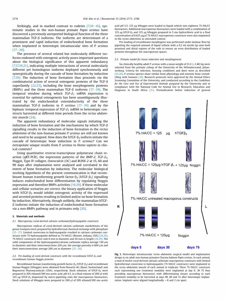
Fig. 1. Heterotopic intramuscular rectus abdominis surgical model and implantationdesign in six adult non-human primates Chacma baboon Papio ursinus. In each animal,a total of twelve coral-derived calcium carbonate macroporous constructs with limitedhydrothermal conversion to hydroxyapatite (7% HA/CC constructs) were implanted inthe rectus abdominis muscle of each animal in triplicate. Three 7% HA/CC constructseach representing one treatment modality were implanted at day 0, 30 75 thusproviding macroporous bioreactors with differentiating tissues according to eachtreatment modality to be harvested at days 90, 60 and 15 after heterotopic implan-tation. Implants were aligned longitudinally w8 and 2 cm apart.
R.M. Klar et al. / Biomaterials 35 (2014) 2773e27882774
Strikingly, and in marked contrast to rodents [7,10e16], sys-tematic studies in the non-human primate Papio ursinus havediscovered a previously unreported biological function of the threemammalian TGF-b isoforms. The isoforms are determinant of aprominent and rapid induction of endochondral bone formationwhen implanted in heterotopic intramuscular sites of P. ursinus[17e20].
The presence of several related but molecularly different iso-forms endowed with osteogenic activity poses important questionsabout the biological significance of this apparent redundancy[7,17,19,21], indicating multiple interactions of several molecularlydifferent yet homologous isoforms deployed synchronously andsynergistically during the cascade of bone formation by induction[7,19]. The induction of bone formation thus proceeds via thecombinatorial action of several osteogenic proteins of the TGF-bsuperfamily [22,23], including the bone morphogenetic proteins(BMPs) and the three mammalian TGF-b isoforms [17e19]. Thetemporal window during which TGF-b1 mRNA expression isessential for optimal osteogenesis has been unambiguously illus-trated by the endochondral osteoinductivity of the threemammalian TGF-b isoforms in P. ursinus [17e19] and by thebiphasic temporal expression of TGF-b1 mRNA in heterotopic con-structs harvested at different time periods from the rectus abdom-inis muscle [24].
The apparent redundancy of molecular signals initiating theinduction of bone formation and the mechanisms by which TGF-bsignalling results in the induction of bone formation in the rectusabdominis of the non-human primate P. ursinus are still not knownand need to be assigned. How does the hTGF-b3 isoform initiate thecascade of heterotopic bone induction in P. ursinus? Can weextrapolate unique results from P. ursinus to Homo sapiens in clin-ical contexts?
Using quantitative reverse-transcriptase polymerase chain re-action (qRT-PCR), the expression patterns of the BMP-2, TGF-b3,Noggin, Type IV collagen, Osteocalcin (OC) and RUNX-2 at 15, 60 and90 days after implantation were analysed and correlated to theextent of bone formation by induction. The molecular biologicalworking hypothesis of the present communication is that recom-binant human transforming growth factor-b3 (hTGF-b3) signallinginduces endochondral bone differentiation by regulating Nogginexpression and therefore BMPs activities [19,20]. If these molecularand cellular scenarios are correct, the binary application of Nogginwith hTGF-b3 would inhibit osteogenic activity of the expressedand secreted proteins resulting in limited and/or no bone formationby induction. Alternatively, though unlikely, the mammalian hTGF-b isoforms initiate the induction of endochondral bone formationvia a non-BMPs pathway and in primates only [20].
2. Materials and methods
2.1. Macroporous coral-derived calcium carbonate/hydroxyapatite constructs
Macroporous replicas of coral-derived calcium carbonate exoskeletons of thegenus Goniporawere prepared by hydrothermal chemical exchangewith phosphate[25e27]. Limited conversion to hydroxyapatite resulted in calcium carbonate con-structs with 7% hydroxyapatite defined as 7% HA/CC (Biomet, Indiana, USA) [28,29].7% HA/CC constructs were rods 8 mm in diameter and 20 mm in length [28,29]. Thesolid components of the hydroxyapatite/calcium carbonate replica average 130 mmin diameter and their interconnections 220 mm; the average porosity is 600 mm andtheir interconnections average 260 mm in diameter [25e29].
2.2. Pre-loading of coral-derived constructs with the recombinant hTGF-b3 andrecombinant human Noggin proteins
Recombinant human transforming growth factor-b3 (hTGF-b3) and recombinanthuman Noggin (hNoggin) were obtained from Novartis AG (Basel, Switzerland) andRegeneron Pharmaceuticals (USA), respectively. Stock solutions of hTGF-b3 wereprepared in 20% ethanol/100 mM acetic acid, pH 4.5, to a final volume of 200 ml with125 mg hTGF-b3 dispensed by micro-pipetting onto the macroporous bioreactors.Stock solutions of hNoggin were prepared in 200 ml of 20% ethanol/100 mM acetic
acid pH 5.5. 125 mg hNoggin were loaded in liquid vehicle into eighteen 7% HA/CCbioreactors. Additional macroporous bioreactors were loaded with a combination of125 mg hTGF-b3 and 125 mg hNoggin prepared in 5 mM hydrochloric acid to a finalconcentration of 0.625 mg/ml 7% HA/CC macroporous constructs were also implantedin the rectus abdominis as untreated control.
Pre-loading of recombinant morphogens was performed under laminar flow bypipetting the required amount of liquid vehicle with a 0.2 ml sterile tip onto bothproximal and distal regions of the rods to ensure an even distribution of loadedproteins throughout the macroporous spaces.
2.3. Primate model for tissue induction and morphogenesis
Six clinically healthy adult P. ursinuswith a meanweight of 21.2 (�1.48) kg wereselected from the primate colony of the University of the Witwatersrand, Johan-nesburg. Criteria for selection, housing conditions and diets were as described[25,26]. P. ursinus species share similar bone physiology and osteonic bone remod-elling with humans [30]. Research protocols were approved by the Animal EthicsScreening Committee of the University, and conducted according to the Guidelinesfor the Care and Use of Experimental Animals prepared by the University and incompliance with the National Code for Animal Use in Research, Education andDiagnosis in South Africa [31]. Premedication before induction of general
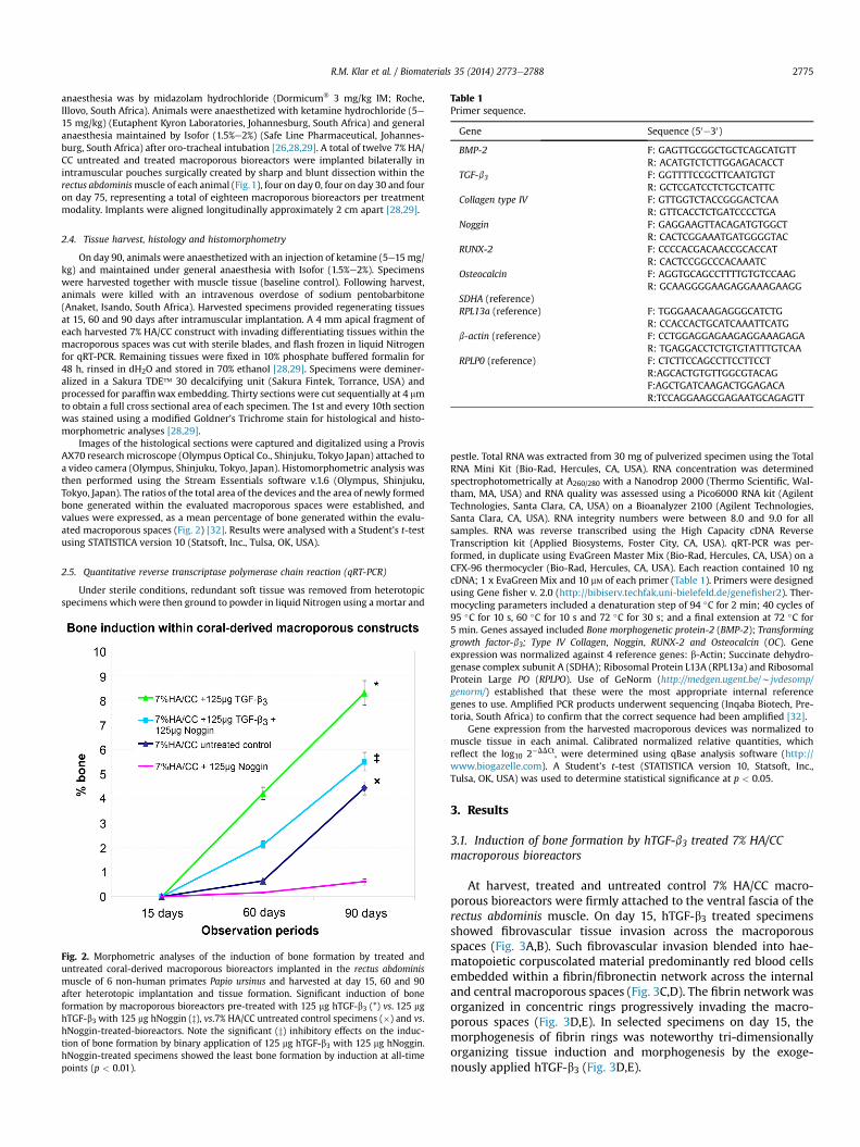
Table 1Primer sequence.
Gene Sequence (50e30)
BMP-2 F: GAGTTGCGGCTGCTCAGCATGTTR: ACATGTCTCTTGGAGACACCT
TGF-b3 F: GGTTTTCCGCTTCAATGTGTR: GCTCGATCCTCTGCTCATTC
Collagen type IV F: GTTGGTCTACCGGGACTCAAR: GTTCACCTCTGATCCCCTGA
Noggin F: GAGGAAGTTACAGATGTGGCTR: CACTCGGAAATGATGGGGTAC
RUNX-2 F: CCCCACGACAACCGCACCATR: CACTCCGGCCCACAAATC
Osteocalcin F: AGGTGCAGCCTTTTGTGTCCAAGR: GCAAGGGGAAGAGGAAAGAAGG
SDHA (reference)RPL13a (reference) F: TGGGAACAAGAGGGCATCTG
R: CCACCACTGCATCAAATTCATGb-actin (reference) F: CCTGGAGGAGAAGAGGAAAGAGA
R: TGAGGACCTCTGTGTATTTGTCAARPLP0 (reference) F: CTCTTCCAGCCTTCCTTCCT
R:AGCACTGTGTTGGCGTACAGF:AGCTGATCAAGACTGGAGACAR:TCCAGGAAGCGAGAATGCAGAGTT
R.M. Klar et al. / Biomaterials 35 (2014) 2773e2788 2775
anaesthesia was by midazolam hydrochloride (Dormicum� 3 mg/kg IM; Roche,Illovo, South Africa). Animals were anaesthetized with ketamine hydrochloride (5e15 mg/kg) (Eutaphent Kyron Laboratories, Johannesburg, South Africa) and generalanaesthesia maintained by Isofor (1.5%e2%) (Safe Line Pharmaceutical, Johannes-burg, South Africa) after oro-tracheal intubation [26,28,29]. A total of twelve 7% HA/CC untreated and treated macroporous bioreactors were implanted bilaterally inintramuscular pouches surgically created by sharp and blunt dissection within therectus abdominismuscle of each animal (Fig.1), four on day 0, four on day 30 and fouron day 75, representing a total of eighteen macroporous bioreactors per treatmentmodality. Implants were aligned longitudinally approximately 2 cm apart [28,29].
2.4. Tissue harvest, histology and histomorphometry
On day 90, animals were anaesthetized with an injection of ketamine (5e15mg/kg) and maintained under general anaesthesia with Isofor (1.5%e2%). Specimenswere harvested together with muscle tissue (baseline control). Following harvest,animals were killed with an intravenous overdose of sodium pentobarbitone(Anaket, Isando, South Africa). Harvested specimens provided regenerating tissuesat 15, 60 and 90 days after intramuscular implantation. A 4 mm apical fragment ofeach harvested 7% HA/CC construct with invading differentiating tissues within themacroporous spaces was cut with sterile blades, and flash frozen in liquid Nitrogenfor qRT-PCR. Remaining tissues were fixed in 10% phosphate buffered formalin for48 h, rinsed in dH2O and stored in 70% ethanol [28,29]. Specimens were deminer-alized in a Sakura TDE� 30 decalcifying unit (Sakura Fintek, Torrance, USA) andprocessed for paraffinwax embedding. Thirty sections were cut sequentially at 4 mmto obtain a full cross sectional area of each specimen. The 1st and every 10th sectionwas stained using a modified Goldner’s Trichrome stain for histological and histo-morphometric analyses [28,29].
Images of the histological sections were captured and digitalized using a ProvisAX70 research microscope (Olympus Optical Co., Shinjuku, Tokyo Japan) attached toa video camera (Olympus, Shinjuku, Tokyo, Japan). Histomorphometric analysis wasthen performed using the Stream Essentials software v.1.6 (Olympus, Shinjuku,Tokyo, Japan). The ratios of the total area of the devices and the area of newly formedbone generated within the evaluated macroporous spaces were established, andvalues were expressed, as a mean percentage of bone generated within the evalu-ated macroporous spaces (Fig. 2) [32]. Results were analysed with a Student’s t-testusing STATISTICA version 10 (Statsoft, Inc., Tulsa, OK, USA).
2.5. Quantitative reverse transcriptase polymerase chain reaction (qRT-PCR)
Under sterile conditions, redundant soft tissue was removed from heterotopicspecimens which were then ground to powder in liquid Nitrogen using a mortar and
Fig. 2. Morphometric analyses of the induction of bone formation by treated anduntreated coral-derived macroporous bioreactors implanted in the rectus abdominismuscle of 6 non-human primates Papio ursinus and harvested at day 15, 60 and 90after heterotopic implantation and tissue formation. Significant induction of boneformation by macroporous bioreactors pre-treated with 125 mg hTGF-b3 (*) vs. 125 mghTGF-b3 with 125 mg hNoggin (z), vs.7% HA/CC untreated control specimens (�) and vs.hNoggin-treated-bioreactors. Note the significant (z) inhibitory effects on the induc-tion of bone formation by binary application of 125 mg hTGF-b3 with 125 mg hNoggin.hNoggin-treated specimens showed the least bone formation by induction at all-timepoints (p < 0.01).
pestle. Total RNA was extracted from 30 mg of pulverized specimen using the TotalRNA Mini Kit (Bio-Rad, Hercules, CA, USA). RNA concentration was determinedspectrophotometrically at A260/280 with a Nanodrop 2000 (Thermo Scientific, Wal-tham, MA, USA) and RNA quality was assessed using a Pico6000 RNA kit (AgilentTechnologies, Santa Clara, CA, USA) on a Bioanalyzer 2100 (Agilent Technologies,Santa Clara, CA, USA). RNA integrity numbers were between 8.0 and 9.0 for allsamples. RNA was reverse transcribed using the High Capacity cDNA ReverseTranscription kit (Applied Biosystems, Foster City, CA, USA). qRT-PCR was per-formed, in duplicate using EvaGreen Master Mix (Bio-Rad, Hercules, CA, USA) on aCFX-96 thermocycler (Bio-Rad, Hercules, CA, USA). Each reaction contained 10 ngcDNA; 1 x EvaGreen Mix and 10 mM of each primer (Table 1). Primers were designedusing Gene fisher v. 2.0 (http://bibiserv.techfak.uni-bielefeld.de/genefisher2). Ther-mocycling parameters included a denaturation step of 94 �C for 2 min; 40 cycles of95 �C for 10 s, 60 �C for 10 s and 72 �C for 30 s; and a final extension at 72 �C for5 min. Genes assayed included Bone morphogenetic protein-2 (BMP-2); Transforminggrowth factor-b3; Type IV Collagen, Noggin, RUNX-2 and Osteocalcin (OC). Geneexpression was normalized against 4 reference genes: b-Actin; Succinate dehydro-genase complex subunit A (SDHA); Ribosomal Protein L13A (RPL13a) and RibosomalProtein Large PO (RPLPO). Use of GeNorm (http://medgen.ugent.be/wjvdesomp/genorm/) established that these were the most appropriate internal referencegenes to use. Amplified PCR products underwent sequencing (Inqaba Biotech, Pre-toria, South Africa) to confirm that the correct sequence had been amplified [32].
Gene expression from the harvested macroporous devices was normalized tomuscle tissue in each animal. Calibrated normalized relative quantities, whichreflect the log10 2�DDCt, were determined using qBase analysis software (http://www.biogazelle.com). A Student’s t-test (STATISTICA version 10, Statsoft, Inc.,Tulsa, OK, USA) was used to determine statistical significance at p < 0.05.
3. Results
3.1. Induction of bone formation by hTGF-b3 treated 7% HA/CCmacroporous bioreactors
At harvest, treated and untreated control 7% HA/CC macro-porous bioreactors were firmly attached to the ventral fascia of therectus abdominis muscle. On day 15, hTGF-b3 treated specimensshowed fibrovascular tissue invasion across the macroporousspaces (Fig. 3A,B). Such fibrovascular invasion blended into hae-matopoietic corpuscolated material predominantly red blood cellsembedded within a fibrin/fibronectin network across the internaland central macroporous spaces (Fig. 3C,D). The fibrin network wasorganized in concentric rings progressively invading the macro-porous spaces (Fig. 3D,E). In selected specimens on day 15, themorphogenesis of fibrin rings was noteworthy tri-dimensionallyorganizing tissue induction and morphogenesis by the exoge-nously applied hTGF-b3 (Fig. 3D,E).
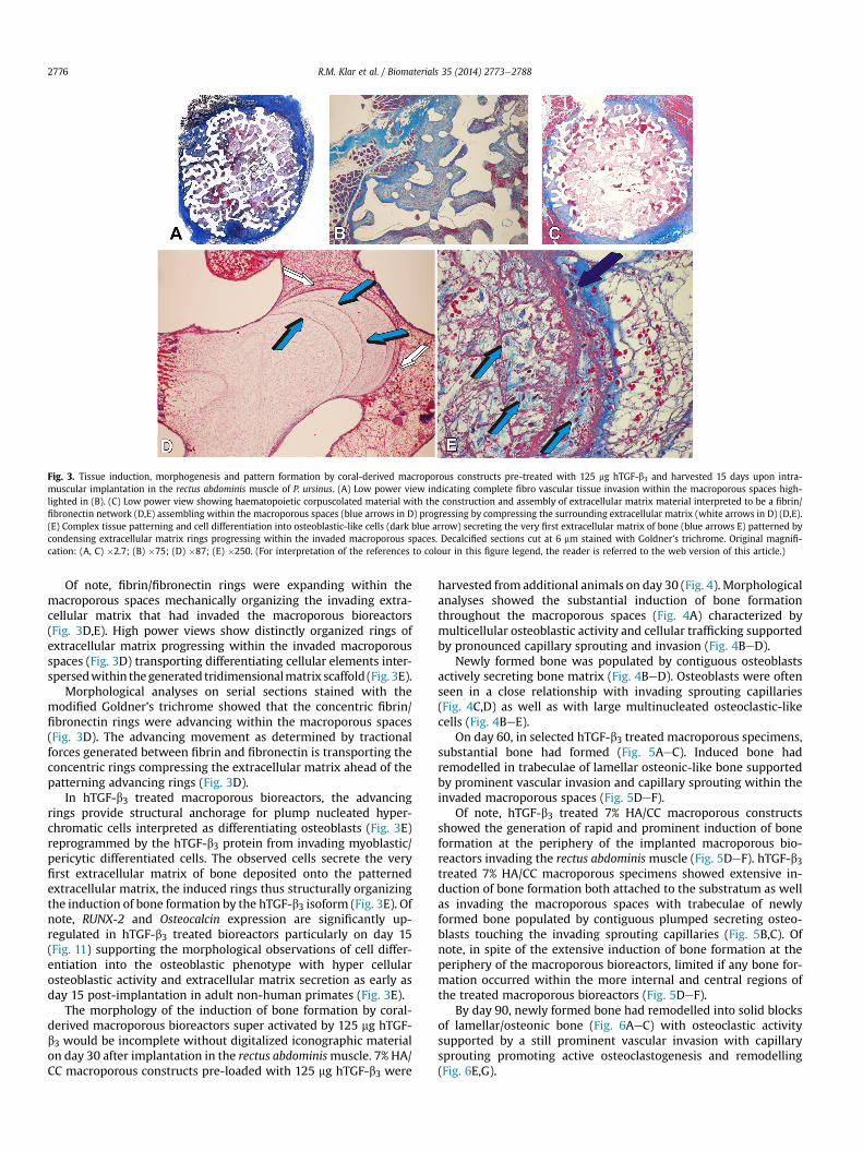
Fig. 3. Tissue induction, morphogenesis and pattern formation by coral-derived macroporous constructs pre-treated with 125 mg hTGF-b3 and harvested 15 days upon intra-muscular implantation in the rectus abdominis muscle of P. ursinus. (A) Low power view indicating complete fibro vascular tissue invasion within the macroporous spaces high-lighted in (B). (C) Low power view showing haematopoietic corpuscolated material with the construction and assembly of extracellular matrix material interpreted to be a fibrin/fibronectin network (D,E) assembling within the macroporous spaces (blue arrows in D) progressing by compressing the surrounding extracellular matrix (white arrows in D) (D,E).(E) Complex tissue patterning and cell differentiation into osteoblastic-like cells (dark blue arrow) secreting the very first extracellular matrix of bone (blue arrows E) patterned bycondensing extracellular matrix rings progressing within the invaded macroporous spaces. Decalcified sections cut at 6 mm stained with Goldner’s trichrome. Original magnifi-cation: (A, C) �2.7; (B) �75; (D) �87; (E) �250. (For interpretation of the references to colour in this figure legend, the reader is referred to the web version of this article.)
R.M. Klar et al. / Biomaterials 35 (2014) 2773e27882776
Of note, fibrin/fibronectin rings were expanding within themacroporous spaces mechanically organizing the invading extra-cellular matrix that had invaded the macroporous bioreactors(Fig. 3D,E). High power views show distinctly organized rings ofextracellular matrix progressing within the invaded macroporousspaces (Fig. 3D) transporting differentiating cellular elements inter-spersedwithin thegenerated tridimensionalmatrix scaffold (Fig. 3E).
Morphological analyses on serial sections stained with themodified Goldner’s trichrome showed that the concentric fibrin/fibronectin rings were advancing within the macroporous spaces(Fig. 3D). The advancing movement as determined by tractionalforces generated between fibrin and fibronectin is transporting theconcentric rings compressing the extracellular matrix ahead of thepatterning advancing rings (Fig. 3D).
In hTGF-b3 treated macroporous bioreactors, the advancingrings provide structural anchorage for plump nucleated hyper-chromatic cells interpreted as differentiating osteoblasts (Fig. 3E)reprogrammed by the hTGF-b3 protein from invading myoblastic/pericytic differentiated cells. The observed cells secrete the veryfirst extracellular matrix of bone deposited onto the patternedextracellular matrix, the induced rings thus structurally organizingthe induction of bone formation by the hTGF-b3 isoform (Fig. 3E). Ofnote, RUNX-2 and Osteocalcin expression are significantly up-regulated in hTGF-b3 treated bioreactors particularly on day 15(Fig. 11) supporting the morphological observations of cell differ-entiation into the osteoblastic phenotype with hyper cellularosteoblastic activity and extracellular matrix secretion as early asday 15 post-implantation in adult non-human primates (Fig. 3E).
The morphology of the induction of bone formation by coral-derived macroporous bioreactors super activated by 125 mg hTGF-b3 would be incomplete without digitalized iconographic materialon day 30 after implantation in the rectus abdominismuscle. 7% HA/CC macroporous constructs pre-loaded with 125 mg hTGF-b3 were
harvested from additional animals on day 30 (Fig. 4). Morphologicalanalyses showed the substantial induction of bone formationthroughout the macroporous spaces (Fig. 4A) characterized bymulticellular osteoblastic activity and cellular trafficking supportedby pronounced capillary sprouting and invasion (Fig. 4BeD).
Newly formed bone was populated by contiguous osteoblastsactively secreting bone matrix (Fig. 4BeD). Osteoblasts were oftenseen in a close relationship with invading sprouting capillaries(Fig. 4C,D) as well as with large multinucleated osteoclastic-likecells (Fig. 4BeE).
On day 60, in selected hTGF-b3 treated macroporous specimens,substantial bone had formed (Fig. 5AeC). Induced bone hadremodelled in trabeculae of lamellar osteonic-like bone supportedby prominent vascular invasion and capillary sprouting within theinvaded macroporous spaces (Fig. 5DeF).
Of note, hTGF-b3 treated 7% HA/CC macroporous constructsshowed the generation of rapid and prominent induction of boneformation at the periphery of the implanted macroporous bio-reactors invading the rectus abdominis muscle (Fig. 5DeF). hTGF-b3treated 7% HA/CC macroporous specimens showed extensive in-duction of bone formation both attached to the substratum as wellas invading the macroporous spaces with trabeculae of newlyformed bone populated by contiguous plumped secreting osteo-blasts touching the invading sprouting capillaries (Fig. 5B,C). Ofnote, in spite of the extensive induction of bone formation at theperiphery of the macroporous bioreactors, limited if any bone for-mation occurred within the more internal and central regions ofthe treated macroporous bioreactors (Fig. 5DeF).
By day 90, newly formed bone had remodelled into solid blocksof lamellar/osteonic bone (Fig. 6AeC) with osteoclastic activitysupported by a still prominent vascular invasion with capillarysprouting promoting active osteoclastogenesis and remodelling(Fig. 6E,G).
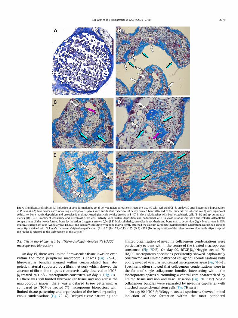
Fig. 4. Significant and substantial induction of bone formation by coral-derived macroporous constructs pre-treated with 125 mg hTGF-b3 on day 30 after heterotopic implantationin P. ursinus. (A) Low power view indicating macroporous spaces with substantial trabeculae of newly formed bone attached to the mineralized substratum (B) with significantcellularity, bone matrix deposition and osteoclastic multinucleated giant cells (white arrows in BeD) in close relationship with both osteoblastic cells (BeD) and sprouting cap-illaries (D). (C,D) Prominent cellularity and osteoblastic-like cells activity with matrix deposition and endothelial cells in close relationship with the cellular osteoblasticcompartment of the newly formed bone by induction (magenta arrows C,D). (E,F) Multicellularity, osteoblastic synthesis and bone matrix deposition (light blue arrows in E,F),multinucleated giant cells (white arrows B,C,D,E) and capillary sprouting with bone matrix tightly attached the calcium carbonate/hydroxyapatite substratum. Decalcified sectionscut at 6 mm stained with Goldner’s trichrome. Original magnification: (A) �2.7; (B) �75; (C, E) �125; (D, F) �175. (For interpretation of the references to colour in this figure legend,the reader is referred to the web version of this article.)
R.M. Klar et al. / Biomaterials 35 (2014) 2773e2788 2777
3.2. Tissue morphogenesis by hTGF-b3/hNoggin-treated 7% HA/CCmacroporous bioreactors
On day 15, there was limited fibrovascular tissue invasion evenwithin the most peripheral macroporous spaces (Fig. 7AeC);fibrovascular bundles merged within corpuscolated haemato-poietic material supported by a fibrin network which showed theabsence of fibrin-like rings as characteristically observed in hTGF-b3 treated 7% HA/CC macroporous constructs. On day 60 (Fig. 7DeG) there was still limited fibrovascular tissue invasion across themacroporous spaces; there was a delayed tissue patterning ascompared to hTGF-b3 treated 7% macroporous bioreactors withlimited tissue patterning and organization of the invading collag-enous condensations (Fig. 7EeG). Delayed tissue patterning and
limited organization of invading collagenous condensations wereparticularly evident within the centre of the treated-macroporousconstructs (Fig. 7D,E). On day 90, hTGF-b3/hNoggin-treated 7%HA/CC macroporous specimens persistently showed haphazardlyconstructed and limited patterned collagenous condensations withpoorly invaded vascularised central macroporous areas (Fig. 7HeJ).Specimens often showed that collagenous condensations were inthe form of single collagenous bundles intersecting within themacroporous spaces surrounding a central core characterized bylimited tissue invasion and vascularisation (Fig. 7H inset). Singlecollagenous bundles were separated by invading capillaries withattached mesenchymal stem cells (Fig. 7H inset).
On day 90, hTGF-b3/hNoggin-treated specimens showed limitedinduction of bone formation within the most peripheral
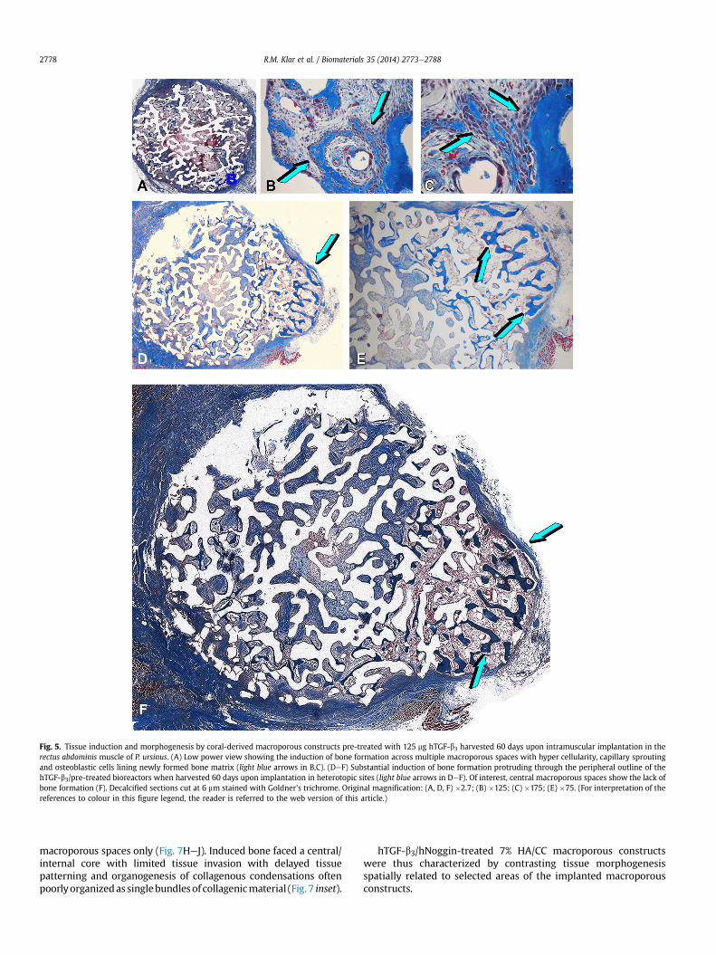
Fig. 5. Tissue induction and morphogenesis by coral-derived macroporous constructs pre-treated with 125 mg hTGF-b3 harvested 60 days upon intramuscular implantation in therectus abdominis muscle of P. ursinus. (A) Low power view showing the induction of bone formation across multiple macroporous spaces with hyper cellularity, capillary sproutingand osteoblastic cells lining newly formed bone matrix (light blue arrows in B,C). (DeF) Substantial induction of bone formation protruding through the peripheral outline of thehTGF-b3/pre-treated bioreactors when harvested 60 days upon implantation in heterotopic sites (light blue arrows in DeF). Of interest, central macroporous spaces show the lack ofbone formation (F). Decalcified sections cut at 6 mm stained with Goldner’s trichrome. Original magnification: (A, D, F) �2.7; (B) �125; (C) �175; (E) �75. (For interpretation of thereferences to colour in this figure legend, the reader is referred to the web version of this article.)
R.M. Klar et al. / Biomaterials 35 (2014) 2773e27882778
macroporous spaces only (Fig. 7HeJ). Induced bone faced a central/internal core with limited tissue invasion with delayed tissuepatterning and organogenesis of collagenous condensations oftenpoorlyorganizedas singlebundlesof collagenicmaterial (Fig. 7 inset).
hTGF-b3/hNoggin-treated 7% HA/CC macroporous constructswere thus characterized by contrasting tissue morphogenesisspatially related to selected areas of the implanted macroporousconstructs.
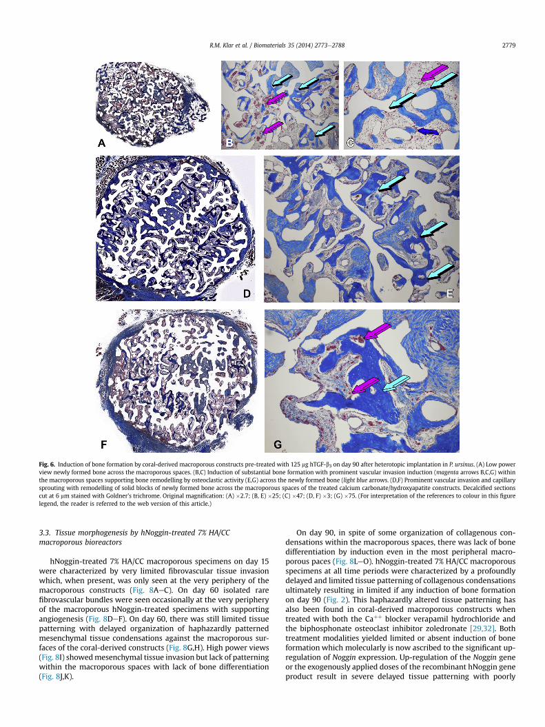
Fig. 6. Induction of bone formation by coral-derived macroporous constructs pre-treated with 125 mg hTGF-b3 on day 90 after heterotopic implantation in P. ursinus. (A) Low powerview newly formed bone across the macroporous spaces. (B,C) Induction of substantial bone formation with prominent vascular invasion induction (magenta arrows B,C,G) withinthe macroporous spaces supporting bone remodelling by osteoclastic activity (E,G) across the newly formed bone (light blue arrows. (D,F) Prominent vascular invasion and capillarysprouting with remodelling of solid blocks of newly formed bone across the macroporous spaces of the treated calcium carbonate/hydroxyapatite constructs. Decalcified sectionscut at 6 mm stained with Goldner’s trichrome. Original magnification: (A) �2.7; (B, E) �25; (C) �47; (D, F) �3; (G) �75. (For interpretation of the references to colour in this figurelegend, the reader is referred to the web version of this article.)
R.M. Klar et al. / Biomaterials 35 (2014) 2773e2788 2779
3.3. Tissue morphogenesis by hNoggin-treated 7% HA/CCmacroporous bioreactors
hNoggin-treated 7% HA/CC macroporous specimens on day 15were characterized by very limited fibrovascular tissue invasionwhich, when present, was only seen at the very periphery of themacroporous constructs (Fig. 8AeC). On day 60 isolated rarefibrovascular bundles were seen occasionally at the very peripheryof the macroporous hNoggin-treated specimens with supportingangiogenesis (Fig. 8DeF). On day 60, there was still limited tissuepatterning with delayed organization of haphazardly patternedmesenchymal tissue condensations against the macroporous sur-faces of the coral-derived constructs (Fig. 8G,H). High power views(Fig. 8I) showedmesenchymal tissue invasion but lack of patterningwithin the macroporous spaces with lack of bone differentiation(Fig. 8J,K).
On day 90, in spite of some organization of collagenous con-densations within the macroporous spaces, there was lack of bonedifferentiation by induction even in the most peripheral macro-porous paces (Fig. 8LeO). hNoggin-treated 7% HA/CC macroporousspecimens at all time periods were characterized by a profoundlydelayed and limited tissue patterning of collagenous condensationsultimately resulting in limited if any induction of bone formationon day 90 (Fig. 2). This haphazardly altered tissue patterning hasalso been found in coral-derived macroporous constructs whentreated with both the Caþþ blocker verapamil hydrochloride andthe biphosphonate osteoclast inhibitor zoledronate [29,32]. Bothtreatment modalities yielded limited or absent induction of boneformation which molecularly is now ascribed to the significant up-regulation of Noggin expression. Up-regulation of the Noggin geneor the exogenously applied doses of the recombinant hNoggin geneproduct result in severe delayed tissue patterning with poorly
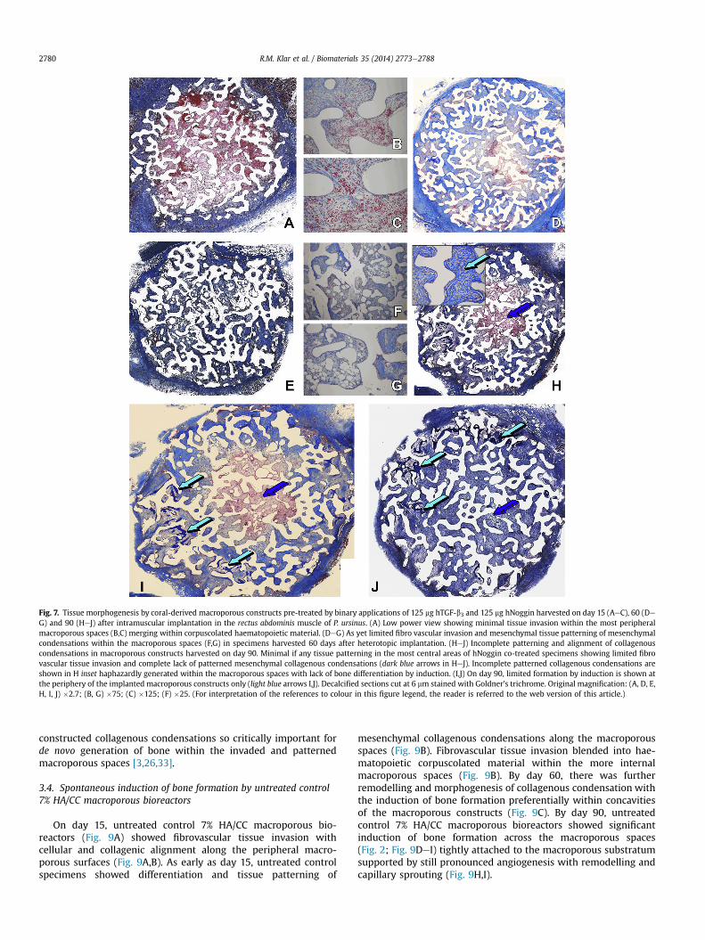
Fig. 7. Tissue morphogenesis by coral-derived macroporous constructs pre-treated by binary applications of 125 mg hTGF-b3 and 125 mg hNoggin harvested on day 15 (AeC), 60 (DeG) and 90 (HeJ) after intramuscular implantation in the rectus abdominis muscle of P. ursinus. (A) Low power view showing minimal tissue invasion within the most peripheralmacroporous spaces (B,C) merging within corpuscolated haematopoietic material. (DeG) As yet limited fibro vascular invasion and mesenchymal tissue patterning of mesenchymalcondensations within the macroporous spaces (F,G) in specimens harvested 60 days after heterotopic implantation. (HeJ) Incomplete patterning and alignment of collagenouscondensations in macroporous constructs harvested on day 90. Minimal if any tissue patterning in the most central areas of hNoggin co-treated specimens showing limited fibrovascular tissue invasion and complete lack of patterned mesenchymal collagenous condensations (dark blue arrows in HeJ). Incomplete patterned collagenous condensations areshown in H inset haphazardly generated within the macroporous spaces with lack of bone differentiation by induction. (I,J) On day 90, limited formation by induction is shown atthe periphery of the implanted macroporous constructs only (light blue arrows I,J). Decalcified sections cut at 6 mm stained with Goldner’s trichrome. Original magnification: (A, D, E,H, I, J) �2.7; (B, G) �75; (C) �125; (F) �25. (For interpretation of the references to colour in this figure legend, the reader is referred to the web version of this article.)
R.M. Klar et al. / Biomaterials 35 (2014) 2773e27882780
constructed collagenous condensations so critically important forde novo generation of bone within the invaded and patternedmacroporous spaces [3,26,33].
3.4. Spontaneous induction of bone formation by untreated control7% HA/CC macroporous bioreactors
On day 15, untreated control 7% HA/CC macroporous bio-reactors (Fig. 9A) showed fibrovascular tissue invasion withcellular and collagenic alignment along the peripheral macro-porous surfaces (Fig. 9A,B). As early as day 15, untreated controlspecimens showed differentiation and tissue patterning of
mesenchymal collagenous condensations along the macroporousspaces (Fig. 9B). Fibrovascular tissue invasion blended into hae-matopoietic corpuscolated material within the more internalmacroporous spaces (Fig. 9B). By day 60, there was furtherremodelling and morphogenesis of collagenous condensation withthe induction of bone formation preferentially within concavitiesof the macroporous constructs (Fig. 9C). By day 90, untreatedcontrol 7% HA/CC macroporous bioreactors showed significantinduction of bone formation across the macroporous spaces(Fig. 2; Fig. 9DeI) tightly attached to the macroporous substratumsupported by still pronounced angiogenesis with remodelling andcapillary sprouting (Fig. 9H,I).
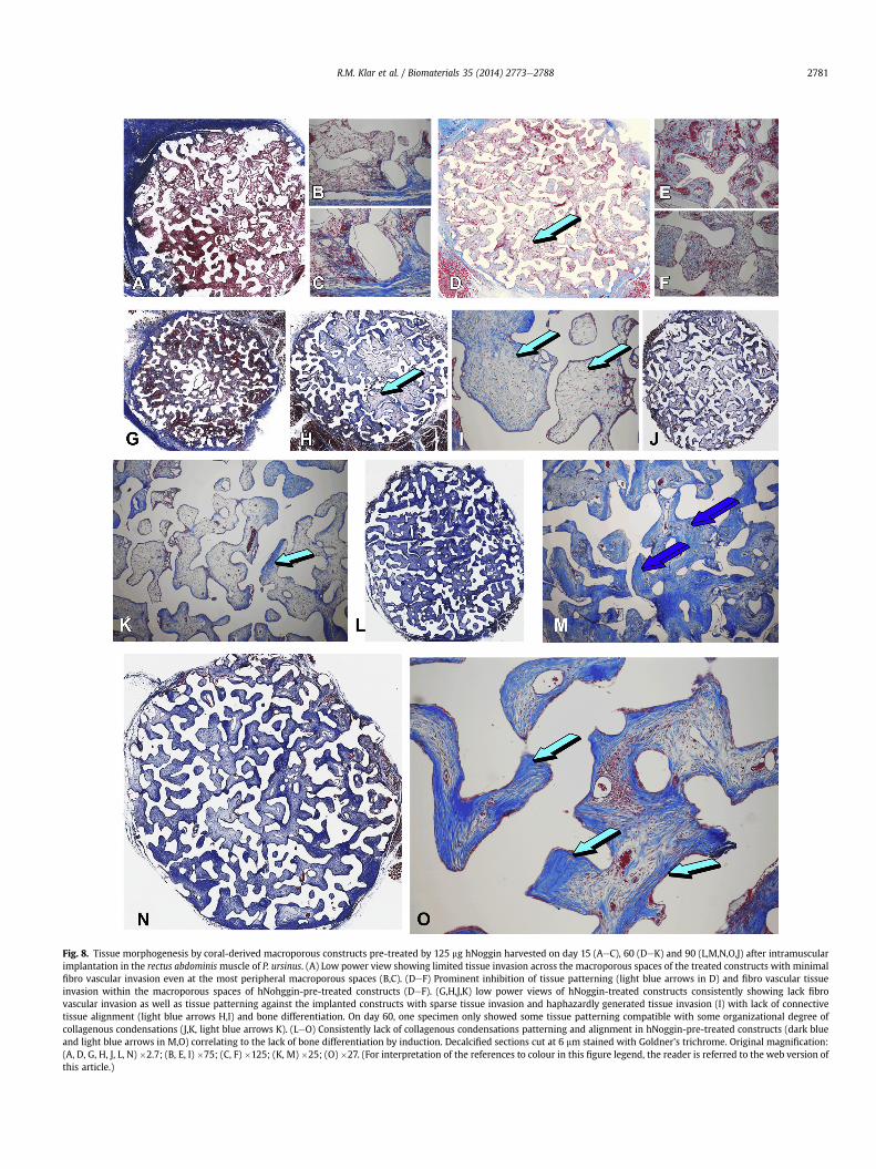
Fig. 8. Tissue morphogenesis by coral-derived macroporous constructs pre-treated by 125 mg hNoggin harvested on day 15 (AeC), 60 (DeK) and 90 (L,M,N,O,J) after intramuscularimplantation in the rectus abdominismuscle of P. ursinus. (A) Low power view showing limited tissue invasion across the macroporous spaces of the treated constructs with minimalfibro vascular invasion even at the most peripheral macroporous spaces (B,C). (DeF) Prominent inhibition of tissue patterning (light blue arrows in D) and fibro vascular tissueinvasion within the macroporous spaces of hNohggin-pre-treated constructs (DeF). (G,H,J,K) low power views of hNoggin-treated constructs consistently showing lack fibrovascular invasion as well as tissue patterning against the implanted constructs with sparse tissue invasion and haphazardly generated tissue invasion (I) with lack of connectivetissue alignment (light blue arrows H,I) and bone differentiation. On day 60, one specimen only showed some tissue patterning compatible with some organizational degree ofcollagenous condensations (J,K, light blue arrows K). (LeO) Consistently lack of collagenous condensations patterning and alignment in hNoggin-pre-treated constructs (dark blueand light blue arrows in M,O) correlating to the lack of bone differentiation by induction. Decalcified sections cut at 6 mm stained with Goldner’s trichrome. Original magnification:(A, D, G, H, J, L, N) �2.7; (B, E, I) �75; (C, F) �125; (K, M) �25; (O) �27. (For interpretation of the references to colour in this figure legend, the reader is referred to the web version ofthis article.)
R.M. Klar et al. / Biomaterials 35 (2014) 2773e2788 2781
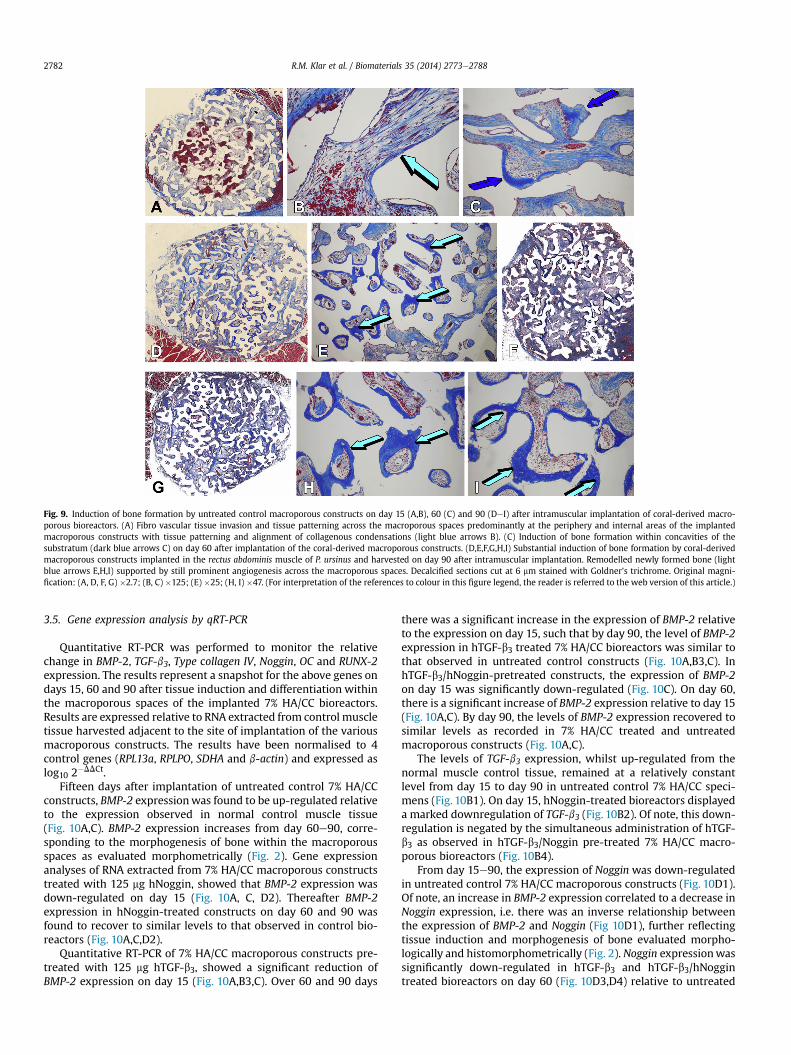
Fig. 9. Induction of bone formation by untreated control macroporous constructs on day 15 (A,B), 60 (C) and 90 (DeI) after intramuscular implantation of coral-derived macro-porous bioreactors. (A) Fibro vascular tissue invasion and tissue patterning across the macroporous spaces predominantly at the periphery and internal areas of the implantedmacroporous constructs with tissue patterning and alignment of collagenous condensations (light blue arrows B). (C) Induction of bone formation within concavities of thesubstratum (dark blue arrows C) on day 60 after implantation of the coral-derived macroporous constructs. (D,E,F,G,H,I) Substantial induction of bone formation by coral-derivedmacroporous constructs implanted in the rectus abdominis muscle of P. ursinus and harvested on day 90 after intramuscular implantation. Remodelled newly formed bone (lightblue arrows E,H,I) supported by still prominent angiogenesis across the macroporous spaces. Decalcified sections cut at 6 mm stained with Goldner’s trichrome. Original magni-fication: (A, D, F, G) �2.7; (B, C) �125; (E) �25; (H, I) �47. (For interpretation of the references to colour in this figure legend, the reader is referred to the web version of this article.)
R.M. Klar et al. / Biomaterials 35 (2014) 2773e27882782
3.5. Gene expression analysis by qRT-PCR
Quantitative RT-PCR was performed to monitor the relativechange in BMP-2, TGF-b3, Type collagen IV, Noggin, OC and RUNX-2expression. The results represent a snapshot for the above genes ondays 15, 60 and 90 after tissue induction and differentiation withinthe macroporous spaces of the implanted 7% HA/CC bioreactors.Results are expressed relative to RNA extracted from control muscletissue harvested adjacent to the site of implantation of the variousmacroporous constructs. The results have been normalised to 4control genes (RPL13a, RPLPO, SDHA and b-actin) and expressed aslog10 2�DDCt.
Fifteen days after implantation of untreated control 7% HA/CCconstructs, BMP-2 expressionwas found to be up-regulated relativeto the expression observed in normal control muscle tissue(Fig. 10A,C). BMP-2 expression increases from day 60e90, corre-sponding to the morphogenesis of bone within the macroporousspaces as evaluated morphometrically (Fig. 2). Gene expressionanalyses of RNA extracted from 7% HA/CC macroporous constructstreated with 125 mg hNoggin, showed that BMP-2 expression wasdown-regulated on day 15 (Fig. 10A, C, D2). Thereafter BMP-2expression in hNoggin-treated constructs on day 60 and 90 wasfound to recover to similar levels to that observed in control bio-reactors (Fig. 10A,C,D2).
Quantitative RT-PCR of 7% HA/CC macroporous constructs pre-treated with 125 mg hTGF-b3, showed a significant reduction ofBMP-2 expression on day 15 (Fig. 10A,B3,C). Over 60 and 90 days
there was a significant increase in the expression of BMP-2 relativeto the expression on day 15, such that by day 90, the level of BMP-2expression in hTGF-b3 treated 7% HA/CC bioreactors was similar tothat observed in untreated control constructs (Fig. 10A,B3,C). InhTGF-b3/hNoggin-pretreated constructs, the expression of BMP-2on day 15 was significantly down-regulated (Fig. 10C). On day 60,there is a significant increase of BMP-2 expression relative to day 15(Fig. 10A,C). By day 90, the levels of BMP-2 expression recovered tosimilar levels as recorded in 7% HA/CC treated and untreatedmacroporous constructs (Fig. 10A,C).
The levels of TGF-b3 expression, whilst up-regulated from thenormal muscle control tissue, remained at a relatively constantlevel from day 15 to day 90 in untreated control 7% HA/CC speci-mens (Fig. 10B1). On day 15, hNoggin-treated bioreactors displayeda marked downregulation of TGF-b3 (Fig. 10B2). Of note, this down-regulation is negated by the simultaneous administration of hTGF-b3 as observed in hTGF-b3/Noggin pre-treated 7% HA/CC macro-porous bioreactors (Fig. 10B4).
From day 15e90, the expression of Nogginwas down-regulatedin untreated control 7% HA/CC macroporous constructs (Fig. 10D1).Of note, an increase in BMP-2 expression correlated to a decrease inNoggin expression, i.e. there was an inverse relationship betweenthe expression of BMP-2 and Noggin (Fig 10D1), further reflectingtissue induction and morphogenesis of bone evaluated morpho-logically and histomorphometrically (Fig. 2). Noggin expressionwassignificantly down-regulated in hTGF-b3 and hTGF-b3/hNoggintreated bioreactors on day 60 (Fig. 10D3,D4) relative to untreated
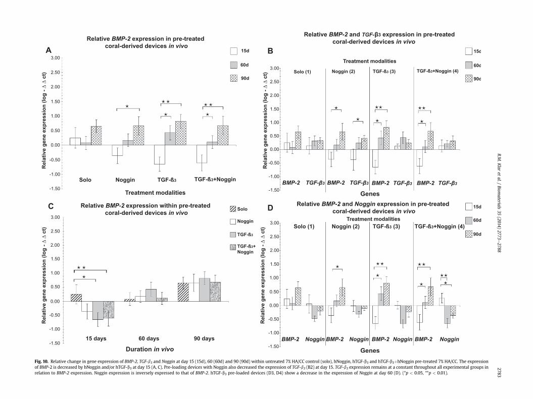
TGF-
Treatment
Treatment
Fig. 10. Relative change in gene expression of BMP-2, TGF-b3 and Noggin at day 15 (15d), 60 (60d) and 90 (90d) within untreated 7% HA/CC control (solo), hNoggin, hTGF-b3 and hTGF-b3þhNoggin pre-treated 7% HA/CC. The expressionof BMP-2 is decreased by hNoggin and/or hTGF-b3 at day 15 (A, C). Pre-loading devices with Noggin also decreased the expression of TGF-b3 (B2) at day 15. TGF-b3 expression remains at a constant throughout all experimental groups inrelation to BMP-2 expression. Noggin expression is inversely expressed to that of BMP-2. hTGF-b3 pre-loaded devices (D3, D4) show a decrease in the expression of Noggin at day 60 (D). (*p < 0.05, **p < 0.01).
R.M.K
laret
al./Biom
aterials35
(2014)2773
e2788
2783
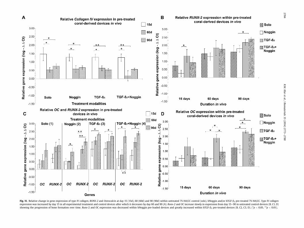
Fig. 11. Relative change in gene expression of type IV collagen, RUNX-2 and Osteocalcin at day 15 (15d), 60 (60d) and 90 (90d) within untreated 7% HA/CC control (solo), hNoggin and/or hTGF-b3 pre-treated 7% HA/CC. Type IV collagenexpression was increased by day 15 in all experimental treatment and control devices after which it decreases by day 60 and 90 (A). Runx-2 and OC increase slowly in expression from day 15e90 in untreated control devices (B, C1, D)showing the progression of bone formation over time. Runx-2 and OC expression was decreased within hNoggin pre-loaded devices and greatly increased within hTGF-b3 pre-treated devices (B, C2, C3, D). (*p < 0.05, **p < 0.01).
R.M.K
laret
al./Biom
aterials35
(2014)2773
e2788
2784
R.M. Klar et al. / Biomaterials 35 (2014) 2773e2788 2785
7% HA/CC control specimens. However, Nogginwas up-regulated inhTGF-b3/Noggin constructs on day 15 (Fig. 10D4).
By day 15, Type IV collagen expression was up-regulated in un-treated control 7% HA/CC macroporous constructs relative tomuscle control tissue. (Fig. 11 A). Of interest, Type IV collagenexpression as observed on day 15 was similar throughout alltreatment modalities (Fig. 11A) suggesting that the coral-derivedmacroporous constructs are per se intrinsically angiogenic whenimplanted in the rectus abdominis muscle of P. ursinus. There washowever a significant decrease in level of Type IV collagen expres-sion on day 60 and 90 after heterotopic implantation.
From day 15 through 90 a steady increase of RUNX-2 and OCexpressionwas recorded in untreated control bioreactors (Fig.11 B).The expression of RUNX-2 was found to be significantly lower onday 15 in hNoggin treated 7% HA/CC constructs than in the un-treated control bioreactors (Fig. 11B1). From day 60e90 there was arecovery in the expression of RUNX-2within Noggin treated 7% HA/CC devices (Fig.11). RUNX-2was significantly up-regulated in hTGF-b3/treated bioreactors on day 15 (Fig. 11B); this up-regulation ismaintained throughout days 60 and 90 (Fig. 11B). On day 15, RUNX-2 expression remained unchanged between hTGF-b3/hNoggintreated and untreated control bioreactors (Fig. 11B). By day 60 and90, RUNX-2 expression reached similar levels in the hTGF-b3/Noggin, and hTGF-b3 treated 7% HA/CC constructs (Fig. 11 B, C).
The expression pattern of the OC gene, in untreated control 7%HA/CC devices and hNoggin/treated macroporous constructs, weresimilar, in that both showed an increased expression at subsequenttime points (Fig. 11C). hTGF-b3 treated 7% HA/CC bioreactorsshowed a highly significant increase in Osteocalcin expression,recorded from day 15 to day 90 (Fig. 11C). In hTGF-b3/hNoggintreated constructs, OC showed limited expression on day 15 and 60,with however high expression on day 90 when compared to hTGF-b3 treated 7% HA/CC macroporous constructs (Fig. 10D4), corre-sponding to the induction of bone formation on day 90 albeitlimited at the periphery of the hTGF-b3/hNoggin treated macro-porous constructs only.
4. Discussion
Understanding gene expression changes during regenerativemedicine is vital to unlock the mechanisms that control, recapitu-late and induce the regenerative potential of morphogeneticmatrices. This study was directed to unravel some of the com-plexities surrounding the signalling events that occur during theinduction of bone formation when hTGF-b3 treated macroporousbioreactors are implanted in heterotopic intramuscular sites ofP. ursinus.
In a number of systematic studies in different microenviron-ments of heterotopic intramuscular and orthotopic craniofacialsites including the rectus abdominis muscle, the calvarium, themandible and periodontal furcation defects, we have shown thatprimate tissues and microenvironments respond remarkablydifferently when compared to rodent and lagomorph tissues whenimplanted in heterotopic sites with identical osteogenic proteins ofthe TGF-b supergene family [7,19,21]. Our data have shown that themammalian TGF-b isoforms are determinant of substantial endo-chondral bone formation by induction in the non-human primate P.ursinus [17e20].
Our previous combined data using the three mammalian hTGF-b isoforms have indicated that the TGF-b proteinsmay act upstreamto the bone morphogenetic proteins and may induce the inductionof heterotopic bone by expressing BMPs-related gene products ul-timately directing the induction of bone formation [17e20].Northern blot analyses of generated tissue by naturally-derivedTGF-b1 and recombinant hTGF-b2 show the expression of BMP-3
and OP-1 mRNAs [18,20,34]. This underlies the importance of theBMPs pathway even when molecularly different but related oste-ogenic proteins induce de novo heterotopic bone formation in pri-mate species.
The results of the present study highlight the modulation ofBMP-2 expression together with the down-regulation of Noggincorrelating with the induction of bone morphogenesis within theheterotopically implanted macroporous bioreactors. The reporteddata indicate that hTGF-b3 signalling induces endochondral bonedifferentiation by regulating Noggin expression and BMPs activities,and thus, the induction of bone formation. Indeed, the binaryapplication of hNoggin with equal doses of the hTGF-b3 proteinsignificantly inhibited the extent of bone formation by inductionwhen compared to coral-derived macroporous bioreactors pre-treated with hTGF-b3.
Importantly, the addition of the hNoggin protein to the coral-derived macroporous constructs profoundly inhibited the sequen-tial morphogenetic cascade of tissue patterning and alignment ofcollagenous condensations so critical for the induction of boneformation as initiated by coral-derived macroporous constructs[3,25,26,33]. Indirectly thus, pre-treatment of hNoggin shows thatthe spontaneous and intrinsic osteoinductivity of coral-derivedmacroporous constructs is initiated via the BMPs pathway duringcollagenous condensations, alignment and tissue patterning beforeosteoblastic cell differentiation with expression of BMPs geneproducts and the deposition of bone as a secondary response [3,33].
The mammalian TGF-b3 is one of the major regulators ofmesenchymal fate choice that control the early stages of osteo-blastic cell differentiation [5,35,36]. Members of the TGF-b super-gene family, BMPs and TGF-bs isoforms, are critical for theinduction, maintenance and expansion of cartilage and bone[2,4,7]. The three mammalian TGF-b isoforms are responsible forthe proliferation, early differentiation and commitment of boneprogenitors by signalling through Smad2/3 and TAK1-MMK-p38[36]. Members of the BMP family via BMP-2, -4, -5, -6 and -7 areosteogenic inducers [7,37]. At the same time, however, it has beenshown that BMP-2 expression is sufficient for full commitment toosteogenesis [38] and even short term expression of BMP-2 is suf-ficient to induce bone formation irreversibly [39].
Our previous and current results have shown that coral-derivedmacroporous bioreactors initiate the induction of bone formationwithout exogenously applied BMPs and/or TGF-b isoforms evenwhen implanted in heterotopic intramuscular sites of P. ursinus[3,28,33]. Of note, a loss of BMP-2 signalling would thus result inlimited osteogenesis; this is clearly supported by the limited in-duction of bone formation by 7% HA/CC macroporous constructspre-treated with hNoggin. Noggin is a known antagonist of BMPssignalling and inhibits the binding of BMP to its receptor [40]. Animportant as yet unreported observation is the loss of TGF-b3expression on day 15 in macroporous bioreactors pre-treated withhNoggin which further shows the complexities of the relationshipsof BMPs with TGF-b3 expression in vivo.
BMP-2 signals independently through type I and II BMP andALK2 receptors to activate the Smad1/5/8 pathway [41]. However,both BMP and TGF-b signals converge to regulate the expression ofRUNX-2, the master transcriptional regulator of osteogenesisthrough Dlx5 [42,43]. There is considerable cross talk in the path-ways that control bone formation. TGF-b signalling interacts withelements of the Wnt, FGF and the pituitary hormone (PTH) path-ways, whilst BMPs interacts with components of the Notch, FGF andWnt signalling pathways [44,45]. Modulation of the vast pleiotropicactivities of BMPs and of the mammalian TGF-b isoforms is highlycomplex, and several extracellular antagonists regulate the pleio-tropic activities of the gene products of the TGF-b supergene familyincluding Noggin, Chordin, Cerberus, and Gremlin [46e52].
R.M. Klar et al. / Biomaterials 35 (2014) 2773e27882786
It is clear that complex signalling dynamics exist during theinduction of bone formation by macroporous bioreactors pre-treated with hTGF-b3 in P. ursinus. The results show that on day15 in TGF-b3 pre-treated bioreactors there is down-regulation ofBMP-2, which is recovered on days 60 and 90. Recent studies[53,54] have made similar observations on the effect of TGF-b onBMP-2 and BMP-7 signalling. It was shown that in cultures of iso-lated primary human osteoblasts the addition of hTGF-b couldcompletely block BMP-2 and -7 signalling by inhibiting Smad1/5/8expression [53,54].
The present results indicate that endogenous TGF-b3 acts up-stream to the bone morphogenetic proteins modulating the tem-poral and spatial patterns of BMPs expression and the rate of boneformation by controlling RUNX-2 and Osteocalcin thus affectingcellular differentiation and proliferation pathways of mesenchymalstem cells into the osteoblastic lineage. This is achieved by regu-lating both inhibitory bone inductive signals, such as Noggin, andpromoting osteoblastic cellular differentiationmarkers RUNX-2 andOsteocalcin, respectively. Firstly BMPs, in context BMP-2, induce theformation of bone within the macroporous bioreactors. TGF-b3 isthen expressed at the same time but at a more constant ratemodulating BMP-2 effect by preventing Noggin expression pro-tecting the bone induction cascade from inhibition. Our resultsclearly show that hTGF-b3 treated coral-derived bioreactors displaya constant decrease in the transcription of Noggin, which is furthernegatively enhanced on day 60 in both hTGF-b3 and hTGF-b3/hNoggin pre-treated macroporous constructs.
The results show that hTGF-b3 controls RUNX-2 and Osteocalcinand thus, as observed by the addition of exogenously applied re-combinant protein, there is significant up-regulation of the aboveosteoblastic cellular differentiation markers on day 15. These data inthe non-human primate P. ursinus indicate that the dose of theexogenously applied hTGF-b3 controls the induction of bone for-mation. In P. ursinus, the hTGF-b3 isoform is a vital component of thisprocess; this is shown by hNoggin pre-treated constructs in whichthere is a profound down-regulation of osteogenic differentiationmarkers RUNX-2 and Osteocalcin, together with the loss of BMP-2 onday 15 corresponding to a profound delay in tissue patterning andmorphogenesis with lack of bone differentiation by induction.
The recruitment of responding stem cells is clearly a phenom-enon that results in the profound and rapid cascade of bone dif-ferentiation by induction when the mammalian hTGF-b3 isoform,combined with coral-derived macroporous bioreactors, is implan-ted in the rectus abdominis muscle of the non-human primateP. ursinus. The available morphological data show that the induc-tion of bone rapidly forms between 15 and 30 days, with osteo-blasts multicellularity and vascular invasion with capillarysprouting by day 30 predated by up-regulation of Collagen type IV,RUNX-2 and Osteocalcin on day 15. Osteoblasts differentiation,secretion and multicellularity as seen and predicted morphologi-cally between 15 and 30 days are molecularly driven by highexpression of Osteocalcin but particularly RUNX-2 on day 15 inhTGF-b3 treaded macroporous bioreactors.
The images presented in Fig. 3 indicate the extent of structuralorganization of the extracellular matrix directly modulated by theof hTGF-b3 isoform. The presented digital images are highly sug-gestive of hTGF-b3 reprogramming resident invading recruitedmyoblastic and/or pericytic/perivascular cells into secreting func-tionalized osteoblasts by day 15 depositing bone matrix ontopatterned extracellular matrix rings induced within the coral-derived macroporous constructs. The induction of advancingextracellular matrix rings within the macroporous bioreactors su-per activated by the hTGF-b3 isoform provides structural anchoragefor hyper chromatic osteoblastic-like cells reprogrammed by thehTGF-b3 from invading myoblastic/pericytic cells.
Taken together, these unique morphological results togetherwith up-regulation of RUNX-2, a master control gene of osteogenicdifferentiation [42,43,55], suggest that the 125 mg dose of the hTGF-b3 protein is successfully reprogramming differentiatedmyoblastic/pericytic cells into the induction of pluripotent stem cells [56,57]for rapid transformation into active secreting osteoblasts. Theessential role of RUNX-2 in osteoblast differentiation [42,43,55] hasbeen shown in null-mutation mice that had a cartilaginous skel-eton with complete absence of osteoblasts [42,55,58]. Transgenicexpression of RUNX-2 in chondrocytes results in heterotopicchondrocytes hypertrophy and endochondral ossification [59]. Theinduction of chondrocytes hypertrophy and endochondral ossifi-cation indicate that RUNX-2 controls chondrocytes and osteoblastdifferentiation ultimately regulating the induction of endochondralossification [42,55,59].
The rapidity of tissue induction and transformation into bone bythe hTGF-b3 protein is further morphologically shown by the in-duction of bone formation well beyond the perimeter of theimplanted macroporous bioreactors with however lack of bonedifferentiation within the central regions of the coral derivedmacroporous constructs as shown on day 60. As previously hy-pothesized using the hTGF-b1 isoform [17], the extensive osteoin-duction surrounding the implanted hTGF-b3 treated bioreactorscould be the result of desorption of the applied morphogen fromthe coral-derived constructs, as suggested in previous experimentsin the non-human primate P. ursinus when using insoluble collag-enous bone matrix as carrier [17] also for the recombinant humanosteogenic protein-1 [22,60], with subsequent diffusion and bio-logical activity into the extracellular space away from the implan-ted matrices [17,22,60]. Whether such an extended range of actionis due to a diffusion gradient or to the initiation of a sequentialchain of cellular induction [61,62] cannot be deduced from theavailable data. The data do however indicate the rapidity of stemcells/responding cells induction into bone long before migratingcells are within the pre-treated bioreactors’ macroporous spacesthus transformed into bone when still at the periphery of theimplanted bioreactors super activated by the adsorbed hTGF-b3signal.
Further molecular andmorphological studies are now necessaryto precisely resolve the remarkable endochondral osteoinductivityof the hTGF-b3 isoform in primates only. Our current and previousexperiments using the hTGF-b3 protein in P. ursinus have shownrapid and substantial bone induction when hTGF-b3 reconstitutedosteogenic device are implanted in heterotopic intramuscularrectus abdominis sites with BMP-2, TGF-b1, TGF-b3, RUNX-2, Osteo-calcin and Type IV collagen expression, hypercellular osteoblasticactivity and capillary sprouting which are now the molecular andmorphological basis to rapidly construct bone in clinical contexts[19,63].
The substantial induction of bone formation by 125 mg hTGF-b3but particularly the prominent induction of bone by 250 mg hTGF-b3by day 20 when combined with coral-derived macroporous bio-reactors [64], has been translated in clinical contexts resulting insignificant induction of bone formation in a large mandibulardefect after hemi-mandibulectomy in a human patient [63,64] nowfurther evaluated by CT-scans 6 months after implantation of the250 mg hTGF-b3 osteogenic device.
Finally, mandatory research will have now to be devoted toaddress the following question: why the mammalian TGF-b3 in-duces prominent bone formation when implanted in heterotopicintramuscular sites of the rectus abdominis muscle of P. ursinus andnot when implanted in heterotopic sites of rodents and lago-morphs? Mechanistically, the question raises rather complicatedavenues of apparent redundancy of genes and related secretedmolecular signals in primate tissues vs. rodents and lagomorphs.
R.M. Klar et al. / Biomaterials 35 (2014) 2773e2788 2787
Molecular redundancy with associated different biological func-tions in primate tissues may simply represent the fine tuning ofspeciation-related molecular evolution in anthropoid apes at theearly Pliocene boundary which resulted in a more capable tuning ofthe bone induction cascade in primate species.
Importantly, a large proportion of molecular evolution acrossmany phylogenies of different clades is associated with speciationevents which have prompted the hypothesis that speciation per secauses a burst of genetic evolution [65,66] which indeed drivesmolecular evolution. Alternatively, the heterotopic induction ofbone formation in P. ursinus as a result of bursts of cladistic evo-lution is now redundant but yet still activated by high doses of therecombinant morphogen to induce prominent and rapid inductionof bone formation.
5. Conclusions
Themammalian TGF-b3 isoform is a vital constituent of the boneinduction cascade in heterotopic sites of the non-human primateP. ursinus. The results unequivocally demonstrate that hTGF-b3elicits the induction of bone formation by up-regulating endoge-nous BMP-2 and is blocked by Noggin, a BMP antagonist. The dataprovocatively suggest a role for hTGF-b3 in recruiting and re-programming resident progenitor cells including pericytic/peri-vascular stem cells into active secreting osteoblasts. The rapid andprominent induction of bone formation has been translated inclinical contexts. The potential manipulation of a pluripotent stemcell niche by the heterotopically applied hTGF-b3 osteogenic deviceis an exciting avenue of fervent future investigation in the uniquemodel system of bone induction that has been provided byP. ursinus.
Acknowledgements
The controversial research on the hTGF-b3 in Papio ursinus and itstranslation in clinical contexts have been continuously supported byad hoc grants of the Bone Research Laboratory and by a grant fromNovartis AG in the late nineties. Aris Economides and Peter ten Dijkeare gratefully acknowledged for the kind donation of the recombi-nant humanNoggin and for suggesting the use of the Nogginproteinto highlight some the mechanisms of this unique apparent redun-dancy of soluble molecular signals initiating the induction of boneformation inprimate species. The significant help of Barbara van denHeever,Manolis Heliotis, Laura Rodennée Yeates andCarlo Ferretti isgratefully acknowledged. A special word of thank to Ruquayya Parakfor thehistological processing andsomeof theevaluation, toCarolineDickens for critical help with the qRT-PCR, and to Mike PonticielloBiomet USA for the kind supply of the implanted coral-derivedmacroporous constructs. Hari Reddi significantly contributed byinitiating the senior author to the inductionof bone formationandbysharply discussing the 2nd last manuscript draft. This paper and thesignificant efforts of this never ending journey on the hTGF-b3 iso-form are dedicated to the Bone Research Laboratory of theUniversityof the Witwatersrand, Johannesburg.
References
[1] Reddi AH. Bone morphogenesis and modelling: soluble signals sculpt osteo-somes in the solid state. Cell 1997;89:159e61.
[2] Reddi AH. Morphogenesis and tissue engineering of bone and cartilage:inductive signals, stem cells, and biomimetic biomaterials. Tissue Eng 2000;6:351e9.
[3] Ripamonti U. Biomimetism, biomimetic matrices and the induction of boneformation. J Cell Mol Med 2009;13:2953e72.
[4] Wozney JM, Rosen V, Celeste AJ, Mitsock LM, Whitters MJ, Kriz RW, et al.Novel regulators of bone formation: molecular clones and activities. Science1988;242:1528e34.
[5] ten Dijke P, Iwata KK, Goddard C, Pieter C, Canalis E, McCarthy TL, et al. Re-combinant transforming growth factor type beta 3: biological activities andreceptor-binding properties in isolated bone cells. Mol Cell Biol 1990;10:4473e9.
[6] Massagué J. How cells read TGF-b signals. Nat Rev Mol Cell Biol 2000;3:169e78.
[7] Ripamonti U. Soluble osteogenic molecular signals and the induction of boneformation. Biomaterials 2006;27:807e22.
[8] Balooch G, Balooch M, Nalla RK, Schilling S, Filvaroff EH, Marshal GW, et al.TGFb regulates the mechanical properties and composition of bone matrix.Proc Natl Acad Sci U S A 2005;102:18813e8.
[9] Spagnoli A, O’Rear L, Chandler RL, Granero-Molto F, Mortlock DP, Gorska AE,et al. TGF-b signalling is essential for joint morphogenesis. J Cell Biol2007;177:1105e17.
[10] Roberts AB, Sporn MB, Assoian RK, Smith JM, Roche NS, Wakefield LM, et al.Transforming growth factor type b: rapid induction of fibrosis and angio-genesis in vivo and stimulation of collagen formation in vitro. Proc Natl AcadSci U S A 1986;83:4167e71.
[11] Saadeh PB, Mehrara BJ, Steinbrech DS, Dudziak ME, Greenwald JA, Luchs JS,et al. Transforming growth factor-b1 modulates the expression of vascularendothelial growth factor by osteoblasts. Am J Phys 1999;277:C628e37.
[12] Sampath TK, Muthukumaran N, Reddi AH. Isolation of osteogenin, an extra-cellular matrix-associated, bone-inductive protein, by heparin affinity chro-matography. Proc Natl Acad Sci U S A 1987;84:7109e13.
[13] Hammonds RG, Schwall R, Dudley A, Berkemeier L, Lai C, Lee J, et al. Boneinducing activity of mature BMP-2b produced from a hybrid BMP-2a/2bprecursor. Mol Endocrinol 1991;5:149e55.
[14] Shinozaki M, Kawara S, Hayashi N, Kakinuma T, Igarashi A, Takehara K. In-duction of subcutaneous tissue fibrosis in newborn mice by transforminggrowth factor b-simultaneous application with basic fibroblast growth factorcauses persistent fibrosis. Biochem Biophys Res Commun 1997;240:292e7.
[15] Shah M, Foreman DM, Ferguson MW. Neutralisation of TGF-b1 and TGF-b2 orexogenous addition of TGF-b3 to cutaneous rat wounds reduces scarring. J CellSci 1995;108:985e1002.
[16] Ripamonti U, Ramoshebi LN, Patton J, Matsaba T, Teare J, Renton L. Solublesignals and insoluble substrata: novel molecular cues instructing the induc-tion of bone. In: Massaro EJ, Rogers JM, editors. The skeleton. New York:Humana Press; 2004. pp. 217e27.
[17] Ripamonti U, Duneas N, van den Heever B, Bosch C, Crooks J. Recombinanttransforming growth factor-b1 induces endochondral bone in the baboon andsynergizes with recombinant osteogenic protein-1 (bone morphogeneticprotein-7) to initiate rapid bone formation. J Bone Miner Res 1997;2:1584e95.
[18] Ripamonti U, Crooks J, Matsaba T, Taskar J. Induction of endochondral boneformation by recombinant human transforming growth factor-b2 in the ba-boon (Papio ursinus). Growth Factors 2000;17:269e85.
[19] Ripamonti U, Ramoshebi LN, Teare J, Renton L, Ferretti C. The induction ofendochondral bone formation by transforming growth factor-b3: experi-mental studies in the non-human primate Papio ursinus. J Cell Mol Med2008;12:1029e48.
[20] Ripamonti U, Roden LC. The induction of bone formation by transforminggrowth factor-b2 in the non-human primate Papio ursinus and its modulationby skeletal muscle responding stem cells. Cell Prolif 2010;43:207e18.
[21] Ripamonti U, Ferretti C, Teare J, Blann L. Transforming growth factor-b iso-forms and the induction of bone formation: Implications for reconstructivecraniofacial surgery. J Craniofac Surg 2009;20:1544e55.
[22] Ripamonti U, Van Den Heever B, Crooks J, Tucker MM, Sampath TK, Rueger DC,et al. Long-term evaluation of bone formation by osteogenic protein 1 in thebaboon and relative efficacy of bone-derived bone morphogenetic proteinsdelivered by irradiated xenogeneic collagenous matrices. J Bone Miner Res2000;9:1798e809.
[23] Ripamonti U. Osteogenic proteins of the transforming growth factor-b su-perfamily. In: Henry HL, Norman AW, editors. Encyclopaedia of hormones.Texas: Austin Academic Press; 2003. pp. 80e6.
[24] Ripamonti U. Bone induction by recombinant human osteogenic protein-1(hOP-1, BMP-7) in the primate Papio ursinus with expression of mRNA ofgene products of the TGF-b superfamily. J Cell Mol Med 2005;9:911e28.
[25] Ripamonti U. Inductive bone matrix and porous hydroxyapatite composites inrodents and non-human primates. In: Yamamuro T, Wilson-Hench J,Hench LL, editors. Handbook of bioactive ceramics, calcium phosphate andhydroxylapatite ceramics, vol. II. Florida: CRC Press; 1990. pp. 245e53.
[26] Ripamonti U. The morphogenesis of bone in replicas of porous hydroxyapatiteobtained from conversion of calcium carbonate exoskeletons of coral. J BoneJoint Surg Am 1991;73:692e703.
[27] Shors EC. Coralline bone graft substitutes. Orthop Clin North Am 1999;30:599e613.
[28] Ripamonti U, Crooks J, Khoali L, Roden L. The induction of bone formation bycoral-derived calcium carbonate/hydroxyapatite constructs. Biomaterials2009;30:1428e39.
[29] Ripamonti U, Klar RM, Renton LF, Ferretti C. Synergistic induction of boneformation by hOP-1, hTGF-b3 and inhibition by zoledronate in macroporouscoral-derived hydroxyapatites. Biomaterials 2010;31:6400e10.
[30] Schnitzler CM, Ripamonti U, Mesquita JM. Histomorphometry of iliac cresttrabecular bone in adult male baboons in captivity. Calcif Tissue Int 1993;52:447e54.
R.M. Klar et al. / Biomaterials 35 (2014) 2773e27882788
[31] Public Service Department. National code for animal use in research educa-tion, diagnosis and testing of drugs and related substances in South Africa.Pretoria, South Africa: Public Service Department; 1990.
[32] Klar RM, Duarte R, Dix-Peek T, Dickens C, Ferretti C, Ripamonti U. Calcium ionsand osteoclastogenesis initiate the induction of bone formation by coral-derived macroporous constructs. J Cell Mol Med 2013. http://dx.doi.org/10.1111/jcmm.12125.
[33] Ripamonti U, van Den Heever B, van Wyk J. Expression of the osteogenicphenotype in porous hydroxyapatite implanted extraskeletally in baboons.Matrix 1993;13:491e502.
[34] Duneas N, Crooks J, Ripamonti U. Transforming growth factor-b1: induction ofbone morphogenetic protein genes expression during endochondral boneformation in the baboon, and synergistic interaction with osteogenic protein-1 (BMP-7). Growth Factors 1998;15:259e77.
[35] Shi Y, Massagué J. Mechanisms of TGF-b signaling from cell membrane to thenucleus. Cell 2003;113:685e700.
[36] Matsunobu T, Torigoe K, Ishikawa M, de Vega S, Kulkarni AB, Iwamoto Y, et al.Critical roles of the TGF-b type I receptor ALK5 in perichondrial formation andfunction, cartilage integrity, and osteoblast differentiation during growthplate development. Dev Biol 2009;2:325e38.
[37] Bandyopadhyay A, Tsuji K, Cox K, Harfe BD, Rosen V, Tabin CJ. Genetic analysisof the roles of BMP2, BMP4, and BMP7 in limb patterning and skeletogenesis.PLoS Genet 2006;12:e216.
[38] Huang Z, Ren PG, Ma T, Smith RL, Goodman SB. Modulating osteogenesis ofmesenchymal stem cells by modifying growth factor availability. Cytokine2010;51:305e10.
[39] Noel D, Gazit D, Bouquet C, Apparailly F, Bony C, Plence P, et al. Short-termBMP-2 expression is sufficient for in vivo osteochondral differentiation ofmesenchymal stem cells. Stem Cells 2004;22:74e85.
[40] Zimmerman LB, De Jesus-Escobar JM, Harland RM. The Spemann organizersignal noggin binds and inactivates bone morphogenetic protein 4. Cell1996;96:599e606.
[41] Zhou Z, Xie J, Lee D, Liu Y, Jung J, Zhou L, et al. Neogenin regulation of BMP-induced canonical Smad signaling and endochondral bone formation. Dev Cell2010;19:90e102.
[42] Ducy P, Zhang R, Geoffroy V, Ridall AL, Karsenty G. Osf2/Cbfa1: a transcrip-tional activator of osteoblast differentiation. Cell 1997;89:747e54.
[43] Pretheeban T, Lemos DR, Paylor B, Zhang RH, Rossi FM. Role of stem/pro-genitor cells in reparative disorders. Fibrogenesis Tissue Repair 2012;5:1e20.
[44] De Robertis EM. Evo-Devo: the merging of evolutionary and developmentalbiology. In: The scientific legacy of the 20th centuryPontif Acad Sci Acta, vol.21; 2011. pp. 221e35.
[45] Chen G, Deng C, Li YP. TGF-b and BMP signaling in osteoblast differentiationand bone formation. Int J Biol Sci 2012;8:272e88.
[46] Miyazono K, Kusanagi K, Inoue H. Divergence and convergence of TGF-b/BMPsignaling. J Cell Physiol 2001;187:265e76.
[47] Heldin CH, Miyazono K, ten Dijke P. TGF-b signalling from cell membrane tonucleus through SMAD proteins. Nature 1997;390:465e71.
[48] Miyazono K, ten Dijke P, Heldin CH. TGF-b signaling by Smad proteins. AdvImmunol 2000;75:115e57.
[49] Miyazono K. Positive and negative regulation of TGF-b signaling. J Cell Sci2000;113:1101e9.
[50] Cooper GM, Usas A, Olshanski A, Mooney MP, Losee JE, Huard J. Ex vivo Noggingene therapy inhibits bone formation in a mouse model of postoperativeresynostosis. Plast Reconstr Surg 2009;123:94Se103S.
[51] Onichtchouk D, Chen YG, Dosch R, Gawantka V, Delius H, Massagué J, et al.Silencing of TGF-beta signalling by the pseudoreceptor BAMBI. Nature1999;401:480e5.
[52] Gazzerro E, Smerdel-Ramoya A, Zanotti S, Stadmeyer L, Durant D,Economides AN, et al. Conditional deletion of gremlin causes a transient in-crease in bone formation and bone mass. J Biol Chem 2007;282:31549e57.
[53] Ehnert S, Baur J, Schmitt A, Neumaier M, Lucke M, Dooley S, et al. TGF-b1 aspossible link between loss of bone mineral density and chronic inflammation.PLoS One 2010;5:e14073.
[54] Ehnert S, Zhao J, Pscherer S, Freude T, Dooley S, Kolk A, et al. Transforminggrowth factor b1 inhibits bone morphogenic protein (BMP)-2 and BMP-7signaling via upregulation of Ski-related novel protein N (SnoN): possiblemechanism for the failure of BMP therapy? BMC Med 2012;10:101.
[55] Komori T, Yagi H, Nomura S, Yamaguchi A, Sasaki K, Deguchi K, et al. Targeteddisruption of Cbfa1 results in a complete lack of bone formation owing tomaturational arrest of osteoblasts. Cell 1997;89:755e64.
[56] Takahashi K, Yamanaka S. Induction of pluripotent stem cells from mouseembryonic and adult fibroblast cultures by defined factors. Cell 2006;126:663e76.
[57] Takahashi K, Tanabe K, Ohnuki M, Narita M, Ichisaka T, Tomoda K, et al. In-duction of pluripotent stem cells from adult human fibroblasts by definedfactors. Cell 2007;131:1e12.
[58] Harada S, Rodan GA. Control of osteoblast function and regulation of bonemass. Nature 2003;423:349e55.
[59] Takeda S, Bonnamy JP, Owen MJ, Ducy P, Karsenty G. Continuous expressionof Cbfa1 in non-hypertrophic chondrocytes uncovers its ability to inducehypertrophic chondrocyte differentiation and partially rescues Cbfa1-deficient mice. Genes Dev 2001;15:467e81.
[60] Ripamonti U, van den Heever B, Sampath TK, Tucker MM, Rueger DC,Reddi AH. Complete regeneration of bone in the baboon by recombinanthuman osteogenic protein-1 (hOP-1, bone morphogenetic protein-7). GrowthFactors 1996;13:273e89.
[61] Slack JMW. Morphogenetic gradients e past and present. Trends Biochem1987;12:200e4.
[62] Lander AD. Morpheus unbound: reimagining the morphogen gradient. Cell2007;128:245e56.
[63] Ripamonti U, Ferretti C. Grand challenges for craniomandibularfacialrecon-struction by human recombinant transforming growth fac tor-b3. In: Tuan RS,Guilak F, Atala A, editors. Keystone symposia on regenerative tissue engi-neering. Breckenridge CO. http://www.keystonesymposia.org; 2012.
[64] Ripamonti U, Teare J, Ferretti C. A macroporous bioreactor super activated bythe recombinant human transforming growth factor-b3. Front Physiol 2012;3:1e18.
[65] Webster AJ, Payne RJH, Pagel M. Molecular phylogenies link rates of evolutionand speciation. Science 2003;301:478.
[66] Ezard THG, Thomas GH, Purvis A. Inclusion of a near-complete fossil recordreveals speciation-related molecular evolution. Methods Ecol Evol 2013;4:745e53.