The Anticancer, Antioxidant and Antimicrobial Properties ...
22
Molecules 2015, 20, 11808-11829; doi:10.3390/molecules200711808 molecules ISSN 1420-3049 www.mdpi.com/journal/molecules Article The Anticancer, Antioxidant and Antimicrobial Properties of the Sesquiterpene β-Caryophyllene from the Essential Oil of Aquilaria crassna Saad S. Dahham 1 , Yasser M. Tabana 1 , Muhammad A. Iqbal 2 , Mohamed B. K. Ahamed 3 , Mohammed O. Ezzat 4 , Aman S. A. Majid 5, * and Amin M. S. A. Majid 1, * 1 EMAN Research and Testing Laboratory, School of Pharmaceutical Sciences, Universiti Sains Malaysia, 11800 Minden, Pulau Pinang, Malaysia; E-Mails: [email protected] (S.S.D.); [email protected] (Y.M.T.) 2 School of Chemical Sciences, Universiti Sains Malaysia, 11800 Minden, Pulau Pinang, Malaysia; E-Mail: [email protected] 3 EMAN Biodiscoveries Sdn. Bhd. EUREKA Complex, Universiti Sains Malaysia (USM) Campus, 11800 Minden, Pulau Pinang, Malaysia; E-Mail: [email protected] 4 Centre for Drug Research, Universiti Sains Malaysia, 11800 Minden, Pulau Pinang, Malaysia; E-Mail: [email protected] 5 Advanced Medical and Dental Institute (IPPT), Universiti Sains Malaysia, Bertam, 13200 Kepala Batas, Penang, Malaysia * Authors to whom correspondence should be addressed; E-Mails: [email protected] (A.S.A.M.); [email protected] (A.M.S.A.M.); Tel.: +60-124-930-842 (A.S.A.M.); +60-124-240-842 (A.M.S.A.M.); Fax: +60-604-562-2468 (A.S.A.M.); +60-604-641-0295 (A.M.S.A.M.). Academic Editor: Derek J. McPhee Received: 24 February 2015 / Accepted: 24 April 2015 / Published: 26 June 2015 Abstract: The present study reports a bioassay-guided isolation of β-caryophyllene from the essential oil of Aquilaria crassna. The structure of β-caryophyllene was confirmed using FT-IR, NMR and MS. The antimicrobial effect of β-caryophyllene was examined using human pathogenic bacterial and fungal strains. Its anti-oxidant properties were evaluated by DPPH and FRAP scavenging assays. The cytotoxicity of β-caryophyllene was tested against seven human cancer cell lines. The corresponding selectivity index was determined by testing its cytotoxicity on normal cells. The effects of β-caryophyllene were studied on a series of in vitro antitumor-promoting assays using colon cancer cells. Results showed OPEN ACCESS
Transcript of The Anticancer, Antioxidant and Antimicrobial Properties ...
Molecules 2015, 20, 11808-11829; doi:10.3390/molecules200711808
molecules ISSN 1420-3049
www.mdpi.com/journal/molecules
Article
The Anticancer, Antioxidant and Antimicrobial Properties of the Sesquiterpene β-Caryophyllene from the Essential Oil of Aquilaria crassna
Saad S. Dahham 1, Yasser M. Tabana 1, Muhammad A. Iqbal 2, Mohamed B. K. Ahamed 3,
Mohammed O. Ezzat 4, Aman S. A. Majid 5,* and Amin M. S. A. Majid 1,*
1 EMAN Research and Testing Laboratory, School of Pharmaceutical Sciences,
Universiti Sains Malaysia, 11800 Minden, Pulau Pinang, Malaysia;
E-Mails: [email protected] (S.S.D.); [email protected] (Y.M.T.) 2 School of Chemical Sciences, Universiti Sains Malaysia, 11800 Minden, Pulau Pinang, Malaysia;
E-Mail: [email protected] 3 EMAN Biodiscoveries Sdn. Bhd. EUREKA Complex, Universiti Sains Malaysia (USM) Campus,
11800 Minden, Pulau Pinang, Malaysia; E-Mail: [email protected] 4 Centre for Drug Research, Universiti Sains Malaysia, 11800 Minden, Pulau Pinang, Malaysia;
E-Mail: [email protected] 5 Advanced Medical and Dental Institute (IPPT), Universiti Sains Malaysia, Bertam,
13200 Kepala Batas, Penang, Malaysia
* Authors to whom correspondence should be addressed;
E-Mails: [email protected] (A.S.A.M.); [email protected] (A.M.S.A.M.);
Tel.: +60-124-930-842 (A.S.A.M.); +60-124-240-842 (A.M.S.A.M.);
Fax: +60-604-562-2468 (A.S.A.M.); +60-604-641-0295 (A.M.S.A.M.).
Academic Editor: Derek J. McPhee
Received: 24 February 2015 / Accepted: 24 April 2015 / Published: 26 June 2015
Abstract: The present study reports a bioassay-guided isolation of β-caryophyllene from the
essential oil of Aquilaria crassna. The structure of β-caryophyllene was confirmed using
FT-IR, NMR and MS. The antimicrobial effect of β-caryophyllene was examined using
human pathogenic bacterial and fungal strains. Its anti-oxidant properties were evaluated by
DPPH and FRAP scavenging assays. The cytotoxicity of β-caryophyllene was tested against
seven human cancer cell lines. The corresponding selectivity index was determined by
testing its cytotoxicity on normal cells. The effects of β-caryophyllene were studied on a
series of in vitro antitumor-promoting assays using colon cancer cells. Results showed
OPEN ACCESS
Molecules 2015, 20 11809
that β-caryophyllene demonstrated selective antibacterial activity against S. aureus (MIC
3 ± 1.0 µM) and more pronounced anti-fungal activity than kanamycin. β-Caryophyllene
also displayed strong antioxidant effects. Additionally, β-caryophyllene exhibited selective
anti-proliferative effects against colorectal cancer cells (IC50 19 µM). The results also
showed that β-caryophyllene induces apoptosis via nuclear condensation and fragmentation
pathways including disruption of mitochondrial membrane potential. Further, β-caryophyllene
demonstrated potent inhibition against clonogenicity, migration, invasion and spheroid
formation in colon cancer cells. These results prompt us to state that β-caryophyllene is
the active principle responsible for the selective anticancer and antimicrobial activities of
A. crassnia. β-Caryophyllene has great potential to be further developed as a promising
chemotherapeutic agent against colorectal malignancies.
Keywords: β-caryophyllene; anti-cancer; apoptosis; anti-clonogenic; nuclear fragmentation;
colorectal cancer
1. Introduction
Aquilaria crassna (Thymelaeaceae) has been used in diverse Chinese and Southeast Asian traditional
medicine systems to treat infectious and inflammatory diseases, arthritis and cardiac disorders. In the
Middle East this woody plant has been widely used in fragrances, incense and cosmetics. The plant,
locally known in Indonesia as gaharu or agarwood, contains biologically active essential oils which have
been used for various medicinal purposes by a number of civilizations due to their phytochemically rich
and pharmacologically active aromatic compounds [1]. It has been widely used by Arabs and Japanese to
treat digestive, neurodegenerative and sedative disorders [2,3]. In Thailand, A. crassna extract known as
one of the ingredients of “Ya-hom”, a traditional Thai herbal formulation for the treatments of various
disorders including inflammation, aging, cancer and cardiovascular disorders [4,5].
A number of scientific studies have revealed that the essential oil extracts of A. crassna possess
antioxidant, antimicrobial, cytotoxic, antipyretic, analgesic, anti-ischemic, laxative and digestive
effects [6–11]. The most important bioactive constituents of the plant are alkaloids, tannins,
flavonoids and phenolic compounds [12,13]. Several scientific studies have showed the presence of a
versatile class of polyphenols in A. crassna. The major polyphenols in the plant are glycosides of
flavonoids, benzophenones, and xanthones. Among the active principles identified were iriflophenone
3,5-C-β-diglucoside, iriflophenone 3-C-β-glucosidemangiferin, iriflophenone 2-O-α-rhamnoside,
genkwanin 5-O-β-glucoside, and genkwanin 4′-methyl ether 5-O-β-primeveroside [14–16]. Sesquiterpenes
are reported to be the main active compounds in agarwood and were thought to be the active principles
of the plant [17].
In the present study, an essential oil mixture from A. crassna bark was subjected to bioactivity-guided
fractionation and repeated column chromatography to afford β-caryophyllene as an active principle. The
identity of the β-caryophyllene was elucidated by physicochemical spectral studies. β-Caryophyllene was
then tested for its inhibitory effect on proliferation of a panel of human cancer and normal cell lines.
In addition, the antimicrobial effect of β-caryophyllene was tested against some human pathogens.
Molecules 2015, 20 11810
Finally, to elucidate the mechanism of action and to characterize the mode of cytotoxicity induced by
β-caryophyllene in human colorectal cancer cells, a series of in vitro assays, such as Hoechst 33342,
rhodamine 123, colony formation, migration and invasion assays were performed.
2. Results and Discussion
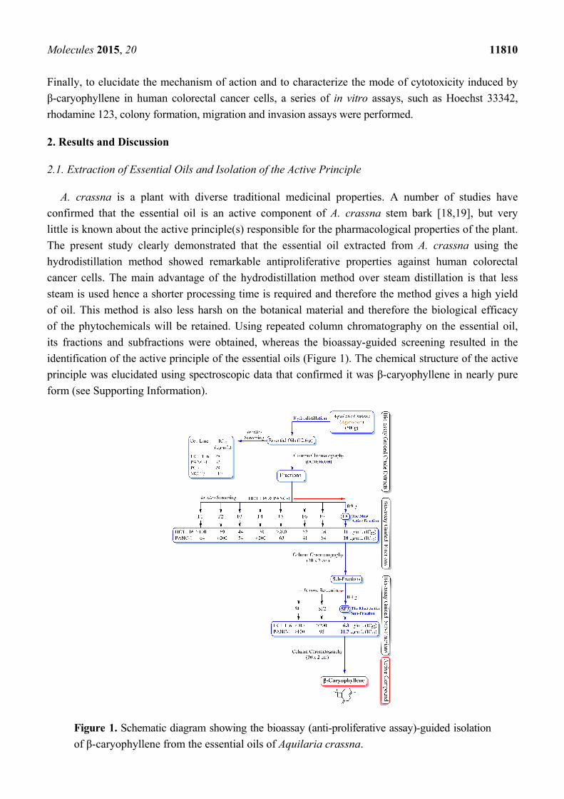
2.1. Extraction of Essential Oils and Isolation of the Active Principle
A. crassna is a plant with diverse traditional medicinal properties. A number of studies have
confirmed that the essential oil is an active component of A. crassna stem bark [18,19], but very
little is known about the active principle(s) responsible for the pharmacological properties of the plant.
The present study clearly demonstrated that the essential oil extracted from A. crassna using the
hydrodistillation method showed remarkable antiproliferative properties against human colorectal
cancer cells. The main advantage of the hydrodistillation method over steam distillation is that less
steam is used hence a shorter processing time is required and therefore the method gives a high yield
of oil. This method is also less harsh on the botanical material and therefore the biological efficacy
of the phytochemicals will be retained. Using repeated column chromatography on the essential oil,
its fractions and subfractions were obtained, whereas the bioassay-guided screening resulted in the
identification of the active principle of the essential oils (Figure 1). The chemical structure of the active
principle was elucidated using spectroscopic data that confirmed it was β-caryophyllene in nearly pure
form (see Supporting Information).
Figure 1. Schematic diagram showing the bioassay (anti-proliferative assay)-guided isolation
of β-caryophyllene from the essential oils of Aquilaria crassna.
Molecules 2015, 20 11811
In the present study, the powdered material (500 g) of A. crassna stem bark was subjected to
hydrodistillation to obtain the essential oil in a yield of 2.52%. The essential oil mixture showed
significant anti-proliferation activity against HCT 116 (IC50 28 µg·mL−1), PANC-1 (IC50 32 µg·mL−1),
PC3 (IC50 79 µg·mL−1) and MCF-7 (IC50 110 µg·mL−1) human cancer cell lines. The mixture of essential
oils was subjected to column chromatography to obtain 12 fractions (F1-F12). Among all the fractions,
fraction 8 (F8) showed most potent activity (HCT 116 IC50 11 µg·mL−1; PANC-1 IC50 18 µg·mL−1;
PC3 IC50 26 µg·mL−1 and MCF-7 IC50 72 µg·mL−1). Further chromatographic separation of the fraction
8 yielded three sub-fractions (SF1-SF3). Among the three sub-fractions, SF3 was found to be the most
active one against the proliferation of HCT 116 (IC50 6.3 µg·mL−1) and PANC-1 (IC50 11.7 µg·mL−1).
Recrystallization of sub-fraction SF3 using hot methanol yielded β-caryophyllene.
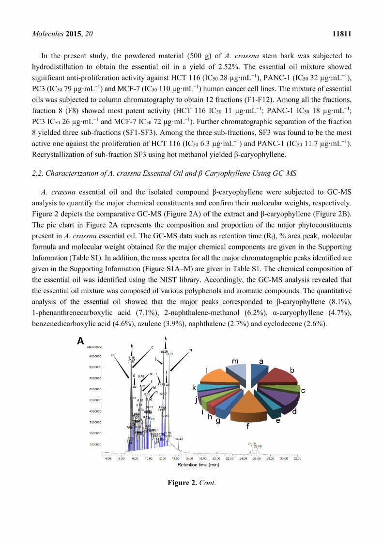
2.2. Characterization of A. crassna Essential Oil and β-Caryophyllene Using GC-MS
A. crassna essential oil and the isolated compound β-caryophyllene were subjected to GC-MS
analysis to quantify the major chemical constituents and confirm their molecular weights, respectively.
Figure 2 depicts the comparative GC-MS (Figure 2A) of the extract and β-caryophyllene (Figure 2B).
The pie chart in Figure 2A represents the composition and proportion of the major phytoconstituents
present in A. crassna essential oil. The GC-MS data such as retention time (Rt), % area peak, molecular
formula and molecular weight obtained for the major chemical components are given in the Supporting
Information (Table S1). In addition, the mass spectra for all the major chromatographic peaks identified are
given in the Supporting Information (Figure S1A–M) are given in Table S1. The chemical composition of
the essential oil was identified using the NIST library. Accordingly, the GC-MS analysis revealed that
the essential oil mixture was composed of various polyphenols and aromatic compounds. The quantitative
analysis of the essential oil showed that the major peaks corresponded to β-caryophyllene (8.1%),
1-phenanthrenecarboxylic acid (7.1%), 2-naphthalene-methanol (6.2%), α-caryophyllene (4.7%),
benzenedicarboxylic acid (4.6%), azulene (3.9%), naphthalene (2.7%) and cyclodecene (2.6%).
Figure 2. Cont.
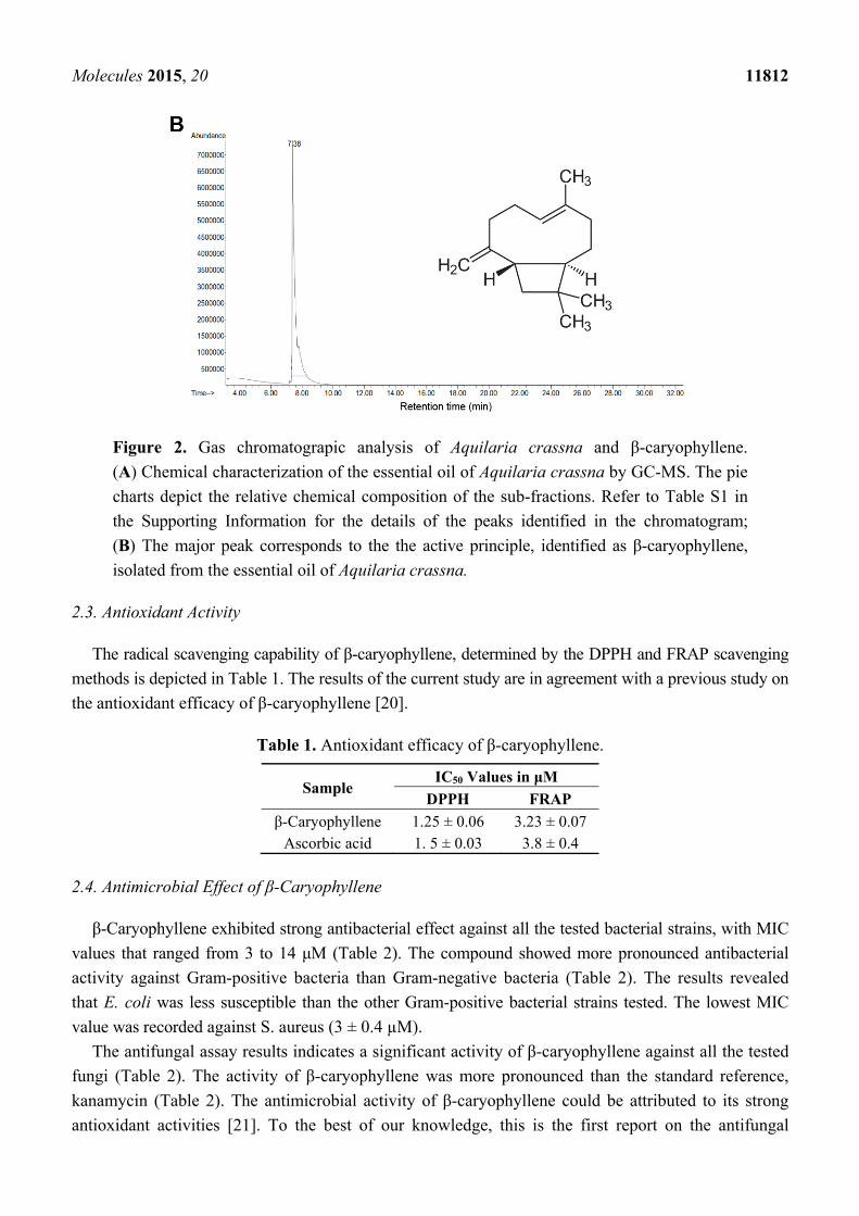
Molecules 2015, 20 11812
Figure 2. Gas chromatograpic analysis of Aquilaria crassna and β-caryophyllene.
(A) Chemical characterization of the essential oil of Aquilaria crassna by GC-MS. The pie
charts depict the relative chemical composition of the sub-fractions. Refer to Table S1 in
the Supporting Information for the details of the peaks identified in the chromatogram;
(B) The major peak corresponds to the the active principle, identified as β-caryophyllene,
isolated from the essential oil of Aquilaria crassna.
2.3. Antioxidant Activity
The radical scavenging capability of β-caryophyllene, determined by the DPPH and FRAP scavenging
methods is depicted in Table 1. The results of the current study are in agreement with a previous study on
the antioxidant efficacy of β-caryophyllene [20].
Table 1. Antioxidant efficacy of β-caryophyllene.
Sample IC50 Values in μM
DPPH FRAP
β-Caryophyllene 1.25 ± 0.06 3.23 ± 0.07 Ascorbic acid 1. 5 ± 0.03 3.8 ± 0.4
2.4. Antimicrobial Effect of β-Caryophyllene
β-Caryophyllene exhibited strong antibacterial effect against all the tested bacterial strains, with MIC
values that ranged from 3 to 14 μM (Table 2). The compound showed more pronounced antibacterial
activity against Gram-positive bacteria than Gram-negative bacteria (Table 2). The results revealed
that E. coli was less susceptible than the other Gram-positive bacterial strains tested. The lowest MIC
value was recorded against S. aureus (3 ± 0.4 µM).
The antifungal assay results indicates a significant activity of β-caryophyllene against all the tested
fungi (Table 2). The activity of β-caryophyllene was more pronounced than the standard reference,
kanamycin (Table 2). The antimicrobial activity of β-caryophyllene could be attributed to its strong
antioxidant activities [21]. To the best of our knowledge, this is the first report on the antifungal
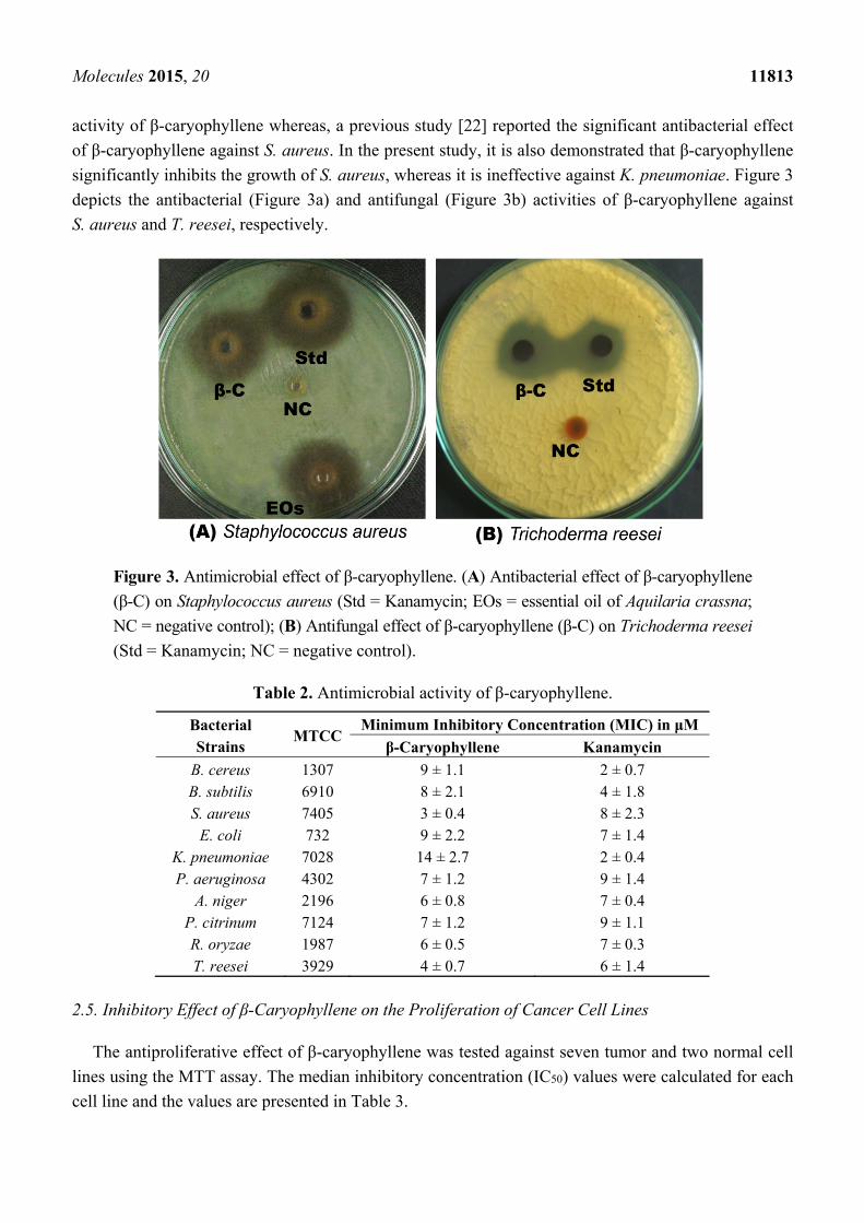
Molecules 2015, 20 11813
activity of β-caryophyllene whereas, a previous study [22] reported the significant antibacterial effect
of β-caryophyllene against S. aureus. In the present study, it is also demonstrated that β-caryophyllene
significantly inhibits the growth of S. aureus, whereas it is ineffective against K. pneumoniae. Figure 3
depicts the antibacterial (Figure 3a) and antifungal (Figure 3b) activities of β-caryophyllene against
S. aureus and T. reesei, respectively.
Figure 3. Antimicrobial effect of β-caryophyllene. (A) Antibacterial effect of β-caryophyllene
(β-C) on Staphylococcus aureus (Std = Kanamycin; EOs = essential oil of Aquilaria crassna;
NC = negative control); (B) Antifungal effect of β-caryophyllene (β-C) on Trichoderma reesei
(Std = Kanamycin; NC = negative control).
Table 2. Antimicrobial activity of β-caryophyllene.
Bacterial Strains
MTCC Minimum Inhibitory Concentration (MIC) in μM
β-Caryophyllene Kanamycin
B. cereus 1307 9 ± 1.1 2 ± 0.7 B. subtilis 6910 8 ± 2.1 4 ± 1.8 S. aureus 7405 3 ± 0.4 8 ± 2.3
E. coli 732 9 ± 2.2 7 ± 1.4 K. pneumoniae 7028 14 ± 2.7 2 ± 0.4 P. aeruginosa 4302 7 ± 1.2 9 ± 1.4
A. niger 2196 6 ± 0.8 7 ± 0.4 P. citrinum 7124 7 ± 1.2 9 ± 1.1 R. oryzae 1987 6 ± 0.5 7 ± 0.3 T. reesei 3929 4 ± 0.7 6 ± 1.4
2.5. Inhibitory Effect of β-Caryophyllene on the Proliferation of Cancer Cell Lines
The antiproliferative effect of β-caryophyllene was tested against seven tumor and two normal cell
lines using the MTT assay. The median inhibitory concentration (IC50) values were calculated for each
cell line and the values are presented in Table 3.
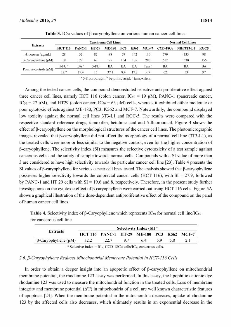
Molecules 2015, 20 11814
Table 3. IC50 values of β-caryophyllene on various human cancer cell lines.
Extracts Carcinoma Cell Lines Normal Cell Lines
HCT 116 PANC-1 HT-29 ME-180 PC3 K562 MCF-7 CCD-18Co NIH/3T3-L1 RGC5
A. crassna (µg/mL) 28 32 82 98 79 142 110 579 133 98
β-Caryophyllene (µM) 19 27 63 95 104 105 285 612 530 156
Positive controls (µM) 5-FU a BA b 5-FU BA BA BA Tam c BA BA BA
12.7 19.4 15 37.1 8.4 17.3 9.5 62 53 97
a 5-fluorouracil; b betulinic acid; c tamoxifen.
Among the tested cancer cells, the compound demonstrated selective anti-proliferative effect against
three cancer cell lines, namely HCT 116 (colon cancer, IC50 = 19 µM), PANC-1 (pancreatic cancer,
IC50 = 27 µM), and HT29 (colon cancer, IC50 = 63 µM) cells, whereas it exhibited either moderate or
poor cytotoxic effects against ME-180, PC3, K562 and MCF-7. Noteworthily, the compound displayed
low toxicity against the normal cell lines 3T3-L1 and RGC-5. The results were compared with the
respective standard reference drugs, tamoxifen, betulinic acid and 5-fluorouracil. Figure 4 shows the
effect of β-caryophyllene on the morphological structures of the cancer cell lines. The photomicrographic
images revealed that β-caryophyllene did not affect the morphology of a normal cell line (3T3-L1), as
the treated cells were more or less similar to the negative control, even for the higher concentration of
β-caryophyllene. The selectivity index (SI) measures the selective cytotoxicity of a test sample against
cancerous cells and the safety of sample towards normal cells. Compounds with a SI value of more than
3 are considered to have high selectivity towards the particular cancer cell line [23]. Table 4 presents the
SI values of β-caryophyllene for various cancer cell lines tested. The analysis showed that β-caryophyllene
possesses higher selectivity towards the colorectal cancer cells (HCT 116), with SI = 27.9, followed
by PANC-1 and HT 29 cells with SI = 19.6 and 8, respectively. Therefore, in the present study further
investigations on the cytotoxic effect of β-caryophyllene were carried out using HCT 116 cells. Figure 5A
shows a graphical illustration of the dose-dependent antiproliferative effect of the compound on the panel
of human cancer cell lines.
Table 4. Selectivity index of β-Caryophyllene which represents IC50 for normal cell line/IC50
for cancerous cell line.
Extracts Selectivity Index (SI) a
HCT 116 PANC-1 HT-29 ME-180 PC3 K562 MCF-7
β-Caryophyllene (µM) 32.2 22.7 9.7 6.4 5.9 5.8 2.1 a Selective index = IC50 CCD-18Co cells/IC50 cancerous cells.
2.6. β-Caryophyllene Reduces Mitochondrial Membrane Potential in HCT-116 Cells
In order to obtain a deeper insight into an apoptotic effect of β-caryophyllene on mitochondrial
membrane potential, the rhodamine 123 assay was performed. In this assay, the lipophilic cationic dye
rhodamine 123 was used to measure the mitochondrial function in the treated cells. Loss of membrane
integrity and membrane potential (∆Ψ) in mitochondria of a cell are well known characteristic features
of apoptosis [24]. When the membrane potential in the mitochondria decreases, uptake of rhodamine
123 by the affected cells also decreases, which ultimately results in an exponential decrease in the
Molecules 2015, 20 11815
fluorescence signal. Results of the present study revealed that the untreated cells displayed a strong
fluorescence intensity indicating their unaffected health with good growth and cell proliferation
(Figure 5B). On the other hand, the fluorescence signal decreased remarkably in the cells treated with
10 μM β-caryophyllene (Figure 5C,D), which suggests a reduced membrane potential due to the
disrupted mitochondrial membrane. In addition, a time-dependent apoptotic effect of β-caryophyllene
was recorded as the intensity of fluorescence signal dropped upon duration of the treatment (Figure 5C,D).
A similar effect can be seen in the cells treated with the standard drug 5-flourouracil (Figure 5E). The
apoptotic index estimated for β-caryophyllene treatment on HCT 116 cells after 24 h treatment was
64 ± 0.04 (Figure 5F). The results can be compared in Figure 5F with those of the standard reference
5-fluorouracil. The results provide additional information about the apoptotic properties of
β-caryophyllene, and suggest that the apoptosis induced by β-caryophyllene in HCT 116 cells occurs via
both the DNA and mitochondrial pathways.
Figure 4. Effect of β-caryophyllene on the cellular morphology of human cancer and
normal cell lines. Photomicrographic images of cancer cell lines, taken under an inverted
phase-contrast microscope at 200× magnification using a digital camera at 48 h after treatment
with β-caryophyllene.
Molecules 2015, 20 11816
Figure 5. (A) Dose-dependent anti-proliferative effect of β-caryophyllene on, HCT 116,
PANC-1, HT-29, MCF-7, PC3, K562, ME-180 and NIH/3T3-L1 cell lines was assessed
by MTT-assay (values are represented as mean ± SD, n = 3); (B) Rhodamine 123 stained
photomicrographic images of HCT 116 cells treated with vehicle (0.1% DMSO);
(C) Rhodamine 123 stained photomicrographic images of HCT 116 cells treated with
β-caryophyllene (10 µM) for 6 h; (D) Rhodamine 123 stained photomicrographic images
of HCT 116 cells treated with β-caryophyllene (10 µM) for 12 h; (E) Rhodamine 123
stained photomicrographic images of HCT 116 cells treated with 5-flourouracil (10 µM)
for 12 h; (F) Graphical representation of percentage of apoptotic indices. The apoptotic
index for each test group was expressed as a percentage of the ratio of number of unstained
cells to the total number of cell in 10 different microscopic fields. Values are presented as
mean ± SD (n = 10), ** represents p < 0.01.
Molecules 2015, 20 11817
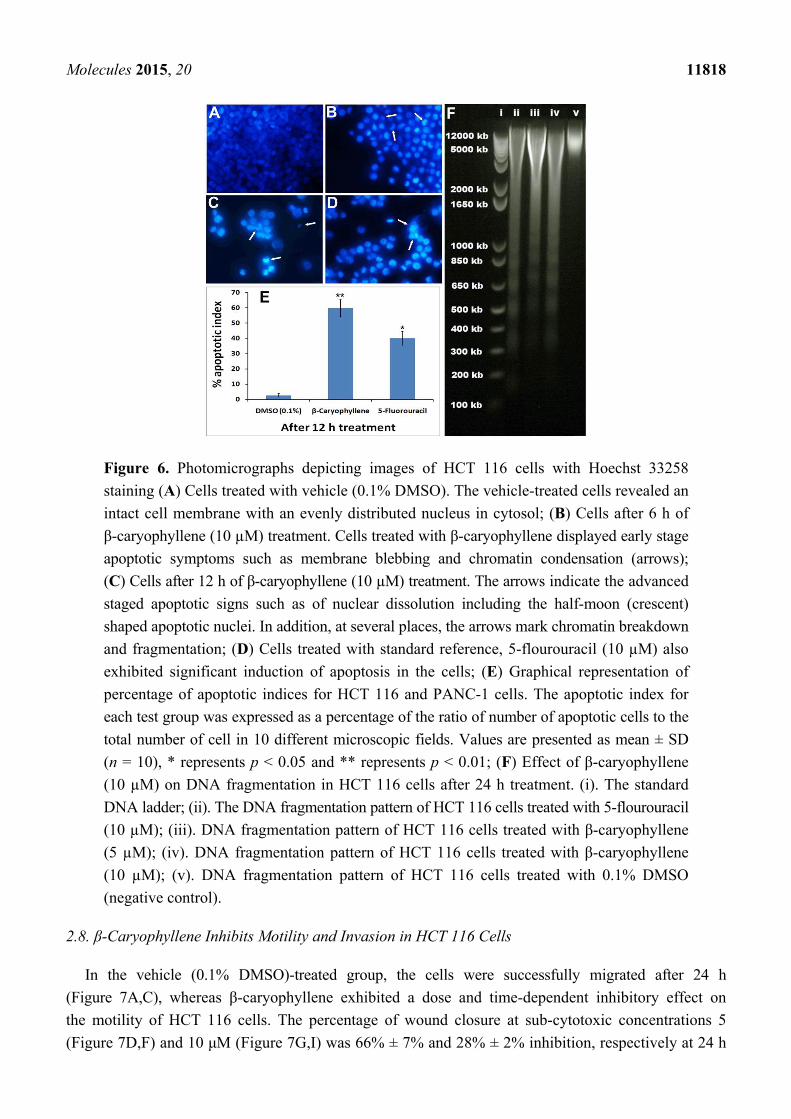
2.7. β-Caryophyllene Induces Chromatin Condensation and DNA Fragmentation in HCT 116 Cells
HCT 116 cells were selected to study the effect of β-caryophyllene on nuclear changes and
condensation using Hoechst 33342 stain. The vehicle-treated cells showed aggressively growing cells
with prominent nuclei (Figure 6A), whereas, the characteristic signs of apoptosis were noticed in the
morphology of the nuclei of the β-caryophyllene-treated cells. β-Caryophyllene affected the nuclear
morphology in a time-dependent manner (Figure 6B,C). β-Caryophyllene at 10 μM concentration,
caused significant nuclei condensation (arrows) after 6 h of treatment (Figure 6B). This was clearly
evidenced by lumping and squeezing of nuclear material to give irregularly distributed chromatin in the
cytosol of the cells, whereas, at a later stage of treatment (after 12 h) the nuclear material is converted
to shrunken and crescent-shaped structures (arrows), indicating the advanced features of apoptosis
(Figure 6C). At this stage, most of the treated cells showed discrete chromatin bodies suggesting the
induction of karyorrhexis caused by β-caryophyllene. These results were compared with the standard
drug, 5-fluorouracil (Figure 6D). β-Caryophyllene displayed more pronounced effects compared to
the standard reference drug. The apoptotic indices (Figure 6E) for untreated HCT 116 cells were
1.7% ± 0.04%. However, following the treatment with β-caryophyllene for 24 h, the apoptotic indices
against HCT 116 cells were significantly increased to 58.4% ± 7% (Figure 6E).
It is well studied phenomenon that apoptotic cells go through a series of morphological
changes namely, membrane blebbing, chromatin condensation, nuclear membrance disruption, nuclear
fragmentation and dissolution [25]. Recently, a study reported that β-caryophyllene induces apoptosis
in a mouse blood cancer cell line through caspase-3 induction [26]. Similarly, another study reported
that a derivative of β-caryophyllene, β-caryophyllene oxide, induces apoptosis through suppression of
PI3K/AKT/mTOR/S6K1 pathways and ROS-mediated MAPKs activation [27]. In the present study,
β-caryophyllene demonstrated clear signs of apoptosis in human colorectal carcinoma (HCT 116) cells.
In order to investigate the effect of β-caryophyllene on advanced stage of apoptosis, isolated DNA
from β-caryophyllene-treated HCT 116 cells was analyzed by agarose gel electrophoresis. The result
showed an obvious DNA fragmentation caused by β-caryophyllene as a dose-dependent laddering
pattern was observed (Figure 6F). The findings of the present study coincide with those of Amiel et al. [26],
who reported a DNA fragmentation effect of β-caryophyllene in a mouse lymphoma cell line. However,
for the first time, the present study demonstrated induction of nuclear condensation and fragmentation by
β-caryophyllene in human cancer cells. The study of Amiel [26] reported that β-caryophyllene activates
the caspase-3 enzyme. Caspase-3 is an executioner enzyme in a caspase-dependent apoptosis cascade.
Induction of caspase-3 activity in turn leads to chromatin condensation, degradation and dissolution.
In the present study these facts were further confirmed and validated by Hoechst 33342 assay in
β-caryophyllene-treated human colorectal cancer cells.
Molecules 2015, 20 11818
Figure 6. Photomicrographs depicting images of HCT 116 cells with Hoechst 33258
staining (A) Cells treated with vehicle (0.1% DMSO). The vehicle-treated cells revealed an
intact cell membrane with an evenly distributed nucleus in cytosol; (B) Cells after 6 h of
β-caryophyllene (10 µM) treatment. Cells treated with β-caryophyllene displayed early stage
apoptotic symptoms such as membrane blebbing and chromatin condensation (arrows);
(C) Cells after 12 h of β-caryophyllene (10 µM) treatment. The arrows indicate the advanced
staged apoptotic signs such as of nuclear dissolution including the half-moon (crescent)
shaped apoptotic nuclei. In addition, at several places, the arrows mark chromatin breakdown
and fragmentation; (D) Cells treated with standard reference, 5-flourouracil (10 µM) also
exhibited significant induction of apoptosis in the cells; (E) Graphical representation of
percentage of apoptotic indices for HCT 116 and PANC-1 cells. The apoptotic index for
each test group was expressed as a percentage of the ratio of number of apoptotic cells to the
total number of cell in 10 different microscopic fields. Values are presented as mean ± SD
(n = 10), * represents p < 0.05 and ** represents p < 0.01; (F) Effect of β-caryophyllene
(10 µM) on DNA fragmentation in HCT 116 cells after 24 h treatment. (i). The standard
DNA ladder; (ii). The DNA fragmentation pattern of HCT 116 cells treated with 5-flourouracil
(10 µM); (iii). DNA fragmentation pattern of HCT 116 cells treated with β-caryophyllene
(5 µM); (iv). DNA fragmentation pattern of HCT 116 cells treated with β-caryophyllene
(10 µM); (v). DNA fragmentation pattern of HCT 116 cells treated with 0.1% DMSO
(negative control).
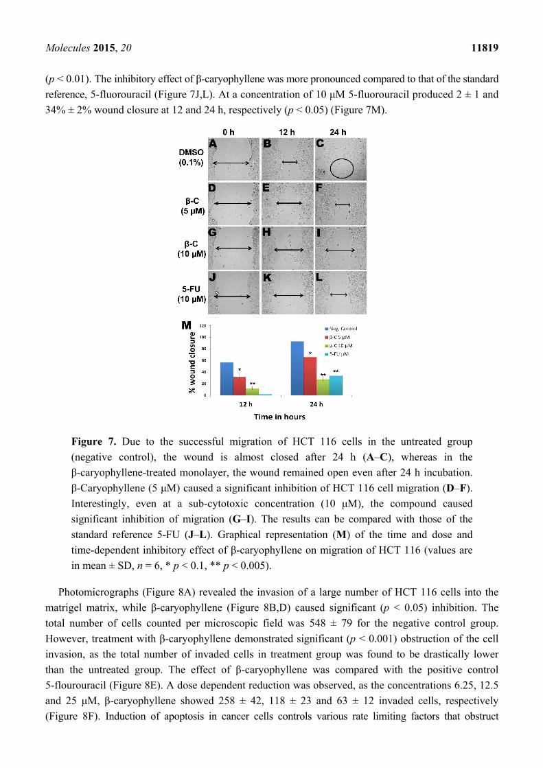
2.8. β-Caryophyllene Inhibits Motility and Invasion in HCT 116 Cells
In the vehicle (0.1% DMSO)-treated group, the cells were successfully migrated after 24 h
(Figure 7A,C), whereas β-caryophyllene exhibited a dose and time-dependent inhibitory effect on
the motility of HCT 116 cells. The percentage of wound closure at sub-cytotoxic concentrations 5
(Figure 7D,F) and 10 μM (Figure 7G,I) was 66% ± 7% and 28% ± 2% inhibition, respectively at 24 h
Molecules 2015, 20 11819
(p < 0.01). The inhibitory effect of β-caryophyllene was more pronounced compared to that of the standard
reference, 5-fluorouracil (Figure 7J,L). At a concentration of 10 μM 5-fluorouracil produced 2 ± 1 and
34% ± 2% wound closure at 12 and 24 h, respectively (p < 0.05) (Figure 7M).
Figure 7. Due to the successful migration of HCT 116 cells in the untreated group
(negative control), the wound is almost closed after 24 h (A–C), whereas in the
β-caryophyllene-treated monolayer, the wound remained open even after 24 h incubation.
β-Caryophyllene (5 μM) caused a significant inhibition of HCT 116 cell migration (D–F).
Interestingly, even at a sub-cytotoxic concentration (10 μM), the compound caused
significant inhibition of migration (G–I). The results can be compared with those of the
standard reference 5-FU (J–L). Graphical representation (M) of the time and dose and
time-dependent inhibitory effect of β-caryophyllene on migration of HCT 116 (values are
in mean ± SD, n = 6, * p < 0.1, ** p < 0.005).
Photomicrographs (Figure 8A) revealed the invasion of a large number of HCT 116 cells into the
matrigel matrix, while β-caryophyllene (Figure 8B,D) caused significant (p < 0.05) inhibition. The
total number of cells counted per microscopic field was 548 ± 79 for the negative control group.
However, treatment with β-caryophyllene demonstrated significant (p < 0.001) obstruction of the cell
invasion, as the total number of invaded cells in treatment group was found to be drastically lower
than the untreated group. The effect of β-caryophyllene was compared with the positive control
5-flourouracil (Figure 8E). A dose dependent reduction was observed, as the concentrations 6.25, 12.5
and 25 μM, β-caryophyllene showed 258 ± 42, 118 ± 23 and 63 ± 12 invaded cells, respectively
(Figure 8F). Induction of apoptosis in cancer cells controls various rate limiting factors that obstruct
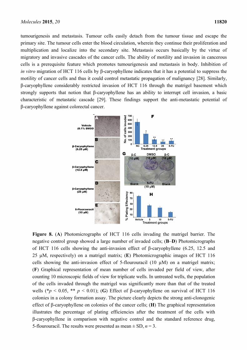
Molecules 2015, 20 11820
tumourigenesis and metastasis. Tumour cells easily detach from the tumour tissue and escape the
primary site. The tumour cells enter the blood circulation, wherein they continue their proliferation and
multiplication and localize into the secondary site. Metastasis occurs basically by the virtue of
migratory and invasive cascades of the cancer cells. The ability of motility and invasion in cancerous
cells is a prerequisite feature which promotes tumourigenesis and metastasis in body. Inhibition of
in vitro migration of HCT 116 cells by β-caryophyllene indicates that it has a potential to suppress the
motility of cancer cells and thus it could control metastatic propagation of malignancy [28]. Similarly,
β-caryophyllene considerably restricted invasion of HCT 116 through the matrigel basement which
strongly supports that notion that β-caryophyllene has an ability to interrupt cell invasion, a basic
characteristic of metastatic cascade [29]. These findings support the anti-metastatic potential of
β-caryophyllene against colorectal cancer.
Figure 8. (A) Photomicrographs of HCT 116 cells invading the matrigel barrier. The
negative control group showed a large number of invaded cells; (B‒D) Photomicrographs
of HCT 116 cells showing the anti-invasion effect of β-caryophyllene (6.25, 12.5 and
25 µM, respectively) on a matrigel matrix; (E) Photomicrographic images of HCT 116
cells showing the anti-invasion effect of 5-flourouracil (10 µM) on a matrigel matrix;
(F) Graphical representation of mean number of cells invaded per field of view, after
counting 10 microscopic fields of view for triplicate wells. In untreated wells, the population
of the cells invaded through the matrigel was significantly more than that of the treated
wells (*p < 0.05, ** p < 0.01); (G) Effect of β-caryophyllene on survival of HCT 116
colonies in a colony formation assay. The picture clearly depicts the strong anti-clonogenic
effect of β-caryophyllene on colonies of the cancer cells; (H) The graphical representation
illustrates the percentage of plating efficiencies after the treatment of the cells with
β-caryophyllene in comparison with negative control and the standard reference drug,
5-flourouracil. The results were presented as mean ± SD, n = 3.
Molecules 2015, 20 11821
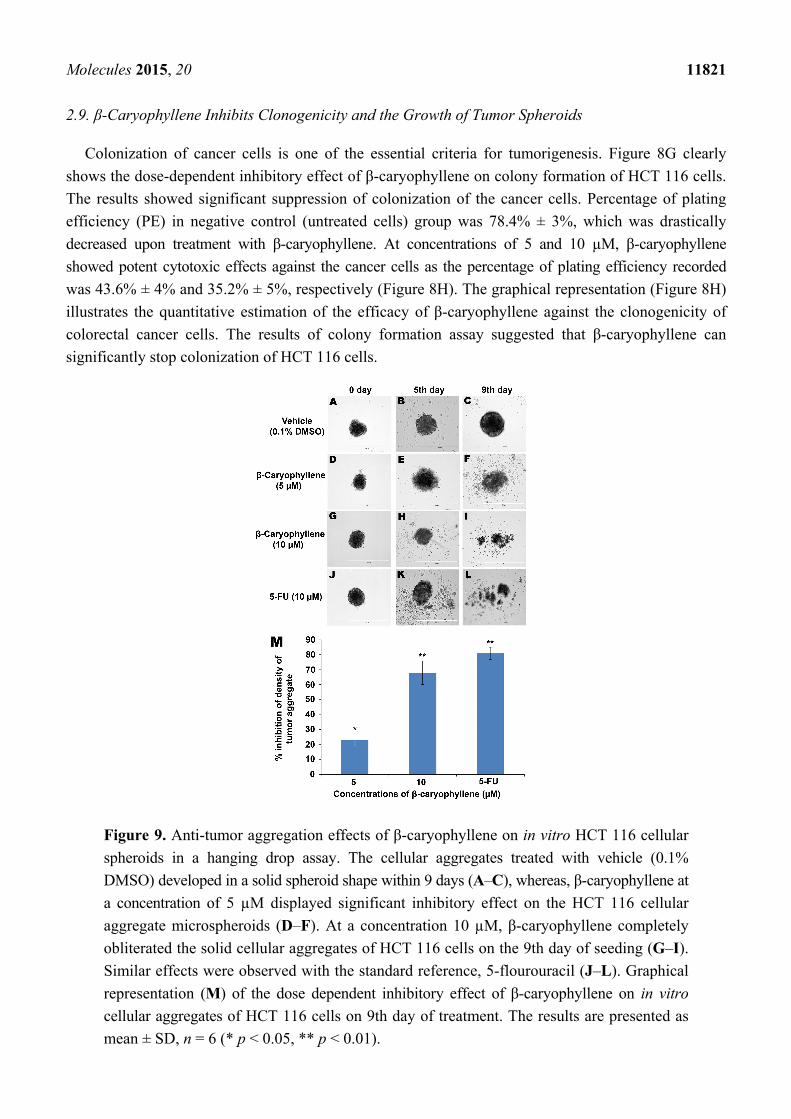
2.9. β-Caryophyllene Inhibits Clonogenicity and the Growth of Tumor Spheroids
Colonization of cancer cells is one of the essential criteria for tumorigenesis. Figure 8G clearly
shows the dose-dependent inhibitory effect of β-caryophyllene on colony formation of HCT 116 cells.
The results showed significant suppression of colonization of the cancer cells. Percentage of plating
efficiency (PE) in negative control (untreated cells) group was 78.4% ± 3%, which was drastically
decreased upon treatment with β-caryophyllene. At concentrations of 5 and 10 µM, β-caryophyllene
showed potent cytotoxic effects against the cancer cells as the percentage of plating efficiency recorded
was 43.6% ± 4% and 35.2% ± 5%, respectively (Figure 8H). The graphical representation (Figure 8H)
illustrates the quantitative estimation of the efficacy of β-caryophyllene against the clonogenicity of
colorectal cancer cells. The results of colony formation assay suggested that β-caryophyllene can
significantly stop colonization of HCT 116 cells.
Figure 9. Anti-tumor aggregation effects of β-caryophyllene on in vitro HCT 116 cellular
spheroids in a hanging drop assay. The cellular aggregates treated with vehicle (0.1%
DMSO) developed in a solid spheroid shape within 9 days (A–C), whereas, β-caryophyllene at
a concentration of 5 µM displayed significant inhibitory effect on the HCT 116 cellular
aggregate microspheroids (D–F). At a concentration 10 µM, β-caryophyllene completely
obliterated the solid cellular aggregates of HCT 116 cells on the 9th day of seeding (G–I).
Similar effects were observed with the standard reference, 5-flourouracil (J–L). Graphical
representation (M) of the dose dependent inhibitory effect of β-caryophyllene on in vitro
cellular aggregates of HCT 116 cells on 9th day of treatment. The results are presented as
mean ± SD, n = 6 (* p < 0.05, ** p < 0.01).
Molecules 2015, 20 11822
In a hanging drop environment, tumour cells aggregate and sediment in the cultures due to the applied
gravitational force on the suspended cells. In the hanging drop assay, the cells were allowed to sediment
and aggregate and later the cellular aggregates were harvested and seeded onto an agar-coated plate.
The cellular aggregates from the hanging drop adopt a three-dimensional spheroid shape on the agar
plate which leads to transformation of suspended cells into cellular aggregates which ultimately from
a solid spheroid shape within the first 24 h (negative control, Figure 9A,C). Figure 9D,F illustrate the
inhibitory effect of β-caryophyllene (5 µM) on the growth of aggregates and spheroids of HCT 116
cells. A stronger inhibitory effect of β-caryophyllene (10 µM) on tumour spheroids was recorded and
is depicted in Figure 9G,I.
Initially, the cellular aggregates were prepared from 20 µL hanging drops (5000 cells/drop) which
later on developed into a thick and dense tumor-shaped spheroid within 24 h as shown in the negative
control group (Figure 9A,C). β-Caryophyllene displayed a dose-dependent inhibitory effect on the
HCT 116 tumour spheroids. At concentrations of 5 and 10 µM, β-caryophyllene produced significant
inhibitory effects of 23% ± 4% and 68% ± 8% on the density of the tumour spheroids, respectively
(Figure 9D,I). The results are compared with that of the standard reference, 5-Fluorouracil which
showed 81% ± 4% inhibition at 10 µM concentration (Figure 9J,L). Figure 9M illustrates the
quantitative data of dose-dependent effect of β-caryophyllene in comparison with 5-fluorouracil. The
inhibition of cellular aggregation is directly associated with the significant anti-proliferation effect on
HCT 116 cells. The MTT assay in the present study supports the findings of the tumour spheroid assay.
3. Experimental Section
3.1. Chemicals and Reagents
Growth medium (RPMI 1640, Dulbecco’s Modified Eagle Medium, McCoy’s 5a modified medium
and F-12K medium), trypsin and heat inactivated foetal bovine serum (HIFBS) were purchased from
GIBCO (Paisley, UK). Methylthiazolyldiphenyltetrazolium bromide (MTT) reagent, phosphate buffer
saline (PBS) and penicillin/streptomycin (PS) were purchased from Sigma-Aldrich (Darmstadt,
Germany). Cell culture grade dimethyl sulfoxide (DMSO) was procured from Fluka (St. Louis, MO,
USA). F-12K medium was obtained from ATCC (Rockville, MD, USA). All the reagents and solvents
used were analytical grade. For biological assays, a stock solution (10 mM) of β-caryophyllene was
prepared using DMSO. Further, various concentrations (3 to 100 µM) of β-caryophyllene were prepared
by serially diluting the stock with respective culture medium.
3.2. Plants Material
Flowers, twigs and stem bark of A. crassna was collected from a local farm in Kajang,
Selangor, Malaysia in 2013. Floral characteristics and taxonomical identification was confirmed by
Mr. V. Shanmogan, a senior taxonomist from School of Biological Sciences, Universti Sains Malaysia.
A herbarium sample (Ref. No. USM/122083) was submitted at the Department of Botany, School of
Biological Sciences, UniversitiSains Malaysia, Pulau Pinang, Malaysia. In addition, the plant name has
been checked and confirmed with The Plant List by the International Botanic Gardens [30].
Molecules 2015, 20 11823
3.3. Extraction of Essential Oils from A. crassna Bark
Fresh sample of stem bark of A. crassna was collected and rinsed with water. The covering layer of
the bark composed of dead tissues was removed. The heartwood was sliced into small pieces and grinded
mechanically. The powdered material of bark (500 g) was soaked in a round-bottom flask with 5 L of
water at room temperature (25 ± 2 °C) for 5 days. Then, hydrodistillation was carried out at the boiling
temperature of water for 48 h. The essential oils were collected for 12 h by a modified Clevenger-type
apparatus, yielding a pale-yellow liquid (about 12.6 g).
3.4. Bioassay Guided Isolation of β-Caryophyllene
The obtained essential oil mixture of A. crassna was subjected to repeated chromatographic
separation. Anti-proliferation (MTT) assay was used as the bioassay to guide the isolation of the
active principle of the essential oil. The details of the chromatographic conditions and fractionation are
given in the Supporting Information. The active compound was obtained as a brown-colored crystalline
compound which was washed with n-hexane several times and recrystallized from hot methanol to
obtain colourless β-caryophyllene (0.4 g, 0.2%).
3.5. Chemical Characterization Techniques
The structure of β-caryophyllene was elucidated using FT-IR and NMR spectral studies. Refer to
the Supporting Information for detailed experimental conditions of the instruments (Figure S2).
3.6. Gas Chromatography-Mass (GC-MS) Spectral Analysis
Quantitative analysis of composition of the essential oils of A. crassna bark and the isolated compound,
β-caryophyllene was carried out using GC-MS with the aim of comparingthe chemical profile of the
crude extract and the pure compound. A detailed description of the assay conditions is given in the
Supporting Information.
3.7. Antioxidant Activity of β-caryophyllene
3.7.1. DPPH Radical Scavenging Activity
The antioxidant activity of β-caryophyllene was assessed based on the radical scavenging effect of
stable 2,2-diphenyl-1-picrylhydrazyl (DPPH) free radical activity according to the method described
previously [31].
3.7.2. Ferric Reducing Antioxidant Power (FRAP) Assay
The FRAP assay was conducted according to the method described before [32]. The results were
expressed as μM·Fe2+·mg−1. All measurements were carried out in triplicate and the mean values
were calculated.
Molecules 2015, 20 11824
3.8. Microbial Strains
Six human pathogenic bacterial strains were procured from the Microbial Type Culture Collection
Centre and Gene Bank (MTCC, Chandigarh, India). The strains used were Bacillus cereus (MTCC
1307), Bacillus subtilis (MTCC 6910), Escherichia coli (MTCC 732), Klebsiella pneumonia (MTCC
7028), Staphylococcus aureus (MTCC 7405) and Pseudomonas aeruginosa (MTCC 4302). Two fungal
strains, Rhizopus oryzae (MTCC 1987) and Tricho dermareesei (MTCC 3929). All the bacterial strains
were cultured on Mueller-Hinton agar medium at 37 °C and the fungal strains on Sabouraud dextrose
agar medium at 28 °C.
3.9. Antimicrobial Assay
The antimicrobial activity of β-caryophyllene was determined by the disk diffusion method [33].
Refer to the Supporting information for the detailed methodology. Further, serial tube dilution
technique [34,35] was used to determine MIC of β-caryophyllene.
3.10. Anticancer Assays
3.10.1. Cell Lines and Culture Conditions
Panel of human cancer cells such as, pancreatic (PANC-1), colorectal (HCT-116 and HT-29), invasive
squamous cell carcinoma (ME-180), leukemia (K562), hormone sensitive and invasive breast cancer
cell line (MCF-7), and prostatic (PC3) adenocarcinoma cell lines were used. In addition, two normal
cell lines, mouse fibroblast (NIH/3T3-L1), and human retinal ganglion cell line (RGC-5) were used as
the model cell lines for normal cells. All the cell lines were purchased from ATCC. HCT 116,
HT-29 and K562 cells were maintained in RPMI; MCF-7 cells were maintained in DMEM; ME-180
cells were maintained in McCoy’s 5a modified medium and PC3 was maintained in F-12K medium.
All the media were additionally supplemented with 5% HIFBS and 1% PS. Cells were incubated in
a humidified CO2 incubator at 37 °C supplied with 5% CO2.
3.10.2. In Vitro Cytotoxic Assay
Inhibitory effect of β-caryophyllene on proliferation of the cell lines was tested using the MTT
assay [36–38]. The selectivity index (SI) for the cytotoxicity of β-caryophyllene was calculated using
the ratio of IC50 of the compound on a normal cell line (NIH-3T3) to the IC50 of the compound on cancer
cell lines.
3.10.3. Chromatin Condensation Assay using Hoechst 33342 Stain
Effect of β-caryophyllene on nuclear chromatin condensation in HCT 116 cells was assessed suing
Hoechst 33258 staining [39] and quantified by fluorescence microscopy. Refer to the Supporting
Information for the detailed methodology.
Molecules 2015, 20 11825
3.10.4. DNA Fragmentation Assay
The assay was performed according to the method as described previously [39], with minor
modifications. Refer to the Supporting Information for the detailed methodology.
3.10.5. Rhodamin 123 Stain Assay
The effect of β-caryophyllene on mitochondrial membrane potential in treated-HCT 116 cells was
studied and quantified by a rhodamine 123 assay [40]. Refer to the Supporting Information for the
detailed methodology.
3.10.6. Anti-Migration (Motility) Assay
The effect of β-caryophyllene on cellular migration (motility) was assessed on HCT 116 cells [41].
Refer to the Supporting Information for the detailed methodology.
3.10.7. Cell Invasion
The effect of β-caryophyllene on HCT 116 cellular invasion was assessed using matrigel as an
artificial basement membrane matrix following a previously described method [42]. Refer to the
Supporting Information for the detailed methodology.
3.10.8. Spheroid-Based in Vitro Anti-Tumor Assay
The effect of β-caryophyllene was tested on micro-spheroids (hanging drops) of HCT 116 cells. The
micro-spheroids in this assay represents in vitro tumor and the assay was performed according to the
previously described method [43]. Refer to the Supporting Information for the detailed methodology.
3.10.9. Colony Formation Assay
The effect of β-caryophyllene on the clonogenicity of HCT 116 cells was investigated by a colony
formation assay [42]. Refer to the Supporting Information for the detailed methodology.
3.11. Statistical Analysis
Statistical analysis was performed by one-way analysis of variance (ANOVA) followed by Tukey’s
multiple comparison test with the help of IBM SPSS software (Version 20). Statistical significance
were considered at p < 0.05 and p < 0.01 and were indicated as * and **, respectively.
4. Conclusions
The findings of the present work provide good evidence for β-caryophyllene being an active
principle of A. crassna essential oil. The present study concludes that in particular β-caryophyllene
could be a potential source of selective antifungal agents. Principally, from the findings of the present
study, it can be concluded that β-caryophyllene has strong selective cytotoxic properties against human
colorectal cancer cells. In addition, the results proved that the cytotoxicity induced by β-caryophyllene
Molecules 2015, 20 11826
can be attributed to its apoptotic properties via DNA fragmentation and mitochondrial pathways. Further
studies indicate that β-caryophyllene has promising capability to suppress tumour motility, cell invasion
and tumour aggregation.
Supplementary Materials
Supplementary materials can be accessed at: http://www.mdpi.com/1420-3049/20/07/11808/s1.
Acknowledgments
The first author Saad S. Dahham wishes to acknowledge Universiti Sains Malaysia (USM) for
a USM Ph.D fellowship. The authors gratefully acknowledge the financial support from the
Malaysian Ministry of Agriculture under NRGS grants No: 304/PFARMASI/650583/K123 and
304/PFARMASI/650737/K123. The authors also wish to acknowledge Universiti Sains Malaysia
(USM) for the Research University Team (RUT) Grant No.: 1001/PFARMASI/851001. Muhammad
Adnan Iqbal is thankful to USM for a postdoctoral research fellowship.
Author Contributions
S.S.D., A.M.S.A.M. and M.B.K.A. designed the experiments. S.S.D. and Y.M.T. carried out the
phytochemical and in vitro biological experiments. M.O.E. and M.A.I. performed the structural
elucidation studies. S.S.D., A.M.S.A.M. and M.B.K.A. interpreted the results. S.S.D., M.A.I., M.B.K.A.
and A.S.A.M. drafted the manuscript. All the authors read and approved the final manuscript.
Conflicts of Interest
The authors declare no conflict of interest.
References and Notes
1. Chen, H.; Yang, Y.; Xue, J.; Wei, J.; Zhang, Z.; Chen, H. Comparison of compositions and
antimicrobial activities of essential oils from chemically stimulated agarwood, wild agarwood and
healthy Aquilaria sinensis (Lour.) Gilg trees. Molecules 2011, 16, 4884–4896.
2. Okugawa, H.; Ueda, R.; Matsumoto, K.; Kawanishi, K.; Kato, A. Effects of agarwood extracts on
the central nervous system in mice. Planta Med. 1993, 59, 32–36.
3. Okugawa, H.; Ueda, R.; Matsumoto, K.; Kawanishi, K.; Kato, A. Effect of jinkoh-eremol and
agarospirol from agarwood on the central nervous system in mice. Planta Med. 1996, 62, 2–6.
4. Suvitayavat, W.; Tunglert, S.; Thirawarapan, S.; Bunyapraphatsara, N. Effects of Ya-hom on blood
pressure in rats. J. Ethnopharmacol. 2005, 97, 503–508.
5. Kumar, V. Potential medicinal plants for CNS disorders: An overview. Phytother. Res. 2006, 20,
1023–1035.
6. Sattayasai, J.; Bantadkit, J.; Aromdee, C.; Lattmann, E.; Airarat, W. Antipyretic, analgesic and
anti-oxidative activities of Aquilaria crassna leaves extract in rodents. J. Ayurveda Integr. Med.
2012, 3, 175–179.
Molecules 2015, 20 11827
7. Jermsri, P.; Jiraviriyakul, A.; Unajak, S.; Kumphune, S. Effect of Aquilaria crassna crude extract
on simulated ischemia induced cardiac cell death. Int. J. Pharm. Biol. Sci. 2012, 3, 604–613.
8. Kamonwannasit, S.; Nantapong, N.; Kumkrai, P.; Luecha, P.; Kupittayanant, S.; Chudapongse, N.
Antibacterial activity of Aquilaria crassna leaf extract against Staphylococcus epidermidis by
disruption of cell wall. Ann. Clin. Microbiol. Antimicrob. 2013, 12, 20, doi:10.1186/1476-0711-12-20.
9. Novriyanti, E.; Santosa, E. the role of phenolics in agarwood formation of Aquilaria crassna
Pierre ex Lecomte AND Aquilaria microcarpa Baill Trees. Indones. J. For. Res. 2011, 8, 101–113.
10. Hideaki, H. Agarwood (Aquilaria crassna) extracts decrease high-protein high-fat diet-induced intestinal
putrefaction toxins in mice. Pharm. Anal. Acta 2012, 3, 152, doi:10.4172/2153-2435.1000152.
11. Majid, A.M.S.A.; Dahham, S.S.; Ahamed, M.B.K.; Saghir, S.M.; Alsuede, F.S.; Iqbal, M.A.
Bioactive essential oils from Aquilaria crassna for cancer prevention and treatment. Glob. J. Adv.
Pure Appl. Sci. 2014, 4, 26–31.
12. Kumar, A.R.; Subburathinam, K.; Prabakar, G. Phytochemical screening of selected medicinal
plants of asclepiadaceae family. Asian J. Microbiol. Biotechnol. Environ. Sci. 2007, 9, 177–180.
13. Syed, H.K.; Iqbal, M.A.; Haque, R.A.; Peh, K.K. Synthesis, characterization and antibacterial
activity of a Curcumin-Silver(I) complex. J. Coord. Chem. 2015, 68, 1–13.
14. Hara, H.; Ise, Y.; Morimoto, N.; Shimazawa, M.; Ichihashi, K.; Ohyama, M.; Iinuma, M. Laxative
effect of agarwood leaves and its mechanism. Biosci. Biotechnol. Biochem. 2008, 72, 335–345.
15. Kakino, M.; Izuta, H.; Ito, T.; Tsuruma, K.; Araki, Y.; Shimazawa, M.; Oyama, M.; Iinuma, M.;
Hara, H. Agarwood induced laxative effects via acetylcholine receptors on loperamide-induced
constipation in mice. Biosci. Biotechnol. Biochem. 2010, 74, 1550–1555.
16. Ito, T.; Kakino, M.; Tazawa, S.; Oyama, M.; Maruyama, H.; Araki, Y.; Hara, H.; Iinuma, M.
Identification of Phenolic Compounds in Aquilaria crassna Leaves via Liquid Chromatography-
Electrospray Ionization Mass Spectroscopy. Food Sci. Technol. Res. 2012, 18, 259–262.
17. Chen, H.Q.; Wei, J.H.; Yang, J.S.; Zhang, Z.; Yang, Y.; Gao, Z.H.; Sui, C.; Gong, B. Chemical
constituents of agarwood originating from the endemic genus Aquilaria plants. Chem. Biodivers.
2012, 9, 236–250.
18. Kumphune, S.; Prompunt, E.; Phaebuaw, K.; Sriudwong, P.; Pankla, R.; Thongyoo, P.
Anti-inflammatory effects of the ethyl acetate extract of Aquilaria crassna inhibits LPS-induced
tumour necrosis factor-alpha production by attenuating P38 MAPK activation. Int. J Green Pharm.
2011, 5, 43.
19. Wetwitayaklung, P.; Thavanapong, N.; Charoenteeraboon, J. Chemical constituents and antimicrobial
activity of essential oil and extracts of heartwood of Aquilaria crassna obtained from water
distillation and supercritical fluid carbon dioxide extraction. Silpakorn Univ. Sci. Technol. J.
2009, 3, 25–33.
20. Calleja, M.A.; Vieites, J.M.; Montero-Meterdez, T.; Torres, M.I.; Faus, M.J.; Gil, A.; Suárez, A.
The antioxidant effect of β-caryophyllene protects rat liver from carbon tetrachloride-induced
fibrosis by inhibiting hepatic stellate cell activation. Br. J. Nutr. 2013, 109, 394–401.
21. Dorman, H.; Deans, S. Antimicrobial agents from plants: antibacterial activity of plant volatile
oils. J. Appl. Microbiol. 2000, 88, 308–316.
Molecules 2015, 20 11828
22. Kim, Y.S.; Park, S.J.; Lee, E.J.; Cerbo, R.M.; Lee, S.M.; Ryu, C.H.; Kim, G.S.; Kim, J.O.; Ha, Y.L.
Antibacterial Compounds from Rose Bengal-Sensitized Photooxidation of β-Caryophyllene.
J. Food Sci. 2008, 73, C540–C545.
23. Prayong, P.; Barusrux, S.; Weerapreeyakul, N. Cytotoxic activity screening of some indigenous
Thai plants. Fitoterapia 2008, 79, 598–601.
24. Skommer, J.; Wlodkowic, D.; Deptala, A. Larger than life: Mitochondria and the Bcl-2 family.
Leuk. Res. 2007, 31, 277–286.
25. Bold, R.J.; Termuhlen, P.M.; McConkey, D.J. Apoptosis, cancer and cancer therapy. Surg. Oncol.
1997, 6, 133–142.
26. Amiel, E.; Ofir, R.; Dudai, N.; Soloway, E.; Rabinsky, T.; Rachmilevitch, S. β-Caryophyllene, a
compound isolated from the biblical balm of gilead (Commiphora gileadensis), is a selective
apoptosis inducer for tumor cell lines. Evid. Based Complement. Alternat. Med. 2012, 2012,
doi:10.1155/2012/872394.
27. Park, K.R.; Nam, D.; Yun, H.M.; Lee, S.G.; Jang, H.J.; Sethi, G.; Cho, S.K.; Ahn, K.S.
β-Caryophyllene oxide inhibits growth and induces apoptosis through the suppression of
PI3K/AKT/mTOR/S6K1 pathways and ROS-mediated MAPKs activation. Cancer Lett. 2011,
312, 178–188.
28. Valster, A.; Tran, N.L.; Nakada, M.; Berens, M.E.; Chan, A.Y.; Symons, M. Cell migration and
invasion assays. Methods 2005, 37, 208–215.
29. Hocman, G., Chemoprevention of cancer: phenolic antioxidants (BHT, BHA). Int. J. Biochem.
1988, 20, 639–651.
30. The Plant List. Available online: http://www.theplantlist.org/ (accessed on 29 March 2015)
(Archived by WebCite® at http://www.webcitation.org/6Y9HaiV43).
31. Khadeer Ahamed, M.B.; Krishna, V.; Dandin, C.J. In vitro antioxidant and in vivo prophylactic
effects of two γ-lactones isolated from Grewia tiliaefolia against hepatotoxicity in carbon
tetrachloride intoxicated rats. Eur. J. Pharmacol. 2010, 631, 42–52.
32. Haque, R.; Iqbal, M.; Asekunowo, P.; Majid, A.M.S.A.; Khadeer Ahamed, M.; Umar, M.;
Al-Rawi, S.; Al-Suede, F. Synthesis, structure, anticancer, and antioxidant activity of para-xylyl
linked bis-benzimidazolium salts and respective dinuclear Ag(I) N-heterocyclic carbene complexes
(Part-II). Med. Chem. Res. 2013, 22, 4663–4676.
33. Ahamed, B.; Krishna, V.; Gowdru, H.B.; Rajanaika, H.; Kumaraswamy, H.; Rajshekarappa, S.;
Dandin, C.J.; Mahadevan, K. Isolation of Bactericidal Constituents from the Stem Bark Extract of
Grewia tiliaefolia Vahl. Res. J. Med. Plant. 2007, 1, 72–82.
34. Rahman, M.M.; Mosaddik, M.A.; Wahed, M.I.I.; Haque, M.E. Antimicrobial activity and
cytotoxicity of Trapa bispinosa. Fitoterapia 2000, 71, 704–706.
35. Vinson, J.A.; Dabbagh, Y.A.; Serry, M.M.; Jang, J. Plant flavonoids, especially tea flavonols, are
powerful antioxidants using an in vitro oxidation model for heart disease. J. Agric. Food Chem.
1995, 43, 2800–2802.
36. Mosmann, T. Rapid colorimetric assay for cellular growth and survival: application to proliferation
and cytotoxicity assays. J. Immunol. Methods 1983, 65, 55–63.
Molecules 2015, 20 11829
37. Ahamed, M.B.K.; Aisha, A.F.; Nassar, Z.D.; Siddiqui, J.M.; Ismail, Z.; Omari, S.; Parish, C.;
Majid, A.A., Cat’s whiskers tea (Orthosiphon stamineus) extract inhibits growth of colon tumor
in nude mice and angiogenesis in endothelial cells via suppressing VEGFR phosphorylation.
Nutr. Cancer 2012, 64, 89–99.
38. Iqbal, M.; Haque, R.; Ahamed, S.; Jafari, S.; Khadeer, A.M.; Abdul, M.A. Crystal Structures
and Cytotoxicity of Ortho-Xylene Linked Bis-benzimidazolium Salts. Med. Chem. 2015,
doi:10.2174/1573406411666150101153115.
39. Nassar, Z.D.; Aisha, A.F.; Idris, N.; Ahamed, K.; Mohamed, B.; Ismail, Z.; Abu-Salah, K.M.;
Alrokayan, S.A.; Majid, A.; Shah, A.M. Koetjapic acid, a natural triterpenoid, induces apoptosis
in colon cancer cells. Oncol. Rep. 2012, 27, 727–733.
40. Hassan, L.E.A.; Ahamed, M.B.K.; Majid, A.S.A.; Iqbal, M.A.; Al Suede, F.S.R.; Haque, R.A.;
Ismail, Z.; Ein, O.C.; Majid, A.M.S.A. Crystal Structure Elucidation and Anticancer Studies of
(−)-Pseudosemiglabrin: A Flavanone Isolated from the Aerial Parts of Tephrosia apollinea.
PLoS ONE 2014, 9, e90806.
41. Al-Suede, F.S.R.; Khadeer Ahamed, M.B.; Abdul Majid, A.S.; Baharetha, H.M.; Hassan, L.E.;
Kadir, M.O.A.; Nassar, Z.D.; Abdul Majid, A. Optimization of Cat’s Whiskers Tea (Orthosiphon
stamineus) Using Supercritical Carbon Dioxide and Selective Chemotherapeutic Potential against
Prostate Cancer Cells. Evid. Based Complement. Alternat. Med. 2014, doi:10.1155/2014/396016.
42. Baharetha, H.M.; Nassar, Z.D.; Aisha, A.F.; Ahamed, M.B.K.; Al-Suede, F.S.R.; Kadir, M.O.A.;
Ismail, Z.; Majid, A.M.S.A. Proapoptotic and Antimetastatic Properties of Supercritical CO2
Extract of Nigella sativa Linn. Against Breast Cancer Cells. J. Med. Food 2013, 16, 1121–1130.
43. Jafari, S.F.; Ahamed, K.; Mohamed, B.; Iqbal, M.A.; Al Suede, F.S.R.; Khalid, S.H.; Haque, R.A.;
Nassar, Z.D.; Umar, M.I.; Majid, A. Increased aqueous solubility and proapoptotic activity of
potassium koetjapate against human colorectal cancer cells. J. Pharm. Pharmacol. 2014, 66,
1394–1409.
Sample Availability: Sample of the compound, β-caryophyllene is available from the authors.
© 2015 by the authors; licensee MDPI, Basel, Switzerland. This article is an open access article
distributed under the terms and conditions of the Creative Commons Attribution license
(http://creativecommons.org/licenses/by/4.0/).