pH-Dependent Anticancer Drug Release From Silk Nanoparticles
6
www.advhealthmat.de www.MaterialsViews.com FULL PAPER © 2013 WILEY-VCH Verlag GmbH & Co. KGaA, Weinheim 1 wileyonlinelibrary.com pH-Dependent Anticancer Drug Release from Silk Nanoparticles F. Philipp Seib, Gregory T. Jones, Jelena Rnjak-Kovacina, Yinan Lin, and David L. Kaplan* Silk has traditionally been used as a suture material because of its excellent mechanical properties and biocompatibility. These properties have led to the development of different silk-based material formats for tissue engineering and regenerative medicine. Although there have been a small number of studies about the use of silk particles for drug delivery, none of these studies have assessed the potential of silk to act as a stimulus-responsive anticancer nanomedicine. This report demonstrates that an acetone precipitation of silk allows the formation of uniform silk nanoparticles (98 nm diameter, polydis- persity index 0.109), with an overall negative surface charge (–33.6 ± 5.8 mV), in a single step. Silk nanoparticles are readily loaded with doxorubicin (40 ng doxorubicin/ μg silk) and show pH-dependent release (pH 4.5 >> 6.0 > 7.4). In vitro studies with human breast cancer cell lines demonstrates that the silk nanoparticles are not cytotoxic (IC 50 > 120 μg mL −1 ) and that doxorubicin- loaded silk nanoparticles are able to overcome drug resistance mechanisms. Live cell fluorescence microscopy studies show endocytic uptake and lyso- somal accumulation of silk nanoparticles. In summary, the pH-dependent drug release and lysosomal accumulation of silk nanoparticles demonstrate the ability of drug-loaded silk nanoparticles to serve as a lysosomotropic anticancer nanomedicine. cancer, diabetes, hypercholesterolemia, sea- sonal influenza, and smoking cessation. [2] The payloads of these nanomedicines differ widely. However, when targeting cancer, there is a universal requirement for nanoscale therapeutics to reach the tumor microenvironment and often to deliver the payload to a specific intracellular compart- ment for the desired therapeutic effect. [3] Because nanosized drug delivery systems only gain access into cells through the process of endocytosis, [4] these carriers often end up in lysosomes where they are exposed to a low pH (typically pH 4.5) and proteolytic enzymes. This lysosomal micro- environment can be exploited to trigger drug release from the carrier (lysosomo- tropic delivery) [5] using stimulus-respon- sive polymers. [3] These synthetic polymers raise concerns for toxicity or the impact of residuals in cells due to their limited biodegradability. In particular, lysosomal accumulation leading to lysosomal storage diseases and/or inadequate renal excre- tion following in vivo dosing are potential caveats with the use of such synthetic poly- mers in these systems. [6] Therefore, second-generation polymers have been engineered to contain proteolytically or hydrolytically cleavable sites in an attempt to overcome these shortcomings. [7] Due to these limitations, biopolymers such as chitosan, alginate, dextran/dextrin, and cyclodextrin have been explored as alterna- tives to synthetic systems. [8] Although many of these systems are regarded as biocompatible, there is a need to chemically modify these polymers to yield stimulus-responsive nanomedicines. For example, chitosan is frequently quaternized to maintain aqueous solubility and to facilitate nanoparticle formation. [9] Additionally, mixtures of chitosan with acrylic acid monomers are in situ- polymerized to generate hollow nanoparticles. [10] Similar strate- gies have been used for other biopolymers, such as alginate, that are functionalized with poly[2-(diethylamino)ethyl methacrylate] to form self-assembling nanoparticles. [11] Although these native biopolymers are regarded as biocompatible, their chemical mod- ification leads to products that have no track record in terms of acute or chronic impact on cells and tissues. Silk proteins offer unique material properties, proven bio- compatibility and versatility to generate various material for- mats such as films, gels, fibres, mats, or composites under mild processing conditions, without chemical modifications, which 1. Introduction Research over the past three decades has facilitated studies such that more than 40 nanomedicines that are now in routine clin- ical use. [1] Nanomedicines are defined as specifically engineered, nanosized drugs and drug delivery systems that are composed of multiple components. [1] Spurred by the success of nanoparticulate imaging agents (SPIONS family), [1] there has been a revival in nan- oparticle research. More than a dozen nanoparticles are in early clinical development for a wide range of indications, including DOI: 10.1002/adhm.201300034 Dr. F. P. Seib, [+] G. T. Jones, Dr. J. Rnjak-Kovacina, Dr. Y. Lin, Prof. D. L. Kaplan Tufts University Department of Biomedical Engineering 4 Colby Street, Medford, MA 02155, USA E-mail: [email protected] [+] Present Address: Strathclyde Institute of Pharmacy and Biomedical Sciences, University of Strathclyde, 161 Cathedral Street Glasgow G4 0RE, UK Adv. Healthcare Mater. 2013, DOI: 10.1002/adhm.201300034
Transcript of pH-Dependent Anticancer Drug Release From Silk Nanoparticles
www.advhealthmat.de
FULL P
APER
www.MaterialsViews.com
pH-Dependent Anticancer Drug Release from Silk Nanoparticles
F. Philipp Seib , Gregory T. Jones , Jelena Rnjak-Kovacina , Yinan Lin , and David L. Kaplan *
Silk has traditionally been used as a suture material because of its excellent mechanical properties and biocompatibility. These properties have led to the development of different silk-based material formats for tissue engineering and regenerative medicine. Although there have been a small number of studies about the use of silk particles for drug delivery, none of these studies have assessed the potential of silk to act as a stimulus-responsive anticancer nanomedicine. This report demonstrates that an acetone precipitation of silk allows the formation of uniform silk nanoparticles (98 nm diameter, polydis-persity index 0.109), with an overall negative surface charge (–33.6 ± 5.8 mV), in a single step. Silk nanoparticles are readily loaded with doxorubicin (40 ng doxorubicin/ μ g silk) and show pH-dependent release (pH 4.5 > > 6.0 > 7.4). In vitro studies with human breast cancer cell lines demonstrates that the silk nanoparticles are not cytotoxic (IC 50 > 120 μ g mL − 1 ) and that doxorubicin-loaded silk nanoparticles are able to overcome drug resistance mechanisms. Live cell fl uorescence microscopy studies show endocytic uptake and lyso-somal accumulation of silk nanoparticles. In summary, the pH-dependent drug release and lysosomal accumulation of silk nanoparticles demonstrate the ability of drug-loaded silk nanoparticles to serve as a lysosomotropic anticancer nanomedicine.
1. Introduction
Research over the past three decades has facilitated studies such that more than 40 nanomedicines that are now in routine clin-ical use. [ 1 ] Nanomedicines are defi ned as specifi cally engineered, nanosized drugs and drug delivery systems that are composed of multiple components. [ 1 ] Spurred by the success of nanoparticulate imaging agents (SPIONS family), [ 1 ] there has been a revival in nan-oparticle research. More than a dozen nanoparticles are in early clinical development for a wide range of indications, including
© 2013 WILEY-VCH Verlag GmbH & Co. KGaA, Weinheim
DOI: 10.1002/adhm.201300034
Dr. F. P. Seib, [+] G. T. Jones, Dr. J. Rnjak-Kovacina, Dr. Y. Lin, Prof. D. L. Kaplan Tufts University Department of Biomedical Engineering4 Colby Street, Medford, MA 02155, USA E-mail: [email protected] [+] Present Address: Strathclyde Institute of Pharmacy and Biomedical Sciences, University of Strathclyde, 161 Cathedral Street Glasgow G4 0RE, UK
Adv. Healthcare Mater. 2013, DOI: 10.1002/adhm.201300034
cancer, diabetes, hypercholesterolemia, sea-sonal infl uenza, and smoking cessation. [ 2 ]
The payloads of these nanomedicines differ widely. However, when targeting cancer, there is a universal requirement for nanoscale therapeutics to reach the tumor microenvironment and often to deliver the payload to a specifi c intracellular compart-ment for the desired therapeutic effect. [ 3 ] Because nanosized drug delivery systems only gain access into cells through the process of endocytosis, [ 4 ] these carriers often end up in lysosomes where they are exposed to a low pH (typically pH 4.5) and proteolytic enzymes. This lysosomal micro-environment can be exploited to trigger drug release from the carrier (lysosomo-tropic delivery) [ 5 ] using stimulus-respon-sive polymers. [ 3 ] These synthetic polymers raise concerns for toxicity or the impact of residuals in cells due to their limited biodegradability. In particular, lysosomal accumulation leading to lysosomal storage diseases and/or inadequate renal excre-tion following in vivo dosing are potential caveats with the use of such synthetic poly-
mers in these systems. [ 6 ] Therefore, second-generation polymers have been engineered to contain proteolytically or hydrolytically cleavable sites in an attempt to overcome these shortcomings. [ 7 ] Due to these limitations, biopolymers such as chitosan, alginate, dextran/dextrin, and cyclodextrin have been explored as alterna-tives to synthetic systems. [ 8 ] Although many of these systems are regarded as biocompatible, there is a need to chemically modify these polymers to yield stimulus-responsive nanomedicines. For example, chitosan is frequently quaternized to maintain aqueous solubility and to facilitate nanoparticle formation. [ 9 ] Additionally, mixtures of chitosan with acrylic acid monomers are in situ-polymerized to generate hollow nanoparticles. [ 10 ] Similar strate-gies have been used for other biopolymers, such as alginate, that are functionalized with poly[2-(diethylamino)ethyl methacrylate] to form self-assembling nanoparticles. [ 11 ] Although these native biopolymers are regarded as biocompatible, their chemical mod-ifi cation leads to products that have no track record in terms of acute or chronic impact on cells and tissues.
Silk proteins offer unique material properties, proven bio-compatibility and versatility to generate various material for-mats such as fi lms, gels, fi bres, mats, or composites under mild processing conditions, without chemical modifi cations, which
1wileyonlinelibrary.com

www.MaterialsViews.com
FULL
PAPER
www.advhealthmat.de
2
Figure 1 . Manufacture and characterization of silk nanoparticles. A) A fl ow diagram depicting the steps required to generate silk nanoparticles. B) Qualitative characterization of the silk nanoparticles by scanning elec-tron microscopy (scale bars 200 nm). C) Size and surface charge of the silk nanoparticles determined by dynamic light scattering and zeta poten-tial measurements.
has allowed silk to expanded beyond its traditional use as a suture material. [ 12 ] There have been a number of studies focused on generating silk particles using the following methods: (1) poly vinyl alcohol blends (particle size range from 300 nm to 10 μ m), [ 13 ] (2) vibrational splitting of a laminar jet (particle size range from 100 to 400 μ m), [ 14 ] (3) spray-drying (5- μ m par-ticles), [ 15 ] (4) salting out (particle size range from 1 to 2 μ m), [ 16 ] (5) capillary microdot printing (particle size range from 25 to 140 nm), [ 17 ] or (6) organic solvents (particle size range from 35 to 170 nm). [ 18 , 19 ] The majority of these studies have focused on the processing parameters to generate the particles, while a small number of studies have examined the ability of silk parti-cles to entrap and release (model) drugs. [ 13 , 16 , 18 ] The majority of these studies have used silk derived from silk cocoons ( Bombyx mori ) [ 13–19 ] ; notable exceptions have been studies that have used recombinantly expressed spider silks to generate microcap-sules [ 20 ] or nanoparticles using chimeric constructs such as silk--elastin-like proteins [ 21 ] or lysine-modifi ed spider silks. [ 22 ]
However, the potential of silk to act as a stimulus-responsive drug delivery system in the context of anticancer nanomedi-cine has not been addressed and was the goal of this present study. To address this goal, a robust and simple method was used to generate nanoparticles with a narrow size distribution. The particles formed in the process were evaluated as a putative anticancer drug delivery vector. The particles were examined for stimulus responsiveness and the intracellular traffi cking was studied in human breast cancer cells.
2. Results
2.1. Silk Nanoparticle Generation, Characterization, and Loading with Doxorubicin
A one-step desolvation procedure was applied to generate silk nanoparticles (23 ± 8% effi ciency) of uniform size (98 nm) that showed a low polydispersity (0.109) and an overall moderate neg-ative net charge of − 33.6 ± 5.8 mV ( Figure 1 ). Qualitative charac-terization by scanning electron microscopy (SEM) indicated that the silk nanoparticles had a spherical geometry (Figure 1 B).
Next, we examined the ability of these silk nanoparticles to serve as a potential drug delivery system ( Figure 2 ). In some cases, silk is known to facilitate the adsorption of weakly basic drugs [ 23 ] ; therefore, we tested the binding and release charac-teristics of silk nanoparticles for doxorubicin. Using either 500 or 125 μ g of the silk nanoparticles, the binding characteristics of doxorubicin was studied (Figure 2 A); at concentrations of up to 0.04 μ g doxorubicin/ μ g of silk, more than 95% of the drug could be absorbed by the silk. Going beyond this ratio by fur-ther increasing the amount of doxorubicin in the system, the percentage of bound drug declined while the total amount of drug loaded per silk weight remained constant at approximately 40 ng doxorubicin/ μ g silk. Next, we examined the release char-acteristics of doxorubicin from the doxorubicin-loaded silk nanoparticles at different pHs (Figure 2 B,C). Doxorubicin-associated fl uorescence was monitored in the buffers with pH values mimicking those of blood plasma (pH 7.4), early endosomes (pH 6.0), and lysosomes (pH 4.5). Throughout the early time points (1 to 24 h) and the 7-day release period, drug
wileyonlinelibrary.com © 2013 WILEY-VCH Verlag G
release was signifi cantly faster (two to threefold) from the doxo-rubicin-loaded silk nanoparticles that were incubated at pH 4.5 than for the other test conditions.
2.2. Cellular Response Toward Silk Nanoparticles
First, we examined the cytotoxicity of the silk nanoparticles using human breast cancer cell lines. Both cell lines under normoxic and hypoxic conditions demonstrated more than 90% viability with no signifi cant difference compared with control cultures at the maximum tested silk nanoparticle con-centration (IC 50 > 120 μ g mL − 1 ) ( Figure 3 A). Next, we exam-ined the long-term response of human breast cancer cell lines to either the doxorubicin-loaded silk nanoparticles or free drug using an equivalent doxorubicin dose (Figure 3 B). MCF-7/ WT cells demonstrated a comparable response (6% viability) to free doxorubicin and silk nanoparticle-delivered doxorubicin under normoxic and hypoxic conditions (Figure 3 B). For MCF-7/ DOX cells, signifi cantly higher doses of doxorubicin were needed to achieve a therapeutic response (IC 50 4 μ g mL − 1 versus 0.1 μ g mL − 1 for MCF-7/ DOX and MCF-7/ WT cells, respectively) (Supplementary Figure 1, Supporting Information). However, when the cells were exposed to the doxorubicin-loaded silk
mbH & Co. KGaA, Weinheim Adv. Healthcare Mater. 2013, DOI: 10.1002/adhm.201300034

www.MaterialsViews.com
FULL P
APER
www.advhealthmat.de
Figure 2 . Ability of the silk nanoparticles to bind and release doxorubicin using pH as a stimulus. A) Binding isotherm of doxorubicin to the silk nanoparticles. B) pH-dependent release of doxorubicin during the fi rst 24 h and subsequent 6 days (C). (Signifi cant differences were determined with ANOVA followed by Bonferroni’s multiple comparison post hoc test, ∗ P < 0.05, ∗ ∗ P < 0.001; ± SD; n ≥ 3).
nanoparticles, the cytotoxicity was signifi cantly improved (76% viability) compared with the equivalent amount of free drug (91% viability) (Figure 3 B). This response was observed for both normoxic and hypoxic culture conditions.
2.3. Cellular Uptake and Traffi cking of Silk Nanoparticles
With the aid of live cell confocal microscopy, the intracellular fate of the silk nanoparticles was examined. Both MCF-7/ WT and MCF-7/ DOX cells demonstrated typical endocytic labeling
© 2013 WILEY-VCH Verlag GmAdv. Healthcare Mater. 2013, DOI: 10.1002/adhm.201300034
of vesicular structures ( Figure 4 ). To discriminate between dif-ferent vesicular types, lysosomes were labeled with LysoTracker Red to highlight these acidic intracellular organelles. Single confocal sections demonstrated that the silk nanoparticles co-localized with the lysosomes (Figure 4 A, B).
3. Discussion
Silk has been used for centuries in humans as a suture mate-rial due to its strength, handling, durability, and biocompat-ibility. [ 12 ] Recently silk has also been approved by the FDA for new medical applications, such as surgical meshes (Allergan Medical, Inc.). The biocompatibility of silk and its remarkable material properties have allowed the development of a diverse spectrum of material formats that can be utilized for a broad range of biomedical applications. For example, we have recently described the fi rst examples of self-assembling silk hydrogels for anticancer drug delivery [ 24 ] and silk fi lms for focal therapy of breast cancer. [ 23 ] To go beyond localized (chemotherapy) drug delivery, we set out to examine the potential of silk to serve as a stimulus-responsive nanomedicine.
First, we tested a number of solvents to generate silk nanoparticles (data not shown) and also found acetone to be useful to yield uniform nanoparticles as detailed elsewhere. [ 19 ] Exposing silk to organic solvents, such as methanol, ethanol, or acetone, dehydrates silk fi broin, facilitating closer chain packing of the hydrophobic Gly-X repeats that are responsible for β -sheet formation (crystalline region) and particle stabili-zation through these physical crosslinks. [ 25 ] Second, we exam-ined the impact of extraction time (5, 20, and 60 min) during fi broin preparation from silk cocoons on nanoparticle forma-tion and found that silk extracted for 60 min (boiling time) provided the best results in terms of particle preparation and handling. During this extraction, the amorphous regions of silk are progressively degraded, resulting in shorter silk frag-ments while sparing the crystalline regions of the protein. [ 26 ] The overall reduced molecular weight of silk likely facilitates nanoparticle formation, which driven in part by more favorable thermodynamics. However, an extraction time of 5 or 20 min during fi broin preparation induced substantial gelation of the silk once it was added to the acetone. The subsequent low par-ticle recovery precluded the set of experiments conducted with the 60 min extracted silk. It is therefore unknown how boiling time impacts particle characteristics, drug binding, and release. Furthermore, we have not attempted to fractionate silk (nano)particles. The above-mentioned caveats are currently being addressed in a separate study.
We fi rst examined the impact of low pH on the ability of silk nanoparticles to deliver a therapeutic payload because nano-medicines often accumulate in lysosomes. Previous electroki-netic measurements on silk model substrates indicated how environmental pH impacts the charge characteristics of silk; at low pH values, silk loses its overall acidic surface proper-ties and negative net charge. [ 27 ] To exploit these characteristics, we selected doxorubicin because it is a weak base and adsorbs to silk (in part) by electrostatic interactions. Silk has a high doxorubicin-binding capacity at pH 7.4, providing a loading effi ciency of 40 ng doxorubicin/ μ g silk, values that correlate
3wileyonlinelibrary.combH & Co. KGaA, Weinheim

FULL
PAPER
www.advhealthmat.de
4 wileyonlinelibrary.com © 2013 WILEY-VCH Verlag GmbH & Co. KGaA, Wein
Figure 4 . Lysosomotropic delivery of the silk nanoparticles in human breast cancer cells. A) Live cell confocal images of MCF-7/ WT exposed to: (i) green fl uorescent silk nanoparticles (SNPs), or (ii) LysosTracker red to stain lys-osomes, and (iii) merged respective green and red fl uorescent channels. B) Magnifi ed images of (i) the silk nanoparticles (SNPs) in endocytic vesi-cles, (ii) stained lysosomes, and (iii) merged images of the respective fl uo-rescent channels. Arrowhead demonstrates lysosomal accumulation of silk nanoparticles. C) Live cell confocal images of MCF-7/ DOX exposed to the silk nanoparticles (SNPs) resulting in endocytic labeling. Scale bars 30 μ m.
Figure 3 . Doxorubicin-loaded silk nanoparticles overcome drug resistance in human breast cancer cells. A) Three-day cytotoxicity of the silk nanoparticles in human breast cancer cell lines under normoxic and hypoxic culture conditions ( ± SD; n ≥ 3), B) Cytotoxicity of the silk nanoparticles, free drug and doxorubicin-loaded silk nanoparticles in sensitive (MCF-7/ WT ) and anthracylcine-resistant (MCF-7/ DOX ) breast cancer cells under normoxic and hypoxic culture conditions. (Signifi cant differences were determined with Student’s t-test, ∗ P < 0.05 ± SD; n ≥ 3).
www.MaterialsViews.com
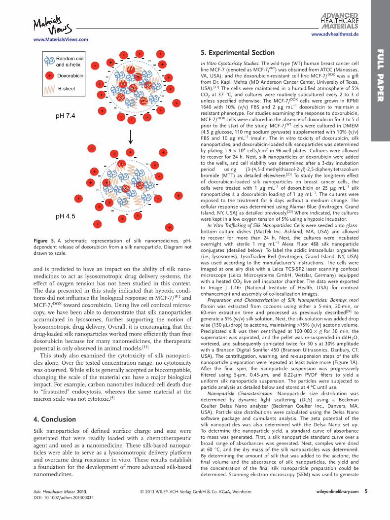
well with those established for silk fi lms. [ 23 ] Studies examining silk–drug interactions by our group [ 23 ] and others [ 28 ] have demon-strated that the log D of weak bases is a crit-ical parameter that governs drug adsorption to silk. Therefore, the low pH values encoun-tered in lysosomes were predicted to increase drug release by changing the electrostatic interaction between the silk and doxorubicin ( Figure 5 ). Next, the lysosomotropic drug delivery potential of silk was simulated using a range of pHs that refl ects the pHs of blood plasma (pH 7.4), early endosomes (pH 6.0), and lysosomes (pH 4.5). The release rates for the pH 4.5 samples were signifi cantly higher than for the other conditions, indicating that silk nanoparticles could potentially serve as a lysosomotropic drug delivery platform. A large number of synthetic- and biopolymer-based delivery systems have been engineered to serve as a lysosomotropic delivery system. However, there is a lack of natural biopoly-mers with the inherent ability to respond to changes in pH to trigger drug release. This is an important outcome of the present study, that a native biopolymer without any chem-ical modifi cations is able to elicit drug release in response to pH.
Targeted drug delivery and overcoming multidrug resistance are major driving forces for the development of nanomedi-cines. For example, packaging small-molec-
ular-weight drugs into nanoparticles changes the cellular and whole body pharmacokinetics of the drugs. [ 29 ] In solid tumors, the leaky blood vessels and reduced lymphatic drainage of the tumor vasculature facilitate the passive accumulation of nanomedicines in the tumor tissue. This phenomenon is called the enhanced permeation and retention (EPR) effect. [ 30 ] The EPR effect is often a key factor that permits (passive) tar-geting of nanomedicines to solid tumors. Once in the extra-cellular space of tumors, nanomedicines are endocytosed by cells and accumulate in intracellular organelles, such as lys-osomes. [ 4 ] The endocytic uptake of nanomedicines provides an opportunity to overcome drug resistance mechanisms, which effectively effl ux small-molecular-weight drugs that normally access the cytoplasm by diffusing across the plasma membrane. [ 29 ] To test the ability of our doxorubicin-loaded silk nanoparticles to serve as a lysosomotropic drug delivery platform that can overcome drug resistance, we have used human breast cancer cells that are resistant to doxorubicin (MCF-7/ DOX ). [ 31 ] The drug-loaded silk nanoparticles were signifi cantly more cyto-toxic than the free drug at the doxorubicin equivalent dose. In contrast, the parental cell lines (MCF-7/ WT ) showed an excel-lent response to doxorubicin regardless of the mechanism of drug delivery. This result suggested that doxorubicin-loaded silk nanoparticles were able to overcome the resistance mechanisms of MCF-7/ DOX cells by changing the cellular uptake mecha-nism. To recapitulate the tumor microenvironment, cytotoxicity studies were also conducted under hypoxic conditions. While intracellular pH is known to affect doxorubicin distribution [ 32 ]
heim Adv. Healthcare Mater. 2013, DOI: 10.1002/adhm.201300034
www.MaterialsViews.com
FULL P
APER
www.advhealthmat.de
Figure 5 . A schematic representation of silk nanomedicines. pH-dependent release of doxorubicin from a silk nanoparticle. Diagram not drawn to scale.
and is predicted to have an impact on the ability of silk nano-medicines to act as lysosomotropic drug delivery systems, the effect of oxygen tension has not been studied in this context. The data presented in this study indicated that hypoxic condi-tions did not infl uence the biological response in MCF-7/ WT and MCF-7/ DOX toward doxorubicin. Using live cell confocal micros-copy, we have been able to demonstrate that silk nanoparticles accumulated in lysosomes, further supporting the notion of lysosomotropic drug delivery. Overall, it is encouraging that the drug-loaded silk nanoparticles worked more effi ciently than free doxorubicin because for many nanomedicines, the therapeutic potential is only observed in animal models. [ 33 ]
This study also examined the cytotoxicity of silk nanoparti-cles alone. Over the tested concentration range, no cytotoxicity was observed. While silk is generally accepted as biocompatible, changing the scale of the material can have a major biological impact. For example, carbon nanotubes induced cell death due to “frustrated” endocytosis, whereas the same material at the micron scale was not cytotoxic. [ 4 ]
4. Conclusion
Silk nanoparticles of defi ned surface charge and size were generated that were readily loaded with a chemotherapeutic agent and used as a nanomedicine. These silk-based nanopar-ticles were able to serve as a lysosomotropic delivery platform and overcame drug resistance in vitro . These results establish a foundation for the development of more advanced silk-based nanomedicines.
© 2013 WILEY-VCH Verlag GAdv. Healthcare Mater. 2013, DOI: 10.1002/adhm.201300034
5. Experimental Section In Vitro Cytotoxicity Studies : The wild-type (WT) human breast cancer cell line MCF-7 (denoted as MCF-7/ WT ) was obtained from ATCC (Manassas, VA, USA), and the doxorubicin-resistant cell line MCF-7/ DOX was a gift from Dr. Kapil Mehta (MD Anderson Cancer Center, University of Texas, USA). [ 31 ] The cells were maintained in a humidifi ed atmosphere of 5% CO 2 at 37 ° C, and cultures were routinely subcultured every 2 to 3 d unless specifi ed otherwise. The MCF-7/ DOX cells were grown in RPMI 1640 with 10% (v/v) FBS and 2 μ g mL − 1 doxorubicin to maintain a resistant phenotype. For studies examining the response to doxorubicin, MCF-7/ DOX cells were cultured in the absence of doxorubicin for 3 to 5 d prior to the start of the study. MCF-7/ WT cells were cultured in DMEM (4.5 g glucose, 110 mg sodium pyruvate) supplemented with 10% (v/v) FBS and 10 μ g mL − 1 insulin. The in vitro toxicity of doxorubicin, silk nanoparticles, and doxorubicin-loaded silk nanoparticles was determined by plating 1.9 × 10 4 cells/cm 2 in 96-well plates. Cultures were allowed to recover for 24 h. Next, silk nanoparticles or doxorubicin were added to the wells, and cell viability was determined after a 3-day incubation period using (3-(4,5-dimethylthiazol-2-yl)-2,5-diphenyltetrazolium bromide (MTT) as detailed elsewhere. [ 23 ] To study the long-term effect of doxorubicin-loaded silk nanoparticles on breast cancer cells, the cells were treated with 1 μ g mL − 1 of doxorubicin or 25 μ g mL − 1 silk nanoparticles ± a doxorubicin loading of 1 μ g mL − 1 . The cultures were exposed to the treatment for 6 days without a medium change. The cellular response was determined using Alamar Blue (Invitrogen, Grand Island, NY, USA) as detailed previously. [ 23 ] Where indicated, the cultures were kept in a low oxygen tension of 5% using a hypoxic incubator.
In Vitro Traffi cking of Silk Nanoparticles : Cells were seeded onto glass-bottom culture dishes (MatTek Inc. Ashland, MA, USA) and allowed to recover for more than 24 h. Next, the cultures were incubated overnight with sterile 1 mg mL − 1 Alexa Fluor 488 silk nanoparticle conjugates (detailed below). To label the acidic intracellular organelles (i.e., lysosomes), LysoTracker Red (Invitrogen, Grand Island, NY, USA) was used according to the manufacturer’s instructions. The cells were imaged at one airy disk with a Leica TCS-SP2 laser scanning confocal microscope (Leica Microsystems GmbH, Wetzlar, Germany) equipped with a heated CO 2 live cell incubator chamber. The data were exported to Image J 1.46r (National Institute of Health, USA) for contrast enhancement and assembly of co-localization images.
Preparation and Characterization of Silk Nanoparticles: Bombyx mori fi broin was extracted from cocoons using either a 5-min, 20-min, or 60-min extraction time and processed as previously described [ 24 ] to generate a 5% (w/v) silk solution. Next, the silk solution was added drop wise (150 μ L/drop) to acetone, maintaining > 75% (v/v) acetone volume. Precipitated silk was then centrifuged at 100 000 × g for 30 min, the supernatant was aspirated, and the pellet was re-suspended in ddH 2 O, vortexed, and subsequently sonicated twice for 30 s at 30% amplitude with a Branson Digital Sonifi er 450 (Branson Ultrasonics, Danbury, CT, USA). The centrifugation, washing, and re-suspension steps of the silk nanoparticle preparation were repeated at least twice more (Figure 1 A). After the fi nal spin, the nanoparticle suspension was progressively fi ltered using 5- μ m, 0.45- μ m, and 0.22- μ m PVDF fi lters to yield a uniform silk nanoparticle suspension. The particles were subjected to particle analysis as detailed below and stored at 4 ºC until use.
Nanoparticle Characterization : Nanoparticle size distribution was determined by dynamic light scattering (DLS) using a Beckman Coulter Delsa Nano analyzer (Beckman Coulter Inc., Danvers, MA, USA). Particle size distributions were calculated using the Delsa Nano software package and cumulants analysis. The zeta potential of the silk nanoparticles was also determined with the Delsa Nano set up. To determine the nanoparticle yield, a standard curve of absorbance to mass was generated. First, a silk nanoparticle standard curve over a broad range of absorbances was generated. Next, samples were dried at 60 ° C, and the dry mass of the silk nanoparticles was determined. By determining the amount of silk that was added to the acetone, the fi nal volume and the absorbance of silk nanoparticles, the yield and the concentration of the fi nal silk nanoparticle preparation could be determined. Scanning electron microscopy (SEM) was used to generate
5wileyonlinelibrary.commbH & Co. KGaA, Weinheim

FULL
PAPER
www.advhealthmat.de
6
qualitative data on the silk nanoparticles. To minimize silk nanoparticle aggregation during the drying process, they were diluted in ultrapure H 2 O, spread on silicon wafers, frozen in the gaseous phase of N 2 , and dehydrated by freeze-drying. The samples were subsequently sputter coated with a thin coat of platinum/palladium and imaged with a Zeiss Supra 55VP fi eld emission scanning electron microscope (Carl Zeiss, Oberkochen, Germany).
Silk Nanoparticles: Loading and Release of Doxorubicin : The loading characteristics of the silk nanoparticles were determined using either 500 μ g or 125 μ g of silk nanoparticles. The ability of the silk nanoparticles to adsorb doxorubicin was evaluated by using a broad range of drug concentrations (10–125 μ g) (N.B. studies performed with ddH 2 O). Following a 20-h adsorption period at room temperature under dynamic conditions (20 revolutions per minute), the particles were pelleted using a 40-min centrifugation at 20 000 × g . The amount of drug remaining in solution was determined by measuring doxorubicin-associated fl uorescence (excitation 480 nm, emission 590 nm). The process of loading the silk nanoparticles with doxorubicin for the subsequent release studies is detailed below.
pH-Dependent Release of Doxorubicin from Silk Nanoparticles : A total of 500 μ g of the silk nanoparticles was loaded with 20 μ g doxorubicin as detailed above. Next, the silk nanoparticle suspension was centrifuged at 20 000 × g for 40 min, yielding a pink pellet. To demonstrate that all samples had been successfully loaded (loading effi ciency > 96%), doxorubicin-associated fl uorescence of the supernatant was determined. The doxorubicin-loaded silk nanoparticle pellet was resuspended in 0.5 mL of phosphate buffer at pH 4.5, 6.0, or 7.4. The samples were loaded into a Slide-A-Lyzer Mini dialysis cartridge (MWCO 3500 g mol − 1 ; Thermo Scientifi c, Waltham, MA, USA), which was inserted into a 1.5-mL receiver chamber containing 1 mL of buffer at the indicated pH. At the indicated time points, doxorubicin-associated fl uorescence in the receiver chamber was monitored, and fresh buffer was added, ensuring that sink conditions were maintained throughout the study.
Labeling Silk Nanoparticles with Alexa Fluor 488 : A total of 2 mg of the silk nanoparticles were resuspended in a 0.2 M NaHCO 3 pH 8.3 buffer to yield a fi nal concentration of 1 mg mL − 1 silk nanoparticles in 0.1 M buffer. Next, 1 mg of Alexa Fluor 488 carboxcylic acid succinimidyl ester (Invitrogen, Grand Island, NY, USA) was dissolved in dimethylsulfoxide, and 100 μ L of the solution was added to the silk nanoparticles and allowed to react for 4 h at room temperature in the dark while stirring. Following the conjugation of Alexa Fluor 488 to the silk nanoparticles, the suspension was centrifuged, and the pellet was washed repeatedly with acidifi ed water (pH 4.6) to remove unbound dye. The silk nanoparticles were washed until no free dye could be detected in the supernatant. Finally, the particles were dialyzed against ddH 2 O for 48 h and processed for in vitro cell culture studies as detailed above.
Supporting Information Supporting Information is available from the Wiley Online Library or from the author.
Acknowledgements The authors would like to thank Dr. Carlo Alonzo for help with the acquisition of live cell confocal images. Particle characterization was performed at the Center for Nanoscale Systems (CNS), a member of the National Nanotechnology Infrastructure Network (NNIN), which is supported by the National Science Foundation under NSF award no. ECS-0335765. CNS is part of Harvard University. This research was supported by NIH grant P41 EB002520-05 (Tissue Engineering Resource Center) (DLK), a Mildred Scheel Postdoctoral fellowship from the German Cancer Aid (FPS) and a Marie Curie FP7 Integration
wileyonlinelibrary.com © 2013 WILEY-VCH Verlag G
www.MaterialsViews.com
Grant 334134 within the 7 th European Union Framework Programme (FPS).
Received: January 22, 2013 Revised: March 4, 2013
Published online:
[ 1 ] R. Duncan , R. Gaspar , Mol. Pharm. 2011 , 8 , 2101 . [ 2 ] C. Sheridan , Nature Biotechnol. 2012 , 30 , 471 . [ 3 ] R. Duncan , Nat. Rev. Cancer. 2006 , 6 , 688 . [ 4 ] R. Duncan , S. C. Richardson , Mol. Pharm. 2012 , 9 , 2380 . [ 5 ] C. de Duve , T. de Barsy , B. Poole , A. Trouet , P. Tulkens , F. Van Hoof ,
Biochem. Pharmacol. 1974 . 23 , 2495 . [ 6 ] A. Bendele , J. Seely , C. Richey , G. Sennello , G. Shopp , Toxicol. Sci.
1998 , 42 , 152 . [ 7 ] N. Kumar , M. N. Ravikumar , A. J. Domb , Adv. Drug Deliv. Rev. 2001 ,
53 , 23 . [ 8 ] P. B. Malafaya , G. A. Silva , R. L. Reis , Adv. Drug Deliv. Rev. 2007 , 59 ,
207 . [ 9 ] M. I. Giannotti , O. Esteban , M. Oliva , M. F. García-Parajo , F. Sanz ,
Biomacromolecules 2011 , 12 , 2524 . [ 10 ] Y. Hu , X. Jiang , Y. Ding , Q. Chen , C. Yang , Adv. Mater. 2004 , 16 , 933 . [ 11 ] Y. Cheng , S. Yu , J. Wang , H. Qian , W. Wu , X. Jiang , Macromol. Biosci.
2012 , 12 , 1326 . [ 12 ] F. G. Omenetto , D. L. Kaplan , Science 2010 , 329 , 528 . [ 13 ] X. Wang , T. Yucel , Q. Lu , X. Hu , D. L. Kaplan , Biomaterials 2010 , 31 ,
1025 . [ 14 ] E. Wenk , A. J. Wandrey , H. P. Merkle , L. Meinel , J. Controlled Release
2008 , 132 , 26 . [ 15 ] T. Hino , S. Himabayashi , A. Nakai , Pharm. Pharmacol. Commun.
2000 , 6 , 335 . [ 16 ] A. S. Lammel , X. Hu , S. H. Park , D. L. Kaplan , T. R. Scheibel , Bioma-
terials 2010 , 31 , 4583 . [ 17 ] V. Gupta , A. Aseh , C. N. Ríos , B. B. Aggarwal , A. B. Mathur , Int. J.
Nanomed. 2009 , 4 , 115 . [ 18 ] J. Kundu , Y. I. Chung , Y. H. Kim , G. Tae , S. C. Kundu , Int. J. Pharm.
2010 , 388 , 242 . [ 19 ] Y. Q. Zhang , W. D. Shen , R. L. Xiang , L. J. Zhuge , W. J. Gao ,
W. B. Wang , J. Nanoparticle Res. 2007 , 9 , 885 . [ 20 ] K. D. Hermanson , D. Huemmerich , T. Scheibel , A. R. Bausch , Adv.
Mater. 2007 , 19 , 1810 . [ 21 ] R. Anumolu , J. A. Gustafson , J. J. Magda , J. Cappello , H. Ghandehari ,
L. F. 3rd Pease , ACS Nano 2011 , 5 , 5374 . [ 22 ] K. Numata , B. Subramanian , H. A. Currie , D. L. Kaplan , Biomaterials
2009 , 30 , 5775 . [ 23 ] F. P. Seib , D. L. Kaplan , Biomaterials 2012 , 33 , 8442 . [ 24 ] F. P. Seib , E. M. Pritchard , D. L. Kaplan , Adv. Funct. Mater. 2013 , 23 ,
58 . [ 25 ] X. Hu , K. Shmelev , L. Sun , E. S. Gil , S. H. Park , P. Cebe , D. L. Kaplan ,
Biomacromolecules 2011 , 12 , 1686 . [ 26 ] L. S. Wray , X. Hu , J. Gallego , I. Georgakoudi , F. G. Omenetto ,
D. Schmidt , D. L. Kaplan , J. Biomed. Mater. Res. B Appl. Biomater. 2011 , 99 , 89 .
[ 27 ] F. P. Seib , M. F. Maitz , X. Hu , C. Werner , D. L. Kaplan , Biomaterials 2012 , 33 , 1017 .
[ 28 ] A. Lammel , M. Schwab , M. Hofer , G. Winterm , T. Scheibel , Biomate-rials 2011 , 32 , 2233 .
[ 29 ] M. E. Davis , Z. G. Chen , D. M. Shin , Nat. Rev. Drug Discov. 2008 , 7 , 771 . [ 30 ] Y. Matsumura , H. Maeda , Cancer Res. 1986 , 46 , 6387 . [ 31 ] K. Mehta , Int. J. Cancer 1994 , 58 , 400 . [ 32 ] S. Simon , D. Roy , M. Schindler , Proc. Natl. Acad. Sci. USA 1994 , 91 , 1128 . [ 33 ] R. Duncan , Nat. Rev. Drug Discov. 2003 , 2 , 347 .
mbH & Co. KGaA, Weinheim Adv. Healthcare Mater. 2013, DOI: 10.1002/adhm.201300034