Synthesis and controlled-release properties of chitosan ...dasan.sejong.ac.kr/~sko/paper/2016...
8
Food Sci. Biotechnol. 25(6): 1583-1590 (2016) DOI 10.1007/s10068-016-0244-y Synthesis and controlled-release properties of chitosan/ β-Lactoglobulin nanoparticles as carriers for oral administration of epigallocatechin gallate Jin Liang 1,2 , Hua Yan 1 , Han-Joo Yang 2 , Hye Won Kim 2 , Xiaochun Wan 1 , Jinhee Lee 3 , and Sanghoon Ko 2, * State Key Laboratory of Tea Plant Biology and Utilization, Anhui Agricultural University, Hefei 230036, China Department of Food Science and Biotechnology, Sejong University, Seoul 05006, Korea Department of Food Science and Biotechnology, Cha University, Seongnam, Gyeonggi 13488, Korea Introduction Epigallocatechin gallate (EGCG) is the major catechin component in green tea, with strong antioxidant, antitumor, and other biological activities (1); EGCG has received extensive attention in the functional foods field because of its health benefits. However, the bioavailability of EGCG after oral administration is low because of its poor stability in the gastrointestinal (GI) tract. Thus, a suitable carrier is needed to enhance the stability and intestinal bioavailablity of EGCG (2). Some bioactives such as tea polyphenols have to be released in the GI tract slowly over time in order to enhance their bioavailability. Recently, sustained-release nano-scale carriers have contributed to the improved stability, cellular uptake, and prolonged clearance time of the enclosed bioactives (3). Chitosan has been extensively used as a sustained-release carrier for delivery of bioactives, such as catechins, because of its mucoadhesive property in the GI tract (4). Chitosan-based carriers can adhere to the intestinal membrane, and therefore, prolong residence time in the GI tract (2). However, the poor solubility of chitosan at neutral pH conditions and its low delivery efficiency for several orally administered bioactives have limited its applications in the food industry. Therefore, it is necessary to design a new type of chitosan-based sustained-release nano-scale carrier with enhanced aqueous solubility and resistance to the harsh conditions of the GI tract. We recently developed chitosan nanoparticles by using two different water-soluble chitosans, carboxymethyl chitosan (CMC) and chitosan hydrochloride (CHC), to improve the bioavailability of tea polyphenols (5,6). These chitosans form nano-sized particles by ionic gelation between carboxyl methyl and amino groups of CMC and CHC, respectively. The water-soluble CMC-CHC nanoparticles are suitable for the sustained release of their core material (7). However, compared with some complex nanoparticles prepared by using CMC or CHC materials, the orally administered CMC-CHC nanoparticles were not stable at low pH in the gastric fluid and underwent rapid acid-induced dissociation and degradation, which could lead to a quick release of their core materials in the stomach (8). In this study, we hypothesized that the limitations of CMC-CHC nanoparticles can be overcome by an additional protective coating against the acidic conditions of the gastric fluid. Therefore, we designed a novel EGCG nano-carrier with a secondary coating resistant to low pH, layered on the primary EGCG-loaded CMC-CHC nanoparticles, to provide additional protection against the acidic gastric environment. This secondary coating is removed in the intestinal fluid, and subsequently, the exposed EGCG-loaded CMC- Received December 12, 2015 Revised October 11, 2016 Accepted October 11, 2016 Published online December 31, 2016 *Corresponding Author Tel: +82-2-3408-3260 Fax: +82-2-3408-4319 E-mail: [email protected] pISSN 1226-7708 eISSN 2092-6456 © KoSFoST and Springer 2016 Abstract A nano-sized double-walled carrier composed of chitosan and β-lactoglobulin (β-Lg) for oral administration of epigallocatechin gallate (EGCG) was developed to achieve a prolonged release of EGCG in the gastrointestinal tract. Carboxymethyl chitosan (CMC) solution was added dropwise to chitosan hydrochloride (CHC) containing EGCG to form a primary coating by ionic complexation. Subsequently, β-Lg was added to create a secondary layer by ionic gelation. The obtained EGCG-loaded chitosan/β-Lg nanoparticles had sizes between 100 and 500 nm and zeta potentials ranging from 10 to 35 mV. FT-IR spectroscopy revealed a high number of hydrogen-bonding sites in the nanoparticles, which could incorporate EGCG, resulting in high encapsulation efficiency. EGCG incorporated in the primary coating was released slowly over time by diffusion from the swollen CMC-CHC matrix after the outer layer of β-Lg was degraded in the intestinal fluid. The sustained-release property makes chitosan/ β-Lg nanoparticles an attractive candidate for effective delivery of EGCG. Keywords: epigallocatechin gallate, chitosan, β-lactoglobulin, double wall formation, controlled release
Transcript of Synthesis and controlled-release properties of chitosan ...dasan.sejong.ac.kr/~sko/paper/2016...
Food Sci. Biotechnol. 25(6): 1583-1590 (2016)
DOI 10.1007/s10068-016-0244-y
Synthesis and controlled-release properties of chitosan/
β-Lactoglobulin nanoparticles as carriers for oral
administration of epigallocatechin gallate
Jin Liang1,2, Hua Yan1, Han-Joo Yang2, Hye Won Kim2, Xiaochun Wan1, Jinhee Lee3, and Sanghoon Ko2,*
1State Key Laboratory of Tea Plant Biology and Utilization, Anhui Agricultural University, Hefei 230036, China2Department of Food Science and Biotechnology, Sejong University, Seoul 05006, Korea3Department of Food Science and Biotechnology, Cha University, Seongnam, Gyeonggi 13488, Korea
Introduction
Epigallocatechin gallate (EGCG) is the major catechin component in
green tea, with strong antioxidant, antitumor, and other biological
activities (1); EGCG has received extensive attention in the functional
foods field because of its health benefits. However, the bioavailability
of EGCG after oral administration is low because of its poor stability
in the gastrointestinal (GI) tract. Thus, a suitable carrier is needed to
enhance the stability and intestinal bioavailablity of EGCG (2). Some
bioactives such as tea polyphenols have to be released in the GI tract
slowly over time in order to enhance their bioavailability. Recently,
sustained-release nano-scale carriers have contributed to the
improved stability, cellular uptake, and prolonged clearance time of
the enclosed bioactives (3).
Chitosan has been extensively used as a sustained-release carrier
for delivery of bioactives, such as catechins, because of its
mucoadhesive property in the GI tract (4). Chitosan-based carriers
can adhere to the intestinal membrane, and therefore, prolong
residence time in the GI tract (2). However, the poor solubility of
chitosan at neutral pH conditions and its low delivery efficiency for
several orally administered bioactives have limited its applications in
the food industry. Therefore, it is necessary to design a new type of
chitosan-based sustained-release nano-scale carrier with enhanced
aqueous solubility and resistance to the harsh conditions of the GI
tract. We recently developed chitosan nanoparticles by using two
different water-soluble chitosans, carboxymethyl chitosan (CMC) and
chitosan hydrochloride (CHC), to improve the bioavailability of tea
polyphenols (5,6). These chitosans form nano-sized particles by ionic
gelation between carboxyl methyl and amino groups of CMC and
CHC, respectively. The water-soluble CMC-CHC nanoparticles are
suitable for the sustained release of their core material (7). However,
compared with some complex nanoparticles prepared by using CMC
or CHC materials, the orally administered CMC-CHC nanoparticles
were not stable at low pH in the gastric fluid and underwent rapid
acid-induced dissociation and degradation, which could lead to a
quick release of their core materials in the stomach (8).
In this study, we hypothesized that the limitations of CMC-CHC
nanoparticles can be overcome by an additional protective coating
against the acidic conditions of the gastric fluid. Therefore, we
designed a novel EGCG nano-carrier with a secondary coating
resistant to low pH, layered on the primary EGCG-loaded CMC-CHC
nanoparticles, to provide additional protection against the acidic
gastric environment. This secondary coating is removed in the
intestinal fluid, and subsequently, the exposed EGCG-loaded CMC-
Received December 12, 2015Revised October 11, 2016Accepted October 11, 2016Published online December 31, 2016
*Corresponding AuthorTel: +82-2-3408-3260Fax: +82-2-3408-4319E-mail: [email protected]
pISSN 1226-7708eISSN 2092-6456
© KoSFoST and Springer 2016
Abstract A nano-sized double-walled carrier composed of chitosan and β-lactoglobulin (β-Lg) for oral
administration of epigallocatechin gallate (EGCG) was developed to achieve a prolonged release of
EGCG in the gastrointestinal tract. Carboxymethyl chitosan (CMC) solution was added dropwise to
chitosan hydrochloride (CHC) containing EGCG to form a primary coating by ionic complexation.
Subsequently, β-Lg was added to create a secondary layer by ionic gelation. The obtained EGCG-loaded
chitosan/β-Lg nanoparticles had sizes between 100 and 500 nm and zeta potentials ranging from 10 to
35 mV. FT-IR spectroscopy revealed a high number of hydrogen-bonding sites in the nanoparticles,
which could incorporate EGCG, resulting in high encapsulation efficiency. EGCG incorporated in the
primary coating was released slowly over time by diffusion from the swollen CMC-CHC matrix after the
outer layer of β-Lg was degraded in the intestinal fluid. The sustained-release property makes chitosan/
β-Lg nanoparticles an attractive candidate for effective delivery of EGCG.
Keywords: epigallocatechin gallate, chitosan, β-lactoglobulin, double wall formation, controlled release
1584 Liang et al.
Food Sci. Biotechnol.
CHC nanoparticles with mucoadhesive properties can achieve a
prolonged release of EGCG in the intestinal tract. Recently published
studies have shown that double-walled chitosan nanoparticles
coated with several food proteins or bioactive peptides, such as soy
protein (9), caseinophosphopeptides (10), could prevent rapid
degradation of bioactives in GI conditions and improve their gastric
stability. Therefore, our second hypothesis was that a biopolymer for
the secondary coating of the primary EGCG-loaded CMC-CHC
nanoparticles would be resistant to peptic digestion and acidic
hydrolysis in the stomach, but would still be enzymatically
degradable in the small intestine.
Beta-lactoglobulin (β-Lg), comprising 50 to 60% of the total whey
protein in cow milk, is resistant to degradation by pepsin hydrolysis in
the stomach (11). Another important property of β-Lg is its ability to
form stable structures with chitosan by ionic gelation (12). We
previously reported the physicochemical and prolonged release
properties of chitosan/β-Lg microcapsules, formed through a simple
ionic gelation process with chitosan and denatured β-Lg (12). Other
studies suggested that chitosan/β-Lg nanoparticles can be used as
carriers for oral administration of nutraceuticals such as quercetin
and vitamin D (9,13).
The main objective of this study was to develop EGCG-loaded
chitosan/β-Lg nanoparticles as a novel carrier to achieve a prolonged
release of orally administered EGCG in the GI tract. Furthermore, the
physicochemical properties and prolonged release performance of
EGCG-loaded chitosan/β-Lg nanoparticles were investigated during
simulated GI digestion. Finally, we propose formation and release
mechanisms for the EGCG-loaded chitosan/β-Lg nanoparticles.
Materials and Methods
Materials EGCG (purity>95%) was provided by Hefei Shangu
Chemical Reagent Company (Hefei, Anhui, China). Two types of
chitosan materials, N-carboxymethyl chitosan (molecular weight
61 kDa and deacetylation degree 83%) and chitosan hydrochloride
(molecular weight 90 kDa and deacetylation degree 85%) were
purchased from Haidebei Marine Bioengineering Company (Jinan,
Shandong, China). β-Lg (93% purity) was obtained from Davisco
Foods International, Inc. (Le Sueur, MN, USA). All other reagents
were of analytical grade.
Preparation of EGCG-loaded chitosan/β-Lg nanoparticles The
EGCG-loaded chitosan/β-Lg nanoparticles with core-shell double-
walled structures were prepared by ionic gelation using CMC, CHC
and β-Lg as wall materials and EGCG as the core material. Different
amounts of EGCG (0, 10, 20, 30, and 40 mg) were added to 30 mL of
CHC 1.0 mg/mL aqueous solution while stirring at 25oC as described
previously (5,6). Next, 15 mL of CMC 2.0 mg/mL aqueous solution
was added dropwisely to the CHC solution containing EGCG while
stirring at 500 rpm at 25oC. EGCG-loaded CMC-CHC (simply called
chitosan) nanoparticles were obtained through this ionic gelation
step.
β-Lg aqueous solutions with different concentrations (0, 10, 20,
30, and 40 mg/mL) were heated at 85oC for 1.5 h to obtain a
denatured β-Lg solution as described previously (12). Finally, the
EGCG-loaded chitosan/β-Lg double-walled nanoparticles were
formed by adding the denatured β-Lg solution to the EGCG-loaded
CMC-CHC nanoparticle solution. The solution containing EGCG-
loaded chitosan/β-Lg double-walled nanoparticles was centrifuged
at 30,000xg for 20 min; the pellet was then collected and freeze-
dried. The EGCG-loaded CMC-CHC nanoparticles without β-Lg were
used as a control.
Particle size and zeta potential measurement Size and zeta potential
of the EGCG-loaded chitosan/β-Lg nanoparticles prepared were
determined by a commercial particle size and zeta potential analyzer
(Delsa Nano C, Beckman Coulter, Inc., Fullerton, CA, USA). For size
and zeta potential measurements, the EGCG-loaded chitosan/β-Lg
nanoparticles prepared were dispersed in distilled water, and then
measured with a scattering angle of 165o at 25oC. The measurement
was repeated three times for every sample.
FT-IR study CMC, CHC, β-Lg, EGCG, and EGCG-loaded chitosan/β-Lg
nanoparticles were analyzed by FT-IR spectroscopy. Each sample was
diluted to a suitable concentration and analyzed with a Spectrum
100 FT-IR spectrometer. The infrared transmittance was acquired at a
wave number from 800 to 4,000 cm−1, and then analyzed using the
OMNIC software.
Encapsulation efficiency The encapsulation efficiency of EGCG-
loaded chitosan/β-Lg nanoparticles was calculated as reported
previously (14). Briefly, the EGCG-loaded chitosan/β-Lg nanoparticles
were filtered through an ultracel membrane. After centrifugation at
4,000xg for 30 min, free EGCG that penetrated through the membrane
was measured using an HPLC-based detection method (15). An
Agilent 1200 series HPLC equipped with a DAD analysis system was
used to quantify EGCG. The mobile phase was composed of (A)
formic acid at pH 2.5 and (B) methanol. The gradient elution was
carried out on an Agilent Eclipse XDB-C18 column (4.6 mmx150 mm,
5 μm) at a flow rate of 1.0 mL/min with a linear gradient as follows:
0–15 min, A from 82 to 40%, and B from 18 to 60%. The sample (10
μL) was filtered through a 0.45-μm cellulose filter prior to injection.
The column was maintained at 40oC, and the detection wavelength
was set at 280 nm. A calibration curve was obtained by analyzing five
concentrations of EGCG standard (1.0–1,000 μg/mL) vs. the peak
area of the eluted peak. The linear regression equation was Y=
11.238X–882.3 (R2=0.9992), where X is the concentration and Y is
the peak area. The EE of EGCG entrapped in double-walled chitosan/
β-Lg nanoparticles was calculated using the following equation:
Chitosan/β-Lactoglobulin nanoparticles for EGCG 1585
December 2016 | Vol. 25 | No. 6
EE (%)=[(Total amount of EGCG−amount of EGCG in ultrafiltrate)/
Total amount of EGCG]x100
Release studies in simulated GI conditions The release characteristics
of EGCG-loaded chitosan/β-Lg nanoparticles in simulated GI tract
conditions were investigated (12). The EGCG-loaded chitosan/β-Lg
nanoparticles were added to 25 mL of 20 mM phosphate buffer, pH
6.8 in a 250 mL flask. After adjusting the pH to 2.0 with a 1 M HCl
solution, 1 mL of porcine pepsin (0.4%, porcine gastric mucosa with
800-2,000 unit/mg protein, Sigma–Aldrich, St. Louis, MO, USA) was
added and the flasks were incubated at 37oC in a shaking water bath
at 150 rpm. Samples were collected at 0, 5, 10, 20, 30, 60, 90, and
120 min to analyze the release characteristics of EGCG from the
chitosan/β-Lg nanoparticles.
Digesta samples with EGCG-loaded chitosan/β-Lg nanoparticles
treated in simulated gastric conditions were used for the release
study in simulated small intestine conditions. The pH of the gastric
digesta was adjusted to 5.3 by adding 25 mM sodium bicarbonate
solution. Next, 1.5 mL of 20 mM phosphate buffer containing 0.2 mg
of lipase, 0.4 mg of pancreatin and 2.4 mg of bile extract from
porcine pancreas was added to the gastric digesta, and subsequently
the pH was adjusted to 6.8 with 1 M NaOH solution. Finally, the flasks
containing the digesta samples with EGCG-loaded chitosan/β-Lg
nanoparticles were incubated at 37oC in a shaking water bath at 150
rpm. Samples were collected at 0, 10, 20, 30, 60, 120, 180, and
240 min to determine the release characteristics of EGCG from the
chitosan/β-Lg nanoparticles. The amounts of EGCG released from
the chitosan/β-Lg nanoparticles at given time intervals were
determined by the same HPLC method used for EE measurement.
EGCG-loaded chitosan nanoparticles without β-Lg were used as
control.
Statistical analysis Statistical analysis was performed using SPSS
software (SPSS ver. 17.0, SPSS Inc., Chicago, IL, USA). A one-way
analysis of variance (ANOVA) with a post-hoc Tukey test was used to
determine statistical significance (p<0.05).
Results and Discussion
Formation of EGCG-loaded chitosan/β-Lg nanoparticle EGCG-
loaded chitosan/β-Lg nanoparticles with a core-shell double-walled
structure were synthesized using chitosan (CMC and CHC) and
denatured β-Lg for encapsulating EGCG. The formation of the inner
core structure was mainly because of CMC-CHC ionotropic interactions
through the opposite charges on their surfaces. EGCG in solution had
a negative charge because of many hydroxyl groups in its structure.
The CMC-CHC ionotropic reaction could make the interior structure
compact and tight. After formation of EGCG-loaded CMC-CHC
nanoparticles, their surface structure also had the ability to form
ionotropic linkages with other molecules, such as β-Lg.
Denatured β-Lg formed the outer layer of the EGCG-loaded
chitosan nanoparticles. β-Lg generally has a negative charge in
aqueous solution at pH>5.2, and a positive charge at pH<5.2 (16). In
this study, β-Lg molecules were negatively charged since the pH of
the aqueous solution containing EGCG-loaded CMC-CHC nanoparticles
was about 6.0-7.0. As a result, the negatively charged denatured β-Lg
molecules formed an external coating on the positively charged
EGCG-loaded CMC-CHC nanoparticles through electrostatic interactions.
In conclusion, the formation of double-walled chitosan/β-Lg nanoparticles
was mainly because of ionotropic interactions between CMC and
CHC molecules for the primary coating, and electrostatic interactions
between the primary CMC-CHC nanoparticles and denatured β-Lg
molecules for the secondary coating.
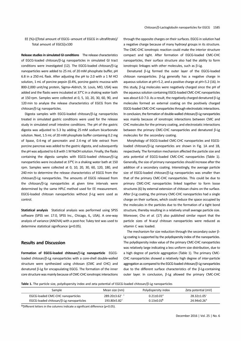
Morphology of EGCG-loaded CMC-CHC nanoparticles and EGCG-
loaded chitosan/β-Lg nanoparticles are shown in Fig. 1A and 1B,
respectively. The formation mechanism affected the particle size and
zeta potential of EGCG-loaded CMC-CHC nanoparticles (Table 1).
Generally, the size of primary nanoparticles should increase after the
addition of a secondary coating. Interestingly, the average particle
size of EGCG-loaded chitosan/β-Lg nanoparticles was smaller than
that of the primary CMC-CHC nanoparticles. This could be due to
primary CMC-CHC nanoparticles linked together to form loose
structures (6) by external extension of chitosan chains on the surface.
After β-Lg coating, the primary CMC-CHC nanoparticles had a single
charge on their surfaces, which could reduce the space occupied by
the molecules in the particles due to the formation of a tight bond
structure, thereby resulting in a relatively small average particle size.
Moreover, Cho et al. (17) also published similar report that the
particle sizes of N-acyl chitosan nanoparticles were reduced as
vitamin C was loaded.
The mechanism for size reduction through the secondary outer β-
Lg coating is supported by the polydispersity index of the nanoparticles.
The polydispersity index value of the primary CMC-CHC nanoparticles
was relatively large indicating a less uniform size distribution, due to
a high degree of particle aggregation (Table 1). The primary CMC-
CHC nanoparticles showed a relatively high degree of inter-particle
aggregation as compared to the EGCG-loaded chitosan/β-Lg nanoparticles
due to the different surface characteristics of the β-Lg-containing
outer layer. In conclusion, β-Lg allowed the primary CMC-CHC
Table 1. The particle size, polydispersity index and zeta potential of EGCG-loaded chitosan/β-Lg nanoparticles
Sample Mean size (nm) Polydispersity index Zeta potential (mV)
EGCG-loaded CMC-CHC nanoparticles 289.20±3.62a1) 0.21±0.01a 28.32±1.05a
EGCG-loaded chitosan/β-Lg nanoparticles 193.80±5.82b 0.13±0.01b 24.94±0.26b
1)Different letters in the columns indicate a significant difference (p<0.05).
1586 Liang et al.
Food Sci. Biotechnol.
nanoparticles to be relatively monodisperse under the preparation
conditions, and subsequently formed a secondary coating on their
surface.
The shape of EGCG-loaded CMC-CHC nanoparticles was relatively
irregular and inconsistent due to inter-particle aggregation, whereas
EGCG-loaded chitosan/β-Lg nanoparticles were mostly spherical in
shape with a relatively uniform size (Fig. 1). The β-Lg molecules
changed the surface characteristics of EGCG-loaded chitosan/β-Lg
nanoparticles. The unfolded β-Lg molecules reduced inter-particle
aggregation among the primary CMC-CHC nanoparticles. Moreover,
the addition of the secondary β-Lg layer reduced the mean size of
EGCG-loaded chitosan/β-Lg nanoparticles as compared to the
primary CMC-CHC nanoparticles. In addition, the interaction between
β-Lg and chitosan molecules improved EE through their stronger
protein-polysaccharide electrostatic interactions.
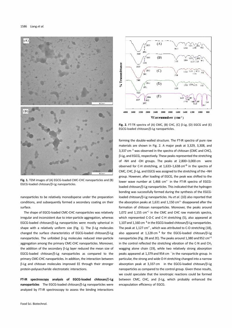
FT-IR spectroscopy analysis of EGCG-loaded chitosan/β-Lg
nanoparticles The EGCG-loaded chitosan/β-Lg nanoparticles were
analyzed by FT-IR spectroscopy to assess the binding interactions
forming the double-walled structure. The FT-IR spectra of pure raw
materials are shown in Fig. 2. A major peak at 3,329, 3,308, and
3,337 cm−1 was observed in the spectra of chitosan (CMC and CHC),
β-Lg, and EGCG, respectively. These peaks represented the stretching
of -NH and -OH groups. The peaks at 2,800–3,000 cm−1 were
observed for C-H stretching, at 1,633–1,638 cm−1 in the spectra of
CMC, CHC, β-Lg, and EGCG was assigned to the stretching of the –NH
group. However, after loading of EGCG, the peak was shifted to the
lower wave number at 1,466 cm−1 in the FT-IR spectra of EGCG-
loaded chitosan/β-Lg nanoparticles. This indicated that the hydrogen
bonding was successfully formed during the synthesis of the EGCG-
loaded chitosan/β-Lg nanoparticles. Hu et al. (10) also reported that
the absorption peaks at 1,631 and 1,150 cm−1 disappeared after the
formation of chitosan nanoparticles. Moreover, the peaks around
1,072 and 1,155 cm−1 in the CMC and CHC raw materials spectra,
which represented C-O-C and C-H stretching (5), also appeared at
1,107 and 1,160 cm−1 in the EGCG-loaded chitosan/β-Lg nanoparticles.
The peak at 1,127 cm−1, which was attributed to C-O stretching (18),
also appeared at 1,128 cm−1 for the EGCG-loaded chitosan/β-Lg
nanoparticles (Fig. 2B and 2E). The peaks around 1,380 and 952 cm−1
in the control reflected the stretching vibration of the C-N and CH3
wagging alone chain (19), while two relatively strong absorption
peaks appeared at 1,379 and 954 cm−1 in the nanoparticle group. In
particular, the strong and wide O-H stretching changed into a narrow
absorption peak at 3,337 cm−1 in the EGCG-loaded chitosan/β-Lg
nanoparticles as compared to the control group. Given these results,
we could speculate that the ionotropic reactions could be formed
between CMC, CHC, and β-Lg, which probably enhanced the
encapsulation efficiency of EGCG.
Fig. 2. FT-TR spectra of (A) CMC, (B) CHC, (C) β-Lg, (D) EGCG and (E)
EGCG-loaded chitosan/β-Lg nanoparticles.
Fig. 1. TEM images of (A) EGCG-loaded CMC-CHC nanoparticles and (B)
EGCG-loaded chitosan/β-Lg nanoparticles.
Chitosan/β-Lactoglobulin nanoparticles for EGCG 1587
December 2016 | Vol. 25 | No. 6
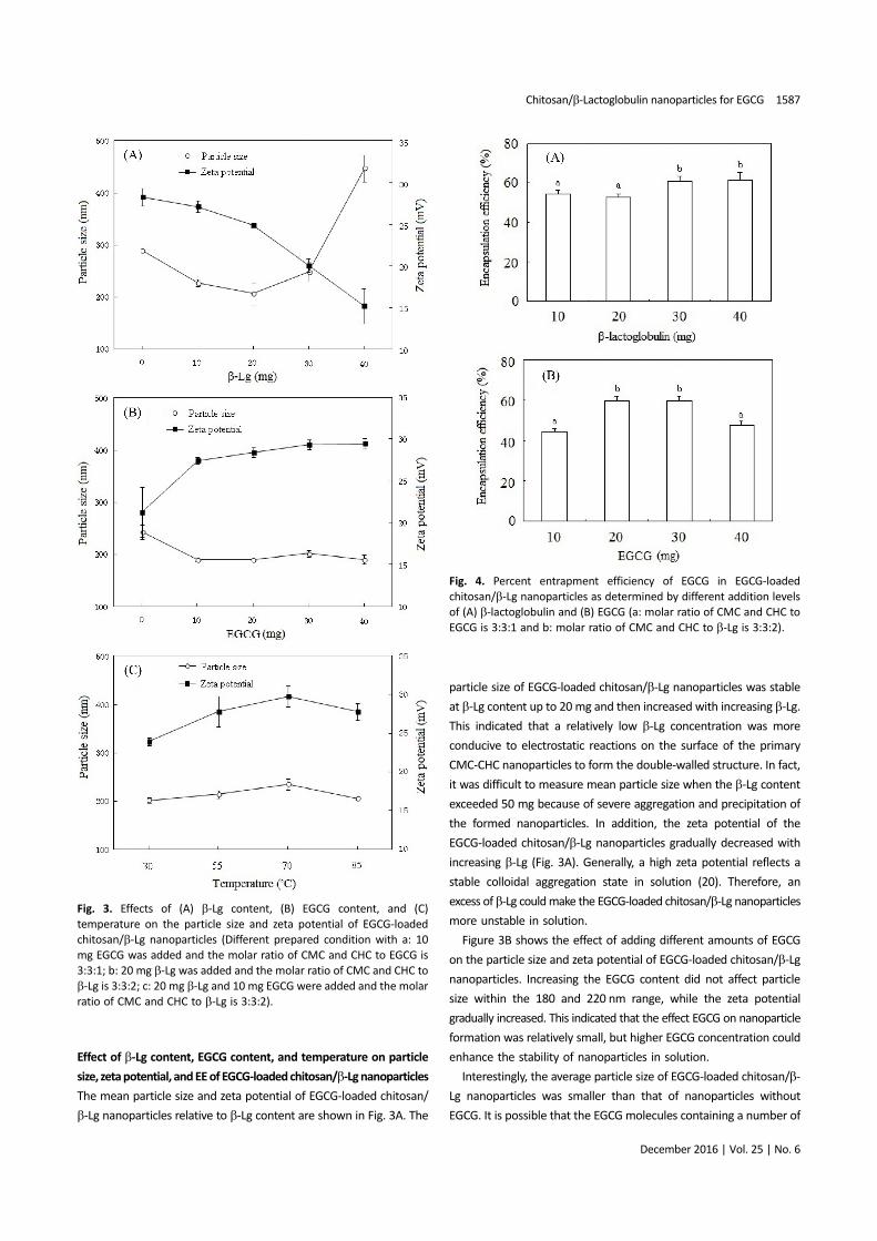
Effect of β-Lg content, EGCG content, and temperature on particle
size, zeta potential, and EE of EGCG-loaded chitosan/β-Lg nanoparticles
The mean particle size and zeta potential of EGCG-loaded chitosan/
β-Lg nanoparticles relative to β-Lg content are shown in Fig. 3A. The
particle size of EGCG-loaded chitosan/β-Lg nanoparticles was stable
at β-Lg content up to 20 mg and then increased with increasing β-Lg.
This indicated that a relatively low β-Lg concentration was more
conducive to electrostatic reactions on the surface of the primary
CMC-CHC nanoparticles to form the double-walled structure. In fact,
it was difficult to measure mean particle size when the β-Lg content
exceeded 50 mg because of severe aggregation and precipitation of
the formed nanoparticles. In addition, the zeta potential of the
EGCG-loaded chitosan/β-Lg nanoparticles gradually decreased with
increasing β-Lg (Fig. 3A). Generally, a high zeta potential reflects a
stable colloidal aggregation state in solution (20). Therefore, an
excess of β-Lg could make the EGCG-loaded chitosan/β-Lg nanoparticles
more unstable in solution.
Figure 3B shows the effect of adding different amounts of EGCG
on the particle size and zeta potential of EGCG-loaded chitosan/β-Lg
nanoparticles. Increasing the EGCG content did not affect particle
size within the 180 and 220 nm range, while the zeta potential
gradually increased. This indicated that the effect EGCG on nanoparticle
formation was relatively small, but higher EGCG concentration could
enhance the stability of nanoparticles in solution.
Interestingly, the average particle size of EGCG-loaded chitosan/β-
Lg nanoparticles was smaller than that of nanoparticles without
EGCG. It is possible that the EGCG molecules containing a number of
Fig. 4. Percent entrapment efficiency of EGCG in EGCG-loaded
chitosan/β-Lg nanoparticles as determined by different addition levels
of (A) β-lactoglobulin and (B) EGCG (a: molar ratio of CMC and CHC to
EGCG is 3:3:1 and b: molar ratio of CMC and CHC to β-Lg is 3:3:2).
Fig. 3. Effects of (A) β-Lg content, (B) EGCG content, and (C)
temperature on the particle size and zeta potential of EGCG-loaded
chitosan/β-Lg nanoparticles (Different prepared condition with a: 10
mg EGCG was added and the molar ratio of CMC and CHC to EGCG is
3:3:1; b: 20 mg β-Lg was added and the molar ratio of CMC and CHC to
β-Lg is 3:3:2; c: 20 mg β-Lg and 10 mg EGCG were added and the molar
ratio of CMC and CHC to β-Lg is 3:3:2).
1588 Liang et al.
Food Sci. Biotechnol.
hydroxyl groups could link electrostatically to the side chains in
chitosan and β-Lg molecules, forming tight intermolecular bonds.
Thus, relatively small particles could be formed as a result of the
electrostatic attraction between EGCG and the wall materials. This
result is consistent with that of a previous study in which the particle
size of thermally induced β-Lg-EGCG nano-vehicles was smaller than
that of the controls without EGCG (21).
The particles size and zeta potential of EGCG-loaded chitosan/β-Lg
nanoparticles were affected by the temperature used for β-Lg heat
treatment (Fig. 3C). Both mean particle size and zeta potential
increased with temperatures up to 70oC beyond which they decreased
due to the severe thermal denaturation of β-Lg (22). β-Lg may undergo
a conformational change after heat treatment at temperatures up to
70oC to expose non-polar side-chains usually buried in the interior
sites of the molecular structure. The change in β-Lg molecular
conformation after the thermal treatment could electrostatically
affect the double-walled structure formation mechanism (23). As a
result, the heat treatment could affect the formation of the EGCG-
loaded chitosan/β-Lg nanoparticle secondary coating and thus
particle size, although the effect was relatively small in the 200-240
nm range.
Figure 4 shows the EE of EGCG-loaded chitosan/β-Lg nanoparticles
with different levels of β-Lg or EGCG. EE increased from 54.15 to
60.67% as β-Lg increased from 10 to 40 mg (Fig. 4A). EGCG molecules
were entrapped primarily by the CMC and CHC matrix and subsequently
the remaining EGCG molecules combined with β-Lg molecules. This
result is in agreement with that of a previous report in which EE of
EGCG-loaded β-Lg nanoparticles increased with an increase in β-Lg
concentration (24).
Figure 4B shows that EE increases at low EGCG content up to 30
mg and subsequently decreases at higher EGCG levels. EE reached a
maximum value of about 60% when the EGCG content was in the 20
to 30 mg range. The improvement in EE was due to the inter- and
intra-molecular electrostatic interactions between EGCG molecules
and wall materials (CMC, CHC, and β-Lg).
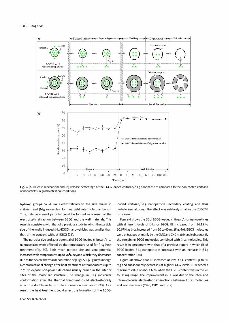
Fig. 5. (A) Release mechanism and (B) Release percentage of the EGCG-loaded chitosan/β-Lg nanoparticles compared to the non-coated chitosan
nanoparticles in gastrointestinal conditions.
Chitosan/β-Lactoglobulin nanoparticles for EGCG 1589
December 2016 | Vol. 25 | No. 6
Prolonged release of EGCG from EGCG-loaded chitosan/β-Lg
nanoparticles in simulated GI conditions The release properties of
double-walled nanoparticles in simulated GI tract conditions were
investigated by changing the pH and adding different digestive
enzymes. Swelling properties and degradation behavior during the
prolonged release process could also be estimated using control
chitosan nanoparticles without β-Lg. A schematic diagram of the
release process for double-walled nanoparticles as compared with
the control is shown in Fig. 5A. The controlled release mechanism of
the EGCG-loaded chitosan/β-Lg nanoparticles maybe involve several
different stages in the stomach (pepsin digestion and partial release)
and the small intestine (swelling, enzymatic degradation, and full
release).
Figure 5B shows the release profiles of EGCG from the EGCG-
loaded chitosan/β-Lg nanoparticles in simulated GI conditions. The
relative release rate of the EGCG-loaded chitosan/β-Lg nanoparticles
was determined by analyzing the release concentration of EGCG in
solution. Compared to the control group the EGCG-loaded chitosan/
β-Lg nanoparticles had a relatively slow release rate in simulated
stomach conditions. It suggested that the β-Lg secondary coating
improved the controlled release of EGCG. Moreover, the double-
walled nanoparticles also had a relatively low release rate as
compared to the control in simulated small intestine conditions. The
controlled release may be due to low external release (Fig. 5A). The
negatively charged β-Lg layered on the outer surface of the primary
CMC-CHC nanoparticles might bind free EGCG resulting in enhanced
EE. Another reason for the sustained release properties of the EGCG-
loaded chitosan/β-Lg nanoparticles could be that the β-Lg outer layer
was pepsin-resistant, which contributed to the relatively slow release
of EGCG in simulated stomach conditions. This is consistent with our
results that the EGCG release rate in the EGCG-loaded chitosan/β-Lg
nanoparticles at 20 min was comparable to that in controls at 5 min
(Fig. 5B). The release rate and degradation of EGCG-loaded chitosan/
β-Lg nanoparticles (20 and 60 min, respectively) in simulated stomach
conditions were slower than those of the control particles (5 and 30
min, respectively) as shown in Fig. 5B. These results suggested that
the CMC-CHC matrix coated with β-Lg could effectively prevent the
degradation of EGCG in the simulated gastric fluids.
Under simulated intestinal conditions, the β-Lg coating on the
EGCG-loaded chitosan/β-Lg nanoparticles was broken down into
small protein molecules with the change of PH value or peptides
chains by swelling and pancreatic enzymes, resulting in disintegration
of EGCG from the wall structure (8,12). Figure 5B clearly shows that
the EGCG amount released from EGCG-loaded chitosan/β-Lg
nanoparticles in the simulated small intestinal fluid is larger than that
released in the simulated stomach conditions. Due to the slower
degradation of the chitosan/β-Lg double-walled structure, the EGCG-
loaded chitosan/β-Lg nanoparticles maintained their prolonged
release characteristics as compared to the controls. Especially, the β-
lactoglobulin could be easily hydrolyzed by trypsin in neutral
aqueous solution (pH 7.7) (25). It may be the digestion of β-Lg
located in the outer of nanocarrier leading to its quick release in the
small intestine condition. In addition, after the external wall
degradation, the exposed chitosan chains can adhere to the
intestinal wall, thereby increasing residence time in the intestinal
tract, resulting in improved bioavailability of the core materials (12).
The prolonged release capabilities and excellent adhesion properties
of EGCG-loaded chitosan/β-Lg nanoparticles could enhance the
effective absorption of EGCG in the human intestine. These results
suggest that the chitosan/β-Lg nanoparticles could be an attractive
carrier for the encapsulation and oral administration of EGCG.
Acknowledgments This work was supported by National Modern
Agriculture Technology System (CARS-23), Anhui Major Demonstration
Project for Leading Talent Team on Tea Chemistry and Health, the
National Natural Science Foundation of China (31301448), the
National Foreign High-end Experts Project (GDW20153400195) and
the Korea-China Young Scientist Exchange Program (2013-2014). In
addition, this research was supported by the Bio-Synergy Research
Project (NRF-2013M3A9C4078159) of the Ministry of Science, ICT
and Future Planning through the National Research Foundation.
Disclosure The authors declare no conflict of interest.
References
1. Du GJ, Zhang Z, Wen XD, Yu C, Calway T, Yuan CS, Wang CZ. Epigallocatechin
Gallate (EGCG) is the most effective cancer chemopreventive polyphenol in
green tea. Nutrition 4: 1679-1691 (2012)
2. Chaudhury A, Das S. Recent advancement of chitosan-based nanoparticles for
oral controlled delivery of insulin and other therapeutic agents. AAPS
PharmSciTech 12: 10-20 (2011)
3. Chen L, Remondetto GE, Subirade M. Food protein-based materials as
nutraceutical delivery systems. Trends Food Sci. Tech. 17: 272-283 (2006)
4. Tang Y, Sun J, Fan H, Zhang X. An improved complex gel of modified gellan
gum and carboxymethyl chitosan for chondrocytes encapsulation. Carbohyd.
Polym. 88: 46-53 (2012)
5. Liang J, Li F, Fang Y, Yang W, An X, Zhao L, Xin Z, Cao L, Hu Q. Synthesis,
characterization and cytotoxicity studies of chitosan-coated tea polyphenols
nanoparticles. Colloid. Surface B 82: 297-301 (2011)
6. Liang J, Li F, Fang Y, Yang WJ, An XX, Zhao LY, Xin ZH, Hu QH. Response surface
methodology in the optimization of tea polyphenols-loaded chitosan
nanoclusters formulations. Eur. Food Res. Technol. 231: 917-924 (2010)
7. Hu B, Pan C, Sun Y, Hou Z, Ye H, Zeng X. Optimization of fabrication parameters
to produce chitosan-tripolyphosphate nanoparticles for delivery of tea
catechins. J. Agr. Food Chem. 56: 7451-7458 (2008)
8. Chen LY, Subirade M. Chitosan/b-lactoglobulin core-shell nanoparticles as
nutraceutical carriers. Biomaterials 26: 6041-6053 (2005)
9. Teng Z, Luo Y, Wang Q. Carboxymethyl chitosan-soy protein complex
nanoparticles for the encapsulation and controlled release of vitamin D3. Food
Chem. 141: 524-532 (2013)
10. Hu B, Ting Y, Zeng X, Huang Q. Cellular uptake and cytotoxicity of chitosan–
caseinophosphopeptides nanocomplexes loaded with epigallocatechin
gallate. Carbohyd. Polym. 89: 362-370 (2012)
11. Peram MR, Loveday SM, Ye A, Singh H. In vitro gastric digestion of heat-
induced aggregates of b-lactoglobulin. J. Dairy Sci. 96: 63-74 (2013)
12. Lee PS, Yim SG, Choi Y, Thi VAH, Ko S. Physiochemical properties and
prolonged release behaviours of chitosan-denatured beta-lactoglobulin
microcapsules for potential food applications. Food Chem. 134: 992-998
(2012)
13. Ha HK, Kim JW, Lee MR, Lee WJ. Formation and characterization of quercetin-
loaded chitosan oligosaccharide/β-lactoglobulin nanoparticle. Food Res. Int.
52: 82-90 (2013)
14. Hu B, Ting YW, Zeng XX, Huang QR. Bioactive peptides/chitosan nanoparticles
1590 Liang et al.
Food Sci. Biotechnol.
enhance cellular antioxidant activity of (-)-Epigallocatechin-3-gallate. J. Agr.
Food Chem. 61: 875-881 (2013)
15. Yang CS, Lambert JD, Sang S. Antioxidative and anti-carcinogenic activities of
tea polyphenols. Arch. Toxicol. 83: 11-21 (2009)
16. Mounsey JS, O'Kennedy BT, Fenelon MA, Brodkorb A. The effect of heating on
beta-lactoglobulin-chitosan mixtures as influenced by pH and ionic strength.
Food Hydrocolloid. 22: 65-73 (2008)
17. Cho Y, Kim JT, Park HJ. Size-controlled self-aggregated N-acyl chitosan
nanoparticles as a vitamin C carrier. Carbohyd. Polym. 88: 1087-1092 (2012)
18. Xing JF, Deng LD, Li J, Dong AJ. Amphiphilic poly{[alpha-maleic anhydride-
omega-methoxy-poly(ethylene glycol)]-co-(ethyl cyanoacrylate)} graft
copolymer nanoparticles as carriers for transdermal drug delivery. Int. J.
Nanomedicine 4: 227-232 (2009)
19. Vino AB, Ramasamy P, Shanmugam V, Shanmugam A. Extraction,
characterization and in vitro antioxidative potential of chitosan and sulfated
chitosan from Cuttlebone of Sepia aculeata Orbigny, 1848. Asian Pac. J. Trop.
Biomed. 2: S334-S341 (2012)
20. Gazori T, Khoshayand MR, Azizi E, Yazdizade P, Nomani A, Haririan I. Evaluation
of Alginate/Chitosan nanoparticles as antisense delivery vector: Formulation,
optimization and in vitro characterization. Carbohyd. Polym. 77: 599-606
(2009)
21. Shpigelman A, Cohen Y, Livney YD. Thermally-induced beta-lactoglobulin-
EGCG nanovehicles: Loading, stability, sensory and digestive-release study.
Food Hydrocolloid. 29: 57-67 (2012)
22. Chanasattru W, Jones OG, Decker EA, McClements DJ. Impact of cosolvents on
formation and properties of biopolymer nanoparticles formed by heat
treatment of beta-lactoglobulin-pectin complexes. Food Hydrocolloid. 23:
2450-2457 (2009)
23. Woo HD, Moon TW, Gunasekaran S, Ko S. Determining the gelation
temperature of â-lactoglobulin using in situ microscopic imaging. J. Dairy Sci.
96: 5565-5574 (2013)
24. Li B, Du WK, Jin JC, Du QZ. Preservation of (-)-Epigallocatechin-3-gallate
antioxidant properties loaded in heat treated b-lactoglobulin nanoparticles. J.
Agr. Food Chem. 60: 3477-3484 (2012)
25. Olsen K, Otte J, Skibsted LH. Steady-state kinetics and thermodynamics of the
hydrolysis of beta-lactoglobulin by trypsin. J. Agr. Food Chem. 48: 3086-3089
(2000)


![Bioresorbable microspheres as devices for the controlled ... · efficacious drug delivery technique is the use of nanoparticles [32] or microspheres [33]. Since the release of PTX](https://static.fdocument.org/doc/165x107/5f74acc2250dba119220991c/bioresorbable-microspheres-as-devices-for-the-controlled-efficacious-drug-delivery.jpg)