Synthesis and Characterization of Conformationally ... and Characterization of Conformationally...
14
rXXXX American Chemical Society A dx.doi.org/10.1021/jo200482d | J. Org. Chem. XXXX, XXX, 000–000 ARTICLE pubs.acs.org/joc Synthesis and Characterization of Conformationally Preorganized, (R)-Diethylene Glycol-Containing γ-Peptide Nucleic Acids with Superior Hybridization Properties and Water Solubility Bichismita Sahu, Iulia Sacui, Srinivas Rapireddy, Kimberly J. Zanotti, Raman Bahal, Bruce A. Armitage, and Danith H. Ly* Department of Chemistry and Center for Nucleic Acids Science and Technology (CNAST), Carnegie Mellon University, 4400 Fifth Avenue, Pittsburgh, Pennsylvania 15213, United States b S Supporting Information ’ INTRODUCTION Peptide nucleic acid (PNA) is a promising class of nucleic acid mimic developed in the past two decades in which the naturally occurring sugar phosphodiester backbone is replaced with N-(2- aminoethyl)glycine units (Chart 1). 1 PNA can hybridize to cDNA or RNA just as the natural counterpart, in accordance with the WatsonCrick base-pairing rules, but with higher affinity and sequence selectivity. 2 Furthermore, PNA can invade selected sequences of double-stranded DNA (dsDNA). 35 The improve- ment in thermodynamic stability has been attributed, in part, to the lack of electrostatic repulsion in the backbone. 1 The other contribution may come from counterion release upon hybridiza- tion, as opposed to condensation taking place with DNA and RNA, resulting in an increase in the overall entropy of the system. 6 The enhancement in sequence selectivity, however, is less well understood. 7 Structural studies suggested that hydration may play a key role in rigidifying the backbone of PNA upon hybridization to DNA or RNA (or PNA), making it less accom- modating to structural mismatches. Structural or spinal water molecules have been observed in the X-ray structures of PNA DNA 8 and PNAPNA 911 duplexes bridging the amide proton in the backbone to the adjacent nucleobase. Besides hybridiza- tion properties, another appealing aspect of PNA is enzymatic stability. Because of its unnatural backbone, PNA is not easily degraded by proteases or nucleases. 12 These properties, along with the ease 13,14 and flexibility 15 of synthesis, together make PNA an attractive reagent for many applications in biology, biotech- nology, and medicine. 16,17 PNA has so far been employed in a number of applications, from molecular tools for probing and manipulating nucleic acid structures and functions 1821 and regulation of gene expression 22,23 to the recognition code for tagging molecules in drug discovery, 24,25 amplifying genetic information in molecular evolution, 26,27 and organizing molecular self-assembly in materials science and nanotechnology. 2832 In spite of its many appealing features, however, PNA has one major setback as compared to other classes of oligonucleotides. Because of the charge-neutral back- bone, PNA is only moderately soluble in aqueous solution. Furthermore, it has a tendency to aggregate and adhere to surfaces and other macromolecules in a nonspecific manner. 33,34 This inherent property posts a considerable technical challenge for the handling and processing of PNA—for instance, in the development of microarrays and related surface-bound applica- tions, because of its propensity to aggregate and collapse onto the surface, causing poor and nonspecific binding, 35,36 and in biology and medicine, in part because of the concerns for off-target binding and cytotoxicity. 34 In attempts to address this concern, several approaches have been taken, including incorporation of charged amino acid Received: March 18, 2011 ABSTRACT: Developed in the early 1990s, peptide nucleic acid (PNA) has emerged as a promising class of nucleic acid mimic because of its strong binding affinity and sequence selectivity toward DNA and RNA and resistance to enzymatic degradation by proteases and nucleases; however, the main draw- backs, as compared to other classes of oligonucleotides, are water solubility and biocompatibility. Herein we show that installation of a relatively small, hydro- philic (R)-diethylene glycol (“miniPEG”, R-MP) unit at the γ-backbone trans- forms a randomly folded PNA into a right-handed helix. Synthesis of optically pure R-MP γPNA monomers is described, which can be accomplished in a few simple steps from a commercially available and relatively cheap Boc-L-serine. Once synthesized, R-MP γPNA oligomers are preorganized into a right-handed helix, hybridize to DNA and RNA with greater affinity and sequence selectivity, and are more water soluble and less aggregating than the parental PNA oligomers. The results presented herein have important implications for the future design and application of PNA in biology, biotechnology, and medicine, as well as in other disciplines, including drug discovery and molecular engineering.
Transcript of Synthesis and Characterization of Conformationally ... and Characterization of Conformationally...
rXXXX American Chemical Society A dx.doi.org/10.1021/jo200482d | J. Org. Chem. XXXX, XXX, 000–000
ARTICLE
pubs.acs.org/joc
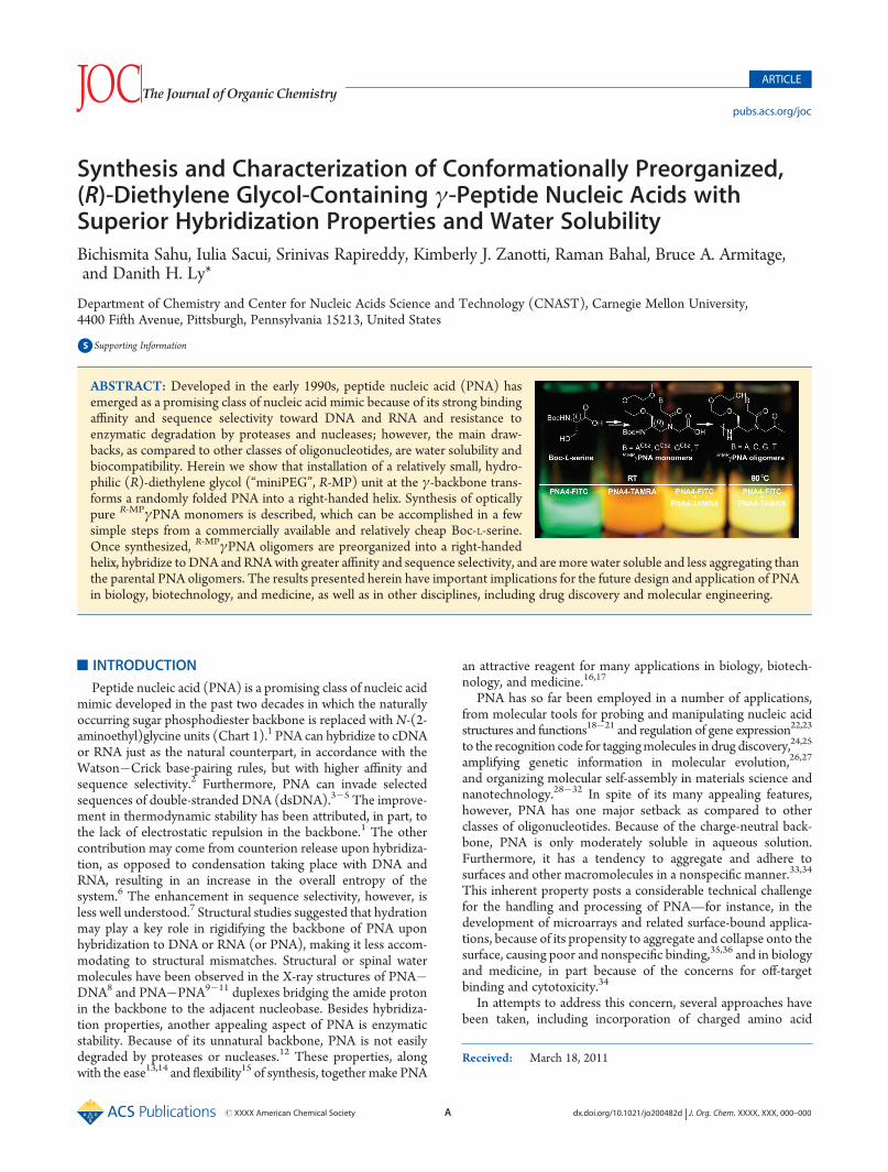
Synthesis and Characterization of Conformationally Preorganized,(R)-Diethylene Glycol-Containing γ-Peptide Nucleic Acids withSuperior Hybridization Properties and Water SolubilityBichismita Sahu, Iulia Sacui, Srinivas Rapireddy, Kimberly J. Zanotti, Raman Bahal, Bruce A. Armitage,and Danith H. Ly*
Department of Chemistry and Center for Nucleic Acids Science and Technology (CNAST), Carnegie Mellon University,4400 Fifth Avenue, Pittsburgh, Pennsylvania 15213, United States
bS Supporting Information
’ INTRODUCTION
Peptide nucleic acid (PNA) is a promising class of nucleic acidmimic developed in the past two decades in which the naturallyoccurring sugar phosphodiester backbone is replaced with N-(2-aminoethyl)glycine units (Chart 1).1 PNA can hybridize to cDNAor RNA just as the natural counterpart, in accordance with theWatson�Crick base-pairing rules, but with higher affinity andsequence selectivity.2 Furthermore, PNA can invade selectedsequences of double-stranded DNA (dsDNA).3�5 The improve-ment in thermodynamic stability has been attributed, in part, tothe lack of electrostatic repulsion in the backbone.1 The othercontribution may come from counterion release upon hybridiza-tion, as opposed to condensation taking place with DNA andRNA, resulting in an increase in the overall entropy of thesystem.6 The enhancement in sequence selectivity, however, isless well understood.7 Structural studies suggested that hydrationmay play a key role in rigidifying the backbone of PNA uponhybridization to DNA or RNA (or PNA), making it less accom-modating to structural mismatches. Structural or spinal watermolecules have been observed in the X-ray structures of PNA�DNA8 and PNA�PNA9�11 duplexes bridging the amide protonin the backbone to the adjacent nucleobase. Besides hybridiza-tion properties, another appealing aspect of PNA is enzymaticstability. Because of its unnatural backbone, PNA is not easilydegraded by proteases or nucleases.12 These properties, alongwith the ease13,14 and flexibility15 of synthesis, together make PNA
an attractive reagent for many applications in biology, biotech-nology, and medicine.16,17
PNA has so far been employed in a number of applications,from molecular tools for probing and manipulating nucleic acidstructures and functions18�21 and regulation of gene expression22,23
to the recognition code for taggingmolecules in drug discovery,24,25
amplifying genetic information in molecular evolution,26,27
and organizing molecular self-assembly in materials science andnanotechnology.28�32 In spite of its many appealing features,however, PNA has one major setback as compared to otherclasses of oligonucleotides. Because of the charge-neutral back-bone, PNA is only moderately soluble in aqueous solution.Furthermore, it has a tendency to aggregate and adhere tosurfaces and other macromolecules in a nonspecific manner.33,34
This inherent property posts a considerable technical challengefor the handling and processing of PNA—for instance, in thedevelopment of microarrays and related surface-bound applica-tions, because of its propensity to aggregate and collapse onto thesurface, causing poor and nonspecific binding,35,36 and in biologyand medicine, in part because of the concerns for off-targetbinding and cytotoxicity.34
In attempts to address this concern, several approaches havebeen taken, including incorporation of charged amino acid
Received: March 18, 2011
ABSTRACT: Developed in the early 1990s, peptide nucleic acid (PNA) hasemerged as a promising class of nucleic acid mimic because of its strong bindingaffinity and sequence selectivity toward DNA and RNA and resistance toenzymatic degradation by proteases and nucleases; however, the main draw-backs, as compared to other classes of oligonucleotides, are water solubility andbiocompatibility. Herein we show that installation of a relatively small, hydro-philic (R)-diethylene glycol (“miniPEG”, R-MP) unit at the γ-backbone trans-forms a randomly folded PNA into a right-handed helix. Synthesis of opticallypure R-MPγPNA monomers is described, which can be accomplished in a fewsimple steps from a commercially available and relatively cheap Boc-L-serine.Once synthesized, R-MPγPNA oligomers are preorganized into a right-handedhelix, hybridize to DNA and RNAwith greater affinity and sequence selectivity, and are more water soluble and less aggregating thanthe parental PNA oligomers. The results presented herein have important implications for the future design and application of PNAin biology, biotechnology, and medicine, as well as in other disciplines, including drug discovery and molecular engineering.
B dx.doi.org/10.1021/jo200482d |J. Org. Chem. XXXX, XXX, 000–000
The Journal of Organic Chemistry ARTICLE
residues, such as lysine at the termini37 or in the interior part ofthe oligomer,38,39 inclusion of polar groups in the backbone,40
carboxymethylene bridge,41 and nucleobases,25,42 replacementof the original (aminoethyl)glycyl backbone skeleton with a nega-tively charged scaffold,43,44 conjugation of high molecular weightpolyethylene glycol (PEG) to one of the termini,36,45 fusion ofPNA toDNA to generate a chimeric oligomer,46�50 and redesignof the backbone architecture.51 These chemical modificationshave led to improvement in solubility, but it is often achieved atthe expense of binding affinity and/or sequence specificity, not tomention the requirement of an elaborate synthetic scheme insome cases. Incorporation of cationic residues can improve watersolubility, but it can also lead to nonspecific binding due to anincrease in charge�charge interaction upon hybridization toDNA or RNA. Conjugation of PNA to DNA (or RNA) can im-prove water solubility as well, but it can compromise bindingaffinity due to an increase in charge�charge repulsion andconformational heterogeneity in the backbone.46,47,49 Hereinwe report the development of a new class of conformationallypreorganized, diethylene glycol-containing γPNAs that exhibitsuperior hybridization properties and water solubility as com-pared to the original PNA design or any other chiral γPNA thathas been developed to date.
’RESULTS AND DISCUSSION
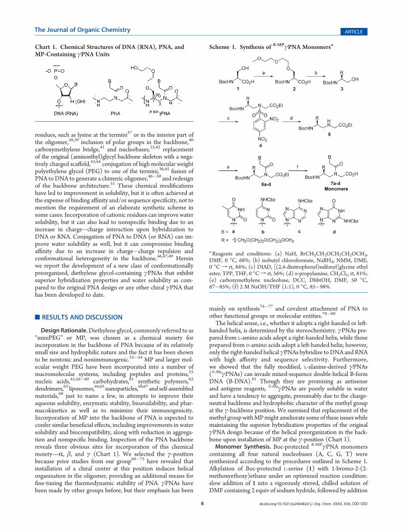
DesignRationale.Diethylene glycol, commonly referred to as“miniPEG” or MP, was chosen as a chemical moiety forincorporation in the backbone of PNA because of its relativelysmall size and hydrophilic nature and the fact it has been shownto be nontoxic and nonimmunogenic.52�54 MP and larger mol-ecular weight PEG have been incorporated into a number ofmacromolecular systems, including peptides and proteins,55
nucleic acids,45,56�60 carbohydrates,61 synthetic polymers,62
dendrimers,63 liposomes,64,65 nanoparticles,66,67 and self-assembledmaterials,68 just to name a few, in attempts to improve theiraqueous solubility, enzymatic stability, bioavailability, and phar-macokinetics as well as to minimize their immunogenicity.Incorporation of MP into the backbone of PNA is expected toconfer similar beneficial effects, including improvements in watersolubility and biocompatibility, along with reduction in aggrega-tion and nonspecific binding. Inspection of the PNA backbonereveals three obvious sites for incorporation of this chemicalmoiety—R, β, and γ (Chart 1). We selected the γ-positionbecause prior studies from our group69�73 have revealed thatinstallation of a chiral center at this position induces helicalorganization in the oligomer, providing an additional means forfine-tuning the thermodynamic stability of PNA. γPNAs havebeen made by other groups before, but their emphasis has been
mainly on synthesis74�77 and covalent attachment of PNA toother functional groups or molecular entities.78�80
The helical sense, i.e., whether it adopts a right-handed or left-handed helix, is determined by the stereochemistry. γPNAs pre-pared from L-amino acids adopt a right-handed helix, while thoseprepared from D-amino acids adopt a left-handed helix; however,only the right-handed helical γPNAs hybridize to DNA and RNAwith high affinity and sequence selectivity. Furthermore,we showed that the fully modified, L-alanine-derived γPNAs(S-AlaγPNAs) can invade mixed-sequence double-helical B-formDNA (B-DNA).81 Though they are promising as antisenseand antigene reagents, S-AlaγPNAs are poorly soluble in waterand have a tendency to aggregate, presumably due to the charge-neutral backbone and hydrophobic character of the methyl groupat the γ-backbone position. We surmised that replacement of themethyl groupwithMPmight ameliorate someof these issues whilemaintaining the superior hybridization properties of the originalγPNA design because of the helical preorganization in the back-bone upon installation of MP at the γ-position (Chart 1).Monomer Synthesis. Boc-protected R-MPγPNA monomers
containing all four natural nucleobases (A, C, G, T) weresynthesized according to the procedures outlined in Scheme 1.Alkylation of Boc-protected L-serine (1) with 1-bromo-2-(2-methoxyethoxy)ethane under an optimized reaction condition:slow addition of 1 into a vigorously stirred, chilled solution ofDMF containing 2 equiv of sodium hydride, followed by addition
Chart 1. Chemical Structures of DNA (RNA), PNA, andMP-Containing γPNA Units
Scheme 1. Synthesis of R-MPγPNA Monomersa
aReagents and conditions: (a) NaH, BrCH2CH2OCH2CH2OCH3,DMF, 0 �C, 68%; (b) isobutyl chloroformate, NaBH4, NMM, DME,0 �C f rt, 84%; (c) DIAD, [(2,4-dinitrophenyl)sulfonyl]glycine ethylester, TPP, THF, 0 �Cf rt, 56%; (d) n-propylamine, CH2Cl2, rt, 81%;(e) carboxymethylene nucleobase, DCC, DhbtOH, DMF, 50 �C,67�85%; (f) 2 M NaOH/THF (1:1), 0 �C, 85�98%.
C dx.doi.org/10.1021/jo200482d |J. Org. Chem. XXXX, XXX, 000–000
The Journal of Organic Chemistry ARTICLE
of 1-bromo-2-(2-methoxyethoxy)ethane afforded compound 2withhigh optical purity (see the section below). The stoichiometryand order of addition were determined to be essential for ob-taining optically pure product. Slow addition of sodium hydridewas necessary to ensure complete deprotonation of the carboxylgroup prior to removal of the hydroxyl proton. Formation of thecarboxylate anion reduces the acidity of the R-proton, making itless susceptible to deprotonation by base. Esterification of thealkylated product 2 followed by reduction with sodium borohy-dride yielded serinol 3. This chemical transformation, conversionof the carboxylic acid to alcohol, renders the R-proton inert todeprotonation and racemization in the subsequent reactionsteps. Coupling 3 toN-[(2,4-dinitrophenyl)sulfonyl]glycine ethylacetate, whichwas prepared according to the published procedure,82
employing the Mitsunobu reaction followed by removal of the(2,4-dinitrophenyl)sulfonyl group with a mild base yieldedcompound 5. DCC-mediated coupling of 5 with the appropriatecarboxymethylene nucleobases (A,83 C,14 G,83 T13) and hydro-lysis of the resulting esters gave the desired monomers 7a�d.Determination of Optical Purities. The optical purities of
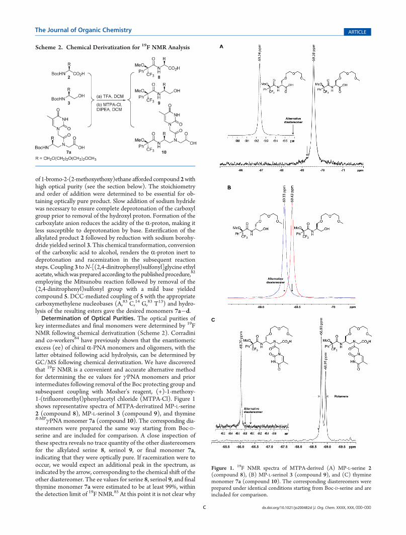
key intermediates and final monomers were determined by 19FNMR following chemical derivatization (Scheme 2). Corradiniand co-workers84 have previously shown that the enantiomericexcess (ee) of chiral R-PNA monomers and oligomers, with thelatter obtained following acid hydrolysis, can be determined byGC/MS following chemical derivatization. We have discoveredthat 19F NMR is a convenient and accurate alternative methodfor determining the ee values for γPNA monomers and priorintermediates following removal of the Boc protecting group andsubsequent coupling with Mosher’s reagent, (+)-1-methoxy-1-(trifluoromethyl)phenylacetyl chloride (MTPA-Cl). Figure 1shows representative spectra of MTPA-derivatized MP-L-serine2 (compound 8), MP-L-serinol 3 (compound 9), and thymineR-MPγPNA monomer 7a (compound 10). The corresponding dia-stereomers were prepared the same way starting from Boc-D-serine and are included for comparison. A close inspection ofthese spectra reveals no trace quantity of the other diastereomersfor the alkylated serine 8, serinol 9, or final monomer 7a,indicating that they were optically pure. If racemization were tooccur, we would expect an additional peak in the spectrum, asindicated by the arrow, corresponding to the chemical shift of theother diastereomer. The ee values for serine 8, serinol 9, and finalthymine monomer 7a were estimated to be at least 99%, withinthe detection limit of 19F NMR.85 At this point it is not clear why
Scheme 2. Chemical Derivatization for 19F NMR Analysis
Figure 1. 19F NMR spectra of MTPA-derived (A) MP-L-serine 2(compound 8), (B) MP-L-serinol 3 (compound 9), and (C) thyminemonomer 7a (compound 10). The corresponding diastereomers wereprepared under identical conditions starting from Boc-D-serine and areincluded for comparison.
D dx.doi.org/10.1021/jo200482d |J. Org. Chem. XXXX, XXX, 000–000
The Journal of Organic Chemistry ARTICLE
the thymine R-MPγPNAmonomer showed two rotamers (Figure 1C),at �68.80 and �68.95 ppm, while thymine S-MPγPNA showedonly one (Figure 1C, inset). (The existence of the two rotamerswas determined by multinuclear and multidimensional NMRanalyses as described in an earlier work;73 they differ from oneanother in the rotation around the C7�N4 tertiary amide bond.)We attribute the high optical purities of the intermediates andfinal monomers (the others not shown) to the optimized, initialalkylation step and the stereoselective conversion of carboxylicacid to alcohol in the second step, on the basis of the procedurereported by Rodriguez and co-workers.86
Oligomer Design and Synthesis. Three sets of PNA oligo-mers were prepared (Table 1). The first set, comprising PNA1�5,was designed to test the effects of MP on the conformation andhybridization properties of PNA. The second set, consisting ofPNA6�10, was designed to test the effect of MP on watersolubility. The hexadecameric sequence was chosen for this studybecause it represents a statistical length that would be required totarget a unique site within the mammalian genome or transcrip-tome. The third set, constituting PNA1 and PNA4, each sepa-rately linked to fluorescein (FITC) at the N-terminus (PNA1Xand PNA4X) and tetramethylrhodamine (TAMRA) at the C-terminus (PNA1Y and PNA4), was designed to test the effect ofMP on self-aggregation on the basis of F€orster resonance energytransfer (FRET). Inclusion of a lysine residue at the C-terminuswas necessary, since prior attempts to synthesize FITC- andTAMRA-containing PNA oligomers without the lysine residueresulted in sticky and poorly water soluble materials that weredifficult to purify and characterize.All PNA oligomers, those with and without MP side chains, were
synthesizedon a solid support according to thepublishedproceduresof Christensen and co-workers .87 Unlike modifications made at theR-backbone, which require further optimization of the reactioncondition to minimize racemization during the on-resin coupling,88
no precaution was necessary for coupling of R-MPγPNA monomerson the resin. Upon completion of the last monomer coupling, theoligomers were cleaved from the resin and precipitated with ethylether. After being air-dried, the crude pellets were dissolved in awater/acetonitrile mixture (80:20), purified by reversed-phaseHPLC, and characterized by MALDI-TOF mass spectrometry.
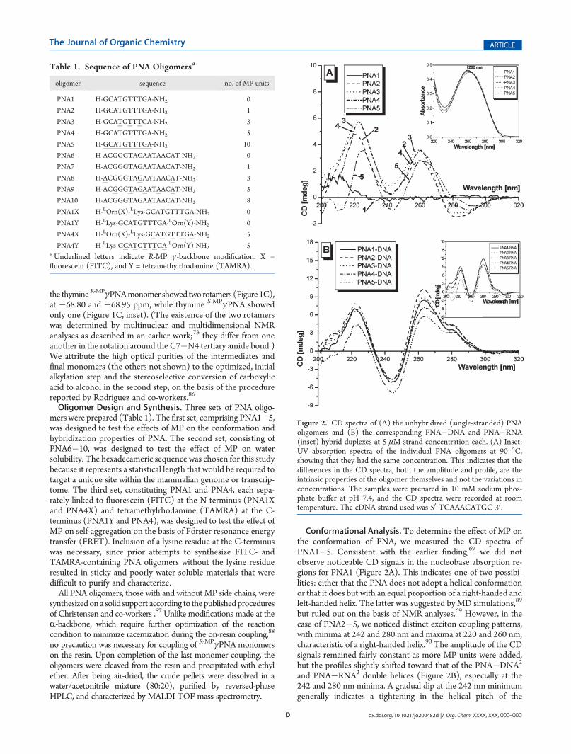
Conformational Analysis. To determine the effect of MP onthe conformation of PNA, we measured the CD spectra ofPNA1�5. Consistent with the earlier finding,69 we did notobserve noticeable CD signals in the nucleobase absorption re-gions for PNA1 (Figure 2A). This indicates one of two possibi-lities: either that the PNA does not adopt a helical conformationor that it does but with an equal proportion of a right-handed andleft-handed helix. The latter was suggested by MD simulations,89
but ruled out on the basis of NMR analyses.69 However, in thecase of PNA2�5, we noticed distinct exciton coupling patterns,with minima at 242 and 280 nm and maxima at 220 and 260 nm,characteristic of a right-handed helix.90 The amplitude of the CDsignals remained fairly constant as more MP units were added,but the profiles slightly shifted toward that of the PNA�DNA2
and PNA�RNA2 double helices (Figure 2B), especially at the242 and 280 nm minima. A gradual dip at the 242 nm minimumgenerally indicates a tightening in the helical pitch of the
Table 1. Sequence of PNA Oligomersa
oligomer sequence no. of MP units
PNA1 H-GCATGTTTGA-NH2 0
PNA2 H-GCATGTTTGA-NH2 1
PNA3 H-GCATGTTTGA-NH2 3
PNA4 H-GCATGTTTGA-NH2 5
PNA5 H-GCATGTTTGA-NH2 10
PNA6 H-ACGGGTAGAATAACAT-NH2 0
PNA7 H-ACGGGTAGAATAACAT-NH2 1
PNA8 H-ACGGGTAGAATAACAT-NH2 3
PNA9 H-ACGGGTAGAATAACAT-NH2 5
PNA10 H-ACGGGTAGAATAACAT-NH2 8
PNA1X H-LOrn(X)-LLys-GCATGTTTGA-NH2 0
PNA1Y H-LLys-GCATGTTTGA-LOrn(Y)-NH2 0
PNA4X H-LOrn(X)-LLys-GCATGTTTGA-NH2 5
PNA4Y H-LLys-GCATGTTTGA-LOrn(Y)-NH2 5aUnderlined letters indicate R-MP γ-backbone modification. X =fluorescein (FITC), and Y = tetramethylrhodamine (TAMRA).
Figure 2. CD spectra of (A) the unhybridized (single-stranded) PNAoligomers and (B) the corresponding PNA�DNA and PNA�RNA(inset) hybrid duplexes at 5 μM strand concentration each. (A) Inset:UV absorption spectra of the individual PNA oligomers at 90 �C,showing that they had the same concentration. This indicates that thedifferences in the CD spectra, both the amplitude and profile, are theintrinsic properties of the oligomer themselves and not the variations inconcentrations. The samples were prepared in 10 mM sodium phos-phate buffer at pH 7.4, and the CD spectra were recorded at roomtemperature. The cDNA strand used was 50-TCAAACATGC-30.
E dx.doi.org/10.1021/jo200482d |J. Org. Chem. XXXX, XXX, 000–000
The Journal of Organic Chemistry ARTICLE
oligomer,91 from one that resembles that of a PNA�PNA92
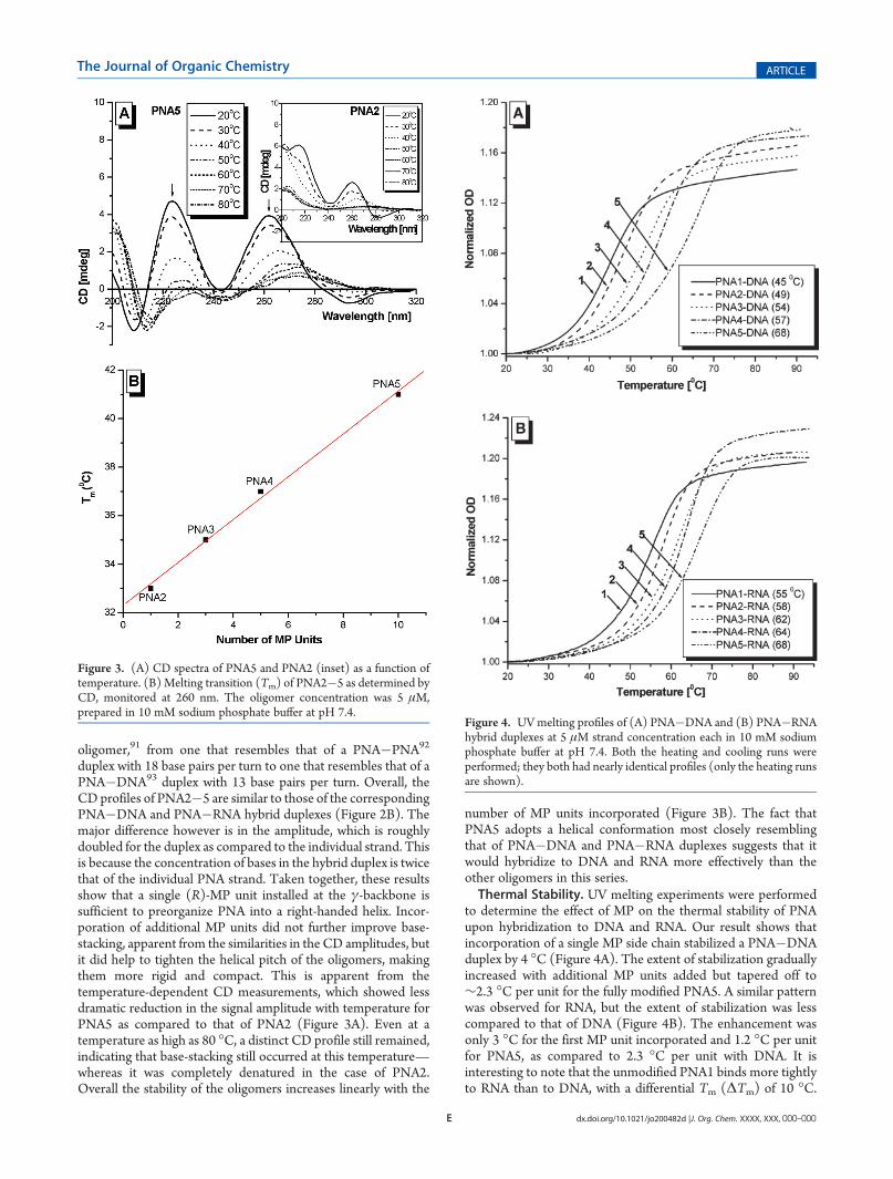
duplex with 18 base pairs per turn to one that resembles that of aPNA�DNA93 duplex with 13 base pairs per turn. Overall, theCD profiles of PNA2�5 are similar to those of the correspondingPNA�DNA and PNA�RNA hybrid duplexes (Figure 2B). Themajor difference however is in the amplitude, which is roughlydoubled for the duplex as compared to the individual strand. Thisis because the concentration of bases in the hybrid duplex is twicethat of the individual PNA strand. Taken together, these resultsshow that a single (R)-MP unit installed at the γ-backbone issufficient to preorganize PNA into a right-handed helix. Incor-poration of additional MP units did not further improve base-stacking, apparent from the similarities in the CD amplitudes, butit did help to tighten the helical pitch of the oligomers, makingthem more rigid and compact. This is apparent from thetemperature-dependent CD measurements, which showed lessdramatic reduction in the signal amplitude with temperature forPNA5 as compared to that of PNA2 (Figure 3A). Even at atemperature as high as 80 �C, a distinct CD profile still remained,indicating that base-stacking still occurred at this temperature—whereas it was completely denatured in the case of PNA2.Overall the stability of the oligomers increases linearly with the
number of MP units incorporated (Figure 3B). The fact thatPNA5 adopts a helical conformation most closely resemblingthat of PNA�DNA and PNA�RNA duplexes suggests that itwould hybridize to DNA and RNA more effectively than theother oligomers in this series.Thermal Stability. UV melting experiments were performed
to determine the effect of MP on the thermal stability of PNAupon hybridization to DNA and RNA. Our result shows thatincorporation of a single MP side chain stabilized a PNA�DNAduplex by 4 �C (Figure 4A). The extent of stabilization graduallyincreased with additional MP units added but tapered off to∼2.3 �C per unit for the fully modified PNA5. A similar patternwas observed for RNA, but the extent of stabilization was lesscompared to that of DNA (Figure 4B). The enhancement wasonly 3 �C for the first MP unit incorporated and 1.2 �C per unitfor PNA5, as compared to 2.3 �C per unit with DNA. It isinteresting to note that the unmodified PNA1 binds more tightlyto RNA than to DNA, with a differential Tm (ΔTm) of 10 �C.
Figure 3. (A) CD spectra of PNA5 and PNA2 (inset) as a function oftemperature. (B) Melting transition (Tm) of PNA2�5 as determined byCD, monitored at 260 nm. The oligomer concentration was 5 μM,prepared in 10 mM sodium phosphate buffer at pH 7.4.
Figure 4. UVmelting profiles of (A) PNA�DNA and (B) PNA�RNAhybrid duplexes at 5 μM strand concentration each in 10 mM sodiumphosphate buffer at pH 7.4. Both the heating and cooling runs wereperformed; they both had nearly identical profiles (only the heating runsare shown).
F dx.doi.org/10.1021/jo200482d |J. Org. Chem. XXXX, XXX, 000–000
The Journal of Organic Chemistry ARTICLE
However, fully modified PNA5 displayed thermal stability iden-tical with that of RNA as well as that of DNA. The lack of apreferential binding for PNA5 is not clearly understood at thispoint, but it may reflect the rigidity of the backbone. BecausePNA5 is more rigid and tightly wound as compared to PNA1, itmay not have the flexibility or conformational freedom to ac-commodate the DNA and RNA template strands. Under suchcircumstances, the DNA and RNA strands would have to under-go a conformational change of their own to accommodatethe R-MPγPNA helix and for hybridization to take place. This mayexplain why S-AlaγPNA�DNA prefers a P-form helix,73 a struc-ture that is intermediate between the A- and B-form DNA, anindication that DNA and RNA accommodate the γPNA ex-igencies rather than the other way around. The RNA strand is lessaccommodating to the R-MPγPNA, when it is fully modified,because the RNA backbone is more rigid as compared to that ofDNA due to the presence of the chiral OH group at the 20-position.94
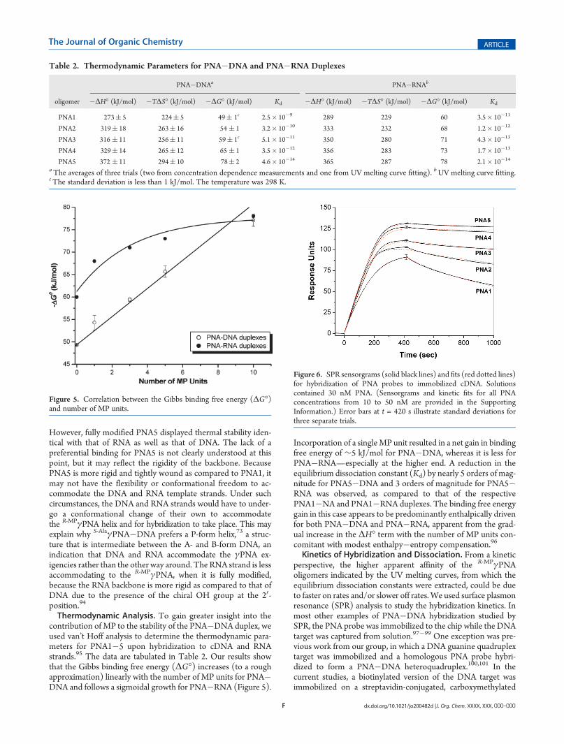
Thermodynamic Analysis. To gain greater insight into thecontribution of MP to the stability of the PNA�DNA duplex, weused van’t Hoff analysis to determine the thermodynamic para-meters for PNA1�5 upon hybridization to cDNA and RNAstrands.95 The data are tabulated in Table 2. Our results showthat the Gibbs binding free energy (ΔG�) increases (to a roughapproximation) linearly with the number of MP units for PNA�DNA and follows a sigmoidal growth for PNA�RNA (Figure 5).
Incorporation of a singleMP unit resulted in a net gain in bindingfree energy of ∼5 kJ/mol for PNA�DNA, whereas it is less forPNA�RNA—especially at the higher end. A reduction in theequilibrium dissociation constant (Kd) by nearly 5 orders of mag-nitude for PNA5�DNA and 3 orders of magnitude for PNA5�RNA was observed, as compared to that of the respectivePNA1�NA and PNA1�RNA duplexes. The binding free energygain in this case appears to be predominantly enthalpically drivenfor both PNA�DNA and PNA�RNA, apparent from the grad-ual increase in the ΔH� term with the number of MP units con-comitant with modest enthalpy�entropy compensation.96
Kinetics of Hybridization and Dissociation. From a kineticperspective, the higher apparent affinity of the R-MPγPNAoligomers indicated by the UV melting curves, from which theequilibrium dissociation constants were extracted, could be dueto faster on rates and/or slower off rates. We used surface plasmonresonance (SPR) analysis to study the hybridization kinetics. Inmost other examples of PNA�DNA hybridization studied bySPR, the PNA probe was immobilized to the chip while the DNAtarget was captured from solution.97�99 One exception was pre-vious work from our group, in which a DNA guanine quadruplextarget was immobilized and a homologous PNA probe hybri-dized to form a PNA�DNA heteroquadruplex.100,101 In thecurrent studies, a biotinylated version of the DNA target wasimmobilized on a streptavidin-conjugated, carboxymethylated
Table 2. Thermodynamic Parameters for PNA�DNA and PNA�RNA Duplexes
PNA�DNAa PNA�RNAb
oligomer �ΔH� (kJ/mol) �TΔS� (kJ/mol) �ΔG� (kJ/mol) Kd �ΔH� (kJ/mol) �TΔS� (kJ/mol) �ΔG� (kJ/mol) Kd
PNA1 273( 5 224( 5 49( 1c 2.5� 10�9 289 229 60 3.5� 10�11
PNA2 319( 18 263( 16 54 ( 1 3.2� 10�10 333 232 68 1.2� 10�12
PNA3 316 ( 11 256( 11 59( 1c 5.1� 10�11 350 280 71 4.3� 10�13
PNA4 329( 14 265( 12 65 ( 1 3.5� 10�12 356 283 73 1.7� 10�13
PNA5 372 ( 11 294( 10 78( 2 4.6� 10�14 365 287 78 2.1� 10�14
aThe averages of three trials (two from concentration dependence measurements and one from UV melting curve fitting). bUV melting curve fitting.cThe standard deviation is less than 1 kJ/mol. The temperature was 298 K.
Figure 5. Correlation between the Gibbs binding free energy (ΔG�)and number of MP units.
Figure 6. SPR sensorgrams (solid black lines) and fits (red dotted lines)for hybridization of PNA probes to immobilized cDNA. Solutionscontained 30 nM PNA. (Sensorgrams and kinetic fits for all PNAconcentrations from 10 to 50 nM are provided in the SupportingInformation.) Error bars at t = 420 s illustrate standard deviations forthree separate trials.
G dx.doi.org/10.1021/jo200482d |J. Org. Chem. XXXX, XXX, 000–000
The Journal of Organic Chemistry ARTICLE
dextran chip. A relatively low surface density (ca. 100 responseunits) of DNA target was used to limit mass transport effects onthe association kinetics. Solutions containing 10�50 nM PNAwere allowed to flow over the chip for 420 s, at which point theflow was switched to a PNA-free buffer to allow net dissociationof the hybridized PNA.Individual sensorgrams for the unmodified (PNA1) and
R-MPγ-modified (PNA2�5) oligomers at 30 nM concentrationare shown in Figure 6. Relatively minor variation is observed inthe association kinetics, although singly modified PNA2 appearsto bind approximately twice as fast as the unmodified PNA.Fitting the data to a 1:1 binding model yields association rateconstants (ka) that range from 4.7 � 105 to 9.7 � 105 M�1 s�1
(Table 3). In contrast, significantly larger effects are seen in thedissociation phase of the experiment, with the dissociation rateconstant (kd) varying by at least a factor of 50. Equilibriumdissociation constants (Kd) calculated from the ratio of the dis-sociation and association rate constants are also given in Table 3.Unmodified PNA1 and fully modified PNA5 have Kd = 2.8 nMand 54 pM, respectively. Note that the Kd values for PNA1�3determined by SPR (Table 3) are similar to those determined byUV melting experiments (Table 2). However, increasing diver-gences are observed for PNA4 and PNA5, with the SPR-derivedvalues being 12- and 1200-fold greater, respectively. We attributethis discrepancy to the very small degrees of dissociationobserved within the time scale of the SPR experiment. This in-troduces a large uncertainty into the fitting of the dissociationphases of the sensorgrams, translating into questionable Kd values.However, we also note that the free energy changes given in Table 2are based on the assumption that the heat capacity change forPNA�DNAmelting is zero, allowing the parameters determined atthe high melting temperature to be extrapolated to lower tempera-tures. Future calorimetric measurements will allow us to determinewhether there is a heat capacity change in these hybridization/melting transitions. Nevertheless, the SPR results clearly demon-strate that the enhanced affinity of the R-MPγPNAs is due almostentirely to the significantly slower dissociation kinetics. Thus, thehelical preorganization of the modified PNA does not translate intosignificantly faster hybridization. This could be due to the fact thatthe cDNA strand is likely to require some structural reorganizationof its own to hybridize to the PNA, negating the preorganization ofthe latter. While the origin of the slower dissociation kinetics is notclear at this time, one possible contributor is the network ofstructuredwatermolecules in theminor groove of the hybrid duplex,which would need to be disrupted to separate the two strands.Sequence Selectivity. On the basis of the CD, NMR,69 and
X-ray73 data which showed that γPNAs derived from L-amino
acids adopt a right-handed helix and that the helix becomes morerigid as more γ-chiral units are added in the backbone, one wouldexpect a fully modified PNA5 to hybridize to DNA and RNAtargets with greater sequence selectivity than PNA1. To verifythis prediction, we determined the thermal stabilities of PNA5�DNA and PNA5�RNA duplexes containing perfectly matched(PM) and single-base-mismatched (MM) targets and comparedthem to those from an earlier study with PNA1�DNA andPNA1�RNA.69 Our results showed that, despite the strong bind-ing affinity, PNA5 is able to discriminate between closely relatedsequences. ΔTm ranges from �17 to �21 �C for PNA5�DNAand from �16 to �20 �C for PNA5�RNA containing a single-base mismatch (X = C, G, T), as compared to�10 to�14 �C forPNA1�DNA and�11 to�18 for PNA1�RNA (Table 4). Thelevel of sequence discrimination is greater for PNA5�DNA thanfor PNA1�DNA, and similar, if not slightly better, for PNA5�RNA as compared to PNA1�RNA. This result is consistent withPNA5 adopting a more rigid helical motif, which is less accom-modating to structural mismatches as compared to PNA1.Water Solubility. To determine whether inclusion of MP in
the backbone has an effect on water solubility, we determined thesaturating concentrations of PNA6�10 (Table 1) in aqueoussolution using UV spectroscopy. It is interesting to note thatincorporation of a single MP unit enhanced the solubility ofPNA6 by nearly 2-fold (Table 5). The solubility of the oligomersis further improved, albeit to a smaller extent, with additional MPunits. For PNA8, which contained eight MP units, we could notdetermine the saturating concentration of this oligomer becauseaddition of a minimum amount of water (1 μL) into a vial con-taining 2.7 mg of fluffy material resulted in complete dissolution.On the basis of this result, we estimated the saturating concen-tration to be at least 500 mM—more than an order of magnitudehigher than that of the unmodified PNA6. Solvation of the MPside chain likely contributes to the enhanced solubility. The othercontributing factor may come from helical organization. Stackingthe nucleobases on top of one another in a helical arrangement
Table 3. Association Rate Constant (ka), Dissociation RateConstant (kd), and Equilibrium Dissociation Constant (Kd)for Hybridization of PNA Probes with a ComplementaryDNA Target
oligomer ka (M�1s�1) kd (s
�1) Kd (M)
PNA1 4.7� 105 13.0� 10�4 2.8� 10�9
PNA2 9.7� 105 4.1� 10�4 4.2� 10�10
PNA3 6.2� 105 1.9� 10�4 3.0� 10�10
PNA4 6.6� 105 0.3� 10�4 a 4.1� 10�11 a
PNA5 8.0� 105 0.4� 10�4 a 5.4� 10�11 a
aUncertainty due to the calculated value approaching the limits ofdetection of the instrument.
Table 4. Sequence Mismatch Discriminationa
Tm (�C) Tm (�C)
X�T PNA1�DNAb PNA5�DNA PNA1�RNAb PNA5�RNA
A�T 45 68 55 68
C<>T 31 (�14)c 47 (�21) 37 (�18) 48 (�20)
G<>T 31 (�14) 48 (�20) 44 (�11) 52 (�16)
T(U)<>T 35 (�10) 51 (�17) 40 (�15) 48 (�20)a Sequences: PNA1, H-GCATGTTTGA-LLys-NH2; PNA5, H-GCA-TGTTTGA-LLys-NH2; DNA, 30-CGTACAXACT-5, X = A, C, G, T;RNA, 30-CGUACAXACU-5, X = A, C, G, U. bThe data for PNA1�DNA and PNA1�RNA mismatched binding were taken from ref 69.cThe value in parentheses indicatesΔTm between the perfect match andmismatch.
Table 5. Saturated Concentrations of PNA Oligomers
oligomer no. of MP units satd concn (mM)
PNA6 0 39
PNA7 1 76
PNA8 3 108
PNA9 5 350
PNA10 8 >500
H dx.doi.org/10.1021/jo200482d |J. Org. Chem. XXXX, XXX, 000–000
The Journal of Organic Chemistry ARTICLE
would shield the hydrophobic core from exposure to solvent. Assuch, only the heteroatoms at the periphery of nucleobases areexposed to and interact with the water molecules. This mayexplain the nonlinear relationship between the number of MPunits and the saturating concentrations of PNA oligomers.This suggestion was corroborated by additional experiments
(Figure S1, Supporting Information) which showed that in-corporation of a single methyl group instead of MP at the γ-backbone of PNA resulted in nearly 1.2-fold improvement inwater solubility, while the reversed trend was observed withadditional methyl groups. This is because addition of the alkylgroup beyond the first unit does not help to preorganize PNAany more than it already has but instead introduces additionalhydrophobic character to the backbone. This would exacerbateself-aggregation, leading to formation of large molecular weight
complexes and precipitation of the aggregates out of thesolution.Self-Aggregation.Next we performed a FRET study to deter-
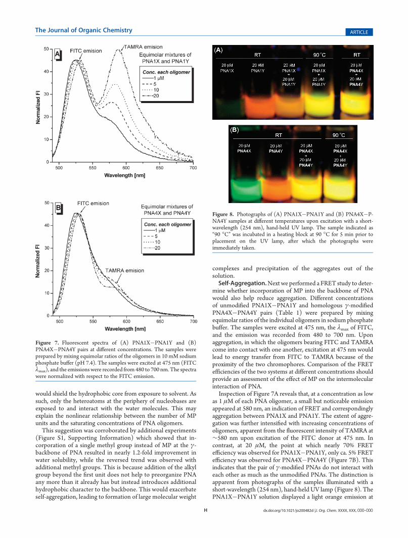
mine whether incorporation of MP into the backbone of PNAwould also help reduce aggregation. Different concentrationsof unmodified PNA1X�PNA1Y and homologous γ-modifiedPNA4X�PNA4Y pairs (Table 1) were prepared by mixingequimolar ratios of the individual oligomers in sodium phosphatebuffer. The samples were excited at 475 nm, the λmax of FITC,and the emission was recorded from 480 to 700 nm. Uponaggregation, in which the oligomers bearing FITC and TAMRAcome into contact with one another, excitation at 475 nm wouldlead to energy transfer from FITC to TAMRA because of theproximity of the two chromophores. Comparison of the FRETefficiencies of the two systems at different concentrations shouldprovide an assessment of the effect of MP on the intermolecularinteraction of PNA.Inspection of Figure 7A reveals that, at a concentration as low
as 1 μM of each PNA oligomer, a small but noticeable emissionappeared at 580 nm, an indication of FRET and correspondinglyaggregation between PNA1X and PNA1Y. The extent of aggre-gation was further intensified with increasing concentrations ofoligomers, apparent from the fluorescent intensity of TAMRA at∼580 nm upon excitation of the FITC donor at 475 nm. Incontrast, at 20 μM, the point at which nearly 70% FRETefficiency was observed for PNA1X�PNA1Y, only ca. 5% FRETefficiency was observed for PNA4X�PNA4Y (Figure 7B). Thisindicates that the pair of γ-modified PNAs do not interact witheach other as much as the unmodified PNAs. The distinction isapparent from photographs of the samples illuminated with ashort-wavelength (254 nm), hand-held UV lamp (Figure 8). ThePNA1X�PNA1Y solution displayed a light orange emission at
Figure 7. Fluorescent spectra of (A) PNA1X�PNA1Y and (B)PNA4X�PNA4Y pairs at different concentrations. The samples wereprepared by mixing equimolar ratios of the oligomers in 10 mM sodiumphosphate buffer (pH 7.4). The samples were excited at 475 nm (FITCλmax), and the emissions were recorded from 480 to 700 nm. The spectrawere normalized with respect to the FITC emission.
Figure 8. Photographs of (A) PNA1X�PNA1Y and (B) PNA4X�P-NA4Y samples at different temperatures upon excitation with a short-wavelength (254 nm), hand-held UV lamp. The sample indicated as“90 �C” was incubated in a heating block at 90 �C for 5 min prior toplacement on the UV lamp, after which the photographs wereimmediately taken.
I dx.doi.org/10.1021/jo200482d |J. Org. Chem. XXXX, XXX, 000–000
The Journal of Organic Chemistry ARTICLE
room temperature and yellow-green hue at 90 �C, an indicationof the aggregate dissociating upon heating. In contrast, the PNA4X�PNA4Y solution displayed the same color, yellow-green, at roomtemperature as well at 90 �C, indicating that the oligomers werewell dispersed even at room temperature. Thus, the R-MP γ-modification not only imparts enhanced solubility to PNA, butalso suppresses aggregation.Off-Target Binding. It has been documented that, at moder-
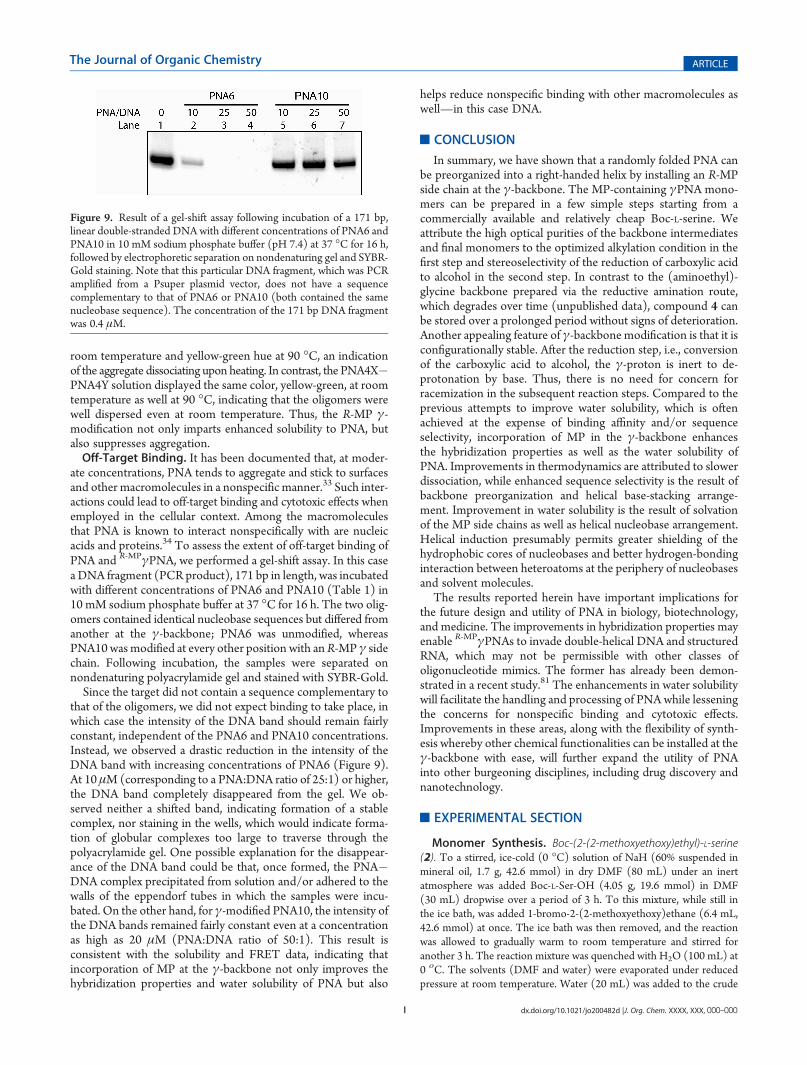
ate concentrations, PNA tends to aggregate and stick to surfacesand other macromolecules in a nonspecific manner.33 Such inter-actions could lead to off-target binding and cytotoxic effects whenemployed in the cellular context. Among the macromoleculesthat PNA is known to interact nonspecifically with are nucleicacids and proteins.34 To assess the extent of off-target binding ofPNA and R-MPγPNA, we performed a gel-shift assay. In this casea DNA fragment (PCR product), 171 bp in length, was incubatedwith different concentrations of PNA6 and PNA10 (Table 1) in10 mM sodium phosphate buffer at 37 �C for 16 h. The two olig-omers contained identical nucleobase sequences but differed fromanother at the γ-backbone; PNA6 was unmodified, whereasPNA10wasmodified at every other position with anR-MP γ sidechain. Following incubation, the samples were separated onnondenaturing polyacrylamide gel and stained with SYBR-Gold.Since the target did not contain a sequence complementary to
that of the oligomers, we did not expect binding to take place, inwhich case the intensity of the DNA band should remain fairlyconstant, independent of the PNA6 and PNA10 concentrations.Instead, we observed a drastic reduction in the intensity of theDNA band with increasing concentrations of PNA6 (Figure 9).At 10 μM (corresponding to a PNA:DNA ratio of 25:1) or higher,the DNA band completely disappeared from the gel. We ob-served neither a shifted band, indicating formation of a stablecomplex, nor staining in the wells, which would indicate forma-tion of globular complexes too large to traverse through thepolyacrylamide gel. One possible explanation for the disappear-ance of the DNA band could be that, once formed, the PNA�DNA complex precipitated from solution and/or adhered to thewalls of the eppendorf tubes in which the samples were incu-bated. On the other hand, for γ-modified PNA10, the intensity ofthe DNA bands remained fairly constant even at a concentrationas high as 20 μM (PNA:DNA ratio of 50:1). This result isconsistent with the solubility and FRET data, indicating thatincorporation of MP at the γ-backbone not only improves thehybridization properties and water solubility of PNA but also
helps reduce nonspecific binding with other macromolecules aswell—in this case DNA.
’CONCLUSION
In summary, we have shown that a randomly folded PNA canbe preorganized into a right-handed helix by installing an R-MPside chain at the γ-backbone. The MP-containing γPNA mono-mers can be prepared in a few simple steps starting from acommercially available and relatively cheap Boc-L-serine. Weattribute the high optical purities of the backbone intermediatesand final monomers to the optimized alkylation condition in thefirst step and stereoselectivity of the reduction of carboxylic acidto alcohol in the second step. In contrast to the (aminoethyl)-glycine backbone prepared via the reductive amination route,which degrades over time (unpublished data), compound 4 canbe stored over a prolonged period without signs of deterioration.Another appealing feature of γ-backbonemodification is that it isconfigurationally stable. After the reduction step, i.e., conversionof the carboxylic acid to alcohol, the γ-proton is inert to de-protonation by base. Thus, there is no need for concern forracemization in the subsequent reaction steps. Compared to theprevious attempts to improve water solubility, which is oftenachieved at the expense of binding affinity and/or sequenceselectivity, incorporation of MP in the γ-backbone enhancesthe hybridization properties as well as the water solubility ofPNA. Improvements in thermodynamics are attributed to slowerdissociation, while enhanced sequence selectivity is the result ofbackbone preorganization and helical base-stacking arrange-ment. Improvement in water solubility is the result of solvationof the MP side chains as well as helical nucleobase arrangement.Helical induction presumably permits greater shielding of thehydrophobic cores of nucleobases and better hydrogen-bondinginteraction between heteroatoms at the periphery of nucleobasesand solvent molecules.
The results reported herein have important implications forthe future design and utility of PNA in biology, biotechnology,and medicine. The improvements in hybridization properties mayenable R-MPγPNAs to invade double-helical DNA and structuredRNA, which may not be permissible with other classes ofoligonucleotide mimics. The former has already been demon-strated in a recent study.81 The enhancements in water solubilitywill facilitate the handling and processing of PNA while lesseningthe concerns for nonspecific binding and cytotoxic effects.Improvements in these areas, along with the flexibility of synth-esis whereby other chemical functionalities can be installed at theγ-backbone with ease, will further expand the utility of PNAinto other burgeoning disciplines, including drug discovery andnanotechnology.
’EXPERIMENTAL SECTION
Monomer Synthesis. Boc-(2-(2-methoxyethoxy)ethyl)-L-serine(2). To a stirred, ice-cold (0 �C) solution of NaH (60% suspended inmineral oil, 1.7 g, 42.6 mmol) in dry DMF (80 mL) under an inertatmosphere was added Boc-L-Ser-OH (4.05 g, 19.6 mmol) in DMF(30 mL) dropwise over a period of 3 h. To this mixture, while still inthe ice bath, was added 1-bromo-2-(2-methoxyethoxy)ethane (6.4 mL,42.6 mmol) at once. The ice bath was then removed, and the reactionwas allowed to gradually warm to room temperature and stirred foranother 3 h. The reaction mixture was quenched with H2O (100 mL) at0 οC. The solvents (DMF and water) were evaporated under reducedpressure at room temperature. Water (20 mL) was added to the crude
Figure 9. Result of a gel-shift assay following incubation of a 171 bp,linear double-stranded DNA with different concentrations of PNA6 andPNA10 in 10 mM sodium phosphate buffer (pH 7.4) at 37 �C for 16 h,followed by electrophoretic separation on nondenaturing gel and SYBR-Gold staining. Note that this particular DNA fragment, which was PCRamplified from a Psuper plasmid vector, does not have a sequencecomplementary to that of PNA6 or PNA10 (both contained the samenucleobase sequence). The concentration of the 171 bp DNA fragmentwas 0.4 μM.
J dx.doi.org/10.1021/jo200482d |J. Org. Chem. XXXX, XXX, 000–000
The Journal of Organic Chemistry ARTICLE
residue and acidified with 5%HCl to pH≈ 3 at 0 �C. The aqueous layerswere extracted with ethyl acetate (5� 100 mL) and dried over Na2SO4.The solvent was evaporated under reduced pressure, and the crudemixture was purified by column chromatography to afford a colorlessliquid (4.1 g, 13.3 mmol): yield 68%; 1H NMR (CDCl3, 300 MHz) δ6.84 (br s, 1H), 5.57 (d, J = 7.4 Hz, 1H), 4.46�4.38 (m, 1H), 3.93 (dd,J1 = 3.9 Hz, J2 = 3.5 Hz, 1H), 3.90�3.49 (m, 9H), 3.37 (s, 3H), 1.41 (s,9H); 13C NMR (CDCl3, 75 MHz) δ 28.2, 53.8, 58.7, 70.2, 70.7, 70.9,71.7, 80.0, 155.8, 173.4; HRMS (ESI/MSm/z)M calcd for C13H25NO7-
Na 330.1529, found 330.1546.Boc-(2-(2-methoxyethoxy)ethyl)-L-serinol (3). To a stirred solution
of Boc-(2-(2-methoxyethoxy)ethyl)-L-serine (2) (4 g, 13.0 mmol) in20 mL of DME in an ice bath was added NMM (1.43 mL, 13.0 mmol)dropwise under an inert atmosphere, followed by isobutyl chloroformate(1.76 mL, 13.0 mmol). The reaction mixture was stirred at the sametemperature for another 0.5 h. The cold solution was filtered, and theprecipitate was washed with DME (2 � 10 mL). To the combinedfiltrate stirred in an ice bath was added NaBH4 (0.741 g, 19.5 mmol in10 mL water) slowly. A strong effervescence occurred. The aqueous layerwas extracted three times with ethyl acetate, and the combined organiclayers were washed with brine and dried over Na2SO4. The solvent wasevaporated under reduced pressure, and the oily residue was purified bycolumn chromatography to give a colorless liquid (3.2 g, 10.9 mmol):yield 84%; 1H NMR (CDCl3, 300 MHz) δ 5.26 (br s, 1H), 3.84�3.50(m, 12 H), 3.36 (s, 3H), 2.93 (br s, 1H), 1.42 (s, 9H); 13C NMR(CDCl3, 75 MHz) δ 28.3, 51.5, 58.9, 63.2, 70.3, 70.4, 70.5, 71.3, 71.8,79.5, 156.0; HRMS (ESI/MSm/z)M calcd for C13H27NO6Na 316.1736,found 316. 1719.Boc-(2-(2-methoxyethoxy)ethyl)-L-serine-Ψ[CH2N(o,p-diNBS)]Gly-
OEt (4). To a stirred, cold solution of o,p-diNBS-Gly-OEt (3.53 g,10.5 mmol), triphenylphosphine (2.73 g, 10.5 mmol), and compound 3(3.1 g, 10.5 mmol) in dried THF (20 mL) under an inert atmospherewas added DIAD (1.48 mL, 10.5 mmol) dropwise over a period of 0.5 h.The reaction mixture was allowed to warm to room temperature andthen stirred overnight. The solvent was evaporated, and the oily residuewas purified by column chromatography to afford a yellow solid (3.6 g,5.91 mmol): yield 56%; 1H NMR (CDCl3, 300 MHz) δ 8.51 (dd, J =2.1Hz, 1H), 8.41 (d, J = 2.1Hz, 1H), 8.32 (d, J = 8.7Hz, 1H), 5.16 (d, J =8.6 Hz, 1H), 4.34 (dd, J = 18.6 Hz, 2H), 4.10 (2 � q, J = 3.6 Hz, 2H),4.01�3.87 (m, 1H), 3.70�3.49 (m, 12H), 3.39 (s, 3H), 1.43 (s, 9H),1.23 (t, J = 7.1 Hz, 3H); 13C NMR (CDCl3, 75 MHz) δ 14.0, 28.2, 48.3,48.7, 49.6, 58.9, 61.6, 70.0, 70.3, 70.4, 70.8, 71.9, 79.7, 119.4, 126.0,132.0, 138.4, 147.9, 149.5, 155.6, 168.6; HRMS (ESI/MSm/z) M calcdfor C23H36N4O13SNa 631.1898, found 631.1840.Boc-(2-(2-methoxyethoxy)ethyl)-L-serine-Ψ[CH2N]Gly-OEt (5). To
a stirred solution of 4 (2.6 g, 4.2mmol) in dichloromethane (20mL)wasadded n-propylamine (7.0 mL, 85.4 mmol) under an inert atmosphere.The reactionmixture was stirred at room temperature for another 20min.The solvent was evaporated, and the crude mixture was purified bycolumn chromatography to afford 5 as a light yellow liquid (1.3 g,3.4 mmol): yield 81%; 1H NMR (CDCl3, 300 MHz) δ 5.13 (br s, 1H),4.12 (q, J = 7.0 Hz, 2H), 3.78�3.63 (m, 1H), 3.62�3.33 (m, 12H), 3.32(s, 3H), 2.71 (2� dd, J1 = J2 = 5.9 Hz, 2H), 1.80 (br s, 1H), 1.39 (s, 9H),1.22 (t, J = 7.3 Hz, 3H); 13C NMR (CDCl3, 75 MHz) δ 14.2, 28.3, 49.9,50.3, 51.0, 58.9, 60.5, 70.4, 70.6, 71.4, 71.9, 79.1, 155.6, 172.4; HRMS(ESI/MSm/z) M calcd for C17H34N2O7Na 401.2264, found 401.2278.Boc-(2-(2-methoxyethoxy)ethyl)-L-serine Thymine Ethyl Ester (6a).
To a stirred solution of thymine acetic acid (0.287 g, 1.56mmol) in driedDMF (15 mL) under an inert atmosphere were added DCC (0.324 g,1.56 mmol) and DhbtOH (0.254 g, 1.56 mmol). The resulting mixturewas stirred at room temperature for 1 h. Compound 5 (0.5 g, 1.32mmol)was dissolved in dried DMF (10 mL) and added to the above reactionmixture, and the resulting mixture was stirred at 50 οC for 24 h. Thesolvent was removed under reduced pressure, and the remaining residue
was dissolved in ethyl acetate (100 mL) and washed with a saturatedsolution ofNaHCO3 (100mL) followed by 10%KHSO4 (100mL). Theorganic layer was washed with brine (50 mL) and dried over Na2SO4.The solvent was removed under reduced pressure. The crude productwas purified by column chromatography to afford a white solid (0.480 g,0.88 mmol): yield 67%; 1H NMR (DMSO-d6, 300 MHz) δ 7.27�7.19[2 � d (Rot1,2), J1 = J2 = 1.0 Hz, 1H], 6.86�6.57 [2 � d (Rot1,2) [tworotamers were found, as determined by multinuclear and multidimen-sional NMR experiments73], J1 = J2 = 8.6 Hz, 1H], 4.80�4.36 [2� ABq(Rot1,2), J1 = J2 = 16.8 Hz, 2H], 4.33�3.65 (m, 5H), 3.58�3.25 (m,12H), 3.22�3.15 [2 � s(Rot1,2), 3H], 1.73 (s, 3H), 1.35 (br s, 9H),1.27�1.11 [2 � t(Rot1,2), J1 = J2 = 7.1 Hz, 3H]; 13C NMRmajor rotamer
(DMSO-d6, 75MHz) δ 12.3, 14.4, 28.6, 47.9, 48.1, 48.6, 49.5, 58.5, 60.9,70.0, 70.3, 71.7, 78.6, 108.5, 142.4, 151.4, 155.8, 164.8, 168.0, 169.3;HRMS (ESI/MSm/z) M calcd for C24H40N4O10Na 567.2642 found,567.2651.
Boc-(2-(2-methoxyethoxy)ethyl)-L-serine Adenine(Cbz) Ethyl Ester(6b). Adenine ester 6b was prepared, purified, and characterized thesame way as described for 6a: 1H NMR (DMSO-d6, 300 MHz) δ 10.56(br s, 1H), 8.59�8.55 [2 � s (Rot1,2), 1H], 8.30�8.24 [2 � s (Rot1,2),1H], 7.55�7.20 (m, 5H), 7.04�6.53 [2 � d (Rot1,2), J1 = J2 = 8.5 Hz,1H ], 5.35 [ABq (Rot1), J = 17.2 Hz, Rot2 appears as a br s at 5.15 ppm,2H], 5.20 (s, 2H), 4.54�3.87 (m, 5H), 3.81�3.31 (m, 12H), 3.23�3.10[2 � s (Rot1,2), 3H], 1.36 (s, 9H), 1.30�1.10 [2 � t (Rot1,2), J1 = J2 =7.1 Hz, 3H]; 13C NMRmajor rotamer (DMSO-d6, 75 MHz) δ 14.4, 28.6,44.3, 48.7, 49.3, 49.9, 58.4, 60.9, 66.7, 70.0, 70.2, 70.4, 71.0, 71.6, 78.7,123.4, 128.3, 128.4, 128.8, 136.8, 145.5, 149.9, 151.9, 152.7, 152.9, 155.8,167.4, 169.2; HRMS (ESI/MSm/z) M calcd for C32H45N7O10Na710.3126, found 710.3110.
Boc-(2-(2-methoxyethoxy)ethyl)-L-serine PNA Guanine(Cbz) EthylEster (6c). Guanine ester 6c was prepared, purified, and characterizedthe same way as described for 6a: 1H NMR (DMSO-d6, 300 MHz) δ7.78�7.74 [2� s (Rot1,2), 1H], 7.45�7.28 (m, 5H), 6.98�6.59 [2� d(Rot1,2), J1 = J2 = 8.5 Hz, 1H], 5.23 (s, 2H), 5.20�4.84 [ABq (Rot1), J =17.1 Hz, m (Rot2), 2H], 4.45�3.77 (m, 5H), 3.76�3.33 (m, 12H),3.22�3.11[2� s (Rot1,2), 3H], 1.38�1.28 [2� s (Rot1,2), 9H], 1.28�1.10 [2 � t (Rot1,2), J1 = J2= 7.2 Hz, 3H ]; 13C NMRmajor rotamer
(DMSO-d6, 75 MHz) δ 14.4, 28.5, 44.1, 48.8, 49.4, 49.8, 58.4, 61.0,67.7, 70.0, 70.1, 70.2, 70.4, 71.6, 78.4, 119.6, 128.5, 128.6, 128.8, 128.9,135.9, 140.8, 147.8, 149.9, 155.0, 155.6, 155.8, 167.4, 169.2; HRMS(ESI/MSm/z) M calcd for C32H45N7O11Na 726.3075, found 726.3070.
Boc-(2-(2-methoxyethoxy)ethyl)-L-serine PNA Cytosine(Cbz) EthylEster (6d). Cytosine ester 6d was prepared, purified, and characterizedthe same way as described for 6a: 1H NMR (DMSO-d6, 300 MHz) δ7.84 (d, J = 7.3 Hz, 1H), 7.46�7.26 (m, 5H), 6.99 (d, J = 7.3 Hz, 1H),6.87�6.57 [2 � d (Rot1,2), J1 = J2 = 8.4 Hz, 1H], 5.17 (s, 2H), 4.93�4.52 [2 � ABq (Rot1,2), J1 = J2 = 15.9 Hz, 2H], 4.37�3.68 (m, 5H),3.63�3.29 (m, 12H), 3.22�3.15 [2 � s (Rot1,2), 3H], 1.35 (s, 9H),1.27�1.09 [2 � t (Rot1,2), J1= J2= 7.1 Hz, 3H]; 13C NMRmajor rotamer
(DMSO-d6, 75MHz) δ 14.4, 28.6, 48.7, 49.9, 58.5, 60.9, 66.9, 70.0, 70.3,71.7, 78.6, 94.3, 128.3, 128.6, 128.9, 136.4, 151.1, 153.6, 155.4, 155.7,163.6, 167.9, 169.2; HRMS (ESI/MSm/z)M calcd for C31H45N5O11Na686.3013, found 686.3006.
Boc-(2-(2-methoxyethoxy)ethyl)-L-serine Thymine Monomer (7a).To a stirred, cold solution of 6a (0.480 g, 0.88 mmol) in THF (10 mL)was added 2 N NaOH (10 mL) dropwise over a period of 15 min. Theresulting mixture was stirred at the same temperature for another 0.5 h.Upon completion of the reaction, as confirmed by TLC, H2O (20 mL)was added, and the resulting mixture was extracted with ethyl acetate(2� 25mL). The aqueous layers were combined, acidified with 5%HClto pH≈ 4 at 0 οC, and then extracted with ethyl acetate (4� 25mL) anddried over Na2SO4. The solvent was evaporated in vacuo, and the crudeproduct was purified by column chromatography to afford a colorlesssolid (0.400 g, 4.5mmol): yield 87%; 1HNMR (DMSO-d6, 300MHz) δ
K dx.doi.org/10.1021/jo200482d |J. Org. Chem. XXXX, XXX, 000–000
The Journal of Organic Chemistry ARTICLE
7.27�7.18 [2 � s (Rot1,2), 1H], 6.88�6.53 [ 2 � d (Rot1,2), J1 = J2 =8.2 Hz, 1H], 4.75�4.33 [ABq (Rot1), J = 17.5 Hz, m (Rot2), 2H],4.01�3.59 (m, 3H), 3.55�3.25 (m, 12H), 3.22�3.19 [2 � s (Rot1,2),3H], 1.73 (s, 3H), 1.35 (s, 9H); 13C NMRmajor rotamer (DMSO-d6,75 MHz) δ 12.3, 28.7, 48.1, 49.3, 51.9, 58.5, 70.0, 70.2, 70.3, 71.0, 71.7,78.2, 78.5, 108.5, 142.6, 151.5, 155.7, 164.8, 167.5, 168.5; HRMS (ESI/MSm/z) M calcd for C22H35N4O10Na2 561.2148, found 561.2115.Data for Boc-(2-(2-methoxyethoxy)ethyl)-L-serine adenine(Cbz)
monomer (7b): 1H NMR (DMSO-d6, 300 MHz) δ 8.57�8.53 [2 � s(Rot1,2), 1H], 8.30�8.16 [2 � s (Rot1,2), 1H], 7.47�7.24 (m, 5H),7.04�6.50 [2� d (Rot1,2), J1 = J2 = 8.7 Hz, 1H], 5.22 [ABq (Rot1), J =16.4 Hz, Rot2 appears as a br s at 5.10 ppm, 2H], 5.20 (s, 2H), 4.50�3.65(m, 5H), 3.63�3.43 (m, 5H), 3.41�3.21 (m, 5H), 3.20�3.14 [2 � s(Rot1,2),3H], 1.41�1.27 [2 � s (Rot1,2),9H];
13C NMRmajor rotamer
(DMSO-d6, 75MHz) δ 28.6, 44.5, 49.3, 58.5, 63.4, 66.7, 70.0, 70.2, 70.4,71.0, 71.7, 78.2, 78.6, 123.3, 126.8, 128.3, 128.4, 128.8, 136.8, 145.7,149.7, 151.8, 152.9, 155.7, 167.8; HRMS (ESI/MSm/z) M calcd forC30H40N7O10Na2 704.2632, found 704.2620.Data for Boc-(2-(2-methoxyethoxy)ethyl)-L-serine guanine(Cbz)
monomer (7c): 1H NMR (DMSO-d6, 300 MHz) δ 7.78�7.72 [2 � s(Rot1,2), 1H], 7.48�7.23 (m, 5H), 7.00�6.61 [2 � d (Rot1,2), J1 = J2 =8.6 Hz, 1H], 5.22�5.19 [2 � s (Rot1,2), 2H ], 5.03�4.84 [ABq (Rot1),J = 17.3 Hz, br s (Rot2), 2H], 3.98�3.62 (m, 4H), 3.59�3.40 (m, 7H),3.41�3.23 (m, 4H), 3.22�3.12 [2 � s (Rot1,2), 3H], 1.35�1.31 [2 � s(Rot1,2), 9H];
13CNMRmajor rotamer (DMSO-d6, 75MHz) δ 28.6, 44.4, 49.3,52.7, 58.5, 67.4, 70.0, 70.1, 70.2, 70.4, 71.0, 71.7, 78.2, 119.4, 128.4, 128.5,128.6, 128.9, 136.1, 140.8, 147.9, 150.2, 155.7, 166.7, 167.9, 171.9; HRMS(ESI/MSm/z) M calcd for C30H40N7O11Na2 720.2581, found 720.2581.Data for Boc-(2-(2-methoxyethoxy)ethyl)-L-serine cytosine(Cbz)
monomer (7d): 1H NMR (DMSO-d6, 300 MHz) δ 7.84 (d, J =7.2 Hz, 1H), 7.43�7.24 (m, 5H), 7.01�6.93 [2 � d (Rot1,2), J1 =J2 = 7.3 Hz, 1H], 6.87�6.58 [2� d (Rot1,2), J1 = J2 = 8.73Hz, 1H ], 5.17(s, 2H), 4.9�4.5 [ABq (Rot1), J = 15.8 Hz, br s (Rot2), 2H], 4.01�3.61(m, 3H), 3.58�3.43 (m, 7H), 3.43�3.23 (m, 5H), 3.22�3.16 [2 � s(Rot1,2), 3H], 1.35 (s, 9H);
13C NMRmajor rotamer (DMSO-d6, 75 MHz)δ 28.1, 48.6, 48.9, 49.7, 52.2, 58.0, 66.4, 69.5, 69.7, 69.8, 70.4, 71.2, 77.8,94.0, 127.9, 128.1, 128.4, 135.9, 150.9, 153.1, 155.1, 155.2, 163.0, 167.8,172.2; HRMS (ESI/MSm/z) M calcd for C29H40N5O11Na2 680.2520,found 680.2546.Determination of Optical Purities. A General Procedure for
Preparing MTPA Derivatives. Compound 8. To a stirred, cold solutionof 2 (100 mg) in DCM (5 mL) was added 5 mL of TFA/m-cresolsolution (95:5). The ice bath was removed, and the reaction mixture wasstirred at room temperature overnight. The solvent was evaporatedunder reduced pressure and triturated with diethyl ether (3 � 5 mL).The remaining residue was dried under high vacuum overnight and usedin the next coupling step without further purification. The crude residuewas dissolved in DCM (5 mL) and chilled at 0 �C. DIPEA (2.0 equiv)and (S)-(+)-R-methoxy-R-(trifluoromethyl)phenylacetyl chloride (MTPA-Cl; 1.1 equiv) were added, and the reaction mixture was stirred at roomtemperature overnight. After completion, the solution was diluted withDCM and washed with water (2� 10 mL) and then brine solution. Theorganic layer was dried on Na2SO4, concentrated under reducedpressure, and purified by column chromatography. Compounds 9 and10 were prepared the same way.Data for 3-[2-(2-methoxyethoxy)ethoxy]-2-[ (3,3,3-trifluoro-2-
methoxy-2-phenylpropionyl)amino]propionic acid (8): 1H NMR(300 MHz, CDCl3) δ 7.78 (br s, 1H), 7.56 (m, 2H), 7.37 (m, 3H),4.47 (m, 1H), 4.03�3.7 (m, 2H), 3.68�3.45 (m, 8H), 3.40 (s, 3H), 3.30(s, 3H); 13C NMR (DMSO-d6, 75 MHz) δ 42.7, 52.1, 54.8, 55.6, 57.9,60.1, 68.9, 69.5, 69.6, 71.2, 72.3, 83.8, 127.2, 127.5, 128.1, 129.4, 165.3,174.7; HRMS (ESI/MSm/z) M calcd for C18H24F3NO7Na 446.1403,found 446.1359; 19F NMR (300MHz, CDCl3) δ�69.58 (s, L-stereomer),�69.24 (s, D-stereomer).
Data for 3,3,3-trifluoro-N-{2-hydroxy-1-[ [2-(2-methoxyethoxy)-ethoxy]methyl]ethyl}-2-methoxy-2-phenylpropionamide (9): 1H NMR(300 MHz, CDCl3) δ 7.55 (m, 2H), 7.44 (br s, 1H), 7.40 (m, 3H), 4.13(m, 1H), 3.83 (m, 1H), 3.70�3.60 (m, 9H), 3.54 (m, 2H), 3.41(m, 3H),3.37 (s, 3H), 3.07 (br s, 1H); 13C NMR (CDCl3, 75 MHz) δ 50.5,54.3, 58.4, 62.2, 69.8, 69.89, 69.9, 70.2, 71.3, 72.1, 83.4, 121.4, 125.2,127.2, 128.0, 128.9, 132.0, 165.9; HRMS (ESI/MSm/z) M calcd forC18H26F3NO6Na 432.1610 found 432. 1595; 19F NMR (300 MHz,CDCl3) δ �69.43 (s, L-stereomer), �69.33 (s, D-stereomer).
Data for {[3-[2-(2-methoxyethoxy)ethoxy]-2-[ (3,3,3-trifluoro-2-methoxy-2-phenylpropionyl)amino]propyl][2-thyminylacetyl]amino}-acetic acid (10): 1H NMR (300 MHz, DMSO-d6) δ 11.22 (br s, 1H),8.33 and 8.25 (2� d, J = 8.5 Hz, 1H), 7.54�7.35 (m, 5H), 7.09 and 6.96(2 � s,1H), 4.52�4.26 (ABq, J = 16.8 Hz, 2H), 4.26�4.16 (m, 1H)3.6�3.34 (m, 16H), 3.21 and 3.20 (2 � s, 3H), 3.10 (ABq, J =14.0 Hz, 2H), 1.70 and 1.68 (2 � s, 3H); 13C NMRmajor rotamer
(DMSO-d6, 75 MHz) δ 11.8, 47.5, 48.0, 48.4, 52.2, 54.8, 58.0, 69.2,69.5, 69.6, 69.9, 71.2, 83.7, 107.9, 127.0, 127.2, 128.3, 129.3, 133.3, 142.0,151.0, 164.3, 165.3, 168.2, 171.6; HRMS (ESI/MSm/z) M calcd forC27H35F3N4O10Na 655.2305 found 655.2299; 19F NMR (300 MHz,CDCl3) δ�68.80 and�68.95 (s, L-stereomer),�68.70 (s, D-stereomer).Oligomer Synthesis.Unmodified, Boc-protected PNAmonomers
were purchased from a commercial supplier (they are no longeravailable). The oligomers were synthesized onMBHA resin according tothe published protocol.87Upon completion of the lastmonomer coupling,the oligomers were cleaved from the resin (and the protecting groupswere simultaneously removed) by immersing the resin in a cocktailcontaining m-cresol/thioanisole/TFA/TFMSA (150/150/900/300 μL—for 100 mg of resin) for 2 h. The crude mixture was eluted andprecipitated in ethyl ether, dissolved in a water/acetonitrile mixture(80:20), purified by reversed-phase HPLC, and characterized by MAL-DI-TOF mass spectrometry. A solution of R-cyano-4-hydroxycinnamicacid (10mg ofR-cyano-4-hydroxycinnamic acid in 500mL of water with0.1% TFA and 500 mL of acetonitrile with 0.1% TFA) was used as thematrix for MALDI-TOF analysis. Concentrations of the oligomers weredetermined by UV absorption at 260 nm at 90 �C in water using thefollowing extinction coefficients: 13 700M�1 cm�1 (A), 11 700M�1 cm�1
(G), 6600 M�1 cm�1 (C), and 8600 M�1 cm�1 (T).Circular Dichroism (CD). All CD data were recorded at room
temperature unless otherwise stated. All spectra represent an average ofat least 15 scans, recorded from 320 to 200 nm at the rate of 100 nm/min.A 1 cm path length cuvette was used, and the temperature was main-tained at 22 �C. All spectra were processed using Origin software,baseline subtracted and, unless otherwise noted, smoothed using a five-point adjacent averaging algorithm.Melting Temperature (Tm). All hybridization experiments were
performed by measuring the change in the absorbance at 260 nm as afunction of temperature. All samples were prepared by mixing astoichiometric amount of each strand (5 μM) in a 10 mM sodiumphosphate at pH 7.4 and annealed prior to each measurement. Therecorded spectra were smoothed using a five-point adjacent averagingalgorithm unless otherwise specified. The melting transitions weredetermined by taking the first derivative of the absorbance�temperatureprofile.Thermodynamic Analysis. Thermodynamic parameters were
determined from concentration-dependent Tmmeasurements followingvan’t Hoff analysis.95 ΔH� and ΔS� were obtained from a plot of 1/Tm
(K�1) vs ln CT, where CT is the total strand concentration. ΔH� wasobtained from the slope of the plot, which should be a linear function,using the relationship slope = (n� 1)R/ΔH, where n is the molecularityof the association interaction (in this case, n = 2) and R is the gasconstant (R = 8.314 J/(mol 3K)). ΔS� was determined from the yintercept (ln CT = 0), where for a non-self-complementary oligonucleo-tide the following relationship stands: slope = [ΔS�� (n� 1)R ln 2n]/
L dx.doi.org/10.1021/jo200482d |J. Org. Chem. XXXX, XXX, 000–000
The Journal of Organic Chemistry ARTICLE
ΔH�.ΔG� was calculated at 298.15 K using the equationΔG� =ΔH��TΔS� and K using the equation ΔG� = �RT ln K.SPR Analysis. DNA and PNA concentrations were calculated by
measuring sample absorbance at 260 nm and 90 �C. At this temperaturethe bases are unstacked, resulting in the extinction coefficient of theoligomer being equal to the sum of the extinction coefficients of its bases.The DNA extinction coefficients were obtained from the literature, whilethe PNA extinction coefficients were provided by the commercial supplier.
SPR experiments were conducted on a Biacore T100 with a four-channel carboxymethylated dextran-coated gold sensor chip (CM5) at25 �C. Approximately 6300 response units (RU) of streptavidin wasimmobilized on the chip’s surface at a flow rate of 5 μL/min usingstandardN-hydroxysuccinimide/1-ethyl-3-[3-(dimethylamino)propyl]-carbodiimide hydrochloride (NHS/EDC) coupling followed by cappingwith ethanolamine. Then 100 RU of 50-biotinylated DNAwas attached tothe chip via noncovalent capture at a flow rate of 2 μL/min and a sampleconcentration of 25 nM. HBS-EP (0.01 M HEPES, 0.15 M NaCl, 3 mMEDTA, and 0.005% Surfactant P-20, pH 7.4) was used as the runningbuffer for both the streptavidin and biotinylated DNA immobilizations.
Kinetic information was obtained from the SPR studies by flowing adilution series (10�50 nM in 10 nM increments) of PNA across thechip. All samples were run in triplicate at a flow rate of 50 μL/min with arunning buffer consisting of 10 mM sodium phosphate, pH 7.4, 100 mMNaCl, 0.1 mM EDTA, and 0.005% Surfactant P-20. The chip wasregenerated with two 30 s pulses of 10 mM NaOH/1 M NaCl. Bufferinjections were conducted before each sample injection to flush anylingering regeneration solution. All samples were reference corrected toaccount for nonspecific binding by subtracting off the signal resultingfrom flowing sample over a streptavidin-coated (i.e., no DNA) flow cell.Kinetic constants were calculated using a 1:1 binding global fit model inBiacore T100 Evaluation Software.Solubility. A saturated solution was prepared for each oligomer by
dissolving the dried pellet in a minimum amount of water so that after,heating at 95 �C for 5 min and thoroughmixing, undissolved material stillremained in the solution. The samples were centrifuged at 12000g for5 min. The concentrations of the saturated solutions were determined bymeasuring the UV absorption at 260 nm following a serial dilution.Self-Aggregation. Samples containing different concentrations of
the PNA1X�PNA1Y or PNA4X�PNA4Y pair at equimolar ratios wereprepared in 10 mM sodium phosphate buffer at pH 7.4. The sampleswere allowed to equilibrate for 1 h. The fluorescence spectrum of eachsample was recorded at room temperature on a fluorescence spectro-meter from 490 to 700 nm upon excitation at 475 nm (FITC). Allspectra were subtracted from the buffer baseline and smoothed using afive-point adjacent averaging algorithm on the Origin software. FRETefficiencies were determined using the relationship E = 1 � F0D/FD,where F0D and FD are the fluorescence intensities with and without theacceptor, respectively. The donor in this case is FITC (X). For thePNA1X�PNA1Y pair, F0D = F0PNA1X�PNA1Y and FD = FPNA1X, and forthe PNA4X�PNA4Y pair, F0D = F0PNA4X�PNA4Y and FD = FPNA4X, at therespective concentration.Off-Target Binding. A PCR product, 171 bp in length (0.4 μM),
was separately incubated with 4, 10, and 20 μM PNA6 and PNA10 insodium phosphate buffer at 37 �C for 16 h. Following incubation, thesamples were separated on nondenaturing polyacrylamide gel andstained with SYBR-Gold. The gel was washed three times with 1�TBE buffer and imaged using the BioDoc-It gel documentation system.
’ASSOCIATED CONTENT
bS Supporting Information. UV absorption spectra ofPNA11�13, 1H and 13C NMR spectra of compounds 1�10,HPLC and MALDI-TOF MS spectra of PNA1�10, UV meltingcurves of PNA5�DNA and PNA5�RNA duplexes containing
perfectly matched and single-base-mismatched targets, CDmelt-ing curves of PNA2�5, and SPR sensorgrams and kinetic fits forPNA1�5. This material is available free of charge via the Internetat http://pubs.acs.org.
’AUTHOR INFORMATION
Corresponding Author*E-mail: [email protected].
’ACKNOWLEDGMENT
Financial support for this work was provided by the NationalInstitutes of Health (Grant GM076251) and the National ScienceFoundation (NSF; Grant CHE-1012467) to D.H.L. SPR instru-mentation was purchased with support from NSF Major Re-search Instrumentaion Award 0821296. K.J.Z. gratefully acknowl-edges support from Department of Defense, Air Force Office ofScientific Research, National Defense Science and EngineeringGraduate Fellowship 32 CFR168a.
’REFERENCES
(1) Nielsen, P. E.; Egholm, M.; Berg, R. H.; Buchardt, O. Science1991, 254, 1497–1500.
(2) Egholm, M.; Buchardt, O.; Christensen, L.; Behrens, C.; Freier,S. M.; Driver, D. A.; Berg, R. H.; Kim, S. K.; Norden, B.; Nielsen, P. E.Nature 1993, 365, 566–568.
(3) Nielsen, P. E. Acc. Chem. Res. 1999, 32, 624–630.(4) Bentin, T.; Larsen, H. J.; Nielsen, P. E. Biochemistry 2003,
42, 13987–13995.(5) Kaihatsu, K.; Shah, R. H.; Zhao, X.; Corey, D. R. Biochemistry
2003, 42, 13996–14003.(6) Tomac, S.; Sarkar, M.; Ratilainen, T.; Wittung, P.; Nielsen, P. E.;
Norden, B.; Graeslund, A. J. Am. Chem. Soc. 1996, 118, 5544–5552.(7) Ratilainen, T.; Holmen, A.; Tuite, E.; Nielsen, P. E.; Norden, B.
Biochemistry 2000, 39, 7781–7791.(8) Menchise, V.; De Simone, G.; Tedeschi, T.; Corradini, R.; Sforza,
S.; Marchelli, R.; Capasso, D.; Saviano, M.; Pedone, C. Proc. Natl. Acad.Sci. U.S.A. 2003, 100, 12021–12026.
(9) Rasmussen, H.; Kastrup, J. S.; Nielsen, J. N.; Nielsen, J. M.;Nielsen, P. E. Nat. Struct. Biol. 1997, 4, 98–101.
(10) Haaima, G.; Rasmussen, H.; Schmidt, G.; Jensen, D. K.; Kastrup,J. S.; Stafshede, P.W.; Norden, B.; Buchardt, O.; Nielsen, P. E.New J. Chem.1999, 23, 833–840.
(11) Eldrup, A. B.; Nielsen, B. B.; Haaima, G.; Rasmussen, H.;Kastrup, J. S.; Christensen, C.; Nielsen, P. E. Eur. J. Org. Chem. 2001,1781–1790.
(12) Demidov, V. V.; Potaman, V. N.; Frank-Kamenetskii, M. D.;Egholm, M.; Buchard, O.; Sonnichsen, S. H.; Nielsen, P. E. Biochem.Pharmacol. 1994, 48, 1310–1313.
(13) Dueholm, K. L.; Egholm, M.; Behrens, C.; Christensen, L.;Hansen, H. F.; Vulpius, T.; Petersen, K. H.; Berg, R. H.; Nielsen, P. E.;Buchardt, O. J. Org. Chem. 1994, 59, 5767–5773.
(14) Thomson, S. A.; Josey, J. A.; Cadilla, R.; Gaul, M. D.; Hassman,C. F.; Luzzio, M. J.; Pipe, A. J.; Reed, K. L.; Ricca, D. J.; Wiethe, R. W.;Noble, S. A. Tetrahedron 1995, 51, 6179–6194.
(15) Beck, F.; Nielsen, P. E. Artificial DNA: Methods and Applica-tions; CRC Press: Boca Raton, FL, 2003; pp 91�114.
(16) Ray, A.; Norden, B. FASEB J. 2000, 14, 1041–1060.(17) Nielsen, P. E. Mol. Biotechnol. 2004, 26, 233–248.(18) Veselkov, A. G.; Demidov, V. V.; Frank-Kamenetskii, M. D.;
Nielsen, P. E. Nature 1996, 379, 214.(19) Komiyama, M.; Aiba, Y.; Yamamoto, Y.; Sumaoka, J. Nat.
Protoc. 2008, 3, 655–622.
M dx.doi.org/10.1021/jo200482d |J. Org. Chem. XXXX, XXX, 000–000
The Journal of Organic Chemistry ARTICLE
(20) Chin, J. Y.; Kuan, J. Y.; Lonkar, P. S.; Krause, D. S.; Seidman,M. M.; Peterson, K. R.; Nielsen, P. E.; Kole, R.; Glazer, P. M. Proc. Natl.Acad. Sci. U.S.A. 2008, 105, 13514–13519.(21) Singer,A.;Wanunu,M.;Morrison,W.;Kuhn,H.;Frank-Kamenetskii,
M.; Meller, A. Nano Lett. 2010, 10, 738–742.(22) Mollegaard, N. E.; Buchardt, O.; Egholm, M.; Nielsen, P. E.
Proc. Natl. Acad. Sci. U.S.A. 1994, 91, 3892–3895.(23) Janowski, B. A.; Kaihatsu, K.; Huffman, K. E.; Schwartz, J. C.;
Ram, R.; Hardy, D.; Mendelson, C. R.; Corey, D. R. Nat. Chem. Biol.2005, 1, 210–215.(24) Winssinger, N.; Ficarro, S.; Schultz, P. G.; Harris, J. L. Proc.
Natl. Acad. Sci. U.S.A. 2002, 99, 11139–11144.(25) Debaene, F.; Da Silva, J. A.; Pianowski, Z.;Duran, F. J.;Winssinger,
N. Tetrahedron 2007, 63, 6577–6586.(26) Bruno, Y.; Birnbaum,M. E.; Kleiner, R. E.; Liu, D. R.Nat. Chem.
Biol. 2010, 6, 148–155.(27) Ura, Y.; Beierle, J. M.; Leman, L. J.; Orgel, L. E.; Ghadiri, M. R.
Science 2009, 325, 73–77.(28) Myers, C. P.; Williams, M. E. Coord. Chem. Rev. 2010, 254,
2416–2428.(29) Chakrabarti, R.; Klibanov, A. M. J. Am. Chem. Soc. 2003, 125,
12531–12540.(30) Janssen, P. G. A.; Meeuwenoord, N.; van der Marel, G.;
Jabbari-Farouji, S.; van der Schoot, P.; Surin,M.; Tomovic, Z.;Meijer, E.W.;Schenning, A. P. H. J. Chem. Commun. 2010, 46, 109–111.(31) Riaz, U.; Ahmad, S.; Ashraf, S. M. J. Nanopart. Res. 2008, 10,
1209–1214.(32) Popescu, D. L.; Parolin, T. J.; Achim, C. J. Am. Chem. Soc. 2003,
125, 6354–6355.(33) Braasch, D. A.; Corey, D. R. Methods 2001, 23, 97–107.(34) Tackett, A. J.; Corey, D. R.; Raney, K. D. Nucleic Acids Res.
2002, 30, 950–957.(35) Masuko, M. Nucleic Acids Res. Suppl. 2003, 145–146.(36) Cattani-Scholz, A.; Pedone, D.; Blobner, F.; Abstreiter, G.;
Schwartz, J.; Tornow, M.; Andruzzi, L. Biomacromolecules 2009, 10,489–496.(37) Egholm, M.; Buchardt, O.; Nielsen, P. E.; Berg, R. H. J. Am.
Chem. Soc. 1992, 114, 1895–1897.(38) Haaima, G.; Lohse, A.; Buchardt, O.; Nielsen, P. E. Angew.
Chem., Int. Ed. Engl. 1996, 35, 1939–1941.(39) Sforza, S.; Tedeschi, T.; Corradini, R.; Marchelli, R. Eur. J. Org.
Chem. 2007, 5879–5885.(40) Boyarskaya, N. P.; Kirillova, Y. G.; Stotland, E. A.; Prokhorov,
D. I.; Zvonkova, E. N.; Shvets, V. I. Dokl. Chem. (Transl. of Dokl. Akad.Nauk) 2006, 408, 57–60.(41) Gildea, B. D.; Casey, S.; MacNeill, J.; Perry-O’Keefe, H.;
Sorensen, D.; Coull, J. M. Tetrahedron Lett. 1998, 39, 7255–7258.(42) Hudson, R. H. E.; Liu, Y.; Wojciechowski, F. Can. J. Chem.
2007, 85, 302–312.(43) Peyman, A.; Uhlmann, E.; Wagner, K.; Augustin, S.; Breipohl,
G.; Will, D. W.; Schafer, A.; Wallmeier, H. Angew. Chem., Int. Ed. Engl.1996, 35, 2636–2638.(44) Efimov, V. A.; Choob, M. V.; Buryakova, A. A.; Kalinkina, A. L.;
Chakhmakhcheva, O. G. Nucleic Acids Res. 1998, 26, 566–575.(45) Bonora, G. M.; Drioli, S.; Ballico, M.; Faccini, A.; Corradini, R.;
Cogoi, S.; Xodo, L. Nucleosides, Nucleotides, Nucleic Acids 2007, 26,661–664.(46) Petersen, K. H.; Jensen, D. K.; Egholm, M.; Nielsen, P. E.;
Buchardt, O. Bioorg. Med. Chem. Lett. 1995, 5, 1119–1124.(47) Bergmann, F.; Bannwarth, W.; Tam, S. Tetrahedron Lett. 1995,
36, 6823–6826.(48) Uhlmann, E.; Will, D. W.; Breipohl, G.; Langner, D.; Ryte, A.
Angew. Chem., Int. Ed. Engl. 1996, 35, 2632–2635.(49) Finn, P. J.; Gibson, N. J.; Fallon, R.; Hamilton, A.; Brown, T.
Nucleic Acids Res. 1996, 24, 3357–3363.(50) vander Laan, A. C.; Brill, R.; Kuimelis, R. G.; KuylYeheskiely, E.;
vanBoom, J. H.; Andrus, A.; Vinayak, R. Tetrahedron Lett. 1997, 38,2249–2252.
(51) Kuwahara, M.; Arimitsu, M.; Sisido, M. J. Am. Chem. Soc. 1999,121, 256–257.
(52) Harris, J. M.; Chess, R. B. Nat. Rev. Drug Discovery 2003,2, 214–221.
(53) Veronese, F. M.; Pasut, G. Drug. Discovery Today 2005, 10,1451–1458.
(54) Knop, K.; Hoogenboom, R.; Fischer, D.; Schubert, U. S. Angew.Chem., Int. Ed. 2010, 49, 6288–6308.
(55) Roberts, M. J.; Benley, M. D.; Harris, J. M. Adv. Drug. DeliveryRev. 2002, 54, 459–476.
(56) Wang, Y.; Chang, F.; Zhang, Y.; Liu, N.; Liu, G.; Gupta, S.;Rusckowski, M.; Hnatowich, D. J. Bioconjugate Chem. 2001, 12, 807–816.
(57) Rapozzi, V.; Cogoi, S.; Spessotto, P.; Risso, A.; Bonora, G. M.;Quadrifoglio, F.; Xodo, L. E. Biochemistry 2002, 41, 502–510.
(58) Bonora, G. M. J. Bioact. Compat. Polym. 2002, 17, 375389.(59) Zhao, H.; Greenwald, R. B.; Reddy, P.; Xia, J.; Peng, P.
Bioconjugate Chem. 2005, 16, 758–766.(60) Liu, B.; Burdine, L.; Kodadek, T. J. Am. Chem. Soc. 2006, 128,
15228–15235.(61) Kikkeri, R.; Lepenies, B.; Adibekian, A.; Laurino, P.; Seeberger,
P. H. J. Am. Chem. Soc. 2009, 131, 2110–2112.(62) Konig, H. M.; Gorelik, T.; Kolb, U.; Kilbinger, A. F. M. J. Am.
Chem. Soc. 2006, 129, 704–708.(63) Gopin, A.; Ebner, S.; Attali, B.; Shabat, D. Bioconjugate Chem.
2006, 17, 1432–1440.(64) Xiong, S.; Li, H.; Yu, B.; Wu, J.; Lee, R. J. J. Pharm. Sci. 2010,
99, 5011–5018.(65) Zhao, H.; Duong, H. H. P.; Yung, L. Y. L. Macromol. Rapid
Commun. 2010, 31, 1163–1169.(66) Nagahori, N.; Abe, M.; Nishimura, S.-I. Biochemistry 2009,
48, 583–594.(67) Li, J.; Chen, Y.-C.; Tseng, Y.-C.; Mozumdar, S.; Huang, L.
J. Controlled Release 2010, 142, 416–421.(68) Arnaud, A.; Belleney, J.; Boue, F.; Bouteiller, L.; Carrot, G.;
Wintgens, V. Angew. Chem., Int. Ed. 2004, 43, 1718–1721.(69) Dragulescu-Andrasi, A.; Rapireddy, S.; Frezza, B. M.; Gayathri,
C.; Gil, R. R.; Ly, D. H. J. Am. Chem. Soc. 2006, 128, 10258–10267.(70) Rapireddy, S.; He, G.; Roy, S.; Armitage, B. A.; Ly, D. H. J. Am.
Chem. Soc. 2007, 129, 15596–15600.(71) Chenna, V.; Rapireddy, S.; Sahu, B.; Ausin, C.; Pedroso, E.;
Ly, D. H. ChemBioChem 2008, 9, 2388–2391.(72) Sahu, B.; Chenna, V.; Lathrop, K. L.; Thomas, S. M.; Zon, G.;
Livak, K. J.; Ly, D. H. J. Org. Chem. 2009, 74, 1509–1516.(73) Yeh, J. I.; Shivachev, B.; Rapireddy, S.; Crawford, M. J.; Gil,
R. R.; Du, S.; Madrid, M.; Ly, D. H. J. Am. Chem. Soc. 2010, 132,10717–10727.
(74) Kosynkina, L.; Wang, W.; Liang, T. C. Tetrahedron Lett. 1994,35, 5173–5176.
(75) Falkiewicz, B.; Kowalska, K.; Kolodziejczyk, A. S.; Wisniewski,K.; Lankiewicz, L. Nucleosides Nucleotides 1999, 18, 353–361.
(76) Wu, Y.; Xu, J. C. Tetrahedron 2001, 57, 8107–8113.(77) Tedeschi, T.; Sforza, S.; Corradini, R.;Marchelli, R.Tetrahedron
Lett. 2005, 46, 8395–8399.(78) Dose, C.; Seitz, O. Org. Lett. 2005, 7, 4365–4368.(79) Englund, E. A.; Appella, D. H. Org. Lett. 2005, 7, 3465–3467.(80) Englund, E. A.; Appella, D. H. Angew. Chem., Int. Ed. 2007,
46, 1414–1418.(81) He, G.; Rapireddy, S.; Bahal, R.; Sahu, B.; Ly, D. H. J. Am. Chem.
Soc. 2009, 131, 12088–12090.(82) Fukuyama, T.; Cheung, M.; Jow, C.-K.; Hidai, Y.; Kan, T.
Tetrahedron Lett. 1997, 38, 5831–5834.(83) Debaene, F.; Winssinger, N. Org. Lett. 2003, 5, 4445–4447.(84) Corradini, R.; Di Silvestro, G.; Sforza, S.; Palla, G.; Dossena, A.;
Nielsen, P. E.;Marchelli, R.Tetrahedron: Asymmetry 1999, 10, 2063–2066.(85) Seco, J. M.; Quinoa, E.; Riguera, R. Chem. Rev. 2004,
104, 17–117.(86) Rodriguez, M.; Llinares, M.; Doulut, S.; Martinez, J. Tetrahedron
Lett. 1991, 32, 923–926.
N dx.doi.org/10.1021/jo200482d |J. Org. Chem. XXXX, XXX, 000–000
The Journal of Organic Chemistry ARTICLE
(87) Christensen, L.; Fitzpatrick, R.; Gildea, B.; Petersen, K. H.;Hansen, H. F.; Koch, T.; Egholm,M.; Buchardt, O.; Nielsen, P. E.; Coull,J.; Berg, R. H. J. Pept. Sci. 1995, 1, 175–183.(88) Tedeschi, T.; Corradini, R.; Marchelli, R.; Pushl, A.; Nielsen,
P. E. Tetrahedron: Asymmetry 2002, 13, 1629–1636.(89) Sen, S.; Nilsson, L. J. Am. Chem. Soc. 2001, 123, 7414–7422.(90) Johnson, W. C. Circular Dichroism: Principles and Applications;
Wiley-VCH: New York, 2000.(91) Nielsen, E. B.; Schellman, J. A. J. Phys. Chem. 1967, 71, 2297–2304.(92) Wittung, P.; Nielsen, P. E.; Buchardt, O.; Egholm, M.; Norden,
B. Nature 1994, 368, 561–563.(93) Eriksson, M.; Nielsen, P. E. Nat. Struct. Biol. 1996, 3, 410–413.(94) Cheatham, T. E. I.; Kollman, P. A. J. Am. Chem. Soc. 1997,
119, 4805.(95) Marky, L.; Breslauer, K. J. Biopolymers 1987, 26, 1601–1620.(96) Gildea, B. D.; Coull, J. M.; Hyldig-Nielsen, J. J.; Fiandaca, M. J.
WO A-9921881, 1999.(97) Ananthanawat, C.; Vilainvan, T.; Hoven, V.; Su, X. Biosens.
Bioelectron. 2010, 25, 1064–1069.(98) Lao, A. I.; Su, X.; Aung, K. M. M. Biosens. Bioelectron. 2009,
24, 1717–1722.(99) Sawata, S.; Kai, E.; Ikebukuro, K.; Iida, T.; Honda, T.; Karube, I.
Biosens. Bioelectron. 1999, 14, 397–404.(100) Roy, S.; Tanious, F. A.; Wilson, W. D.; Ly, D. H.; Armitage,
B. A. Biochemistry 2007, 46, 10433–10443.(101) Lusvarghi, S.; Murphy, C. T.; Roy, S.; Tanious, F. A.; Sacui, I.;
Wilson, W.; Ly, D. H.; Armitage, B. A. J. Am. Chem. Soc. 2009,131, 18415–18424.