Symbiont-derived β-1,3-glucanases in a social insect ... · Another possible reason for the...
11
ORIGINAL RESEARCH ARTICLE published: 21 November 2014 doi: 10.3389/fmicb.2014.00607 Symbiont-derived β-1,3-glucanases in a social insect: mutualism beyond nutrition Rebeca B. Rosengaus 1 *, Kelley F. Schultheis 1 , Alla Yalonetskaya 1 , Mark S. Bulmer 2 , William S. DuComb 3 , Ryan W. Benson 3† ,John P. Thottam 1 and Veronica Godoy-Carter 3 1 Department of Marine and Environmental Sciences, Northeastern University, Boston, MA, USA 2 Department of Biological Sciences,Towson University,Towson, MD, USA 3 Department of Biology, Northeastern University, Boston, MA, USA Edited by: Spencer V. Nyholm, University of Connecticut, USA Reviewed by: Robert W.Thacker, University of Alabama at Birmingham, USA Kenneth Noll, University of Connecticut, USA *Correspondence: Rebeca B. Rosengaus, Department of Marine and Environmental Sciences, Northeastern University, 134 Mugar Life Sciences Building, 360 Huntington Avenue, Boston, MA 02115-5000, USA e-mail: [email protected] † Present address: Ryan W. Benson, Forsyth Institute, Cambridge, MA, USA Termites have had a long co-evolutionary history with prokaryotic and eukaryotic gut microbes. Historically, the role of these anaerobic obligate symbionts has been attributed to the nutritional welfare of the host. We provide evidence that protozoa (and/or their associated bacteria) colonizing the hindgut of the dampwood termite Zootermopsis angusticollis, synthesize multiple functional β-1,3-glucanases, enzymes known for breaking down β-1,3-glucans, the main component of fungal cell walls.These enzymes, we propose, may help in both digestion of ingested fungal hyphae and protection against invasion by fungal pathogens.This research points to an additional novel role for the mutualistic hindgut microbial consortia of termites, an association that may extend beyond lignocellulolytic activity and nitrogen fixation to include a reduction in the risks of mycosis at both the individual- and colony-levels while nesting in and feeding on microbial-rich decayed wood. Keywords: gut protozoa, disease resistance, β-1,3-glucanases, termites, social immunity, mycosis INTRODUCTION The impacts of mutualistic gut microorganisms may extend beyond the confines of the digestive system, affecting not only aspects related to the animal’s nutrition, weight control, and metabolism, but also behavior, neuronal development, learn- ing, and memory (Grenham et al., 2011; Kinross et al., 2011). Recently, the study of gut microbiomes and their repercussion on vertebrate and invertebrate host longevity, overall health status and ability to fight systemic disease has received much attention (Grenham et al., 2011; Kinross et al., 2011). Insects are no exception; their diverse microbiota has been implicated in their resistance against pathogens and parasites (Scarbor- ough et al., 2005; Moran, 2006; Haine, 2008; Ryu et al., 2008; Feldhaar, 2011; Mattila et al., 2012; Parker et al., 2013; Oliver et al., 2014). Given the long coevolutionary history between termites and their diverse gut microbiota (likely established at least ∼100 million years ago, Wier et al., 2002; Poinar, 2009), this social insect group is an excellent candidate to study the nature and dynamics of this mutualism at both the individual- and colony-levels. The dampwood termite Zootermopsis angusticollis nests at high densities in homeothermic and humid microclimatic conditions within their decomposed wood. These settings favor high risks of infection, particularly by entomopathogenic fungi (Blackwell and Rossi, 1986; Rosengaus et al., 2011) which are known to affect the individual’s and colony’s survival (Rosengaus et al., 1998a; Rosengaus and Traniello, 2001). One common soil fungus, Metarhizium anisopliae, has received significant attention as a potential biological control agent of termites (Chouvenc et al., 2011). However, this entomopathogen has had limited success in the control of pest species in the field (Rath, 2000; Chouvenc et al., 2011). One possible reason for such failure involves the refractory nature of the termite’s gut against fungal infection following coni- dia ingestion during and after bouts of mutual grooming between infected nestmates (Chouvenc et al., 2009; Rosengaus et al., 2011). Thus, we asked what is (are) the compound(s) along the termite’s alimentary canal responsible for the reduction of fungal conidia viability? Several candidate antifungal compounds exist, including norharmane which is likely produced by Actinomycete bacteria (Siderhurst et al., 2005; Chouvenc et al., 2008). While norharmane is toxic against several organisms (Rivas et al., 1999), in vitro tests at termite physiological concentrations showed a limited effect on M. anisopliae mycelial growth (Chouvenc et al., 2008). Alter- natively, termicins in salivary glands and other termite tissues have been shown to have powerful antifungal properties (Lam- berty et al., 2001; Hamilton and Bulmer, 2012). These proteins could be swallowed along with the groomed-off conidia and/or may be synthesized in the gut itself, which could explain why the termite’s alimentary canal appears inhospitable to fungus. Another possible reason for the refractory nature of the termite gut may include the presence of β-1,3 glucanases (β-1,3GLUs) known for breaking-down β-1,3 glucans, the main components of fungal cell walls (Brown and Gordon, 2005). In termites, these www.frontiersin.org November 2014 | Volume 5 | Article 607 | 1
Transcript of Symbiont-derived β-1,3-glucanases in a social insect ... · Another possible reason for the...
ORIGINAL RESEARCH ARTICLEpublished: 21 November 2014
doi: 10.3389/fmicb.2014.00607
Symbiont-derived β-1,3-glucanases in a social insect:mutualism beyond nutritionRebeca B. Rosengaus1*, Kelley F. Schultheis1, Alla Yalonetskaya1, Mark S. Bulmer 2 , William S. DuComb 3,
Ryan W. Benson 3 †, John P. Thottam1 and Veronica Godoy-Carter 3
1 Department of Marine and Environmental Sciences, Northeastern University, Boston, MA, USA2 Department of Biological Sciences, Towson University, Towson, MD, USA3 Department of Biology, Northeastern University, Boston, MA, USA
Edited by:
Spencer V. Nyholm, University ofConnecticut, USA
Reviewed by:
Robert W. Thacker, University ofAlabama at Birmingham, USAKenneth Noll, University ofConnecticut, USA
*Correspondence:
Rebeca B. Rosengaus, Department ofMarine and Environmental Sciences,Northeastern University, 134 MugarLife Sciences Building,360 Huntington Avenue, Boston,MA 02115-5000, USAe-mail: [email protected]
†Present address:
Ryan W. Benson, Forsyth Institute,Cambridge, MA, USA
Termites have had a long co-evolutionary history with prokaryotic and eukaryotic gutmicrobes. Historically, the role of these anaerobic obligate symbionts has been attributedto the nutritional welfare of the host. We provide evidence that protozoa (and/or theirassociated bacteria) colonizing the hindgut of the dampwood termite Zootermopsisangusticollis, synthesize multiple functional β-1,3-glucanases, enzymes known for breakingdown β-1,3-glucans, the main component of fungal cell walls.These enzymes, we propose,may help in both digestion of ingested fungal hyphae and protection against invasion byfungal pathogens.This research points to an additional novel role for the mutualistic hindgutmicrobial consortia of termites, an association that may extend beyond lignocellulolyticactivity and nitrogen fixation to include a reduction in the risks of mycosis at both theindividual- and colony-levels while nesting in and feeding on microbial-rich decayed wood.
Keywords: gut protozoa, disease resistance, β-1,3-glucanases, termites, social immunity, mycosis
INTRODUCTIONThe impacts of mutualistic gut microorganisms may extendbeyond the confines of the digestive system, affecting not onlyaspects related to the animal’s nutrition, weight control, andmetabolism, but also behavior, neuronal development, learn-ing, and memory (Grenham et al., 2011; Kinross et al., 2011).Recently, the study of gut microbiomes and their repercussionon vertebrate and invertebrate host longevity, overall healthstatus and ability to fight systemic disease has received muchattention (Grenham et al., 2011; Kinross et al., 2011). Insectsare no exception; their diverse microbiota has been implicatedin their resistance against pathogens and parasites (Scarbor-ough et al., 2005; Moran, 2006; Haine, 2008; Ryu et al., 2008;Feldhaar, 2011; Mattila et al., 2012; Parker et al., 2013; Oliveret al., 2014). Given the long coevolutionary history betweentermites and their diverse gut microbiota (likely established atleast ∼100 million years ago, Wier et al., 2002; Poinar, 2009),this social insect group is an excellent candidate to study thenature and dynamics of this mutualism at both the individual-and colony-levels.
The dampwood termite Zootermopsis angusticollis nests at highdensities in homeothermic and humid microclimatic conditionswithin their decomposed wood. These settings favor high risksof infection, particularly by entomopathogenic fungi (Blackwelland Rossi, 1986; Rosengaus et al., 2011) which are known toaffect the individual’s and colony’s survival (Rosengaus et al.,1998a; Rosengaus and Traniello, 2001). One common soil fungus,
Metarhizium anisopliae, has received significant attention as apotential biological control agent of termites (Chouvenc et al.,2011). However, this entomopathogen has had limited success inthe control of pest species in the field (Rath, 2000; Chouvenc et al.,2011). One possible reason for such failure involves the refractorynature of the termite’s gut against fungal infection following coni-dia ingestion during and after bouts of mutual grooming betweeninfected nestmates (Chouvenc et al., 2009; Rosengaus et al., 2011).Thus, we asked what is (are) the compound(s) along the termite’salimentary canal responsible for the reduction of fungal conidiaviability?
Several candidate antifungal compounds exist, includingnorharmane which is likely produced by Actinomycete bacteria(Siderhurst et al., 2005; Chouvenc et al., 2008). While norharmaneis toxic against several organisms (Rivas et al., 1999), in vitro testsat termite physiological concentrations showed a limited effecton M. anisopliae mycelial growth (Chouvenc et al., 2008). Alter-natively, termicins in salivary glands and other termite tissueshave been shown to have powerful antifungal properties (Lam-berty et al., 2001; Hamilton and Bulmer, 2012). These proteinscould be swallowed along with the groomed-off conidia and/ormay be synthesized in the gut itself, which could explain whythe termite’s alimentary canal appears inhospitable to fungus.Another possible reason for the refractory nature of the termitegut may include the presence of β-1,3 glucanases (β-1,3GLUs)known for breaking-down β-1,3 glucans, the main componentsof fungal cell walls (Brown and Gordon, 2005). In termites, these
www.frontiersin.org November 2014 | Volume 5 | Article 607 | 1
Rosengaus et al. Symbiont-derived β-1,3-glucanases in termite hindguts
functional enzymes were first reported in Nasutitermes (Bulmeret al., 2009) and subsequently in Reticulitermes (Hamilton et al.,2011; Hamilton and Bulmer, 2012). Termites are classified as“lower” or “higher” termites based on the type of symbionts foundin their hindguts. Notably, Nasutitermes (a “higher” termite har-boring only bacteria in its hindgut; Ohkuma and Brune, 2011)has the same two β-1,3GLUs in both the body (minus the gut)and the gut, whereas Reticulitermes (a “lower” termite with itshindgut colonized by both protozoa and bacteria, Brune, 2014)has two β-1,3GLUs in its body and multiple additional β-1,3GLUsin its gut (Bulmer et al., 2009, 2010; Hamilton et al., 2011; Hamil-ton and Bulmer, 2012). Such differences in the gut β-1,3GLUprofiles between the “higher” (lacking protozoa symbionts) and“lower” termites (associated with an abundant and varied hindgutprotozoa) led us to hypothesize that the multiple β-1,3GLUs ofReticulitermes guts (and perhaps those of other “lower” termites)are synthesized by its protozoa community.
We used the “lower” dampwood termite Z. angusticollis toestablish whether β-1,3GLUs also exist in this species and system-atically test the source of origin and function of such additionalenzymes. Through a series of experiments using chromogenicgels for the visualization of functional β-1,3GLUs of differ-ent termite tissues, as well as fractionation, ex vivo protozoaculturing and defaunation assays, we show that the protozoa(and/or its associated bacteria) colonizing the hindgut of Z.angusticollis are the most likely candidates for the synthesisof these enzymes. Moreover, in vitro, enzyme-inhibition andin vivo experiments point to the possibility that both the termite-derived and symbiont-derived β-1,3GLUs have fungistatic activitywhich may translate into lower susceptibility to mycosis for thetermite hosts. Hence, a potential novel role for the mutual-istic association between termites and their hindgut microbialconsortia is proposed, one that goes beyond their historicallyaccepted role in ligno-cellulose digestion and nitrogen metabolism(Brune, 2014).
MATERIALS AND METHODSTERMITE COLLECTIONA total of eight colonies of the dampwood termite, Z. angusticollis,were collected from the Huddart Park (San Mateo County) andthe Redwood East Bay Regional Park in Oakland, CA, USA. Thesestock colonies, maintained in our USDA permitted containmentroom at 25◦C inside closed plastic containers, were the sourceof insects for all of the different experiments described below.Colonies were given water and birch wood as food ad libitum andthe termites were used within the same year they were collectedfrom the field.
GUT DISSECTIONS AND PREPARATION OF TERMITE EXTRACTSTermites (nymphs and pseudergates, also known as false workers)were cold immobilized and their entire guts were pulled throughthe anus by using sterile fine tipped forceps. Crude extracts ofdifferent termite tissues [degutted bodies, entire guts, differentgut regions (fore, mid, and hindgut) and liquid feces] were firstprepared by homogenizing the respective sample in acetate buffer(0.2 M, pH 5.0, 4 μL/termite) inside a Biomasher (pore size 80–145 μm; Cartagen) according to Bulmer et al. (2009). Biomashers
were then centrifuged for 1 min at 4◦C and 14,000 × g. To controlfor differences in the size of the various tissues amongst termites,extracts were standardized as a function of total protein content byrunning a Quick Start Bradford Protein Assay (Bio-Rad, USA). Theadjusted extract volumes were then diluted with Native SampleBuffer (2:1) and subsequently loaded in chromogenic gels for thevisualization of β-1,3GLUs (see below).
VISUALIZATION OF β-1,3GLUsTo visualize β-1,3GLUs in the various termite tissue extracts, Car-boxymethyl Curdlan Remazol Brilliant Blue (Loewe Biochemica)gels were prepared following previously published methods (Kalixand Buchenauer, 2005; Bulmer et al., 2009). Briefly, the curdlangel matrix consists of β-(1,3)-linked glucose residues covalentlybound to the Ramazol blue dye. If tissue extracts contain β-1,3GLUs activity, the enzymes, following migration within the gel,digest the gel matrix and release the dye after gel rinsing. There-fore, the presence of active β-1,3GLUs in our crude tissue extractscan be visualized as light clearing zones in the gel (Figures 1A–E).The gels were run at 50 V for 20 min, and then run at 150 V untilcompletion (approximately 60 min). The gel was then incubatedwith a 100 mM NaAc buffer (pH = 5.0) for 18–20 h on a shakingplatform (50 rpm). All gels were rinsed with deionized water afterincubation. Gels were photographed using a Kodak Gel Logic 10camera.
GUT FRACTIONATIONTo establish whether the sources of the multiple β-1,3GLUswere the protozoa, their associated bacteria (ecto and endosym-bionts) or the free-living bacteria found in the lumen of thetermite’s alimentary canal (Brune, 2006), samples were separatedbased on density (Morgavi et al., 1994) and subsequently run onCM-curdlan-RBB gels. The contents of two termite guts weretransferred to 1000 μL of sterile Trager U solution (Trager, 1934).One of the emptied guts was gently washed in sterile Trager Uand rinsed with a syringe in order to test for β-1,3GLU activityof the rinsed gut tissue. The gut contents of the second dis-sected gut were first centrifuged at a low speed (4◦C, 550 × g,5 min) to pellet down the protozoa and associated bacteria,while leaving the free-living bacteria in suspension (Supplemen-tary Figure 1). The supernatant was then removed and placedinto a separate tube and centrifuged at a high speed (4◦C, 20800 × g, 30 min) to collect the lumen free-living bacterial pel-let (Supplementary Figure 1). The supernatant from the secondspin was saved as a cell-free fraction (Supplementary Figure 1).All symbiont fractions were examined microscopically for pres-ence of bacteria and/or protozoa. The lumen bacteria fractionswere determined to be protist-free, while the mainly protozoafraction still contained visible bacteria. Native sample buffer (Bio-Rad) was added to the protozoa/bacteria pellet, free-living lumenbacterial pellet, and the cell free fraction prior to loading in theCM-curdlan-RBB gel.
EX VIVO SYMBIONT CULTURESTo test if the multiple β-1,3GLUs visualized from the hindgut(Figure 1B) were of symbiont-origin, the symbionts were cul-tured ex vivo. The protocol from Coleman (1991) was followed
Frontiers in Microbiology | Microbial Symbioses November 2014 | Volume 5 | Article 607 | 2
Rosengaus et al. Symbiont-derived β-1,3-glucanases in termite hindguts
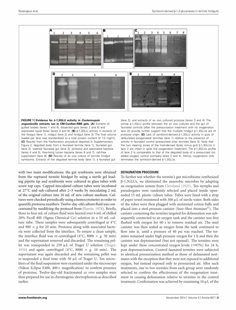
FIGURE 1 | Evidence for β-1,3GLU activity in Zootermopsis
angusticollis extracts run in CM-Curdlan-RBB gels. (A) Extracts ofgutted bodies (lanes 1 and 4), dissected guts (lanes 2 and 5) andexpressed liquid feces (lanes 3 and 6). (B) β-1,3GLU activity in extracts ofthe foregut (lane 1), midgut (lane 2) and hindgut (lane 3). The final volumeloaded per lane was standardized to a total protein content of 1.5 mg/mL.(C) Results from the fractionation procedure depicted in SupplementaryFigure 2, degutted body from a faunated termite (lane 1), faunated gut(lane 2), washed faunated gut (lane 3), protozoa and associated bacteria(lanes 4 and 5), free-living lumen bacteria (lanes 6 and 7), cell-freesupernatant (lane 8). (D) Results of ex vivo culture of termite hindgutsymbionts. Extracts of the degutted termite body (lane 1), a faunated gut
(lane 2), and extracts of ex vivo cultured protozoa (lanes 3 and 4). Thesimilar β-1,3GLU profile between the ex vivo cultures and the gut offaunated controls [after the pressurization treatment with no oxygenationlane 2)] provide further support that the multiple hindgut β-1,3GLUs are ofprotozoa origin. (E) Lack of symbiont-derived β-1,3GLU activity in guts ofdefaunated (oxygenated) termites (lane 1) relative to the presence ofactivity in faunated control (pressurized only) termites (lane 3). Note thatthe two clearing zones of the host-derived (body minus gut) β-1,3GLUs inlane 2 are intact in spite the oxygenation treatment. The β-1,3GLUs profileof lane 2 is comparable to that of the degutted body of a pressurized (noadded oxygen) control (compare lanes 2 and 4). Hence, oxygenation onlyeliminates the symbiont-derived β-1,3GLUs.
with two main modifications: the gut symbionts were obtainedfrom the ruptured termite hindgut by using a sterile gel load-ing pipette tip and symbionts were cultured in glass tubes withscrew top caps. Capped inoculated culture tubes were incubatedat 27◦C and sub-cultured after 2–3 weeks by inoculating 2 mLof the original culture into 10 mL of new culture medium. Cul-tures were checked periodically using a hemocytometer in order toquantify protozoa numbers. Twelve-day-old culture fluid was con-centrated by modifying the protocol from (Yamin, 1978). Briefly,three to four mL of culture fluid were layered over 6 mL of chilled20% Ficoll 400 (Sigma Chemical Co) solution in a 10 mL cul-ture tube. These samples were subsequently centrifuged at 4◦Cand 900 × g for 20 min. Protozoa along with associated bacte-ria were collected from the interface. To ensure a clean sample,the interface fluid was re-centrifuged (4◦C, 8000 × g, 30 min)and the supernatant removed and discarded. The remaining pel-let was resuspended in 250 μL of Trager U solution (Trager,1934) and again centrifuged (4◦C, 8000 × g, 10 min). Thesupernatant was again discarded and the remaining pellet wasre-suspended a final time with 50 μL of Trager U. Ten micro-liters of the final suspension were examined under the microscope(Nikon Eclipse E400, 400× magnification) to confirm presenceof protozoa. Twelve-day-old fractionated ex vivo samples werethen prepared for use in chromogenic electrophoresis as describedearlier.
DEFAUNATION PROCEDURETo further test whether the termite’s gut microbiome synthesizedβ-1,3GLUs, we eliminated the anaerobic microbes by adaptingan oxygenation system from Cleveland (1925). Ten nymphs andpseudergates were randomly selected and placed inside open-ended 15 mL plastic culture tubes. Tubes were lined with a stripof paper towel moistened with 300 μL of sterile water. Both sidesof the tubes were then plugged with moistened cotton-balls andplaced into a steel pressure canister (Sure-Shot Atomizer©). Thecanister containing the termites targeted for defaunation was sub-sequently connected to an oxygen tank and the canister was firstflushed with oxygen for 60 s to remove residual air. The steelcanister was then sealed as oxygen from the tank continued toflow into it, until a pressure of 40 psi was reached. The ter-mites remained under high pressure oxygen for 1 h and then thecanister was depressurized (but not opened). The termites werekept under these concentrated oxygen levels (≈97%) for 24 h,post depressurization. Control faunated termites were subjectedto identical pressurization method as those of defaunated nest-mates with the exception that they were not exposed to additionaloxygen, but instead exposed only to pressurized air. After suchtreatments, one to two termites from each group were randomlyselected to confirm the effectiveness of the oxygenation treat-ment in causing defaunation relative to termites in the controltreatment. Confirmation was achieved by examining 10 μL of the
www.frontiersin.org November 2014 | Volume 5 | Article 607 | 3
Rosengaus et al. Symbiont-derived β-1,3-glucanases in termite hindguts
hindgut fluid of a dissected termite on a hemocytometer using acompound light microscope (Nikon Eclipse E400) at 400× mag-nification and enumerating the number of intact protozoa in thesample (Supplementary Figure 2).
IN VITRO ANTIFUNGAL ASSAYTwo different methods were used to test in vitro the effect of β-1,3GLUs on conidia viability. We first quantified the long-lastingeffects (at 96 h of plating) of β-1,3GLUs on conidia viability byenumerating colony forming units (CFUs). The effects on coni-dia viability earlier during fungus development were also testedby quantifying conidia germination at 18 h following plating.Although the protocols for each of these methods differ (seebelow and Supplementary Figure 3), both provide accurate mea-surement of antifungal properties of termite extracts (and theiraccompanying β-1,3GLU s) at different stages of fungus develop-ment (Supplementary Figure 3). Long-lasting effects can be tracedback to effects during the early stages of fungus development:fewer CFUs four days post-plating result from lower germinationrates18 h post-plating.
To test the long-lasting fungistatic properties of termite tissue,extracts of both faunated and defaunated termites (as describedabove) were incubated with fungal conidia, then plated and fungalCFUs enumerated, following the methods outlined below. Imme-diately after the pressure only (faunated controls) and pressureplus oxygen (defaunated) treatments, termites were placed intoglass dishes with a plaster of Paris substrate that was moistenedwith 1000 μL of sterile water and maintained there for 24 h. Theplaster permitted termites to be kept under high moisture whileeliminating feeding on cellulose material (filter paper or wood).Hence, both defaunated and faunated treatments experienced thesame degree of starvation and therefore we controlled for the effectof nutritional status on antifungal properties of the gut. Through-out the 24 h on the moistened plaster, any residual glucanasesfrom the hindgut symbionts were likely flushed from the diges-tive system of the defaunated termites. Extracts of these termiteguts were prepared and then incubated with 10 μL of a 50 mg/mLsolution of ampicillin (to control bacterial overgrowth) and 10 μLof a 1 × 10⁴ conidia/mL suspension of M. anisopliae for 24 h at25◦C while gently shaken at 50 rpm. Conidia initiate germinationonly after plating onto the agar medium and hence, we are certainno germination occurred during the incubation of conidia withthe termite extracts. Control samples (lacking termite extracts)were created with 10 μL NaAc buffer, 10 μL sterile distilled water,10 μL of ampicillin, and 10 μL of a 1 × 10⁴ conidia/mL suspen-sion of M. anisopliae. These control samples were also incubatedand shaken alongside the experimental groups. After 24 h of incu-bation, 60 μL of sterile distilled water was added to each tubeand 100 μL of the resulting solution was plated using sterile glassbeads onto PDA plates (100 mm × 15 mm) supplemented with50 μg/mL of ampicillin and incubated at 25◦C for 96 h after whichCFUs were counted (Figure 2, Supplementary Figures 3A,B).
IN VITRO ANTIFUNGAL ASSAY, RESCUE EFFECT OFD-GLUCONO1,5-LACTONE (GDL)In a parallel study and to establish if fungistatic effects of ter-mite tissue extracts affected fungus viability earlier during its
development, we quantified conidia germination rates. To thisend, 5 μL of a 20 mM solution of the glucanase inhibitorGDL (Bulmer et al., 2009) were added to the tissue extracts.If β-1,3GLUs had fungistatic effect on conidia, then blockingthe β-1,3GLUs active sites should enhance or completely res-cue fungal viability relative to un-blocked β-1,3GLUs. Entire
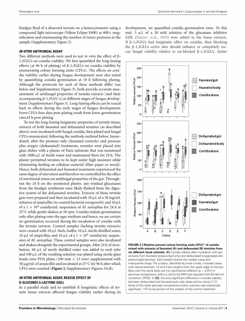
FIGURE 2 | Relative percent colony forming units (CFU)* of conidia
mixed with extracts of faunated (A) and defaunated (B) termites from
six different stock colonies. (C) Conidia viability after incubation with gutextracts from faunated (pressurized only) and defaunated (oxygenated andpressurized) termites. Each boxplot shows the median value andinterquartile range. The outliers, identified by small circles, included caseswith values between 1.5 and 3 box lengths from the upper edge of the box.Bars with the same letter are not significantly different (p > 0.017) inpairwise comparisons within a colony (by MW test adjusted with Bonferronicorrection, SPSS). In (C), the only significant difference in conidia viabilitybetween defaunated and faunated guts was observed for colony C (†).None of the other pairwise comparisons within colonies was statisticallysignificant. *CFUs as percent of the median of the control treatment.
Frontiers in Microbiology | Microbial Symbioses November 2014 | Volume 5 | Article 607 | 4
Rosengaus et al. Symbiont-derived β-1,3-glucanases in termite hindguts
guts (fore-, mid-, hingut, rectum) of 11 nymphs were placedinto a Biomasher (Cartagen) fitted with a filter (pore size 80–145 μm). The corresponding degutted bodies were placed into asecond Biomasher. To each tube, 60 μL of sterile distilled waterwas added. The contents of the tubes were then ground witha pestle and centrifuged (4◦C, 14,000 × g, 1 min). The liquidflow-through was transferred to Costar Spin-X 0.22 μm (Corn-ing) filters (20 μL each filter). Twenty microliter of 200 mMNaAc (pH 5.0) was then added to the filter and centrifuged(4◦C, 12,000 × g, 4 min) to filter out any bacteria contaminantsand remaining termite tissue. The flow-through of the sampleswas then pooled and 4 μL were observed under a microscope(400×) to determine if there was any termite tissue. If termitetissue was present, repeated filtration continued until the flow-through was clean. The filtrate (20 μL) was mixed with 10 μLof ampicillin (50 μg/mL), 10 μL of M. anisopliae (108 coni-dia/mL), and 5 μL of GDL (100 mM; or distilled water for noGDL control).
Control samples (lacking termite extracts) were similarly pre-pared with 10 μL NaAc buffer, 10 μL sterile distilled water,10 μL of ampicillin, and 10 μL of M. anisopliae conidia suspen-sion (108 conidia/mL) and 5 μL of GDL (100 mM). A secondset of controls were set up in an identical fashion except forthe GDL which was substituted by distilled water. Samples wereincubated at 25◦C while shaking for 6 h. Following incubation,10 μL of the conidia-extract suspension was seeded onto a micro-scope slide containing a 1 mL of solidified PDA layer (n = 3replicates/treatment; Rosengaus et al., 1998b). The slides wereincubated for 18 h at 25◦C (Supplementary Figure 3). Percentgermination was estimated by counting the number of coni-dia with visible germ tubes (Supplementary Figure 3D) out ofthe total number of conidia for each of 10 fields of vision perslide [a total of 30 fields of vision (Rosengaus et al., 1998b)].The entire experiment was replicated two or three times. Weensured that the addition of GDL inactivated the two host-derivedβ-1,3GLUs of the termite’s degutted body as well as the multi-ple β-1,3GLUs of the gut by running chromogenic gels with thesesamples (Figure 3).
Whether the in vitro antifungal assays were recorded asCFUs (long-lasting effects) or germination rates (early effects),fungistasis was analyzed through the use of non-parametrictests (Mann–Whitney [MW] and Kruskal–Wallis tests, SPSS,19.0) as both measures of fungal growth (either CFUs or coni-dia germination) were not normally distributed. Bonferronicorrections were applied when running any multiple pairwisecomparisons.
IN VIVO SUSCEPTIBILITY ASSAYSThree different colonies were used for testing whether termiteswith intact hindgut symbiont communities had reduced suscepti-bility to mycosis. Defaunated insects (by oxygenation) or pressurecontrol (faunated) termites were allowed to walk freely for onehour inside a Petri dish (60 × 15 mm) lined with filter paper(Whatman #5) moistened with 374 μL of either a 1 × 10⁵ coni-dia/mL suspension of M. anisopliae or a 0.1% Tween 80 solutionlacking conidia (Rosengaus et al., 1998b). In addition, a group ofnaïve termites (taken directly from the corresponding colonies)
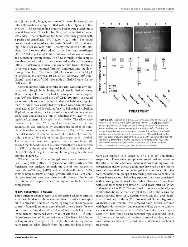
FIGURE 3 | (A) Inactivation of β-1,3GLUs in the presence of 100 mM of theinhibitor GDL relative to the same samples without GDL (B). (C) Medianpercent germination of fungal conidia following incubation with termitetissues and termite tissues treated with the inhibitor GDL. Each boxplotshows the median value and interquartile range. The outliers, identified bysmall circles, included cases with values between 1.5 and 3 box lengthsfrom the upper or lower edges of the box. Bars with the same letter are notsignificantly different (p > 0.002) in pairwise comparisons (by MW testadjusted with Bonferroni correction). n indicates the number of field ofvision on which the median germination rate was estimated.
were also exposed to a Tween 80 or a 1 × 10⁵ conidia/mLsuspension. These naïve groups were established to determinethe effects that the additional manipulations resulting from theoxygenation and/or pressurization, may have had on the insect’ssurvival beyond those due to fungal infection alone. Termiteswere maintained in groups of ten during exposure to conidia orTween 80 suspensions. Following exposure, they were transferredin these same groups to sterile Petri dishes (60 mm × 15 mm) linedwith clean filter paper (Whatman # 1) and given water ad libitumand maintained at 25◦C. The measured parameters included, sur-vival distributions, percent survival at the end of the census, themedian survival time [LT50; Kaplan-Meir (KM) Test] and the rel-ative hazard ratio of death (Cox Proportional Hazard RegressionAnalysis). Dead termites were removed daily; surface sterilizedwith hypochlorite (6.0%), and plated on (PDA) plates to confirmcause of death (Rosengaus et al., 1998b). Survival analyses [KaplanMeir (KM) tests] and a Cox proportional regression model (SPSS,19.0) were used to estimate the time course of survival, mediansurvival times, and relative hazard ratios of death as a function oftreatment.
www.frontiersin.org November 2014 | Volume 5 | Article 607 | 5
Rosengaus et al. Symbiont-derived β-1,3-glucanases in termite hindguts
ONTOGENY OF β-1,3GLUs EXPRESSIONTo establish the time frame during termite development atwhich the expression of β-1,3GLUs begins, we created extractsof termite embryos (fertilized eggs) as well as first and secondinstar larvae by following the same protocols described earlier.Given their small size, these young larvae were not degutted.Instead, the entire animal (with its gut) was processed to cre-ate the extracts. The extracts were subsequently loaded ontochromogenic gels.
SURVEY OF THE PRESENCE OF β-1,3GLUs ACROSS TERMITE SPECIESTo compare profiles of β-1,3GLUs in the body and gut tissuesacross species spanning the termite phylogeny, we loaded side-by-side extract samples of termites representing different specieswith differing nesting, feeding, and foraging habits (Abe, 1987)of the families Termopsidae (Z. angusticollis), Rhinotermitidae(Reticulitermes flavipes), Kalotermitidae (Cryptotermes secundus),and Termitidae (Nasutitermes corniger).
RESULTSZootermopsis angusticollis have two host-derived active β-1,3GLUsin their bodies (Figure 1A). Additionally, they possess several extraβ-1,3GLUs in their gut contents and liquid feces (Figure 1A).Such multiple β-1,3GLUs were observed in the hindgut regionof the termite’s alimentary canal (Figure 1B), the same site wheremost symbiotic protozoa and their associated ecto- and endo-symbionts reside (Brune, 2014). In sharp contrast, the fore- andmid-gut regions of the same termites lacked the multiple clear-ing zones and instead only exhibited the same two β-1,3GLUs asthose observed in the degutted body (Figures 1A,B). Furthermore,hindgut fractions separated by centrifugation (SupplementaryFigure 1) and analyzed in chromogenic gels indicated that thehighest β-1,3GLUs signature (brightest clearing zones) were inthe protozoa (and its associated bacteria) samples (Figure 1C).In comparison, the fractions containing free-living bacteria aswell as the cell free supernatant had negligible β-1,3GLU activity(Figure 1C). That the washed gut tissue showed some enzymaticactivity suggests that the protozoa and associated bacteria con-sortia were not completely flushed out of the tissue during thewash. Otherwise, we would have only observed the two termite-derived β-1,3GLUs (as seen in Figure 1C). Twelve-day-old ex vivoprotozoa cultures of Z. angusticollis hindgut, consisting of mostlyTrichomitopsis termopsidis (and their associated bacteria), alsohad β-1,3GLU activity which approximated that of the multi-ple clearing zones of faunated termite hindguts (Figure 1D).The differences in these β-1,3GLUs profiles likely reflect differ-ences in the diversity and/or density of protozoa (and associatedbacteria) between the ex vivo cultures and the in vivo samples.Finally, in the absence of the normal hindgut microbial com-munity (following oxygenation treatments which destroys theanaerobic gut symbionts; Supplementary Figure 2), there was aconsistent loss of the multiple β-1,3GLUs activity while the two β-1,3GLUs of bodies from the same oxygenated insects remainedactive (Figure 1E). Based on these multiple lines of evidencewe are confident that the observed multiple β-1,3GLUs in thetermite hindguts are of protozoa origin and/or their associatedbacteria.
IN VITRO ANTIFUNGAL ASSAYOur results indicate that incubation of fungal conidia with eitherentire gut or degutted body extracts from faunated termitesreduced fungal growth after 96 h form plating (Figure 2A). Thedegutted body extract reduced the median number of CFUs inall colonies except in colony D; the reduction in CFUs betweendegutted bodies and the corresponding controls was significant incolonies B and C (Figure 2A; MW = 6.0, p = 0.011; MW = 21.0,p = 0.003, respectively). In all six termite colonies examined, gutextracts significantly reduced fungal viability relative to the coni-dia alone. Furthermore, in four out of six colonies, the fungistaticeffects of the faunated gut extracts were significantly greater thanthe inhibition caused by the degutted body (Figure 2A).
To test if the gut symbionts played a role in the fungistatic natureof the gut, the extracts of defaunated termites were also incubatedwith fungal conidia. If symbionts produce the fungistatic metabo-lite, removal of the symbionts (and therefore removal of theirfunctional β-1, 3GLUs, as demonstrated in lane 1 of Figure 1Ein the main text) should rescue conidia viability. To our surprise,the gut extracts of defaunated termites continued to reduce coni-dia viability relative to controls (Figure 2B). A comparison of thefungistatic nature of defaunated vs. faunated guts showed thatthe presence or absence of aerobic and facultative aerobic gutsymbionts did not affect conidia viability (Figure 2C). Extractsof termites from Colony C were the only ones where faunatedguts were significantly more fungistatic than the defaunated guts(Figure 2C, MW = 36.0, p = 0.037), as originally predicted.These results suggest that the two host-derived gut β-1,3GLUs(not affected by the oxygenation treatment, Figure 1E, main text)are sufficient to reduce conidia viability.
IN VITRO ANTIFUNGAL ASSAY, RESCUE EFFECT OFD-GLUCONO1,5-LACTONE (GDL)Given the visual confirmation of the loss of enzymatic functionwhen gels were incubated with GDL (compare Figures 3A,B), weexpected termite extracts/conidia suspensions to have increasedconidia viability in the presence of GDL relative to conidia incu-bated with extracts containing functional β-1,3GLUs. The sodiumacetate (NaAc) buffer itself, used in the preparation of conidiasuspensions with tissue extracts had no negative effects on coni-dia viability relative to the conidia in 0.1% Tween 80 suspension.Neither did the addition of GDL or the combination of NaACand GDL (Figure 3C). Therefore, we conclude that the multi-ple functional β-1,3GLUs of hindgut extracts were responsiblefor reducing germination rates by approximately 20%. Conidiaincubated with the degutted body extracts containing the two host-derived β-1,3GLUs reduced conidia germination by ∼12% relativeto conidia alone, conidia with NaAc, conidia with GDL and coni-dia with both NaAC and GDL, all of which had median percentgermination of 100 (Figure 3C). That conidia germination ratesof the different controls (conida + NaAC, conidia + GDL, coni-dia + NaAC + GDL) were equivalent to those of conidia incubatedwith GDL-inhibited termite tissue extracts demonstrates that theantifungal activity in the hindgut was indeed due to the presence ofboth host- and symbiont-derived functional β-1,3GLUs and thatthese enzymes affect fungal growth at the initial stages of fungusdevelopment (i.e., conidia germination).
Frontiers in Microbiology | Microbial Symbioses November 2014 | Volume 5 | Article 607 | 6
Rosengaus et al. Symbiont-derived β-1,3-glucanases in termite hindguts
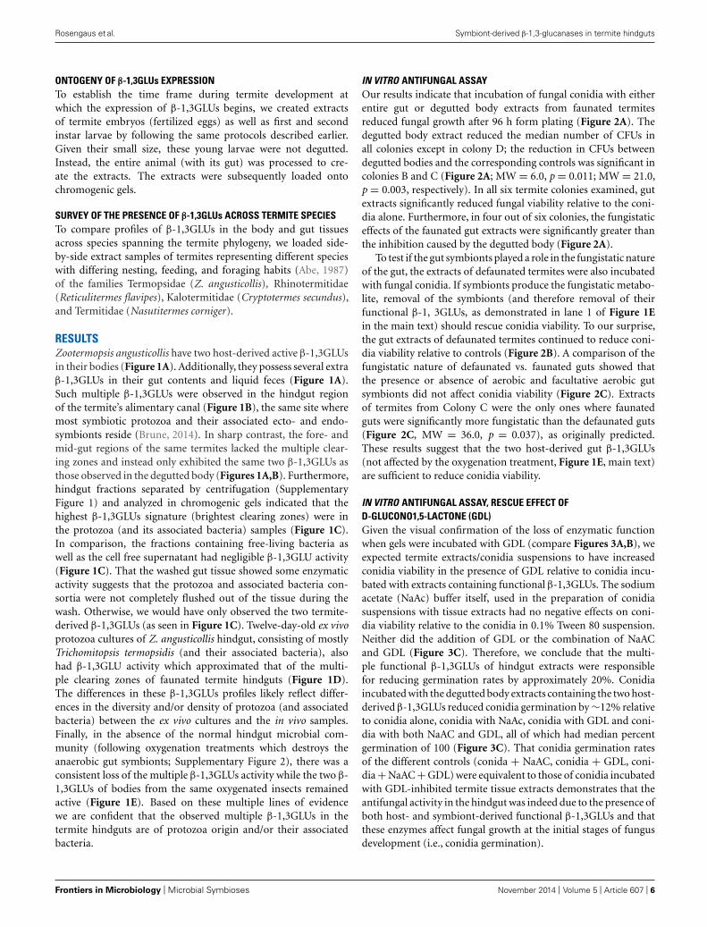
IN VIVO SUSCEPTIBILITY ASSAYSA Cox proportional regression model showed that the colonyof origin from which termites originated was not a significantand independent predictor of termite survival [Wald statistic(WS) = 4.9, df = 2, p > 0.05]. Hence, data from all threecolonies were pooled for further statistical analyses. Treatment[faunated (pressurized only) or defaunated (pressurized and oxy-genated)], on the other hand, was a significant and independentpredictor of termite survival (WS = 227.4, df = 3, p < 0.0001).Pairwise comparisons of the combined effect of treatment andconidia exposure (Tween 80 only or Tween 80 + conidia) revealedsignificant differences in the time course of survival (i.e., sur-vival distributions) between all groups [faunated controls (opendiamond), faunated termites exposed to 105 conidia suspension(filled diamond), defaunated controls (open inverted triangle)and defaunated termites exposed to 105 conidia suspension (filledinverted triangle); Figure 4; KM test]. Defaunated controls expe-rienced significantly higher mortality than faunated controls [LogRank (LR) X2 = 107.1, p < 0.0001, Figure 4; KM test]. The combi-nation of defaunation and fungal exposure significantly increasedtermite mortality relative to fungal exposure with pressure treat-ment alone (LR X2 = 62.2, p < 0.0001; Figure 4; KM test). Hence,in the face of pathogenic pressures, oxygenation (and its resultingdefaunation) had an added effect beyond the stressors of handlingand pressurization, resulting in a 27.5% difference in survival bythe end of the census period. The defaunated control termites had6.6 times the hazard ratio of death, but no confirmations of myco-sis relative to faunated controls (WS = 1.9, df = 1, p < 0.0001).The defaunated fungal-exposed termites had the highest hazard
FIGURE 4 | Survival distributions of defaunated (inverted triangles)
and faunated (diamonds) termites exposed to either a 0.1%Tween 80
solution lacking fungal conidia (open symbols) or a 0.1%Tween 80
suspension containing 1 × 10⁵ conidia/mL (filled symbols). Differentletters (at the right of each survival curve) indicate significant differences(after a Bonferroni correction; p < 0.0001) in the time course of survival(i.e., survival distributions; by Log Rank Test, Survival Analysis). Numbers inparenthesis represent (relative hazard ratio of death, percent confirmationof Metarhizium anisopliae infection). Ref indicates the reference treatmentagainst which all other hazard ratios of death were compared to.
ratio of death (20.7 times) relative to the faunated control treat-ment (WS = 3.0, df = 1, p < 0.0001) with 35% confirmationrates. Fungal-exposed faunated termites had a hazard ratio ofdeath 9.4 times that of their control counterparts (WS = 2.2,df = 1, p < 0.0001), with 29% of the deaths confirmed for M.anisopliae.
ONTOGENY OF β-1,3GLUs EXPRESSIONIndividual embryos (fertilized eggs) had undetectable levels ofβ-1,3GLUs when assayed in chromogenic gels. To reach a minimal(yet noticeable) β-1,3GLU signature, at least 10 pooled embryoswere required, resulting in one faint clearing zone (SupplementaryFigure 4A). Extracts containing five or 10 pooled first or secondinstar larvae also showed detectable β-1,3GLU activity (Supple-mentary Figures 4A,B). Interestingly, β-1,3GLU activity of extractscontaining first instar larvae were highly variable, with some sam-ples showing little evidence while others clearly demonstratingmultiple clearing zones (Supplementary Figures 4A,B). The vari-ation across extract samples of second instar larvae was reducedrelative to that of first instar larvae.
SURVEY OF THE PRESENCE OF β-1,3GLUs ACROSS TERMITE SPECIESTo compare profiles of β-1,3GLUs in the body and gut tis-sues across species spanning the termite phylogeny, we loadedside-by-side extract samples of termites representing differentfamilies [Termopsidae: Z. angusticollis (Z.a.)], [Rhinotermitidae:R. flavipes (R.f.)], [Kalotermitidae: C. secundus (C.s.)] and families[Termitidae: N. corniger (N.c.); Supplementary Figure 5]. Whileall species exhibited two termite-derived β-1,3GLUs in their body(minus the gut) tissue, these enzymes were not identical given theirdifferent migration patterns in the gel. Interestingly, the fact thatdissected guts of the “higher” termite N. corniger (Bulmer et al.,2009; Supplementary Figure 5) have only two β-1,3GLUs providesfurther support to our conclusion that the source of these multiplehindgut enzymes of termites are the protozoa and their associatedbacteria. N.c. lack the typical hindgut protozoa communities ofthe “lower” termites (Ohkuma and Brune, 2011).
DISCUSSIONThrough a combination of chromogenic gels, tissue dissec-tions and fractionation, ex vivo culturing of termite hindgutsymbionts and defaunation experiments, we provide, for thefirst time, evidence that the multiple functionally active β-1,3GLUs in Z. angusticollis hindguts are synthesized by pro-tozoa and/or their associated bacteria. In contrast, the twoβ-1,3GLUs found in tissues other than the hindgut are likelyof termite-origin. This is supported by data from the recentZ. nevadensis genome (Terrapon et al., 2014) which shows thatout of the six Gram-negative binding proteins (GNBPs), twocorrespond to β-1,3GLUs identified in other termites (Hamil-ton et al., 2011; Gao et al., 2012; Hamilton and Bulmer,2012; Hussain et al., 2013) and Cryptocercus wood cockroaches(Bulmer et al., 2012).
It remains to be determined whether the additional β-1,3GLUs’of the termite’s hindgut are synthesized by the protozoa them-selves or by their associated bacteria. Many bacteria are inti-mately associated with the protozoa either on their surface or
www.frontiersin.org November 2014 | Volume 5 | Article 607 | 7
Rosengaus et al. Symbiont-derived β-1,3-glucanases in termite hindguts
as intracellular endo-symbionts (Brune, 2006, 2014; Strassertet al., 2012) and potentially, they too could be the source of themultiple β-1,3GLUs. Unfortunately, because of their inherent co-dependency, the experimental elimination of bacterial ecto- andendo-symbionts also negatively affected the protozoa population.Hence, further studies are needed to identify which microbes inthe protozoa/bacteria partnership are responsible for the synthesisof β-1,3GLUs.
Our results also point to the likelihood that both thetermite- and symbiont-derived β-1,3GLUs have fungistatic activ-ity (Figure 2). Such an effect is likely due to the enzymes’ abilityto break β-1,3 glycosidic linkages of β-1,3 glucans, the principalcomponent of fungal cell walls (Brown and Gordon, 2005). Wehave previously demonstrated that β-1,3GLUs cause Metarhiziumconidia to collapse, decreasing conidia volume by 25% (Bulmeret al., 2009; Rosengaus et al., 2011). The possibility exist that suchconidia shrinkage resulted from using natural and commerciallyavailable β-1,3GLUs at concentrations above the physiologicalconcentrations normally used by termites. Nevertheless, theseresults indicate that β-1,3GLUs can damage and affect both conidiaintegrity and its germination, resulting in prolonged fungistasis (atleast at 96 h post-plating).
The possibility exists that compounds other than (or incombination with) the multiple β-1,3GLUs, including termicin(Hamilton and Bulmer, 2012), may be responsible for the refrac-tive nature of termite guts against fungus growth (Siderhurstet al., 2005; Chouvenc et al., 2008, 2009, 2010). The loss of activ-ity of β-1,3GLUs through the addition of GDL, a competitiveinhibitor, and its accompanying rescue effects on conidia via-bility (Figure 3) together with our observation that body alone(minus the gut) and gut extracts have similar fungistatic prop-erties (Figure 2) provide indirect evidence that β-1,3GLUs canhelp control fungal germination. Our in vivo data also suggestthat termites with an intact gut microbial community are lesssusceptible to mycosis. Regrettably, because immune defensesand susceptibility to mycosis are probably influenced by nutri-tional status, we cannot pull apart the independent effects ofthese coupled factors. Yet, in the face of pathogenic pressures,oxygenation (and its resulting defaunation) had an added effectbeyond the stressors of handling and pressurization, particu-larly within the first 10 days of the census period (Figure 4),when the effects on malnourishment (due to the lack of gutsymbionts) should have had lower impacts on survival. Ulti-mately, by day 20 post-exposure to fungal conidia, there wasa 27.5% difference in the survival of defaunated (oxygenated)and faunated (pressurized) termites. Results from other termitespecies demonstrate that GDL-treated insects (i.e., with inhib-ited β-1,3GLUs’) are more susceptible to mycosis (Bulmer et al.,2009; Hamilton et al., 2011; Hamilton and Bulmer, 2012). Takentogether, all these results strongly suggest that the functionalsymbiont-derived β-1,3GLUs play some role in the control ofmycosis.
It is unlikely that the gut’s hostile environment toward coni-dia germination in previous (Chouvenc et al., 2009, 2010) and ourcurrent experiments was due to factors such as gut pH or low oxy-gen levels. The pH of the termite’s paunch, the hindgut regionwhere the protozoa and their ecto- and endosymbionts reside, is
within the range of M. anisopliae germination (Dillon and Charn-ley, 1986; Brune et al., 1995). Furthermore, while an oxygen deficitis normally inhibitory to the germination of conidia (Cochraneet al., 1963), the termite gut is not completely anoxic as the perime-ter regions of the alimentary canal (where conidia would likely begerminating) are oxic (Ebert and Brune, 1997). Remarkably, thefungistatic effects of termite guts persist even after termites die(Chouvenc et al., 2009) suggesting that whichever antifungal com-pound(s) exist in the termite’s guts, they are environmentally stableand not easily degradable. These same traits are characteristic ofthe multiple hindgut β-1,3GLUs which retain robust activity longafter their incorporation into the termite’s nest structure (Bulmeret al., 2009; Hamilton et al., 2011; Hamilton and Bulmer, 2012).Although the gut microbiota of lower termites has been histori-cally associated with cellulose degradation and nitrogen fixation(Scharf et al., 2011; Brune, 2014), our results suggest that theirfunction may extend beyond nutrition. In conjunction with otherfactors, the gut microbiome may also provide protection againstmycosis.
Notably, the putative protective role of the multiple hindgutβ-1,3GLUs may extend beyond the individual termites. The bene-fits derived from harboring an intact gut microbiome (along withthe functional β-1,3GLUs) can be also appreciated at the com-munal level. Because termites use their liquid and solid fecesin nest construction (Rosengaus et al., 1998b) and given thatthe liquid feces have β-1,3GLU activity [Figure 1A, but not thefecal pellets (Rosengaus et al., 2013)], the excretion and incor-poration of β-1,3GLUs from the liquid feces into the termite’snest structure likely expands mycosis protection to the entirecolony. Although previous research has demonstrated that ter-mite feces have potent antifungal properties (Rosengaus et al.,1998b; Chouvenc et al., 2013) and that Actinobacteria may beresponsible for such negative effects (Chouvenc et al., 2013), noinformation exists on whether microbes colonizing the termitefecal pellets produce β-1,3GLUs. Similarly, at the communal level,the active β-1,3GLU secreted by the salivary glands of some ter-mites (Bulmer et al., 2009; Hamilton et al., 2011), in conjunctionwith termicin (Lamberty et al., 2001), may be playing an anti-fungal role when these compounds are smeared onto the cuticleof nestmates during mutual grooming. The combined effects ofperforming social interactions and the deposition of biochem-ical secretions with antifungal properties (i.e., termicins andβ-1,3GLUs) likely increase resistance against mycosis throughoutthe entire colony.
Embryos have one β-1,3GLU, albeit in extremely low quanti-ties. This suggests that the expression of at least one host-derivedβ-1,3GLU starts early during the termite’s development. Giventhat liquid feces of Z. angusticollis have identical β-1,3GLUs pro-files as those from faunated hindguts (Figure 1A), and that firstand second instar larvae exhibit the typical multiple symbiont-derived β-1,3GLUs, we are confident that these young individualsacquire the additional enzymes along with the full complement ofhindgut microbes through common proctodeal exchanges (anus-to-mouth feeding; Supplementary Figure 4). These exchangesensure that young nestmates benefit not only from attaining cel-lulolytic activity and nitrogen supplementation (Machida et al.,2001), but also from protection against pathogenic fungi during
Frontiers in Microbiology | Microbial Symbioses November 2014 | Volume 5 | Article 607 | 8
Rosengaus et al. Symbiont-derived β-1,3-glucanases in termite hindguts
the insects’ development. If the hindgut β-1,3GLUs of other lowertermites (Bulmer et al., 2010; Hamilton et al., 2011; Hamilton andBulmer, 2012) are also of microbial origin and are similarly sharedamong nestmates via proctodeal exchanges, this and the work byKoch and Schmid-Hempel (2011) may represent prime examplesof widespread symbiont-mediated social immunity across the ter-mites (Traniello et al., 2002; Cremer et al., 2007; Rosengaus et al.,2011). Termites, and their microbiome, are therefore excellent testorganisms to address ecological immunology questions against thebackground of eusociality.
The mutualistic association with hindgut microbes that syn-thesize β-1,3GLUs was likely established before the evolution oftermites. Cryptocercus punctulatus, the closest extant roach rel-ative of termites (Nalepa and Bandi, 2000; Lo and Eggleton,2011), also contains β-1,3GLU activity in its hindgut (Bul-mer et al., 2012) and its liquid feces (Rosengaus et al., 2013).They also share many of the same gut microbiota with ter-mites (Brune, 2014). The most parsimonious explanation forthe presence of multiple β-1,3GLUs in both Cryptocercus and“lower” termites’ hindguts as well as their liquid feces, is thattheir common ancestor already possessed a microbial commu-nity capable of synthesizing β-1,3GLUs, which could be thenshared among family members via proctodeal exchanges. Thepresence of such enzymes could have served as an importantpre-adaptation for the evolution of group-living as family unitsof Cryptocercus and early termite-like ancestors likely exhibitedenhanced resistance against pathogenic fungi while nesting in andfeeding on decayed wood even before the inception of eusocial-ity (Rosengaus et al., 2011). Based on the few termite speciestested, the existence of multiple hindgut β-1,3GLUs appears tobe conserved across the “lower” termites (Bulmer et al., 2010;Hamilton et al., 2011), although given their different migrationpatterns in gels (Supplementary Figure 5), these enzymes likelyunderwent an independent diversification within each termitelineage.
CONCLUSIONOur data show that the multiple β-1,3GLUs in Z. angusticollisare synthesized by the hindgut protozoa (and/or its associatedbacteria). The presence of such enzymes may help explain inpart, why the termite’s alimentary canal appears hostile to fun-gal development. Additional in vivo experiments indicate thattermites with an intact hindgut microbiota (and its accompa-nying β-1,3GLUs) are less susceptibility to mycosis. Throughproctodeal feedings, termites effectively share and distribute theirhindgut microbial consortia along with their fungistatic activ-ity, a prime example of symbiont-mediated social immunity.Studying the holobiont in general, and the microbiome’s impactson host resistance to disease in particular, is critical in under-standing the role that microorganisms play in the evolution ofimportant ecologically relevant host traits (Margulis and Fester,1991; Feldhaar, 2011), including perhaps the evolution of insecteusociality.
AUTHOR CONTRIBUTIONSRebeca B. Rosengaus, Kelley F. Schultheis, Mark S. Bulmer par-ticipated in all aspects of this research. Alla Yalonetskaya ran
the defaunation and in vivo susceptibility assays while WilliamS. DuComb, Ryan W. Benson, and Veronica Godoy-Carter carriedout the chromogenic gel separation for GDL experiments. John P.Thottam and Rebeca B. Rosengaus performed the antifungal activ-ity assays using the GDL inhibitor. Rebeca B. Rosengaus fundedthe work while Rebeca B. Rosengaus, Kelley F. Schultheis, Mark S.Bulmer, and Veronica Godoy-Carter wrote the manuscript.
ACKNOWLEDGMENTSThis research was funded by NSF CAREER award (DEB 0447316,Rosengaus RB, PI) and an NSF REU supplement award for AllaYalonetskaya. Additional funds were available from a NortheasternUniversity TIER1 award and the The Gustavus and Louise Pfeif-fer Research Foundation. We appreciate the thoughtful commentsmade by two referees. Their suggestions improved the quality andclarity of this manuscript. We are indebted to the administra-tors of Huddart Park (San Mateo County) and the Redwood EastBay Regional Park in Oakland, CA, USA for permission to collecttermites.
SUPPLEMENTARY MATERIALThe Supplementary Material for this article can be found online at:http://www.frontiersin.org/journal/10.3389/fmicb.2014.00607/abstract
REFERENCESAbe, T. (1987). “Evolution of life types in termites,” in Evolution and Coadaptation in
Biotic Communities, eds S. Kawano, J. H. Connel, and T. Hidaka (Tokyo: Universityof Tokyo Press), 125–148.
Blackwell, M., and Rossi, W. (1986). Biogeography of fungal ectoparasites oftermites. Mycotaxon 25, 581–601.
Brown, G. D., and Gordon, S. (2005). Immune recognition of fungalbeta-glucans. Cell Microbiol. 7, 471–479. doi: 10.1111/j.1462-5822.2005.00505.x
Brune, A. (2006). “Symbiotic associations between termites and prokaryotes”,in The Prokaryotes, eds M. S. Workin, S. Falkow, E. Rosenberg, K. Schleifer,and E. Stackebrandt (New York: Springer), 439–474. doi: 10.1007/0-387-30741-9-17
Brune, A. (2014). Symbiotic digestion of lignocellulose in termite guts. Nat. Rev.Microbiol. 12, 168–180. doi: 10.1038/nrmicro3182
Brune, A., Emerson, D., and Breznak, J. A. (1995). The termite gut microfloraas an oxygen sink: microelectrode determination of oxygen and pH gra-dients in guts of lower and higher termites. App. Environ. Microbiol. 61,2681–2687.
Bulmer, M. S., Bachelet, I., Raman, R., Rosengaus, R. B., and Sasisekharan, R.(2009). Targeting an antimicrobial effector function in insect immunity as apest control strategy. Proc. Natl. Acad. Sci. U.S.A. 106, 12652–12657. doi:10.1073/pnas.0904063106
Bulmer, M. S., Denier, D., Velenovsky, J., and Hamilton, C. (2012). A commonantifungal defense strategy in Cryptocercus woodroaches and termites. InsectesSoc. 59, 469–478. doi: 10.1007/s00040-012-0241-y
Bulmer, R. M. S., Lay, F., and Hamilton, C. (2010). Adaptive evolution insubterranean termite antifungal peptides. Insect Mol. Biol. 19, 669–674. doi:10.1111/j.1365-2583.2010.01023.x
Chouvenc, T., Efstathion, C. A., Elliot, M. L., and Su, N.-Y. (2013). Extended diseaseresistance emerging from the faecal nest of a subterranean termite. Proc. R. Soc.B 280, 1–9. doi: 10.1098/rspb.2013.1885
Chouvenc, T., Su, N.-Y., and Elliot, M. I. (2008). Antifungal activity of thetermite alkaloid norharmane against the mycelial growth of Metarhiziumanisopliae and Aspergillus nominus. J. Invertebr. Pathol. 99, 345–347. doi:10.1016/j.jip.2008.07.003
Chouvenc, T., Su, N.-Y., and Grace, J. K. (2011). Fifty years of attempted bio-logical control of termites – analysis of a failure. Biol. Control 59, 69–82. doi:10.1016/j.biocontrol.2011.06.015
www.frontiersin.org November 2014 | Volume 5 | Article 607 | 9
Rosengaus et al. Symbiont-derived β-1,3-glucanases in termite hindguts
Chouvenc, T., Su, N.-Y., and Robert, A. (2009). Inhibition of Metarhizium anisopliaein the alimentary tract of the eastern subterranean termite Reticulitermes flavipes.J. Invertebr. Pathol. 101, 130–136. doi: 10.1016/j.jip.2009.04.005
Chouvenc, T., Su, N.-Y., and Robert, A. (2010). Inhibition of the fungal pathogenMetarhizium anisopliae in the alimentary tracts of five termite (Isoptera) species.Fl. Entomol. 93, 467–469. doi: 10.1653/024.093.0327
Cleveland, L. R. (1925). Toxicity of oxygen for protozoa in vivo and in vitro: animalsdefaunated without injury. Biol. Bull. 48, 455–468. doi: 10.2307/1536555
Cochrane, V. W., Cochrane, J. C., Collins, C. B., and Serafin, F. G. (1963). Spore ger-mination and carbon metabolism in Fusarium solani. II. Endogenous respirationin relation to germination. Am. J. Bot. 50, 806–814. doi: 10.2307/2440199
Coleman, G. S. (1991). Medium Preparation and Cultivation of TrichomitopsisTermopsidis in the Presence of Bacteria. New York: Oxford University Press.
Cremer, S., Armitage, S. A. O., and Schmid-Hempel, P. (2007). Social immunity.Curr. Biol. 17, R693–R702. doi: 10.1016/j.cub.2007.06.008
Dillon, R. J., and Charnley, A. K. (1986). Inhibition of Metarhizium anisopliaeby the gut bacterial flora of the desert locust, Schistocerca gregaria: evidencefor an antifungal toxin. J. Invertebr. Pathol. 47, 350–360. doi: 10.1016/0022-2011(86)90106-0
Ebert, A., and Brune, A. (1997). Hydrogen concentration profiles at the oxic-anoxicinterface: a microsensor study of the hindgut of the wood-feeding lower termiteReticulitermes flavipes (Kollar). Appl. Environ. Microbiol. 63, 4039–4046.
Feldhaar, H. (2011). Bacterial symbionts as mediators of ecologically impor-tant traits of insect hosts. Ecol. Entomol. 36, 533–543. doi: 10.1111/j.1365-2311.2011.01318.x
Gao, Q., Tancredi, S. E., and Thompson, G. J. (2012). Identification of mycosis-related genes in the eastern subterranean termite by suppression subtractivehybridization. Arch. Insect Biochem. Physiol. 80, 63–76. doi: 10.1002/arch.21026
Grenham, S., Clarke, G., Cryan, J. F., and Dinan, T. G. (2011). Brain-gut-microbe communication in health and disease. Front. Physiol. 2:94. doi:10.3389/fphys.2011.00094
Haine, E. R. (2008). Symbiont-mediated protection. Proc. R. Soc. B 275, 353–361.doi: 10.1098/rspb.2007.1211
Hamilton, C., and Bulmer, M. S. (2012). Molecular antifungal defenses in subter-ranean termites: RNA interference reveals in vivo roles of termicins and GNBPsagainst a naturally encountered pathogen. Dev. Comp. Immunol. 36, 372–377.doi: 10.1016/j.dci.2011.07.008
Hamilton, C., Lay, F., and Bulmer, M. S. (2011). Subterranean termite prophylacticsecretions and external antifungal defenses. J. Insect Physiol. 57, 1259–1266. doi:10.1016/j.jinsphys.2011.05.016
Hussain, A., Li, Y.-F., Cheng, Y., Liu, Y., and Chen, C.-C. (2013). Immune-related transcriptome of Coptotermes formosanus shiraki workers: the defensemechanism. PLoS ONE 87:e69543. doi: 10.1371/journal.pone.0069543
Kalix, S., and Buchenauer, H. (2005). Direct detection of beta-1,3-glucanase in plantextracts by polyacrylamide gel electrophoresis. Electrophoresis 16, 1016–1018. doi:10.1002/elps.11501601170
Kinross, J. M., Darzi, A. W., and Nicholson, J. K. (2011). Gut microbiome-hostinteractions in health and disease. Genome Med. 3, 14. doi: 10.1186/gm228
Koch, H., and Schmid-Hempel, P. (2011). Socially transmitted gut microbiota pro-tect bumble bees against an intestinal parasite. Proc. Natl. Acad. Sci. U.S.A. 108,19288–11929. doi: 10.1073/pnas.1110474108
Lamberty, M., Zachary, D., Lanot, R., Bordereau, C., Robert, A., Hoffmann, J.A., et al. (2001). Insect immunity - Constitutive expression of a cysteine-richantifungal and a linear antibacterial peptide in a termite insect. J. Biol. Chem.276, 4085–4092. doi: 10.1074/jbc.M002998200
Lo, N., and Eggleton, P. (2011). “Termite phylogenetics and cocladogenesis withsymbionts,” in Biology of Termites: A Modern Synthesis, eds D. E. Bignell, Y. Roisin,and N. Lo (Heidelberg: Springer), 27–50.
Machida, M., Kitade, O., Miura, T., and Matsumoto, T. (2001). Nitrogenrecycling through proctodeal trophallaxis in the Japanese damp-wood termiteHodotermopsis japonicus (Isoptera, Termopsidae). Insectes Soc. 48, 52–56. doi:10.1007/PL00001745
Margulis, L., and Fester, R. (1991). Symbiosis as a Source of Evolutionary Innovation.Cambridge, MA: MIT Press.
Mattila, H. R., Rios, D., Walker-Sperling, V. E., Roeselers, G., and Newton, I. L.G. (2012). Characterization of the active microbiotas associated with honey beesreveals healthier and broader communities when colonies are genetically diverse.PLoS ONE 7:e32962. doi: 10.1371/journal.pone.0032962
Moran, N. A. (2006). Symbiosis. Curr. Biol. 16, R866–R871. doi:10.1016/j.cub.2006.09.019
Morgavi, D. P., Sakurada, M., Tomita, Y., and Onodera, R. (1994). Pres-ence in rumen bacterial and protozoal populations of enzymes capable ofdegrading fungal cell walls. Microbiol. 140, 631–636. doi: 10.1099/00221287-140-3-631
Nalepa, C. A., and Bandi, C. (2000). “Characterizing the ancestors: paedomorphosisand termite evolution,” in Termites: Evolution, Sociality, Symbioses, Ecology, edsT. Abe, D. E. Bignell, and M. Higashi (Dordrecht: Kluwer Academic Publishers),53–76. doi: 10.1007/978-94-017-3223-9_3
Ohkuma, M., and Brune, A. (2011). “Diversity, structure, and evolution of termitegut microbial community,” in Biology of Termites: a Modern Synthesis, eds D. E.Bignell, Y. Roisin, and N. Lo (Berlin: Springer-Verlag), 413–438.
Oliver, K. M., Smith, A. H., and Russell, J. A. (2014). Defensive symbiosis in the realworld- advancing ecological studies of heritable, protective bacteria in aphids andbeyond. Funct. Ecol. 28, 341–355. doi: 10.1111/1365-2435.12133
Parker, B. J., Chelsea, J., Spragg, C. J., Altincicek, B., and Gerardo, N. M.(2013). Symbiont-mediated protection against fungal pathogens in pea aphids:a role for pathogen specificity? Appl. Environ. Microbiol. 79, 2455–2458. doi:10.1128/AEM.03193-12
Poinar, G. O. (2009). Description of an early Cretaceous termite (Isoptera:Kalotermitidae) and its associated intestinal protozoa, with comments on theirco-evolution. Parasit. Vectors 2, 1–17. doi: 10.1186/1756-3305-2-12
Rath, A. C. (2000). The use of entomopathogenic fungi for control oftermites. Biocontrol Sci. Technol. 10, 563–581. doi: 10.1080/095831500750016370
Rivas, P., Cassels, B. K., Morello, A., and Repetto, Y. (1999). Effects of some [beta]-carboline alkaloids on intact Trypanosoma cruzi epimastigotes. Comp. Biochem.Physiol. C. 122, 27–31.
Rosengaus, R. B., Maxmen, A. B., Coates, L. E., and Traniello, J. F. A. (1998a).Disease resistance: a benefit of sociality in the dampwood termite Zootermopsisangusticollis (Isoptera: Termopsidae). Behav. Ecol. Sociobiol. 44, 125–134. doi:10.1007/s002650050523
Rosengaus, R. B., Guldin, M. R., and Traniello, J. F. A. (1998b). Inhibitory effect oftermite fecal pellets on fungal spore germination. J. Chem. Ecol. 24, 1697–1706.doi: 10.1023/A:1020872729671
Rosengaus, R. B., Mead, K., Du Comb, W. S., Benson, R. W., and Godoy, V. G. (2013).Nest sanitation through defecation: antifungal properties of wood cockroachfeces. Naturwissenschaften 100, 1051–1059. doi: 10.1007/s00114-013-1110-x
Rosengaus, R. B., and Traniello, J. F. A. (2001). Disease susceptibility and theadaptive nature of colony demography in the dampwood termite Zootermopsisangusticollis. Behav. Ecol. Sociobiol. 50, 546–556. doi: 10.1007/s002650100394
Rosengaus, R. B., Traniello, J. F. A., and Bulmer, M. S. (2011). “Ecology, behaviorand evolution of disease resistance in termites,” in Biology of Termites: A ModernSynthesis, eds D. E. Bignell, Y. Roisin, and N. Lo (Berlin: Springer-Verlag), 165–192.
Ryu, J.-R., Kim, S.-H., Lee, H.-Y., Bai, J. Y., Nam, Y.-D., Bae, J.-W., et al. (2008).Innate immune homeostasis by the homeobox gene caudal and commensal-gutmutualism in Drosophila. Science 319, 777–782. doi: 10.1126/science.1149357
Scarborough, C. L., Ferrari, J., and Godfray, H. C. J. (2005). Aphid protected frompathogen by endosymbiont. Science 310, 1781. doi: 10.1126/science.1120180
Scharf, M. E., Karl, Z. J., Sethi, A., and Boucias, D. G. (2011). Multiple levels ofsynergistic collaboration in termite lignocellulose digestion. PLoS ONE 6:e21709.doi: 10.1371/journal.pone.0021709
Siderhurst, M. S., James, D. M., Blunt, T. D., and Bjostad, L. B. (2005). Antimicro-bial activity of norharmane against the entomopathogenic fungus Metarhiziumanisopliae (Metsch.) and the caste and phylogenetic distribution of this defensein termites (Insecta: Isoptera). Sociobiology 46, 563–577.
Strassert, J. F. H., Köhler, T., Wienemann, T. H. G., Ikeda-Ohtsubo, W., Faivre,N., Franckenberg, S., et al. (2012). ‘Candidatus Ancillula trichonymphae’, a novellineage of endosymbiotic Actinobacteria in termite gut flagellates of the genusTrichonympha. Environ. Microbiol. 14, 3259–3270. doi: 10.1111/1462-2920.12012
Terrapon, N., Li, C., Robertson, H. M., Ji, L., Meng, X., Booth, W., et al. (2014).Molecular traces of alternative social organization in a termite genome. Nat.Commun. 5, 3636. doi: 10.1038/ncomms4636
Trager, W. (1934). The cultivation of a cellulose-digesting flagellate, Trichomonastermopsidis, and of certain other termite protozoa. Biol. Bull. 66, 182–190. doi:10.2307/1537331
Frontiers in Microbiology | Microbial Symbioses November 2014 | Volume 5 | Article 607 | 10
Rosengaus et al. Symbiont-derived β-1,3-glucanases in termite hindguts
Traniello, J. F. A., Rosengaus, R. B., and Savoie, K. (2002). The developmentof immunity in a social insect: evidence for the group facilitation of diseaseresistance. Proc. Natl. Acad. Sci. U.S.A. 99, 6838–6842. doi: 10.1073/pnas.102176599
Wier, A., Dolan, M., Grimaldi, D., Guerrero, R., Wagensberg, J., and Margulis, L.(2002). Spirochete and protist symbionts of a termite (Mastotermes electrodo-minicus) in Miocene amber. Proc. Natl. Acad. Sci. U.S.A. 99, 1410–1413. doi:10.1073/pnas.022643899
Yamin, M. A. (1978). Axenic cultivation of the cellulolytic flagellate Trichomitopsistermopsidis (Cleveland) from the termite Zootermopsis. J. Protozool. 25, 535–538.doi: 10.1111/j.1550-7408.1978.tb04181.x
Conflict of Interest Statement: The authors declare that the research was conductedin the absence of any commercial or financial relationships that could be construedas a potential conflict of interest.
Received: 01 September 2014; accepted: 24 October 2014; published online: 21November 2014.Citation: Rosengaus RB, Schultheis KF, Yalonetskaya A, Bulmer MS, DuComb WS,Benson RW, Thottam JP and Godoy-Carter V (2014) Symbiont-derived β-1,3-glucanases in a social insect: mutualism beyond nutrition. Front. Microbiol. 5:607.doi: 10.3389/fmicb.2014.00607This article was submitted to Microbial Symbioses, a section of the journal Frontiers inMicrobiology.Copyright © 2014 Rosengaus, Schultheis, Yalonetskaya, Bulmer, DuComb, Benson,Thottam and Godoy-Carter. This is an open-access article distributed under the termsof the Creative Commons Attribution License (CC BY). The use, distribution or repro-duction in other forums is permitted, provided the original author(s) or licensor arecredited and that the original publication in this journal is cited, in accordance withaccepted academic practice. No use, distribution or reproduction is permitted whichdoes not comply with these terms.
www.frontiersin.org November 2014 | Volume 5 | Article 607 | 11