![Supporting Information · S1 Supporting Information Facile Synthesis of 9H-Pyrrolo[1,2-α]indoles Via Brønsted Acid Catalyzed Cascade Reaction Kunhua Xu,a Wenming Chen,b Jin Lin,a](https://static.fdocument.org/doc/165x107/605455892ce0f4683a341586/supporting-s1-supporting-information-facile-synthesis-of-9h-pyrrolo12-indoles.jpg)
Supporting Information Facile Synthesis of a Novel ...
17
Supporting Information Facile Synthesis of a Novel Genetically Encodable Fluorescent α-Amino Acid Emitting Greenish Blue Light Aakash Gupta, a Brian P. Garreffi, a and Maolin Guo* ab a) Department of Chemistry and Biochemistry, UMass Cranberry Health Research Center, University of Massachusetts Dartmouth, 285 Old Westport Road, North Dartmouth, MA 02747 (USA), E-mail: [email protected]. b) Department of Chemistry, University of Massachusetts Amherst, 710 North Pleasant Street, Amherst, MA 01003 (USA), E-mail: [email protected]. Electronic Supplementary Material (ESI) for ChemComm. This journal is © The Royal Society of Chemistry 2020
Transcript of Supporting Information Facile Synthesis of a Novel ...
Supporting Information
Facile Synthesis of a Novel Genetically Encodable Fluorescent α-Amino Acid Emitting Greenish Blue Light
Aakash Gupta,a Brian P. Garreffi,a and Maolin Guo*ab
a) Department of Chemistry and Biochemistry, UMass Cranberry Health Research Center, University of Massachusetts Dartmouth, 285 Old Westport Road, North Dartmouth, MA 02747 (USA), E-mail: [email protected].
b) Department of Chemistry, University of Massachusetts Amherst, 710 North Pleasant Street, Amherst, MA 01003 (USA), E-mail: [email protected].
Electronic Supplementary Material (ESI) for ChemComm.This journal is © The Royal Society of Chemistry 2020

SI Table of Contents
1. Experimental Section ................................................................................................... 1
2. Supplemental Figures ................................................................................................. 9
3. References................................................................................................................... 14
1
1. Experimental Section
Chemicals: N-(tert-Butoxycarbonyl)-4-iodo-L-phenylalanine, 9-Phenanthracenylboronic
acid, N,N-Dimethyformamide, and Bis(triphenylphosphine) palladium (II) dichloride were
purchased from Sigma-Aldrich. Methyl iodide was purchased from Alfa Aesar. Rhodamine B was
purchased from Sigma-Aldrich. All chemicals were used without further purification. The reagents
not mentioned as well as the solvents used in the syntheses were purchased commercially and were
ACS grade.
Instrumentation and characterization: The 1H NMR and 13C NMR spectra were
obtained on a Bruker AVANCE III HD 400 MHz High-Performance Digital NMR spectrometer.
Chemical shifts were analyzed on MestReNova software and reported in delta (δ) unit parts per
million (ppm) downfield tetramethylsilane (TMS). The spectra were obtained in Methanol-d4 or
DMSO-d6 olvent with 0.03 % of TMS as the internal reference. The abbreviation for splitting
patterns represents, s = singlet, d = doublet, m = multiplet and coupling constants are reported in
Hertz (Hz). The mass spectra were obtained on a Waters ACQUITY UPLC (Ultra-Pure Liquid
chromatograph) Xevo QTOF (Quadrupole Time of Flight) high resolution mass spectrometer in a
mobile phase of Acetonitrile:Water with a solvent gradient of 100% to 50% over 10 minutes of
time. The product was purified by HPLC using an Agela Technologies Cheetah HP100 equiped
with a 12g Flash Column. Flow rate was set to 8.0 mL min-1.The mobile phase solvent system
consisted of water (A)/Methanol (B) gradient system (0% B for 5 min, 0-40% for 10 min, 40-100%
for 1 min, and 100% for 9 min). UV-Vis spectra were obtained on a Perkin-Elmer Lambda 25
spectrometer over wavelength range of 200-800 nm. Fluorescence spectra were recorded on a
Perkin-Elmer LS55 luminescence spectrometer at 293 K. The excitation wavelengths and filters
used were indicated in the figures. The relative quantum yield was measured using rhodamine B
2
as a standard. The pH measurements were carried out on a Corning pH meter equipped with a
Sigma-Aldrich micro combination electrode calibrated with standard buffer solutions. Optical
rotation was measured by optical spectrophotometry using a Rudolph Research Analytical Autopol
III Automatic Polarimeter. Zeiss LSM 710 laser scanning confocal microscope with excitation
lasers at 405 nm, 458 nm, 488 nm, 514 nm, 543 nm, and 633 nm was used in the imaging
experiments. The reuse function of the Zeiss software enables the use of previous experiment
perimeter to minimize differences between different batch of experiments if needed. 35 mm glass
bottom dishes (P35G-1.5-14-C) with cover slips for confocal cell experiments were purchased
from Mattek.
Synthesis and Characterization
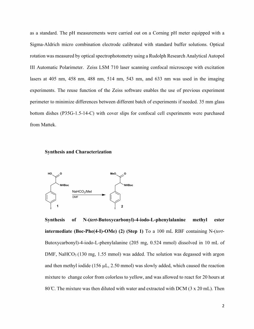
Synthesis of N-(tert-Butoxycarbonyl)-4-iodo-L-phenylalanine methyl ester
intermediate (Boc-Phe(4-I)-OMe) (2) (Step 1) To a 100 mL RBF containing N-(tert-
Butoxycarbonyl)-4-iodo-L-phenylalanine (205 mg, 0.524 mmol) dissolved in 10 mL of
DMF, NaHCO3 (130 mg, 1.55 mmol) was added. The solution was degassed with argon
and then methyl iodide (156 µL, 2.50 mmol) was slowly added, which caused the reaction
mixture to change color from colorless to yellow, and was allowed to react for 20 hours at
80 C. The mixture was then diluted with water and extracted with DCM (3 x 20 mL). Then
I
HO O
NHBoc
NaHCO3/MeIDMF
I
MeO O
NHBoc
1 2
3
the organic layers were combined and washed with a saturated brine solution (1 x 10 mL)
before the addition of anhydrous MgSO4 to remove any traces of water. The solvent was
removed using roto-evaporation set at 40-50 C to afford a yellow oily compound. The
product was purified with silica gel flash chromatography using a Hexane: Ethyl acetate
solvent gradient of 9:1. The product obtained (N-(tert-Butoxycarbonyl)-4-iodo-L-
phenylalanine methyl ester) (2) is a white fibrous solid (190 mg). HRMS (ESI): calcd for
[C15H20INO4+H]+: 406.0437, found: m/z: 406.0450.
I
MeO O
NHBoc
2
Cs2CO3/Pd(PPh3)2Cl2Dioxane
B
OH
HO
BocHN O
OMe
4
3
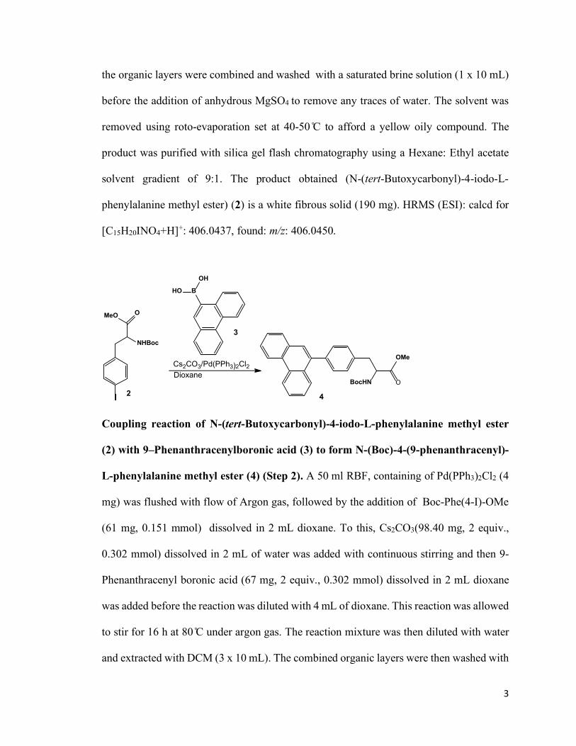
Coupling reaction of N-(tert-Butoxycarbonyl)-4-iodo-L-phenylalanine methyl ester
(2) with 9–Phenanthracenylboronic acid (3) to form N-(Boc)-4-(9-phenanthracenyl)-
L-phenylalanine methyl ester (4) (Step 2). A 50 ml RBF, containing of Pd(PPh3)2Cl2 (4
mg) was flushed with flow of Argon gas, followed by the addition of Boc-Phe(4-I)-OMe
(61 mg, 0.151 mmol) dissolved in 2 mL dioxane. To this, Cs2CO3(98.40 mg, 2 equiv.,
0.302 mmol) dissolved in 2 mL of water was added with continuous stirring and then 9-
Phenanthracenyl boronic acid (67 mg, 2 equiv., 0.302 mmol) dissolved in 2 mL dioxane
was added before the reaction was diluted with 4 mL of dioxane. This reaction was allowed
to stir for 16 h at 80 C under argon gas. The reaction mixture was then diluted with water
and extracted with DCM (3 x 10 mL). The combined organic layers were then washed with
4
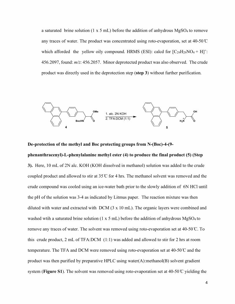
a saturated brine solution (1 x 5 mL) before the addition of anhydrous MgSO4 to remove
any traces of water. The product was concentrated using roto-evaporation, set at 40-50 C
which afforded the yellow oily compound. HRMS (ESI): calcd for [C29H29NO4 + H]+:
456.2097, found: m/z: 456.2057. Minor deprotected product was also observed. The crude
product was directly used in the deprotection step (step 3) without further purification.
De-protection of the methyl and Boc protecting groups from N-(Boc)-4-(9-
phenanthracenyl)-L-phenylalanine methyl ester (4) to produce the final product (5) (Step
3). Here, 10 mL of 2N alc. KOH (KOH dissolved in methanol) solution was added to the crude
coupled product and allowed to stir at 35 C for 4 hrs. The methanol solvent was removed and the
crude compound was cooled using an ice-water bath prior to the slowly addition of 6N HCl until
the pH of the solution was 3-4 as indicated by Litmus paper. The reaction mixture was then
diluted with water and extracted with DCM (3 x 10 mL). The organic layers were combined and
washed wtih a saturated brine solution (1 x 5 mL) before the addition of anhydrous MgSO4 to
remove any traces of water. The solvent was removed using roto-evaporation set at 40-50 C. To
this crude product, 2 mL of TFA:DCM (1:1) was added and allowed to stir for 2 hrs at room
temperature. The TFA and DCM were removed using roto-evaporation set at 40-50 C and the
product was then purified by preparative HPLC using water(A):methanol(B) solvent gradient
system (Figure S1). The solvent was removed using roto-evaporation set at 40-50 C yielding the
OMe
OBocHN
OH
OH3N
1. alc. 2N KOH2. TFA:DCM (1:1)
54
5
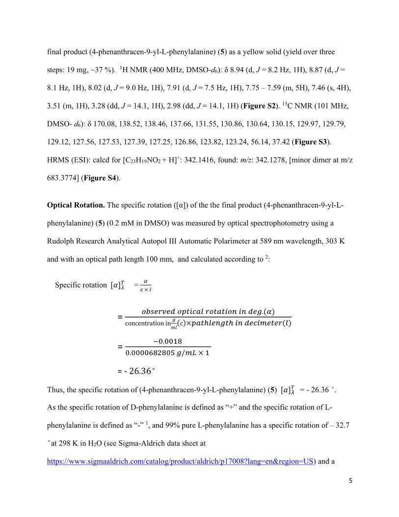
final product (4-phenanthracen-9-yl-L-phenylalanine) (5) as a yellow solid (yield over three
steps: 19 mg, ~37 %). 1H NMR (400 MHz, DMSO-d6): δ 8.94 (d, J = 8.2 Hz, 1H), 8.87 (d, J =
8.1 Hz, 1H), 8.02 (d, J = 9.0 Hz, 1H), 7.91 (d, J = 7.5 Hz, 1H), 7.75 – 7.59 (m, 5H), 7.46 (s, 4H),
3.51 (m, 1H), 3.28 (dd, J = 14.1, 1H), 2.98 (dd, J = 14.1, 1H) (Figure S2). 13C NMR (101 MHz,
DMSO- d6): δ 170.08, 138.52, 138.46, 137.66, 131.55, 130.86, 130.64, 130.15, 129.97, 129.79,
129.12, 127.56, 127.53, 127.39, 127.25, 126.86, 123.82, 123.24, 56.14, 37.42 (Figure S3).
HRMS (ESI): calcd for [C23H19NO2 + H]+: 342.1416, found: m/z: 342.1278, [minor dimer at m/z
683.3774] (Figure S4).
Optical Rotation. The specific rotation ([α]) of the the final product (4-phenanthracen-9-yl-L-
phenylalanine) (5) (0.2 mM in DMSO) was measured by optical spectrophotometry using a
Rudolph Research Analytical Autopol III Automatic Polarimeter at 589 nm wavelength, 303 K
and with an optical path length 100 mm, and calculated according to 2:
Specific rotation [𝛼𝛼]𝜆𝜆𝑇𝑇 = 𝛼𝛼c × 𝑙𝑙
= 𝑜𝑜𝑜𝑜𝑜𝑜𝑜𝑜𝑜𝑜𝑜𝑜𝑜𝑜𝑜𝑜 𝑜𝑜𝑜𝑜𝑜𝑜𝑜𝑜𝑜𝑜𝑜𝑜𝑙𝑙 𝑜𝑜𝑜𝑜𝑜𝑜𝑜𝑜𝑜𝑜𝑜𝑜𝑜𝑜𝑟𝑟 𝑜𝑜𝑟𝑟 𝑜𝑜𝑜𝑜𝑑𝑑.(𝛼𝛼)concentration in 𝑔𝑔
𝑚𝑚𝑚𝑚(c)×𝑜𝑜𝑜𝑜𝑜𝑜ℎ𝑙𝑙𝑜𝑜𝑟𝑟𝑑𝑑𝑜𝑜ℎ 𝑜𝑜𝑟𝑟 𝑜𝑜𝑜𝑜𝑜𝑜𝑜𝑜𝑑𝑑𝑜𝑜𝑜𝑜𝑜𝑜𝑜𝑜(𝑙𝑙)
= −0.0018 0.0000682805 𝑑𝑑/𝑑𝑑𝑚𝑚 × 1
= - 26.36
Thus, the specific rotation of (4-phenanthracen-9-yl-L-phenylalanine) (5) [𝛼𝛼]𝜆𝜆𝑇𝑇 = - 26.36 .
As the specific rotation of D-phenylalanine is defined as “+” and the specific rotation of L-
phenylalanine is defined as “-” 1, and 99% pure L-phenylalanine has a specific rotation of – 32.7
at 298 K in H2O (see Sigma-Aldrich data sheet at
https://www.sigmaaldrich.com/catalog/product/aldrich/p17008?lang=en®ion=US) and a
6
value of -34.3 in an earlier literature 3, the (4-phenanthracen-9-yl-L-phenylalanine) (5) is thus
in its L-form. Previous studies using similar Suzuki coupling to synthesize fluorescent unnatural
amino acids have been verified that these reaction conditions didn’t cause epimerization 4.
Fluorescence quantum yield measurement. Rhodamine B (0.1 M) solution was prepared by
dissolving Rhodamine B (0.047 g) into ethanol (0.001 L). Rhodamine B solution (10 µM) was
prepared by diluting the Rhodamine B solution (0.1 M). Rhodamine B solutions (1 µM, 0.8 µM,
0.6 µM, 0.4 µM, 0.2 µM) were prepared by diluting the Rhodamine B solution (10 µM).
Solutions for the fluorescent amino acid 4-phenanthracen-9-yl-L-phenylalanine(Phen-AA) were
prepared in dimethyl sulfoxide (DMSO). Each solution was mixed using a vortex. Fluorescence
spectra for rhodamine B were carried out with excitation and emission wavelengths set at 420
nm and 450-800 nm, respectively, and using an excitation slit width of 15 nm and an emission
slit width of 2.5 nm, while excitation and emission wavelengths set at 380 nm and 400-800 nm,
respectively, were used for Phen-AA. A plot of the base-line corrected integrated fluorescence
intensity vs. the absorbance for each concentration was prepared and the positive slope of the
linear fit was calculated. The data were compared to the rhodamine standard using the following
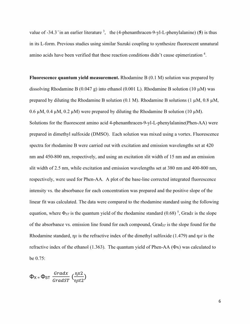
equation, where ΦST is the quantum yield of the rhodamine standard (0.68) 5, Gradx is the slope
of the absorbance vs. emission line found for each compound, GradST is the slope found for the
Rhodamine standard, ƞx is the refractive index of the dimethyl sulfoxide (1.479) and ƞst is the
refractive index of the ethanol (1.363). The quantum yield of Phen-AA (Фx) was calculated to
be 0.75:
ΦX = ΦST 𝐺𝐺𝑜𝑜𝑜𝑜𝑜𝑜𝐺𝐺𝐺𝐺𝑜𝑜𝑜𝑜𝑜𝑜𝐺𝐺𝑇𝑇 ( ɳ𝐺𝐺2ɳ𝑜𝑜𝑜𝑜2
)
7
= 0.68 (34.63837.141
) (1.4792
1.3632)
= 0.75
Photostability study. A series of fluorescence emission spectra (λex=380 nm, excitation slit 15
nm and emission slit 2.5 nm) of 4-phenanthracen-9-yl-L-phenylalanine(Phen-AA) solution
(0.001 M in DMSO-SPB buffer, pH 7.0, 0.05 M) was recorded at 293 K over a 5 h period. The
fluorescence spectrometer was set to scan the sample and record an emission spectrum every 180
seconds. 100 spectra were recorded over 5 hours. Therefore, a spectrum was recorded 120
seconds after the previous spectrum until 100 spectra were recorded.
Solvachromatic study. Due to low solubility of Phen-AA in some solvents, stock solution of
Phen-AA was prepared in DMSO and diluted 10 times in the specified solvents. Phen-AA
(0.001 M) solutions were prepared in a 9:1 mixture of solvents by diluting aliquots of a Phen-AA
(0.01 M) solution in DMSO, DMF, MeOH, SPB buffer (pH 7.0, 0.05 M), EtOH, acetone, and
isopropanol, respectively. Each solution was mixed using a vortex. Fluorescence emission
spectra (λex=380 nm, excitation slit 15 nm and emission slit 2.5 nm) were recorded at 293 K.
Cell culture and confocal imaging. Hela cells (ATCC) were cultured and maintained with the
complete growth medium composing of DMEM with supplement of 10% FBS in a humid 37 C
incubator under 5% CO2 level. To prepare for confocal experiments, Hela cells were cultured in
the flask until the confluency reached about ~70%. After the flask was rinsed with PBS twice, 2
ml 0.25% trypsin-EDTA solution was added followed by incubation in the incubator. Upon cell
dissociation from the flask’s wall, 2 ml complete growth medium was added to neutralize the
enzyme and stop the digestion. The mixture was then transferred into a 15 mL tube and
8
centrifuged for 3 min at 1,000 g. The medium was discarded to remove the residual enzyme and
other waste products. 5 ml of fresh complete medium was added to the tube to resuspend the cell
pellet. The same amount of cell containing medium from the tube was transferred into each of
the 35-mm glass bottom dishes, respectively, and 1 ml fresh complete medium was added.
Change the medium when necessary or every two days to support cell maintenance and growth
until experiment. To test the biocompatibility and imaging potential of the fluorescent amino
acid Phen-AA in live cells, Phen-AA (0.010 mM or 0.020 mM) was added to the medium of the
experimental dish with Hela cells for 1 hour. Before imaging, all dishes were washed three times
with fresh warm complete medium in a period of 45 mins to remove the excessive free Phen-AA
in the medium. Confocal fluorescent images of the Phen-AA incubated and control cells were
then acquired and analyzed. All dishes were excited with 405 nm, and emissions were collected
in the range of 410-550 nm.
2. Supplemental Figures
9
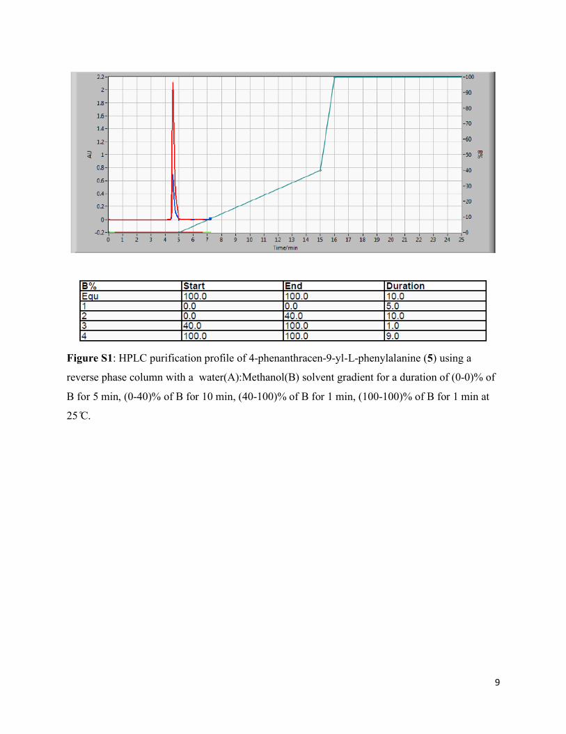
Figure S1: HPLC purification profile of 4-phenanthracen-9-yl-L-phenylalanine (5) using a
reverse phase column with a water(A):Methanol(B) solvent gradient for a duration of (0-0)% of
B for 5 min, (0-40)% of B for 10 min, (40-100)% of B for 1 min, (100-100)% of B for 1 min at
25 C.
10
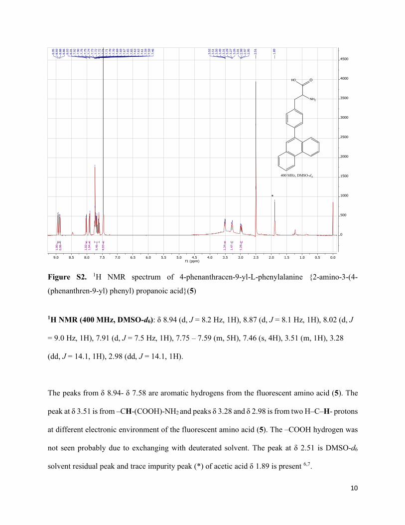
Figure S2. 1H NMR spectrum of 4-phenanthracen-9-yl-L-phenylalanine {2-amino-3-(4-
(phenanthren-9-yl) phenyl) propanoic acid}(5)
1H NMR (400 MHz, DMSO-d6): δ 8.94 (d, J = 8.2 Hz, 1H), 8.87 (d, J = 8.1 Hz, 1H), 8.02 (d, J
= 9.0 Hz, 1H), 7.91 (d, J = 7.5 Hz, 1H), 7.75 – 7.59 (m, 5H), 7.46 (s, 4H), 3.51 (m, 1H), 3.28
(dd, J = 14.1, 1H), 2.98 (dd, J = 14.1, 1H).
The peaks from δ 8.94- δ 7.58 are aromatic hydrogens from the fluorescent amino acid (5). The
peak at δ 3.51 is from –CH-(COOH)-NH2 and peaks δ 3.28 and δ 2.98 is from two H–C–H- protons
at different electronic environment of the fluorescent amino acid (5). The –COOH hydrogen was
not seen probably due to exchanging with deuterated solvent. The peak at δ 2.51 is DMSO-d6
solvent residual peak and trace impurity peak (*) of acetic acid δ 1.89 is present 6,7.
0.00.51.01.52.02.53.03.54.04.55.05.56.06.57.07.58.08.59.0f1 (ppm)
0
500
1000
1500
2000
2500
3000
3500
4000
4500
1.28
1.47
1.34
4.03
5.46
1.04
1.02
0.98
1.00
1.89
2.51
2.95
2.97
2.99
3.01
3.26
3.27
3.29
3.31
3.49
3.50
3.51
3.52
7.46
7.59
7.59
7.61
7.63
7.63
7.65
7.65
7.67
7.69
7.69
7.70
7.70
7.71
7.71
7.72
7.72
7.73
7.74
7.75
7.75
7.90
7.92
8.01
8.03
8.86
8.88
8.93
8.95
*
NH2
OHO
400 MHz, DMSO-d6
11
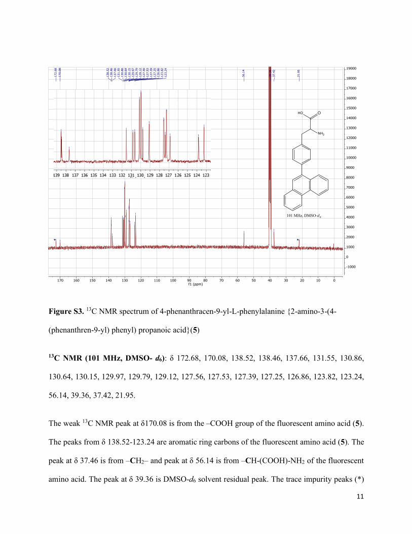
Figure S3. 13C NMR spectrum of 4-phenanthracen-9-yl-L-phenylalanine {2-amino-3-(4-
(phenanthren-9-yl) phenyl) propanoic acid}(5)
13C NMR (101 MHz, DMSO- d6): δ 172.68, 170.08, 138.52, 138.46, 137.66, 131.55, 130.86,
130.64, 130.15, 129.97, 129.79, 129.12, 127.56, 127.53, 127.39, 127.25, 126.86, 123.82, 123.24,
56.14, 39.36, 37.42, 21.95.
The weak 13C NMR peak at δ170.08 is from the –COOH group of the fluorescent amino acid (5).
The peaks from δ 138.52-123.24 are aromatic ring carbons of the fluorescent amino acid (5). The
peak at δ 37.46 is from –CH2– and peak at δ 56.14 is from –CH-(COOH)-NH2 of the fluorescent
amino acid. The peak at δ 39.36 is DMSO-d6 solvent residual peak. The trace impurity peaks (*)
0102030405060708090100110120130140150160170f1 (ppm)
-1000
0
1000
2000
3000
4000
5000
6000
7000
8000
9000
10000
11000
12000
13000
14000
15000
16000
17000
18000
19000
21.9
5
37.4
239
.36
56.1
4
123.
2412
3.82
126.
8612
7.25
127.
3912
7.53
127.
5612
9.12
129.
7912
9.97
130.
1513
0.64
130.
8613
1.55
137.
6613
8.46
138.
52
170.
0817
2.68
**
NH2
OHO
101 MHz, DMSO-d6
12
of acetic acid (δ 21.95 and δ 172.68)6,7 were observed in 13C NMR. The trace amount of acetic
acid impurity was also seen in the 1H NMR spectrum (Fig. S2).
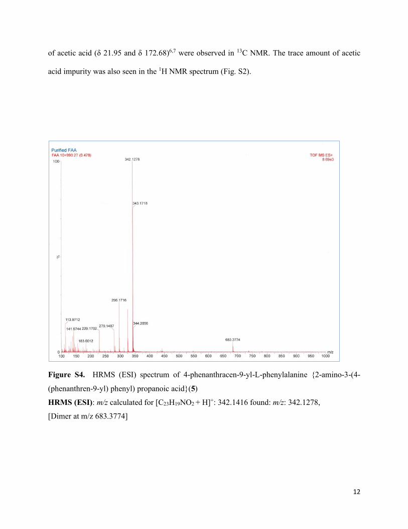
Figure S4. HRMS (ESI) spectrum of 4-phenanthracen-9-yl-L-phenylalanine {2-amino-3-(4-
(phenanthren-9-yl) phenyl) propanoic acid}(5)
HRMS (ESI): m/z calculated for [C23H19NO2 + H]+: 342.1416 found: m/z: 342.1278,
[Dimer at m/z 683.3774]
13
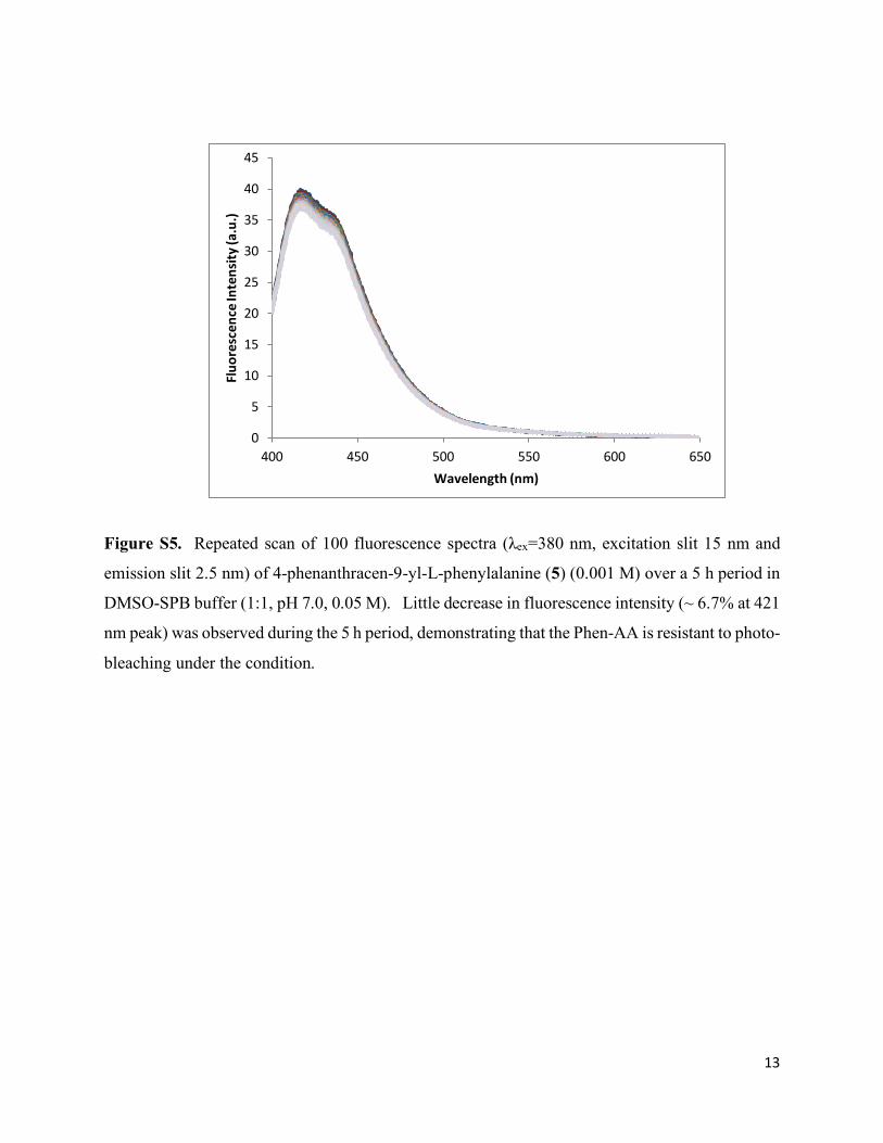
Figure S5. Repeated scan of 100 fluorescence spectra (λex=380 nm, excitation slit 15 nm and
emission slit 2.5 nm) of 4-phenanthracen-9-yl-L-phenylalanine (5) (0.001 M) over a 5 h period in
DMSO-SPB buffer (1:1, pH 7.0, 0.05 M). Little decrease in fluorescence intensity (~ 6.7% at 421
nm peak) was observed during the 5 h period, demonstrating that the Phen-AA is resistant to photo-
bleaching under the condition.
0
5
10
15
20
25
30
35
40
45
400 450 500 550 600 650
Fluo
resc
ence
Inte
nsity
(a.u
.)
Wavelength (nm)
14
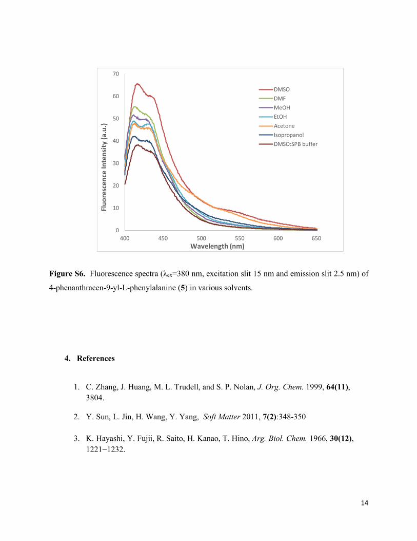
Figure S6. Fluorescence spectra (λex=380 nm, excitation slit 15 nm and emission slit 2.5 nm) of
4-phenanthracen-9-yl-L-phenylalanine (5) in various solvents.
4. References
1. C. Zhang, J. Huang, M. L. Trudell, and S. P. Nolan, J. Org. Chem. 1999, 64(11), 3804.
2. Y. Sun, L. Jin, H. Wang, Y. Yang, Soft Matter 2011, 7(2):348-350
3. K. Hayashi, Y. Fujii, R. Saito, H. Kanao, T. Hino, Arg. Biol. Chem. 1966, 30(12), 1221−1232.
0
10
20
30
40
50
60
70
400 450 500 550 600 650
Fluo
resc
ence
Inte
nsity
(a.u
.)
Wavelength (nm)
DMSODMFMeOHEtOHAcetoneIsopropanolDMSO:SPB buffer
15
4. L. W. K. Moodie, S. Chammaa, T. Kindahl, and C. Hedberg, Org. Lett. 2017, 19,
2797−2800
5. M. J. Snare, F. E. Treloar, K. P. Ghiggino, P. J. Thistlethwaite, J. Photochem., 1982, 18(4), 335-346.
6. G. R. Fulmer, A. J. M. Miller, N. H. Sherden, H. E. Gottileb, A. Nudelman, B. M. Stoltz, J. E. Bercaw, and K. I. Goldberg, Organometallics 2010, 29, 2176.
7. N. R. Babij, E. O. McCusker, G. T. Whiteker, B. Canturk, N. Choy, L. C. Creemer, C. V. De Amicis, N. M. Hewlett, P. L. Johnson, J. A. Knobelsdorf, F. Li, B. A. Lorsbach, B. M. Nugent, S. J. Ryan, M. R. Smith, and Q. Yang, Org. Process Res. Dev. 2016, 20, 661.