Studies toward bivalent κ opioids derived from - Beilstein Journals
9
2916 Studies toward bivalent κ opioids derived from salvinorin A: heteromethylation of the furan ring reduces affinity Thomas A. Munro *1,2 , Wei Xu 3 , Douglas M. Ho 4 , Lee-Yuan Liu-Chen 3 and Bruce M. Cohen 1 Full Research Paper Open Access Address: 1 McLean Hospital, Belmont, MA 02478, USA and Department of Psychiatry, Harvard Medical School, Boston, MA 02215, USA, 2 School of Chemistry and Bio21 Institute, University of Melbourne, Parkville 3010, Australia, 3 Center for Substance Abuse Research and Department of Pharmacology, Temple University School of Medicine, Philadelphia, PA 19140, USA and 4 Department of Chemistry and Chemical Biology, Harvard University, Cambridge MA 02138, USA Email: Thomas A. Munro * - [email protected] * Corresponding author Keywords: allotopic; bivalent ligand; designed multiple ligand; JDTic; κ-opioid receptor; natural products; Salvinorin A Beilstein J. Org. Chem. 2013, 9, 2916–2924. doi:10.3762/bjoc.9.328 Received: 01 September 2013 Accepted: 21 November 2013 Published: 20 December 2013 This article is part of the Thematic Series "Natural products in synthesis and biosynthesis". Guest Editor: J. S. Dickschat © 2013 Munro et al; licensee Beilstein-Institut. License and terms: see end of document. Abstract The recent crystal structure of the κ-opioid receptor (κ-OR) revealed, unexpectedly, that the antagonist JDTic is a bivalent ligand: in addition to the orthosteric pocket occupied by morphinans, JDTic also occupies a distinct (allotopic) pocket. Mutagenesis data suggest that salvinorin A (1) also binds to this allotopic pocket, adjacent to the aspartate residue that anchors the basic nitrogen atom of classical opiates (Asp138). It has been suggested that an H-bond donor appended to 1 might interact with Asp138, increasing affinity. Such a bivalent ligand might also possess altered functional selectivity. Based on modeling and known N-furanylmethyl opioid antagonists, we appended H-bond donors to the furan ring of 1. (Dimethylamino)methyl groups at C-15 or C-16 abolished affinity for κ-OR. Hydroxymethylation at C-16 was tolerated, but 15,16-bis-hydroxymethylation was not. Since allosteric modulators may go undetected in binding assays, we also tested these and other low-affinity derivatives of 1 for allosteric modulation of dynorphin A in the [ 35 S]GTPγS assay. No modulation was detected. As an alternative attachment point for bivalent derivatives, we prepared the 2-(hydroxyethoxy)methyl ether, which retained high affinity for κ-OR. We discuss alternative design strategies for linked, fused or merged bivalent derivatives of 1. 2916
Transcript of Studies toward bivalent κ opioids derived from - Beilstein Journals
2916
Studies toward bivalent κ opioids derived fromsalvinorin A: heteromethylation of the
furan ring reduces affinityThomas A. Munro*1,2, Wei Xu3, Douglas M. Ho4, Lee-Yuan Liu-Chen3
and Bruce M. Cohen1
Full Research Paper Open Access
Address:1McLean Hospital, Belmont, MA 02478, USA and Department ofPsychiatry, Harvard Medical School, Boston, MA 02215, USA,2School of Chemistry and Bio21 Institute, University of Melbourne,Parkville 3010, Australia, 3Center for Substance Abuse Research andDepartment of Pharmacology, Temple University School of Medicine,Philadelphia, PA 19140, USA and 4Department of Chemistry andChemical Biology, Harvard University, Cambridge MA 02138, USA
Email:Thomas A. Munro* - [email protected]
* Corresponding author
Keywords:allotopic; bivalent ligand; designed multiple ligand; JDTic; κ-opioidreceptor; natural products; Salvinorin A
Beilstein J. Org. Chem. 2013, 9, 2916–2924.doi:10.3762/bjoc.9.328
Received: 01 September 2013Accepted: 21 November 2013Published: 20 December 2013
This article is part of the Thematic Series "Natural products in synthesisand biosynthesis".
Guest Editor: J. S. Dickschat
© 2013 Munro et al; licensee Beilstein-Institut.License and terms: see end of document.
AbstractThe recent crystal structure of the κ-opioid receptor (κ-OR) revealed, unexpectedly, that the antagonist JDTic is a bivalent ligand: in
addition to the orthosteric pocket occupied by morphinans, JDTic also occupies a distinct (allotopic) pocket. Mutagenesis data
suggest that salvinorin A (1) also binds to this allotopic pocket, adjacent to the aspartate residue that anchors the basic nitrogen
atom of classical opiates (Asp138). It has been suggested that an H-bond donor appended to 1 might interact with Asp138,
increasing affinity. Such a bivalent ligand might also possess altered functional selectivity. Based on modeling and known
N-furanylmethyl opioid antagonists, we appended H-bond donors to the furan ring of 1. (Dimethylamino)methyl groups at C-15 or
C-16 abolished affinity for κ-OR. Hydroxymethylation at C-16 was tolerated, but 15,16-bis-hydroxymethylation was not. Since
allosteric modulators may go undetected in binding assays, we also tested these and other low-affinity derivatives of 1 for allosteric
modulation of dynorphin A in the [35S]GTPγS assay. No modulation was detected. As an alternative attachment point for bivalent
derivatives, we prepared the 2-(hydroxyethoxy)methyl ether, which retained high affinity for κ-OR. We discuss alternative design
strategies for linked, fused or merged bivalent derivatives of 1.
2916
Beilstein J. Org. Chem. 2013, 9, 2916–2924.
2917
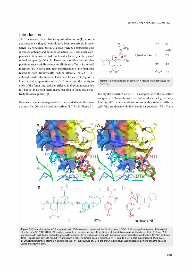
Figure 2: Crystal structure of κ-OR in complex with JDTic compared to naltrindole’s binding pose in δ-OR. A: Cross-eyed stereoview of the crystalstructure of κ-OR (PDB 4DJH). All residues known to be required for high-affinity binding of 1 (mutation reproducibly reduces affinity ≥10-fold [7-9])are shown with thick bonds and water-accessible surfaces. JDTic is shown in green with the hydroxyphenylpiperidine substructure (HPP) in light blue.Ionic H-bonds from JDTic to Asp1383.32 are shown in red. The binding pose of naltrindole (NTI, pink) to δ-OR is also superimposed (PDB 4EJ4).B: Structural similarities: atoms in 1 common to the HPP substructure of JDTic are shown in light blue; superimposable portions of naltrindole andJDTic are shown in pink.
IntroductionThe structure–activity relationships of salvinorin A (1), a potent
and selective κ (kappa) opioid, have been extensively investi-
gated [1]. Modifications at C-2 have yielded compounds with
increased potency and duration of action [2,3], and other com-
pounds with unprecedented functional selectivity at the μ (mu)
opioid receptor (μ-OR) [4]. However, modifications at other
positions substantially reduce or eliminate affinity for opioid
receptors [1]. In particular, most modifications of the furan ring
tested to date dramatically reduce affinity for κ-OR [1],
although small substituents at C-16 have little effect (Figure 1).
Unexpectedly, epimerization at C-12, inverting the configur-
ation of the furan ring, reduces efficacy in G-protein activation
[5], but not in arrestin recruitment, resulting in functional selec-
tivity (biased agonism) [6].
Extensive receptor mutagenesis data are available on the inter-
actions of κ-OR with 1 and derivatives [1,7-9]. In Figure 2A,
Figure 1: Binding affinities of salvinorin A (1) and furan derivatives forκ-OR [5].
the crystal structure of κ-OR in complex with the selective
antagonist JDTic is shown. Essential residues for high affinity
binding of 1, where mutation reproducibly reduces affinity
≥10-fold, are shown with thick bonds for emphasis [7-9]. These
Beilstein J. Org. Chem. 2013, 9, 2916–2924.
2918
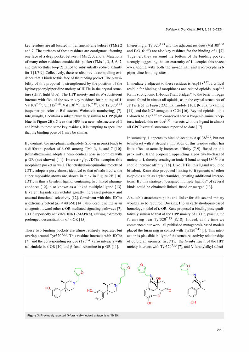
Figure 3: Previously reported N-furanylalkyl opioid antagonists [19,20].
key residues are all located in transmembrane helices (TMs) 2
and 7. The surfaces of these residues are contiguous, forming
one face of a deep pocket between TMs 2, 3, and 7. Mutations
of many other residues outside this pocket (TMs 1, 3, 5, 6, 7,
and extracellular loop 2) failed to substantially reduce affinity
for 1 [1,7-9]. Collectively, these results provide compelling evi-
dence that 1 binds to this face of the binding pocket. The plausi-
bility of this proposal is strengthened by the position of the
hydroxyphenylpiperidine moiety of JDTic in the crystal struc-
ture (HPP, light blue). The HPP moiety and its N-substituent
interact with five of the seven key residues for binding of 1:
Val1082.53, Gln1152.60, Val1182.63, Ile3167.39, and Tyr3207.43
(superscripts refer to Ballesteros–Weinstein numbering) [7].
Intriguingly, 1 contains a substructure very similar to HPP (light
blue in Figure 2B). Given that HPP is a near substructure of 1
and binds to these same key residues, it is tempting to speculate
that the binding pose of 1 may be similar.
By contrast, the morphinan naltrindole (shown in pink) binds to
a different pocket of δ-OR among TMs 3, 6, and 7 [10];
β-funaltrexamine adopts a near-identical pose in complex with
μ-OR (not shown) [11]. Interestingly, JDTic occupies this
morphinan pocket as well. The tetrahydroisoquinoline moiety of
JDTic adopts a pose almost identical to that of naltrindole; the
superimposable atoms are shown in pink in Figure 2B [10].
JDTic is thus a bivalent ligand, containing two linked pharma-
cophores [12], also known as a linked multiple ligand [13].
Bivalent ligands can exhibit greatly increased potency and
unusual functional selectivity [12]. Consistent with this, JDTic
is extremely potent (Ke < 40 pM) [14]; also, despite acting as an
antagonist toward other κ-OR-mediated signaling pathways [7],
JDTic reportedly activates JNK1 (MAPK8), causing extremely
prolonged desensitization of κ-OR [15].
These two binding pockets are almost entirely separate, but
overlap around Tyr3207.43. This residue interacts with JDTic
[7], and the corresponding residue (Tyr7.43) also interacts with
naltrindole in δ-OR [10] and β-funaltrexamine in μ-OR [11].
Interestingly, Tyr3207.43 and two adjacent residues (Val1082.53
and Ile3167.39) are also key residues for the binding of 1 [7].
Together, they surround the bottom of the binding pocket,
strongly suggesting that an extremity of 1 occupies this space,
overlapping with both the morphinan and hydroxyphenyl-
piperidine binding sites.
Immediately adjacent to these residues is Asp1383.32, a critical
residue for binding of morphinans and related opioids. Asp3.32
forms strong ionic H-bonds (‘salt bridges’) to the basic nitrogen
atoms found in almost all opioids, as in the crystal structures of
JDTic (red in Figure 2A), naltrindole [10], β-funaltrexamine
[11], and the NOP antagonist C-24 [16]. Beyond opioids, ionic
H-bonds to Asp3.32 are conserved across biogenic amine recep-
tors; indeed, this residue3.32 interacts with the ligand in almost
all GPCR crystal structures reported to date [17].
In summary, 1 appears to bind adjacent to Asp1383.32, but not
to interact with it strongly: mutation of this residue either has
little effect or actually increases affinity [7-9]. Based on this
proximity, Kane proposed appending a positively-charged
moiety to 1, thereby creating an ionic H bond to Asp1383.32 that
should increase affinity [18]. Like JDTic, this ligand would be
bivalent. Kane also proposed linking to fragments of other
κ-opioids such as arylacetamides, creating additional interac-
tions. By this strategy, “designed multiple ligands” of several
kinds could be obtained: linked, fused or merged [13].
A suitable attachment point and linker for this second moiety
would also be required. Docking 1 to an early rhodopsin-based
homology model of κ-OR, Kane proposed a binding pose quali-
tatively similar to that of the HPP moiety of JDTic, placing the
furan ring near Tyr3207.43 [8,18]. Indeed, at the time we
commenced our work, all published mutagenesis-based models
placed the furan ring in contact with Tyr3207.43 [1]. This inter-
action is plausible in light of the structure–activity relationships
of opioid antagonists. In JDTic, the N-substituent of the HPP
moiety interacts with Tyr3207.43 [7], and N-furanylalkyl substi-
Beilstein J. Org. Chem. 2013, 9, 2916–2924.
2919
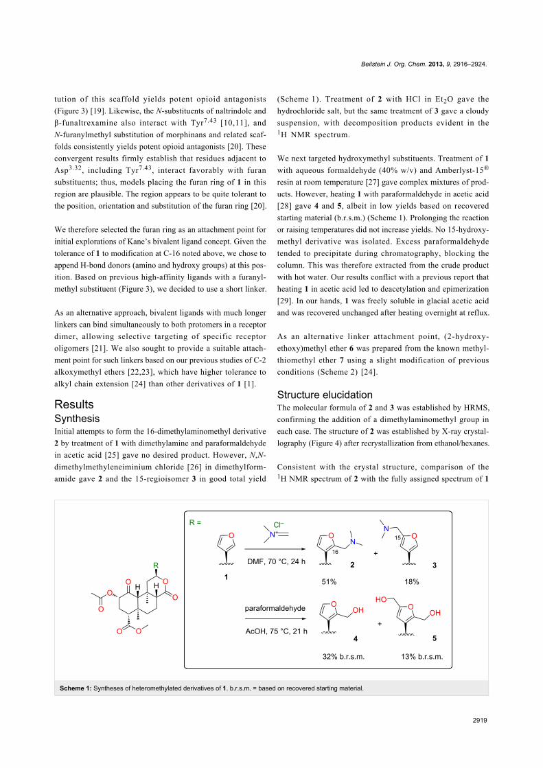
Scheme 1: Syntheses of heteromethylated derivatives of 1. b.r.s.m. = based on recovered starting material.
tution of this scaffold yields potent opioid antagonists
(Figure 3) [19]. Likewise, the N-substituents of naltrindole and
β-funaltrexamine also interact with Tyr7.43 [10,11], and
N-furanylmethyl substitution of morphinans and related scaf-
folds consistently yields potent opioid antagonists [20]. These
convergent results firmly establish that residues adjacent to
Asp3.32, including Tyr7.43, interact favorably with furan
substituents; thus, models placing the furan ring of 1 in this
region are plausible. The region appears to be quite tolerant to
the position, orientation and substitution of the furan ring [20].
We therefore selected the furan ring as an attachment point for
initial explorations of Kane’s bivalent ligand concept. Given the
tolerance of 1 to modification at C-16 noted above, we chose to
append H-bond donors (amino and hydroxy groups) at this pos-
ition. Based on previous high-affinity ligands with a furanyl-
methyl substituent (Figure 3), we decided to use a short linker.
As an alternative approach, bivalent ligands with much longer
linkers can bind simultaneously to both protomers in a receptor
dimer, allowing selective targeting of specific receptor
oligomers [21]. We also sought to provide a suitable attach-
ment point for such linkers based on our previous studies of C-2
alkoxymethyl ethers [22,23], which have higher tolerance to
alkyl chain extension [24] than other derivatives of 1 [1].
ResultsSynthesisInitial attempts to form the 16-dimethylaminomethyl derivative
2 by treatment of 1 with dimethylamine and paraformaldehyde
in acetic acid [25] gave no desired product. However, N,N-
dimethylmethyleneiminium chloride [26] in dimethylform-
amide gave 2 and the 15-regioisomer 3 in good total yield
(Scheme 1). Treatment of 2 with HCl in Et2O gave the
hydrochloride salt, but the same treatment of 3 gave a cloudy
suspension, with decomposition products evident in the1H NMR spectrum.
We next targeted hydroxymethyl substituents. Treatment of 1
with aqueous formaldehyde (40% w/v) and Amberlyst-15®
resin at room temperature [27] gave complex mixtures of prod-
ucts. However, heating 1 with paraformaldehyde in acetic acid
[28] gave 4 and 5, albeit in low yields based on recovered
starting material (b.r.s.m.) (Scheme 1). Prolonging the reaction
or raising temperatures did not increase yields. No 15-hydroxy-
methyl derivative was isolated. Excess paraformaldehyde
tended to precipitate during chromatography, blocking the
column. This was therefore extracted from the crude product
with hot water. Our results conflict with a previous report that
heating 1 in acetic acid led to deacetylation and epimerization
[29]. In our hands, 1 was freely soluble in glacial acetic acid
and was recovered unchanged after heating overnight at reflux.
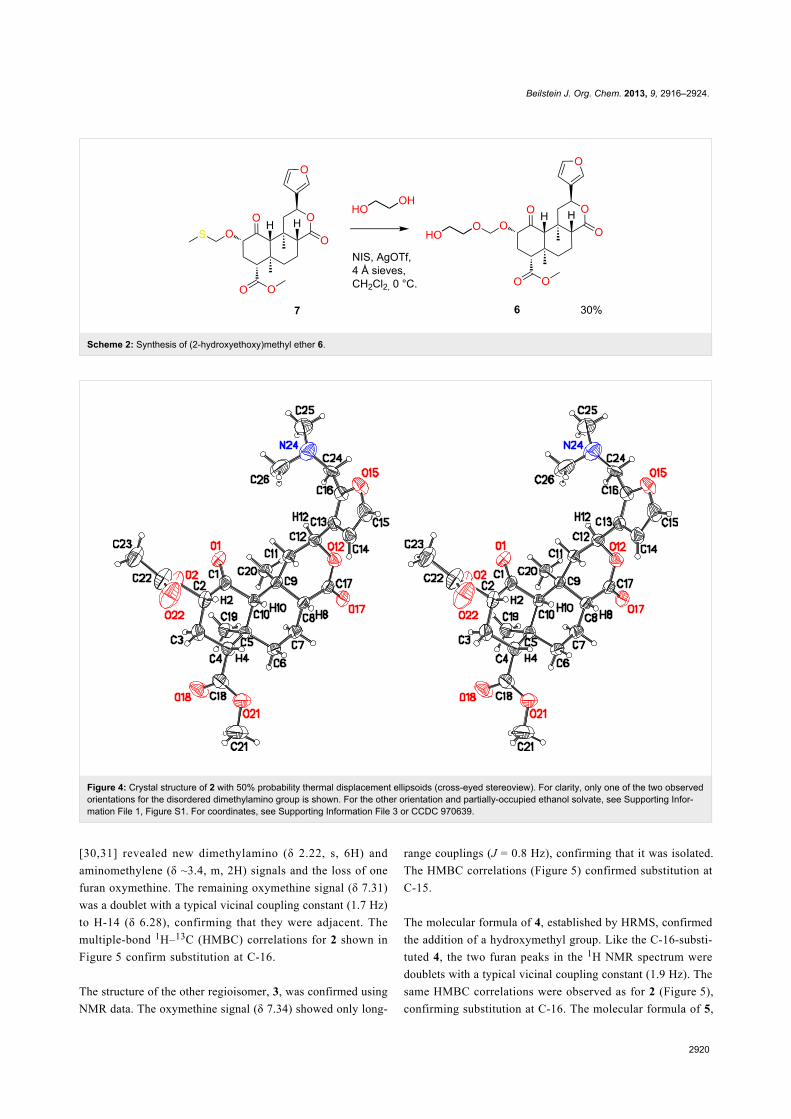
As an alternative linker attachment point, (2-hydroxy-
ethoxy)methyl ether 6 was prepared from the known methyl-
thiomethyl ether 7 using a slight modification of previous
conditions (Scheme 2) [24].
Structure elucidationThe molecular formula of 2 and 3 was established by HRMS,
confirming the addition of a dimethylaminomethyl group in
each case. The structure of 2 was established by X-ray crystal-
lography (Figure 4) after recrystallization from ethanol/hexanes.
Consistent with the crystal structure, comparison of the1H NMR spectrum of 2 with the fully assigned spectrum of 1
Beilstein J. Org. Chem. 2013, 9, 2916–2924.
2920
Scheme 2: Synthesis of (2-hydroxyethoxy)methyl ether 6.
Figure 4: Crystal structure of 2 with 50% probability thermal displacement ellipsoids (cross-eyed stereoview). For clarity, only one of the two observedorientations for the disordered dimethylamino group is shown. For the other orientation and partially-occupied ethanol solvate, see Supporting Infor-mation File 1, Figure S1. For coordinates, see Supporting Information File 3 or CCDC 970639.
[30,31] revealed new dimethylamino (δ 2.22, s, 6H) and
aminomethylene (δ ~3.4, m, 2H) signals and the loss of one
furan oxymethine. The remaining oxymethine signal (δ 7.31)
was a doublet with a typical vicinal coupling constant (1.7 Hz)
to H-14 (δ 6.28), confirming that they were adjacent. The
multiple-bond 1H–13C (HMBC) correlations for 2 shown in
Figure 5 confirm substitution at C-16.
The structure of the other regioisomer, 3, was confirmed using
NMR data. The oxymethine signal (δ 7.34) showed only long-
range couplings (J = 0.8 Hz), confirming that it was isolated.
The HMBC correlations (Figure 5) confirmed substitution at
C-15.
The molecular formula of 4, established by HRMS, confirmed
the addition of a hydroxymethyl group. Like the C-16-substi-
tuted 4, the two furan peaks in the 1H NMR spectrum were
doublets with a typical vicinal coupling constant (1.9 Hz). The
same HMBC correlations were observed as for 2 (Figure 5),
confirming substitution at C-16. The molecular formula of 5,
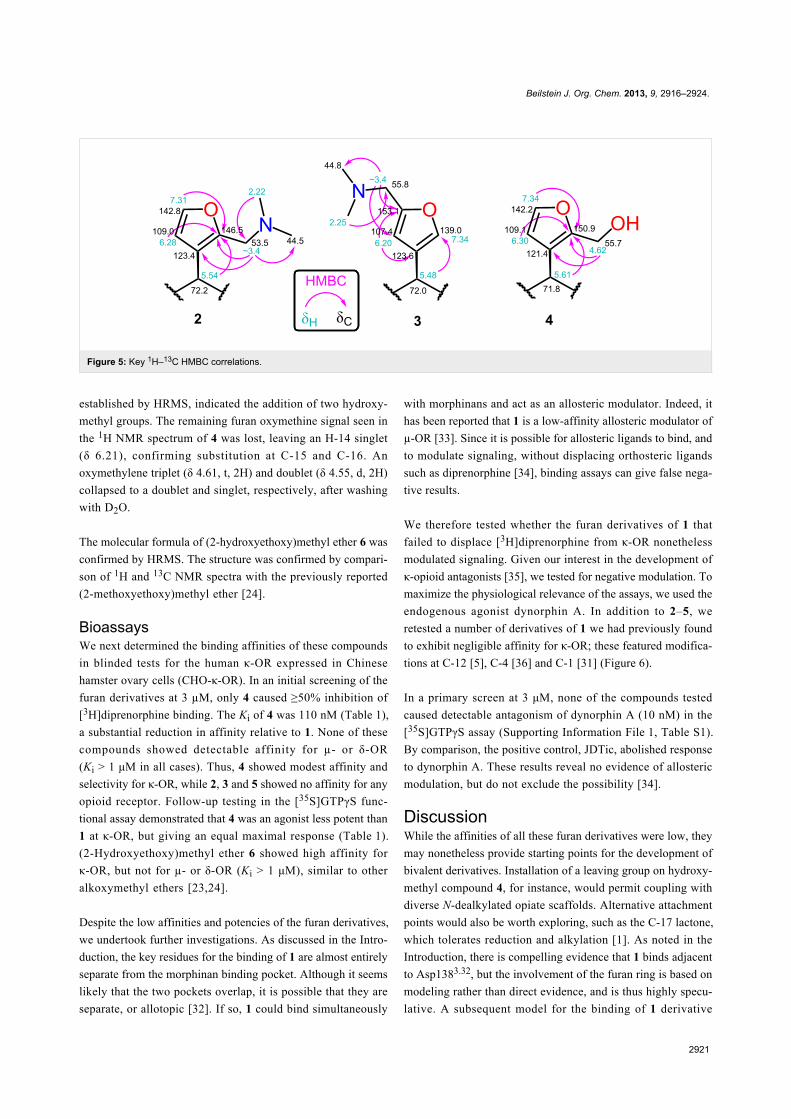
Beilstein J. Org. Chem. 2013, 9, 2916–2924.
2921
Figure 5: Key 1H–13C HMBC correlations.
established by HRMS, indicated the addition of two hydroxy-
methyl groups. The remaining furan oxymethine signal seen in
the 1H NMR spectrum of 4 was lost, leaving an H-14 singlet
(δ 6.21), confirming substitution at C-15 and C-16. An
oxymethylene triplet (δ 4.61, t, 2H) and doublet (δ 4.55, d, 2H)
collapsed to a doublet and singlet, respectively, after washing
with D2O.
The molecular formula of (2-hydroxyethoxy)methyl ether 6 was
confirmed by HRMS. The structure was confirmed by compari-
son of 1H and 13C NMR spectra with the previously reported
(2-methoxyethoxy)methyl ether [24].
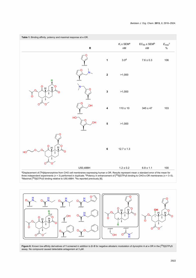
BioassaysWe next determined the binding affinities of these compounds
in blinded tests for the human κ-OR expressed in Chinese
hamster ovary cells (CHO-κ-OR). In an initial screening of the
furan derivatives at 3 µM, only 4 caused ≥50% inhibition of
[3H]diprenorphine binding. The Ki of 4 was 110 nM (Table 1),
a substantial reduction in affinity relative to 1. None of these
compounds showed detectable affinity for µ- or δ-OR
(Ki > 1 μM in all cases). Thus, 4 showed modest affinity and
selectivity for κ-OR, while 2, 3 and 5 showed no affinity for any
opioid receptor. Follow-up testing in the [35S]GTPγS func-
tional assay demonstrated that 4 was an agonist less potent than
1 at κ-OR, but giving an equal maximal response (Table 1).
(2-Hydroxyethoxy)methyl ether 6 showed high affinity for
κ-OR, but not for µ- or δ-OR (Ki > 1 μM), similar to other
alkoxymethyl ethers [23,24].
Despite the low affinities and potencies of the furan derivatives,
we undertook further investigations. As discussed in the Intro-
duction, the key residues for the binding of 1 are almost entirely
separate from the morphinan binding pocket. Although it seems
likely that the two pockets overlap, it is possible that they are
separate, or allotopic [32]. If so, 1 could bind simultaneously
with morphinans and act as an allosteric modulator. Indeed, it
has been reported that 1 is a low-affinity allosteric modulator of
µ-OR [33]. Since it is possible for allosteric ligands to bind, and
to modulate signaling, without displacing orthosteric ligands
such as diprenorphine [34], binding assays can give false nega-
tive results.
We therefore tested whether the furan derivatives of 1 that
failed to displace [3H]diprenorphine from κ-OR nonetheless
modulated signaling. Given our interest in the development of
κ-opioid antagonists [35], we tested for negative modulation. To
maximize the physiological relevance of the assays, we used the
endogenous agonist dynorphin A. In addition to 2–5, we
retested a number of derivatives of 1 we had previously found
to exhibit negligible affinity for κ-OR; these featured modifica-
tions at C-12 [5], C-4 [36] and C-1 [31] (Figure 6).
In a primary screen at 3 μM, none of the compounds tested
caused detectable antagonism of dynorphin A (10 nM) in the
[35S]GTPγS assay (Supporting Information File 1, Table S1).
By comparison, the positive control, JDTic, abolished response
to dynorphin A. These results reveal no evidence of allosteric
modulation, but do not exclude the possibility [34].
DiscussionWhile the affinities of all these furan derivatives were low, they
may nonetheless provide starting points for the development of
bivalent derivatives. Installation of a leaving group on hydroxy-
methyl compound 4, for instance, would permit coupling with
diverse N-dealkylated opiate scaffolds. Alternative attachment
points would also be worth exploring, such as the C-17 lactone,
which tolerates reduction and alkylation [1]. As noted in the
Introduction, there is compelling evidence that 1 binds adjacent
to Asp1383.32, but the involvement of the furan ring is based on
modeling rather than direct evidence, and is thus highly specu-
lative. A subsequent model for the binding of 1 derivative
Beilstein J. Org. Chem. 2013, 9, 2916–2924.
2922
Table 1: Binding affinity, potency and maximal response at κ-OR.
Ki ± SEMa EC50 ± SEMb Emaxc
R nM nM %
1 3.0d 7.6 ± 0.3 106
2 >1,000
3 >1,000
4 110 ± 10 345 ± 47 103
5 >1,000
6 12.7 ± 1.3
U50,488H 1.2 ± 0.2 6.8 ± 1.1 100aDisplacement of [3H]diprenorphine from CHO cell membranes expressing human κ-OR. Results represent mean ± standard error of the mean forthree independent experiments (n = 3) performed in duplicate. bPotency in enhancement of [35S]GTPγS binding to CHO-κ-OR membranes (n = 3–5).cMaximal [35S]GTPγS binding relative to U50,488H. dAs reported previously [6].
Figure 6: Known low-affinity derivatives of 1 screened in addition to 2–5 for negative allosteric modulation of dynorphin A at κ-OR in the [35S]GTPγSassay. No compound caused detectable antagonism at 3 μM.
Beilstein J. Org. Chem. 2013, 9, 2916–2924.
2923
RB-64 places the C-18 methyl ester rather than the furan ring
close to Asp1383.32 [7]. In our previous studies, however,
substitution of amine groups for the C-18 methyl ester abol-
ished affinity for κ-OR [36]. Indeed, all modifications of this
group tested to date greatly reduce affinity, in most cases abol-
ishing binding altogether [1]. C-18 is thus not a promising
attachment point. At the opposite extreme, the C-2 alkoxy-
methyl ether series is highly tolerant to alkyl chain extension.
Compound 6 and the synthetic route used may prove useful for
the attachment of very long linkers, as used with other scaf-
folds to bridge receptor dimers [21].
Rather than linking pharmacophores, another approach to biva-
lent ligands is to design hybrid structures, or merged multiple
ligands [13]. As noted above, the HPP moiety of JDTic inter-
acts with many of the key residues for binding of 1, and shares a
very similar substructure, suggesting that the binding pose of 1
may be similar. If so, converting the lactone to a cyclic amine
may provide the desired H-bond to Asp1383.32 without the need
for additional functional groups.
Our results provide no evidence of negative allosteric modula-
tion of κ-OR by low-affinity derivatives of 1, and the mutagen-
esis results discussed in the Introduction suggest some overlap
with other opioids. However, given the report that 1 is an
allosteric modulator of μ-OR [33], high-affinity μ-selective
derivatives such as herkinorin [1] also represent potential scaf-
folds for bivalent and possibly bitopic (orthosteric–allosteric)
derivatives [12].
ConclusionIn conclusion, compelling evidence suggests that 1 binds to an
allotopic pocket of κ-OR, leaving the classical opiate binding
site largely unoccupied. This suggests the possibility of creating
bivalent derivatives by linking these scaffolds, with the poten-
tial for dramatic increases in potency and altered functional
selectivity. A suitable attachment point and linker remain to be
determined. Our preliminary modifications at C-2, C-15 and
C-16 yielded no potent derivatives, but may prove useful as
intermediates in further exploration of this concept.
Author ContributionsT.A.M. designed and prepared the compounds, wrote the manu-
script and prepared the figures. W.X. and L.Y.L.C. performed
and analyzed the bioassays. D.M.H. obtained the crystal struc-
ture. B.M.C. supervised and coordinated the project.
Competing InterestsB.M.C., L.Y.L.C., and T.A.M. hold US patents on the prepar-
ation and therapeutic use of salvinorin A derivatives (7,629,475
and 8,492,564).
Supporting InformationSupporting Information File 1Synthetic procedures and 1H and 13C NMR spectra of all
new compounds; HMQC and HMBC spectra from
Figure 5; supporting Figure S1 and Table S1.
[http://www.beilstein-journals.org/bjoc/content/
supplementary/1860-5397-9-328-S1.pdf]
Supporting Information File 21H and 13C NMR spectra of all new compounds in
jcamp-dx format.
[http://www.beilstein-journals.org/bjoc/content/
supplementary/1860-5397-9-328-S2.zip]
Supporting Information File 3X-ray crystal structure of 2.
[http://www.beilstein-journals.org/bjoc/content/
supplementary/1860-5397-9-328-S3.cif]
Supporting Information File 4Structure factors for 2.
[http://www.beilstein-journals.org/bjoc/content/
supplementary/1860-5397-9-328-S4.hkl]
AcknowledgementsFunding for this work was provided by the National Institute of
Drug Abuse (P30 DA13429 to LYLC). Cécile Béguin
suggested compounds for the negative modulation assay.
References1. Cunningham, C. W.; Rothman, R. B.; Prisinzano, T. E.
Pharmacol. Rev. 2011, 63, 316–347. doi:10.1124/pr.110.0032442. Hooker, J. M.; Munro, T. A.; Béguin, C.; Alexoff, D.; Shea, C.; Xu, Y.;
Cohen, B. M. Neuropharmacology 2009, 57, 386–391.doi:10.1016/j.neuropharm.2009.06.044
3. Peet, M. M.; Baker, L. E. Behav. Pharmacol. 2011, 22, 450–457.doi:10.1097/FBP.0b013e328349fc1b
4. Lamb, K.; Tidgewell, K.; Simpson, D. S.; Bohn, L. M.; Prisinzano, T. E.Drug Alcohol Depend. 2012, 121, 181–188.doi:10.1016/j.drugalcdep.2011.10.026
5. Béguin, C.; Duncan, K. K.; Munro, T. A.; Ho, D. M.; Xu, W.;Liu-Chen, L.-Y.; Carlezon, W. A., Jr.; Cohen, B. M. Bioorg. Med. Chem.2009, 17, 1370–1380. doi:10.1016/j.bmc.2008.12.012
6. Béguin, C.; Potuzak, J.; Xu, W.; Liu-Chen, L.-Y.; Streicher, J. M.;Groer, C. E.; Bohn, L. M.; Carlezon, W. A., Jr.; Cohen, B. M.Bioorg. Med. Chem. Lett. 2012, 22, 1023–1026.doi:10.1016/j.bmcl.2011.11.128
7. Wu, H.; Wacker, D.; Mileni, M.; Katritch, V.; Han, G. W.; Vardy, E.;Liu, W.; Thompson, A. A.; Huang, X.-P.; Carroll, F. I.;Mascarella, S. W.; Westkaemper, R. B.; Mosier, P. D.; Roth, B. L.;Cherezov, V.; Stevens, R. C. Nature 2012, 485, 327–332.doi:10.1038/nature10939
Beilstein J. Org. Chem. 2013, 9, 2916–2924.
2924
8. Kane, B. E.; McCurdy, C. R.; Ferguson, D. M. J. Med. Chem. 2008, 51,1824–1830. doi:10.1021/jm701040v
9. Vardy, E.; Mosier, P. D.; Frankowski, K. J.; Wu, H.; Katritch, V.;Westkaemper, R. B.; Aubé, J.; Stevens, R. C.; Roth, B. L.J. Biol. Chem. 2013, 288, 34470–34483. doi:10.1074/jbc.M113.515668
10. Granier, S.; Manglik, A.; Kruse, A. C.; Kobilka, T. S.; Thian, F. S.;Weis, W. I.; Kobilka, B. K. Nature 2012, 485, 400–404.doi:10.1038/nature11111
11. Manglik, A.; Kruse, A. C.; Kobilka, T. S.; Thian, F. S.; Mathiesen, J. M.;Sunahara, R. K.; Pardo, L.; Weis, W. I.; Kobilka, B. K.; Granier, S.Nature 2012, 485, 321–326. doi:10.1038/nature10954
12. Valant, C.; Lane, J. R.; Sexton, P. M.; Christopoulos, A.Annu. Rev. Pharmacol. Toxicol. 2012, 52, 153–178.doi:10.1146/annurev-pharmtox-010611-134514
13. Morphy, R. J. Historical Strategies for Lead Generation. In DesigningMulti-Target Drugs; Morphy, R. J.; Harris, C. J., Eds.; RSC: Cambridge,2012; pp 111–129. doi:10.1039/9781849734912-00111
14. Béguin, C.; Cohen, B. M. Medicinal Chemistry of Kappa OpioidReceptor Antagonists. In Opiate Receptors and Antagonists: fromBench to Clinic; Dean, R. L.; Bilsky, E. J.; Negus, S. S., Eds.; HumanaPress: New York, 2009; pp 99–118. doi:10.1007/978-1-59745-197-0_6
15. Melief, E. J.; Miyatake, M.; Carroll, F. I.; Béguin, C.;Carlezon, W. A., Jr.; Cohen, B. M.; Grimwood, S.; Mitch, C. H.;Rorick-Kehn, L.; Chavkin, C. Mol. Pharmacol. 2011, 80, 920–929.doi:10.1124/mol.111.074195
16. Thompson, A. A.; Liu, W.; Chun, E.; Katritch, V.; Wu, H.; Vardy, E.;Huang, X.-P.; Trapella, C.; Guerrini, R.; Calo, G.; Roth, B. L.;Cherezov, V.; Stevens, R. C. Nature 2012, 485, 395–399.doi:10.1038/nature11085
17. Venkatakrishnan, A. J.; Deupi, X.; Lebon, G.; Tate, C. G.;Schertler, G. F.; Babu, M. M. Nature 2013, 494, 185–194.doi:10.1038/nature11896
18. Kane, B. E. Molecular recognition of G-protein coupled receptorligands: Insights into salvinorin A and xanomeline. Ph.D. Thesis,University of Minnesota, 2006.http://gradworks.umi.com/32/38/3238362.html
19. Zimmerman, D. M.; Leander, J. D.; Cantrell, B. E.; Reel, J. K.;Snoddy, J.; Mendelsohn, L. G.; Johnson, B. G.; Mitch, C. H.J. Med. Chem. 1993, 36, 2833–2841. doi:10.1021/jm00072a001
20. Merz, H.; Langbein, A.; Stockhaus, K.; Walther, G.; Wick, H.Adv. Biochem. Psychopharmacol. 1974, 8, 91–107.
21. Hiller, C.; Kühhorn, J.; Gmeiner, P. J. Med. Chem. 2013, 56,6542–6559. doi:10.1021/jm4004335
22. Munro, T. A.; Ho, D. M.; Cohen, B. M.Acta Crystallogr., Sect. E: Struct. Rep. Online 2012, 68, o3225–o3226.doi:10.1107/S1600536812043449
23. Wang, Y.; Chen, Y.; Xu, W.; Lee, D. Y. W.; Ma, Z.; Rawls, S. M.;Cowan, A.; Liu-Chen, L. Y. J. Pharmacol. Exp. Ther. 2008, 324,1073–1083. doi:10.1124/jpet.107.132142
24. Munro, T. A.; Duncan, K. K.; Xu, W.; Wang, Y.; Liu-Chen, L.-Y.;Carlezon, W. A., Jr.; Cohen, B. M.; Béguin, C. Bioorg. Med. Chem.2008, 16, 1279–1286. doi:10.1016/j.bmc.2007.10.067
25. Chernov, S. V.; Shul'ts, E. E.; Shakirov, M. M.; Tolstikov, G. A.Russ. J. Org. Chem. 2000, 36, 1455–1464.
26. Roman, G. Mini-Rev. Org. Chem. 2013, 10, 27–39.doi:10.2174/1570193x11310010003
27. Iovel, I.; Goldberg, Yu.; Shymanska, M. J. Mol. Catal. 1989, 57,91–103. doi:10.1016/0304-5102(89)80129-4
28. Pevzner, L. M.; Ignat'ev, V. M. J. Org. Chem. USSR 1987, 23,1169–1170.
29. Harding, W. W.; Schmidt, M.; Tidgewell, K.; Kannan, P.; Holden, K. G.;Dersch, C. M.; Rothman, R. B.; Prisinzano, T. E.Bioorg. Med. Chem. Lett. 2006, 16, 3170–3174.doi:10.1016/j.bmcl.2006.03.062
30. Ortega, A.; Blount, J. F.; Manchand, P. S.J. Chem. Soc., Perkin Trans. 1 1982, 2505–2508.doi:10.1039/P19820002505
31. Munro, T. A. The Chemistry of Salvia divinorum. Ph.D. Thesis,University of Melbourne, Australia, 2006.http://pqdtopen.proquest.com/#abstract?dispub=3304556
32. Neubig, R. R.; Spedding, M.; Kenakin, T.; Christopoulos, A.Pharmacol. Rev. 2003, 55, 597–606. doi:10.1124/pr.55.4.4
33. Rothman, R. B.; Murphy, D. L.; Xu, H.; Godin, J. A.; Dersch, C. M.;Partilla, J. S.; Tidgewell, K.; Schmidt, M.; Prisinzano, T. E.J. Pharmacol. Exp. Ther. 2007, 320, 801–810.doi:10.1124/jpet.106.113167
34. May, L. T.; Leach, K.; Sexton, P. M.; Christopoulos, A.Annu. Rev. Pharmacol. Toxicol. 2007, 47, 1–51.doi:10.1146/annurev.pharmtox.47.120505.105159
35. Knoll, A. T.; Carlezon, W. A., Jr. Brain Res. 2010, 1314, 56–73.doi:10.1016/j.brainres.2009.09.074
36. Béguin, C.; Richards, M. R.; Li, J.-G.; Wang, Y.; Xu, W.;Liu-Chen, L.-Y.; Carlezon, W. A., Jr.; Cohen, B. M.Bioorg. Med. Chem. Lett. 2006, 16, 4679–4685.doi:10.1016/j.bmcl.2006.05.093
License and TermsThis is an Open Access article under the terms of the
Creative Commons Attribution License
(http://creativecommons.org/licenses/by/2.0), which
permits unrestricted use, distribution, and reproduction in
any medium, provided the original work is properly cited.
The license is subject to the Beilstein Journal of Organic
Chemistry terms and conditions:
(http://www.beilstein-journals.org/bjoc)
The definitive version of this article is the electronic one
which can be found at:
doi:10.3762/bjoc.9.328