Structure of the measles virus hemagglutinin bound to the ......arrangement of the two MV-H...
9
Supporting Figures and Table for Structure of the measles virus hemagglutinin bound to the CD46 receptor César Santiago, María L. Celma, Thilo Stehle and José M. Casasnovas This PDF file includes: Supplementary Figures 1 to 6 Supplementary Table 1 References for Supplementary Materials Nature Neuroscience: doi:10.1038/nn.1726
Transcript of Structure of the measles virus hemagglutinin bound to the ......arrangement of the two MV-H...
Supporting Figures and Table for
Structure of the measles virus hemagglutinin bound to the CD46 receptor
César Santiago, María L. Celma, Thilo Stehle and José M. Casasnovas
This PDF file includes: Supplementary Figures 1 to 6 Supplementary Table 1 References for Supplementary Materials
Nature Neuroscience: doi:10.1038/nn.1726
β5 a β6
β4
β1
β2 β3
b β5 β6
β4
β1
β2 β3
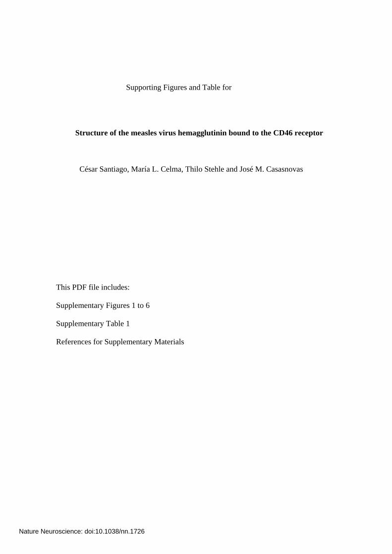
Supplementary Figure 1. Dimeric structures of the MV-H protein. Ribbon
representations of the protein structures are shown using a rainbow coloring, with the
six-blades (β1-β6) labeled. a. The MV-H dimer subunit present in the asymmetric unit
of the MV-H/CD46 crystals. b. Dimer formed by two symmetry related molecules in
the crystals of a disulfide-linked homodimeric MV-H protein (PDB 2ZB6)1. The
arrangement of the two MV-H protomers in the dimers is almost identical in both
structures, even though our protein lacks the Cys154 residue engaged in MV-H
homodimerization. Nevertheless, a similar MV-H protein lacking the cysteine residue
was monomeric (PDB 2RKC)2, quite likely because of the distinct crystallization
conditions used.
Nature Neuroscience: doi:10.1038/nn.1726
a
Supspanthe ashowtip oCD4deteAde(blu
Nature Neuroscie
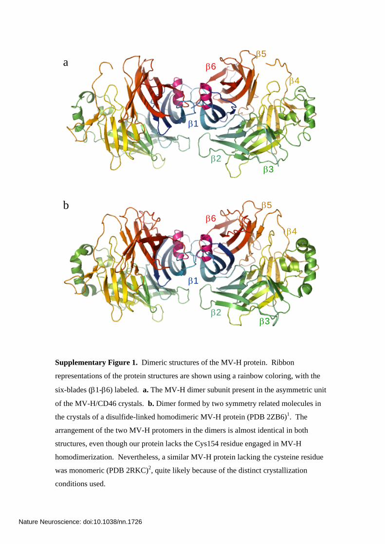
SCR1 SCR1
Pro39 Pro39
SCR2 SCR2
b
plementary Figure 2. Interdomain flexibility among structures of a CD46 protein ning SCR1 and SCR2. a. Superimposition of the two CD46 molecules present in symmetric unit of the MV-H/CD46 crystals based on SCR2. One molecule is n in light blue and the other molecule in dark blue. The side chain of Pro39 at the
f the D'D loop in SCR1 is shown with spheres and labeled. b. Superimposed 6 structures. The structures of the same two domain protein fragment were rmined in the ligand-free state (black, PDB 1CKL)3, in complex with the novirus type 11 knob (red, PDB 2O39)4 and in complex with the MV-H protein e). They are superimposed based on SCR2.
nce: doi:10.1038/nn.1726
5.5 Å 5.5 Å
3 Å 3 Å
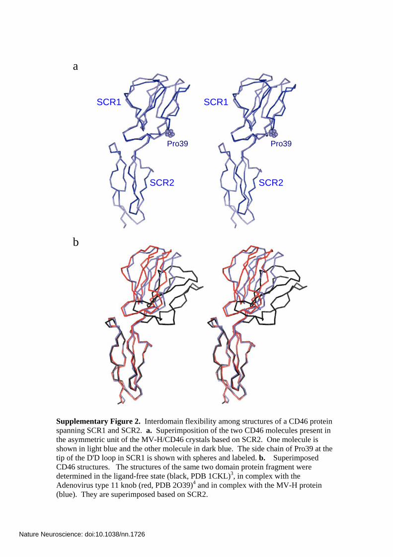
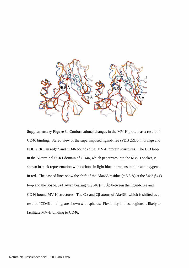
Supplementary Figure 3. Conformational changes in the MV-H protein as a result of
CD46 binding. Stereo view of the superimposed ligand-free (PDB 2ZB6 in orange and
PDB 2RKC in red)1,2 and CD46 bound (blue) MV-H protein structures. The D'D loop
in the N-terminal SCR1 domain of CD46, which penetrates into the MV-H socket, is
shown in stick representation with carbons in light blue, nitrogens in blue and oxygens
in red. The dashed lines show the shift of the Ala463 residue (~ 5.5 Å) at the β4s2-β4s3
loop and the β5s3-β5s4 β-turn bearing Gly546 (~ 3 Å) between the ligand-free and
CD46 bound MV-H structures. The Cα and Cβ atoms of Ala463, which is shifted as a
result of CD46 binding, are shown with spheres. Flexibility in these regions is likely to
facilitate MV-H binding to CD46.
Nature Neuroscience: doi:10.1038/nn.1726
Tyr481 Tyr481
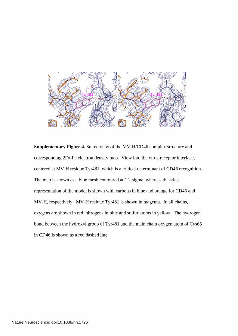
Supplementary Figure 4. Stereo view of the MV-H/CD46 complex structure and
corresponding 2Fo-Fc electron density map. View into the virus-receptor interface,
centered at MV-H residue Tyr481, which is a critical determinant of CD46 recognition.
The map is shown as a blue mesh contoured at 1.2 sigma, whereas the stick
representation of the model is shown with carbons in blue and orange for CD46 and
MV-H, respectively. MV-H residue Tyr481 is shown in magenta. In all chains,
oxygens are shown in red, nitrogens in blue and sulfur atoms in yellow. The hydrogen
bond between the hydroxyl group of Tyr481 and the main chain oxygen atom of Cys65
in CD46 is shown as a red dashed line.
Nature Neuroscience: doi:10.1038/nn.1726
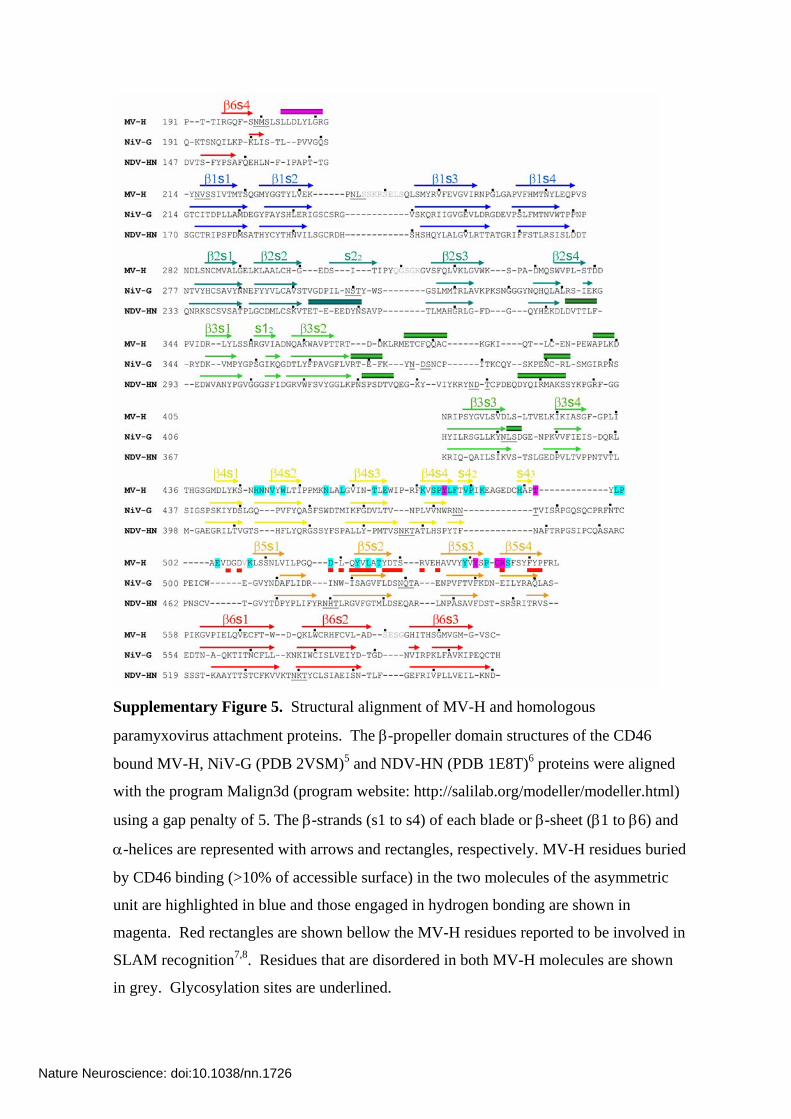
Supplementary Figure 5. Structural alignment of MV-H and homologous
paramyxovirus attachment proteins. The β-propeller domain structures of the CD46
bound MV-H, NiV-G (PDB 2VSM)5 and NDV-HN (PDB 1E8T)6 proteins were aligned
with the program Malign3d (program website: http://salilab.org/modeller/modeller.html)
using a gap penalty of 5. The β-strands (s1 to s4) of each blade or β-sheet (β1 to β6) and
α-helices are represented with arrows and rectangles, respectively. MV-H residues buried
by CD46 binding (>10% of accessible surface) in the two molecules of the asymmetric
unit are highlighted in blue and those engaged in hydrogen bonding are shown in
magenta. Red rectangles are shown bellow the MV-H residues reported to be involved in
SLAM recognition7,8. Residues that are disordered in both MV-H molecules are shown
in grey. Glycosylation sites are underlined.
Nature Neuroscience: doi:10.1038/nn.1726
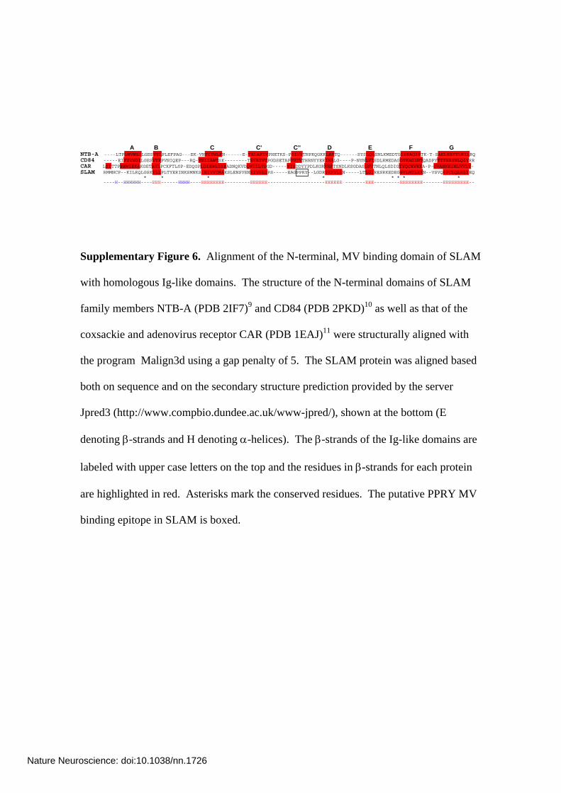
A B C C' C'' D E F G NTB-A ----LTPLMVNGILGESVTLPLEFPAG---EK-VNFITWLFN------E-TSLAFIVPHETKS-PEIHVTNPKQGKRLNFTQ------SYSLQLSNLKMEDTGSYRAQISTK-T-SAKLSSYTLRILRQ CD84 -----EIFTVNGILGESVTFPVNIQEP---RQ-VKIIAWTSK--------TSVAYVTPGDSETAPVVTVTHRNYYERIHALG----P-NYNLVISDLRMEDAGDYKADINTQADPYTTTKRYNLQIYRR CAR LSITTPEEMIEKAKGETAYLPCKFTLSP-EDQGPLDIEWLISPADNQKVDQVIILYSGD-----KIYDDYYPDLKGRVHFTSNDLKSGDASINVTNLQLSDIGTYQCKVKKA-P-GVANKKIHLVVLV- SLAM RMMNCP--KILRQLGSKVLLPLTYERINKSMNKSIHIVVTMAKSLENSVENKIVSLDPS-----EAGPPRY--LGDRYKFYLEN-----LTLGIRESRKEDEGWYLMTLEKN--VSVQRFCLQLRLYEQ * * * * * * * * ----H--HHHHHH---- EEE---- EEEEEEEE--- EEEEEE-- EEEEEE EEE------- EEEEEEEE----- EEEEEEEEE-- --HHHH---- ------ ------------------ -------- -- --
Supplementary Figure 6. Alignment of the N-terminal, MV binding domain of SLAM
with homologous Ig-like domains. The structure of the N-terminal domains of SLAM
family members NTB-A (PDB 2IF7)9 and CD84 (PDB 2PKD)10 as well as that of the
coxsackie and adenovirus receptor CAR (PDB 1EAJ)11 were structurally aligned with
the program Malign3d using a gap penalty of 5. The SLAM protein was aligned based
both on sequence and on the secondary structure prediction provided by the server
Jpred3 (http://www.compbio.dundee.ac.uk/www-jpred/), shown at the bottom (E
denoting β-strands and H denoting α-helices). The β-strands of the Ig-like domains are
labeled with upper case letters on the top and the residues in β-strands for each protein
are highlighted in red. Asterisks mark the conserved residues. The putative PPRY MV
binding epitope in SLAM is boxed.
Nature Neuroscience: doi:10.1038/nn.1726
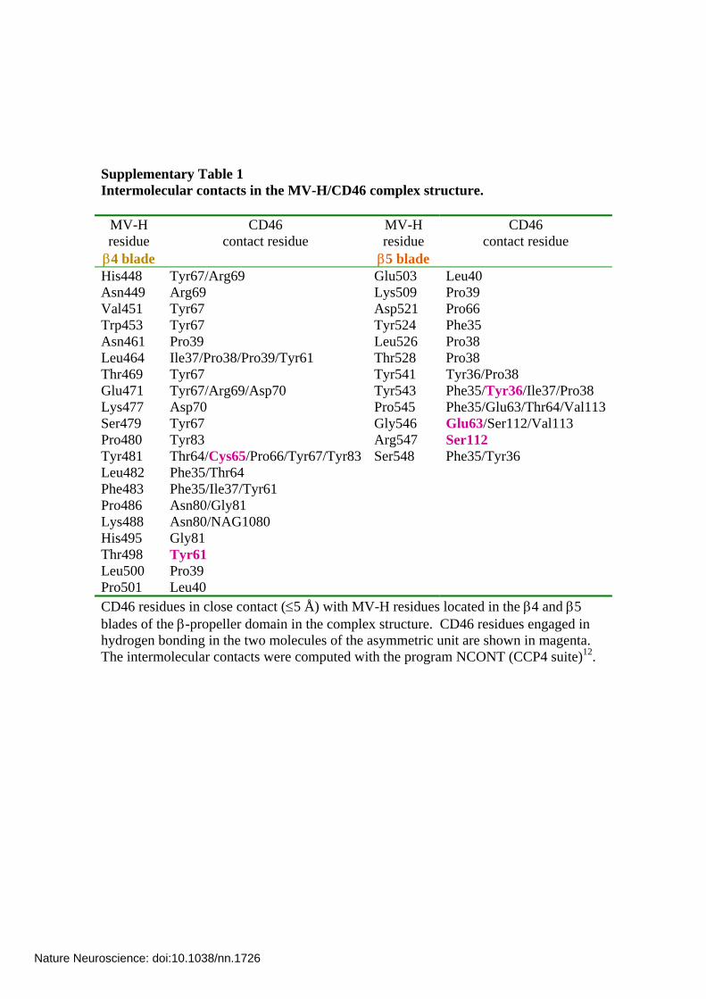
Supplementary Table 1 Intermolecular contacts in the MV-H/CD46 complex structure.
MV-H residue β4 blade
CD46 contact residue
MV-H residue β5 blade
CD46 contact residue
His448 Tyr67/Arg69 Glu503 Leu40 Asn449 Arg69 Lys509 Pro39 Val451 Tyr67 Asp521 Pro66 Trp453 Tyr67 Tyr524 Phe35 Asn461 Pro39 Leu526 Pro38 Leu464 Ile37/Pro38/Pro39/Tyr61 Thr528 Pro38 Thr469 Tyr67 Tyr541 Tyr36/Pro38 Glu471 Tyr67/Arg69/Asp70 Tyr543 Phe35/Tyr36/Ile37/Pro38 Lys477 Asp70 Pro545 Phe35/Glu63/Thr64/Val113Ser479 Tyr67 Gly546 Glu63/Ser112/Val113 Pro480 Tyr83 Arg547 Ser112 Tyr481 Thr64/Cys65/Pro66/Tyr67/Tyr83 Ser548 Phe35/Tyr36 Leu482 Phe35/Thr64 Phe483 Phe35/Ile37/Tyr61 Pro486 Asn80/Gly81 Lys488 Asn80/NAG1080 His495 Gly81 Thr498 Tyr61 Leu500 Pro39 Pro501 Leu40 CD46 residues in close contact (≤5 Å) with MV-H residues located in the β4 and β5 blades of the β-propeller domain in the complex structure. CD46 residues engaged in hydrogen bonding in the two molecules of the asymmetric unit are shown in magenta. The intermolecular contacts were computed with the program NCONT (CCP4 suite)12.
Nature Neuroscience: doi:10.1038/nn.1726
References for Supplementary Materials
1. Hashiguchi, T. et al. Crystal structure of measles virus hemagglutinin provides insight into effective vaccines. Proceedings of the National Academy of Sciences 104, 19535-19540 (2007).
2. Colf, L.A., Juo, Z.S. & Garcia, K.C. Structure of the measles virus hemagglutinin. Nat Struct Mol Biol 14, 1227-1228 (2007).
3. Casasnovas, J.M., Larvie, M. & Stehle, T. Crystal structure of two CD46 domains reveals an extended measles virus-binding surface. The EMBO J. 18, 2911-2922 (1999).
4. Persson, B.D. et al. Adenovirus type 11 binding alters the conformation of its receptor CD46. Nat Struct Mol Biol 14, 164-166 (2007).
5. Bowden, T.A. et al. Structural basis of Nipah and Hendra virus attachment to their cell-surface receptor ephrin-B2. Nat Struct Mol Biol 15, 567-72 (2008).
6. Crennell, S., Takimoto, T., Portner, A. & Taylor, G. Crystal structure of the multifunctional paramyxovirus hemagglutinin-neuraminidase. Nat. Struct. Biol. 7, 1068-1074 (2000).
7. Masse, N. et al. Measles Virus (MV) Hemagglutinin: Evidence that Attachment Sites for MV Receptors SLAM and CD46 Overlap on the Globular Head. J. Virol. 78, 9051-9063 (2004).
8. Vongpunsawad, S., Oezgun, N., Braun, W. & Cattaneo, R. Selectively Receptor-Blind Measles Viruses: Identification of Residues Necessary for SLAM- or CD46-Induced Fusion and Their Localization on a New Hemagglutinin Structural Model. J. Virol. 78, 302-313 (2004).
9. Cao, E. et al. NTB-A receptor crystal structure: insights into homophilic interactions in the signaling lymphocytic activation molecule receptor family. Immunity 25, 559-70 (2006).
10. Yan, Q. et al. Structure of CD84 provides insight into SLAM family function. Proc Natl Acad Sci U S A 104, 10583-8 (2007).
11. van Raaij, M.J., Chouin, E., van der Zandt, H., Bergelson, J.M. & Cusack, S. Dimeric Structure of the Coxsackievirus and Adenovirus Receptor D1 Domain at 1.7 A Resolution. Structure 8, 1147-1155 (2000).
12. Collaborative Computational Project, N. The CCP4 Suite: Programs for Protein Crystallography. Acta Cryst. D 50, 760-763 (1994).
Nature Neuroscience: doi:10.1038/nn.1726