STRUCTURAL ANALYSIS AND MODELING COMPARISON OF...
29
ii STRUCTURAL ANALYSIS AND MODELING COMPARISON OF PRIMATES’ AMYLOID βA4 PROTEIN FAHIMEH JALALI A dissertation submitted in partial fulfillment of the requirements for the award of the degree of Master of Science (Biotechnology) Faculty of Bioscience and Bioengineering University Technology Malaysia January 2013
-
Upload
truongcong -
Category
Documents
-
view
216 -
download
0
Transcript of STRUCTURAL ANALYSIS AND MODELING COMPARISON OF...
ii
STRUCTURAL ANALYSIS AND MODELING COMPARISON OF PRIMATES’
AMYLOID βA4 PROTEIN
FAHIMEH JALALI
A dissertation submitted in partial fulfillment of the
requirements for the award of the degree of
Master of Science (Biotechnology)
Faculty of Bioscience and Bioengineering
University Technology Malaysia
January 2013

iv
To my beloved family and fiancé

v
ACKNOWLEDGMENT
I would like to begin by sincerely thanking my supervisor, Associated Prof Dr.
Shahir Shamsir for his constant support, guidance and mentorship over the course of this
thesis. He gave me the freedom to define my thesis statements. I am very grateful to
having the opportunity for work on this thesis.
I am heartily thankful to my parents, for their unconditional supports and
encouraging me to continue my education. I am very grateful to my lovely fiance for his
professional gaudiness and emotional supports.

vi
ABSTRACT
Amyloid protein originally termed "beta-protein" or "amyloid A4" which is
indicated as "beta A4." Amyloid beta A4 protein is proteolytically derived from a
transmembrane protein named amyloid precursor protein (APP) which is encoded by an
extensively expressed gene on chromosome 21. Mutations in Amyloid βA4 gene cause
the plaques which are composed of a tangle of regularly ordered fibrillar aggregates
called amyloid fibers, and abnormal accumulation of amyloid fibrils make protein
misfolding diseases so that it leads to amyloidosis and neurodegenerative disorder like
Alzheimer diseases (AD) or Parkinson diseases (PD).Currently there are 91 structure of
Amyloid beta A4 protein have been predicted in various species, but there is no
presently concrete evidence that shows Amyloid beta A4 model in primates. This study
was focus on Amyloid beta A4 protein information and analyzed using bioinformatics
software such as: Uniprot, Deep View, PDB (protein data bank), Swiss Model, Jal View,
BLAST, VMD(Visual molecular dynamics), NCBI(National center for biotechnology
information) therefore those of programs help us to predict and create a new model of
3D structure for this protein and they are useful for analysis multiple alignment,
simulation , image processing for βA4 protein sequence, illustrate conserved regions and
residues of protein between different species (human being and primates) to indicate
comparison of their structural features and their gen properties to this end 8 primates
were chosen. Based on the analysis of this comparison demonstrated that some of the
primates are highly conserved with their template (Homo sapiens) and they have similar
primary and tertiary structure with template .The reason of this issue is that in all of
them the protein’s gene location is on chromosome 21 as same as human gen location.
On the other hand rest of chosen primates is not conserved with template and their
structures are totally different with Homo sapiens due to APP gene location is on
chromosome 3 instead of chromosome 21. This information was gathered from
GenBank which is the genetic sequence database, an annotated collection of all publicly
available DNA sequences.
vii
ABSTRAK
Amiloid protein asalnya dipanggil "beta-protein" atau "A4 amyloid" yang
ditandakan sebagai "A4 beta." Beta amiloid protein A4 proteolytically diperolehi
daripada protein transmembran pelopor protein amiloid dinamakan (APP) yang
dikodkan oleh gene meluas dinyatakan pada 21 kromosom. Mutasi dalam gen βA4
amiloid menyebabkan plak yang terdiri daripada kekusutan agregat kerap diperintahkan
berhubung dgn urat saraf dipanggil serat amiloid, dan pengumpulan abnormal gentian
halus amiloid membuat penyakit misfolding protein supaya ia membawa kepada
amyloidosis dan gangguan neurodegenerative seperti penyakit Alzheimer (AD) atau
Parkinson penyakit (PD). Pada masa ini terdapat 91 struktur protein amiloid beta A4
telah diramalkan dalam pelbagai spesis, tetapi tidak ada bukti yang kini konkrit yang
menunjukkan amiloid beta A4 model dalam primat. Kajian ini adalah memberi tumpuan
kepada protein amiloid beta maklumat A4 dan dianalisis menggunakan perisian
bioinformatik seperti: Uniprot, Deep View, PDB (protein data bank), Model
Switzerland, Jal View, letupan, VMD (Visual molekul dinamik), NCBI (National pusat
maklumat bioteknologi) itu mereka program membantu kita untuk meramalkan dan
mewujudkan satu model baru struktur 3D protein ini dan mereka adalah berguna untuk
penjajaran pelbagai analisis, simulasi, pemprosesan imej untuk βA4 urutan protein,
menggambarkan kawasan terpelihara dan sisa protein antara spesies yang berbeza
(manusia dan primat) untuk menunjukkan perbandingan ciri-ciri struktur mereka dan
sifat-sifat gen mereka untuk tujuan ini 8 primat telah dipilih. Berdasarkan analisis
perbandingan ini menunjukkan bahawa beberapa primat sangat dipelihara dengan
template (Homo sapiens) mereka dan mereka mempunyai struktur yang serupa utama
dan tertiari dengan template. Sebab isu ini adalah bahawa dalam semua daripada mereka
lokasi gen-protein ini adalah pada 21 kromosom yang sama sebagai lokasi gen manusia.
Pada rehat tangan lain primat yang dipilih tidak dipelihara dengan template dan struktur
mereka adalah sama sekali berbeza dengan Homo sapiens disebabkan lokasi gen APP
adalah pada 3 kromosom bukannya kromosom 21. Maklumat ini dikumpulkan dari
GenBank yang merupakan pangkalan data jujukan genetik, koleksi beranotasi semua
jujukan DNA yang boleh didapati oleh orang ramai.

viii
TABLE OF CONTENTS
CHAPTER TITLE PAGE
DECLARATION III
DEDICATION IV
ACKNOWLEDGE V
ABSTRACT VI
ABSTRAK VII
TABLE OF CONTENTS VIII
LIST OF TABLES XII
LIST OF FIGURES XIII
LIST OF ABBREVIATION XV
LIST OF SYMBOLS XVII
LIST OF APPENDIX XIX
1 INTRODUCTION
1.1 Background 1
1.2 Problem Statement 6
1.3 Objectives 6
1.4 Research Scope 7

ix
2 LITERATURE REVIEWS
2.1: Amyloid β in Down’s syndrome (DS) 8
2.2 Amyloid β peptide as a Predictor of Synaptic 9
Change in Alzheimer’s disease
2.3 Comparison of protein by laboratory methods 12
2.3.1 A Surface of Minimum Area Metric for 12
the Structural Comparison of Proteins
2.4 Comparison of proteins by bioinformatics methods 13
2.4.1 Structural prediction of a novel chitinase 13
2.4.1.1 Model development and evaluation 14
2.4.1.2 Molecular dynamic (MD) simulations 15
of CHI II
2.4.1.3 Structural comparison of CHI II 16
3 RESEARCH METHODOLOGY
3.1 Experimental design 18
3.2 Identify protein in various species 20
3.3 Collection protein sequences 21
3.3.1 PDB (protein data bank) 23
3.4 View and edit Multiple Sequence Alignment (MSA) 24
3.5 Prediction and modeling 3D structure 25
3.6 Evaluation of homology modeling 26
3.7 Comparison primary sequences and tertiary structures 26
x
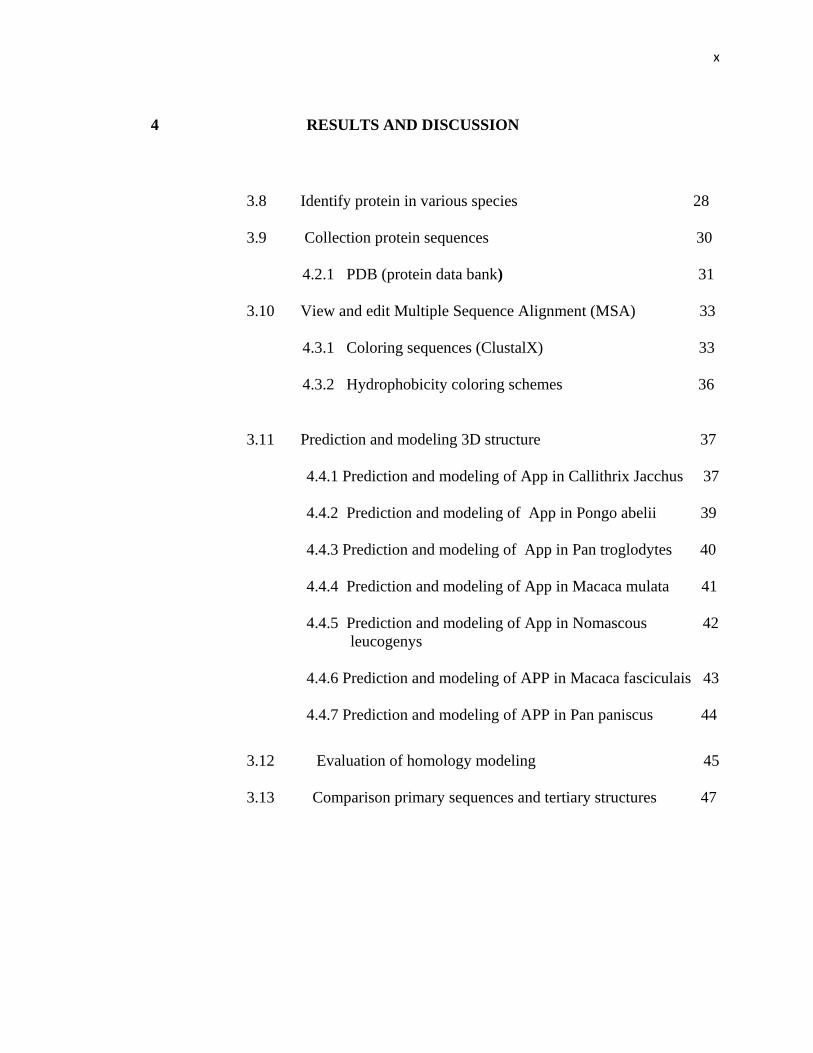
4 RESULTS AND DISCUSSION
3.8 Identify protein in various species 28
3.9 Collection protein sequences 30
4.2.1 PDB (protein data bank) 31
3.10 View and edit Multiple Sequence Alignment (MSA) 33
4.3.1 Coloring sequences (ClustalX) 33
4.3.2 Hydrophobicity coloring schemes 36
3.11 Prediction and modeling 3D structure 37
4.4.1 Prediction and modeling of App in Callithrix Jacchus 37
4.4.2 Prediction and modeling of App in Pongo abelii 39
4.4.3 Prediction and modeling of App in Pan troglodytes 40
4.4.4 Prediction and modeling of App in Macaca mulata 41
4.4.5 Prediction and modeling of App in Nomascous 42
leucogenys
4.4.6 Prediction and modeling of APP in Macaca fasciculais 43
4.4.7 Prediction and modeling of APP in Pan paniscus 44
3.12 Evaluation of homology modeling 45
3.13 Comparison primary sequences and tertiary structures 47

xi
5 CONCLUSION
3.14 Conclusion 52
3.15 Future work 53
REFERENCES 54
Appendix A~D 59~68
xii
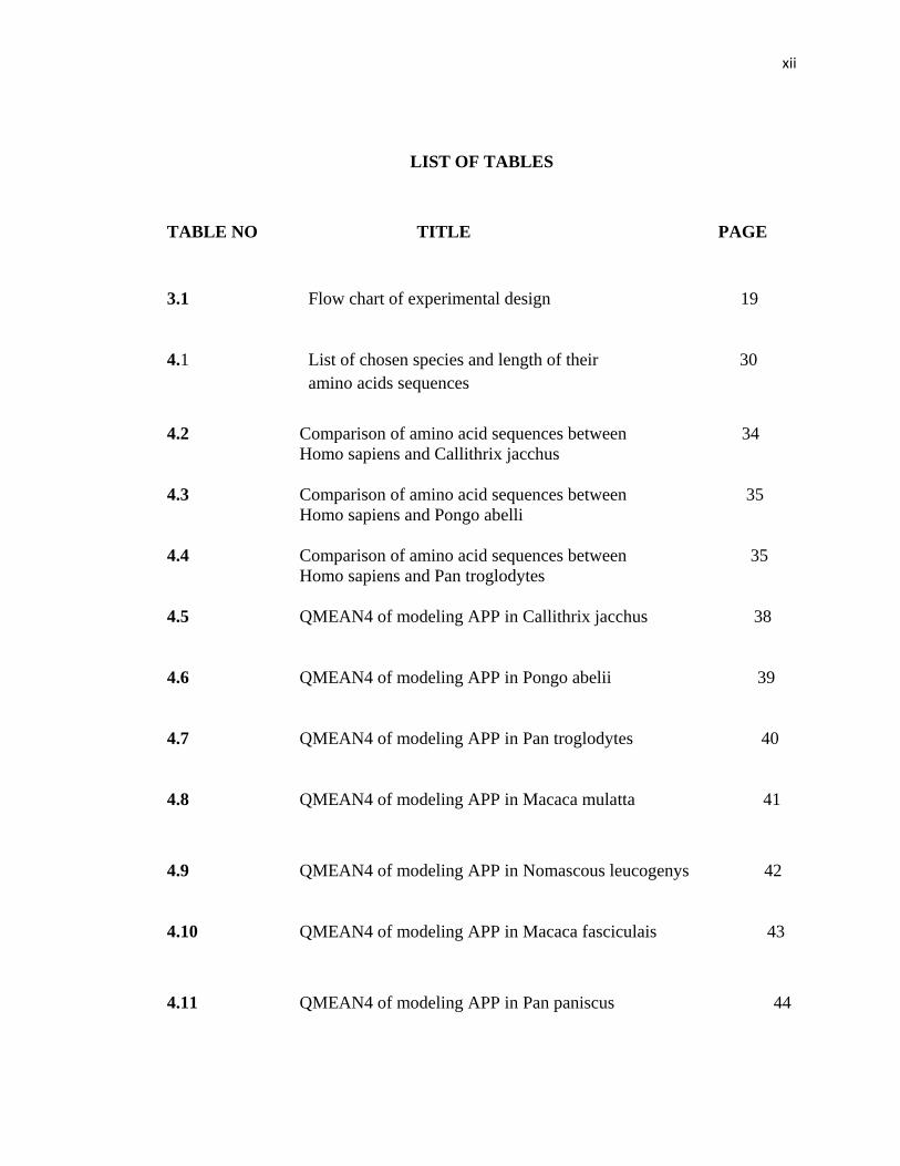
LIST OF TABLES
TABLE NO TITLE PAGE
3.1 Flow chart of experimental design 19
4.1 List of chosen species and length of their 30
amino acids sequences
4.2 Comparison of amino acid sequences between 34
Homo sapiens and Callithrix jacchus
4.3 Comparison of amino acid sequences between 35
Homo sapiens and Pongo abelli
4.4 Comparison of amino acid sequences between 35
Homo sapiens and Pan troglodytes
4.5 QMEAN4 of modeling APP in Callithrix jacchus 38
4.6 QMEAN4 of modeling APP in Pongo abelii 39
4.7 QMEAN4 of modeling APP in Pan troglodytes 40
4.8 QMEAN4 of modeling APP in Macaca mulatta 41
4.9 QMEAN4 of modeling APP in Nomascous leucogenys 42
4.10 QMEAN4 of modeling APP in Macaca fasciculais 43
4.11 QMEAN4 of modeling APP in Pan paniscus 44
xiii
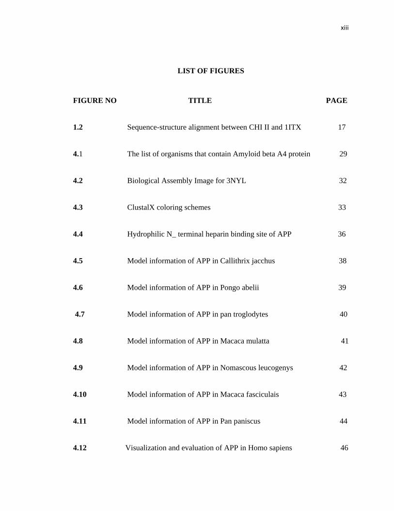
LIST OF FIGURES
FIGURE NO TITLE PAGE
1.2 Sequence-structure alignment between CHI II and 1ITX 17
4.1 The list of organisms that contain Amyloid beta A4 protein 29
4.2 Biological Assembly Image for 3NYL 32
4.3 ClustalX coloring schemes 33
4.4 Hydrophilic N_ terminal heparin binding site of APP 36
4.5 Model information of APP in Callithrix jacchus 38
4.6 Model information of APP in Pongo abelii 39
4.7 Model information of APP in pan troglodytes 40
4.8 Model information of APP in Macaca mulatta 41
4.9 Model information of APP in Nomascous leucogenys 42
4.10 Model information of APP in Macaca fasciculais 43
4.11 Model information of APP in Pan paniscus 44
4.12 Visualization and evaluation of APP in Homo sapiens 46

xiv
4.13 Comparison 3D structure of App in homo sapiens 47
and Callithrix jacchus
4.14 Comparison 3D structure of App in Homo sapiens 48
and Macaca fasilarus
4.15 Comparison 3D structure of App in Homo sapiens 48
and Pan troglodyetes
4.16 Comparison 3D structure of App in Homo sapiens 49
and Pongo abelii
4.17 Visualization 3D structure of App in Macaca mulatta 49
4.18 Observation 3D structure of App in Nomascous leucogenys 50
4.19 Observation 3D structure of App in Pan paniscus 50
xv
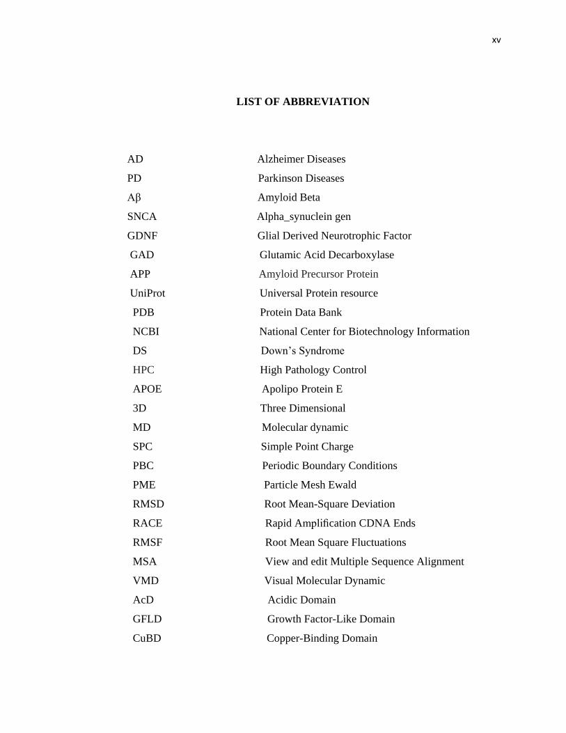
LIST OF ABBREVIATION
AD Alzheimer Diseases
PD Parkinson Diseases
Aβ Amyloid Beta
SNCA Alpha_synuclein gen
GDNF Glial Derived Neurotrophic Factor
GAD Glutamic Acid Decarboxylase
APP Amyloid Precursor Protein
UniProt Universal Protein resource
PDB Protein Data Bank
NCBI National Center for Biotechnology Information
DS Down’s Syndrome
HPC High Pathology Control
APOE Apolipo Protein E
3D Three Dimensional
MD Molecular dynamic
SPC Simple Point Charge
PBC Periodic Boundary Conditions
PME Particle Mesh Ewald
RMSD Root Mean-Square Deviation
RACE Rapid Amplification CDNA Ends
RMSF Root Mean Square Fluctuations
MSA View and edit Multiple Sequence Alignment
VMD Visual Molecular Dynamic
AcD Acidic Domain
GFLD Growth Factor-Like Domain
CuBD Copper-Binding Domain
xvi
AAV_GDNF Adeno_Associated Viral vector _ Glial Derived
Neurotrophic Factor
GABA Gamma-Aminobutyric Acid
BLAST Basic Local Alignment Search Tool
ND Neurodegenerative Disease
HMM Hidden Markor Models
GROMACS Groningen Machine for Chemical Simulation
NMR Nuclear Magnetic Resonance spectroscopy
QMEAN4 Qualitative Model Energy Analysis
RACE Rapid Amplification of CDNA

xvii
LIST OF SYMBOLS
DNA Deoxyribonucleic Acid
RNA Reoxyribonucleic Acid
CHI Chitinase
Ca Calcium
cDNA Complementary DNA
G. antarctica PI12 Glaciozyma antarctica PI12
MPTP Methyl_Phenyl_Tetrahydro Pyridine
K0. Kelvin
GLU Glutamic acid
Asp Aspartic acid
Arg Argenine
A° Angstrom
aa amino acid
Z score Standard score
TM score Template modelling score
Gly Glycine
Phe Phenylalanin
Ile Isoleucine
Ala Alanine
Thr Theronine
His Histidine
Lys Lysine
H_bond hydrogen bond
NE Noradrenaline neuron
1D2k The Xray structure of a chitinase from the
Pathogenic fungus Coccidioides

xviii
NH Neuron
3NYL PDB cod of The X-ray structure of
an antiparallel dimer of the humaamyloid
precursor protein E2 domain
°C Celsius

xix
LIST OF APPENDICES
APPENDIX TITLE PAGE
A FASTA format of Amyloid βA4 protein
Sequence in selected primates
B A rooted phylogenic tree of Amyloid beta
A4 protein in selected primates
C Alignment between Homo sapiens (Query)
with Callithrix jacchus (Sbjct)
D Gene resource of Amyloid beta A4 protein in primates
1
CHAPTER 1
INTRODUCTION
1.0 Background of Studies
Protein can be defined as biochemical compounds which are consisting of one or
more polypeptide generally folded into globular or fibrous form. Some of them are
insoluble fibrous protein named amyloid so that they increase from minimum eighteen
unsuitable folded proteins and polypeptides that exist normally in the body. The
misfolded structure of these proteins and polypeptides have been incorporated with the
pathology of more than twenty severe human illnesses in that unnatural reposition of
amyloid fibrils in organ might cause amyloidosis and neurodegenerative disorder, like
Alzheimer diseases (AD) and Parkinson diseases (PD).
2
1.0.1 Description of protein
There are different type of amyloid and amyloid beta or (Aβ) is one of significant
ones that is the most important portion of amyloid plaques which detected in the brain of
patients with AD, in addition same plaques emerge in some versions of lewy body dementia
and inclusion body myositis ( muscle disease).
The plaques are contained of a tangle of orderly arranged fibrillar accumulation
named amyloid fibers, a protein folded partake by other peptides like the prions related
within protein misfolding diseases, Tha soluble oligomeric shaped of the peptide might be
causative factor in the progress of AD diseases.
1.0.2 Disease and exclusive mutations
As mentioned Alzheimer’s and Parkinson’s disease are associated with the
formation in the brain of amyloid fibrils from β_amyloid and α_synuclein proteins
respectively. Alpha_synuclein is a protein in human is encoded by SNCA gene, usually
found in an amyloid fraction is shown to be a fragment of its precursor protein which cause
Alzheimer diseases amyloid.
Actually amyloid’s mutants such as: (A53T,A3OP,E22G) coupled with familial
Alzheimer’s and Parkinson’s diseases from morphologically identical annular protofibrils
that look like a class of pore_forming bacterial toxin, suggesting that inappropriate

3
membrane permeabilization might be the cause of cell dysfunction and even cell death in
amyloid dieases.
According to Lang,A.E & Lozano,A.M (1998) Parkinson disease is a common
neurodegenerative disorder characterized by the loss of dopaminergic neurons and the
presence of intracytoplasmic-ubiquitinated inclusions (Lewy bodies). Mutations in α-
synuclein (A53T, A30P) and parkin cause familial Parkinson disease. Both these proteins
are found in Lewy bodies.(Lewy bodies are abnormal aggregates of protein that develop
inside nerve cells in Parkinson's disease). The absence of Lewy bodies in patients with
parkin mutations suggests that parkin might be required for the formation of Lewy bodies.
(Lang & Lozano, 1998)
.
PD is a destructive and complicated disease that interferes with movement more and
more as time is passing by. Signs of the disease are vary divers, but they may consist of
problems with swallowing, speech disorders, chewing, urinary problems or constipation,
extreme sweating, skin problems, depression, other emotional changes, and difficulties with
sleep or insomnia.
Nobody can predict which of these symptoms will have an effect on specific patient,
and the strength of the symptoms is different from person to person. None of these
secondary symptoms is mortal, although swallowing problems can lead to choking. The
development of symptoms in Parkinson’s disease may take 20 years or more. In some
people however, the disease advancement is greatly faster. In fact, it is one of the most
usually used systems for explaining how the symptoms of PD progress are.
4
1.0.3 Treatments and solutions
Despite of there are currently two main types of treatment for PD: drug treatments
and surgical treatments, but there are no treatments to curb the destruction of neurons in the
substantia nigra. In prior studies of animal models of Parkinson’s, researchers saw glimmers
of success by injecting proteins that simulate neuronal growth into the animals’ brains. A
protein called glial-derived neurotrophic factor (GDNF) showed the most promise. In
clinical trials, however, GDNF injections have failed to slow the course of the disease and in
some cases; the injections have caused side effects such as severe weight loss. (Kim, et al.,
2002)
In the new study of Gene therapy, monkeys were given a toxin (called MPTP) that
destroys the same cells that are lost in Parkinson’s disease. Several months after the
monkeys developed Parkinson’s-like symptoms, they received AAV-GDNF gene therapy by
injections into the putamen, a brain region that connects to the subsantia nigra.
(The substantia nigra is a brain structure located in the mesencephalon (midbrain) that plays
an important role in reward, addiction, and movement).For more precise delivery, the
researchers used a high pressure injection system, unlike previous studies where GDNF was
injected under low pressure and allowed to spread by passive flow. As a control, other
monkeys received saline injections which are gene therapy injections. Monkeys given the
saline injections remained symptomatic, but monkeys that received the GDNF-virus
injections got better. They showed an average 50 percent improvement on a symptom rating
scale after 9 months, with smaller continued improvements out to 2 years. (Takagi, et al.,
2005)
Japanese researchers have been capable to cure the signs of Parkinson's disease in
monkeys by transplanting nerve cells isolated from embryonic stem cells into their brains,
The finding is the world's first published success of its kind with a primate, pursuant to the
5
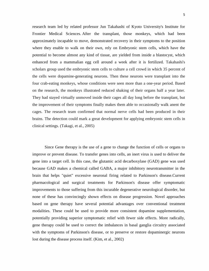
research team led by related professor Jun Takahashi of Kyoto University's Institute for
Frontier Medical Sciences. After the transplant, those monkeys, which had been
approximately incapable to move, demonstrated recovery in their symptoms to the position
where they enable to walk on their own, rely on Embryonic stem cells, which have the
potential to become almost any kind of tissue, are yielded from inside a blastocyst, which
enhanced from a mammalian egg cell around a week after it is fertilized. Takahashi's
scholars group used the embryonic stem cells to culture a cell crowd in which 35 percent of
the cells were dopamine-generating neurons. Then these neurons were transplant into the
four crab-eating monkeys, whose conditions were seen more than a one-year period. Based
on the research, the monkeys illustrated reduced shaking of their organs half a year later.
They had stayed virtually unmoved inside their cages all day long before the transplant, but
the improvement of their symptoms finally makes them able to occasionally walk anent the
cages. The research team confirmed that normal nerve cells had been produced in their
brains. The detection could mark a great development for applying embryonic stem cells in
clinical settings. (Takagi, et al., 2005)
Since Gene therapy is the use of a gene to change the function of cells or organs to
improve or prevent disease. To transfer genes into cells, an inert virus is used to deliver the
gene into a target cell. In this case, the glutamic acid decarboxylase (GAD) gene was used
because GAD makes a chemical called GABA, a major inhibitory neurotransmitter in the
brain that helps "quiet" excessive neuronal firing related to Parkinson's disease.Current
pharmacological and surgical treatments for Parkinson's disease offer symptomatic
improvements to those suffering from this incurable degenerative neurological disorder, but
none of these has convincingly shown effects on disease progression. Novel approaches
based on gene therapy have several potential advantages over conventional treatment
modalities. These could be used to provide more consistent dopamine supplementation,
potentially providing superior symptomatic relief with fewer side effects. More radically,
gene therapy could be used to correct the imbalances in basal ganglia circuitry associated
with the symptoms of Parkinson's disease, or to preserve or restore dopaminergic neurons
lost during the disease process itself. (Kim, et al., 2002)

6
1.1 Problem Statement
Currently there are 91 structure of Amyloid beta A4 protein have been predicted in
various species, but there is no presently concrete evidence that shows Amyloid beta A4
model in primates. However findings demonstrate that primates provide a close animal
model for examining the early transcriptional and post transcriptional processing of APP
(Amyloid precursor protein) that precedes it during aging in Alzheimer’s diseases but in
organisms like primates there isn’t predicted model or sequence structure for amyloid β A4
protein.
1.2 Project Objectives
1. To develop a structural model for Amyloid beta A4 protein in primates.
2. To analyse the differences in the amino acid sequences and composition between
different primates and human.
3. To compare 3D structure and investigation the implication of the differences in
amino acid sequences of Amyloid beta A4 protein between human and primates.
7
1.3 Project Scope
The work will focus on Amyloid beta A4 protein information and analyzed using
bioinformatics software: UniProt, PDB (protein data bank), BLAST, NCBI (National center
for biotechnology information), and GenBank.
54
REFERENCES
Adams, P. D., Grosse-Kunstleve, R. W., Hung, L. W., Ioerger, T. R., McCoy, A. J.,
Moriarty, N. W., et al. (2002). PHENIX: building new software for automated
crystallographic structure determination. Acta Crystallographica Section D:
Biological Crystallography, 58(11), 1948-1954.
Altschul, S. F., Madden, T. L., Schäffer, A. A., Zhang, J., Zhang, Z., Miller, W., et al.
(1997). Gapped BLAST and PSI-BLAST: a new generation of protein database
search programs. Nucleic acids research, 25(17), 3389-3402.
Bard, F., Cannon, C., Barbour, R., Burke, R. L., Games, D., Grajeda, H., et al. (2000).
Peripherally administered antibodies against amyloid beta-peptide enter the
central nervous system and reduce pathology in a mouse model of Alzheimer
disease. Nature medicine, 6(8), 916-919.
Bowes, M. P., Masliah, E., Otero, D. A. C., Zivin, J. A., & Saitoh, T. (1994). Reduction
of neurological damage by a peptide segment of the amyloid β/A4 protein
precursor in a rabbit spinal cord ischemia model. Experimental neurology,
129(1), 112-119.
55
Cohen, F. E. & Sternberg, M. J. E. (1980). On the prediction of protein structure: the
significance of the root-mean-square deviation. Journal of molecular biology,
138(2), 321-333.
Cuff, J. A., Clamp, M. E., Siddiqui, A. S., Finlay, M., & Barton, G. J. (1998). JPred: a
consensus secondary structure prediction server. Bioinformatics, 14(10), 892-
893.
Dev, C. O. G. (1974). 1. Kornberg RD, Thomas JO: Chromatin structure; oligomers of
histones. Science, 184(2), 865-868.
Falicov, A., & Cohen, F. E. (1996). A surface of minimum area metric for the
structural comparison of proteins. Journal of molecular biology, 258(5), 871-
892.
Fraser, P. E., Nguyen, J. T., Inouye, H., Surewicz, W. K., Selkoe, D. J., Podlisny, M. B.,
et al. (1992). Fibril formation by primate, rodent, and Dutch-hemorrhagic
analogs of Alzheimer amyloid. beta.-protein. Biochemistry, 31(44), 10716-10723.
Games, D., Adams, D., Alessandrini, R., Barbour, R., Borthelette, P., Blackwell, C., et
al. (1995). Alzheimer-type neuropathology in transgenic mice overexpressing
V717F β-amyloid precursor protein. 34(2), 324-453.
Gough, J., Karplus, K., Hughey, R., & Chothia, C. (2001). Assignment of homology to
genome sequences using a library of hidden Markov models that represent all
proteins of known structure. Journal of molecular biology, 313(4), 903-1154.
56
Guex, N., & Peitsch, M. C. (1997). SWISS‐MODEL and the Swiss‐Pdb Viewer: An
environment for comparative protein modeling. Electrophoresis, 18(15), 2714-
2723.
Hess, B., Kutzner, C., van der Spoel, D., & Lindahl, E. (2008). GROMACS 4:
Algorithms for highly efficient, load-balanced, and scalable molecular
simulation. Journal of chemical theory and computation, 4(3), 435-447.
Holm, L. & Sander, C. (1993). Protein structure comparison by alignment of distance
matrices. Journal of molecular biology, 233(1), 123-138.
Kim, J. H., Auerbach, J. M., Rodríguez-Gómez, J. A., Velasco, I., Gavin, D., Lumelsky,
N., et al. (2002). Dopamine neurons derived from embryonic stem cells function
in an animal model of Parkinson's disease. Nature, 418(6893), 50-56.
Lang, A. E., & Lozano, A. M. (1998). Parkinson's disease. New England Journal of
Medicine, 339(15), 1044-1053.
Lee, S., Xue, Y., Hu, J., Wang, Y., Liu, X., Demeler, B., et al. (2011). The E2 domains of
APP and APLP1 share a conserved mode of dimerization. Biochemistry, 50(24),
5453-5464.

57
Lustbader, J. W., Cirilli, M., Lin, C., Xu, H. W., Takuma, K., Wang, N., et al. (2004).
ABAD Directly Links Aß to Mitochondrial Toxicity in Alzheimer's Disease.
Science, 304(5669), 448-452.
Ramli, A. N. M., Mahadi, N. M., Rabu, A., Murad, A. M. A., Bakar, F. D. A., & Illias,
R. M. (2011). Molecular cloning, expression and biochemical characterisation of
a cold-adapted novel recombinant chitinase from Glaciozyma antarctica PI12.
Microbial cell factories, 10(1), 94-134.
Ramli, A. N. M., Mahadi, N. M., Shamsir, M. S., Rabu, A., Joyce-Tan, K. H., Murad,
A. M. A (2012). Structural prediction of a novel chitinase from the
psychrophilic Glaciozyma antarctica PI12 and an analysis of its structural
properties and function. Journal of computer-aided molecular design, 10(3), 1-
15.
Sun, X., Zhulin, I. & Wartell, R. M. (2002). Predicted structure and phyletic
distribution of the RNA‐binding protein Hfq. Nucleic acids research, 30(17),
3662-3671.
Takagi, Y., Takahashi, J., Saiki, H., Morizane, A., Hayashi, T., Kishi, Y., et al. (2005).
Dopaminergic neurons generated from monkey embryonic stem cells function
in a Parkinson primate model. J Clin Invest, 115(1), 102-109.
Teller, J. K., Russo, C., Debusk, L. M., Angelini, G., Zaccheo, D., Dagna-Bricarelli, F.,
et al. (1996). Presence of soluble amyloid β–peptide precedes amyloid plaque
formation in Down's syndrome. Nature medicine, 2(1), 93-95.

58
Wang, Y., & Ha, Y. (2004). The X-ray structure of an antiparallel dimer of the human
amyloid precursor protein E2 domain. Molecular cell, 15(3), 343-353.