SERUM FREE LIGHT CHAIN TESTING AND WALDENSTRÖM MACROGLOBULINEMIA · PDF...
7
Dr. Neal Makens Diagram of Immunoglobulin (Antibody) Molecule SERUM FREE LIGHT CHAIN TESTING AND WALDENSTRÖM MACROGLOBULINEMIA by Neal Makens, MD Introduction Antibodies (immunoglobulins) are proteins produced by plasma cells (and to a limited extent by their immediate B-lymphocyte precursors) in response to antigens, which are foreign substances from microbes and other sources. A basic immunoglobulin (Ig) molecule is composed of 2 identical longer chains of amino acids called heavy chains and 2 identical shorter chains of amino acids called light chains. The resulting structure has a roughly Y-shaped configuration in which each arm of the Y is formed by an entire light chain bound to about half of a heavy chain (see diagram). Each tip of the arms of the Y constitutes an antigen binding site; the stem of the Y, consisting of the bound “lower” halves of the heavy chains, can attach to the surface of an immune cell or to an immune system protein such as complement. The light chains can be either of two types designated by the Greek letters kappa (κ) or lambda (λ). Intact immunoglobulin (Ig) molecules can be any of five types, each determined by the type of heavy chain that it contains (γ = gamma in IgG, α = alpha in IgA, μ = mu in IgM, δ = delta in IgD, or ε = epsilon in IgE). A heavy chain may be paired with either of the two light chain types, but any given Ig molecule will contain only κ or only λ light chains. Likewise, the heavy chains in an individual Ig molecule will be of only one type. In the plasma, IgG is a “monomer,” a single unit that
Transcript of SERUM FREE LIGHT CHAIN TESTING AND WALDENSTRÖM MACROGLOBULINEMIA · PDF...
Dr. Neal Makens
Diagram of Immunoglobulin (Antibody) Molecule
SERUM FREE LIGHT CHAIN TESTINGAND WALDENSTRÖM MACROGLOBULINEMIA
by Neal Makens, MD
IntroductionAntibodies (immunoglobulins) areproteins produced by plasma cells(and to a limited extent by theirimmediate B-lymphocyte precursors)in response to antigens, which areforeign substances from microbes andother sources. A basicimmunoglobulin (Ig) molecule iscomposed of 2 identical longer chainsof amino acids called heavy chainsand 2 identical shorter chains ofamino acids called light chains. The
resulting structure has a roughly Y-shaped configuration in which each armof the Y is formed by an entire light chain bound to about half of a heavychain (see diagram). Each tip of the arms of the Y constitutes an antigenbinding site; the stem of the Y, consisting of the bound “lower” halves of theheavy chains, can attach to the surface of an immune cell or to an immunesystem protein such as complement.
The light chains can be either oftwo types designated by the Greekletters kappa (κ) or lambda (λ).Intact immunoglobulin (Ig)molecules can be any of five types,each determined by the type ofheavy chain that it contains(γ = gamma in IgG, α = alpha inIgA, µ = mu in IgM, δ = delta inIgD, or ε = epsilon in IgE). A heavychain may be paired with either ofthe two light chain types, but anygiven Ig molecule will contain onlyκ or only λ light chains. Likewise,the heavy chains in an individualIg molecule will be of only onetype. In the plasma, IgG is a“monomer,” a single unit that
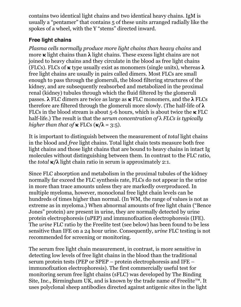
contains two identical light chains and two identical heavy chains. IgM isusually a “pentamer” that contains 5 of these units arranged radially like thespokes of a wheel, with the Y “stems” directed inward.
Free light chainsPlasma cells normally produce more light chains than heavy chains andmore κ light chains than λ light chains. These excess light chains are notjoined to heavy chains and they circulate in the blood as free light chains(FLCs). FLCs of κ type usually exist as monomers (single units), whereas λfree light chains are usually in pairs called dimers. Most FLCs are smallenough to pass through the glomeruli, the blood filtering structures of thekidney, and are subsequently reabsorbed and metabolized in the proximalrenal (kidney) tubules through which the fluid filtered by the glomerulipasses. λ FLC dimers are twice as large as κ FLC monomers, and the λ FLCstherefore are filtered through the glomeruli more slowly. (The half-life of λFLCs in the blood stream is about 5-6 hours, which is about twice the κ FLChalf-life.) The result is that the serum concentration of λ FLCs is typicallyhigher than that of κ FLCs (κ/λ = 3:5).
It is important to distinguish between the measurement of total light chainsin the blood and free light chains. Total light chain tests measure both freelight chains and those light chains that are bound to heavy chains in intact Igmolecules without distinguishing between them. In contrast to the FLC ratio,the total κ/λ light chain ratio in serum is approximately 2:1.
Since FLC absorption and metabolism in the proximal tubules of the kidneynormally far exceed the FLC synthesis rate, FLCs do not appear in the urinein more than trace amounts unless they are markedly overproduced. Inmultiple myeloma, however, monoclonal free light chain levels can behundreds of times higher than normal. (In WM, the range of values is not asextreme as in myeloma.) When abnormal amounts of free light chain (“BenceJones” protein) are present in urine, they are normally detected by urineprotein electrophoresis (uPEP) and immunofixation electrophoresis (IFE).The urine FLC ratio by the Freelite test (see below) has been found to be lesssensitive than IFE on a 24 hour urine. Consequently, urine FLC testing is notrecommended for screening or monitoring.
The serum free light chain measurement, in contrast, is more sensitive indetecting low levels of free light chains in the blood than the traditionalserum protein tests (PEP or SPEP – protein electrophoresis and IFE –immunofixation electrophoresis). The first commercially useful test formonitoring serum free light chains (sFLC) was developed by The BindingSite, Inc., Birmingham UK, and is known by the trade name of Freelite™. Ituses polyclonal sheep antibodies directed against antigenic sites in the light
chain that are hidden in intact Ig molecules, but exposed in free light chains.The reference (normal) ranges for this test are given below and are thosemost commonly cited:
Serum Free Light Chain Reference Ranges:κ: 3.3–19.4 mg/Lλ: 5.7–26.3 mg/Lκ/λ ratio: 0.26 –1.65
In addition to the above test, the Siemens company has developed amonoclonal antibody sFLC test that is currently available in Europe, but notin the United States. Patients who are being monitored should be followedwith only one type of test, preferably performed by the same laboratory, inorder to maintain the continuity of test results from one office visit to thenext.
κ/λ ratios that are increased above normal are considered as presumptiveevidence of a κ monoclonal gammopathy (a disorder in which Ig moleculesare produced by a single abnormal clone of lymphoid cells), while ratios thatare decreased are interpreted as evidence of a λ monoclonal gammopathy.However, it must be kept in mind that patients with polyclonalhypergammaglobulinemia (a broad elevation of many different Igs due toinfections, autoimmune diseases, and other chronic inflammatoryconditions) can have elevations of both light chain classes, and the κ/λ ratiocan sometimes be slightly abnormal even though no monoclonal process ispresent. There can also be lot-to-lot variations in reagents and other technicalproblems that could affect the test results. In the presence of renal disease,the κ/λ ratio may also be altered, usually in an upward direction, due to aslower than normal rate of renal clearance of κ monomers. It has beenrecommended that FLC test results in patients with renal impairment beevaluated using a different reference range for the κ/λ ratio: 0.37–3.1. Forthese reasons, serum light chain individual κ and λ values and κ/λ ratio testvalues that are slightly outside of their reference ranges should be interpretedcautiously.
The κ/λ ratio should be interpreted together with the individual κ and λvalues and not be considered in isolation:
If one of the light chain measurements were elevated and the serum lightchain ratio were significantly elevated, then a monoclonal gammopathy (inour case, IgM-MGUS or WM) would be likely.
If both light chain values were elevated and the ratio were normal orborderline, then a polyclonal gammopathy due to a chronic inflammatorycondition or kidney impairment would be suspected. (In rare cases, abiclonal gammopathy with one κ and one λ clone could also produce thispattern.)If both individual light chain values were low and the ratio were normal,bone marrow failure would be suspected.If all values were within the normal ranges, a monoclonal gammopathycould still be possible if the monoclonal cells were producing only an intactmonoclonal Ig without a detectable increase in monoclonal free light chain.(This should be evident by PEP and IFE, however.)
Serum free light chains and Waldenström macroglobulinemia (WM)Serum FLC (sFLC) testing is recommended as part of the initial workup forall MGUS (monoclonal gammopathy of undetermined significance) patients.The sFLC κ/λ ratio value is one of the 3 major risk factors used in stratifyingMGUS patients into 4 groups for risk of progression to one of the plasma cell-related malignancies. sFLC testing is also recommended forscreening/diagnostic evaluation for multiple myeloma (in its various forms),light chain (AL) amyloidosis, and a related condition known as light chaindeposition disease (LCDD). It is recommended for ongoing monitoring ofpatients with oligosecretory multiple myeloma (in which the amount ofsecreted monoclonal protein is very low), light chain myeloma, light chain(AL) amyloidosis, and LCDD. It is also recommended for verifying completeremission in multiple myeloma, and may have a broader role in determiningthe response to treatment in myeloma.
Because FLCs have a substantially shorter half-life in the blood (up to 6hours) than IgM (4-5 days), it has been hypothesized that sFLC testing mightprovide more rapid indications of changes in the course of WM that wouldfacilitate monitoring of the disease. In 2011, Leleu and colleagues noted (in astudy of sFLC monitoring of patients treated with bortezomib and rituximab)that sFLC analysis showed promising results as a sensitive marker of WMtumor measurement that correlated well with IgM and M-spikemeasurements, showed a more rapid change in test results in patients whoresponded to treatment, and also provided an earlier indication of diseaseprogression (by about 1 month) than the results observed with traditionalIgM or M-spike measurements. The article ended with a comment that theauthors proposed to study sFLC measurement in future large, prospectivetrials to confirm whether or not early determination of sFLC would help indecision-making during the course of therapy. Kyrtsonis and colleagues in2012 reported that the level of the involved (monoclonal) FLC and the FLCκ/λ ratio correlated with time to first treatment and adverse survival in WM.To date, however, there have been relatively few FLC studies in WM. As a
consequence, the status of sFLC testing in WM remains uncertain. In reviewarticles on WM in 2015 by Drs. Gertz and Treon, sFLC testing was included inlists of tests that may be ordered as part of the initial workup for WM.However, at the present time, it has not been recommended for ongoingmonitoring of patients with WM who can be followed with conventionaltesting.
Screening for AL amyloidosis and LCDDIt has been recommended that MGUS (monoclonal gammopathy ofundetermined significance) patients whose baseline κ/λ ratio is abnormaland whose involved (monoclonal) FLC is elevated be monitored periodicallyfor the development of heart or kidney damage due to amyloidosis or arelated condition, light chain deposition disease. The recommended tests areserum NT-proBNP, which is a sensitive indicator of cardiac injury, and urinealbumin (or microalbumin) to detect renal injury at an early stage. Suchmonitoring has also been recommended for selected WM patients. Signs andsymptoms of amyloidosis can develop insidiously. Many of these (such asperipheral neuropathy, constipation or diarrhea, feeling lightheaded uponstanding, peripheral edema, etc.) may also be seen with other diseases, so thediagnosis requires the demonstration by biopsy of one or more sites or byabdominal fat aspiration (at medical centers skilled in its detection by thistechnique). The need for such testing increases with additional risk factors,such as the presence of λ light chain as a monoclonal component of IgMgammopathy. It has been recommended that WM patients with peripheralneuropathy be considered for evaluation for amyloidosis, particularly if theyhave an IgMλ monoclonal protein. (While most WM patients have κmonoclonal light chain, the majority of patients with light chain amyloidosishave λ light chain. The ratio of λ to κ monoclonal light chain involvement isnearly 4:1.) For a fuller discussion of amyloidosis in WM, see Dr. Merlini’sprevious article in the April 2013 edition of the Torch newsletter.
Kidney injury from WM may involve infiltration by lymphoplasmacytic WMcells, deposition of intact IgM in glomerular capillaries, cryoglobulin injury,or deposition of monoclonal light chains. Light chain deposition diseasealways involves the kidney. The liver is the organ next most commonlyinvolved, but numerous other organs can be involved as well. It typicallypresents with hypertension (high blood pressure), protein in the urine, andmicroscopic blood in the urine. There are microscopic and clinical differencesbetween LCDD and light chain amyloidosis. At least half of the patients withLCDD have multiple myeloma but LCDD can occur by itself, in associationwith MGUS, and occasionally in WM patients. Treatment is aimed atlowering the monoclonal light chain to the maximum extent possible, whichwould be monitored by sFLC analysis and other tests.
ConclusionMy personal opinion is that it would be reasonable to order sFLC testing atthe time of initial diagnostic evaluation to establish a baseline for futurereference. At present, however, it is not considered necessary for determiningresponse to therapy or for routine monitoring for most patients with WM. Itcould be useful for selected patients who are being evaluated for light chainamyloidosis or light chain deposition disease as well as those who alreadyhave either of those conditions.
For those of you who are having periodic FLC testing:A rising involved (monoclonal) individual kappa or lambda light chain levelwell above the normal range associated with a rising ratio of the involvedFLC/uninvolved FLC suggests that your WM is producing more monoclonalprotein and may be proliferating. The reverse suggests that you areresponding to treatment. These values may precede changes in your IgM byseveral weeks. The trend is important. They need to be correlated over timewith the level of your M-spike by PEP and IgM by nephelometry, hemoglobin,platelet count, white count, kidney function tests, bone marrow findings, yourgeneral level of energy for daily tasks, the status of other illnesses that youmay have, and your oncologist’s impression of the status of your WM.
The future:Over the last few years, articles have been appearing in the literature thatevaluate a newer test developed by the same company that developed thesFLC test (The Binding Site). The new test is known generically as theheavy/light chain (HLC) test and is known by the trade name of Hevylite™.The HLC test uses antibodies that target junctional binding sites between theconstant regions of heavy and light chains of Ig molecules, thereby measuringheavy/light chain pairs. Thus, there is simultaneous measurement both of aparticular type of heavy chain (gamma, alpha, or mu) and the κ or λ lightchains that are associated with that heavy chain. For example, in a case ofIgMκ WM, the level of IgMκ would be increased, while the level of IgMλmight be normal or decreased. It may be that the HLC test could serve as away to monitor the level of the M-spike. Some studies have evaluated bothsFLC and HLC testing to see if the combination offers advantages over HLCalone. At the present time, only the IgG and IgA HLC tests are approved foruse in the United States. In Europe, the IgM HLC test is also available. Beforethe HLC test is recommended for use in the diagnosis and monitoring of WMpatients, additional prospective studies will be needed.
Select Bibliography1. Gertz M. Waldenström macroglobulinemia: 2015 update on diagnosis, risk
stratification, and management. Am J Hematol 2015;90:347–354.2. Jimenez J et al. Clinical recommendations for the measurement of serum
free light chains and the emerging role of heavy/light chain pair analysis inthe management of monoclonal gammopathies: When and how to use it? Jof Applied Hematol 2015;6(2):43-52 .
3. Kyrtsonis M-C et al. Prognostic Contribution of the New Immunoglobulin (Ig)Biomarkers (Freelite™ and Hevylite™) in Waldenstrom’s Macroglobulinemia(WM). Am J Med Medical Sciences 2012;2(6):136-143.
4. Leleu X et al. The Role of Serum Immunoglobulin Free Light Chain inResponse and Progression in Waldenstrom Macroglobulinemia. Clin CancerRes 2011;17(9):3013–8.
5. Merlini G. Amyloidosis Associated with Waldenström Disease or IgM-MGUS. IWMF Torch, volume 14.2 (April 2013) pages 1-4.
6. Sherwood G. Basic Immunology in Waldenstrom’s Macroglobulinemia.March 2014, copyright IWMF.
7. Treon S. How I treat Waldenström macroglobulinemia. Blood2015;126(6):721-732.
8. International Myeloma Foundation 2011. Understanding Serum Free LightChain Assays.
Neal Makens, MD, completed his medical training in 1969 at St. LouisUniversity, St. Louis MO, followed by an internship in internal medicine anda residency in anatomic and clinical pathology, both at Naval MedicalCenter, San Diego CA. From 1974-81, Dr. Makens served as a staffpathologist and later as the Head of the Laboratory Department, NavalHospital, Camp Pendelton CA. From 1981-88 he served at Naval MedicalCenter, Portsmouth VA, first as a staff pathologist and subsequently asLaboratory Director and Chairman of the Department of Pathology. In1988, Dr. Makens retired from the United States Navy with the rank ofCaptain.
For the next fifteen years he was affiliated with Robinson MemorialHospital in Ravenna OH, where he worked both as a staff pathologist andas Director of the Laboratory Department. He retired from practice in2003.
Dr. Makens is an emeritus Fellow of the American Society of ClinicalPathologists and the College of American Pathologists. He is currently amember of the IWMF Research Committee.
This article was published in the IWMF Torch, volume 17.2 (April 2016)pages 1-4.