Scavenger Hunt Bingo - National Park Service
15
JOURNAL OF VIROLOGY, July 2002, p. 6800–6814 Vol. 76, No. 13 0022-538X/02/$04.000 DOI: 10.1128/JVI.76.13.6800–6814.2002 Copyright © 2002, American Society for Microbiology. All Rights Reserved. Identification of NF-B-Dependent Gene Networks in Respiratory Syncytial Virus-Infected Cells Bing Tian, 1 Yuhong Zhang, 1 Bruce A. Luxon, 2,3 Roberto P. Garofalo, 4,5 Antonella Casola, 4 Mala Sinha, 2,3 and Allan R. Brasier 1,6 * Departments of Medicine, 1 Human Biological Chemistry and Genetics, 3 Pediatrics, 4 and Microbiology and Immunology, 5 Sealy Center for Structural Biology, 2 and Sealy Center for Molecular Sciences, 6 The University of Texas Medical Branch, Galveston, Texas 77555-1060 Received 5 November 2001/Accepted 28 March 2002 Respiratory syncytial virus (RSV) is a mucosa-restricted virus that is a leading cause of epidemic respiratory tract infections in children. In epithelial cells, RSV replication activates nuclear translocation of the inducible transcription factor nuclear factor B (NF-B) through proteolysis of its cytoplasmic inhibitor, IB. In spite of a putative role in mediating virus-inducible gene expression, the spectrum of NF-B-dependent genes induced by RSV infection has not yet been determined. To address this, we developed a tightly regulated cell system expressing a nondegradable, epitope-tagged IB isoform (Flag-IB Mut) whose expression could be controlled by exogenous addition of nontoxic concentrations of doxycycline. Flag-IB Mut expression potently inhibited IB proteolysis, NF-B binding, and NF-B-dependent gene transcription in cells stimulated with the prototypical NF-B-activating cytokine tumor necrosis factor alpha (TNF-) and in response to RSV infection. High-density oligonucleotide microarrays were then used to profile constitutive and RSV-induced gene expression in the absence or presence of Flag-IB Mut. Comparison of these profiles revealed 380 genes whose expression was significantly changed by the dominant-negative NF-B. Of these, 236 genes were constitutive (not RSV regulated), and surprisingly, only 144 genes were RSV regulated, representing numer- ically 10% of the total population of RSV-inducible genes at this time point. Hierarchical clustering of the 144 RSV- and Flag-IB Mut-regulated genes identified two discrete gene clusters. The first group had high constitutive expression, and its expression levels fell in response to RSV infection. In this group, constitutive mRNA expression was increased by Flag-IB Mut expression, and the RSV-induced decrease in expression was partly inhibited. In the second group, constitutive expression was very low (or undetectable) and, after RSV infection, expression levels strongly increased. In this group, NF-B was required for RSV-inducible expression because Flag-IB Mut expression blocked their induction by RSV. This latter cluster includes chemokines, transcriptional regulators, intracellular proteins regulating translation and proteolysis, and secreted proteins (complement components and growth factor regulators). These data suggest that NF-B action induces global cellular responses after viral infection. NF-B is a family of inducible transcription factors control- ling expression of genes important for pathogen- or cytokine- induced inflammation, immune response, and cellular survival (5, 21, 37). The prototypical NF-B complex, composed of 50-kDa NF-B1 and 65-kDa RelA heterodimers, is regulated by its association with a family of cytoplasmic inhibitors, IBs, whose members bind and specifically inactivate NF-B mem- bers by masking their nuclear localization sequence, thereby preventing nuclear entry (27, 32; reviewed in reference 7). NF-B activation is a sequential process whose final result is to induce nuclear translocation of the inactivated cytoplasmic NF-B complex through targeted proteolysis of the IB inhib- itors. Intracellular NF-B-activating signals converge on the multiprotein cytoplasmic IB kinase complex (IKK), a com- plex that phosphorylates IB on two serine residues (Ser 32 and Ser 36 ) in its NH 2 -regulatory domain (reviewed in reference 37). Phospho-IB is then specifically bound by the SCF-type E3 ubiquitin ligase E3RS, initiating IB ubiquitination and proteolysis through the proteasome (10, 37, 38). A parallel pathway important in viral infection produces IB degradation through cytoplasmic calpains (30, 34). Following IB proteol- ysis, liberated NF-B enters the nucleus to activate target gene transcription. Because of the large number of inducible cytokine, chemo- kine, acute-phase reactant, and adhesion molecule genes that contain NF-B binding sites in their promoters, NF-B ap- pears to play an essential role in innate immunity and the inflammatory (host) response to infectious agents (8, 11, 40, 52; reviewed in reference 56). Our laboratory has been inves- tigating the mechanisms for chemokine expression induced by respiratory syncytial virus (RSV). RSV is a ubiquitous human respiratory tract pathogen known to produce severe lower re- spiratory tract infections (bronchiolitis) in infants, accounting for 100,000 hospitalizations in the United States annually (50; reviewed in reference 26). Human RSV infections char- acteristically show virus-induced damage to the epithelial sur- face (1, 15, 19) and a pronounced inflammatory response, consisting of a perivascular mononuclear and lymphocytic in- filtrate (1, 15). Other features of immune activation that can be detected include neutrophil recruitment into the bronchoal- veolar lavage (14) and eosinophilic and basophilic degranula- * Corresponding author. Mailing address: Division of Endocrinol- ogy, MRB 8.138, The University of Texas Medical Branch, 301 Uni- versity Blvd., Galveston, TX 77555-1060. Phone: (409) 772-2824. Fax: (409) 772-8709. E-mail: [email protected]. 6800 on April 16, 2019 by guest http://jvi.asm.org/ Downloaded from
Transcript of Scavenger Hunt Bingo - National Park Service
JOURNAL OF VIROLOGY, July 2002, p. 6800–6814 Vol. 76, No. 130022-538X/02/$04.00�0 DOI: 10.1128/JVI.76.13.6800–6814.2002Copyright © 2002, American Society for Microbiology. All Rights Reserved.
Identification of NF-�B-Dependent Gene Networks in RespiratorySyncytial Virus-Infected Cells
Bing Tian,1 Yuhong Zhang,1 Bruce A. Luxon,2,3 Roberto P. Garofalo,4,5 Antonella Casola,4Mala Sinha,2,3 and Allan R. Brasier1,6*
Departments of Medicine,1 Human Biological Chemistry and Genetics,3 Pediatrics,4 and Microbiology andImmunology,5 Sealy Center for Structural Biology,2 and Sealy Center for Molecular Sciences,6
The University of Texas Medical Branch, Galveston, Texas 77555-1060
Received 5 November 2001/Accepted 28 March 2002
Respiratory syncytial virus (RSV) is a mucosa-restricted virus that is a leading cause of epidemic respiratorytract infections in children. In epithelial cells, RSV replication activates nuclear translocation of the inducibletranscription factor nuclear factor �B (NF-�B) through proteolysis of its cytoplasmic inhibitor, I�B. In spiteof a putative role in mediating virus-inducible gene expression, the spectrum of NF-�B-dependent genesinduced by RSV infection has not yet been determined. To address this, we developed a tightly regulated cellsystem expressing a nondegradable, epitope-tagged I�B� isoform (Flag-I�B� Mut) whose expression could becontrolled by exogenous addition of nontoxic concentrations of doxycycline. Flag-I�B� Mut expression potentlyinhibited I�B� proteolysis, NF-�B binding, and NF-�B-dependent gene transcription in cells stimulated withthe prototypical NF-�B-activating cytokine tumor necrosis factor alpha (TNF-�) and in response to RSVinfection. High-density oligonucleotide microarrays were then used to profile constitutive and RSV-inducedgene expression in the absence or presence of Flag-I�B� Mut. Comparison of these profiles revealed 380 geneswhose expression was significantly changed by the dominant-negative NF-�B. Of these, 236 genes wereconstitutive (not RSV regulated), and surprisingly, only 144 genes were RSV regulated, representing numer-ically �10% of the total population of RSV-inducible genes at this time point. Hierarchical clustering of the 144RSV- and Flag-I�B� Mut-regulated genes identified two discrete gene clusters. The first group had highconstitutive expression, and its expression levels fell in response to RSV infection. In this group, constitutivemRNA expression was increased by Flag-I�B� Mut expression, and the RSV-induced decrease in expressionwas partly inhibited. In the second group, constitutive expression was very low (or undetectable) and, after RSVinfection, expression levels strongly increased. In this group, NF-�B was required for RSV-inducible expressionbecause Flag-I�B� Mut expression blocked their induction by RSV. This latter cluster includes chemokines,transcriptional regulators, intracellular proteins regulating translation and proteolysis, and secreted proteins(complement components and growth factor regulators). These data suggest that NF-�B action induces globalcellular responses after viral infection.
NF-�B is a family of inducible transcription factors control-ling expression of genes important for pathogen- or cytokine-induced inflammation, immune response, and cellular survival(5, 21, 37). The prototypical NF-�B complex, composed of50-kDa NF-�B1 and 65-kDa RelA heterodimers, is regulatedby its association with a family of cytoplasmic inhibitors, I�Bs,whose members bind and specifically inactivate NF-�B mem-bers by masking their nuclear localization sequence, therebypreventing nuclear entry (27, 32; reviewed in reference 7).
NF-�B activation is a sequential process whose final result isto induce nuclear translocation of the inactivated cytoplasmicNF-�B complex through targeted proteolysis of the I�B inhib-itors. Intracellular NF-�B-activating signals converge on themultiprotein cytoplasmic I�B kinase complex (IKK), a com-plex that phosphorylates I�B on two serine residues (Ser32 andSer36) in its NH2-regulatory domain (reviewed in reference37). Phospho-I�B is then specifically bound by the SCF-typeE3 ubiquitin ligase E3RS, initiating I�B ubiquitination and
proteolysis through the proteasome (10, 37, 38). A parallelpathway important in viral infection produces I�B degradationthrough cytoplasmic calpains (30, 34). Following I�B proteol-ysis, liberated NF-�B enters the nucleus to activate target genetranscription.
Because of the large number of inducible cytokine, chemo-kine, acute-phase reactant, and adhesion molecule genes thatcontain NF-�B binding sites in their promoters, NF-�B ap-pears to play an essential role in innate immunity and theinflammatory (host) response to infectious agents (8, 11, 40,52; reviewed in reference 56). Our laboratory has been inves-tigating the mechanisms for chemokine expression induced byrespiratory syncytial virus (RSV). RSV is a ubiquitous humanrespiratory tract pathogen known to produce severe lower re-spiratory tract infections (bronchiolitis) in infants, accountingfor �100,000 hospitalizations in the United States annually(50; reviewed in reference 26). Human RSV infections char-acteristically show virus-induced damage to the epithelial sur-face (1, 15, 19) and a pronounced inflammatory response,consisting of a perivascular mononuclear and lymphocytic in-filtrate (1, 15). Other features of immune activation that can bedetected include neutrophil recruitment into the bronchoal-veolar lavage (14) and eosinophilic and basophilic degranula-
* Corresponding author. Mailing address: Division of Endocrinol-ogy, MRB 8.138, The University of Texas Medical Branch, 301 Uni-versity Blvd., Galveston, TX 77555-1060. Phone: (409) 772-2824. Fax:(409) 772-8709. E-mail: [email protected].
6800
on April 16, 2019 by guest
http://jvi.asm.org/
Dow
nloaded from
tion products in nasopharyngeal secretions whose levels corre-late with disease severity (20, 31, 53).
Because RSV is a mucosally restricted virus, the airwayepithelium is thought to play an important role in initiatingairway inflammation. Here RSV replication induces expressionand secretion of cytokines (16, 21, 44), chemokines (6, 47, 57),arachidonic acid metabolites (22), reactive oxygen species (11),and cell surface display of major histocompatibility complex(MHC) class I (36) and CD18 (46). We recently extendedthese observations by profiling chemokine expression patternsby using macro- and high-density oligonucleotide arrays (57).These studies suggested that RSV profoundly influenced geneexpression profiles in the cells in which it replicates; focusedanalysis of virus-induced chemokines showed a time-depen-dent expression of CC (I-309, Exodus-1, TARC, RANTES,MCP-1, MDC, and MIP-1 �/�), CXC (GRO �/�/�, ENA-78,IL-8, and I-TAC), and CX3C (fractalkine) subclasses.
Promoter analysis studies suggest that NF-�B is likely to playa central role mediating inducible cytokine and chemokinegene expression. For example, we have shown that RSV in-duces NF-�B translocation through a mechanism involvingproteolysis of the RelA-associated inhibitor I�B� through adual pathway involving the 26S proteasome and an as yetuncharacterized protease (34) stimulated by active viral repli-cation (17, 34) and mediated upstream by activation of the IKKcomplex (25). Mutation of the NF-�B binding site or inhibitionof its translocation blocks RSV-inducible interleukin-8 (IL-8)and RANTES expression (8, 12, 52). However, the spectrum ofNF-�B-dependent genes expressed by replicating RSV has notbeen reported.
To address this, we developed a cell system expressing anondegradable epitope-tagged I�B� (I�B� Ser32Ala/Ser36Ala,termed Flag-I�B� Mut) whose expression is strongly inhibitedafter addition of nontoxic concentrations of doxycycline to theculture medium. Stable clonal cell lines expressing the tetra-cycline-controlled transactivator and I�B� Mut were isolatedand screened for doxycycline-dependent expression. Upondoxycycline withdrawal, Flag-I�B� Mut expression potentlyinhibited NF-�B-dependent transcription in response to theprototypical NF-�B-activating cytokine TNF-� and in responseto RSV infection. High-density oligonucleotide arrays werethen used to assay for profiles of gene expression in responseto RSV infection in the presence of Flag-I�B� Mut versusthose induced in its absence. Our data suggest a role for NF-�Bin controlling constitutive gene expression and further specif-ically identify members of the RSV-inducible NF-�B-depen-dent gene network.
MATERIALS AND METHODS
RSV preparation. The human Long strain of RSV (A2) was grown in Hep-2cells and purified by centrifugation on discontinuous sucrose gradients (57). Thevirus titer of the purified RSV pools was 7.5 to 8.5 log PFU/ml, determined bymethylcellulose plaque assay. No contaminating cytokines, including IL-1,TNF-�, IL-6, IL-8, granulocyte-macrophage colony-stimulating factor, and in-terferon, were found in these sucrose-purified viral preparations (21). Lipopoly-saccharide (LPS), assayed by using the Limulus hemocyanin agglutination assay,was also not detected. Virus pools were aliquoted, quick-frozen on dry ice-alcohol, and stored at �70°C until used.
Plasmid construction. Plasmids pUHD10-4, a plasmid containing seven copiesof the tetracycline operator (tetO) upstream of the cytomegalovirus minimalpromoter, and pUHD15-1 Neo, a plasmid expressing the tetracycline transacti-vator (tTA), were gifts of M. Gossen and H. Bujard (University of Heidelberg,
Germany) (23). The doxycycline-regulated NF-�B dominant-negative expressionplasmid encoding I�B� with serine residues 32 and 36 substituted for alanine(I�B� Mut) under control of tetO sequences was generated in two steps. ThepUHD10-4 multiple cloning site was modified to encode a Flag epitope tagdownstream of an optimized Kozak initiation site. This plasmid was constructedby inserting the double-stranded oligonucleotide produced by annealing thesequence 5�-AATT CGC CAT ATG GAT TAC AAA GAC GAT GAC GATACC ATG GTT AGA TCT GAT ATC G-3� (initiation codon indicated bydouble underline, BglII site indicated by single underline) with 5�-GATCC GATATC AGA TCT AAC CAT GGT ATC GTC ATC GTC TTT GTA ATC CATATG GC-3� into EcoRI- and BamHI-restricted pUHD10-4 to generate pUHD-FLAG.
The I�B� Ser32Ala/Ser36Ala mutation was produced by PCR by using muta-genic primers as previously described by us (12). The I�B� Mut cDNA wasisolated containing upstream BamHI and downstream HindIII (made bluntended by Klenow polymerase) and ligated into BglII and BamHI-restrictedpUHDFLAG (the BamHI site made blunt ended by Klenow polymerase) togenerate the expression vector pUHDFLAG-I�B� Mut for use in transienttransfection assays. For stable cell isolation, the Flag-I�B� Mut cDNA wasexcised, made blunt ended, and cloned into the PvuII site of the plasmid pBI-EGFP plasmid (Clontech, Palo Alto, Calif.). pBI-EGFP-I�B� Mut expresses theenhanced green fluorescent protein (EGFP) and Flag-I�B� Mut from a bidirec-tional tetO-driven promoter for rapid visual screening of stable transfectants.
The firefly luciferase reporter genes driven by the human IL-8 (hIL-8) nativepromoter, �162/�44 hIL-8/LUC, and the IL-6 native promoter, �310/�25 hIL-6/LUC, have been described (8, 29). Constructs carrying three copies of theangiotensinogen RelA/NF-�B1 binding site or three copies of the IL-6 RelA/NF-�B1 site cloned upstream of an inert TATA box driving expression of fireflyluciferase, (APREWT)3-p59rAT/LUC and (IL-6�BE)3-p59rAT/LUC, respec-tively, have been described (8, 29, 35). pTK-Hyg, encoding the hygromycinresistance marker, was obtained commercially (Clontech, Palo Alto, Calif.).Plasmids were purified by ion exchange chromatography (Qiagen, Chatsworth,Calif.) and sequenced across cloning junctions to verify authenticity prior totransfection.
Cell culture and transfection. The human cervical epithelioid carcinoma cellline HeLa expressing tTA (HeLa Tet-Off) was constructed earlier (23). HeLaTet-Off cells were grown in medium containing 90% Dulbecco’s modified Eagle’smedium, 10% heat-inactivated fetal bovine serum, 4 mM L-glutamine, 0.1 mMnonessential amino acids, and 1 mM sodium pyruvate plus 100 g of G418, 100U of penicillin G sodium, and 100 g of streptomycin sulfate per ml in ahumidified atmosphere of 5% CO2.
To establish stable cell lines in which I�B� Mut is regulated by exogenousdoxycycline, HeLa Tet-Off cells were cotransfected with pBI-EGFP-I�B� Mutand pTK-Hyg plasmids by using Lipofectamine (Life Technologies, Inc.). At 24 hafter transfection, cells were changed into medium containing 250 g of hygro-mycin B and 2 g of doxycycline per ml. Hygromycin-resistant clones wereisolated and screened after doxycycline withdrawal by culture in tetracycline-freefetal bovine serum (Clontech) for 7 days. Only strongly fluorescent clones wereselected further. These clones were further screened for I�B� Mut expression byWestern blotting (with either anti-�B� or anti-Flag antibodies) or by inhibitionof TNF-�-stimulated NF-�B binding in electrophoretic mobility shift assays.Only the clones whose expression of I�B� Mut was tightly regulated by doxycy-cline were selected for further experiments.
For TNF-� stimulation, recombinant TNF-� (rTNF-�) was added directly tothe culture medium for the indicated times (25 ng/ml, final concentration). ForRSV infection, freshly isolated cells were split into two cultures; one group wasmaintained in doxycycline, and the other was maintained without for 7 days, atime at which I�B� Mut expression was maximal. Cells were then changed intoDulbecco’s modified Eagle’s medium containing 2% (vol/vol) fetal bovine serum.Cell monolayers were infected with sucrose cushion-purified RSV at a multiplic-ity of infection of 1.
For transient transfections, cells freshly plated into triplicate 60-mm plateswere transfected by using Lipofectamine (Life Technologies, Inc.) with 8 g ofindicated luciferase reporter plasmid and 3 g of the constitutive alkaline phos-phatase internal control plasmid pSV2PAP (35). Cells were cultured for anadditional 40 h and, where indicated, stimulated with rTNF-� for the indicatedtimes. For reporter assays, cells were harvested 6 h after rTNF-� stimulation,cytoplasmic lysates were prepared, and luciferase activity was measured (35). Asan internal control for transfection efficiency, alkaline phosphatase activity wasmeasured in 50 g of cell lysate as described previously (35). Fold induction ofreporter activity was calculated by dividing the mean normalized luciferase ac-tivity from three treated cultures by the mean normalized luciferase activity fromthree untreated cultures.
VOL. 76, 2002 NF-�B-DEPENDENT GENE NETWORK 6801
on April 16, 2019 by guest
http://jvi.asm.org/
Dow
nloaded from
Preparation of subcellular extracts. Cells were washed two times with phos-phate-buffered saline and disrupted by hypotonic nonionic detergent lysis byaddition of 2 cell volumes of buffer A (50 mM HEPES [pH 7.4], 10 mM KCl, 1mM EDTA, 1 mM EGTA, 1 mM dithiothreitol plus 0.1 mg of phenylmethylsul-fonyl fluoride, 1 g of pepstatin A, 1 g of leupeptin, 10 g of soybean trypsininhibitor, and 10 g of aprotinin per ml, and 0.5% IGEPAL CA-630 [(octylphe-noxy)polyethoxyethanol]). After 10 min on ice, the nuclei were pelleted bycentrifugation at 4,000 � g for 4 min at 4°C (the supernatant constituting thecytoplasmic extract). The nuclear pellets were further purified by centrifugationthrough a discontinuous sucrose cushion by resuspending in buffer B (1.7 Msucrose, 50 mM HEPES [pH 7.4], 10 mM KCl, 1 mM EDTA, 1 mM EGTA, 1mM dithiothreitol plus 0.1 mg of phenylmethylsulfonyl fluoride, 1 g of pepstatinA, 1 g of leupeptin, 10 g of soybean trypsin inhibitor, and 10 g of aprotininper ml), and centrifuging at 12,000 � g for 10 min at 4°C.
Proteins in the pelleted nuclei were extracted by incubating in buffer C (10%glycerol, 50 mM HEPES [pH 7.4], 400 mM KCl, 1 mM EDTA, 1 mM EGTA, 1mM dithiothreitol plus 0.1 mg of phenylmethylsulfonyl fluoride, 1 g of pepstatinA, 1 g of leupeptin, 10 g of soybean trypsin inhibitor, and 10 g of aprotininper ml) with frequent vortexing for 30 min at 4°C. After centrifugation at 12,000� g for 30 min at 4°C, the supernatant was frozen at �70°C. With this technique,the nuclear fractions are free of cytoplasmic contamination and the cytoplasmicfractions lack nuclear markers (9). Both cytoplasmic and nuclear extracts werenormalized for protein concentration by using bovine serum albumin as a stan-dard (Bio-Rad, Hercules, Calif.).
Western immunoblot. For Western immunoblot, a constant amount of cyto-plasmic or nuclear extracts (200 to 300 g as indicated) was boiled in Laemmlibuffer, fractionated by sodium dodecyl sulfate–10% polyacrylamide gel electro-phoresis (SDS–10% PAGE), and transferred to polyvinylidene difluoride mem-branes (Millipore, Bedford, Mass.) in 3-[cyclohexylamino]-1-propanesulfonicacid (CAPS)–methanol (35). Membranes were blocked in 5% milk–Tris-bufferedsaline (TBS)–0.1% Tween for 1 h and immunoblotted with either anti-Flag M2monoclonal antibody-peroxidase conjugate (Sigma Chemical, no. A8592) or af-finity-purified rabbit polyclonal antibody anti-�B� (Santa Cruz Biotechnology,Santa Cruz, Calif.) for 1 h at 4°C. Membranes were washed four times inTBS–0.1% Tween 20. Immune complexes were detected by reaction in theenhanced chemiluminescence assay (ECL; Amersham) according to the manu-facturer’s recommendations. Protein loading controls were performed by prob-ing the blot with monoclonal antibody to �-actin (9).
EMSAs. Electrophoretic mobility shift assays (EMSAs) were performed asdescribed previously (9) with minor modifications. Nuclear extracts (15 g) wereincubated with 40,000 cpm of 32P-labeled APRE WT duplex oligonucleotideprobe and 2 g of poly(dA-dT) in a buffer containing 8% glycerol, 100 mM NaCl,5 mM MgCl2, 5 mM dithiothreitol, and 0.1 g of phenylmethylsulfonyl fluorideper ml in a final volume of 20 l for 20 min at room temperature. The complexeswere fractionated on 6% or 7% native polyacrylamide gels run in 1� TBE buffer(89 mM Tris, 89 mM boric acid, and 2.0 mM EDTA), dried, and exposed toKodak X-AR film at 70°C. Competition was performed by the addition of a100-fold molar excess of nonradioactive double-stranded oligonucleotide com-petitor at the time of addition of radioactive probe. The sequences of the APREdouble-stranded oligonucleotides are shown below:
APRE WT: GATCCACCACAGTTGGGATTTCCCAACCTGACCAGTGGTGTCAACCCTAAAGGGTTGGACTGGTCTAG
APRE M6: GATCCACCACATGTTGGATTTCCGATACTGACCAGTGGTGTACAACCTAAAGGCTATGACTGGTCTAG
Antibody supershift assays were performed by adding to the binding reaction1 l of affinity-purified polyclonal antibodies and incubating for 1 h on ice. All ofthe antibodies used in these assays were obtained commercially (Santa CruzBiotechnology.
RNase protection assay. Steady-state mRNA levels in RSV-infected cells wereanalyzed by RNase protection assay by using the RiboQuant multiprobe kit(PharMingen, San Diego, Calif.) following the manufacturer’s recommenda-tions. In brief, 32P-labeled specific antisense RNA probes were synthesized by invitro transcription from multiple DNA template sets. An equal amount of totalRNA (15 g per sample) was hybridized overnight to the 32P-labeled RNAprobes and digested with RNases A and T1. The protected cRNA probes wereprecipitated with ethanol and resolved on QuickPoint precast denaturing poly-acrylamide gels (Novex, San Diego, Calif.). The gel was dried and then exposedto X-ray film. The abundance of each mRNA transcript was quantified on aPhosphorImaging screen and normalized to housekeeping L32 transcripts in thesame reaction.
Northern blot. Total cellular RNA was extracted by acid guanidium-phenolextraction (Tri Reagent; Sigma). RNA (20 g) was denatured, fractionated byelectrophoresis on a 1.2% agarose–formaldehyde gel, capillary transferred to anitrocellulose membrane (Zeta-Probe GT; Bio-Rad), and prehybridized as de-scribed previously (8). The 1.1-kb RSV N cDNA probe was produced by PCR byusing poly(A)-primed cDNA as a template from RSV-infected A549 cells byusing the upstream primer 5�-CAAATGGATCCATGGCTCTTAGCAAAGTCAAG-3� and the downstream primer 5�-TTCCCGGTTCAAAGCTCTACATCATTATC-3�. The membrane was hybridized with 1 � 106 to 2 � 106 cpm of32P-RSV-N cDNA probe per ml at 60°C overnight in 5% sodium dodecyl sulfate(SDS) hybridization buffer. The membrane was washed with a buffer containing5% SDS and 1� saline-sodium citrate (0.15 M NaCl and 0.015 M sodium citrate)for 20 min at room temperature, followed by 30 min at 60°C. Internal controlhybridization was performed with 18S RNA. The membrane was exposed toXAR film (Kodak) for 24 to 48 h at 70°C and quantified by exposure to Phos-phorImager cassette.
Measurement of apoptotic index. DNA fragmentation was assayed by using acommercially available enzyme-linked immunosorbent assay (ELISA) (Boehr-inger Mannheim Biochemicals, Indianapolis, Ind.). This assay detects small nu-cleosomal fragments in the cytoplasmic fractions of affected cells that ariseduring apoptosis but not as a result of necrosis. After the cells were infected withRSV for the indicated time period, cells were harvested by trypsinization, and 5� 105 cells were lysed for 30 min at room temperature and cleared of cellulardebris for 20 min at 200 � g. The resulting cytoplasmic fraction was used as asource of nucleosomal fragments. Following incubation of this supernatant withcapture antibodies and detection reagent, the absorbance was measured at 405nm by Multiskan (model MCC/340; Titertek Instruments, Inc., Irvine, Calif.) andcompared with substrate solution as a blank. The apoptotic index was calculatedaccording to the manufacturer’s instructions by dividing the absorbance of stim-ulated cells by the absorbance of control cells.
Oligonucleotide probe-based microarrays. Hu95A GeneChip (Affymetrix Inc.,Santa Clara, Calif.) containing 12,626 sequenced human genes was used aspreviously described (57). Briefly, first-strand cDNA synthesis was performedby using total RNA (10 to 25 g), a T7-(dT)24 oligomer [5�-GGCCAGTGAATTGTAATACGACTCACTATAGGGAGGCGG-(dT)24-3�] and SuperScriptII reverse transcriptase (Life Technologies). The T7 promoter introduced duringfirst-strand cDNA synthesis is then used to direct the synthesis of cRNA by usingbacteriophage T7 RNA polymerase. The biotin-labeled target RNAs are frag-mented to a mean size of 200 bases and initially hybridized to a test arraycontaining a set of probes representing genes that are commonly expressed in themajority of cells (actin, glyceraldehyde-3-phosphate dehydrogenase [GAPDH],transferrin receptor, transcription factor ISGF-3, 18S RNA, 28S RNA, and alu)to confirm their successful labeling.
Hybridization was performed at 45°C for 16 h in hybridization buffer (0.1 Mmorpholineethanesulfonic acid [MES, pH 6.6], 1 M NaCl, 0.02 M EDTA, and0.01% Tween 20). Four prokaryotic genes (bioB, bioC, and bioD from theEscherichia coli biotin synthesis pathway and cre, the recombinase gene frombacteriophage P1) were added to the hybridization cocktail as internal controls.Arrays were washed by using both nonstringent (1 M NaCl, 25°C) and stringent(1 M NaCl, 50°C) conditions prior to staining with phycoerythrin-streptavidin (10g/ml final). Gene Chip arrays were scanned by using a Gene Array Scanner(Hewlett Packard) and analyzed by using the Gene Chip Analysis Suite 3.3software (Affymetrix Inc.). For each gene, 16 to 20 probe pairs were immobilizedas �25-mer oligonucleotides that hybridized throughout the mRNA; each probepair was represented as a perfect match oligonucleotide and a mismatch oligo-nucleotide as a hybridization control.
The average intensity of each probe cell was calculated after subtraction of thelocal background (the lowest 2% intensity of each sector; each probe cell isdivided into 16 sectors). The normalized average intensity value is used todetermine the number of positive and negative probe pairs. Based on the posi-tive/negative ratio, positive fraction and log average ratio of the perfect match tomismatch, the absolute call [e.g., the gene is detected (present) or not (absent)]is determined (57, 58). Finally the average difference is determined by calculatingthe difference intensity between the perfect match and mismatch of every probepair and averaging the differences over the entire probe set.
Data analysis. Three independent experiments were performed identically,including control without doxycycline treatment, control with doxycycline treat-ment, RSV 12-h infection without doxycycline treatment, and RSV 12-h infectionwith doxycycline treatment. For comparison of the fluorescent intensity (averagedifference) values among multiple experiments, the average difference values foreach “experimental” GeneChip were scaled to that of the “base” GeneChip. Thiswas done first by calculating the “2% trimmed mean” (a measurement of globalsignal intensity) for each GeneChip. The trimmed mean is obtained by calculat-
6802 TIAN ET AL. J. VIROL.
on April 16, 2019 by guest
http://jvi.asm.org/
Dow
nloaded from
ing the mean signal intensity of the chip after discarding the top and bottom 2%average difference values (representing the outliers). Next, scaling was per-formed by multiplying all the average differences in the “experimental” array bya scaling factor defined as the ratio of the base trimmed mean to the experi-mental trimmed mean (the base array was defined to be the control plus doxy-cycline GeneChip).
Because both RSV and doxycycline can be considered experimental treat-ments, the scaled average difference values were then subjected to a two-wayanalysis of variance (ANOVA; Splus 6; Insightful Inc.) with replications todetermine which genes were significantly influenced by either the RSV or doxy-cycline treatment. Genes (probe sets) with a P value [Pr(F)] at the �0.05confidence level as a result of either treatment were deemed highly significantand were selected for further analysis. Agglomerative hierarchical clustering byusing the unweighted pair-group method with arithmetic mean (43) was per-formed on the indicated genes (Spotfire Array Explorer, v. 3.01; Spotfire Inc.,Cambridge, Mass.). The data are graphically presented as heat maps in whichfluorescence intensity is represented by a color gradient. For the heat mapsshown, green represents the minimum average difference value (5 scaled units),black represents the mid average difference value (5,000 scaled units), and redrepresents the maximum average difference value (10,000 scaled units). Inves-tigators may obtain the primary data at our website http://bioinfo.utmb.edu/Brasier_Lab/.
RESULTS
To identify the gene network under control of the NF-�Btranscription factor in the setting of RSV replication, we ex-amined methods for expressing dominant negative inhibitorsthat would be suitable for high-density microarray analysis.Although others have had success with transducing cells withreplication-deficient adenovirus encoding the nonproteolyzedI�B� Mut (52), we sought alternative approaches as adenovi-rus infection may have significant confounding effects on hostcell gene expression or unrecognized interactions with RSVreplication or signaling. Additionally, isolating a cell line con-stitutively expressing an NF-�B inhibitor would be problematicas we and others have shown that NF-�B is required for cellsurvival (4, 41).
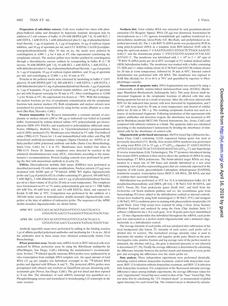
For these reasons, we employed a tightly regulated system inwhich expression of the nonproteolyzable I�B� Mut could becontrolled by exogenously added tetracycline (doxycycline).Clonal isolates of Flag epitope-tagged I�B� Mut under tetOcontrol selected by hygromycin resistance in HeLa cells con-taining the tetracycline transactivator were selected and char-acterized. To determine the kinetics of I�B� Mut expression,cells were withdrawn from doxycycline for various times from0 to 7 days, and cytoplasmic lysates were prepared. Figure 1shows a Western immunoblot of I�B� Mut expression de-tected by probing with monoclonal antibody to the Flag
epitope. Undetectable in control cells, steady-state levels of thespecific �42-kDa Flag-I�B� Mut protein plateaued 4 daysafter doxycycline withdrawal, and its expression remained con-stant for the duration of the experiment. These data suggestedthat Flag-I�B� Mut expression is tightly controlled and wouldbe suitable for further analysis.
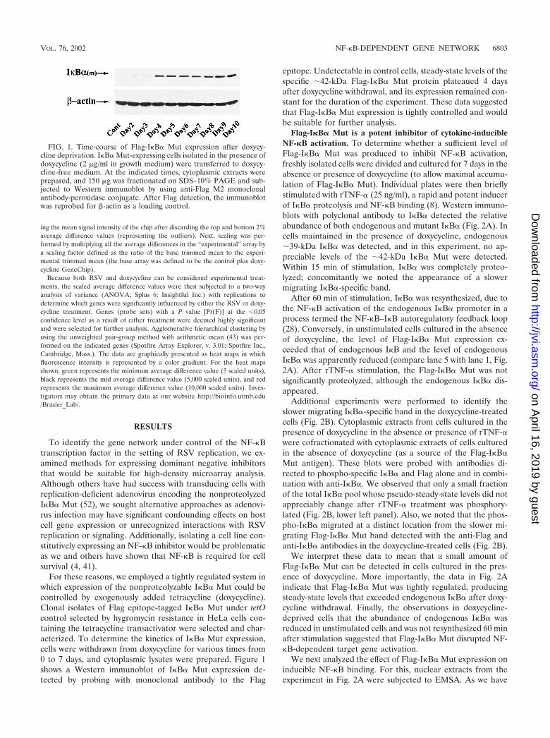
Flag-I�B� Mut is a potent inhibitor of cytokine-inducibleNF-�B activation. To determine whether a sufficient level ofFlag-I�B� Mut was produced to inhibit NF-�B activation,freshly isolated cells were divided and cultured for 7 days in theabsence or presence of doxycycline (to allow maximal accumu-lation of Flag-I�B� Mut). Individual plates were then brieflystimulated with rTNF-� (25 ng/ml), a rapid and potent inducerof I�B� proteolysis and NF-�B binding (8). Western immuno-blots with polyclonal antibody to I�B� detected the relativeabundance of both endogenous and mutant I�B� (Fig. 2A). Incells maintained in the presence of doxycycline, endogenous�39-kDa I�B� was detected, and in this experiment, no ap-preciable levels of the �42-kDa I�B� Mut were detected.Within 15 min of stimulation, I�B� was completely proteo-lyzed; concomitantly we noted the appearance of a slowermigrating I�B�-specific band.
After 60 min of stimulation, I�B� was resynthesized, due tothe NF-�B activation of the endogenous I�B� promoter in aprocess termed the NF-�B–I�B autoregulatory feedback loop(28). Conversely, in unstimulated cells cultured in the absenceof doxycycline, the level of Flag-I�B� Mut expression ex-ceeded that of endogenous I�B and the level of endogenousI�B� was apparently reduced (compare lane 5 with lane 1, Fig.2A). After rTNF-� stimulation, the Flag-I�B� Mut was notsignificantly proteolyzed, although the endogenous I�B� dis-appeared.
Additional experiments were performed to identify theslower migrating I�B�-specific band in the doxycycline-treatedcells (Fig. 2B). Cytoplasmic extracts from cells cultured in thepresence of doxycycline in the absence or presence of rTNF-�were cofractionated with cytoplasmic extracts of cells culturedin the absence of doxycycline (as a source of the Flag-I�B�Mut antigen). These blots were probed with antibodies di-rected to phospho-specific I�B� and Flag alone and in combi-nation with anti-I�B�. We observed that only a small fractionof the total I�B� pool whose pseudo-steady-state levels did notappreciably change after rTNF-� treatment was phosphory-lated (Fig. 2B, lower left panel). Also, we noted that the phos-pho-I�B� migrated at a distinct location from the slower mi-grating Flag-I�B� Mut band detected with the anti-Flag andanti-I�B� antibodies in the doxycycline-treated cells (Fig. 2B).
We interpret these data to mean that a small amount ofFlag-I�B� Mut can be detected in cells cultured in the pres-ence of doxycycline. More importantly, the data in Fig. 2Aindicate that Flag-I�B� Mut was tightly regulated, producingsteady-state levels that exceeded endogenous I�B� after doxy-cycline withdrawal. Finally, the observations in doxycycline-deprived cells that the abundance of endogenous I�B� wasreduced in unstimulated cells and was not resynthesized 60 minafter stimulation suggested that Flag-I�B� Mut disrupted NF-�B-dependent target gene activation.
We next analyzed the effect of Flag-I�B� Mut expression oninducible NF-�B binding. For this, nuclear extracts from theexperiment in Fig. 2A were subjected to EMSA. As we have
FIG. 1. Time-course of Flag-I�B� Mut expression after doxycy-cline deprivation. I�B� Mut-expressing cells isolated in the presence ofdoxycycline (2 g/ml in growth medium) were transferred to doxycy-cline-free medium. At the indicated times, cytoplasmic extracts wereprepared, and 150 g was fractionated on SDS–10% PAGE and sub-jected to Western immunoblot by using anti-Flag M2 monoclonalantibody-peroxidase conjugate. After Flag detection, the immunoblotwas reprobed for �-actin as a loading control.
VOL. 76, 2002 NF-�B-DEPENDENT GENE NETWORK 6803
on April 16, 2019 by guest
http://jvi.asm.org/
Dow
nloaded from
described before, rTNF-� is a potent and rapid inducer ofnuclear translocation and DNA binding of the NF-�B het-erodimer RelA/NF-�B1(p50), whose characteristic migrationcould be distinguished from constitutive NF-�B isoforms bynondenaturing electrophoresis (8, 21, 34). The kinetics ofRelA/NF-�B1 DNA binding exactly parallels the pattern ofI�B� degradation in cells cultured in the presence of doxycy-cline (Fig. 2C). In contrast, no inducible RelA/NF-�B1 com-plexes could be detected in nuclear extracts from cells culturedin the absence of doxycycline (Fig. 2C).
Confirmation of the identity of the inducible RelA/NF-�B1complexes was determined in competition analysis in EMSA(Fig. 2D). Here, the inclusion of unlabeled wild-type probestrongly competed for inducible RelA/NF-�B1 DNA binding,whereas the mutant did not. The constitutive NF-�B1 ho-modimer (p50)2 competes with lower affinity, being partly in-hibited with the mutant probe (9). Supershift analysis in
EMSA was further used to identify the presence of RelA in therTNF-�-inducible NF-�B binding complex. Addition of anti-RelA or anti-p50 completely disrupted the RelA/p50 complexand produced a corresponding supershift (Fig. 2E). These dataare consistent with our earlier demonstrations that the pre-dominant rTNF-�-inducible NF-�B complex is composed ofRelA/NF-�B1 in epithelial cells (8, 21). Together, these dataindicate that Flag-I�B� Mut completely blocks inducibleRelA/NF-�B1 binding in response to the potent cytokineTNF-�.
Flag-I�B� Mut is a potent inhibitor of RSV-inducibleNF-�B activation. Although the Ser32Ala/Ser36Ala site muta-tion is a potent inhibitor of cytokine-inducible I�B� proteolysisthrough the IKK-proteasome pathway, we have observed sig-nificant differences between RSV- and cytokine-inducibleI�B� proteolysis. For example, the kinetics of RSV-inducibleI�B� degradation is distinctly slower than that of rTNF-�
FIG. 2. Effect of doxycycline withdrawal on rTNF-�-induced I�B� proteolysis and NF-�B binding. (A) Time course of I�B� degradation afterrTNF-� stimulation. Flag-I�B� Mut-expressing cells were cultured with or without doxycycline for 7 days. The cells were then treated without (lane0) or with rTNF-� (25 ng/ml) for the indicated times (minutes). Top panel, cytoplasmic extracts of the cells were prepared and subjected to Westernimmunoblot with anti-I�B� antibody. �, migration of phospho-I�B� and Flag-I�B� Mut; ---, migration of endogenous I�B�. Bottom panel, similarprotein loading was confirmed by stripping the blot and reprobing with anti-�-actin. (B) Analysis of I�B� isoforms in doxycycline-treated cells.Cytoplasmic lysates corresponding to lanes 1, 2, and 5 from panel A were fractionated and probed with anti-phospho-specific I�B� and anti-Flagalone and in combination with anti-I�B�. Although the Flag-I�B� Mut protein migrated slower than the native I�B�, it was clearly separable fromphospho-I�B� and represents the slower migrating band observed in A. (C) Time course of NF-�B binding. Sucrose cushion-purified nuclearextracts were prepared from the same experiment and subjected to EMSA by using 32P-labeled APRE WT duplex oligonucleotide probe. Thecomplexes were fractionated on 6% native polyacrylamide gels; an autoradiographic exposure is shown. The relative migration of the RelA/NF-�B1(p50) heterodimer and the p50 homodimer (p50)2 complexes are indicated (34). (D) Competition assay in EMSA. Nuclear extracts fromrTNF-�-stimulated cells (15 min, cultured in the presence of doxycycline) were subjected to competition analysis by the addition of a molar excessof nonradioactive double-stranded oligonucleotide competitor at the time of addition of radioactive probe. Competitors used were APRE WT andAPRE M2 (Mut). (E) Antibody supershift in EMSA. Nuclear extracts from rTNF-�-stimulated cells (15 min, plus doxycycline) were mixed with1 l of affinity-purified polyclonal antibodies for 1 h at 4°C prior to EMSA analysis. PI, preimmune IgG. Bottom panel, brief autoradiographicexposure of the native NF-�B binding complexes. Top panel, longer exposure to detect the supershifted complexes (indicated by asterisk). Additionof either anti-RelA or anti-NF-�B1(p50) antibodies completely disrupted the RelA/NF-�B1(p50) complex.
6804 TIAN ET AL. J. VIROL.
on April 16, 2019 by guest
http://jvi.asm.org/
Dow
nloaded from
(being detectable after 12 to 24 h) and is only weakly blockedby pretreatment with specific proteasome inhibitors, and nosignificant I�B� resynthesis occurs (34). Consequently, we de-termined whether Flag-I�B� Mut would interfere with RSV-inducible NF-�B activation.
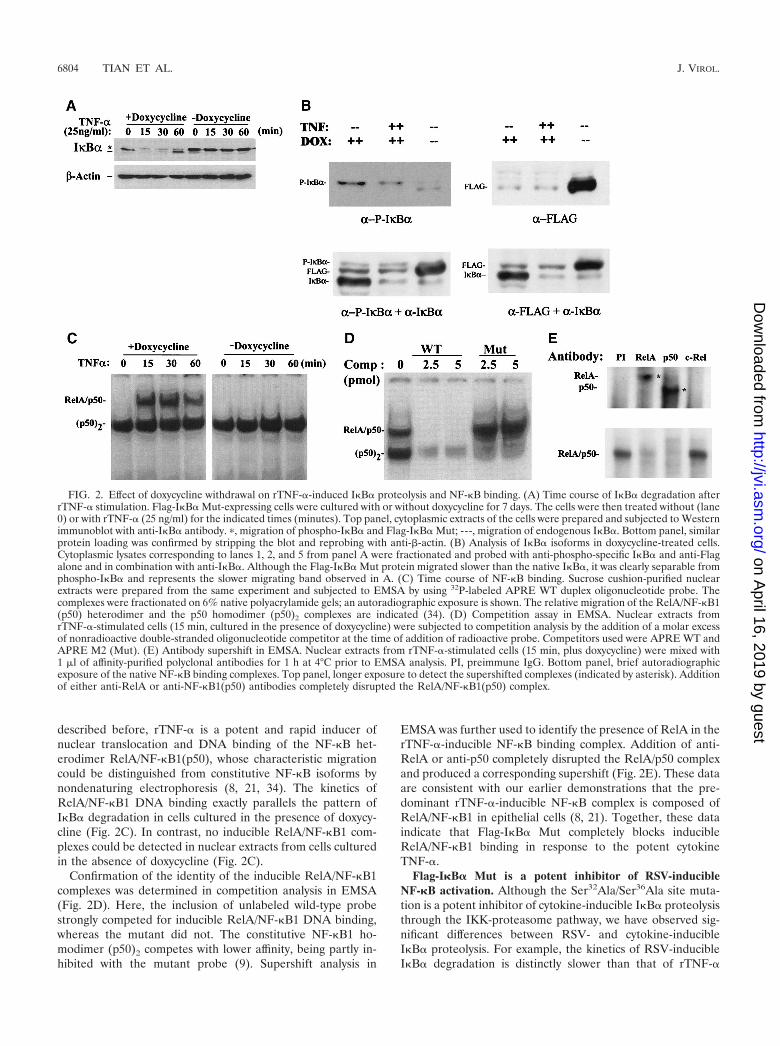
Freshly isolated Flag-I�B� Mut-expressing cells were di-vided and cultured for 7 days in the absence or presence ofdoxycycline. Individual plates were then infected with sucrosecushion-purified RSV (multiplicity of infection, 1), after whichcytoplasmic and nuclear extracts were prepared. Western im-munoblots of the cytoplasmic extracts for I�B� detected afourfold greater relative abundance of endogenous I�B� rel-ative to that of the Flag-I�B� Mut (Fig. 3A). After RSVinfection, a significant amount of I�B� proteolysis was de-tected 24 h later, a time course consistent with our earlierdescriptions (34). In cells maintained in the presence of doxy-cycline, like that seen in Fig. 2, loss of endogenous �39-kDaI�B� was noted, and no significant degradation of the Flag-I�B� Mut occurred at 24 h. These data suggest that RSVdegrades Flag-I�B� Mut less efficiently than the endogenousform.
Nuclear extracts were subjected to EMSA to detect theeffect of Flag-I�B� Mut expression on RSV-inducible NF-�Bbinding. In cells cultured in the presence of doxycycline, sig-nificant RelA/NF-�B1 DNA binding was observed at 12 h and
continued to increase in abundance 24 h after RSV infection(Fig. 3B). Conversely, RelA/NF-�B1 DNA binding was com-pletely abolished at all points in the cells cultured in the ab-sence of doxycycline (Fig. 3B). These data suggest that Flag-I�B� Mut is also a potent inhibitor of RSV-inducible RelA/NF-�B1 binding.
Flag-I�B� Mut blocks NF-�B-dependent promoter transac-tivation. We next determined whether Flag-I�B� Mut expres-sion was sufficient to interfere with NF-�B-dependent tran-scription. For this, several strategies were employed. We firstdetermined whether Flag-I�B� Mut expression interfered withinducible activity of a purely NF-�B-dependent reportergene. Multimeric RelA/NF-�B1 binding sites from eitherthe angiotensinogen (AGT) gene or the IL-6 gene clonedupstream of the inert rAGT minimal promoter driving fireflyluciferase [termed (APREWT)3-p59rAT/LUC and (IL-6�BE)3-p59rAT/LUC, respectively] (29, 35) were transfectedinto Flag-I�B� Mut-expressing cells maintained in the absenceor presence of doxycycline.
Figure 4A shows the results of a representative transfectionof (APREWT)3-p59rAT/LUC in the absence or presence ofacute stimulation with rTNF-�. In the presence of doxycycline,rTNF-� induced a 22-fold increase in normalized luciferasereporter activity (P � 0.05, n 3 independent experiments);however, in the absence of doxycycline, no statistically signifi-cant induction could be recorded (a 1.1-fold increase). Similarresults were obtained for the (IL-6�BE)3-p59rAT/LUC re-porter activity, in which an 8.2-fold increase was observedrelative to the control in the presence of doxycycline and a1.3-fold increase was observed in its absence (Fig. 4B).
Because (APREWT)3-p59rAT/LUC and (IL-6�BE)3-p59rAT/LUC are artificial constructs taken from genes not normallyexpressed by HeLa cells, we determined if Flag-I�B� Mutexpression would block transcriptional activation of a nativechemokine gene by testing its effect on the IL-8 promoter, ahighly RSV-inducible gene in epithelial cells. We selected thistarget because we have previously demonstrated that rTNF-�activates hIL-8 transcription in a manner requiring RelA/NF-�B1 binding to a site in its proximal promoter (8). Flag-I�B�Mut-expressing cells maintained in the absence or presence ofdoxycycline were transfected with �162/�44 hIL-8/LUC, andreporter activity was determined after rTNF-� stimulation.Flag-I�B� Mut expression completely inhibited reporter activ-ity driven by �162/�44 hIL-8/LUC (Fig. 4C).
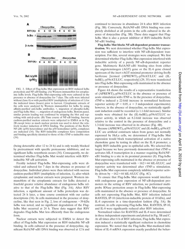
To ensure that Flag-I�B� Mut expression would interferewith endogenous gene expression of NF-�B-dependent pro-moters in the setting of RSV infection, we performed multi-probe RNase protection assays in Flag-I�B� Mut-expressingcells maintained in the absence or presence of doxycycline. Incells not expressing Flag-I�B� Mut, RSV infection inducedRANTES, IP-10 (interferon-inducible protein of 10 kDa), andIL-8 expression in a time-dependent fashion (Fig. 5A). Bycontrast, in cells expressing Flag-I�B� Mut, RANTES, IP-10,and IL-8 were significantly reduced after RSV infection.
Normalized values for RANTES and IL-8 were determinedin three independent experiments and plotted in Fig. 5B and C.At all times after 6 h of RSV infection, Flag-I�B� Mut expres-sion induced a statistically significant reduction in chemokineexpression. We noted that the Flag-I�B� Mut-mediated inhi-bition of IL-8 mRNA expression exactly paralleled the behav-
FIG. 3. Effect of Flag-I�B� Mut expression on RSV-induced I�B�proteolysis and NF-�B binding. (A) Western immunoblot for cytoplas-mic I�B� levels. Flag-I�B� Mut-expressing cells were cultured in thepresence or absence of doxycycline for 7 days. The cells were infectedwithout (lane 0) or with purified RSV (multiplicity of infection of 1) forthe indicated times (hours) prior to harvest. Cytoplasmic extracts ofthe cells were analyzed by Western immunoblot for I�B� by usingaffinity-purified anti-I�B� antibodies. �, migration of phospho-I�B�and Flag-I�B� Mut; ---, migration of endogenous I�B�. Bottom panel,similar protein loading was confirmed by stripping the blot and rep-robing with anti-�-actin. (B) Time course of NF-�B binding. Sucrosecushion-purified nuclear extracts were subjected to EMSA as in Fig.2B except twice as much nuclear protein was used to detect the rela-tively weaker induction of DNA binding. The positions of the RelA/NF-�B1 (p50) heterodimer and the p50 homodimer (p50)2 complexesare indicated (34). The RSV-inducible complexes have compositionand binding specificity identical to those of the rTNF-�-inducible ones(34).
VOL. 76, 2002 NF-�B-DEPENDENT GENE NETWORK 6805
on April 16, 2019 by guest
http://jvi.asm.org/
Dow
nloaded from
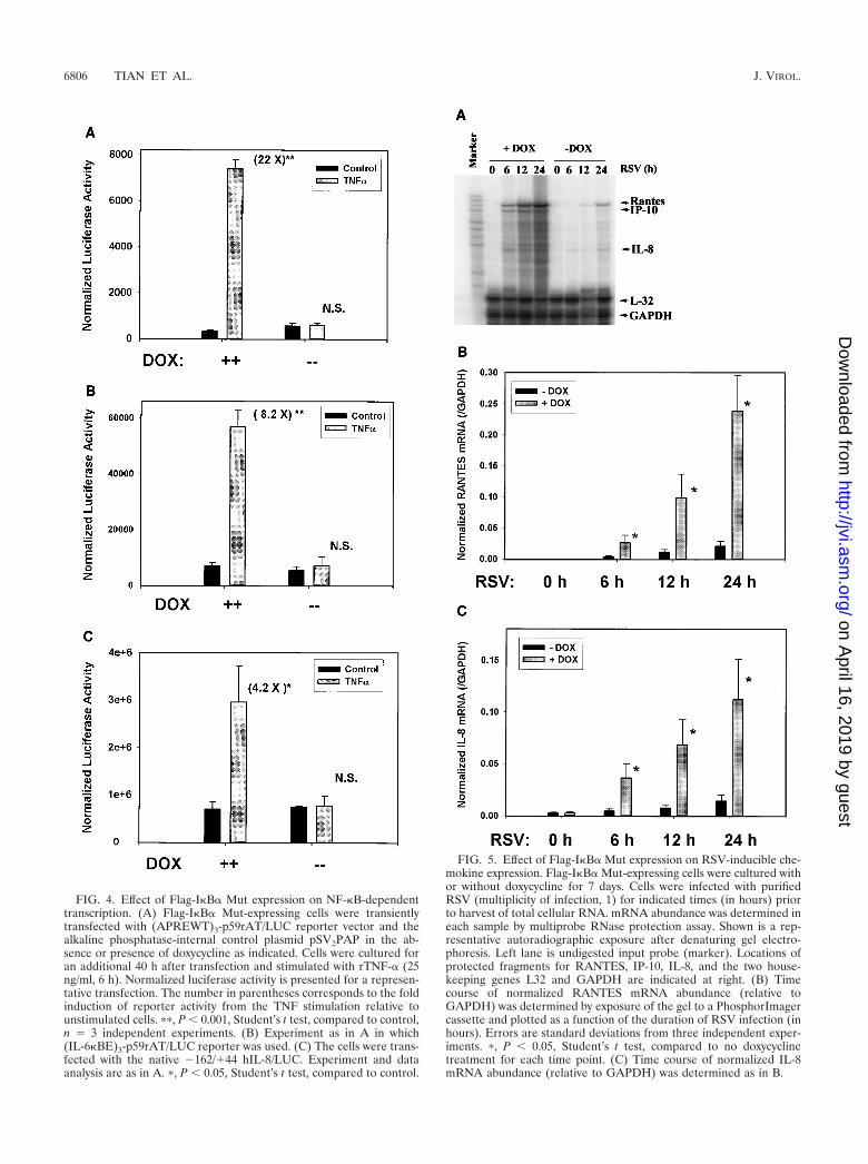
FIG. 4. Effect of Flag-I�B� Mut expression on NF-�B-dependenttranscription. (A) Flag-I�B� Mut-expressing cells were transientlytransfected with (APREWT)3-p59rAT/LUC reporter vector and thealkaline phosphatase-internal control plasmid pSV2PAP in the ab-sence or presence of doxycycline as indicated. Cells were cultured foran additional 40 h after transfection and stimulated with rTNF-� (25ng/ml, 6 h). Normalized luciferase activity is presented for a represen-tative transfection. The number in parentheses corresponds to the foldinduction of reporter activity from the TNF stimulation relative tounstimulated cells. ��, P � 0.001, Student’s t test, compared to control,n 3 independent experiments. (B) Experiment as in A in which(IL-6�BE)3-p59rAT/LUC reporter was used. (C) The cells were trans-fected with the native �162/�44 hIL-8/LUC. Experiment and dataanalysis are as in A. �, P � 0.05, Student’s t test, compared to control.
FIG. 5. Effect of Flag-I�B� Mut expression on RSV-inducible che-mokine expression. Flag-I�B� Mut-expressing cells were cultured withor without doxycycline for 7 days. Cells were infected with purifiedRSV (multiplicity of infection, 1) for indicated times (in hours) priorto harvest of total cellular RNA. mRNA abundance was determined ineach sample by multiprobe RNase protection assay. Shown is a rep-resentative autoradiographic exposure after denaturing gel electro-phoresis. Left lane is undigested input probe (marker). Locations ofprotected fragments for RANTES, IP-10, IL-8, and the two house-keeping genes L32 and GAPDH are indicated at right. (B) Timecourse of normalized RANTES mRNA abundance (relative toGAPDH) was determined by exposure of the gel to a PhosphorImagercassette and plotted as a function of the duration of RSV infection (inhours). Errors are standard deviations from three independent exper-iments. �, P � 0.05, Student’s t test, compared to no doxycyclinetreatment for each time point. (C) Time course of normalized IL-8mRNA abundance (relative to GAPDH) was determined as in B.
6806 TIAN ET AL. J. VIROL.
on April 16, 2019 by guest
http://jvi.asm.org/
Dow
nloaded from
ior of the native IL-8 promoter in transient transfection assays(Fig. 4C). These data suggest that the cell system expressingFlag-I�B� Mut was indeed tightly regulated and could be usedto block RSV-inducible NF-�B-dependent expression of genenetworks.
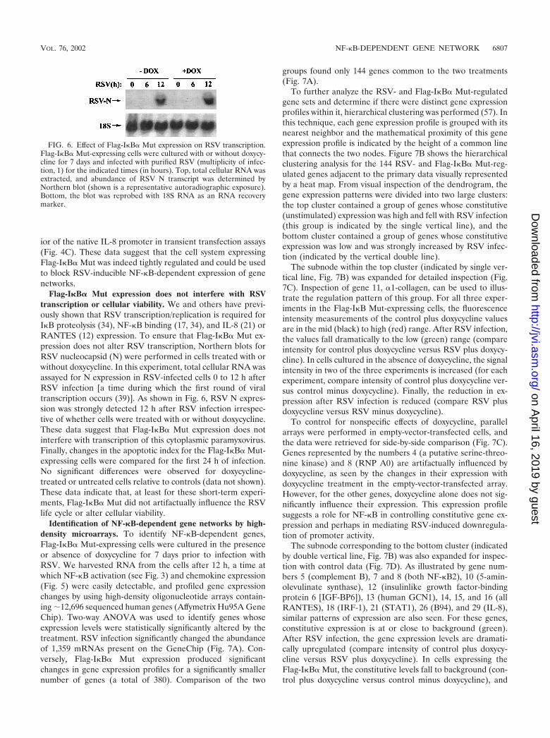
Flag-I�B� Mut expression does not interfere with RSVtranscription or cellular viability. We and others have previ-ously shown that RSV transcription/replication is required forI�B proteolysis (34), NF-�B binding (17, 34), and IL-8 (21) orRANTES (12) expression. To ensure that Flag-I�B� Mut ex-pression does not alter RSV transcription, Northern blots forRSV nucleocapsid (N) were performed in cells treated with orwithout doxycycline. In this experiment, total cellular RNA wasassayed for N expression in RSV-infected cells 0 to 12 h afterRSV infection [a time during which the first round of viraltranscription occurs (39)]. As shown in Fig. 6, RSV N expres-sion was strongly detected 12 h after RSV infection irrespec-tive of whether cells were treated with or without doxycycline.These data suggest that Flag-I�B� Mut expression does notinterfere with transcription of this cytoplasmic paramyxovirus.Finally, changes in the apoptotic index for the Flag-I�B� Mut-expressing cells were compared for the first 24 h of infection.No significant differences were observed for doxycycline-treated or untreated cells relative to controls (data not shown).These data indicate that, at least for these short-term experi-ments, Flag-I�B� Mut did not artifactually influence the RSVlife cycle or alter cellular viability.
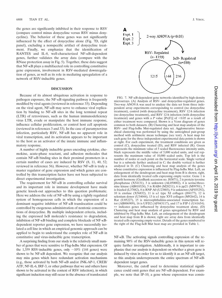
Identification of NF-�B-dependent gene networks by high-density microarrays. To identify NF-�B-dependent genes,Flag-I�B� Mut-expressing cells were cultured in the presenceor absence of doxycycline for 7 days prior to infection withRSV. We harvested RNA from the cells after 12 h, a time atwhich NF-�B activation (see Fig. 3) and chemokine expression(Fig. 5) were easily detectable, and profiled gene expressionchanges by using high-density oligonucleotide arrays contain-ing �12,696 sequenced human genes (Affymetrix Hu95A GeneChip). Two-way ANOVA was used to identify genes whoseexpression levels were statistically significantly altered by thetreatment. RSV infection significantly changed the abundanceof 1,359 mRNAs present on the GeneChip (Fig. 7A). Con-versely, Flag-I�B� Mut expression produced significantchanges in gene expression profiles for a significantly smallernumber of genes (a total of 380). Comparison of the two
groups found only 144 genes common to the two treatments(Fig. 7A).
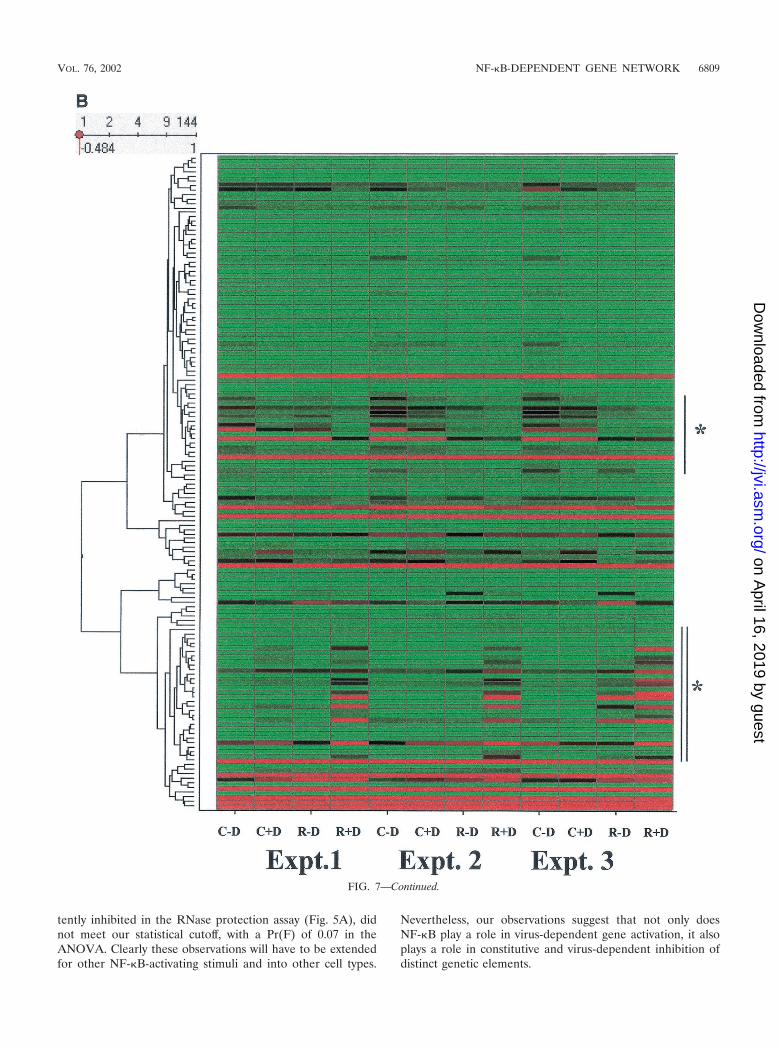
To further analyze the RSV- and Flag-I�B� Mut-regulatedgene sets and determine if there were distinct gene expressionprofiles within it, hierarchical clustering was performed (57). Inthis technique, each gene expression profile is grouped with itsnearest neighbor and the mathematical proximity of this geneexpression profile is indicated by the height of a common linethat connects the two nodes. Figure 7B shows the hierarchicalclustering analysis for the 144 RSV- and Flag-I�B� Mut-reg-ulated genes adjacent to the primary data visually representedby a heat map. From visual inspection of the dendrogram, thegene expression patterns were divided into two large clusters:the top cluster contained a group of genes whose constitutive(unstimulated) expression was high and fell with RSV infection(this group is indicated by the single vertical line), and thebottom cluster contained a group of genes whose constitutiveexpression was low and was strongly increased by RSV infec-tion (indicated by the vertical double line).
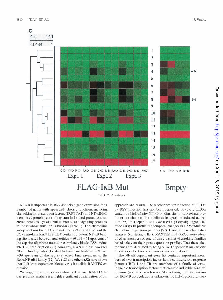
The subnode within the top cluster (indicated by single ver-tical line, Fig. 7B) was expanded for detailed inspection (Fig.7C). Inspection of gene 11, �1-collagen, can be used to illus-trate the regulation pattern of this group. For all three exper-iments in the Flag-I�B Mut-expressing cells, the fluorescenceintensity measurements of the control plus doxycycline valuesare in the mid (black) to high (red) range. After RSV infection,the values fall dramatically to the low (green) range (compareintensity for control plus doxycycline versus RSV plus doxycy-cline). In cells cultured in the absence of doxycycline, the signalintensity in two of the three experiments is increased (for eachexperiment, compare intensity of control plus doxycycline ver-sus control minus doxycycline). Finally, the reduction in ex-pression after RSV infection is reduced (compare RSV plusdoxycycline versus RSV minus doxycycline).
To control for nonspecific effects of doxycycline, parallelarrays were performed in empty-vector-transfected cells, andthe data were retrieved for side-by-side comparison (Fig. 7C).Genes represented by the numbers 4 (a putative serine-threo-nine kinase) and 8 (RNP A0) are artifactually influenced bydoxycycline, as seen by the changes in their expression withdoxycycline treatment in the empty-vector-transfected array.However, for the other genes, doxycycline alone does not sig-nificantly influence their expression. This expression profilesuggests a role for NF-�B in controlling constitutive gene ex-pression and perhaps in mediating RSV-induced downregula-tion of promoter activity.
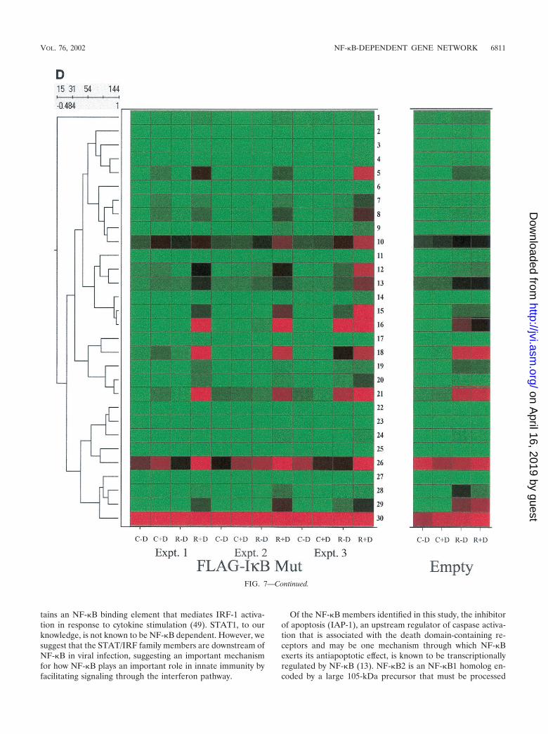
The subnode corresponding to the bottom cluster (indicatedby double vertical line, Fig. 7B) was also expanded for inspec-tion with control data (Fig. 7D). As illustrated by gene num-bers 5 (complement B), 7 and 8 (both NF-�B2), 10 (5-amin-olevulinate synthase), 12 (insulinlike growth factor-bindingprotein 6 [IGF-BP6]), 13 (human GCN1), 14, 15, and 16 (allRANTES), 18 (IRF-1), 21 (STAT1), 26 (B94), and 29 (IL-8),similar patterns of expression are also seen. For these genes,constitutive expression is at or close to background (green).After RSV infection, the gene expression levels are dramati-cally upregulated (compare intensity of control plus doxycy-cline versus RSV plus doxycycline). In cells expressing theFlag-I�B� Mut, the constitutive levels fall to background (con-trol plus doxycycline versus control minus doxycycline), and
FIG. 6. Effect of Flag-I�B� Mut expression on RSV transcription.Flag-I�B� Mut-expressing cells were cultured with or without doxycy-cline for 7 days and infected with purified RSV (multiplicity of infec-tion, 1) for the indicated times (in hours). Top, total cellular RNA wasextracted, and abundance of RSV N transcript was determined byNorthern blot (shown is a representative autoradiographic exposure).Bottom, the blot was reprobed with 18S RNA as an RNA recoverymarker.
VOL. 76, 2002 NF-�B-DEPENDENT GENE NETWORK 6807
on April 16, 2019 by guest
http://jvi.asm.org/
Dow
nloaded from
the genes are significantly inhibited in their response to RSV(compare control minus doxycycline versus RSV minus doxy-cycline). The behavior of these genes was not significantlyinfluenced by the effect of doxycycline alone (Fig. 7D, rightpanel), excluding a nonspecific artifact of doxycycline treat-ment. Finally, we emphasize that the identification ofRANTES and IL-8, well-characterized NF-�B-dependentgenes, further validates the array data (compare with theRNase protection assay in Fig. 5). Together, these data suggestthat NF-�B plays a multifaceted role in controlling constitutivegene expression, involvement in RSV-mediated downregula-tion of genes, as well as its role in mediating upregulation of anetwork of RSV-inducible genes.
DISCUSSION
Because of its almost ubiquitous activation in response topathogen exposure, the NF-�B signaling pathway is frequentlymodified by viral agents (reviewed in reference 33). Dependingon the viral agent, NF-�B may serve to enhance viral replica-tion by binding to NF-�B sites in the long terminal repeat(LTR) of retroviruses, such as the human immunodeficiencyvirus LTR, evade or manipulate the host immune response,influence cellular proliferation, or control host cell apoptosis(reviewed in references 3 and 33). In the case of paramyxovirusinfection, particularly RSV, NF-�B has no apparent role inviral transcription, and its activation appears to be importantto the host as an activator of the innate immune and inflam-matory response.
A number of highly inducible genes encoding cytokine, che-mokine, acute-phase reactant, and adhesion molecules thatcontain NF-�B binding sites in their proximal promoters in acertain number of cases are induced by RSV (8, 11, 40, 52;reviewed in reference 56). However, whether NF-�B is truly amaster regulator of gene expression and which genes are con-trolled by this transcription factor have not been subjected todirect experimental investigation.
The requirement for NF-�B in controlling cellular survivaland its important role in immune development have madegenetic knock-out approaches to this question problematic.Here we address the role of NF-�B by using a tightly regulatedsystem of homogeneous cells in which the expression of adominant negative inhibitor of NF-�B translocation could becontrolled by exogenous administration of nontoxic concentra-tions of doxycycline. By multiple independent criteria, includ-ing the expressed I�B molecule’s resistance to degradation,inhibition of NF-�B binding and complete blockade of NF-�B-dependent reporter gene expression, we have successfully iso-lated a cell line in which an empirical genomic approach can beapplied to begin to understand the complex role of NF-�B inconstitutive and virus-inducible gene transcription.
A surprising finding from our study is the relatively small num-ber of genes that were sensitive to Flag-I�B� Mut expression. Ofthe 1,359 RSV-inducible genes, only �10% (144 genes) wereshown to be NF-�B dependent. We recognize that our analysismay miss genes which have redundant activation mechanisms(e.g., those activated by both NF-�B and/or JNK-AP-1, CREB/ATF, NF-IL-6, IRF-3, or other pathways that we and others haveshown to be activated in the context of RSV infection), in whichsignificant induction may still occur in the absence of translocated
NF-�B. The activating signals controlling expression of the re-maining 90% of the RSV-inducible genes in this system will re-quire further investigation. Additionally, it is important to em-phasize that our analysis is dependent on whether a given gene isinduced by virus in order for us to identify it as an NF-�B target,so this analysis underrepresents the entire spectrum of NF-�B-dependent target genes.
Moreover, the cutoff used to determine statistical signifi-cance could omit genes that are NF-�B dependent. For exam-ple, we note that IP-10, a gene whose expression was consis-
FIG. 7. NF-�B-dependent gene networks identified by high-densitymicroarrays. (A) Analysis of RSV- and doxycycline-regulated genes.Two-way ANOVA was used to analyze the data set from three inde-pendent array experiments corresponding to control (no doxycyclinetreatment), control (with doxycycline treatment), RSV 12-h infection(no doxycycline treatment), and RSV 12-h infection (with doxycyclinetreatment) and genes with a P value [Pr(F)] of �0.05 as a result ofeither treatment were compared. Shown is a Venn diagram of genescommon to both datasets. (B) Clustering and heat map analysis of theRSV- and Flag-I�B� Mut-regulated data set. Agglomerative hierar-chical clustering was performed by using the unweighted pair-groupmethod with arithmetic mean technique (see text). A heat map foreach gene for the three independent experimental data points is shownat right. For each experiment, the treatment conditions are given ascontrol (C), doxycycline treated (D), and RSV infected (R). Greenrepresents the minimum value of 5 scaled fluorescence intensity units,black represents the middle value of 5,000 scaled units, and red rep-resents the maximum value of 10,000 scaled units. Top left is thenumber of nodes at each point on the horizontal scale. Single verticalbar is a subnode further analyzed in C; the double vertical is furtheranalyzed in D. (C) Clustering and heat map analysis of the geneswhose constitutive expression is influenced by Flag-I�B� Mut. Left, anenlargement of the dendrogram and heat map from B is shown; right,data from identically treated cells expressing empty vector. Gene 1 isgrowth hormone (GenBank accession no. J03071), 2 is protein tyrosinephosphatase (X54131), 3 is unknown (AL050002), 4 is serine/threo-nine kinase (AB018324), 5 is RAB4 (M28211), 6 is pp21 (M99701), 7is frizzled (L37882), 8 is RNP A0 (U23803), 9 is unknown (AF035292),10 is enolase (X56832), 11 is �1 type XI collagen (J04177), 12 isselenium donor (U34044), 13 is �1 type XVI collagen (M92642), 14 isRar (U05227), 15 is microophthalmia-associated transcription fac-tor (AB006909), 16 is UFD2 (AF043117), and 17 is FIP-1 (U41654).�� indicates genes influenced by doxycycline treatment alone. (D)Clustering and heat map analysis of genes upregulated by RSV andinhibited by Flag-I�B� Mut. Left, an enlargement of the dendrogramand heat map from B is shown; right are array data from identicallytreated cells expressing empty vector. Details of the genes numbered tothe right of the Flag-I�B Mut heat map are provided in Table 1.
6808 TIAN ET AL. J. VIROL.
on April 16, 2019 by guest
http://jvi.asm.org/
Dow
nloaded from
tently inhibited in the RNase protection assay (Fig. 5A), didnot meet our statistical cutoff, with a Pr(F) of 0.07 in theANOVA. Clearly these observations will have to be extendedfor other NF-�B-activating stimuli and into other cell types.
Nevertheless, our observations suggest that not only doesNF-�B play a role in virus-dependent gene activation, it alsoplays a role in constitutive and virus-dependent inhibition ofdistinct genetic elements.
FIG. 7—Continued.
VOL. 76, 2002 NF-�B-DEPENDENT GENE NETWORK 6809
on April 16, 2019 by guest
http://jvi.asm.org/
Dow
nloaded from
NF-�B is important in RSV-inducible gene expression for anumber of genes with apparently diverse functions, includingchemokines, transcription factors (IRF/STATs and NF-�B/I�Bmembers), proteins controlling translation and proteolysis, se-creted proteins, cytoskeletal elements, and signaling proteins,in those whose function is known (Table 1). The chemokinegroup contains the CXC chemokines GRO� and IL-8 and theCC chemokine RANTES. IL-8 contains a potent NF-�B bind-ing site located between nucleotides �80 and �71 upstream ofthe cap site (8) whose mutation completely blocks RSV-induc-ible IL-8 transcription (21). Similarly, RANTES has two suchNF-�B binding sites (located between nucleotides �71 and�39 upstream of the cap site) which bind members of theRelA/NF-�B1 family (12). We (12) and others (52) have shownthat I�B Mut expression blocks virus-inducible RANTES ex-pression.
We suggest that the identification of IL-8 and RANTES byour genomic analysis is a highly significant confirmation of our
approach and results. The mechanism for induction of GRO�by RSV infection has not been reported; however, GRO�contains a high-affinity NF-�B binding site in its proximal pro-moter, an element that mediates its cytokine-induced activa-tion (55). In a separate study we used high-density oligonucle-otide arrays to profile the temporal changes in RSV-induciblechemokine expression patterns (57). Using similar informaticsanalyses (clustering), IL-8, RANTES, and GRO� were iden-tified as members of one of three distinct chemokine familiesbased solely on their gene expression profiles. That these che-mokines are all related by being NF-�B dependent may be oneexplanation for their common expression pattern.
The NF-�B-dependent gene list contains important mem-bers of two transcription factor families. Interferon responsefactors (IRF) 1 and 7B are members of a family of virus-inducible transcription factors that mediate inducible gene ex-pression (reviewed in reference 51). Although the mechanismfor IRF-7B upregulation is unknown, the IRF-1 promoter con-
FIG. 7—Continued.
6810 TIAN ET AL. J. VIROL.
on April 16, 2019 by guest
http://jvi.asm.org/
Dow
nloaded from
tains an NF-�B binding element that mediates IRF-1 activa-tion in response to cytokine stimulation (49). STAT1, to ourknowledge, is not known to be NF-�B dependent. However, wesuggest that the STAT/IRF family members are downstream ofNF-�B in viral infection, suggesting an important mechanismfor how NF-�B plays an important role in innate immunity byfacilitating signaling through the interferon pathway.
Of the NF-�B members identified in this study, the inhibitorof apoptosis (IAP-1), an upstream regulator of caspase activa-tion that is associated with the death domain-containing re-ceptors and may be one mechanism through which NF-�Bexerts its antiapoptotic effect, is known to be transcriptionallyregulated by NF-�B (13). NF-�B2 is an NF-�B1 homolog en-coded by a large 105-kDa precursor that must be processed
FIG. 7—Continued.
VOL. 76, 2002 NF-�B-DEPENDENT GENE NETWORK 6811
on April 16, 2019 by guest
http://jvi.asm.org/
Dow
nloaded from
into its 50-kDa binding form; although regulation of NF-�B2expression has not been studied, we suggest that there may beNF-�B binding sites in its proximal promoter.
A well-established NF-�B-dependent target is the I�B in-hibitors themselves. Activation of the I�B members is an au-toregulatory feedback loop in which NF-�B induces the syn-thesis of its own inhibitor to terminate its action. Previously weshowed that the NF-�B-I�B� autoregulatory loop was im-paired in RSV-infected cells; this finding explains why I�B�was not identified in the present analysis (34). I�B�, in contrastto I�B�, is strongly upregulated by RSV infection and suggestsstimulus-specific differences in the NF-�B-dependent expres-sion control of individual I�B subunits. Notwithstanding, theinduction of I�B� may compensate for a relative deficiency inI�B�; the existence of multiple independent NF-�B-I�B andNF-�B-BCL-3 inhibitory loops suggests that unregulatedNF-�B activation is highly deleterious (9, 28, 34).
Although there is strong bias towards a role of NF-�B inmediating chemokine and acute-phase reactant gene expres-sion, we have identified a number of other highly NF-�B-dependent genes which do not easily fit into these categories(Table 1). The functional consequences of enhanced expres-sion of 5�-aminolevulinate synthase, a rate-limiting enzyme in
heme biosynthesis, are unknown to us but suggest a role forNF-�B in virus-regulated heme metabolism in nonerythroidcells. Conversely, the NF-�B dependence of the E2 ubiquitin-conjugating enzyme suggests that NF-�B activation may havean important role in determining cellular capacity to breakdown proteins regulated through the ubiquitin-proteasomepathway, a process important in cell surface presentation ofviral antigens in the context of MHC class I molecules.
We were surprised to identify the human homolog of theSaccharomyces cerevisiae GCN1 gene, whose protein productcontrols translational efficiency through modifying upstreamactivation of the eIF2 protein kinase (42). Gene expression andregulation studies of human GCN1 in the setting of viral in-fection have not been reported to our knowledge, althoughviral infections are known to profoundly influence translationalregulation. Complement factor B, a hepatic acute-phase re-sponse factor important in the alternative complement path-way, is well known to be NF-�B inducible (45); however, viralinduction of the alternative complement pathway and its rolein response to infection have not been investigated. The in-duction of IGF-BP6 suggests that virus-infected cells exertparacrine control on the mitogenic actions of insulin-likegrowth factor II (18). Perhaps IGF-BP6 expression is beneficial
TABLE 1. Genes in the RSV-regulated chemokine subnodea
Function Name Geneno.
GenBankaccession no. Locus Avg fold
change
Comparisonb
A549 SAE
Chemokines GRO� 17 X54489 4q21 5.5 I NCIL-8 28 M17017 4q13-q21 7.4 I IIL-8 29 M28130 4q13-q21 22.7 I IRANTES 14 M21121 17q11.2-q12 5.5 I NCRANTES 15 M21121 17q11.2-q12 30.6 I IRANTES 16 M21121 17q11.2-q12 53 I I
Interferon signaling IRF-1 18 M87503 14q11.2 10.5 I NCIRF-7B 19 U53831 11p15.5. 3.5 I ISTAT1 21 M97936 2q32.2 4.6 I I
Metabolism Cholesterol 25-hydroxylase 20 AF059214 10q23 10 I NCCytochrome P-450 23 X55764 8q21 3.3 NC NC
NF-�B signaling IAP-1 2 U45878 11q22 1.8 I INF-�B2 7 X61498 10q24 4 I INF-�B2 8 S76638 4.3 I INF-�B2 9 S76638 6.5 I II�Bε 24 U91616 6pter-p24.1 3.5 I I
Protein synthesis/turnover E2 Ubiquitin conjugating 11 AA883502 11q12 2.3 I IGCN1 13 D50919 9p24.1-q22.33 2.1 I I
Secreted proteins Complement B 5 L15702 6p21.3 16 I IIGF-BP6 12 M62402 12q13 4.9 I NC
Structural Membrane protein 1 AB015633 1 1.4 D NCNuclear phosphoprotein 4 L22342 2q37.1 3 I I
Tyrosine kinase Tyrosine phosphatase 3 M33684 20q13.1-q13.2 0.5 I NCOther 5-Aminolevulinate synthase 10 Y00451 3p21.1 1.6 I I
IL-15R� 6 AF035279 10p15-p14 3.6 I IGAGE-2c 22 U19143 Xp11.4-p11.2 1.8 NC NCCadherin-like 22 25 AF035300 20 1.3 I NCB94 26 M92357 14q32 1.7 I IUnknown 27 AF070549 7 2.5 I NCUnknown 30 W27517 11 1.1 NC NC
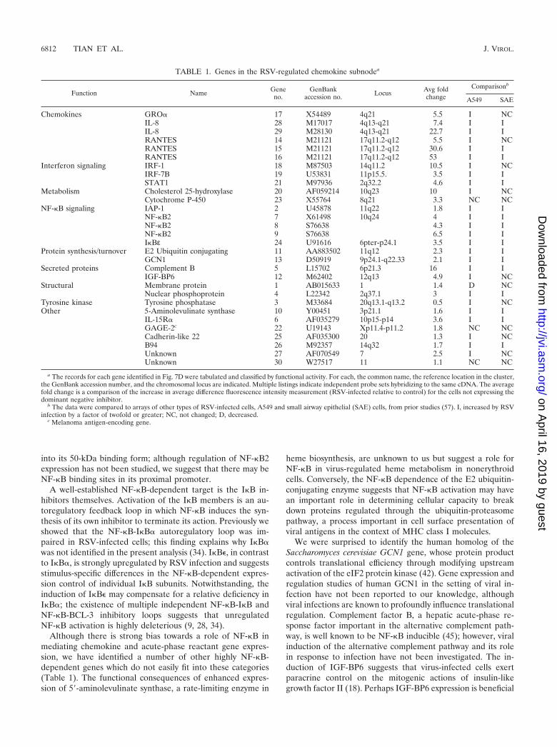
a The records for each gene identified in Fig. 7D were tabulated and classified by functional activity. For each, the common name, the reference location in the cluster,the GenBank accession number, and the chromosomal locus are indicated. Multiple listings indicate independent probe sets hybridizing to the same cDNA. The averagefold change is a comparison of the increase in average difference fluorescence intensity measurement (RSV-infected relative to control) for the cells not expressing thedominant negative inhibitor.
b The data were compared to arrays of other types of RSV-infected cells, A549 and small airway epithelial (SAE) cells, from prior studies (57). I, increased by RSVinfection by a factor of twofold or greater; NC, not changed; D, decreased.
c Melanoma antigen-encoding gene.
6812 TIAN ET AL. J. VIROL.
on April 16, 2019 by guest
http://jvi.asm.org/
Dow
nloaded from
to prevent local cellular proliferation in the presence of aninfecting viral agent.
Upregulation of the � subunit of the IL-15 receptor maysuggest that virus-infected cells have distinct signaling pheno-types to cytokines as a result of NF-�B action. This will requirefurther investigation. Finally, B94 is a protein of unknownfunction, perhaps important in angiogenesis, that was identi-fied by differential display to be a highly cytokine- and LPS-inducible transcript (54). We suggest that B94 is also a virus-inducible transcript in epithelial cells through an NF-�B-dependent mechanism.
Recent work suggests that NF-�B has genomic actions evenin the absence of exogenous stimuli. This “constitutive” NF-�Bactivation may be important to inhibit apoptosis (reviewed inreference 4). For example, NF-�B appears to be required tomaintain low levels of the Bcl-2 protein A1 at levels to preventloss of mitochondrial transmembrane potential and apoptosisin macrophages (48). Alternatively, constitutive NF-�B activitymay be important in cellular immortalization (2). Although ourexperimental design cannot distinguish these or other potentialroles for constitutive NF-�B activity, we interpret our data tomean that constitutive NF-�B appears to downregulate expres-sion of the collagen genes and others. The mechanism (tran-scriptional or posttranscriptional) by which NF-�B influencesthe abundance of these genes will require further investigation.In this regard, we note that a recent study has implicatedNF-�B in the posttranscriptional control of MyoD mRNAabundance (24); perhaps collagen is regulated in a similar way.
In this report, we have identified 380 genes which are con-trolled by NF-�B by using microarray analysis of a tetracycline-regulated expression system controlling expression of the non-proteolyzable I�B� inhibitor. These data suggest that NF-�Bregulates expression of distinct genetic networks of constitutivegenes whose expression is further inhibited by viral infection aswell as controlling a distinct subset of those that are virusinducible. Moreover, these data suggest that NF-�B is an up-stream regulator of RSV-inducible gene expression throughcontrolling expression of the proteins involved in interferonsignaling, STAT/IRF, perhaps providing insights into mecha-nisms for how NF-�B controls the innate immune response.
ACKNOWLEDGMENTS
We thank M. Gossen and H. Bujard (University of Heidelberg) fortTA plasmids and the UTMB Genomics Core Laboratory (T. Wood,Director) for performing the arrays.
This project was supported by grant R21 AI48163 from the NIAIDand in part by grants R01 AI40218 (to A.R.B.) and AI 15939 (toR.P.G.), Child Health and Human Development (R30HD 27841), andP30 ES06676 from the NIEHS (to R. S. Lloyd, UTMB).
REFERENCES
1. Aherne, W. T., T. Bird, S. D. B. Court, P. S. Gardner, and J. McQuillin. 1970.Pathological changes in virus infections of the lower respiratory tract inchildren. J. Clin. Pathol. 23:7–18.
2. Arsura, M., F. Mercurio, A. L. Oliver, S. S. Thorgeirsson, and G. E. Sonen-shein. 2000. Role of the I�B kinase complex in oncogenic Ras- and Raf-mediated transformation of rat liver epithelial cells. Mol. Cell. Biol. 20:5381–5391.
3. Baldwin, A. S. J. 2001. The transcription factor NF-�B and human disease.J. Clin. Investig. 107:3–6.
4. Barkett, M., and T. Gilmore. 1999. Control of apoptosis by Rel/NF-�Btranscription factors. Oncogene 18:6910–6924.
5. Barnes, P. J., and M. Karin. 1997. Nuclear factor-�B: a pivotal transcriptionfactor in chronic inflammatory diseases. N. Engl. J. Med. 336:1066–1071.
6. Becker, S., W. Reed, F. W. Henderson, and T. L. Noah. 1997. RSV infectionof human airway epithelial cells causes production of the �-chemokineRANTES. Am. J. Physiol. 272:L512–L520.
7. Beg, A. A., and A. S. J. Baldwin. 1993. The I�B proteins: multifunctionalregulators of Rel/NF-�B transcription factors. Genes Dev. 7:2064–2070.
8. Brasier, A. R., M. Jamaluddin, A. Casola, W. Duan, Q. Shen, and R. Garo-falo. 1998. A promoter recruitment mechanism for TNF-�-induced IL-8transcription in type II pulmonary epithelial cells: dependence on nuclearabundance of RelA, NF-�B1 and c-Rel transcription factors. J. Biol. Chem.273:3551–3561.
9. Brasier, A. R., M. Lu, T. Hai, Y. Lu, and I. Boldogh. 2001. NF-�B inducibleBCL-3 expression is an autoregulatory loop controlling nuclear p50/NF-�B1residence. J. Biol. Chem. 276:32080–32093.
10. Brown, K., S. Gerstberger, L. Carlson, G. Franzoso, and U. Siebenlist. 1995.Control of I�B-alpha proteolysis by site-specific, signal-induced phosphory-lation. Science 267:1485–1488.
11. Casola, A., N. Burger, T. Liu, M. Jamaluddin, A. R. Brasier, and R. P.Garofalo. 2001. Oxidant tone regulates RANTES gene expression in airwayepithelial cells infected with RSV. J. Biol. Chem. 276:19715–19722.
12. Casola, A., R. P. Garofalo, H. Haeberle, T. Elliott, M. Jamaluddin, and A. R.Brasier. 2000. Multiple inducible cis elements control RANTES promoteractivation in alveolar epithelial cells infected with respiratory syncytial virus.J. Virol. 75:6428–6439.
13. Erl, W., G. Hansson, R. de Martin, G. Draude, K. Weber, and C. Weber.1999. NF-�B regulates induction of apoptosis and inhibitor of apoptosisprotein-1 expression in vascular smooth muscle cells. Circ. Res. 84:668–677.
14. Everard, M. L., A. Swarbrick, M. Wrightham, J. McIntyre, C. Dunkley, P. D.James, H. F. Sewell, and A. D. Milner. 1994. Analysis of cells obtained bybronchial lavage of infants with respiratory syncytial virus infection. Arch.Dis. Child. 71:428–432.
15. Ferris, J. A., W. A. Aherne, and W. S. Locke. 1973. Sudden and unexpecteddeaths to infants: histology and virology. Br. Med. J. 2:439–449.
16. Fiedler, M. A., K. Wernke-Dollries, and J. M. Stark. 1995. Respiratorysyncytial virus increases IL-8 gene expression and protein release in A549cells. Am. J. Physiol. 269:L865–L872.
17. Fiedler, M. A., K. Wernke-Dollries, and J. M. Stark. 1996. Inhibition of viralreplication reverses respiratory syncytial virus-induced NF-�B activation andinterleukin-8 gene expression in A549 cells. J. Virol. 70:9079–9082.
18. Gabbitas, B., and E. Canalis. 1997. Growth factor regulation of insulin-likegrowth factor binding protein-6 expression in osteoblasts. J. Cell. Biochem.66:77–86.
19. Gardner, P. S., J. McQuillin, and S. D. Court. 1970. Speculation on patho-genesis of death from respiratory syncytial virus infection. Br. Med. J. 1:327–330.
20. Garofalo, R., J. L. L. Kimpen, R. C. Welliver, and P. L. Ogra. 1992. Eosin-ophil degranulation in the respiratory tract during naturally acquired respi-ratory syncytial virus infection. J. Pediatr. 120:28–32.
21. Garofalo, R., M. Sabry, M. Jamaluddin, R. K. Yu, A. Casola, P. L. Ogra, andA. R. Brasier. 1996. Transcriptional activation of the interleukin-8 gene byrespiratory syncytial virus infection in alveolar epithelial cells: nuclear trans-location of the RelA transcription factor as a mechanism producing airwaymucosal inflammation. J. Virol. 70:8773–8781.
22. Garofalo, R., R. C. Welliver, and P. L. Ogra. 1991. Concentrations of LTB4,LTC4, LTD4 and LTE4 in bronchiolitis due to respiratory syncytial virus.Pediatr. Allergy Immunol. 2:30–37.
23. Gossen, M., and H. Bujard. 1992. Tight control of gene expression in mam-malian cells by tetracycline-responsive promoters. Proc. Natl. Acad. Sci.USA 89:5547–5551.
24. Guttridge, D. C., M. W. Mayo, L. V. Madrid, C. Y. Wang, and A. S. Baldwin,Jr. 2000. NF-�B-induced loss of MyoD messenger RNA: possible role inmuscle decay and cachexia. Science 289:2363–2366.
25. Haeberle, H., A. Casola, Z. Gatalica, H.-J. Dieterich, P. B. Ernst, A. R.Brasier, and R. Garofalo. 2000. Respiratory syncytial virus-inducible activa-tion of the transcription factor NF-�B mediates pulmonary inflammation.Pediatr. Res. 47:17A.
26. Hall, C. B., and C. A. McCarthy. 1995. Respiratory syncytial virus, p. 1501–1519. In G. L. Mandel, J. E. Bennett, and R. Dolin (ed.), Principles andpractice of infectious diseases. Churchill Livingston, New York, N.Y.
27. Han, Y., and A. R. Brasier. 1997. Mechanism for biphasic RelA:NF-�B1nuclear translocation in tumor necrosis factor �-stimulated hepatocytes.J. Biol. Chem. 272:9823–9830.
28. Han, Y., T. Meng, N. R. Murray, A. P. Fields, and A. R. Brasier. 1999. IL-1induced NF-�B-I�Ba autoregulatory feedback loop in hepatocytes: a role forPKC� in posttranscriptional regulation of �B� resynthesis. J. Biol. Chem.274:939–947.
29. Han, Y., M. S. Runge, and A. R. Brasier. 1999. Angiotensin II induces IL-6transcription in vascular smooth muscle cells through pleiotropic activationof NF-�B transcription factors. Circ. Res. 84:695–703.
30. Han, Y., S. A. Weinman, S. Boldogh, and A. R. Brasier. 1999. TNF-�-inducible I�B� proteolysis and NF-�B activation mediated by cytosolic m-calpain. J. Biol. Chem. 274:787–794.
31. Harrison, A. M., C. A. Bonville, H. F. Rosenberg, and J. B. Domachowske.
VOL. 76, 2002 NF-�B-DEPENDENT GENE NETWORK 6813
on April 16, 2019 by guest
http://jvi.asm.org/
Dow
nloaded from
1999. Respiratory syncytial virus-induced chemokine expression in the lowerairways: eosinophil recruitment and degranulation. Am. J. Respir. Crit. CareMed. 159:1918–1924.
32. Henkel, T., T. Machleidt, I. Alkalay, M. Kronke, Y. Ben-Neriah, and P. A.Baeuerle. 1993. Rapid proteolysis of I�B-alpha is necessary for activation oftranscription factor NF-�B. Nature 365:182–185.
33. Hiscott, J., H. Kwon, and P. Genin. 2001. Hostile takeovers: viral appropri-ation of the NF-�B pathway. J. Clin. Investig. 107:143–151.
34. Jamaluddin, M., A. Casola, R. P. Garofalo, Y. Han, T. Elliott, P. L. Ogra,and A. R. Brasier. 1998. The major component of �B� proteolysis occursindependently of the proteasome pathway in respiratory syncytial virus-infected pulmonary epithelial cells. J. Virol. 72:4849–4857.
35. Jamaluddin, M., T. Meng, J. Sun, I. Boldogh, Y. Han, and A. R. Brasier.2000. Angiotensin II induces nuclear factor (NF)-�B1 isoforms to bind theangiotensinogen gene acute-phase response element: a stimulus-specificpathway for NF-�B activation. Mol. Endocrinol. 14:99–113.
36. Jamaluddin, M., S. Wang, R. Garofalo, T. Elliott, A. Casola, S. Baron, andA. R. Brasier. 2001. IFN� mediates coordinate expression of antigen-pro-cessing genes in RSV infected pulmonary epithelial cells. Am. J. Physiol.Lung Cell Mol. Physiol. 280:L248–L257.
37. Karin, M. 1999. The beginning of the end: I�B kinase (IKK) and NF-�Bactivation. J. Biol. Chem. 274:27339–27342.
38. Karin, M., and Y. Ben Neriah. 2000. Phosphorylation meets ubiquitination:the control of NF-�B activity. Annu. Rev. Immunol. 18:621–663.
39. Levine, S., and R. Hamilton. 1969. Kinetics of the respiratory syncytial virusgrowth cycle in HeLa cells. Arch. Ges. Virusforsch. 28:122–132.
40. Li, J., and A. R. Brasier. 1996. Angiotensinogen gene activation by AII ismediated by the RelA (NF-�B p65) transcription factor: one mechanism forthe renin angiotensin system (RAS) positive feedback loop in hepatocytes.Mol. Endocrinol. 10:252–264.
41. Lu, Y., L. Jamieson, A. R. Brasier, and A. P. Fields. 2001. NF-�B/RelAtransactivation is required for atypical protein kinase Ciota-mediated cellsurvival. Oncogene 20:4777–4792.
42. Marton, M. J., C. R. Vazques de Aldana, H. Qium, K. Chakraburtty, andA. G. Hinnebusch. 1997. Evidence that GCN1 and GCN20, translationalregulators of GCN4, function on elongating ribosome in activation of eIF2�kinase GCN2. Mol. Cell. Biol. 17:4474–4489.
43. Mirkin, B. 1996. Mathematical classification and clusterin: nonconvex opti-mization and its applications. Kluwer Academic Publishers, Dordrecht, TheNetherlands.
44. Noah, T. L., and S. Becker. 1993. Respiratory syncytial virus-induced cyto-kine production by a human bronchial epithelial cell line. Am. J. Physiol.265:L472–L478.
45. Nonaka, M., and Z. M. Huang. 1990. Interleukin-1-mediated enhancementof mouse factor B gene expression via NF-�B-like hepatoma nuclear factor.Mol. Cell. Biol. 10:6283–6289.
46. Olszewska-Pazdrak, B., K. Pazdrak, P. L. Ogra, and R. Garofalo. 1998.
Respiratory syncytial virus-infected pulmonary epithelial cells induce eosin-ophil degranulation by a CD18-mediated mechanism. J. Immunol. 160:4889–4895.
47. Olszewska-Pazdrak, B., A. Casola, T. Saito, R. Alam, S. E. Crowe, F. Mei,P. L. Ogra, and R. P. Garofalo. 1998. Cell-specific expression of RANTES,MCP-1, and MIP-1� by lower airway epithelial cells and eosinophils infectedwith respiratory syncytial virus. J. Virol. 72:4756–4764.
48. Pagliari, L. J., H. Perlman, H. Liu, and R. M. Pope. 2000. Macrophagesrequire constitutive NF-�B activation to maintain A1 expression and mito-chondrial homeostasis. Mol. Cell. Biol. 20:8855–8865.
49. Pine, R. 1997. Convergence of TNF-� and IFN� signalling pathways throughsynergistic induction of IRF-1/ISGF-2 is mediated by a composite GAS/�Bpromoter element. Nucleic Acids Res. 25:4346–4354.
50. Shay, D. K., R. C. Holman, R. D. Newman, L. L. Liu, J. W. Stout, and L. J.Anderson. 1999. Bronchiolitis-associated hospitalizations among US chil-dren, 1980–1996. JAMA 282:1440–1446.
51. Taniguchi, T., K. Ogasawara, A. Takaoka, and N. Tanaka. 2001. IRF familyof transcription factors as regulators of host defense. Annu. Rev. Immunol.19:623–655.
52. Thomas, L. H., J. S. Friedland, M. Sharland, and S. Becker. 1998. Respi-ratory syncytial virus-induced RANTES production from human bronchialepithelial cells is dependent on nuclear factor-�B nuclear binding and isinhibited by adenovirus-mediated expression of inhibitor of �b alpha. J. Im-munol. 161:1007–1016.
53. Welliver, R. C., D. T. Wong, M. Sun, E. Middleton, Jr., R. S. Vaughan, andP. L. Ogra. 1981. The development of respiratory syncytial virus-specific IGEand the release of histamine in nasopharyngeal secretions after infection.N. Engl. J. Med. 305:841–846.
54. Wolf, F. W., V. Sarma, M. Seldin, S. Drake, S. J. Suchard, H. Shao, K.O’Shea, and V. Dixit. 1994. B94, a primary response gene inducible by tumornecrosis factor-alpha, is expressed in developing hematopoietic tissues andthe sperm acrosome. J. Biol. Chem. 269:3633–3640.
55. Wood, L. D., and A. Richmond. 1995. Constitutive and cytokine-inducedexpression of the melanoma growth stimulatory activity/GRO alpha generequires both NF-�B and novel constitutive factors. J. Biol. Chem. 270:30619–30626.
56. Zhang, G., and S. Ghosh. 2001. Toll-like receptor-mediated NF-�B activa-tion: a phylogenetically conserved paradigm in innate immunity. J. Clin.Investig. 107:13–19.
57. Zhang, Y., B. A. Luxon, A. Casola, R. P. Garofalo, M. Jamaluddin, and A. R.Brasier. 2001. Expression of respiratory syncytial virus-induced chemokinegene networks in lower airway epithelial cells revealed by cDNA microar-rays. J. Virol. 75:9044–9058.
58. Zhu, H., J.-P. Cong, G. Mamtora, T. Gingera, and T. Shenk. 1998. Cellulargene expression altered by human cytomegalovirus: global monitoring witholigonucleotide arrays. Proc. Natl. Acad. Sci. USA 95:14470–14475.
6814 TIAN ET AL. J. VIROL.
on April 16, 2019 by guest
http://jvi.asm.org/
Dow
nloaded from
![ORIGINAL ARTICLE Open Access β-Keto esters from ketones ...tory and antiphlogistic properties. Especially, a pyrazolone derivative (edaravone) [3] acts as a radical scavenger to interrupt](https://static.fdocument.org/doc/165x107/608fba6ac49a6d7592273fd2/original-article-open-access-keto-esters-from-ketones-tory-and-antiphlogistic.jpg)