SBT1103 - Sathyabama Institute of Science and Technology ...
61
SBT1103 - MICROBIOLOGY - UNIT I / BIOTECH / BIOINFO / BIOMED II SEMESTER / I YEAR
Transcript of SBT1103 - Sathyabama Institute of Science and Technology ...
SBT1103 - MICROBIOLOGY - UNIT I / BIOTECH / BIOINFO / BIOMED II SEMESTER / I YEAR
SBT1103 - MICROBIOLOGY - UNIT I / BIOTECH / BIOINFO / BIOMED II SEMESTER / I YEAR
UNIT – I - Introduction to Microbiology
Microbiology (from Greek μῑκρος, mīkros, "small"; βίος, bios, "life"; and -λογία, -logia) is the
study of microscopic organisms, those being unicellular (single cell), multicellular (cell colony),
or acellular (lacking cells).[1] Microbiology encompasses numerous sub-disciplines
including virology, mycology, parasitology, and bacteriology.
Eukaryotic micro-organisms possess membrane-bound cell organelles and
include fungi and protists, whereas prokaryoticorganisms—which all are microorganisms—are
conventionally classified as lacking membrane-bound organelles and
includeeubacteria and archaebacteria. Microbiologists traditionally relied on culture, staining,
and microscopy. However, less than 1% of the microorganisms present in common environments
can be cultured in isolation using current means.[2]Microbiologists often rely on extraction or
detection of nucleic acid, either DNA or RNA sequences.
Viruses have been variably classified as organisms,[3] as they have been considered either as very
simple microorganisms or very complex molecules. Prions, never considered microorganisms,
have been investigated by virologists, however, as the clinical effects traced to them were
originally presumed due to chronic viral infections, and virologists took search—discovering
"infectious proteins".
As an application of microbiology, medical microbiology is often introduced with medical
principles of immunology as microbiology and immunology. Otherwise, microbiology, virology,
and immunology as basic sciences have greatly exceeded the medical variants, applied
sciences.[4][5][6]
Branches
The branches of microbiology can be classified into pure and applied sciences.[7] Microbiology
can be also classified based on taxonomy, in the cases of bacteriology, mycology, protozoology,
and phycology. There is considerable overlap between the specific branches of microbiology
with each other and with other disciplines, and certain aspects of these branches can extend
beyond the traditional scope of microbiology.
Pure microbiology
Taxonomic arrangement
Bacteriology: The study of bacteria.
Mycology: The study of fungi.
Protozoology: The study of protozoa.
Phycology/algology: The study of algae.
Parasitology: The study of parasites.
Immunology: The study of the immune system.
Virology: The study of viruses.
Nematology: The study of nematodes.
SBT1103 - MICROBIOLOGY - UNIT I / BIOTECH / BIOINFO / BIOMED II SEMESTER / I YEAR
Microbial cytology: The study of microscopic and submicroscopic details of
microorganisms.
Microbial physiology: The study of how the microbial cell functions biochemically. Includes
the study of microbial growth, microbial metabolism and microbial cell structure.
Microbial ecology: The relationship between microorganisms and their environment.
Microbial genetics: The study of how genes are organized and regulated in microbes in
relation to their cellular functions. Closely related to the field of molecular biology.
Cellular microbiology: A discipline bridging microbiology and cell biology.
Evolutionary microbiology: The study of the evolution of microbes. This field can be
subdivided into:
Microbial taxonomy: The naming and classification of microorganisms.
Microbial systematic: The study of the diversity and genetic relationship of
microorganisms.
Generation microbiology: The study of those microorganisms that have the same characters
as their parents.
Systems microbiology: A discipline bridging systems biology and microbiology.
Molecular microbiology: The study of the molecular principles of the physiological
processes in microorganisms.
Other
Nano microbiology: The study of those organisms on nano level.
Exo microbiology (or Astro microbiology): The study of microorganisms in outer space
(see: List of microorganisms tested in outer space)
Biological agent: The study of those microorganisms which are being used in weapon
industries.
Applied microbiology
Medical microbiology: The study of the pathogenic microbes and the role of microbes in
human illness. Includes the study of microbial pathogenesis andepidemiology and is related
to the study of disease pathology and immunology. This area of microbiology also covers the
study of human microbiota, cancer, and the tumor microenvironment.
Pharmaceutical microbiology: The study of microorganisms that are related to the production
of antibiotics, enzymes, vitamins,vaccines, and other pharmaceutical products and that cause
pharmaceutical contamination and spoil.
Industrial microbiology: The exploitation of microbes for use in industrial processes.
Examples include industrial fermentation and wastewater treatment. Closely linked to
the biotechnology industry. This field also includes brewing, an important application of
microbiology.
Microbial biotechnology: The manipulation of microorganisms at the genetic and molecular
level to generate useful products.
Food microbiology: The study of microorganisms causing food spoilage and foodborne
illness. Using microorganisms to produce foods, for example by fermentation.
Agricultural microbiology: The study of agriculturally relevant microorganisms. This field
can be further classified into the following:
SBT1103 - MICROBIOLOGY - UNIT I / BIOTECH / BIOINFO / BIOMED II SEMESTER / I YEAR
Plant microbiology and Plant pathology: The study of the interactions between
microorganisms and plants and plant pathogens.
Soil microbiology: The study of those microorganisms that are found in soil.
Veterinary microbiology: The study of the role of microbes in veterinary medicine or
animal taxonomy.
Environmental microbiology: The study of the function and diversity of microbes in their
natural environments. This involves the characterization of key bacterial habitats such as
the rhizosphere and phyllosphere, soil and groundwater ecosystems, open oceans or extreme
environments (extremophiles). This field includes other branches of microbiology such as:
Microbial ecology
Microbially mediated nutrient cycling
Geomicrobiology
Microbial diversity
Bioremediation
Water microbiology (or Aquatic microbiology): The study of those microorganisms that are
found in water.
Aeromicrobiology (or Air microbiology): The study of airborne microorganisms.
Benefits
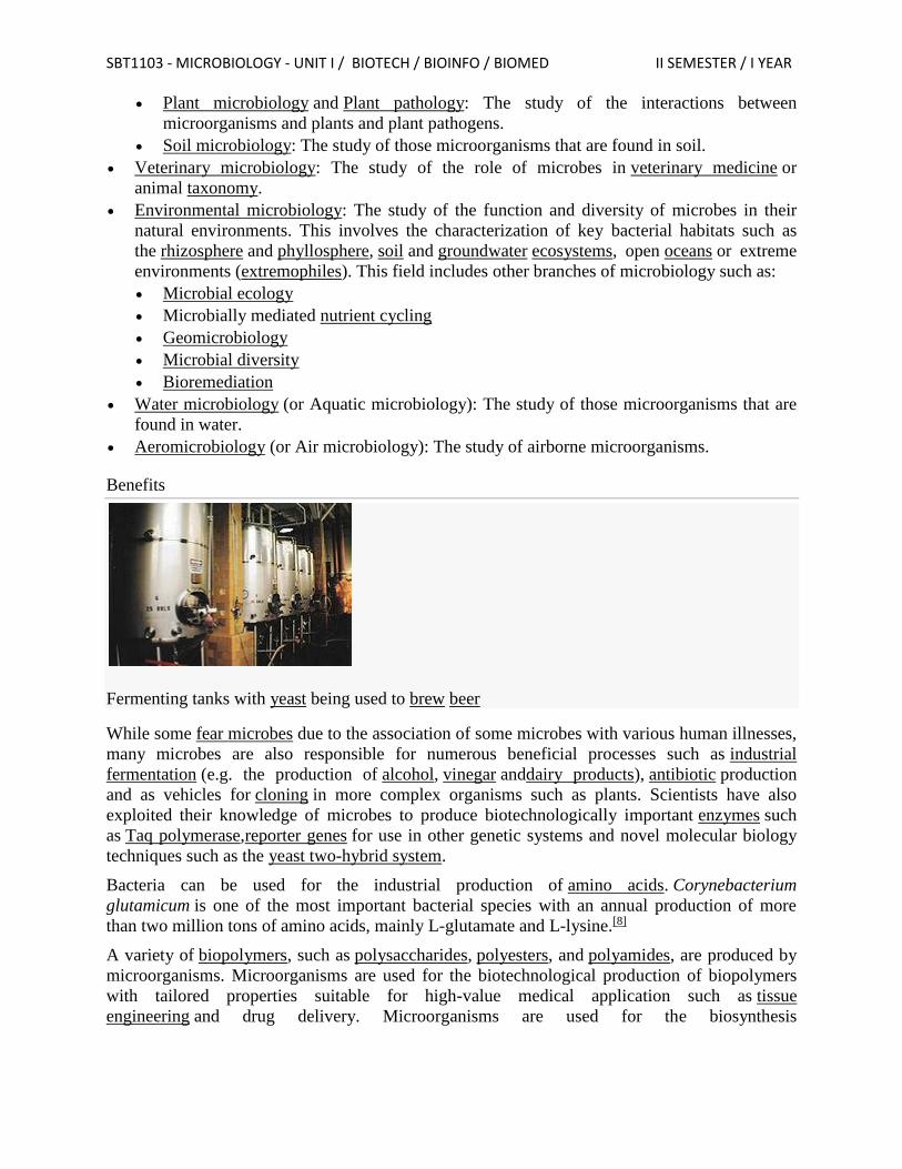
Fermenting tanks with yeast being used to brew beer
While some fear microbes due to the association of some microbes with various human illnesses,
many microbes are also responsible for numerous beneficial processes such as industrial
fermentation (e.g. the production of alcohol, vinegar anddairy products), antibiotic production
and as vehicles for cloning in more complex organisms such as plants. Scientists have also
exploited their knowledge of microbes to produce biotechnologically important enzymes such
as Taq polymerase,reporter genes for use in other genetic systems and novel molecular biology
techniques such as the yeast two-hybrid system.
Bacteria can be used for the industrial production of amino acids. Corynebacterium
glutamicum is one of the most important bacterial species with an annual production of more
than two million tons of amino acids, mainly L-glutamate and L-lysine.[8]
A variety of biopolymers, such as polysaccharides, polyesters, and polyamides, are produced by
microorganisms. Microorganisms are used for the biotechnological production of biopolymers
with tailored properties suitable for high-value medical application such as tissue
engineering and drug delivery. Microorganisms are used for the biosynthesis
SBT1103 - MICROBIOLOGY - UNIT I / BIOTECH / BIOINFO / BIOMED II SEMESTER / I YEAR
of xanthan,alginate, cellulose, cyanophycin, poly(gamma-glutamic acid), levan, hyaluronic acid,
organic acids, oligosaccharides and polysaccharide, and polyhydroxyalkanoates.[9]
Microorganisms are beneficial for microbial biodegradation or bioremediation of domestic,
agricultural and industrial wastes and subsurface pollution in soils, sediments and marine
environments. The ability of each microorganism to degrade toxic waste depends on the nature
of each contaminant. Since sites typically have multiple pollutant types, the most effective
approach to microbial biodegradation is to use a mixture of bacterial and fungal species and
strains, each specific to thebiodegradation of one or more types of contaminants.[10]
Symbiotic microbial communities are known to confer various benefits to their human and
animal hosts health including aiding digestion, production of beneficial vitamins and amino
acids, and suppression of pathogenic microbes. Some benefit may be conferred by consuming
fermented foods, probiotics (bacteria potentially beneficial to the digestive system)
and/or prebiotics (substances consumed to promote the growth of probiotic
microorganisms).[11][12] The ways the microbiome influences human and animal health, as well as
methods to influence the microbiome are active areas of research.[13]
Research has suggested that microorganisms could be useful in the treatment of cancer. Various
strains of non-pathogenic clostridia can infiltrate and replicate within solid tumors. Clostridial
vectors can be safely administered and their potential to deliver therapeutic proteins has been
demonstrated in a variety of preclinical models.[14]
Scope of microbiology There is vast scope in the field of microbiology due to the advancement in the field of science
and technology. The scope in this field is immense due to the involvement of microbiology in
many fields like medicine, pharmacy, diary, industry, clinical research, water industry,
agriculture, chemical technology and nanotechnology. The study of microbiology contributes
greatly to the understanding of life through enhancements and intervention of microorganisms.
There is an increase in demand for microbiologists in India and globally. A microbiologist can
innovate new diagnostic kits, discover new drugs, teach, research, etc.
Since the microbes are living, it follows that microbiology deals with a group of particular life
forms and it comes under the broad domain of biology which includes the study of all aspects of
living beings including man.
Where can we fit in microbes in the hierarchy of living beings? Traditionally living beings are
divided into plants and animals. But members of microbes can be accommodated in both plants
(fungi) and animals (protozoa) and some cannot be accommodated in either plants or animals as
they share the characters of both. (For instance Euglenawas a disputed property till recently
between botanists and zoologists.)
In one of the earlier attempts to resolve this problem, Haeckel (1866) a German Zoologist
suggested that there should be a third kingdom besides Plantae (plants) and Animalia (animals)
to include all the microorganisms. He gave the name Protista to this kingdom to include all
unicellular microorganisms that are neither plants nor animals.
SBT1103 - MICROBIOLOGY - UNIT I / BIOTECH / BIOINFO / BIOMED II SEMESTER / I YEAR
Haeckel's classification raised some questions like how to distinguish a fungus from a bacterium
or from an alga. The discovery in late 1940s of the prokaryotic and eukaryotic nature of the cells
rendered the three kingdom classification unsatisfactory.
A recent and comprehensive classification proposed by R.H. Whittaker (1969) has five kingdoms
of living beings
Kingdom Monera
Kingdom Protista
Kingdom Fungi
Kingdom Animalia
Kingdom Plantae
Microorganisms include three of the (Monera, Protista and Fungi) five kingdoms mentioned
above. At present it is agreed that within the preview of microbiology, five major groups of
microorganisms-viruses, bacteria, fungi, algae and protozoa are dealt with.
As it will be evident from the above discussion, the scope of microbiology extends to both
eukaryotic as well as prokaryotic microbes. While discussing the scope of microbiology it should
be evident to us that it does not deal with merely the enumeration of structural diversity or
classification but extends to all aspects of microbial life. Microbiology is concerned with their
form, structure, reproduction, physiology, metabolism classification and most important their
economic importance. In other words, what the microbes can do and should not be allowed to
do (some times) as for as human beings are concerned is one of the vital aspects of microbiology
on which rests human destiny.
History
Ancient
The existence of microorganisms was hypothesized for many centuries before their actual
discovery. The existence of unseen microbiological life was postulated byJainism which is based
on Mahavira’s teachings as early as 6th century BCE.[15] Paul Dundas notes that Mahavira
asserted existence of unseen microbiological creatures living in earth, water, air and fire.[16] Jain
scriptures also describe nigodas which are sub-microscopic creatures living in large clusters and
having a very short life and are said to pervade each and every part of the universe, even in
tissues of plants and flesh of animals.[17]
SBT1103 - MICROBIOLOGY - UNIT I / BIOTECH / BIOINFO / BIOMED II SEMESTER / I YEAR
SBT1103 - MICROBIOLOGY - UNIT I / BIOTECH / BIOINFO / BIOMED II SEMESTER / I YEAR
SBT1103 - MICROBIOLOGY - UNIT I / BIOTECH / BIOINFO / BIOMED II SEMESTER / I YEAR
SBT1103 - MICROBIOLOGY - UNIT I / BIOTECH / BIOINFO / BIOMED II SEMESTER / I YEAR
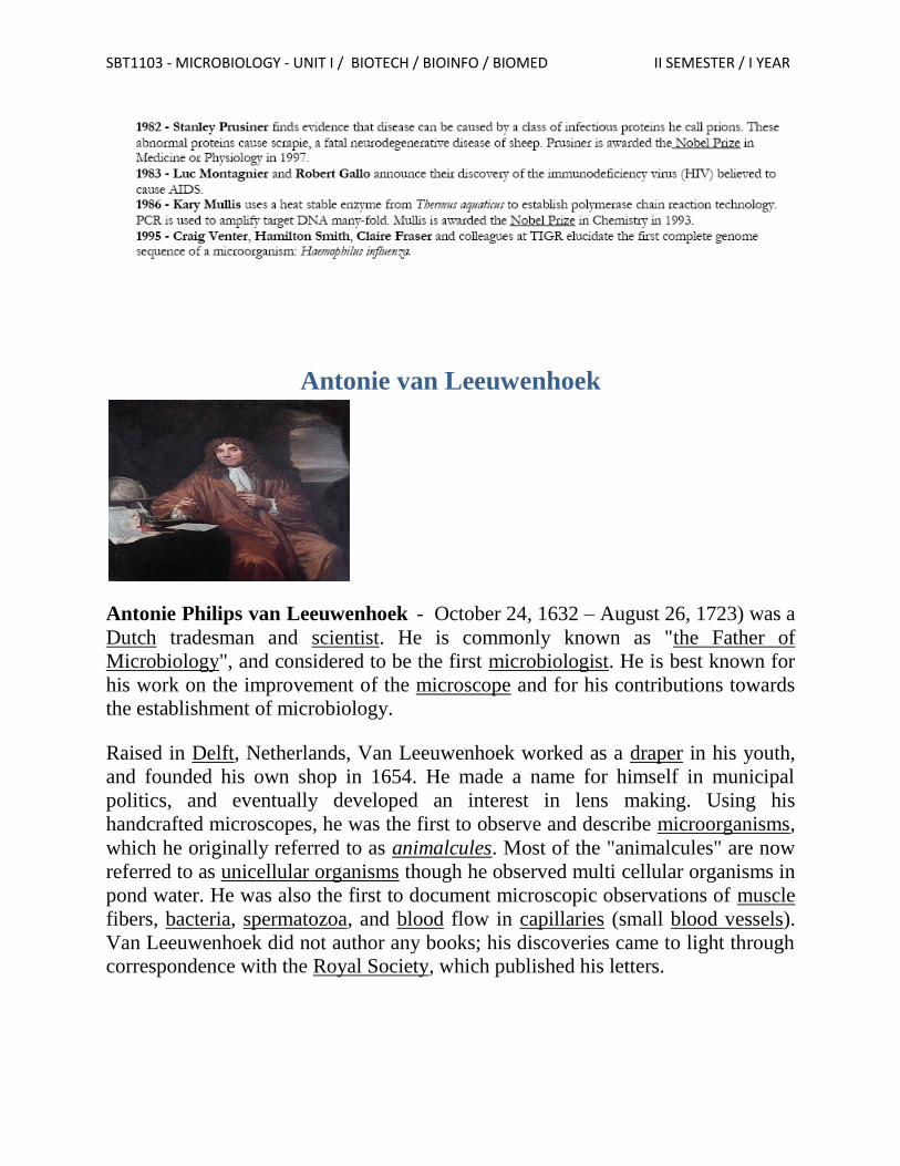
Antonie van Leeuwenhoek
Antonie Philips van Leeuwenhoek - October 24, 1632 – August 26, 1723) was a
Dutch tradesman and scientist. He is commonly known as "the Father of
Microbiology", and considered to be the first microbiologist. He is best known for
his work on the improvement of the microscope and for his contributions towards
the establishment of microbiology.
Raised in Delft, Netherlands, Van Leeuwenhoek worked as a draper in his youth,
and founded his own shop in 1654. He made a name for himself in municipal
politics, and eventually developed an interest in lens making. Using his
handcrafted microscopes, he was the first to observe and describe microorganisms,
which he originally referred to as animalcules. Most of the "animalcules" are now
referred to as unicellular organisms though he observed multi cellular organisms in
pond water. He was also the first to document microscopic observations of muscle
fibers, bacteria, spermatozoa, and blood flow in capillaries (small blood vessels).
Van Leeuwenhoek did not author any books; his discoveries came to light through
correspondence with the Royal Society, which published his letters.
SBT1103 - MICROBIOLOGY - UNIT I / BIOTECH / BIOINFO / BIOMED II SEMESTER / I YEAR
Early life and career
Antonie van Leeuwenhoek was born in Delft, Dutch Republic, on October 24,
1632. On 4 November he was baptized as Thonis. His father, Philips Antonisz van
Leeuwenhoek, was a basket maker who died when Antonie was only five years
old. His mother, Margaretha (Bel van den Berch), came from a well-to-do brewer's
family, and remarried Jacob Jansz Molijn, a painter. Van Leeuwenhoek married
Barbara de Mey in July 1654, with whom he would have one surviving daughter,
Maria (four other children died in infancy). That same year he returned to Delft,
where he would live and study for the rest of his life. He opened a draper's shop,
which he ran throughout the 1650s. His wife died in 1666, and in 1671 Van
Leeuwenhoek remarried, to Cornelia Swalmius, with whom he had no children.[6]
His status in Delft had grown throughout the years. In 1660 he received a lucrative
job as chamberlain for the Delft sheriffs' assembly chamber in the City Hall, a
position which he would hold for almost 40 years. In 1669 he was appointed as a
land surveyor by the Court of Holland; at some time he combined it with another
municipal job, being the official "wine-gauger" of Delft and in charge of the city's
wine imports[7] and (wine) taxation..
Microscopic study
While running his draper's shop, Van Leeuwenhoek wanted to see the quality of
the thread, better than the then-current magnifying lenses available. Therefore, he
began to develop an interest in lensmaking, although few records exist of his early
activity. Van Leeuwenhoek's interest in microscopes and a familiarity with glass
processing led to one of the most significant, and simultaneously well-hidden,
technical insights in the history of science. By placing the middle of a small rod of
soda lime glass in a hot flame, Van Leeuwenhoek could pull the hot section apart
to create two long whiskers of glass. Then, by reinserting the end of one whisker
into the flame, he could create a very small, high-quality glass sphere. These
spheres became the lenses of his microscopes, with the smallest spheres providing
the highest magnifications.
Recognition by the Royal Society
After developing his method for creating powerful lenses and applying them to the
study of the microscopic world, Van Leeuwenhoek introduced his work to his
friend, the prominent Dutch physician Reinier de Graaf. When the Royal Society
in London published the groundbreaking work of an Italian lensmaker in their
journal Philosophical Transactions of the Royal Society, De Graaf wrote to the
journal's editor Henry Oldenburg with a ringing endorsement of Van
SBT1103 - MICROBIOLOGY - UNIT I / BIOTECH / BIOINFO / BIOMED II SEMESTER / I YEAR
Leeuwenhoek's microscopes which, he claimed, "far surpass those which we have
hitherto seen". In response the Society published in 1673 a letter from Van
Leeuwenhoek, which included his microscopic observations on mold, bees, and
lice.[9]
Antonie van Leeuwenhoek was elected to the Royal Society in February 1680 on
the nomination of William Croone, a then-prominent physician.[note 3] Van
Leeuwenhoek was "taken aback" by the nomination, which he considered a high
honor, although he did not attend the induction ceremony in London, nor did he
ever attend a Royal Society meeting.[16]
Scientific fame
By the end of the 17th century, Van Leeuwenhoek had a virtual monopoly on
microscopic study and discovery. His contemporary Robert Hooke, an early
microscope pioneer, bemoaned that the field had come to rest entirely on one man's
shoulders.[17] He made about 200 microscopes with different magnification.
On this occasion Van Leeuwenhoek presented the Tsar an "eel-viewer", so Peter
could study the blood circulation, whenever he wanted.
Techniques and discoveries
Antonie van Leeuwenhoek made more than 500 optical lenses. He also created at
least 25 single-lens microscopes, of differing types, of which only nine survived.
These microscopes were made of silver or copper frames, holding hand-made
lenses. Those that have survived are capable of magnification up to 275 times. It is
suspected that Van Leeuwenhoek possessed some microscopes that could magnify
up to 500 times. Although he has been widely regarded as a dilettante or amateur,
his scientific research was of remarkably high quality.[20]
The single-lens microscopes of Van Leeuwenhoek were relatively small devices,
the biggest being about 5 cm long.[21] They are used by placing the lens very close
in front of the eye, while looking in direction of the sun. The other side of the
microscope had a pin, where the sample was attached in order to stay close to the
lens. There were also three screws that allowed to move the pin, and the sample,
along three axes: one axis to change the focus, and the two other axes to navigate
through the sample.
Van Leeuwenhoek maintained throughout his life that there are aspects of
microscope construction "which I only keep for myself", in particular his most
SBT1103 - MICROBIOLOGY - UNIT I / BIOTECH / BIOINFO / BIOMED II SEMESTER / I YEAR
critical secret of how he made the lenses. For many years no-one was able to
reconstruct Van Leeuwenhoek's design techniques. However, in 1957 C.L. Stong
used thin glass thread fusing instead of polishing, and successfully created some
working samples of a Van Leeuwenhoek design microscope.[22] Such a method was
also discovered independently by A. Mosolov and A. Belkin at the Russian
Novosibirsk State Medical Institute.[23]
Van Leeuwenhoek used samples and measurements to estimate numbers of
microorganisms in units of water.[24][25] He also made good use of the huge lead
provided by his method. He studied a broad range of microscopic phenomena, and
shared the resulting observations freely with groups such as the British Royal
Society.[26] Such work firmly established his place in history as one of the first and
most important explorers of the microscopic world. Antonie van Leeuwenhoek was
one of the first people to observe cells, much like Robert Hooke.
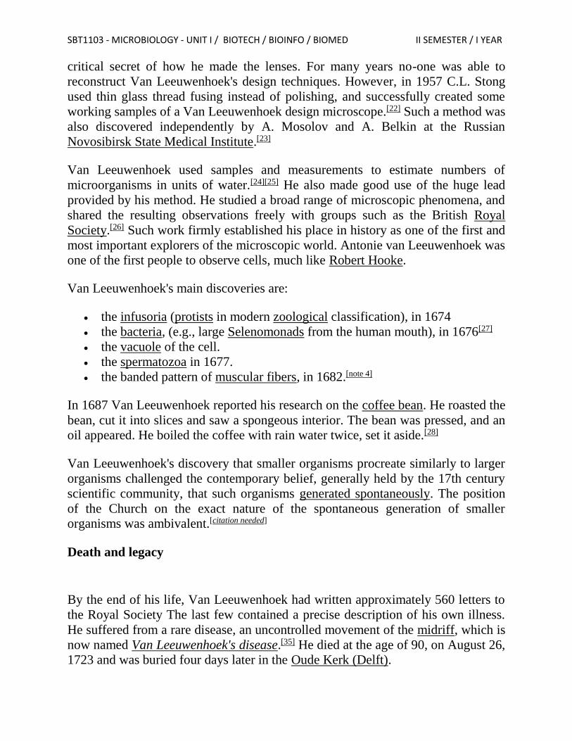
Van Leeuwenhoek's main discoveries are:
the infusoria (protists in modern zoological classification), in 1674
the bacteria, (e.g., large Selenomonads from the human mouth), in 1676[27]
the vacuole of the cell.
the spermatozoa in 1677.
the banded pattern of muscular fibers, in 1682.[note 4]
In 1687 Van Leeuwenhoek reported his research on the coffee bean. He roasted the
bean, cut it into slices and saw a spongeous interior. The bean was pressed, and an
oil appeared. He boiled the coffee with rain water twice, set it aside.[28]
Van Leeuwenhoek's discovery that smaller organisms procreate similarly to larger
organisms challenged the contemporary belief, generally held by the 17th century
scientific community, that such organisms generated spontaneously. The position
of the Church on the exact nature of the spontaneous generation of smaller
organisms was ambivalent.[citation needed]
Death and legacy
By the end of his life, Van Leeuwenhoek had written approximately 560 letters to
the Royal Society The last few contained a precise description of his own illness.
He suffered from a rare disease, an uncontrolled movement of the midriff, which is
now named Van Leeuwenhoek's disease.[35] He died at the age of 90, on August 26,
1723 and was buried four days later in the Oude Kerk (Delft).
SBT1103 - MICROBIOLOGY - UNIT I / BIOTECH / BIOINFO / BIOMED II SEMESTER / I YEAR
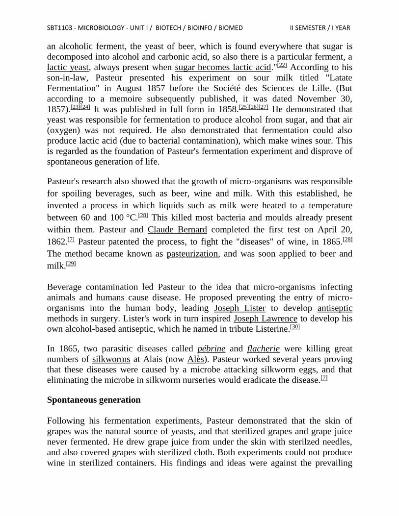
Louis Pasteur
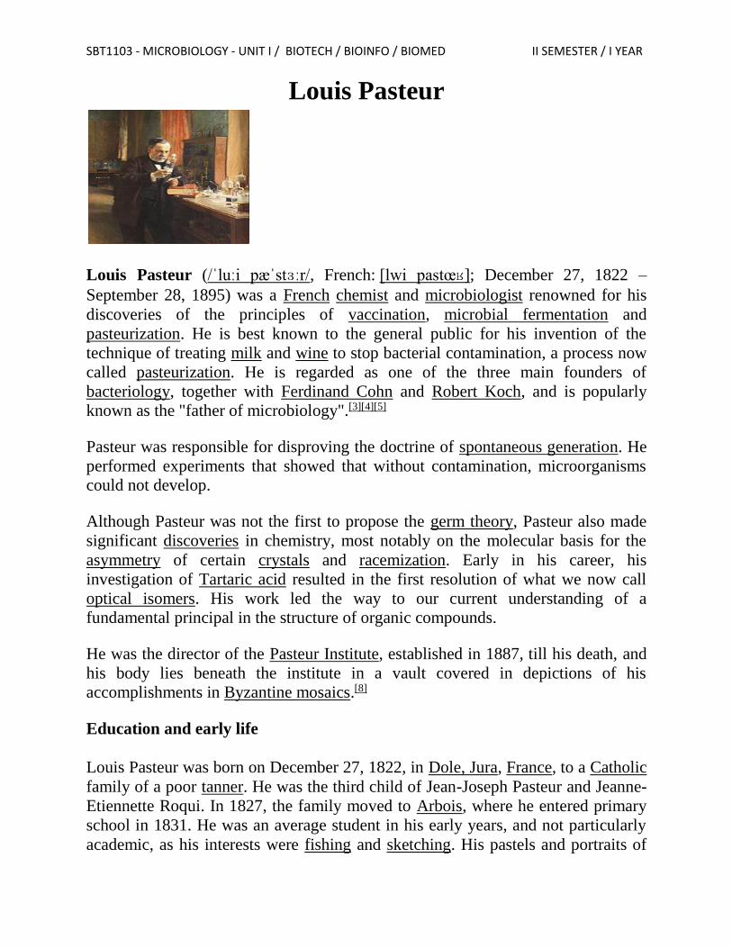
Louis Pasteur (/ˈluːi pæˈstɜːr/, French: [lwi pastœʁ]; December 27, 1822 –
September 28, 1895) was a French chemist and microbiologist renowned for his
discoveries of the principles of vaccination, microbial fermentation and
pasteurization. He is best known to the general public for his invention of the
technique of treating milk and wine to stop bacterial contamination, a process now
called pasteurization. He is regarded as one of the three main founders of
bacteriology, together with Ferdinand Cohn and Robert Koch, and is popularly
known as the "father of microbiology".[3][4][5]
Pasteur was responsible for disproving the doctrine of spontaneous generation. He
performed experiments that showed that without contamination, microorganisms
could not develop.
Although Pasteur was not the first to propose the germ theory, Pasteur also made
significant discoveries in chemistry, most notably on the molecular basis for the
asymmetry of certain crystals and racemization. Early in his career, his
investigation of Tartaric acid resulted in the first resolution of what we now call
optical isomers. His work led the way to our current understanding of a
fundamental principal in the structure of organic compounds.
He was the director of the Pasteur Institute, established in 1887, till his death, and
his body lies beneath the institute in a vault covered in depictions of his
accomplishments in Byzantine mosaics.[8]
Education and early life
Louis Pasteur was born on December 27, 1822, in Dole, Jura, France, to a Catholic
family of a poor tanner. He was the third child of Jean-Joseph Pasteur and Jeanne-
Etiennette Roqui. In 1827, the family moved to Arbois, where he entered primary
school in 1831. He was an average student in his early years, and not particularly
academic, as his interests were fishing and sketching. His pastels and portraits of
SBT1103 - MICROBIOLOGY - UNIT I / BIOTECH / BIOINFO / BIOMED II SEMESTER / I YEAR
his parents and friends, made when he was 15, were later kept in the museum of
the Pasteur Institute in Paris. In 1838, he left for Paris to join the Institution Barbet,
but became homesick and returned in November. In 1839, he entered the Collège
Royal de Besançon and earned his baccalauréat (BA) degree in 1840. He was
appointed teaching assistant at the Besançon college while continuing a degree
science course with special mathematics. He failed his first examination in 1841.
He managed to pass the baccalauréat scientifique (general science) degree in 1842
from Dijon but with a poor grade in chemistry. After one failed attempt for the
entrance test for the École Normale Supérieure in Paris in 1842, he succeeded in
1844. In 1845 he received the licencié ès sciences (Bachelor of Science) degree.
After serving briefly as professor of physics at the Dijon Lycée in 1848, he became
professor of chemistry at the University of Strasbourg, where he met and courted
Marie Laurent, daughter of the university's rector in 1849. They were married on
May 29, 1849, and together had five children, only two of whom survived to
adulthood; the other three died of typhoid. These personal tragedies were his
motivations for curing infectious diseases.[3][11]
Career
Pasteur was appointed to the Chair of Chemistry in the faculty of sciences of the
University of Strasbourg in 1848. In 1854, he was named dean of the new faculty
of sciences at Lille University,)
In 1857, he moved to Paris as the director of scientific studies at the École
Normale Supérieure where he took control from 1858 to 1867 and introduced a
series of reforms to improve the standard of scientific work. In 1862, he was
appointed professor of geology, physics, and chemistry at the École nationale
supérieure des Beaux-Arts, the position which held until his resignation in 1867. In
Paris, he established the Pasteur Institute in 1887, in which he was its director for
the rest of his life.[4][5][11]
Fermentation and germ theory of diseases
Pasteur demonstrated that fermentation is caused by the growth of micro-
organisms, and the emergent growth of bacteria in nutrient broths is due not to
spontaneous generation, but rather to biogenesis (Omne vivum ex vivo "all life from
life"). He was motivated to investigate the matter while working at Lille. In 1856 a
local wine manufacturer, M. Bigot, the father of his student, sought for his advice
on the problems of making beetroot alcohol and souring after long storage.[21] In
1857 he developed his ideas stating that: "I intend to establish that, just as there is
SBT1103 - MICROBIOLOGY - UNIT I / BIOTECH / BIOINFO / BIOMED II SEMESTER / I YEAR
an alcoholic ferment, the yeast of beer, which is found everywhere that sugar is
decomposed into alcohol and carbonic acid, so also there is a particular ferment, a
lactic yeast, always present when sugar becomes lactic acid."[22] According to his
son-in-law, Pasteur presented his experiment on sour milk titled "Latate
Fermentation" in August 1857 before the Société des Sciences de Lille. (But
according to a memoire subsequently published, it was dated November 30,
1857).[23][24] It was published in full form in 1858.[25][26][27] He demonstrated that
yeast was responsible for fermentation to produce alcohol from sugar, and that air
(oxygen) was not required. He also demonstrated that fermentation could also
produce lactic acid (due to bacterial contamination), which make wines sour. This
is regarded as the foundation of Pasteur's fermentation experiment and disprove of
spontaneous generation of life.
Pasteur's research also showed that the growth of micro-organisms was responsible
for spoiling beverages, such as beer, wine and milk. With this established, he
invented a process in which liquids such as milk were heated to a temperature
between 60 and 100 °C.[28] This killed most bacteria and moulds already present
within them. Pasteur and Claude Bernard completed the first test on April 20,
1862.[7] Pasteur patented the process, to fight the "diseases" of wine, in 1865.[28]
The method became known as pasteurization, and was soon applied to beer and
milk.[29]
Beverage contamination led Pasteur to the idea that micro-organisms infecting
animals and humans cause disease. He proposed preventing the entry of micro-
organisms into the human body, leading Joseph Lister to develop antiseptic
methods in surgery. Lister's work in turn inspired Joseph Lawrence to develop his
own alcohol-based antiseptic, which he named in tribute Listerine.[30]
In 1865, two parasitic diseases called pébrine and flacherie were killing great
numbers of silkworms at Alais (now Alès). Pasteur worked several years proving
that these diseases were caused by a microbe attacking silkworm eggs, and that
eliminating the microbe in silkworm nurseries would eradicate the disease.[7]
Spontaneous generation
Following his fermentation experiments, Pasteur demonstrated that the skin of
grapes was the natural source of yeasts, and that sterilized grapes and grape juice
never fermented. He drew grape juice from under the skin with sterilzed needles,
and also covered grapes with sterilized cloth. Both experiments could not produce
wine in sterilized containers. His findings and ideas were against the prevailing
SBT1103 - MICROBIOLOGY - UNIT I / BIOTECH / BIOINFO / BIOMED II SEMESTER / I YEAR
notion of spontaneous generation. He received a particularly stern criticism from
Félix Archimède Pouchet, who was director of the Rouen Museum of Natural
History. To settle the debate between the eminent scientists, the French Academy
of Sciences offered Alhumbert Prize carrying 2,500 francs to whoever could
experimentally demonstrate for or against the doctrine.[31][32][33]
To prove himself correct, Pasteur exposed boiled broths to air in swan-neck flasks
that contained a filter to prevent all particles from passing through to the growth
medium, and even in flasks with no filter at all, with air being admitted via a long
tortuous tube that would not allow dust particles to pass. Nothing grew in the
broths unless the flasks were broken open, showing that the living organisms that
grew in such broths came from outside, as spores on dust, rather than
spontaneously generated within the broth. This was one of the last and most
important experiments disproving the theory of spontaneous generation for which
Pasteur won the Alhumbert Prize in 1862. He concluded that:[34][35]
Never will the doctrine of spontaneous generation recover from the mortal blow of
this simple experiment. There is no known circumstance in which it can be
confirmed that microscopic beings came into the world without germs, without
parents similar to themselves.
Immunology and vaccination
Pasteur's later work on diseases included work on chicken cholera. During this
work, a culture of the responsible bacteria had spoiled and failed to induce the
disease in some chickens he was infecting with the disease. Upon reusing these
healthy chickens, Pasteur discovered he could not infect them, even with fresh
bacteria; the weakened bacteria had caused the chickens to become immune to the
disease, though they had caused only mild symptoms.[3][7]
His assistant, Charles Chamberland (of French origin), had been instructed to
inoculate the chickens after Pasteur went on holiday. Chamberland failed to do
this, but instead went on holiday himself. On his return, the month-old cultures
made the chickens unwell, but instead of the infections being fatal, as they usually
were, the chickens recovered completely. Chamberland assumed an error had been
made, and wanted to discard the apparently faulty culture when Pasteur stopped
him. Pasteur guessed the recovered animals now might be immune to the disease,
as were the animals at Eure-et-Loir that had recovered from anthrax.[36]
In the 1870s, he applied this immunization method to anthrax, which affected
cattle, and aroused interest in combating other diseases.
SBT1103 - MICROBIOLOGY - UNIT I / BIOTECH / BIOINFO / BIOMED II SEMESTER / I YEAR
Pasteur publicly claimed he had made the anthrax vaccine by exposing the bacilli
to oxygen. His laboratory notebooks, now in the Bibliothèque Nationale in Paris, in
fact show that he used the method of rival Jean-Joseph-Henri Toussaint, a
Toulouse veterinary surgeon, to create the anthrax vaccine.[20][37] This method used
the oxidizing agent potassium dichromate. Pasteur's oxygen method did eventually
produce a vaccine but only after he had been awarded a patent on the production of
an anthrax vaccine.
The notion of a weak form of a disease causing immunity to the virulent version
was not new; this had been known for a long time for smallpox. Inoculation with
smallpox (Variolation) was known to result in far less scarring, and greatly reduced
mortality, in comparison with the naturally acquired disease. Edward Jenner had
also discovered vaccination using cowpox (Vaccinia) to give cross-immunity to
smallpox in 1796, and by Pasteur's time this had generally replaced the use of
actual smallpox (Variola) material in inoculation. The difference between smallpox
vaccination and anthrax or chicken cholera vaccination was that the weakened
form of the latter two disease organisms had been "generated artificially", so a
naturally weak form of the disease organism did not need to be found. This
discovery revolutionized work in infectious diseases, and Pasteur gave these
artificially weakened diseases the generic name of "vaccines", in honour of
Jenner's discovery. Pasteur produced the first vaccine for rabies by growing the
virus in rabbits, and then weakening it by drying the affected nerve tissue.[38]
The rabies vaccine was initially created by Emile Roux, a French doctor and a
colleague of Pasteur who had been working with a killed vaccine produced by
desiccating the spinal cords of infected rabbits. The vaccine had been tested in 50
dogs before its first human trial.[39][40] This vaccine was first used on 9-year old
Joseph Meister, on July 6, 1885, after the boy was badly mauled by a rabid
dog.[20][38] This was done at some personal risk for Pasteur, since he was not a
licensed physician and could have faced prosecution for treating the boy. After
consulting with colleagues, he decided to go ahead with the treatment. Three
months later he examined Meister and found that he was in good health.[41] Pasteur
was hailed as a hero and the legal matter was not pursued. The treatment's success
laid the foundations for the manufacture of many other vaccines. The first of the
Pasteur Institutes was also built on the basis of this achievement.[20]
Legal risk was not the only kind Pasteur undertook. In The Story of San Michele,
Axel Munthe writes of the rabies vaccine research:
Pasteur himself was absolutely fearless. Anxious to secure a sample of saliva
straight from the jaws of a rabid dog, I once saw him with the glass tube held
SBT1103 - MICROBIOLOGY - UNIT I / BIOTECH / BIOINFO / BIOMED II SEMESTER / I YEAR
between his lips draw a few drops of the deadly saliva from the mouth of a rabid
bull-dog, held on the table by two assistants, their hands protected by leather
gloves.
Because of his study in germs, Pasteur encouraged doctors to sanitize their hands
and equipment before surgery. Prior to this, few doctors or their assistants
practiced these procedures.
Fermentation
He regarded himself as the first to show the role of microorganisms in
fermentation.[48] Pasteur started his experiments only in 1857 and published his
findings in 1858 (April issue of Comptes Rendus Chimie, Béchamp's paper
appeared in January issue), which, as Béchamp noted, did not bring any novel idea
or experiments that earlier works had not shown. Particularly on the spontaneous
generation because Pasteur in his 1858 paper explicitly stated that the lactic acid
bacteria (he named them "lactic yeasts"), which caused wine souring, "takes birth
spontaneously, as easily as beer yeast every time that the conditions are
favourable." This statement directly implied that Pasteur did believe in
spontaneous generation. He condemned the ideas of Pasteur as "'the greatest
scientific silliness of the age".[22] However, Béchamp was on the losing side, as the
BMJ obituary remarked: His name was associated with bygone controversies as to
priority which it would be unprofitable to recall.[51] Pasteur and Béchamp believed
that fermentation was exclusively cellular activity, that is, it was only due to living
cells. But later extraction of enzymes such as invertase by Marcelin Barthelot in
1860 showed that it was simply an enzymatic reaction.[52]
Anthrax vaccine
Pasteur had given a misleading account of the preparation of the anthrax vaccine
used in the experiment at Pouilly-le-Fort.[9] The fact is that Pasteur publicly
claimed his success in developing anthrax vaccine in 1881.[41] However, his
admirer-turned-rival Toussaint was the one who developed the first vaccine.
Toussaint isolated the Gram-negative bacteria cholera des poules (later named – to
add irony – Pasteurella in honour of Pasteur) in 1879 and gave samples to Pasteur
who used for his own works. In 1880 with his publishing on July 12 at the French
Academy of Sciences, Toussaint presented his successful result with an attenuated
vaccine against anthrax in dogs and sheep.[53] Pasteur purely on grounds of
jealousy contested the discovery by publicly displaying his vaccination method in
Pouilly-le-Fort on 5 May 1881. The promotional experiment was a success and
helped Pasteur sell his products, getting all the benefits and glory.[54][55][56]
SBT1103 - MICROBIOLOGY - UNIT I / BIOTECH / BIOINFO / BIOMED II SEMESTER / I YEAR
Experimental ethics
Pasteur experiments are often cited as against medical ethics, especially on his
vaccination of Meister. Firstly, he did not have any experience in medical practice,
and more importantly, a medical license. This is often cited as a serious threat to
his professional and personal reputation.[57][58]
Awards and honours
Pasteur was awarded the prize of 1,500 francs in 1853 by the Pharmaceutical
Society for the synthesis of racemic acid. In 1856 the Royal Society of London
presented him the Rumford Medal for his discovery of the nature of racemic acid
and its relations to polarized light, and the Copley Medal in 1874 for his work on
fermentation.
Pasteur Institute
The Pasteur Institute was established by Pasteur to perpetuate his commitment to
basic research and its practical applications.
Death
Pasteur was frequently stricken by strokes beginning in 1868, and the one in 1894
severely impaired his health. Failing to fully recover, he died in 1895, near
Paris.[20] He was given a state funeral and was buried in the Cathedral of Notre
Dame, but his remains were reinterred in a crypt in the Pasteur Institute in Paris,
where the crypt is engraved with his life-saving work.
Robert Koch
Robert Heinrich Herman Koch (/ˈkɔːx/;[3] German: [ˈkɔχ]; 11 December 1843 – 27 May 1910)
was a celebrated German physician and pioneering microbiologist. As the founder of modern
bacteriology, he is known for his role in identifying the specific causative agents of tuberculosis,
cholera, and anthrax and for giving experimental support for the concept of infectious disease.[4]
In addition to his trail-blazing studies on these diseases, Koch created and improved laboratory
technologies and techniques in the field of microbiology, and made key discoveries in public
health.[5] His research led to the creation of Koch’s postulates, a series of four generalized
principles linking specific microorganisms to specific diseases that remain today the "gold
standard" in medical microbiology.[5] As a result of his groundbreaking research on tuberculosis,
Koch received the Nobel Prize in Physiology or Medicine in 1905.[5]
SBT1103 - MICROBIOLOGY - UNIT I / BIOTECH / BIOINFO / BIOMED II SEMESTER / I YEAR
Personal life
Robert Koch was born in Clausthal, Hanover, Germany, on 11 December 1843, to Hermann
Koch and Mathilde Julie Henriette Biewand.[6] Koch excelled in academics from an early age.
Before entering school in 1848, he had taught himself how to read and write.[4] He graduated
from high school in 1862, having excelled in science and maths.[4] At the age of 19, Koch
entered the University of Göttingen, studying natural science.[7] However, after three semesters,
Koch decided to change his area of study to medicine, as he aspired to be a physician.[4] During
his fifth semester of medical school, Jacob Henle, an anatomist who had published a theory of
contagion in 1840, asked him to participate in his research project on uterine nerve structure.[4] In
his sixth semester, Koch began to conduct research at the Physiological Institute, where he
studied Succinic acid secretion.[4] This would eventually form the basis of his dissertation.[5] In
January 1866, Koch graduated from medical school, earning honors of the highest distinction.[4]
In July 1867, Koch married Emma Adolfine Josephine Fraatz, and the two had a daughter,
Gertrude, in 1868.[5] After his graduation in 1866, he worked as a surgeon in the Franco-Prussian
War, and following his service, worked as a physician in Wollstein, Posen.[7] Koch’s marriage to
Emma Fraatz ended in 1893, and later that same year, he married actress Hedwig Freiberg.[5]
From 1885 to 1890, he served as an administrator and professor at Berlin University.[4] Koch
suffered a heart attack on 9 April 1910, and never made a complete recovery.[4] On 27 May, only
three days after giving a lecture on his tuberculosis research at the Prussian Academy of
Sciences, Robert Koch died in Baden-Baden at the age of 66.[7] Following his death, the Institute
named its establishment after him in his honour.[4]
Research contributions
Anthrax
Robert Koch is widely known for his work with anthrax, discovering the causative agent of the
fatal disease to be Bacillus anthracis.[8] Koch discovered the formation in anthrax bacteria of
spores that could remain dormant under specific conditions.[7] However, under optimal
conditions, the spores were activated and caused disease.[7] To determine this causative agent, he
dry-fixed bacterial cultures onto glass slides, used dyes to stain the cultures, and observed them
through a microscope.[4] Koch’s work with anthrax is notable in that he was the first to link a
specific microorganism with a specific disease, rejecting the idea of spontaneous generation and
supporting the germ theory of disease.[8]
Koch's four postulates
Koch accepted a position as government advisor with the Imperial Department of Health in
1880.[9] During his time as government advisor, he published a report in which he stated the
importance of pure cultures in isolating disease-causing organisms and explained the necessary
steps to obtain these cultures, methods which are summarized in Koch’s four postulates.[10]
Koch’s discovery of the causative agent of anthrax led to the formation of a generic set of
postulates which can be used in the determination of the cause of most infectious diseases.[8]
These postulates, which not only outlined a method for linking cause and effect of an infectious
disease but also established the significance of laboratory culture of infectious agents, are listed
here:[8]
SBT1103 - MICROBIOLOGY - UNIT I / BIOTECH / BIOINFO / BIOMED II SEMESTER / I YEAR
1. The organism must always be present, in every case of the disease.
2. The organism must be isolated from a host containing the disease and grown in pure
culture.
3. Samples of the organism taken from pure culture must cause the same disease when
inoculated into a healthy, susceptible animal in the laboratory.
4. The organism must be isolated from the inoculated animal and must be identified as the
same original organism first isolated from the originally diseased host.
Isolating pure culture on solid media
Koch began conducting research on microorganisms in a laboratory connected to his patient
examination room.[7] Koch’s early research in this laboratory proved to yield one of his major
contributions to the field of microbiology, as it was there that he developed the technique of
growing bacteria. Koch's second postulate calls for the isolation and growth of a selected
pathogen in pure laboratory culture.[11] In an attempt to grow bacteria, Koch began to use solid
nutrients such as potato slices.[11] Through these initial experiments, Koch observed individual
colonies of identical, pure cells.[11] Coming to the conclusion that potato slices were not suitable
media for all organisms, Koch later began to use nutrient solutions with gelatin.[11] However, he
soon realized that gelatin, like potato slices, was not the optimal medium for bacterial growth, as
it did not remain solid at 37 °C, the ideal temperature for growth of most human pathogens.[11]
As suggested to him by Walther and Fanny Hesse, Koch began to utilize agar to grow and isolate
pure cultures, as this polysaccharide remains solid at 37 °C, is not degraded by most bacteria,
and results in a transparent medium.[11][12]
Cholera
Koch next turned his attention to cholera, and began to conduct research in Egypt in the hopes of
isolating the causative agent of the disease.[7] However, he was not able to complete the task
before the epidemic in Egypt ended, and subsequently traveled to India to continue with the
study.[4] In India, Koch was indeed able to determine the causative agent of cholera, isolating
Vibrio cholerae.[4][13] The bacterium had originally been isolated in 1854 by Italian anatomist
Filippo Pacini,[14] but its exact nature and his results were not widely known.
Tuberculosis
During his time as the government advisor with the Imperial Department of Health in Berlin in
the 1880s, Robert Koch became interested in tuberculosis research.[4] At the time, it was widely
believed that tuberculosis was an inherited disease.[4] However, Koch was convinced that the
disease was caused by a bacterium and was infectious, and tested his four postulates using guinea
pigs.[4] Through these experiments, he found that his experiments with tuberculosis satisfied all
four of his postulates.[4] In 1882, he published his findings on tuberculosis, in which he reported
the causative agent of the disease to be the slow-growing Mycobacterium tuberculosis.[11] His
work with this disease won Koch the Nobel Prize in Physiology and Medicine in 1905.[4]
Additionally, Koch's research on tuberculosis, along with his studies on tropical diseases, won
him the Prussian Order Pour le Merite in 1906 and the Robert Koch medal, established to honour
the greatest living physicians, in 1908.[4]
SBT1103 - MICROBIOLOGY - UNIT I / BIOTECH / BIOINFO / BIOMED II SEMESTER / I YEAR
Awards and honours
In addition to being awarded a Nobel Prize, Koch was elected a Foreign Member of the Royal
Society (ForMemRS) in 1897.[2] His microbial postulates are named in his honour, Koch's
postulates.
Edward Jenner
Edward Jenner, FRS (/ˈdʒɛnər/; 17 May 1749 – 26 January 1823) was an English physician and
scientist who was the pioneer of smallpox vaccine, the world's first vaccine.[1][2] He is often
called "the father of immunology", and his work is said to have "saved more lives than the work
of any other human".[3][4][5]
He was also the first person to describe the brood parasitism of the cuckoo.
Early life
Edward Anthony Jenner was born on 17 May 1749[6] (6 May Old Style) in Berkeley,
Gloucestershire, as the eighth of nine children. His father, the Reverend Stephen Jenner, was the
vicar of Berkeley, so Jenner received a strong basic education.[6]
He went to school in Wotton-under-Edge and Cirencester.[6] During this time, he was inoculated
for smallpox, which had a lifelong effect upon his general health.[6] At the age of 14, he was
apprenticed for seven years to Mr Daniel Ludlow, a surgeon of Chipping Sodbury, South
Gloucestershire, where he gained most of the experience needed to become a surgeon himself.[6]
In 1770, Jenner became apprenticed in surgery and anatomy under surgeon John Hunter and
others at St George's Hospital.[7] William Osler records that Hunter gave Jenner William
Harvey's advice, very famous in medical circles (and characteristic of the Age of
Enlightenment), "Don't think; try."[8] Hunter remained in correspondence with
Jenner over natural history and proposed him for the Royal Society. Returning to
his native countryside by 1773, Jenner became a successful family doctor and
surgeon, practising on dedicated premises at Berkeley.
Jenner and others formed the Fleece Medical Society or Gloucestershire Medical
Society, so called because it met in the parlour of the Fleece Inn, Rodborough (in
Gloucestershire), meeting to dine together and read papers on medical subjects.
Jenner contributed papers on angina pectoris, ophthalmia, and cardiac valvular
disease and commented on cowpox. He also belonged to a similar society which
met in Alveston, near Bristol.[9]
SBT1103 - MICROBIOLOGY - UNIT I / BIOTECH / BIOINFO / BIOMED II SEMESTER / I YEAR
He became a master mason 30 December 1802, in Lodge of Faith and Friendship
#449. From 1812–1813, he served as worshipful master of Royal Berkeley Lodge
of Faith and Friendship.[10]
Zoology
Jenner was elected fellow of the Royal Society in 1788, following his publication
of a careful study of the previously misunderstood life of the nested cuckoo, a
study that combined observation, experiment, and dissection.
Marriage and human medicine
Jenner married Catharine Kingscote (died 1815 from tuberculosis) in March 1788.
He might have met her while he and other fellows were experimenting with
balloons. Jenner's trial balloon descended into Kingscote Park, Gloucestershire,
owned by Anthony Kingscote, one of whose daughters was Catharine.[15]
He earned his MD from the University of St Andrews in 1792. He is credited with
advancing the understanding of angina pectoris.[16] In his correspondence with
Heberden, he wrote, "How much the heart must suffer from the coronary arteries
not being able to perform their functions."
Invention of the vaccine
Inoculation was already a standard practice, but involved serious risks. In 1721,
Lady Mary Wortley Montagu had imported variolation to Britain after having
observed it in Istanbul, where her husband was the British ambassador. Voltaire,
writing of this, estimates that at this time 60% of the population caught smallpox
and 20% of the population died of it.[17] Voltaire also states that the Circassians
used the inoculation from times immemorial, and the custom may have been
borrowed by the Turks from the Circassians.[18]
Jenner's Hypothesis:
The initial source of infection was a disease
of horses, called "the grease", which was
transferred to cattle by farm workers,
transformed, and then manifested as cowpox.
SBT1103 - MICROBIOLOGY - UNIT I / BIOTECH / BIOINFO / BIOMED II SEMESTER / I YEAR
Noting the common observation that milkmaids were generally immune to
smallpox, Jenner postulated that the pus in the blisters that milkmaids received
from cowpox (a disease similar to smallpox, but much less virulent) protected them
from smallpox.
On 14 May 1796, Jenner tested his hypothesis by inoculating James Phipps, an
eight-year-old boy who was the son of Jenner's gardener. He scraped pus from
cowpox blisters on the hands of Sarah Nelmes, a milkmaid who had caught
cowpox from a cow called Blossom,[23] whose hide now hangs on the wall of the St
George's medical school library (now in Tooting). Phipps was the 17th case
described in Jenner's first paper[24] on vaccination.
Jenner inoculated Phipps in both arms that day, subsequently producing in Phipps a
fever and some uneasiness, but no full-blown infection. Later, he injected Phipps
with variolous material, the routine method of immunization at that time. No
disease followed. The boy was later challenged with variolous material and again
showed no sign of infection.
Donald Hopkins has written, "Jenner's unique contribution was not that he
inoculated a few persons with cowpox, but that he then proved [by subsequent
challenges] that they were immune to smallpox. Moreover, he demonstrated that
the protective cowpox pus could be effectively inoculated from person to person,
not just directly from cattle.[25] Jenner successfully tested his hypothesis on 23
additional subjects.
Jenner continued his research and reported it to the Royal Society, which did not
publish the initial paper. After revisions and further investigations, he published his
findings on the 23 cases. Some of his conclusions were correct, some erroneous;
modern microbiological and microscopic methods would make his studies easier to
reproduce. The medical establishment, cautious then as now, deliberated at length
over his findings before accepting them. Eventually, vaccination was accepted, and
in 1840, the British government banned variolation – the use of smallpox to induce
immunity – and provided vaccination using cowpox free of charge. (See
Vaccination acts). The success of his discovery soon spread around Europe and,
for example, was used en masse in the Spanish Balmis Expedition,[26] a three-year-
long mission to the Americas, the Philippines, Macao, China, and Saint Helena
Island led by Dr. Francisco Javier de Balmis with the aim of giving thousands the
smallpox vaccine. The expedition was successful, and Jenner wrote, "I don’t
imagine the annals of history furnish an example of philanthropy so noble, so
extensive as this."
SBT1103 - MICROBIOLOGY - UNIT I / BIOTECH / BIOINFO / BIOMED II SEMESTER / I YEAR
Jenner's continuing work on vaccination prevented him from continuing his
ordinary medical practice. He was supported by his colleagues and the King in
petitioning Parliament, and was granted £10,000 in 1802 for his work on
vaccination. In 1807, he was granted another £20,000 after the Royal College of
Physicians had confirmed the widespread efficacy of vaccination.
In 1803 in London, he became president of the Jennerian Society, concerned with
promoting vaccination to eradicate smallpox. The Jennerian ceased operations in
1809. In 1808, with government aid, the National Vaccine Establishment was
founded, but Jenner felt dishonoured by the men selected to run it and resigned his
directorship.[27] Jenner became a member of the Medical and Chirurgical Society
on its founding in 1805 and presented a number of papers there. The society is now
the Royal Society of Medicine. He was elected a foreign honorary member of the
American Academy of Arts and Sciences in 1802.[28] In 1806, Jenner was elected a
foreign member of the Royal Swedish Academy of Sciences.
Returning to London in 1811, Jenner observed a significant number of cases of
smallpox after vaccination. He found that in these cases the severity of the illness
was notably diminished by previous vaccination. In 1821, he was appointed
physician extraordinary to King George IV, a great national honour, and was also
made mayor of Berkeley and justice of the peace. He continued to investigate
natural history, and in 1823, the last year of his life, he presented his "Observations
on the Migration of Birds" to the Royal Society.
Jenner was found in a state of apoplexy on 25 January 1823, with his right side
paralysed. He never fully recovered and eventually died of an apparent stroke, his
second, on 26 January 1823, aged 73. He was buried in the Jenner family vault at
the Church of St. Mary's, Berkeley, Gloucestershire.[29]
Edward Jenner was survived by one son and one daughter, his elder son having
died of tuberculosis at the age of 21.
Legacy
In 1979, the World Health Organization declared smallpox an eradicated
disease.[33] This was the result of coordinated public health efforts by many people,
but vaccination was an essential component.
SBT1103 - MICROBIOLOGY - UNIT I / BIOTECH / BIOINFO / BIOMED II SEMESTER / I YEAR
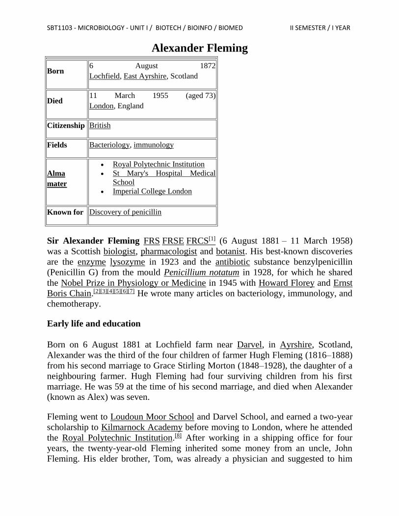
Alexander Fleming
Born 6 August 1872
Lochfield, East Ayrshire, Scotland
Died 11 March 1955 (aged 73)
London, England
Citizenship British
Fields Bacteriology, immunology
Alma
mater
Royal Polytechnic Institution
St Mary's Hospital Medical
School
Imperial College London
Known for Discovery of penicillin
Sir Alexander Fleming FRS FRSE FRCS[1] (6 August 1881 – 11 March 1958)
was a Scottish biologist, pharmacologist and botanist. His best-known discoveries
are the enzyme lysozyme in 1923 and the antibiotic substance benzylpenicillin
(Penicillin G) from the mould Penicillium notatum in 1928, for which he shared
the Nobel Prize in Physiology or Medicine in 1945 with Howard Florey and Ernst
Boris Chain.[2][3][4][5][6][7] He wrote many articles on bacteriology, immunology, and
chemotherapy.
Early life and education
Born on 6 August 1881 at Lochfield farm near Darvel, in Ayrshire, Scotland,
Alexander was the third of the four children of farmer Hugh Fleming (1816–1888)
from his second marriage to Grace Stirling Morton (1848–1928), the daughter of a
neighbouring farmer. Hugh Fleming had four surviving children from his first
marriage. He was 59 at the time of his second marriage, and died when Alexander
(known as Alex) was seven.
Fleming went to Loudoun Moor School and Darvel School, and earned a two-year
scholarship to Kilmarnock Academy before moving to London, where he attended
the Royal Polytechnic Institution.[8] After working in a shipping office for four
years, the twenty-year-old Fleming inherited some money from an uncle, John
Fleming. His elder brother, Tom, was already a physician and suggested to him
SBT1103 - MICROBIOLOGY - UNIT I / BIOTECH / BIOINFO / BIOMED II SEMESTER / I YEAR
that he should follow the same career, and so in 1903, the younger Alexander
enrolled at St Mary's Hospital Medical School in Paddington; he qualified with an
MBBS degree from the school with distinction in 1906.
Fleming had been a private in the London Scottish Regiment of the Volunteer
Force since 1900,[2] and had been a member of the rifle club at the medical school.
The captain of the club, wishing to retain Fleming in the team suggested that he
join the research department at St Mary's, where he became assistant bacteriologist
to Sir Almroth Wright, a pioneer in vaccine therapy and immunology. In 1908, he
gained a BSc degree with Gold Medal in Bacteriology, and became a lecturer at St
Mary's until 1914. Fleming served throughout World War I as a captain in the
Royal Army Medical Corps, and was Mentioned in Dispatches. He and many of
his colleagues worked in battlefield hospitals at the Western Front in France. In
1918 he returned to St Mary's Hospital, where he was elected Professor of
Bacteriology of the University of London in 1928. In 1951 he was elected the
Rector of the University of Edinburgh for a term of 3 years.
Research
Following World War I, Fleming actively searched for anti-bacterial agents,
having witnessed the death of many soldiers from sepsis resulting from infected
wounds. Antiseptics killed the patients' immunological defences more effectively
than they killed the invading bacteria. In an article he submitted for the medical
journal The Lancet during World War I, Fleming described an ingenious
experiment, which he was able to conduct as a result of his own glass blowing
skills, in which he explained why antiseptics were killing more soldiers than
infection itself during World War I. Antiseptics worked well on the surface, but
deep wounds tended to shelter anaerobic bacteria from the antiseptic agent, and
antiseptics seemed to remove beneficial agents produced that protected the patients
At St Mary’s Hospital Fleming continued his investigations into antibacterial
substances. Testing the nasal secretions from a patient with a heavy cold, he found
that nasal mucus had an inhibitory effect on bacterial growth.[10] This was the first
recorded discovery of lysozyme, an enzyme present in many secretions including
tears, saliva, human milk as well as mucus. Lysozyme degrades the bonds in
bacterial peptidoglycan cell walls, particularly in Gram-positive organisms.
Unfortunately, lysozyme had little therapeutic potential.
SBT1103 - MICROBIOLOGY - UNIT I / BIOTECH / BIOINFO / BIOMED II SEMESTER / I YEAR
Accidental discovery
"When I woke up just after dawn on September 28, 1928, I certainly didn't plan to
revolutionise all medicine by discovering the world's first antibiotic, or bacteria
killer," Fleming would later say, "But I suppose that was exactly what I did."[11]
By 1927, Fleming had been investigating the properties of staphylococci. He was
already well-known from his earlier work, and had developed a reputation as a
brilliant researcher, but his laboratory was often untidy. On 3 September 1928,
Fleming returned to his laboratory having spent August on holiday with his family.
Before leaving, he had stacked all his cultures of staphylococci on a bench in a
corner of his laboratory. On returning, Fleming noticed that one culture was
contaminated with a fungus, and that the colonies of staphylococci immediately
surrounding the fungus had been destroyed, whereas other staphylococci colonies
farther away were normal, famously remarking "That's funny".[12] Fleming showed
the contaminated culture to his former assistant Merlin Price, who reminded him,
"That's how you discovered lysozyme."[13] Fleming grew the mould in a pure
culture and found that it produced a substance that killed a number of disease-
causing bacteria. He identified the mould as being from the Penicillium genus, and,
after some months of calling it "mould juice", named the substance it released
penicillin on 7 March 1929.[14] The laboratory in which Fleming discovered and
tested penicillin is preserved as the Alexander Fleming Laboratory Museum in St.
Mary's Hospital, Paddington.
He investigated its positive anti-bacterial effect on many organisms, and noticed
that it affected bacteria such as staphylococci and many other Gram-positive
pathogens that cause scarlet fever, pneumonia, meningitis and diphtheria, but not
typhoid fever or paratyphoid fever, which are caused by Gram-negative bacteria,
for which he was seeking a cure at the time. It also affected Neisseria gonorrhoeae, which causes gonorrhoea although this bacterium is Gram-negative.
Fleming published his discovery in 1929, in the British Journal of Experimental
Pathology,[15] but little attention was paid to his article. Fleming continued his
investigations, but found that cultivating penicillium was quite difficult, and that
after having grown the mould, it was even more difficult to isolate the antibiotic
agent. Fleming's impression was that because of the problem of producing it in
quantity, and because its action appeared to be rather slow, penicillin would not be
important in treating infection. Fleming also became convinced that penicillin
would not last long enough in the human body (in vivo) to kill bacteria effectively.
Many clinical tests were inconclusive, probably because it had been used as a
surface antiseptic. They started mass production after the bombing of Pearl Harbor.
SBT1103 - MICROBIOLOGY - UNIT I / BIOTECH / BIOINFO / BIOMED II SEMESTER / I YEAR
By D-Day in 1944, enough penicillin had been produced to treat all the wounded
with the Allied forces.
Purification and stabilisation
In Oxford, Ernst Boris Chain and Edward Abraham discovered how to isolate and
concentrate penicillin. Abraham was the first to propose the correct structure of
penicillin.[17][18] Shortly after the team published its first results in 1940, Fleming
telephoned Howard Florey, Chain's head of department, to say that he would be
visiting within the next few days. When Chain heard that Fleming was coming, he
remarked "Good God! I thought he was dead."
Norman Heatley suggested transferring the active ingredient of penicillin back into
water by changing its acidity. This produced enough of the drug to begin testing on
animals. There were many more people involved in the Oxford team, and at one
point the entire Dunn School was involved in its production.
After the team had developed a method of purifying penicillin to an effective first
stable form in 1940, several clinical trials ensued, and their amazing success
inspired the team to develop methods for mass production and mass distribution in
1945.
Fleming was modest about his part in the development of penicillin, describing his
fame as the "Fleming Myth" and he praised Florey and Chain for transforming the
laboratory curiosity into a practical drug. Fleming was the first to discover the
properties of the active substance, giving him the privilege of naming it: penicillin.
He also kept, grew, and distributed the original mould for twelve years, and
continued until 1940 to try to get help from any chemist who had enough skill to
make penicillin. But Sir Henry Harris said in 1998: "Without Fleming, no Chain;
without Chain, no Florey; without Florey, no Heatley; without Heatley, no
penicillin."[19]
Antibiotics
Fleming's accidental discovery and isolation of penicillin in September 1928 marks
the start of modern antibiotics. Before that, several scientists had published or
pointed out that mould or penicillium sp. were able to inhibit bacterial growth, and
even to cure bacterial infections in animals. Ernest Duchesne in 1897 in his thesis
"Contribution to the study of vital competition in micro-organisms: antagonism
between moulds and microbes",[20] or also Clodomiro Picado Twight whose work
at Institut Pasteur in 1923 on the inhibiting action of fungi of the "Penicillin sp"
SBT1103 - MICROBIOLOGY - UNIT I / BIOTECH / BIOINFO / BIOMED II SEMESTER / I YEAR
genre in the growth of staphylococci drew little interest from the direction of the
Institut at the time. Fleming was the first to push these studies further by isolating
the penicillin, and by being motivated enough to promote his discovery at a larger
scale. Fleming also discovered very early that bacteria developed antibiotic
resistance whenever too little penicillin was used or when it was used for too short
a period. Almroth Wright had predicted antibiotic resistance even before it was
noticed during experiments. Fleming cautioned about the use of penicillin in his
many speeches around the world. He cautioned not to use penicillin unless there
was a properly diagnosed reason for it to be used, and that if it were used, never to
use too little, or for too short a period, since these are the circumstances under
which bacterial resistance to antibiotics develops.
Personal life
On 24 December 1915, Fleming married a trained nurse, Sarah Marion McElroy of
Killala, County Mayo, Ireland. Their only child, Robert Fleming, (1924 - 2 July
2015) became a general medical practitioner. Robert married Kathleen, a trained
radiographer on 10 September 1955 and had two children: Andrew (b. 1956) and
Sarah (b. 1959).
Fleming has two great grandsons (James and Christopher) and one great
granddaughter (Claire). After Sarah's death in 1949, Fleming married Dr. Amalia
Koutsouri-Vourekas, a Greek colleague at St. Mary's, on 9 April 1953; she died in
1986.[citation needed]
Death
On 11 March 1955, Fleming died at his home in London of a heart attack. He was
buried in St Paul's Cathedral
SBT1103 - MICROBIOLOGY - UNIT I / BIOTECH / BIOINFO / BIOMED II SEMESTER / I YEAR
Classification of microorganisms
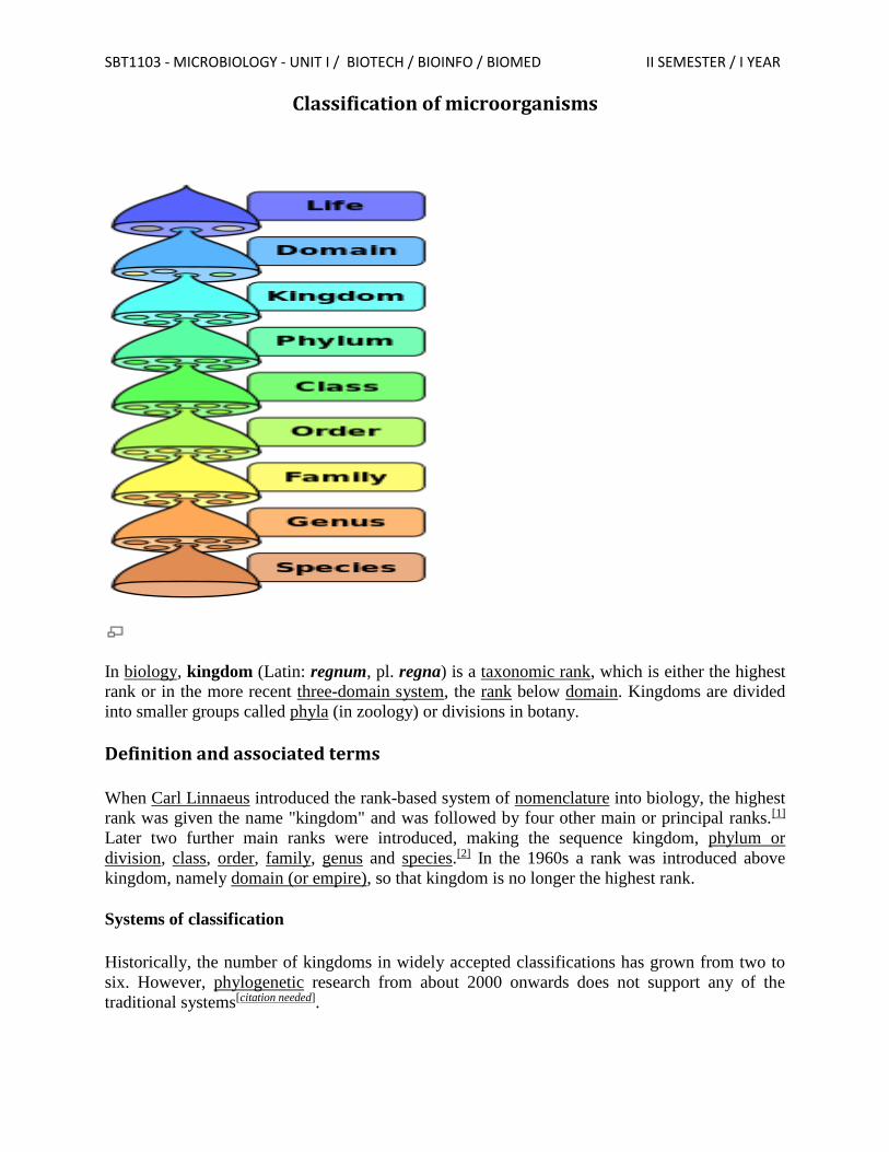
In biology, kingdom (Latin: regnum, pl. regna) is a taxonomic rank, which is either the highest
rank or in the more recent three-domain system, the rank below domain. Kingdoms are divided
into smaller groups called phyla (in zoology) or divisions in botany.
Definition and associated terms
When Carl Linnaeus introduced the rank-based system of nomenclature into biology, the highest
rank was given the name "kingdom" and was followed by four other main or principal ranks.[1]
Later two further main ranks were introduced, making the sequence kingdom, phylum or
division, class, order, family, genus and species.[2] In the 1960s a rank was introduced above
kingdom, namely domain (or empire), so that kingdom is no longer the highest rank.
Systems of classification
Historically, the number of kingdoms in widely accepted classifications has grown from two to
six. However, phylogenetic research from about 2000 onwards does not support any of the
traditional systems[citation needed].
SBT1103 - MICROBIOLOGY - UNIT I / BIOTECH / BIOINFO / BIOMED II SEMESTER / I YEAR
Classical classification
An initial dichotomy: Two kingdoms
The classification of living things into animals and plants is an ancient one. Aristotle (384–322
BC) classified animal species in his work The History of Animals, and his pupil Theophrastus (c.
371–c. 287 BC) wrote a parallel work on plants (Historia Plantarum (The History of Plants)).[4]
Carolus Linnaeus (1707–1778) laid the foundations for modern biological nomenclature, now
regulated by the Nomenclature Codes. He distinguished two kingdoms of living things: Regnum
Animale ('animal kingdom') for animals and Regnum Vegetabile ('vegetable kingdom') for plants.
(Linnaeus also included minerals, placing them in a third kingdom, Regnum Lapideum.)
Linnaeus divided each kingdom into classes, later grouped into phyla for animals and divisions
for plants.
life Regnum Vegetabile Regnum Animalia
An increasing number of kingdoms
Three kingdoms
In 1674, Antonie van Leeuwenhoek, often called the "father of microscopy", sent the Royal
Society of London a copy of his first observations of microscopic single-celled organisms. Until
then, the existence of such microscopic organisms was entirely unknown. In 1866, following
earlier proposals by Richard Owen and Ernst Haeckel proposed a third kingdom of life. Haeckel
revised the content of this kingdom a number of times before settling on a division based on
whether organisms were unicellular (Protista) or multicellular (animals and plants).[5]
life Kingdom Protista Kingdom Plantae Kingdom Animalia
Four kingdoms
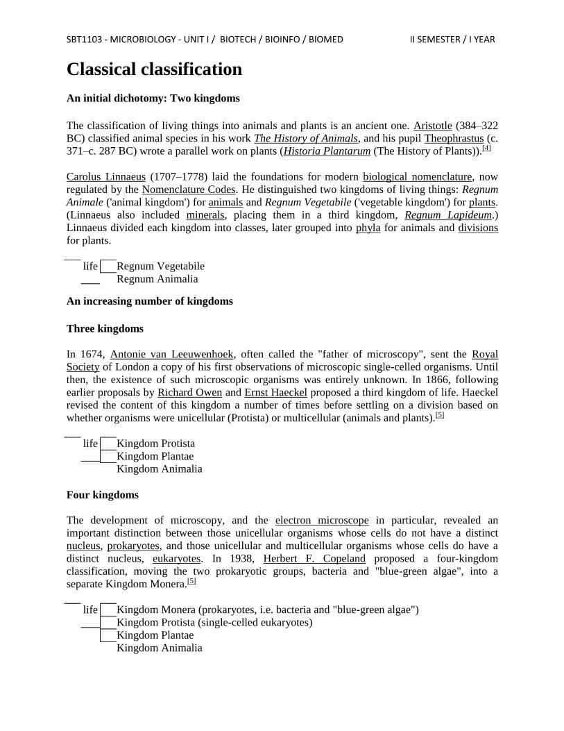
The development of microscopy, and the electron microscope in particular, revealed an
important distinction between those unicellular organisms whose cells do not have a distinct
nucleus, prokaryotes, and those unicellular and multicellular organisms whose cells do have a
distinct nucleus, eukaryotes. In 1938, Herbert F. Copeland proposed a four-kingdom
classification, moving the two prokaryotic groups, bacteria and "blue-green algae", into a
separate Kingdom Monera.[5]
life Kingdom Monera (prokaryotes, i.e. bacteria and "blue-green algae") Kingdom Protista (single-celled eukaryotes) Kingdom Plantae Kingdom Animalia
SBT1103 - MICROBIOLOGY - UNIT I / BIOTECH / BIOINFO / BIOMED II SEMESTER / I YEAR
Five kingdoms (Whittaker system )
The differences between fungi and other organisms regarded as plants had long been recognized.
For example, at one point Haeckel moved the fungi out of Plantae into Protista, before changing
his mind.[5] Robert Whittaker recognized an additional kingdom for the Fungi. The resulting
five-kingdom system, proposed in 1969 by Whittaker, has become a popular standard and with
some refinement is still used in many works and forms the basis for new multi-kingdom systems.
It is based mainly on differences in nutrition; his Plantae were mostly multicellular autotrophs,
his Animalia multicellular heterotrophs, and his Fungi multicellular saprotrophs. The remaining
two kingdoms, Protista and Monera, included unicellular and simple cellular colonies.[7] The five
kingdom system may be combined with the two empire system.
life Empire Prokaryota Kingdom Monera
Empire Eukaryota Kingdom Fungi Kingdom Protista Kingdom Plantae Kingdom Animalia
In the Whittaker system, Plantae included some algae. In other systems (e.g., Margulis system),
Plantae included just the land plants (Embryophyta).
Recent developments: six kingdoms or more?
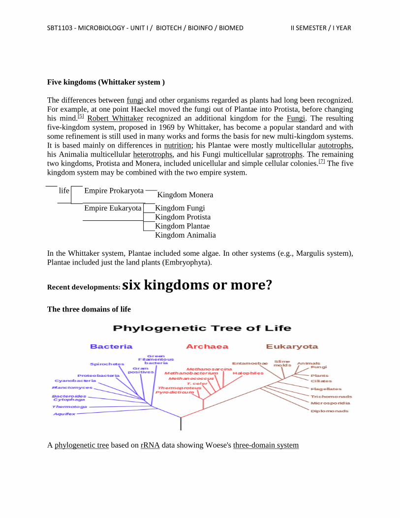
The three domains of life
A phylogenetic tree based on rRNA data showing Woese's three-domain system
SBT1103 - MICROBIOLOGY - UNIT I / BIOTECH / BIOINFO / BIOMED II SEMESTER / I YEAR
From around the mid-1970s onwards, there was an increasing emphasis on comparisons of genes
on the molecular level (initially ribosomal RNA genes) as the primary factor in classification;
genetic similarity was stressed over outward appearances and behavior. Taxonomic ranks,
including kingdoms, were to be groups of organisms with a common ancestor, whether
monophyletic (all descendants of a common ancestor) or paraphyletic (only some descendants of
a common ancestor).
Based on such RNA studies, Carl Woese divided the prokaryotes (hitherto classified as the
Kingdom Monera) into two groups, called Eubacteria and Archaebacteria, stressing that there
was as much genetic difference between these two groups as between either of them and all
eukaryotes. Similarly, though eukaryote groups such as plants, fungi and animals may look
different, they are more closely related to each other from a genetic standpoint than they are to
either the Eubacteria or Archaebacteria. It was also found that the eukaryotes are more closely
related, genetically, to the Archaebacteria than they are to the Eubacteria.
Although the primacy of the eubacteria-archaebacteria divide has been questioned, it has been
upheld by subsequent research.[8]
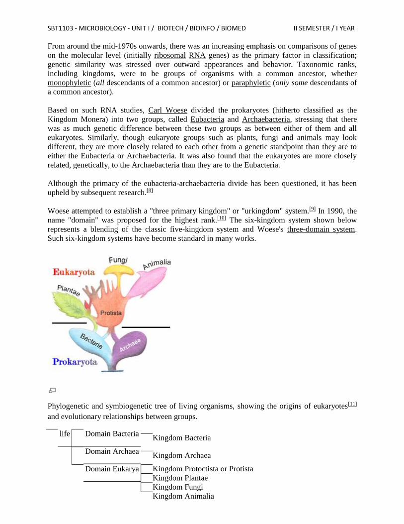
Woese attempted to establish a "three primary kingdom" or "urkingdom" system.[9] In 1990, the
name "domain" was proposed for the highest rank.[10] The six-kingdom system shown below
represents a blending of the classic five-kingdom system and Woese's three-domain system.
Such six-kingdom systems have become standard in many works.
Phylogenetic and symbiogenetic tree of living organisms, showing the origins of eukaryotes[11]
and evolutionary relationships between groups.
life Domain Bacteria Kingdom Bacteria
Domain Archaea Kingdom Archaea
Domain Eukarya Kingdom Protoctista or Protista Kingdom Plantae Kingdom Fungi Kingdom Animalia
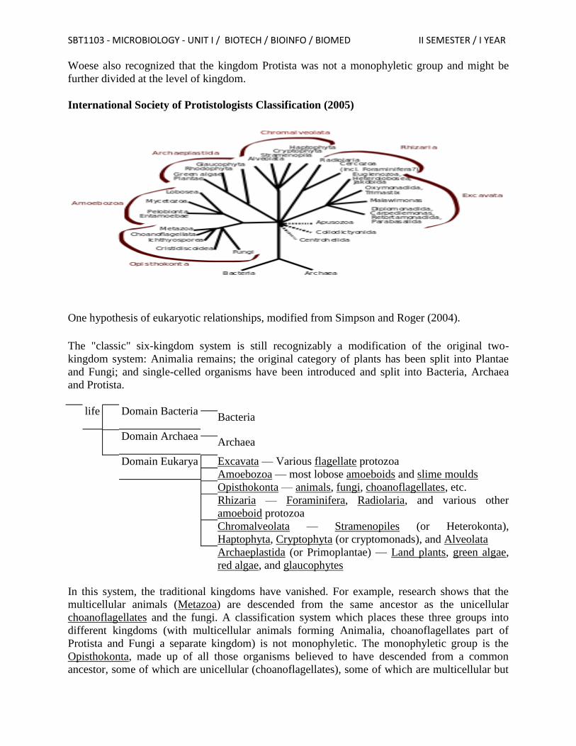
SBT1103 - MICROBIOLOGY - UNIT I / BIOTECH / BIOINFO / BIOMED II SEMESTER / I YEAR
Woese also recognized that the kingdom Protista was not a monophyletic group and might be
further divided at the level of kingdom.
International Society of Protistologists Classification (2005)
One hypothesis of eukaryotic relationships, modified from Simpson and Roger (2004).
The "classic" six-kingdom system is still recognizably a modification of the original two-
kingdom system: Animalia remains; the original category of plants has been split into Plantae
and Fungi; and single-celled organisms have been introduced and split into Bacteria, Archaea
and Protista.
life Domain Bacteria Bacteria
Domain Archaea Archaea
Domain Eukarya Excavata — Various flagellate protozoa Amoebozoa — most lobose amoeboids and slime moulds Opisthokonta — animals, fungi, choanoflagellates, etc. Rhizaria — Foraminifera, Radiolaria, and various other
amoeboid protozoa
Chromalveolata — Stramenopiles (or Heterokonta),
Haptophyta, Cryptophyta (or cryptomonads), and Alveolata
Archaeplastida (or Primoplantae) — Land plants, green algae,
red algae, and glaucophytes
In this system, the traditional kingdoms have vanished. For example, research shows that the
multicellular animals (Metazoa) are descended from the same ancestor as the unicellular
choanoflagellates and the fungi. A classification system which places these three groups into
different kingdoms (with multicellular animals forming Animalia, choanoflagellates part of
Protista and Fungi a separate kingdom) is not monophyletic. The monophyletic group is the
Opisthokonta, made up of all those organisms believed to have descended from a common
ancestor, some of which are unicellular (choanoflagellates), some of which are multicellular but
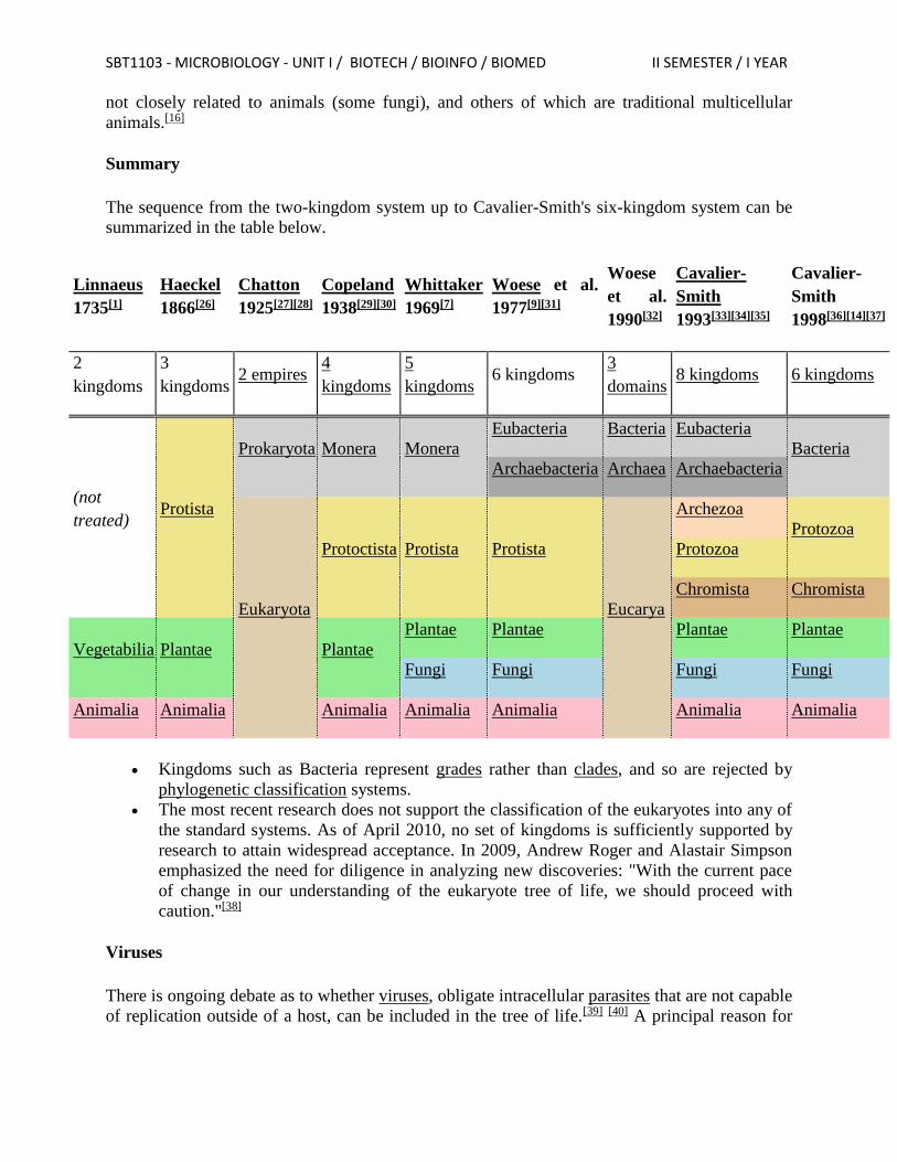
SBT1103 - MICROBIOLOGY - UNIT I / BIOTECH / BIOINFO / BIOMED II SEMESTER / I YEAR
not closely related to animals (some fungi), and others of which are traditional multicellular
animals.[16]
Summary
The sequence from the two-kingdom system up to Cavalier-Smith's six-kingdom system can be
summarized in the table below.
Linnaeus
1735[1]
Haeckel
1866[26]
Chatton
1925[27][28]
Copeland
1938[29][30]
Whittaker
1969[7]
Woese et al.
1977[9][31]
Woese
et al.
1990[32]
Cavalier-
Smith
1993[33][34][35]
Cavalier-
Smith
1998[36][14][37]
2
kingdoms
3
kingdoms 2 empires
4
kingdoms
5
kingdoms
6 kingdoms 3
domains
8 kingdoms 6 kingdoms
(not
treated) Protista
Prokaryota Monera Monera
Eubacteria Bacteria Eubacteria
Bacteria
Archaebacteria Archaea Archaebacteria
Eukaryota
Protoctista Protista Protista
Eucarya
Archezoa
Protozoa
Protozoa
Chromista Chromista
Vegetabilia Plantae Plantae
Plantae Plantae Plantae Plantae
Fungi Fungi Fungi Fungi
Animalia Animalia Animalia Animalia Animalia Animalia Animalia
Kingdoms such as Bacteria represent grades rather than clades, and so are rejected by
phylogenetic classification systems.
The most recent research does not support the classification of the eukaryotes into any of
the standard systems. As of April 2010, no set of kingdoms is sufficiently supported by
research to attain widespread acceptance. In 2009, Andrew Roger and Alastair Simpson
emphasized the need for diligence in analyzing new discoveries: "With the current pace
of change in our understanding of the eukaryote tree of life, we should proceed with
caution."[38]
Viruses
There is ongoing debate as to whether viruses, obligate intracellular parasites that are not capable
of replication outside of a host, can be included in the tree of life.[39] [40] A principal reason for
SBT1103 - MICROBIOLOGY - UNIT I / BIOTECH / BIOINFO / BIOMED II SEMESTER / I YEAR
inclusion comes from the discovery of unusually large and complex viruses, such as Mimivirus,
that possess typical cellular genes.[41]
Current classification
Knowledge of the phenotypic, genotypic and biological characteristics of a microorganism is
imperative in differentiating it from its pathogenic and/or toxigenic relatives or other
microorganisms that are detrimental to the health of plants, animals, humans and the
environment.
Polyphasic approach to microbial identification
The selection of method(s) used for microbial identification depends on the type and nature of
the microorganism. The method(s) chosen should be well-described in scientific literature and
consistent with those currently used in the field of microbial identification and taxonomic
classification and they must enable identification of the organisms to the genus and species and,
if possible, strain level. The robustness, precision and validity of the methodologies used to
identify the microorganism are critical elements in the assessment of the safety of the product.
The choice of methods for microbial identification is at the discretion of the product proponent.
However, the Fertilizer Safety Office recommends that applicants adopt an integrated polyphasic
approach that includes classical microbiological and phenotypic analysis along with molecular
tools, to accurately identify the active microorganism(s). The strengths and weaknesses of the
various identification methods should be taken into consideration, such that the methods chosen
complement each other to result in a conclusive and definitive identification of the
microorganism, and allow for clear differentiation of the organism from any closely related
pathogenic and/or toxigenic species and strains. The methods commonly used in identification
and substantiation of taxonomic classification of microorganisms are summarized below.
1. Phenotypic analysis
Preliminary analysis in microbial identification often involves one or more phenotypic methods.
Phenotypic methods are suitable for microorganisms that are culturable (i.e., can grow as pure
culture on artificial media), have well-established growth parameters, and physiological and
biochemical profiles.
a. Analysis of morphological traits
These methods utilize colony and cell morphology to obtain an initial identification of a
microorganism. This is accomplished through simple isolation and culturing of the
microorganism and subsequent visual observation using microscopy. The morphological
properties include:
1. shape,
2. size,
3. surface characteristics and pigmentation,
4. cell wall characteristics (Gram-staining),
5. sporulation characteristics,
6. mechanisms of motility, and
7. other cellular inclusions and ultrastructural characteristics.
SBT1103 - MICROBIOLOGY - UNIT I / BIOTECH / BIOINFO / BIOMED II SEMESTER / I YEAR
b. Analysis of biochemical, physiological and metabolic characteristics
Phenotypic identification methods include the study of the biochemical profile and metabolic
properties of a microorganism by testing its growth requirements, enzymatic activities and
cellular fatty acid composition.
The biochemical tests use specific growth media, nutrients, chemicals or growth conditions to
elicit an observable or measurable biochemical response from the microorganism, thereby
enabling its identification and characterization. These tests include: utilization of carbon and
nitrogen sources, growth requirements (anaerobic or aerobic; temperature-optimum and
range, pH optimum and range), preferred osmotic conditions, generation of fermentation
products, production of enzymes, production of antimicrobial compounds, as well as sensitivity
to metabolic inhibitors and antibiotics. Examples of recognized tests include: phenol red
carbohydrate, catalase and oxidase tests, oxidation-fermentation tests, methyl red tests, Voges-
Proskauer tests, nitrate reduction, starch hydrolysis, tryptophan hydrolysis, hydrogen sulfide
production, citrate utilization, litmus milk reactions, etc. Several miniaturized and automated
commercial systems are currently available with well-defined quality control procedures that
allow for rapid identification of microorganisms.
c. Analysis of Fatty Acid Methyl Ester composition (FAME analysis)
Microorganisms can be identified by analysing the fatty acid profiles of whole cells or cell
membranes using gas-liquid chromatography or mass spectrometry. The data on the type,
content, proportion and variation in the fatty acid profile are used to identify and characterize the
genus and species by comparing it against the fatty acid profiles of known organisms.
The expression of microbial phenotypes is highly dependent on environmental variables (e.g.,
culture pH, temperature, selective vs non-selective media, depletion of nutrients, presence of
stressors etc.), and thus, may introduce inconsistencies in the identification process. The
phenotypic methods are only acceptable if the response criteria are sufficient to identify the
microorganism with a high level of confidence and distinguish it from phylogenetically close
relatives that potentially pose safety concerns. Also, the applicability of the method is based on
the robustness of information in reference databases. As such, results from phenotypic methods
may require supporting data from other methods to accurately identify a microorganism.
2. Molecular Methods
Development of molecular methods has greatly improved the ability to rapidly detect, identify
and classify microorganisms and also establish the taxonomic relationship among closely related
genera and species. Identification, using molecular methods, relies on the comparison of the
nucleic acid sequences (DNA, RNA) or protein profiles of a microorganism with documented
data on known organisms. The molecular methods are considered sensitive enough to allow
detection of low concentrations of viable or non-viable microorganisms in both pure cultures and
complex samples (e.g., soil, peat, wateretc.).
a. Genotypic methods
These include methods such as nucleic acid hybridization (Southern blot analysis or Solution-
phase hybridization) and amplification-based or polymerase chain reaction (PCR) technologies.
The latter consist of sequence comparisons of conserved genomic regions such as 16S or
18S rRNA, or comparisons of restriction fragment length polymorphisms (RFLP), amplified
SBT1103 - MICROBIOLOGY - UNIT I / BIOTECH / BIOINFO / BIOMED II SEMESTER / I YEAR
fragment length polymorphisms (AFLP) or G+C % content in the genomic DNA with
corresponding data on known organisms.
Reliable genotypic identification requires databases with accurate and complete sequence
information from a large number of taxa. The commonly used gene sequence databases include:
GenBank®Footnote 2;
Ribosomal Database Project (RDP)Footnote 3;
Europe's collection of nucleotide sequence data (EMBL)Footnote 4; and
Universal Protein Resource (UniProt)Footnote 5.
Product proponents are not limited to using the reference materials listed above; these are
intended as guidance only.
Some of the limitations associated with genotypic methods also include: difficulties in
differentiating between species that share identical and/or similar conserved region sequences,
limited information on the quality of sequence data available in public databases and the
complexity of taxonomic nomenclature overall. Considering the above, it is important to validate
the results of genotypic microbial identification methods with data from other sources (e.g.,
morphological and/or phenotypic analysis).
b. Protein based methods
Serological methods such as Western blotting, Immuno-precipitation and Enzyme Linked
Immuno-sorbant Assay (ELISA) use antibodies to detect specific proteins that are unique and/or
characteristic of a microorganism. The applicability of serological methods is dependent on the
availability, sensitivity and specificity of the antibodies used. There are commercial kits
available for immuno-detection of several microorganisms. Protein based methods also include
gel electrophoresis (SDS-PAGE, 2D-gels, etc.) that can separate cellular proteins on a defined
matrix and identify microbial proteins of interest by comparing with microorganisms with known
protein profiles.
3. Genomics
More recently, complete profiling of the transcriptome, genome, proteome or metabolome have
been used to identify and characterise organisms. Several modern technologies such as DNA and
protein microarray analyses, mass spectral protein profiling, nuclear magnetic resonance (NMR)
spectral analysis, in-silico microbial metabolome platforms are increasingly used in
identification and characterization of microorganisms.
The knowledge of the sensitivity and specificity of genomic tools and their application in
microbial identification is rapidly evolving. However, challenges related to standardization of
genomics methodologies (including optimization of protocols and bioinformatics tools for
reliable data annotation, interpretation etc.) continue to hinder their applicability in safety (risk)
assessment and regulatory decision making. The Fertilizer Safety Office will consider data
generated by genomics-methods on a case-by-case basis. However, validation of genomics data
using alternate methods is currently required to substantiate the identification and taxonomic
classification of an active microorganism(s) in a supplement product.
SBT1103 - MICROBIOLOGY - UNIT I / BIOTECH / BIOINFO / BIOMED II SEMESTER / I YEAR
II. Taxonomic Classification and Nomenclature:
The taxonomic identification of the microorganism(s) should be based on the currently used and
internationally accepted taxonomic classification system. The description of the
microorganism(s) in the product and its characteristics must correspond to the characteristics
described in standard resources and/or references that are commonly used by the scientific
community to validate taxonomic classification. These can include but are not limited to:
textbooks such as the Bergey's Manual of Systematic BacteriologyFootnote 6;
The ProkaryotesFootnote 7;
Applied Microbial SystematicsFootnote 8;
Principles of fungal taxonomyFootnote 9 etc.;
online resources such as the Catalogue of LifeFootnote 10;
PubMed TaxonomyFootnote 11 and UniProt TaxonomyFootnote 12 etc.; and
peer reviewed journals.
The taxonomic name should follow the nomenclature code officially recognized by the
International Committee on Systematics of Prokaryotes (ICSP). Applicants should verify the
"Approved List of Bacterial Names" to ensure that the nomenclature is in accordance with the
latest Validation List developed and updated by the International Journal of Systematic and
Evolutionary Microbiology (IJSEM).
Please note that microbial taxonomic classification and nomenclature, particularly for bacteria, is
in a constant state of flux as methodologies evolve to generate more reliable information to
identify/classify and/or reclassify the current taxonomic scheme. Cross referencing more than
one resource/reference will help in validating the current taxonomic designation and
classification of a microorganism.
Methods of Identifying microbes
Based on
1. Morphological characteristics – shape and size – staining techniques
2. Chemical characteristics - microscopic observation
3. Cultural characteristics - spread , pour and streak plate method
4. Metabolic characteristics – biochemical tests
5. Antigenic characteristics – Ag –Ab reactions
6. Genetic characteristics - DNA sequencing
7. Pathogenic characteristics - Infection to human and plants
8. Ecological characteristics - climatic conditions
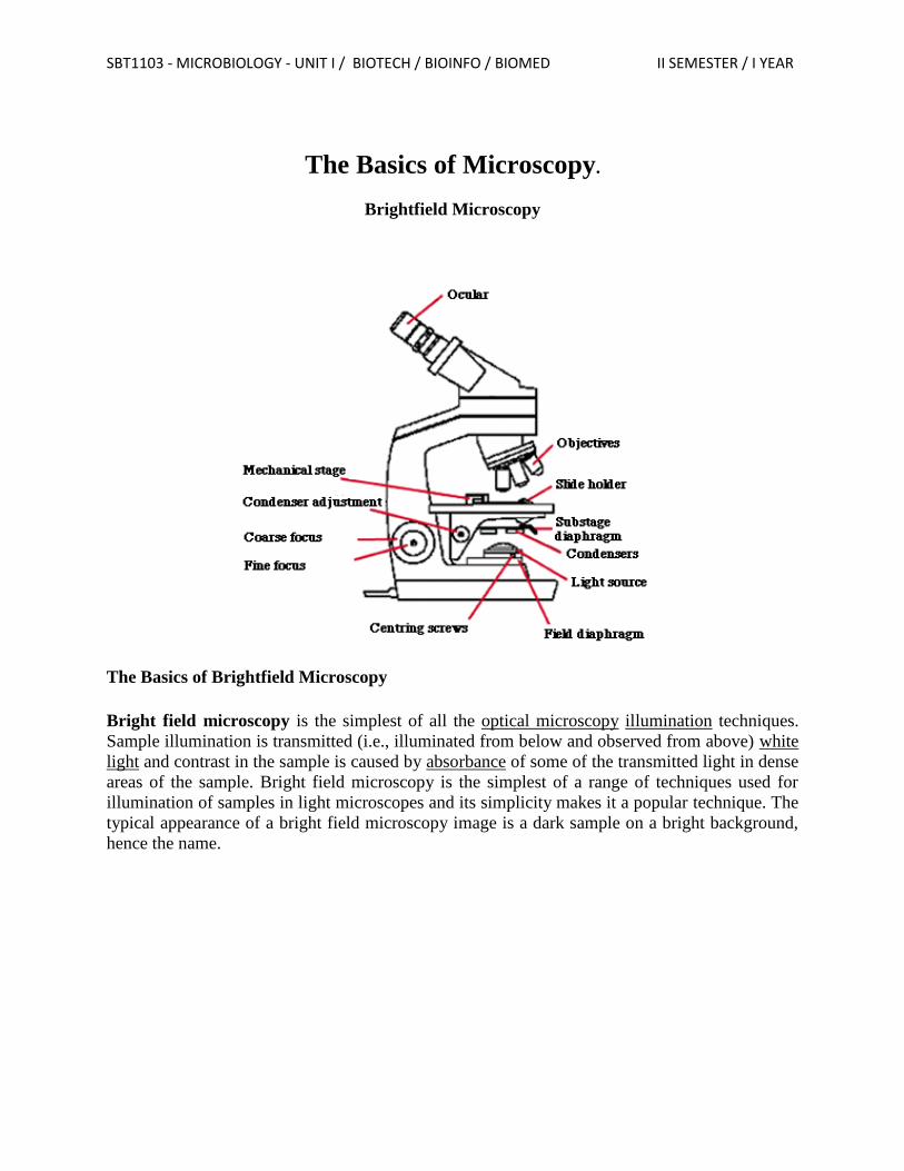
SBT1103 - MICROBIOLOGY - UNIT I / BIOTECH / BIOINFO / BIOMED II SEMESTER / I YEAR
The Basics of Microscopy.
Brightfield Microscopy
The Basics of Brightfield Microscopy
Bright field microscopy is the simplest of all the optical microscopy illumination techniques.
Sample illumination is transmitted (i.e., illuminated from below and observed from above) white
light and contrast in the sample is caused by absorbance of some of the transmitted light in dense
areas of the sample. Bright field microscopy is the simplest of a range of techniques used for
illumination of samples in light microscopes and its simplicity makes it a popular technique. The
typical appearance of a bright field microscopy image is a dark sample on a bright background,
hence the name.
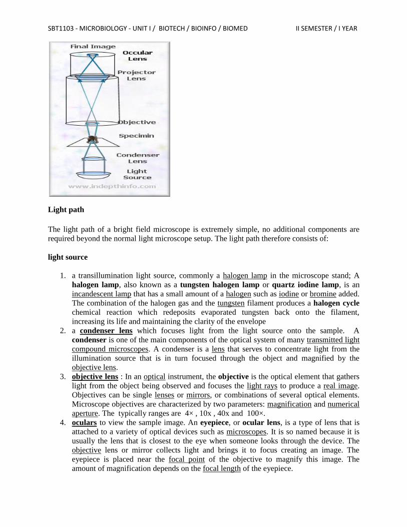
SBT1103 - MICROBIOLOGY - UNIT I / BIOTECH / BIOINFO / BIOMED II SEMESTER / I YEAR
Light path
The light path of a bright field microscope is extremely simple, no additional components are
required beyond the normal light microscope setup. The light path therefore consists of:
light source
1. a transillumination light source, commonly a halogen lamp in the microscope stand; A
halogen lamp, also known as a tungsten halogen lamp or quartz iodine lamp, is an
incandescent lamp that has a small amount of a halogen such as iodine or bromine added.
The combination of the halogen gas and the tungsten filament produces a halogen cycle
chemical reaction which redeposits evaporated tungsten back onto the filament,
increasing its life and maintaining the clarity of the envelope
2. a condenser lens which focuses light from the light source onto the sample. A
condenser is one of the main components of the optical system of many transmitted light
compound microscopes. A condenser is a lens that serves to concentrate light from the
illumination source that is in turn focused through the object and magnified by the
objective lens.
3. objective lens : In an optical instrument, the objective is the optical element that gathers
light from the object being observed and focuses the light rays to produce a real image.
Objectives can be single lenses or mirrors, or combinations of several optical elements.
Microscope objectives are characterized by two parameters: magnification and numerical
aperture. The typically ranges are 4× , 10x , 40x and 100×.
4. oculars to view the sample image. An eyepiece, or ocular lens, is a type of lens that is
attached to a variety of optical devices such as microscopes. It is so named because it is
usually the lens that is closest to the eye when someone looks through the device. The
objective lens or mirror collects light and brings it to focus creating an image. The
eyepiece is placed near the focal point of the objective to magnify this image. The
amount of magnification depends on the focal length of the eyepiece.
SBT1103 - MICROBIOLOGY - UNIT I / BIOTECH / BIOINFO / BIOMED II SEMESTER / I YEAR
Magnification is the process of enlarging something only in appearance, not in physical size.
Typically magnification is related to scaling up visuals or images to be able to see more detail,
increasing resolution.
Resolving power is the ability of an imaging device to separate (i.e., to see as distinct) points of
an object that are located at a small angular distance..
In optics, the numerical aperture (NA) of an optical system is a dimensionless number that
characterizes the range of angles over which the system can accept or emit light. In most areas of
optics, and especially in microscopy, the numerical aperture of an optical system such as an
objective lens is defined by
where n is the index of refraction of the medium in which the lens is working (1.0 for air, 1.33
for pure water, and up to 1.56 for oils; see also list of refractive indices), and θ is the half-angle
of the maximum cone of light that can enter or exit the lens. In general, this is the angle of the
real marginal ray in the system
Working Performance
Bright field microscopy typically has low contrast with most biological samples as few absorb
light to a great extent. Staining is often required to increase contrast, which prevents use on live
cells in many situations. Bright field illumination is useful for samples which have an intrinsic
colour, for example chloroplasts in plant cells.
Light is first emitted by the light source and is directed by the condenser lens on to the
specimen, which might be a loose object, a prepared plate or almost anything. A microscope can
even be applied to small parts of larger objects, though with a bit more difficulty. (The light does
not absolutely need to originate below the specimen.)
The light from the specimen then passes through the objective lens. This lens is often selected
from among three or four and is the main determinant for the level of magnification. It bends the
light rays and in the case of this example sends them to a projector lens, which reverses their
direction so that when the image reaches the eye it will not appear "upside-down". Not all
microscopes have a projector lens, so the viewer may be seeing a reverse image. In these cases,
when the slide is moved, it will appear to be moving in the opposite direction to the viewer.
The light rays then travel to the oracular lens or "eye piece". This is often a 10X magnification
lens, meaning it magnifies the magnified image an additional ten times. The image is then
projected into the eye. It is very seldom that a specimen is in focus the moment it is placed
beneath a microscope. This means that some adjustment will have to be made. Unlike in
telescopes, the focal length between lenses remains constant when adjusting the focus. The lens
apparatus is brought closer to or further from the object. The focus adjustment is often along the
SBT1103 - MICROBIOLOGY - UNIT I / BIOTECH / BIOINFO / BIOMED II SEMESTER / I YEAR
neck of the tube containing the lenses, but it might just as well move the slide up and down. The
best way to make this adjustment is to make a course adjustment so that it is too close to the
object and then back off with the fine adjustment2. This helps to ensure that the specimen is not
inadvertently smashed by the lens.
Advantages
The name "brightfield" is derived from the fact that the specimen is dark and contrasted by the
surrounding bright viewing field. Simple light microscopes are sometimes referred to as bright
field microscopes.
Brightfield microscopy is very simple to use with fewer adjustments needed to be made to view
specimens.
Some specimens can be viewed without staining and the optics used in the brightfield technique
don’t alter the color of the specimen.
It is adaptable with new technology and optional pieces of equipment can be implemented with
brightfield illumination to give versatility in the tasks it can perform.
Disadvantages
Certain disadvantages are inherent in any optical imaging technique.
By using an aperture diaphragm for contrast, past a certain point, greater contrast adds
distortion. However, employing an iris diaphragm will help compensate for this problem.
Brightfield microscopy can’t be used to observe living specimens of bacteria, although
when using fixed specimens, bacteria have an optimum viewing magnification of 1000x.
Brightfield microscopy has very low contrast and most cells absolutely have to be stained to be
seen; staining may introduce extraneous details into the specimen that should not be present.
Also, the user will need to be knowledgeable in proper staining techniques.
Lastly, this method requires a strong light source for high magnification applications and intense
lighting can produce heat that will damage specimens or kill living microorganisms.
The most important feature of a microscope is its resolution, the ability to see fine details. Once
you can resolve fine details then you can magnify them.
Every optical system has a finite resolution, if you magnify objects beyond the resolution the
result will be empty magnification. So, the actual purpose of a microscope is to see small things
clearly.
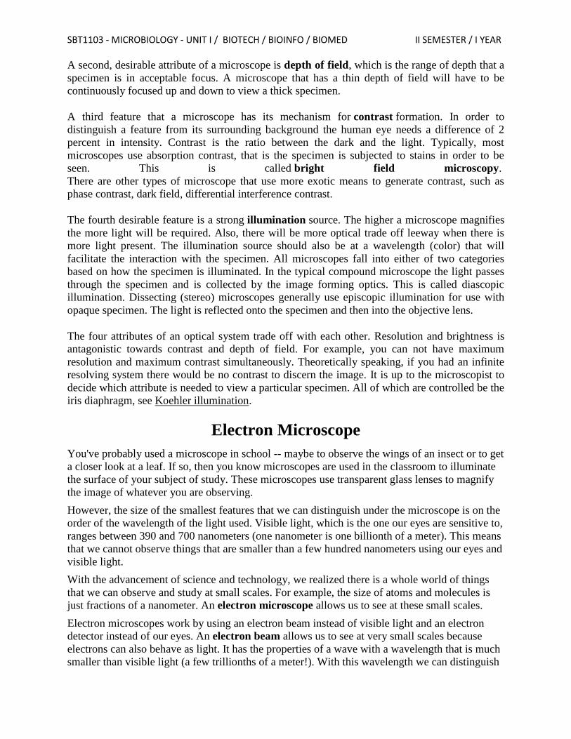
SBT1103 - MICROBIOLOGY - UNIT I / BIOTECH / BIOINFO / BIOMED II SEMESTER / I YEAR
A second, desirable attribute of a microscope is depth of field, which is the range of depth that a
specimen is in acceptable focus. A microscope that has a thin depth of field will have to be
continuously focused up and down to view a thick specimen.
A third feature that a microscope has its mechanism for contrast formation. In order to
distinguish a feature from its surrounding background the human eye needs a difference of 2
percent in intensity. Contrast is the ratio between the dark and the light. Typically, most
microscopes use absorption contrast, that is the specimen is subjected to stains in order to be
seen. This is called bright field microscopy.
There are other types of microscope that use more exotic means to generate contrast, such as
phase contrast, dark field, differential interference contrast.
The fourth desirable feature is a strong illumination source. The higher a microscope magnifies
the more light will be required. Also, there will be more optical trade off leeway when there is
more light present. The illumination source should also be at a wavelength (color) that will
facilitate the interaction with the specimen. All microscopes fall into either of two categories
based on how the specimen is illuminated. In the typical compound microscope the light passes
through the specimen and is collected by the image forming optics. This is called diascopic
illumination. Dissecting (stereo) microscopes generally use episcopic illumination for use with
opaque specimen. The light is reflected onto the specimen and then into the objective lens.
The four attributes of an optical system trade off with each other. Resolution and brightness is
antagonistic towards contrast and depth of field. For example, you can not have maximum
resolution and maximum contrast simultaneously. Theoretically speaking, if you had an infinite
resolving system there would be no contrast to discern the image. It is up to the microscopist to
decide which attribute is needed to view a particular specimen. All of which are controlled be the
iris diaphragm, see Koehler illumination.
Electron Microscope
You've probably used a microscope in school -- maybe to observe the wings of an insect or to get
a closer look at a leaf. If so, then you know microscopes are used in the classroom to illuminate
the surface of your subject of study. These microscopes use transparent glass lenses to magnify
the image of whatever you are observing.
However, the size of the smallest features that we can distinguish under the microscope is on the
order of the wavelength of the light used. Visible light, which is the one our eyes are sensitive to,
ranges between 390 and 700 nanometers (one nanometer is one billionth of a meter). This means
that we cannot observe things that are smaller than a few hundred nanometers using our eyes and
visible light.
With the advancement of science and technology, we realized there is a whole world of things
that we can observe and study at small scales. For example, the size of atoms and molecules is
just fractions of a nanometer. An electron microscope allows us to see at these small scales.
Electron microscopes work by using an electron beam instead of visible light and an electron
detector instead of our eyes. An electron beam allows us to see at very small scales because
electrons can also behave as light. It has the properties of a wave with a wavelength that is much
smaller than visible light (a few trillionths of a meter!). With this wavelength we can distinguish

SBT1103 - MICROBIOLOGY - UNIT I / BIOTECH / BIOINFO / BIOMED II SEMESTER / I YEAR
features down to a fraction of a nanometer. Let's explore the different types of electron
microscopes, how they work and some of their applications.
Types of Electron Microscopes
Scanning Electron Microscope (SEM)
In a scanning electron microscope or SEM, a beam of electrons scans the surface of a sample
(Figure 1). The electrons interact with the material in a way that triggers the emission
of secondary electrons. These secondary electrons are captured by a detector, which forms an
image of the surface of the sample. The direction of the emission of the secondary electrons
depends on the orientation of the features of the surface. There, the image formed will reflect the
characteristic feature of the region of the surface that was exposed to the electron beam.
Figure 1. A Scanning Electron Microscope focuses a beam of electrons on the surface of a
sample. Secondary electrons are emitted from the sample surface. A detector is used to form
an image from the secondary electrons.
Transmission Electron Microscope (TEM)
In a transmission electron microscope or TEM, a beam of electrons hits a very thin sample
(usually no more than 100 nm thick). The electrons are transmitted through the sample (Figure
2). After the sample, the electrons hit a fluorescence screen that forms an image with the
electrons that were transmitted. You can better understand this process by imagining how a
movie projector works. In a projector, you have a film that has the negative image that will be
projected. The projector shines white light on the negative and the light transmitted forms the
image contained in the negative.
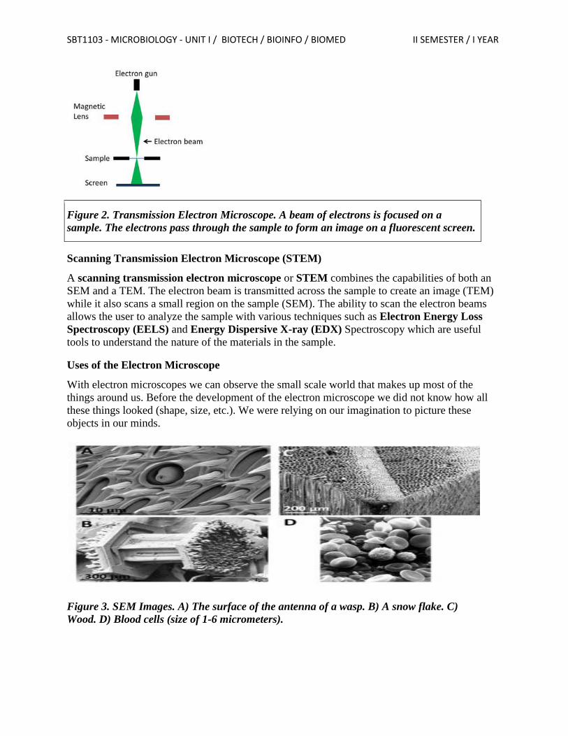
SBT1103 - MICROBIOLOGY - UNIT I / BIOTECH / BIOINFO / BIOMED II SEMESTER / I YEAR
Figure 2. Transmission Electron Microscope. A beam of electrons is focused on a
sample. The electrons pass through the sample to form an image on a fluorescent screen.
Scanning Transmission Electron Microscope (STEM)
A scanning transmission electron microscope or STEM combines the capabilities of both an
SEM and a TEM. The electron beam is transmitted across the sample to create an image (TEM)
while it also scans a small region on the sample (SEM). The ability to scan the electron beams
allows the user to analyze the sample with various techniques such as Electron Energy Loss
Spectroscopy (EELS) and Energy Dispersive X-ray (EDX) Spectroscopy which are useful
tools to understand the nature of the materials in the sample.
Uses of the Electron Microscope
With electron microscopes we can observe the small scale world that makes up most of the
things around us. Before the development of the electron microscope we did not know how all
these things looked (shape, size, etc.). We were relying on our imagination to picture these
objects in our minds.
Figure 3. SEM Images. A) The surface of the antenna of a wasp. B) A snow flake. C)
Wood. D) Blood cells (size of 1-6 micrometers).
SBT1103 - MICROBIOLOGY - UNIT I / BIOTECH / BIOINFO / BIOMED II SEMESTER / I YEAR
Simple Staining Technique
In a simple staining technique, a basic, cationic dye is flooded across a sample, adding color to
the cells. Before we move on, let's define the word cationic. A cation is simply a positively
charged ion. The molecules that make up basic dyes have a positive charge. This is important
because the cell wall and cytoplasm of bacterial cells have a negative charge. The positively
charged dye is attracted to the negatively charged cells, enhancing the ability of the stain to stick
to and color the cells. Now, those nearly colorless cells should pop off the slide in any number of
colors.
It is important to note that before a sample can be stained with a simple stain, it must be heat
fixed to the slide. During heat fixation, a glass slide is waved over an open flame. This kills the
bacteria, attaches the cells to the slide, and enhances the stain uptake. This process makes
staining more effective but can damage or distort the cells, changing their appearance from a
truly natural, free-living state.
Methylene blue is a classic example of a simple stain. This blue stain will color all cells blue,
making them stand out against the bright background of the light microscope. Notice how the
background remains generally clear, while the bacterial cells are a deep blue.
Procedure:
1. Clean and dry microscope slides thoroughly.
2. Flame the surface in which the smear is to be spread.
3. Flame the inoculating loop.
4. Transfer a loop full of tap water to the flamed slide surface.
5. Reflame the loop making sure the entire length of the wire that will enter the tube has been
heated to redness
6. Remove the tube cap with the fingers of the hand holding the loop.
7. Flame the tube mouth.
8. Touch the inoculating loop to the inside of the tube to make sure it is not so hot that it will
distort the bacterial cells; then pick up a pinhead size sample of the bacterial growth without
digging into the agar
9. Reflame the tube mouth, replace the can, and put the tube back in the holder.
10. Disperse the bacteria on the loop in the drop of water on the slide and spread the drop over
an area the siz of a dime. It should be a thin, even smear.
11. Reflame the inoculating loop to redness including the entire length that entered the tube.
12. Allow the smear to dry thoroughly.
13. Heat-fix the smear cautiously by passing the underside of the slide through the burner flame
two or three times. Test the temperature of the slide after each pass against the back of the
hand. It has been heated sufficiently when it feels hot but can still be held against the skin for
several seconds. Overheating will distort the cells
14. Stain the smear by flooding it with one of the staining solutions and allowing it to remain
covered with the stain for the time designated below.
Methylene blue - 1 minute
Crystal violet - 30 seconds
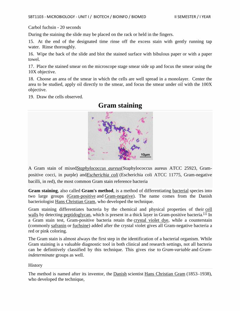
SBT1103 - MICROBIOLOGY - UNIT I / BIOTECH / BIOINFO / BIOMED II SEMESTER / I YEAR
Carbol fuchsin - 20 seconds
During the staining the slide may be placed on the rack or held in the fingers.
15. At the end of the designated time rinse off the excess stain with gently running tap
water. Rinse thoroughly.
16. Wipe the back of the slide and blot the stained surface with bibulous paper or with a paper
towel.
17. Place the stained smear on the microscope stage smear side up and focus the smear using the
10X objective.
18. Choose an area of the smear in which the cells are well spread in a monolayer. Center the
area to be studied, apply oil directly to the smear, and focus the smear under oil with the 100X
objective.
19. Draw the cells observed.
Gram staining
A Gram stain of mixedStaphylococcus aureus(Staphylococcus aureus ATCC 25923, Gram-
positive cocci, in purple) andEscherichia coli (Escherichia coli ATCC 11775, Gram-negative
bacilli, in red), the most common Gram stain reference bacteria
Gram staining, also called Gram's method, is a method of differentiating bacterial species into
two large groups (Gram-positive and Gram-negative). The name comes from the Danish
bacteriologist Hans Christian Gram, who developed the technique.
Gram staining differentiates bacteria by the chemical and physical properties of their cell
walls by detecting peptidoglycan, which is present in a thick layer in Gram-positive bacteria.[1] In
a Gram stain test, Gram-positive bacteria retain the crystal violet dye, while a counterstain
(commonly safranin or fuchsine) added after the crystal violet gives all Gram-negative bacteria a
red or pink coloring.
The Gram stain is almost always the first step in the identification of a bacterial organism. While
Gram staining is a valuable diagnostic tool in both clinical and research settings, not all bacteria
can be definitively classified by this technique. This gives rise to Gram-variable and Gram-
indeterminate groups as well.
History
The method is named after its inventor, the Danish scientist Hans Christian Gram (1853–1938),
who developed the technique,
SBT1103 - MICROBIOLOGY - UNIT I / BIOTECH / BIOINFO / BIOMED II SEMESTER / I YEAR
Process
Step 1. Prepare a slide with the culture by transferring the specimen to be examined onto a
drop of suspension medium (distilled water) using an inoculation loop. Spread the specimen
on the slide to ensure that it is not clumped.
Step 2. Fix the culture by heating the slide over a Bunsen burner to evaporate the water --
make sure not to hold the slide over the flame too long or it will denature the specimen.
Step 3. Drop a few drops of crystal violet stain onto the fixed culture (enough to cover the
specimen) and let it stand for 20 seconds. Then pour off the crystal violet stain and gently
rinse the excess stain with distilled water.
The objective of this step is:
To allow the crystal violet stain to bind to the peptidoglycan molecules of the Gram
+ bacteria (if present). Remember, Gram + bacteria have a large peptidoglycan layer
located outside the bacterial "inner membrane".
To allow the crystal violet stain to bind to the lipopolysaccharide molecules attached
to the "outer membrane" of the Gram - bacteria (if present). Remember, while Gram
+ bacteria have no "outer membrane", Gram - bacteria have lipopolysaccharide
molecules attached to their bacterial "outer membrane".
Step 4. Drop a few drops of iodine solution on the smear and let it stand for 20 seconds.
Then pour off the iodine solution and rinse the slide with distilled water.
The objective of this step is to fix the crystal violet to the peptidoglycan molecules on the
Gram + bacteria (if present).
Step 5. Drop a few drops of decolorizer (acetone or ethanol) and let the solution trickle down
off the slide until the decolorizer has removed enough of the color to drip off clear. Then
IMMEDIATELY rinse the slide off with distilled water after 5 seconds. Note that pouring
too much decolorier will cause the decolorization of the Gram + bacterial cells (in addition to
the Gram - bacteria), and the purpose of staining will be defeated.
The objective of this step is to dissolve the lipopolysaccharide membraine in the Gram -
bacteria and expose the thin peptidoglycan layer below.
Step 6. Drop a few drops of basic counterstain (fuchsin or safranin) on the slide and let it sit
for 20 seconds, then wash off the solution with distilled water.
The objective of this step is to stain the peptidoglycan layer of the Gram - bacteria a pink
/ red color. Remember that the addition of iodine to the crystal violet in Step 4 binds the
crystal violet stain in the Gram + bacteria, so the counterstain is unable to bind to the
peptidoglycan wall in the Gram + bacteria in the specimen.
Step 7. Observe the slide under light microscope.
Uses
Gram staining is a bacteriological laboratory technique[4] used to differentiate bacterial species
into two large groups (gram-positive and gram-negative) based on the physical properties of
SBT1103 - MICROBIOLOGY - UNIT I / BIOTECH / BIOINFO / BIOMED II SEMESTER / I YEAR
their cell walls.[5] Gram staining is not used to classify archaea, formerly archaeabacteria, since
these microorganisms yield widely varying responses that do not follow their phylogenetic
groups.[6]
Staining mechanism
Gram-positive bacteria have a thick mesh-like cell wall made of peptidoglycan (50–90% of cell
envelope), and as a result are stained purple by crystal violet, whereas Gram-negative bacteria
have a thinner layer (10% of cell envelope), so do not retain the purple stain and are counter-
stained pink by the Safranin. There are four basic steps of the Gram stain:
Applying a primary stain (crystal violet) to a heat-fixed smear of a bacterial culture. Heat
fixation kills some bacteria but is mostly used to affix the bacteria to the slide so that they
don't rinse out during the staining procedure.
The addition of iodide, which binds to crystal violet and traps it in the cell,
Rapid decolorization with ethanol or acetone, and
Counterstaining with safranin.[9] Carbol fuchsin is sometimes substituted for safranin since it
more intensely stains anaerobic bacteria, but it is less commonly used as a counterstain.[10]
Crystal violet (CV) dissociates in aqueous solutions into CV+ and chloride (Cl−) ions. These
ions penetrate through the cell wall and cell membrane of both Gram-positive and Gram-
negative cells. The CV+ ion interacts with negatively charged components of bacterial cells and
stains the cells purple. Iodide (I−or I−
3) interacts with CV+
and forms large complexes of crystal violet and iodine (CV–I) within the inner and outer layers
of the cell. Iodine is often referred to as a mordant, but is a trapping agent that prevents the
removal of the CV–I complex and, therefore, color the cell.[11]
When a decolorizer such as alcohol or acetone is added, it interacts with the lipids of the cell
membrane. A gram-negative cell loses its outer lipopolysaccharide membrane, and the inner
peptidoglycan layer is left exposed. The CV–I complexes are washed from the gram-negative
cell along with the outer membrane. In contrast, a gram-positive cell becomes dehydrated from
an ethanol treatment. The large CV–I complexes become trapped within the gram-positive cell
due to the multilayered nature of its peptidoglycan. The decolorization step is critical and must
be timed correctly; the crystal violet stain is removed from both gram-positive and negative cells
if the decolorizing agent is left on too long (a matter of seconds).
After decolorization, the gram-positive cell remains purple and the gram-negative cell loses its
purple color. Counterstain, which is usually positively charged safranin or basic fuchsine, is
applied last to give decolorized gram-negative bacteria a pink or red color.[12][13]
Some bacteria, after staining with the Gram stain, yield a gram-variable pattern: a mix of pink
and purple cells are seen. The
genera Actinomyces, Arthobacter,Corynebacterium, Mycobacterium,
and Propionibacterium have cell walls particularly sensitive to breakage during cell division,
resulting in gram-negative staining of these gram-positive cells. In cultures
of Bacillus, Butyrivibrio, and Clostridium, a decrease in peptidoglycan thickness during growth
coincides with an increase in the number of cells that stain gram-negative.[14] In addition, in all
bacteria stained using the Gram stain, the age of the culture may influence the results of the stain.
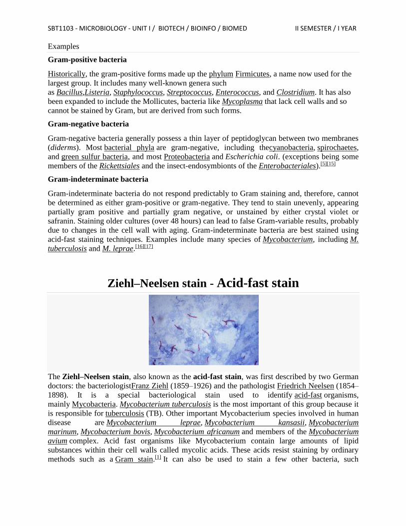
SBT1103 - MICROBIOLOGY - UNIT I / BIOTECH / BIOINFO / BIOMED II SEMESTER / I YEAR
Examples
Gram-positive bacteria
Historically, the gram-positive forms made up the phylum Firmicutes, a name now used for the
largest group. It includes many well-known genera such
as Bacillus,Listeria, Staphylococcus, Streptococcus, Enterococcus, and Clostridium. It has also
been expanded to include the Mollicutes, bacteria like Mycoplasma that lack cell walls and so
cannot be stained by Gram, but are derived from such forms.
Gram-negative bacteria
Gram-negative bacteria generally possess a thin layer of peptidoglycan between two membranes
(diderms). Most bacterial phyla are gram-negative, including thecyanobacteria, spirochaetes,
and green sulfur bacteria, and most Proteobacteria and Escherichia coli. (exceptions being some
members of the Rickettsiales and the insect-endosymbionts of the Enterobacteriales).[5][15]
Gram-indeterminate bacteria
Gram-indeterminate bacteria do not respond predictably to Gram staining and, therefore, cannot
be determined as either gram-positive or gram-negative. They tend to stain unevenly, appearing
partially gram positive and partially gram negative, or unstained by either crystal violet or
safranin. Staining older cultures (over 48 hours) can lead to false Gram-variable results, probably
due to changes in the cell wall with aging. Gram-indeterminate bacteria are best stained using
acid-fast staining techniques. Examples include many species of Mycobacterium, including M.
tuberculosis and M. leprae.[16][17]
Ziehl–Neelsen stain - Acid-fast stain
The Ziehl–Neelsen stain, also known as the acid-fast stain, was first described by two German
doctors: the bacteriologistFranz Ziehl (1859–1926) and the pathologist Friedrich Neelsen (1854–
1898). It is a special bacteriological stain used to identify acid-fast organisms,
mainly Mycobacteria. Mycobacterium tuberculosis is the most important of this group because it
is responsible for tuberculosis (TB). Other important Mycobacterium species involved in human
disease are Mycobacterium leprae, Mycobacterium kansasii, Mycobacterium
marinum, Mycobacterium bovis, Mycobacterium africanum and members of the Mycobacterium
avium complex. Acid fast organisms like Mycobacterium contain large amounts of lipid
substances within their cell walls called mycolic acids. These acids resist staining by ordinary
methods such as a Gram stain.[1] It can also be used to stain a few other bacteria, such
SBT1103 - MICROBIOLOGY - UNIT I / BIOTECH / BIOINFO / BIOMED II SEMESTER / I YEAR
as Nocardia. The reagents used are Ziehl–Neelsen carbol fuchsin, acid alcohol, andmethylene
blue. Acid-fast bacilli will be bright red after staining.
A variation on this staining method is used in mycology to differentially stain acid-fast
incrustations in the cuticular hyphae of certain species of fungi in the genus Russula.[2][3] It is
also useful in the identification of some protozoa, namelyCryptosporidium and Isospora. The
Ziehl–Neelsen stain can also hinder diagnosis in the case of paragonimiasis because the eggs in
an ovum and parasite sputum sample (OnP) can be dissolved by the stain, and is often used in
this clinical setting because signs and symptoms of paragonimiasis closely resemble those of TB.
Procedure
A typical AFB stain procedure involves dropping the cells in suspension onto a slide, then air
drying the liquid and heat fixing the cells. The slide is flooded with Carbol Fuchsin, which is
then heated to dry and rinsed off in tap water. The slide is then flooded with a 1% solution of
hydrochloric acid in isopropyl alcohol (or methanol) to remove the carbol fuchsin, thus removing
the stain from cells that are unprotected by a waxy lipid layer. Thereafter, the cells are stained in
methylene blue and viewed on a microscope under oil immersion.
Studies have shown that an AFB stain without a culture has a poor negative predictive value. An
AFB Culture should be performed along with an AFB stain; this has a much higher negative
predictive value.
Mechanism explanation
Initially, Carbol Fuchsin stains every cell. When they are destained with acid-alcohol, only non-
acid-fast bacteria get destained since they do not have a thick, waxy lipid layer like acid-fast
bacteria. When counter stain is applied, non-acid-fast bacteria pick it up and become blue when
viewed under the microscope. Acid-fast bacteria retain Carbol Fuchsin so they appear red.
Modifications
1% sulfuric acid alcohol for actinomycetes, nocardia.
0.5-1% sulfuric acid alcohol for oocysts of isospora, cyclospora.
0.25–0.5% sulfuric acid alcohol for bacterial endospores.
Brucella differential stain – glacial acetic acid used, no heat applied, secondary stain is
loeffler's methylene blue.
Kinyoun modification (or cold Ziehl–Neelsen technique) is also available.
A protocol in which a detergent is substituted for the highly toxic phenol in the fuchsin
staining solution
Endospore staining
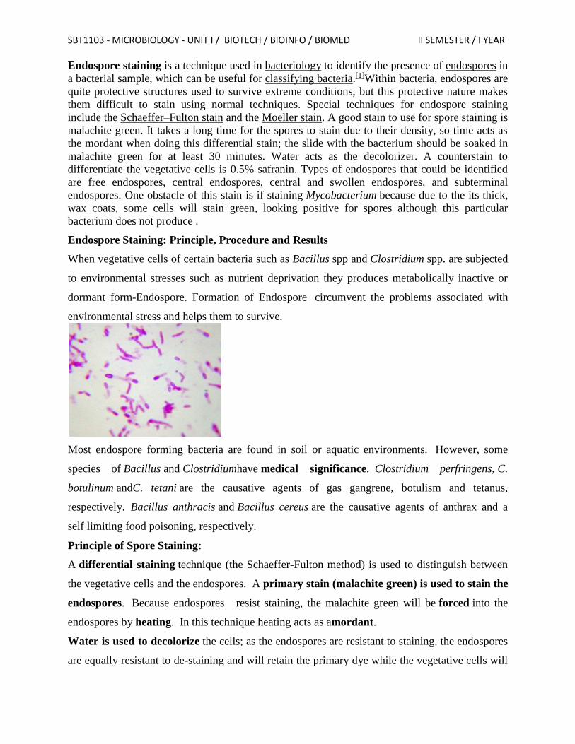
SBT1103 - MICROBIOLOGY - UNIT I / BIOTECH / BIOINFO / BIOMED II SEMESTER / I YEAR
Endospore staining is a technique used in bacteriology to identify the presence of endospores in
a bacterial sample, which can be useful for classifying bacteria.[1]Within bacteria, endospores are
quite protective structures used to survive extreme conditions, but this protective nature makes
them difficult to stain using normal techniques. Special techniques for endospore staining
include the Schaeffer–Fulton stain and the Moeller stain. A good stain to use for spore staining is
malachite green. It takes a long time for the spores to stain due to their density, so time acts as
the mordant when doing this differential stain; the slide with the bacterium should be soaked in
malachite green for at least 30 minutes. Water acts as the decolorizer. A counterstain to
differentiate the vegetative cells is 0.5% safranin. Types of endospores that could be identified
are free endospores, central endospores, central and swollen endospores, and subterminal
endospores. One obstacle of this stain is if staining Mycobacterium because due to the its thick,
wax coats, some cells will stain green, looking positive for spores although this particular
bacterium does not produce .
Endospore Staining: Principle, Procedure and Results
When vegetative cells of certain bacteria such as Bacillus spp and Clostridium spp. are subjected
to environmental stresses such as nutrient deprivation they produces metabolically inactive or
dormant form-Endospore. Formation of Endospore circumvent the problems associated with
environmental stress and helps them to survive.
Most endospore forming bacteria are found in soil or aquatic environments. However, some
species of Bacillus and Clostridiumhave medical significance. Clostridium perfringens, C.
botulinum andC. tetani are the causative agents of gas gangrene, botulism and tetanus,
respectively. Bacillus anthracis and Bacillus cereus are the causative agents of anthrax and a
self limiting food poisoning, respectively.
Principle of Spore Staining:
A differential staining technique (the Schaeffer-Fulton method) is used to distinguish between
the vegetative cells and the endospores. A primary stain (malachite green) is used to stain the
endospores. Because endospores resist staining, the malachite green will be forced into the
endospores by heating. In this technique heating acts as amordant.
Water is used to decolorize the cells; as the endospores are resistant to staining, the endospores
are equally resistant to de-staining and will retain the primary dye while the vegetative cells will
SBT1103 - MICROBIOLOGY - UNIT I / BIOTECH / BIOINFO / BIOMED II SEMESTER / I YEAR
lose the stain. The addition of a counterstain or secondary stain (safranin) is used to stain the
decolorized vegetative cells.
When visualized under microscopy the cells should have three characteristics:
1. the vegetative cells should appear pink,
2. the vegetative cells that contain endospores should stain pink while the spores should be
seen as green ellipses within the cells.
3. Mature, free endospores should not be associated with the vegetative bacteria and should
be seen as green ellipses.
Procedure of endospore stain:
1. Prepare smears of organisms to be tested for endospores on a clean microscope slide and
air dry it.
2. Heat fix the smear.
2. Place a small piece of blotting paper (absorbent paper) over the smear and place the slide
(smear side up) on a wire gauze on a ring stand.
3. Heat the slide gently till it starts to evaporate (either by putting the slide on a staining
rack that has been placed over a boiling water bath or via bunsen burner).
4. Remove the heat and reheat the slide as needed to keep the slide steaming for about 3-
5 minutes. As the paper begins to dry add a drop or two of malachite green to keep it
moist, but don’t add so much at one time that the temperature is appreciably
reduced. DO NOT OVERHEAT. The process is steaming and not baking.
7. After 5 minutes carefully remove the slide from the rack using a clothespin
8. Remove the blotting paper and allow the slide to cool to room temperature for 2 minutes.
9. Rinse the slide thoroughly with tap water (to wash the malachite green from both sides
of the microscope slide).
8. Stain the smear with safranin for 2 minutes.
9. Rinse both side of the slide to remove the secondary stain and blot the slide/ air dry.
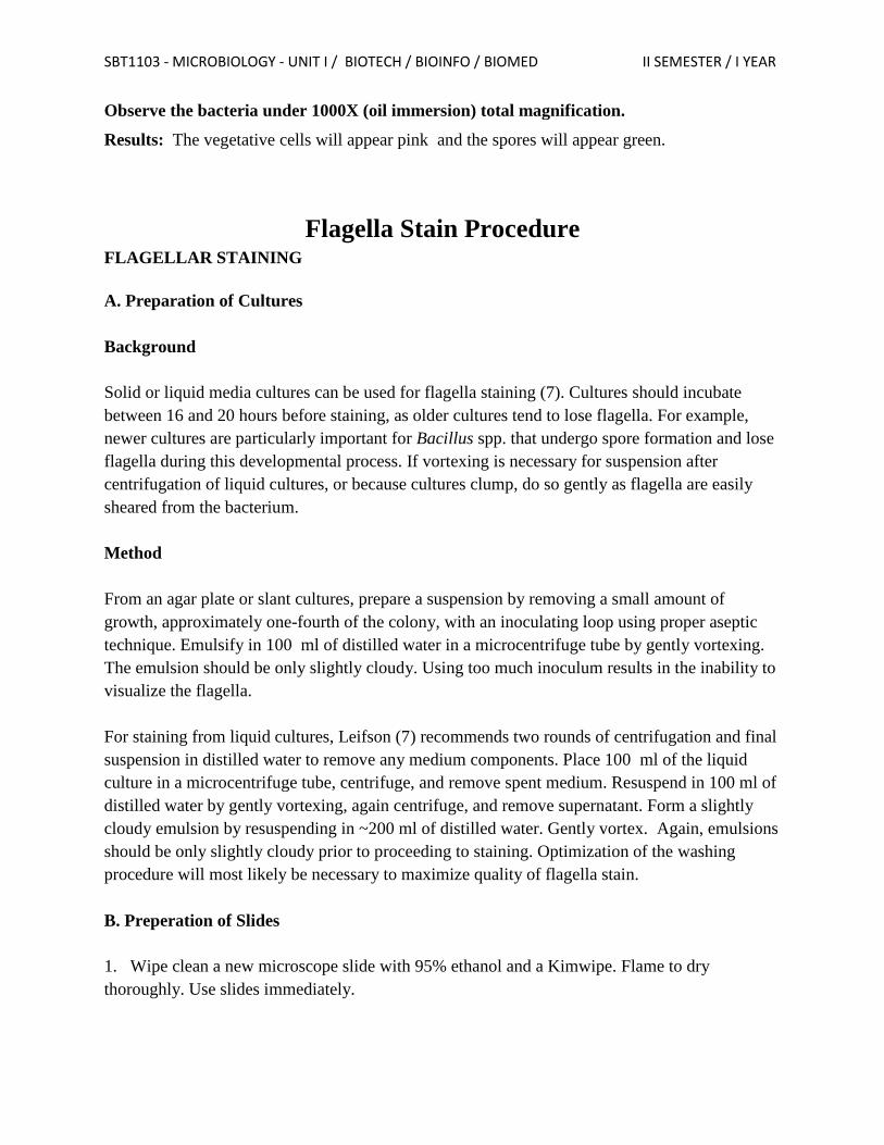
SBT1103 - MICROBIOLOGY - UNIT I / BIOTECH / BIOINFO / BIOMED II SEMESTER / I YEAR
Observe the bacteria under 1000X (oil immersion) total magnification.
Results: The vegetative cells will appear pink and the spores will appear green.
Flagella Stain Procedure FLAGELLAR STAINING
A. Preparation of Cultures
Background
Solid or liquid media cultures can be used for flagella staining (7). Cultures should incubate
between 16 and 20 hours before staining, as older cultures tend to lose flagella. For example,
newer cultures are particularly important for Bacillus spp. that undergo spore formation and lose
flagella during this developmental process. If vortexing is necessary for suspension after
centrifugation of liquid cultures, or because cultures clump, do so gently as flagella are easily
sheared from the bacterium.
Method
From an agar plate or slant cultures, prepare a suspension by removing a small amount of
growth, approximately one-fourth of the colony, with an inoculating loop using proper aseptic
technique. Emulsify in 100 ml of distilled water in a microcentrifuge tube by gently vortexing.
The emulsion should be only slightly cloudy. Using too much inoculum results in the inability to
visualize the flagella.
For staining from liquid cultures, Leifson (7) recommends two rounds of centrifugation and final
suspension in distilled water to remove any medium components. Place 100 ml of the liquid
culture in a microcentrifuge tube, centrifuge, and remove spent medium. Resuspend in 100 ml of
distilled water by gently vortexing, again centrifuge, and remove supernatant. Form a slightly
cloudy emulsion by resuspending in ~200 ml of distilled water. Gently vortex. Again, emulsions
should be only slightly cloudy prior to proceeding to staining. Optimization of the washing
procedure will most likely be necessary to maximize quality of flagella stain.
B. Preperation of Slides
1. Wipe clean a new microscope slide with 95% ethanol and a Kimwipe. Flame to dry
thoroughly. Use slides immediately.
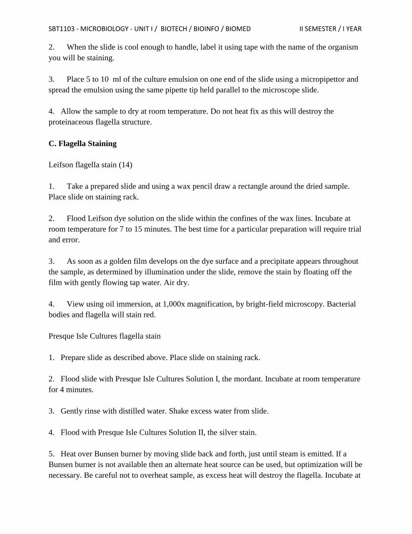
SBT1103 - MICROBIOLOGY - UNIT I / BIOTECH / BIOINFO / BIOMED II SEMESTER / I YEAR
2. When the slide is cool enough to handle, label it using tape with the name of the organism
you will be staining.
3. Place 5 to 10 ml of the culture emulsion on one end of the slide using a micropipettor and
spread the emulsion using the same pipette tip held parallel to the microscope slide.
4. Allow the sample to dry at room temperature. Do not heat fix as this will destroy the
proteinaceous flagella structure.
C. Flagella Staining
Leifson flagella stain (14)
1. Take a prepared slide and using a wax pencil draw a rectangle around the dried sample.
Place slide on staining rack.
2. Flood Leifson dye solution on the slide within the confines of the wax lines. Incubate at
room temperature for 7 to 15 minutes. The best time for a particular preparation will require trial
and error.
3. As soon as a golden film develops on the dye surface and a precipitate appears throughout
the sample, as determined by illumination under the slide, remove the stain by floating off the
film with gently flowing tap water. Air dry.
4. View using oil immersion, at 1,000x magnification, by bright-field microscopy. Bacterial
bodies and flagella will stain red.
Presque Isle Cultures flagella stain
1. Prepare slide as described above. Place slide on staining rack.
2. Flood slide with Presque Isle Cultures Solution I, the mordant. Incubate at room temperature
for 4 minutes.
3. Gently rinse with distilled water. Shake excess water from slide.
4. Flood with Presque Isle Cultures Solution II, the silver stain.
5. Heat over Bunsen burner by moving slide back and forth, just until steam is emitted. If a
Bunsen burner is not available then an alternate heat source can be used, but optimization will be
necessary. Be careful not to overheat sample, as excess heat will destroy the flagella. Incubate at
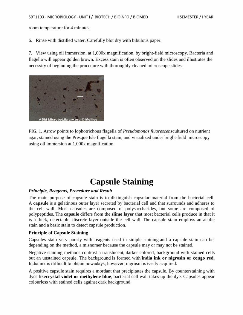
SBT1103 - MICROBIOLOGY - UNIT I / BIOTECH / BIOINFO / BIOMED II SEMESTER / I YEAR
room temperature for 4 minutes.
6. Rinse with distilled water. Carefully blot dry with bibulous paper.
7. View using oil immersion, at 1,000x magnification, by bright-field microscopy. Bacteria and
flagella will appear golden brown. Excess stain is often observed on the slides and illustrates the
necessity of beginning the procedure with thoroughly cleaned microscope slides.
FIG. 1. Arrow points to lophotrichous flagella of Pseudomonas fluorescenscultured on nutrient
agar, stained using the Presque Isle flagella stain, and visualized under bright-field microscopy
using oil immersion at 1,000x magnification.
Capsule Staining
Principle, Reagents, Procedure and Result
The main purpose of capsule stain is to distinguish capsular material from the bacterial cell.
A capsule is a gelatinous outer layer secreted by bacterial cell and that surrounds and adheres to
the cell wall. Most capsules are composed of polysaccharides, but some are composed of
polypeptides. The capsule differs from the slime layer that most bacterial cells produce in that it
is a thick, detectable, discrete layer outside the cell wall. The capsule stain employs an acidic
stain and a basic stain to detect capsule production.
Principle of Capsule Staining
Capsules stain very poorly with reagents used in simple staining and a capsule stain can be,
depending on the method, a misnomer because the capsule may or may not be stained.
Negative staining methods contrast a translucent, darker colored, background with stained cells
but an unstained capsule. The background is formed with india ink or nigrosin or congo red.
India ink is difficult to obtain nowadays; however, nigrosin is easily acquired.
A positive capsule stain requires a mordant that precipitates the capsule. By counterstaining with
dyes likecrystal violet or methylene blue, bacterial cell wall takes up the dye. Capsules appear
colourless with stained cells against dark background.
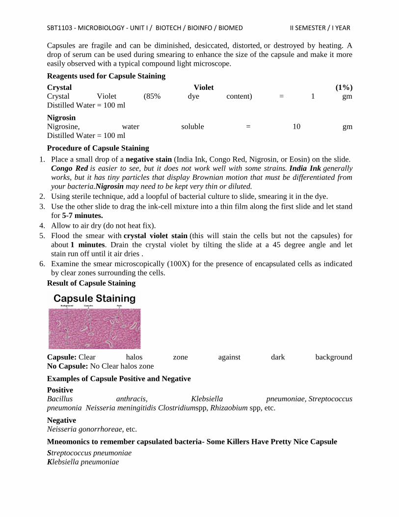
SBT1103 - MICROBIOLOGY - UNIT I / BIOTECH / BIOINFO / BIOMED II SEMESTER / I YEAR
Capsules are fragile and can be diminished, desiccated, distorted, or destroyed by heating. A
drop of serum can be used during smearing to enhance the size of the capsule and make it more
easily observed with a typical compound light microscope.
Reagents used for Capsule Staining
Crystal Violet (1%) Crystal Violet (85% dye content) = 1 gm
Distilled Water = 100 ml
Nigrosin Nigrosine, water soluble = 10 gm
Distilled Water = 100 ml
Procedure of Capsule Staining
1. Place a small drop of a negative stain (India Ink, Congo Red, Nigrosin, or Eosin) on the slide.
Congo Red is easier to see, but it does not work well with some strains. India Ink generally
works, but it has tiny particles that display Brownian motion that must be differentiated from
your bacteria.Nigrosin may need to be kept very thin or diluted.
2. Using sterile technique, add a loopful of bacterial culture to slide, smearing it in the dye.
3. Use the other slide to drag the ink-cell mixture into a thin film along the first slide and let stand
for 5-7 minutes.
4. Allow to air dry (do not heat fix).
5. Flood the smear with crystal violet stain (this will stain the cells but not the capsules) for
about 1 minutes. Drain the crystal violet by tilting the slide at a 45 degree angle and let
stain run off until it air dries .
6. Examine the smear microscopically (100X) for the presence of encapsulated cells as indicated
by clear zones surrounding the cells.
Result of Capsule Staining
Capsule: Clear halos zone against dark background
No Capsule: No Clear halos zone
Examples of Capsule Positive and Negative
Positive Bacillus anthracis, Klebsiella pneumoniae, Streptococcus
pneumonia Neisseria meningitidis Clostridiumspp, Rhizaobium spp, etc.
Negative
Neisseria gonorrhoreae, etc.
Mneomonics to remember capsulated bacteria- Some Killers Have Pretty Nice Capsule
Streptococcus pneumoniae
Klebsiella pneumoniae

SBT1103 - MICROBIOLOGY - UNIT I / BIOTECH / BIOINFO / BIOMED II SEMESTER / I YEAR
Haemophilus influenzae
Pseudomonas aeruginosa
Neisseria meningitidis
Cryptococcus neoformans