
Ruoli Bai, David G. Covell, George F. Taylor, John A. Kepler, Terry D ...
49
1 DIRECT PHOTOAFFINITY LABELING BY DOLASTATIN 10 OF THE AMINO TERMINAL PEPTIDE OF b -TUBULIN CONTAINING CYSTEINE-12 Ruoli Bai, David G. Covell, George F. Taylor, John A. Kepler, Terry D. Copeland, Nga Y. Nguyen, George R. Pettit, and Ernest Hamel Screening Technologies Branch Developmental Therapeutics Program Division of Cancer Treatment and Diagnosis National Cancer Institute at Frederick National Institutes of Health Frederick, Maryland 21702 (RB, DGC, EH) Research Triangle Institute Research Triangle Park, North Carolina 27709 (GFT, JAK) Laboratory of Protein Dynamics and Signaling Center for Cancer Research National Cancer Institute at Frederick National Institutes of Health Frederick, Maryland 21702 (TDC) Facility for Biotechnology Resources Center for Biologics Evaluation and Research Food and Drug Administration Bethesda, Maryland 20892 (NYN) and Cancer Research Institute and Department of Chemistry and Biochemistry Arizona State University Tempe, Arizona 85287 (GRP) Address correspondence to Dr. Ernest Hamel, Building 469, Room 104, P. O. Box B, National Cancer Institute at Frederick, Frederick MD 21702 Phone: 301-846-1678 FAX: 301-846-6014 Email: [email protected] JBC Papers in Press. Published on April 29, 2004 as Manuscript M402110200 by guest on March 17, 2018 http://www.jbc.org/ Downloaded from
Transcript of Ruoli Bai, David G. Covell, George F. Taylor, John A. Kepler, Terry D ...
1
DIRECT PHOTOAFFINITY LABELING BY DOLASTATIN 10 OF THE AMINO
TERMINAL PEPTIDE OF β-TUBULIN CONTAINING CYSTEINE-12
Ruoli Bai, David G. Covell, George F. Taylor, John A. Kepler, Terry D. Copeland,
Nga Y. Nguyen, George R. Pettit, and Ernest Hamel
Screening Technologies Branch
Developmental Therapeutics Program Division of Cancer Treatment and Diagnosis
National Cancer Institute at Frederick National Institutes of Health Frederick, Maryland 21702
(RB, DGC, EH)
Research Triangle Institute Research Triangle Park, North Carolina 27709
(GFT, JAK)
Laboratory of Protein Dynamics and Signaling Center for Cancer Research
National Cancer Institute at Frederick National Institutes of Health Frederick, Maryland 21702
(TDC)
Facility for Biotechnology Resources Center for Biologics Evaluation and Research
Food and Drug Administration Bethesda, Maryland 20892
(NYN)
and
Cancer Research Institute and Department of Chemistry and Biochemistry Arizona State University Tempe, Arizona 85287
(GRP) Address correspondence to Dr. Ernest Hamel, Building 469, Room 104, P. O. Box B,
National Cancer Institute at Frederick, Frederick MD 21702 Phone: 301-846-1678 FAX: 301-846-6014
Email: [email protected]
JBC Papers in Press. Published on April 29, 2004 as Manuscript M402110200 by guest on M
arch 17, 2018http://w
ww
.jbc.org/D
ownloaded from
2
SUMMARY
Tubulin with bound [5-3H]dolastatin 10 was exposed to ultraviolet light, and 8-
10% of the bound drug cross-linked to the protein, most of it specifically. The primary
cross- link was to the peptide spanning amino acid residues 2-31 of β-tubulin, but the
specific amino acid could not be identified. Indirect studies indicated that cross- link
formation occurred between cysteine-12 and the thiazole moiety of dolastatin 10. An
equipotent analog of dolastatin 10, lacking the thiazole ring, did not form an ultraviolet
light- induced cross- link to β-tubulin. Preillumination of tubulin with ultraviolet light,
known to induce cross- link formation between cysteine-12 and exchangeable site
nucleotide, inhibited the binding of [5-3H]dolastatin 10 and cross- link formation more
potently than it inhibited the binding of colchicine or vinblastine to tubulin. Conversely,
binding of dolastatin 10 to tubulin inhibited formation of the cross- link between cysteine-
12 and exchangeable site nucleotide. Dithiothreitol inhibited formation of the β-
tubulin/dolastatin 10 cross- link but not the β-tubulin/exchangeable site nucleotide cross-
link. Modeling studies revealed a highly favored binding site for dolastatin 10 at the +-
end of β-tubulin in proximity to the exchangeable site GDP. Computational docking of
an energy-minimized dolastatin 10 conformation at this site placed the thiazole ring of
dolastatin 10 8-9 Å from the sulfur atom of cysteine-12. Dolastatin 15 and cryptophycin
1 could also be docked into positions that overlapped more extensively with the docked
dolastatin 10 than with each other. This result was consistent with the observed binding
properties of these peptides.
_______________________________________________________________________
by guest on March 17, 2018
http://ww
w.jbc.org/
Dow
nloaded from
3
The subunit protein of microtubules, the αβ-tubulin heterodimer, has a number of
ligand binding sites. These include the exchangeable and nonexchangeable GTP binding
sites and at least three reasonably well-characterized sites that bind antimitotic drugs.
The electron crystallographic model of the tubulin sheet polymer formed in the presence
of zinc and paclitaxel and composed of antiparallel protofilaments has provided relatively
detailed information about the locations of the nucleotide binding sites and the paclitaxel
site on the αβ-dimer (1).
In contrast, drugs that inhibit tubulin polymerization will not bind to structurally
normal tubulin protofilaments, so their binding sites have only been approached
indirectly through biochemical and genetic techniques. For example, without exception,
drugs that bind in the colchicine site inhibit formation of a sulfhydryl cross- link between
Cys-239 and Cys-354 of β-tubulin (2). These two cysteine residues are also alkylated by
A ring chloroacetyl derivatives of thiocolchicine (3, 4). Cys-239 and Cys-354 are near
the interface between the subunits (1), and such a location for the colchicine site would
explain the ability of different photoactive groups attached to the colchicine B ring to
preferentially alkylate either the α- or the β-subunit (5, 6).
A large number of structurally diverse compounds, including many unusual
peptides and depsipeptides, interfere with the binding of vinca alkaloids to tubulin.
Despite near total inhibition of vinca alkaloid binding, many of these drugs display
noncompetitive patterns when the data they yield are examined by the classic formulas of
enzyme kinetic analysis, while other inhibitors display competitive patterns. We have
suggested that the noncompetitive inhibitors bind near the vinca site, interfering sterically
with vinca alkaloid binding to tubulin, and proposed that this region of tubulin be called
by guest on March 17, 2018
http://ww
w.jbc.org/
Dow
nloaded from
4
the vinca domain (7). This region of tubulin appears to be close to the exchangeable GTP
site on β-tubulin, based on two observations. First, a unique sulfhydryl cross- link can be
formed in nucleotide-depleted tubulin, between Cys-12 and, probably, Cys-211 (1, 2, 8),
while direct photoaffinity labeling of tubulin by exchangeable site guanosine nucleotide
leads to alkylation primarily of Cys-12 (9) and secondarily of Cys-211 (10). All vinca
domain drugs that have been examined inhibit formation of the Cys-12/Cys-211 cross-
link, with vinblastine having the weakest effect (2, 11, 12). Second, all compounds that
inhibit vinca alkaloid binding to tubulin also inhibit nucleotide exchange on β-tubulin (7,
13-15). Vinblastine, however, only weakly inhibits nucleotide exchange (16), while
rhizoxin is moderately inhibitory (7).
There have been several published studies in which cross- links were induced
between a vinca domain drug and tubulin. By direct photoaffinity labeling with
vinblastine (17) and with two photoactive derivatives of vinblastine (18, 19), there was
greater labeling of α-tubulin than β-tubulin, ranging from 57-75% of the incorporated
radiolabel being in the α-subunit. A photoaffinity analog of maytansine was incorporated
in about a 4:5 ratio into α- and β-tubulin, respectively (20). More specific labeling of β-
tubulin has also been observed. A photoreactive vinb lastine analog specifically labeled a
peptide containing residues 175-213 (21), and a photoreactive rhizoxin derivative labeled
a peptide containing residues 363-379 (22).
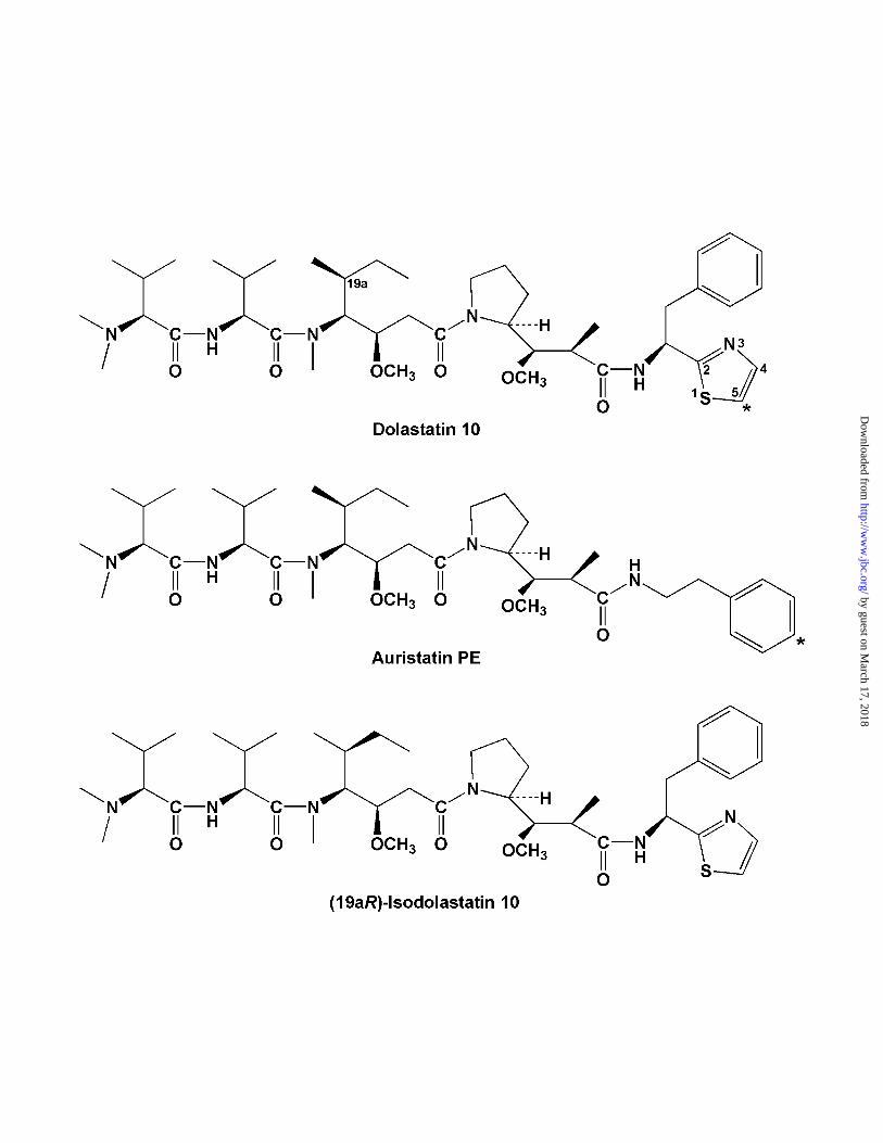
We have been studying the interaction of the marine peptide dolastatin 10
(structure in Fig. 1) with tubulin. Besides potent cytotoxicity, dolastatin 10 inhibits
microtubule assembly while inducing formation of ring and spiral polymers,
noncompetively inhibits the binding of vinca alkaloids to tubulin, and inhibits nucleotide
by guest on March 17, 2018
http://ww
w.jbc.org/
Dow
nloaded from
5
exchange on β-tubulin. With radiolabeled dolastatin 10 we showed that the peptide
bound tenaciously to tubulin, with negligible dissociation during gel filtration
chromatography. The nominal Kd value for binding of drug to tubulin was about 25 nM.
The binding was reversible, however, since an active analog could displace radiolabel
from tubulin. The ability of dolastatin 10 to induce aberrant assembly reactions was so
potent that we were not able to demonstrate binding of the radiolabeled drug to any
tubulin species smaller than 200 kDa (7, 23).
Besides the natural product, a number of peptide analogs of dolastatin 10 have
equivalent activity in both cell-based and tubulin-based assays. One of the more
interesting, which we also prepared in a radiolabeled form, is auristatin PE (24; structure
in Fig. 1), also prepared independently as TZT-1027 (25). We began to study the
potential of direct photoaffinity labeling with the [3H]dolastatin 101 and the [3H]auristatin
PE to provide information about the binding site for these peptides on tubulin. Our
findings are presented here.
by guest on March 17, 2018
http://ww
w.jbc.org/
Dow
nloaded from
6
EXPERIMENTAL PROCEDURES
Materials. Purified bovine brain tubulin with nonradiolabeled GDP (26) or [8-
14C]GDP (27) in the exchangeable site, nonradiolabeled dolastatin 10 (28),
nonradiolabeled auristatin PE (29), (19aR)- isodolastatin 10 (30), and hemiasterlin (31)
were prepared as described previously. Phomopsin A was generously provided by Dr. C.
C. J. Culvenor (CSIRO Division of Animal Health, Parkville, Victoria, Australia) or
obtained from Calbiochem. Cryptophycin 1 was a generous gift from Merck Research
Laboratories. [8-14C]GTP was from Moravek Biochemicals and repurified by
triethylammonium bicarbonate gradient chromatography on DEAE-cellulose.
Nonradiolabeled colchicine was from Sigma; [3H]colchicine and EN3HANCE spray from
Perkin Elmer Life Sciences; [3H]vinblastine from Amersham Biosciences; precast 16%
polyacrylamide Tricine-SDS gels, Tricine-SDS solutions, and PVDF membranes from
Invitrogen; and Biomax MR film from Kodak.
Synthesis of [5-3H]dolastatin 10. Dolastatin 10 was first converted to 5-bromo-
dolastatin 10. The dolastatin 10 (30 mg) was dissolved in 0.2 ml of acetic acid with 40
mg of silver trifluoroacetate. A solution of bromine in acetic acid (0.114 mmol in 0.6 ml)
was added, and the mixture was stirred for 18 h at ambient temperature. Chloroform (2
ml) was added, and the solution was filtered through celite. The solvents were removed
by evaporation under reduced pressure. The residue was chromatographed on a Dupont
Zorbax ODS column (0.75 x 26 cm), which was developed with 10 mM triethylamine in
75% methanol at 1 ml/min. The column effluent was monitored at 240 nm. The solvent
fraction containing the largest peak, with a retention time of 33 min, was concentrated to
dryness, and 2.6 mg of product was obtained. The [1H]-NMR spectrum, when compared
by guest on March 17, 2018
http://ww
w.jbc.org/
Dow
nloaded from
7
with that of dolastatin 10, showed collapse of a doublet at δ 7.4, corresponding to the
hydrogen atom at position C-4, to a singlet, indicating bromination at position C-5 on the
thiazole ring. The 5-bromo-dolastatin 10 was dissolved in 0.3 ml of ethyl acetate
containing 25 µl of triethylamine and 2 mg of 10% Pd on carbon. The reaction mixture
was exposed to carrier-free tritium gas at 630 mm Hg for 2 h at ambient temperature.
The catalyst was removed by filtration, and the filtrate was exchanged three times with
ethanol. The crude product was chromatographed on a 5 x 20 cm silica gel 60F plate
with 3:2 acetone-hexane. The dolastatin 10 band was eluted from the silica with 1:1
CHCl3-ethanol. The solvent was removed by evaporation under vacuum and the residue
redissolved in 50 ml of ethanol. The product was 96% pure and its mobility identical to
that of dolastatin 10 by TLC and HPLC. Specific activity was 5.4 Ci/mmol, and the yield
was 20.7 mCi. The [3H]-NMR spectrum showed a doublet at δ 7.29, J = 3 Hz, indicating
that the tritium was at the C-5 position in the thiazole ring. Back exchange for 24 h at
ambient temperature in pH 7 phosphate buffer showed 0.2% exchangeable tritium.
Synthesis of [phenyl-4-3H]auristatin PE. An analog of auristatin PE (2.5 mg),
with a chlorine atom at position C-4 in the phenyl ring (prepared as described in ref. 24),
was mixed with 1.5 mg of 10% Pd on carbon and 2 µl of triethylamine in 0.4 ml of ethyl
acetate under nitrogen at ambient temperature and exposed to carrier-free tritium gas for
4 h. The catalyst was removed by filtration, and the solution was back exchanged four
times with ethanol on a vacuum line. The crude material was chromatographed on an
analytical 20 x 20 cm silica gel 60F TLC plate with acetone-hexane-methanol 5:4:1. The
band corresponding to auristatin PE was collected and eluted from the silica with 1:1
CHCl3-ethanol. The TLC mobility of the radiolabeled material was identical with that of
by guest on March 17, 2018
http://ww
w.jbc.org/
Dow
nloaded from
8
nonradiolabeled auristatin PE. Material from two identical procedures was combined,
and HPLC indicated that the radiopurity of the combined product was 81%. The pooled
material was applied to a Waters 8 x 10 C8 Novapak radial compression column, which
was developed with 10 mM triethylamine in 75% methanol. Column flow rate was 1
ml/min, and the effluent was monitored at 230 nm. The fractions containing
[3H]auristatin PE were combined, and the solvent was removed by evaporation to yield
18.6 mCi of product with a specific activity of 8.46 Ci/mmol. Radiopurity by HPLC and
TLC were 98% and 99%, respectively, with mobilities identical with those of
nonradiolabeled auristatin PE.
Photolabeling of tubulin with [3H]dolastatin 10. Reaction mixtures contained 1.0
mg/ml (10 µM) tubulin, 10 µM [3H]dolastatin 10 (or, in some experiments, 10 µM
[3H]auristatin PE), 0.1 M Mes (pH of 1.0 M stock solution adjusted to pH 6.9 with
NaOH), and 0.5 mM MgCl2. In some experiments, as indicated, other components were
included in reaction mixtures, and incubations prior to exposure to UV light were as
described for individual experiments. Reaction mixtures with volumes up to 0.25 ml,
with volumes between 0.25 and 1.0 ml, and with volumes over 1.0 ml were placed in
wells in Costar polystyrene tissue culture plates, with well diameters of 1.5, 2.5, and 3.5
cm, respectively. Samples were placed on ice at a distance of 10 cm from the UV lamp
and exposed to 254 nm light for times as indicated. Light intensity at 10 cm was 2.5
mW/cm2.
Measurement of ligand bound to tubulin (total bound and covalently bound). The
centrifugal gel filtration method, as described previously (32), was used, except that all
centrifugations were for 4 min at 2,000 rpm in a Beckman Allegra 6KR centrifuge
by guest on March 17, 2018
http://ww
w.jbc.org/
Dow
nloaded from
9
equipped with a GH-3.8A horizontal rotor. The Sephadex G-50 (superfine) columns
were prepared in tuberculin syringes. When total binding of a ligand was measured,
aliquots of the reaction mixture were applied directly to the pre-centrifuged Sephadex,
which had been swollen in a solution containing 0.1 M Mes (pH 6.9) and 0.5 mM MgCl2.
When covalently bound ligand was measured, the reaction mixture was mixed with 1.5
parts 8 M guanidine-HCl, and aliquots of the resulting solution were applied to syringe-
columns containing Sephadex that had been swollen in 4 M guanidine-HCl. Each assay
mixture was evaluated in triplicate, and both radiolabel and protein in column filtrates
was measured.
Peptide sequencing. Samples for sequencing were radiolabeled as described
above, although in some experiments the tubulin and [3H]dolastatin 10 concentrations
were increased to 25 µM. Reaction mixtures were initially incubated for 15 min at room
temperature (22°C) and subsequently irradiated for 5-10 min on ice. Two volumes of
ethanol were added to precipitate the tubulin. This removed the noncovalently bound,
soluble dolastatin 10. The protein was harvested by centrifugation, washed twice with
70% ethanol, and either dried under vacuum for later use or immediately dissolved in
75% formic acid. Formic acid digestion (96 h at 37°C in the dark), cyanogen bromide
digestion, peptide recovery by lyophilization, peptide separation by SDS-PAGE and
transfer of resolved peptides to PVDF membranes, and automated Edman degradation
were performed as described previously (4), except that after formic acid digestions the
peptides were dissolved in the Tricine-SDS sample buffer solution instead of
concentrated Tris buffer. PVDF membranes were sprayed with EN3HANCE, which was
by guest on March 17, 2018
http://ww
w.jbc.org/
Dow
nloaded from
10
allowed to dry for 15 min prior to exposure to the Biomax MR film. Autoradiograms
were prepared over a 5-7 day exposure at - 70°C.
Molecular modeling. Modeling consisted of identifying acceptable conformations
for the docked ligands, scanning the solvent accessible surface of the tubulin dimer (1)
for potential binding sites, and computational docking of the candidate ligands to these
sites. Conformations of dolastatin 10, dolastatin 15, and cryptophycin 1 were obtained by
sampling the lowest energy geometries derived from in vacuo minimization and
molecular dynamics using the CVFF force field of the Discover2002 tool in the Accelrys
molecular package (BIOSYM, MSI). A sample set of the five lowest energy
conformations for each molecule was selected for docking studies. Candidate docking
sites were identified using a method developed for assigning docking sites of ligands
against their Protein Data Bank crystal structures (see ref. 33 for details). Briefly, the
method scans the solvent accessible surface of the electron crystallographic structure of
the tubulin dimer (1) using a set of residue-based molecular probes that had previously
been identified as the crystal packing geometries of proteins within the Protein Data Bank
(34). Each surface point is scored according to the energy associated with docking each
of the molecular probes at that point. Calculated binding strengths are then scored by
summing the interaction energies for the most favorable binding interactions within this
suite of molecular probes. Using this method, potential binding sites are identified for
subsequent docking. The interaction energies of this method are derived from extensive
study of residue-residue-based potentials (35) and, as such, represent coarse assessments
of candidate binding sites. This method proved effective in correctly assigning binding
sites in subsequent studies (36, 37). Docking studies were completed with a
by guest on March 17, 2018
http://ww
w.jbc.org/
Dow
nloaded from
11
computational method developed for placing candidate ligands into possible binding
sites. Previous calibration of this procedure found the correct binding position for over
93% of the known crystal complexes studies at the time of analysis (38, 39). Docking is
achieved in three successive steps, each with increasing demands for scoring acceptable
binding positions. The initial step is sufficiently crude, so that all candidate binding sites
on the tubulin dimer can be scanned for docking. Following this step, candidate binding
sites with the greatest predicted binding strength are passed along for more exact
placement into each potential binding site. Completion of all computational steps for
docking yields the minimum energy position for each test molecule into its most
favorable binding location. The molecular coordinates for all binding conformations
described in this paper are available on request to D. G. Covell at [email protected].
by guest on March 17, 2018
http://ww
w.jbc.org/
Dow
nloaded from
12
RESULTS
Direct photoaffinity labeling of tubulin by [3H]dolastatin 10 and identification of
the predominant radiolabeled peptide. We decided to explore the potential of the direct
photoaffinity labeling technique for providing information about the dolastatin 10 binding
site on tubulin based on the success that had been had, especially, with colchicine (17),
paclitaxel (40), and GTP (9). Only in the latter case was a specific amino acid residue,
Cys-12 of β-tubulin, identified, and Shivanna et al. (9) proposed that exposure of tubulin
to ultraviolet radiation resulted in generation of sulfhydryl free radicals as the reactive
species.
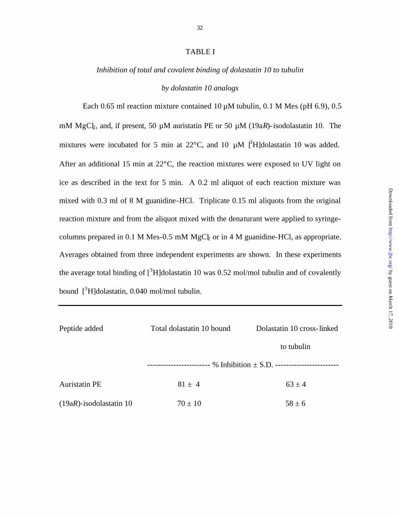
Table 1 summarizes our initial experiments, in which we found that about 8% of
the bound [3H]dolastatin 10 reacted covalently with tubulin, and this covalent reaction, as
well as the total binding reaction, was substantially inhibited by preincubating the tubulin
with a 5-fold molar excess of two highly active analogs of dolastatin 10, auristatin PE
(24, 25), and (19aR)- isodolastatin 10 (12, 41). The cross- linking reaction therefore
appeared to be largely specific and to require initial binding of the peptide to the protein.
In the experiments summarized in Table 1 and elsewhere in this study, the
stoichiometry of cross- linking ranged from 0.040 to 0.054 mol dolastatin 10/mol tubulin.
While this stoichiometry is relatively low compared to the results obtained by direct
photoaffinity labelling with exchangeable site GDP or GTP (refs. 9, 42; also see below),
it differs little from results obtained with other antimitotic drugs. Direct photoaffinity
labeling experiments with [3H]paclitaxel2 (40) and with [3H]colchicine3 and
[3H]vinblastine3 (17) all yielded cross- linking stoichiometries of less than 0.05 mol
ligand/mol tubulin.
by guest on March 17, 2018
http://ww
w.jbc.org/
Dow
nloaded from
13
Initial attempts to identify the tubulin subunit cross- linked to dolastatin 10 were
unsuccessful. We found it difficult to resolve the tubulin subunits after UV irradiation
either by SDS-PAGE or by hydrophobic chromatography on decylagarose (43). There
was, however, no significant intersubunit cross- link formation during the brief irradiation
period, as no higher molecular weight species were visualized on the gels. The usual
tubulin doublet was replaced by a broad band, with the greatest amount of protein in the
usual β-tubulin position (data not shown).
Therefore, unresolved tubulin that had been irradiated in the presence of
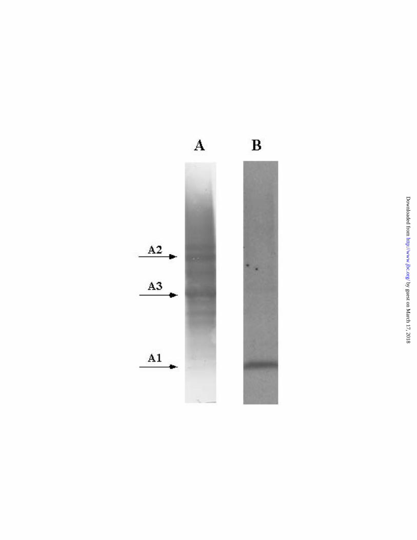
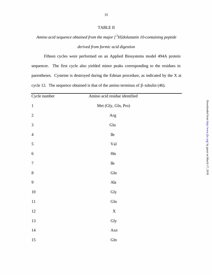
[3H]dolastatin 10 was subjected to formic acid digestion. Fig. 2 presents a typical PVDF
membrane blot transferred from a polyacrylamide gel, with track A showing the
Coomassie blue stained peptides and track B the autoradiograph of the same membrane.
The primary cleavage site for formic acid is an aspartyl-proline bond (44). There are
only three such sites in tubulin. One is located in the α-subunit (positions 306-307) (45),
and two in the β-subunit (positions 31-32 and positions 304-305) (46). In our previous
studies with decylagarose-purified β-tubulin cross- linked to colchicine analogs (3, 4), we
had noted a number of secondary cleavage sites derived from the initially formed larger
peptides. This undoubtedly accounts for the multiple bands in the Coomassie blue
stained track A, but the striking feature of the corresponding autoradiograph (track B), is
that there is only one prominent band. This corresponds to the small A1 peptide [in the
terminology of Rao et al. (47)] at the amino terminus of β-tubulin, as shown when the
peptide was eluted from the membrane and subjected to sequential Edman degradation
(Table II).
by guest on March 17, 2018
http://ww
w.jbc.org/
Dow
nloaded from

14
We next attempted to localize the radiolabel to a specific residue, but the bond
formed between the [3H]dolastatin 10 and β-tubulin was unstable to the harsh conditions
of sequential Edman degradation. A large amount of radiolabel was recovered only in
the first cycle (it is unlikely that the initial methionine was the residue linked to the drug,
see below), even when 31 digestive cycles were performed, and little residual radiolabel
was found when the membrane itself was counted after being removed from the
sequencing apparatus.
The modeling studies described below and the finding of Shivanna et al. (9) that
ultraviolet light specifically activates sulfhydryl residues indicate that a likely covalent
interaction between dolastatin 10 and β-tubulin could be between Cys-12 and the thiazole
ring of the drug, perhaps even with formation of a disulfide bond and disruption of the
thiazole ring. We therefore performed additional protein degradation studies in attempts
to better define the amino acid residue cross- linked to the peptide. Cyanogen bromide
digestion allowed us to exclude Met-1 as the reactive residue [the next methionine
residues in β-tubulin are at positions 73 and 147 (46)], since this technique yielded large
radiolabeled peptides (data not shown). Multiple attempts at enzymatic digestions with
trypsin and the endopeptidases Glu-C and Lys-C failed to yield a radiolabeled peptide
that could be sequenced. This could be due to the low stoichiometry of cross-linking
between dolastatin 10 and β-tubulin and/or to the small size of the anticipated
radiolabeled peptides (17-19 amino acid residues).
Indirect studies suggesting that the cross- link between [3H]dolastatin 10 and β-
tubulin occurs at Cys-12. Shivanna et al. (9) reported, and we confirmed (10), that direct
photoaffinity labeling of exchangeable site guanine nucleotide occurs primarily at Cys-
by guest on March 17, 2018
http://ww
w.jbc.org/
Dow
nloaded from
15
12. Moreover, dolastatin 10 potently inhibits nucleotide exchange on β-tubulin (7).
When we modeled the energy minimized conformation of dolastatin 10 into the most
promising potential binding pocket on the electron crystallographic model of tubulin (1),
the S atom of Cys-12 and the thiazole ring of dolastatin 10 were in reasonable proximity
(see below). These observations suggested several experiments that might strengthen
the case for UV-mediated cross-link formation between Cys-12 and dolastatin 10. A
simple prediction was that auristatin PE could not form a similar cross- link to β-tubulin.
Despite lacking the thiazole ring, this analog of dolastatin 10 has activity comparable to
the natural product in all biochemical, cytological, and in vivo antitumor systems where
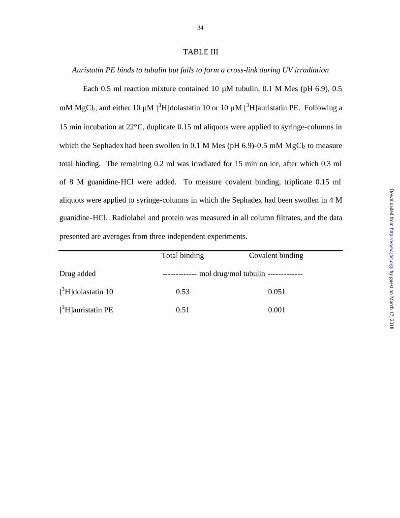
the two compounds have been examined (24, 25, 29, 48). Table III demonstrates that
[3H]auristatin PE bound as avidly to tubulin as [3H]dolastatin 10, but, as predicted, little
or no cross- linking to the protein occurred.
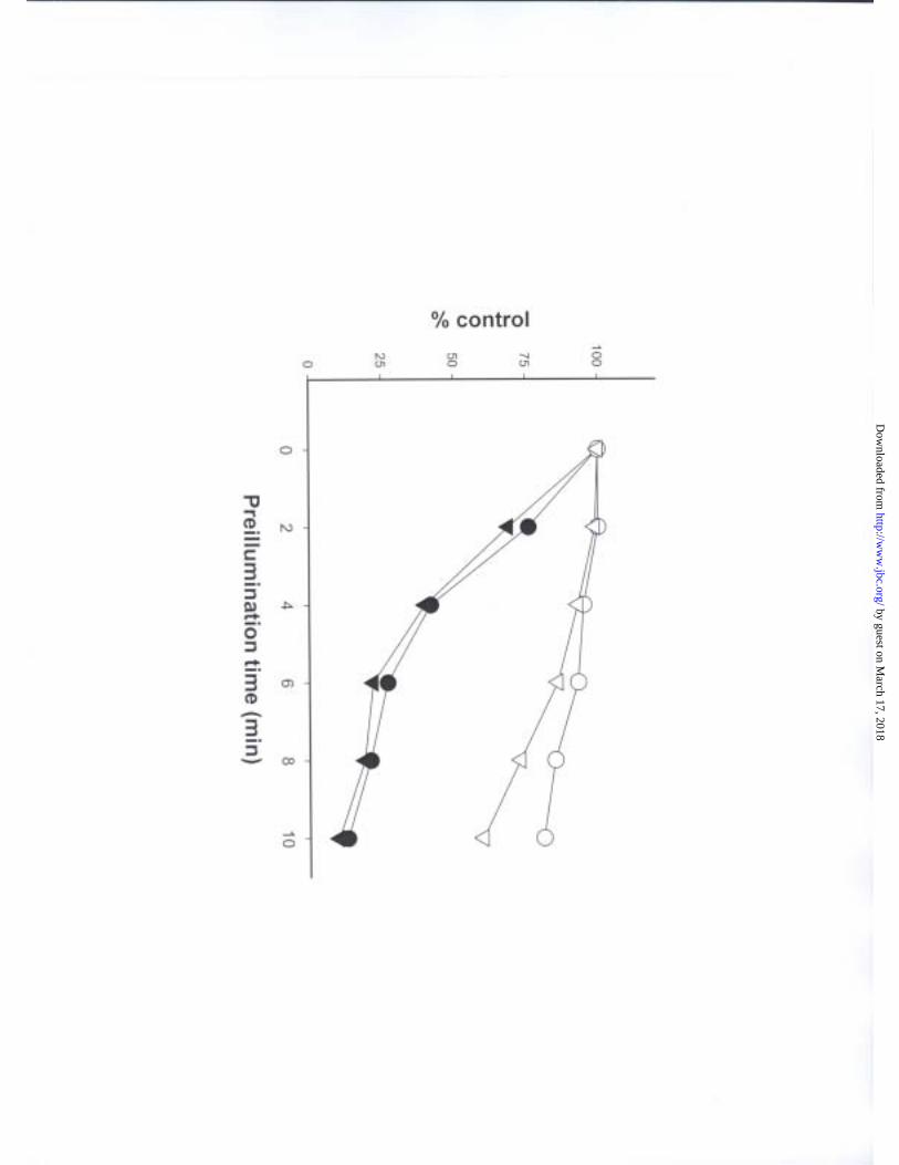
In an earlier study, using a photolabeling methodology similar to that used here,
we found that cross-link formation between [8-14C]GDP bound in the exchangeable site
{prepared by performing two cycles of assembly/disassembly with [8-14C]GTP (27)} and
β-tubulin occurred rapidly. With tubulin at 5 mg/ml, the cova lent reaction was 75%
complete within 5 min (42). Therefore, if Cys-12 were the amino acid that formed the
covalent bond with dolastatin 10, brief prior irradiation of tubulin to form the GDP-Cys-
12 cross-link (9) should inhibit subsequent cross- link formation with dolastatin 10. This
proved to be the case, and irradiation of tubulin also dramatically reduced the ability of
dolastatin 10 to even bind to tubulin (Fig. 3). With about 3 min of prior irradiation the
amount of dolastatin 10 bound to tubulin was reduced by 50%. The ability of a second
irradiation to cause formation of the dolastatin 10-β-tubulin cross-link showed a similar
by guest on March 17, 2018
http://ww
w.jbc.org/
Dow
nloaded from
16
time course for the inhibitory effect of the first irradiation period when this was done in
the absence of the drug (data not presented). This, like the inhibitory effect of dolastatin
10 analogs shown in Table I, confirmed that binding of dolastatin 10 to tubulin was a
requirement for cross- link formation. Prior irradiation had almost an identical effect on
the binding of exogenous [8-14C]GTP to tubulin (Fig. 3), as would be expected if the
endogenous GDP in the exchangeable site had been cross- linked to β-tubulin. In
contrast, prior irradiation of tubulin had a significantly smaller effect on the binding of
either [3H]vinblastine or [3H]colchicine to tubulin (Fig. 3).
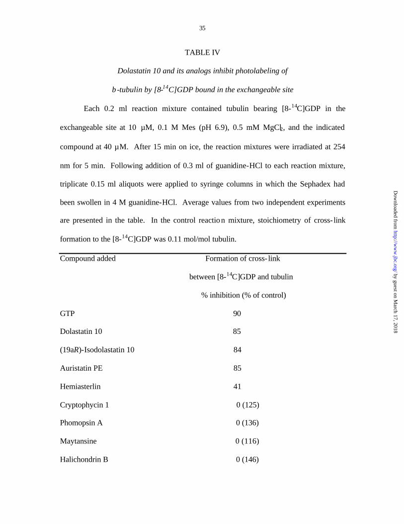
The converse was also true. As shown in Table IV, dolastatin 10 strongly
inhibited covalent bond formation between [8-14C]GDP and β-tubulin when we used the
tubulin preparation with the radiolabeled GDP prebound in the exchangeable site (27).
We also examined the effects of a number of other vinca domain drugs that strongly
inhibit nucleotide exchange on covalent bond formation between tubulin and prebound
exchangeable site [8-14C]GDP (Table IV). Of the compounds examined, only the
dolastatin 10 analogs (19aR)- isodolastatin 10 and auristatin PE had a similar inhibitory
effect. Even other peptide antimitotic agents that competitively inhibit the binding of
[3H]dolastatin 10 to tubulin were either noninhibitory (phomopsin A, cryptophycin 1) or
less inhibitory than dolastatin 10 and its analogs (hemiasterlin).4 In fact, there was an
apparent stimulation of covalent bond formation by phomopsin A and cryptophycin 1, as
well as by maytansine and halichondrin B.5 This difference between dolastatin 10 and
the other three antimitotic peptides was unexpected, since they all inhibit nucleotide
exchange on β-tubulin and bind tightly to the protein as indicated by the equivalent
persistence of apparent drug-tubulin complexes during gel filtration HPLC (7, 15, 49).
by guest on March 17, 2018
http://ww
w.jbc.org/
Dow
nloaded from
17
Such a result may imply significant differences in contact points between the different
peptides and β-tubulin (i.e., different proximities than dolastatin 10 to Cys-12 or,
possibly, Cys-211).
In addition, if irradiation produces a disulfide bond between Cys-12 and the sulfur
atom of the thiazole ring of dolastatin 10, dithiothreitol should release radiolabel derived
from dolastatin 10 from the tubulin. In contrast, if, as mass spectrometry data indicate (9,
42), the cross- link formed by irradiation between cysteine residues and guanosine
nucleotides results from attack by a sulfhydryl-derived free radical on C-5 of the guanine
moiety with formation of C-S bonds, dithiothreitol should not release nucleotide-derived
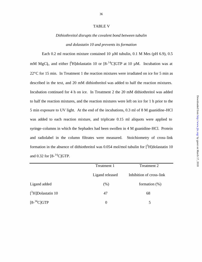
radiolabel from tubulin. This was in fact what was observed when dithiothreitol was
added to the reaction mixtures after irradiation (Table V, Treatment 1). Moreover,
adding dithiothreitol to reaction mixtures prior to irradiation produced the same result: a
sharp reduction in the amount of [3H]dolastatin cross- linked by UV light to tubulin, but
little effect on the amount of exogenously added [8-14C]GTP cross- linked to tubulin
(Table V, Treatment 2).
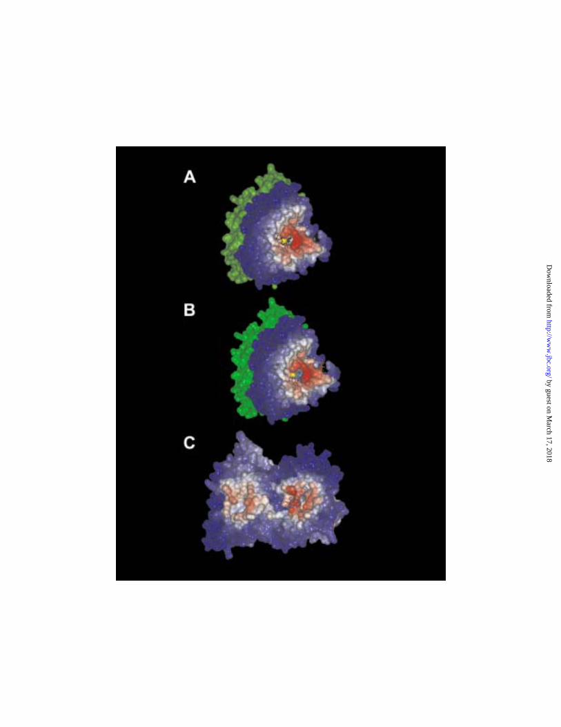
Molecular modeling analysis. The electron crystallographic model of tubulin
derived from paclitaxel-stabilized, zinc-induced sheets of antiparallel protofilaments (1)
was used in this analysis. Currently, there is no analogous structural information about
the unpolymerized αβ-heterodimer that is probably the actual binding species for
dolastatin 10 and other inhibitors of tubulin assembly. Initially we performed a global
scanning of the solvent accessible surface of β-tubulin in the isolated αβ-heterodimer for
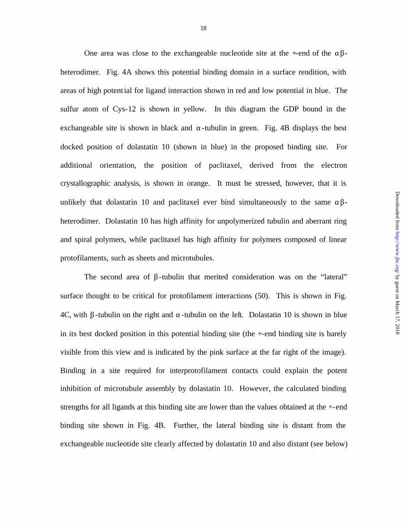
potential ligand binding sites. Two areas were identified in this step as candidate binding
sites (Fig. 4).
by guest on March 17, 2018
http://ww
w.jbc.org/
Dow
nloaded from
18
One area was close to the exchangeable nucleotide site at the +-end of the αβ-
heterodimer. Fig. 4A shows this potential binding domain in a surface rendition, with
areas of high potent ial for ligand interaction shown in red and low potential in blue. The
sulfur atom of Cys-12 is shown in yellow. In this diagram the GDP bound in the
exchangeable site is shown in black and α-tubulin in green. Fig. 4B displays the best
docked position of dolastatin 10 (shown in blue) in the proposed binding site. For
additional orientation, the position of paclitaxel, derived from the electron
crystallographic analysis, is shown in orange. It must be stressed, however, that it is
unlikely that dolastatin 10 and paclitaxel ever bind simultaneously to the same αβ-
heterodimer. Dolastatin 10 has high affinity for unpolymerized tubulin and aberrant ring
and spiral polymers, while paclitaxel has high affinity for polymers composed of linear
protofilaments, such as sheets and microtubules.
The second area of β-tubulin that merited consideration was on the “lateral”
surface thought to be critical for protofilament interactions (50). This is shown in Fig.
4C, with β-tubulin on the right and α-tubulin on the left. Dolastatin 10 is shown in blue
in its best docked position in this potential binding site (the +-end binding site is barely
visible from this view and is indicated by the pink surface at the far right of the image).
Binding in a site required for interprotofilament contacts could explain the potent
inhibition of microtubule assembly by dolastatin 10. However, the calculated binding
strengths for all ligands at this binding site are lower than the values obtained at the +-end
binding site shown in Fig. 4B. Further, the lateral binding site is distant from the
exchangeable nucleotide site clearly affected by dolastatin 10 and also distant (see below)
by guest on March 17, 2018
http://ww
w.jbc.org/
Dow
nloaded from
19
from the peptide spanning residues 2-31 to which dolastatin 10 can be cross- linked by
UV light.
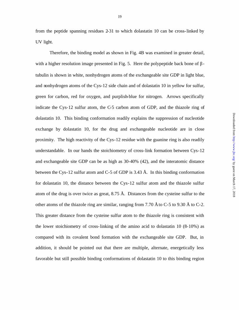
Therefore, the binding model as shown in Fig. 4B was examined in greater detail,
with a higher resolution image presented in Fig. 5. Here the polypeptide back bone of β-
tubulin is shown in white, nonhydrogen atoms of the exchangeable site GDP in light blue,
and nonhydrogen atoms of the Cys-12 side chain and of dolastatin 10 in yellow for sulfur,
green for carbon, red for oxygen, and purplish-blue for nitrogen. Arrows specifically
indicate the Cys-12 sulfur atom, the C-5 carbon atom of GDP, and the thiazole ring of
dolastatin 10. This binding conformation readily explains the suppression of nucleotide
exchange by dolastatin 10, for the drug and exchangeable nucleotide are in close
proximity. The high reactivity of the Cys-12 residue with the guanine ring is also readily
understandable. In our hands the stoichiometry of cross- link formation between Cys-12
and exchangeable site GDP can be as high as 30-40% (42), and the interatomic distance
between the Cys-12 sulfur atom and C-5 of GDP is 3.43 Å. In this binding conformation
for dolastatin 10, the distance between the Cys-12 sulfur atom and the thiazole sulfur
atom of the drug is over twice as great, 8.75 Å. Distances from the cysteine sulfur to the
other atoms of the thiazole ring are similar, ranging from 7.70 Å to C-5 to 9.30 Å to C-2.
This greater distance from the cysteine sulfur atom to the thiazole ring is consistent with
the lower stoichiometry of cross- linking of the amino acid to dolastatin 10 (8-10%) as
compared with its covalent bond formation with the exchangeable site GDP. But, in
addition, it should be pointed out that there are multiple, alternate, energetically less
favorable but still possible binding conformations of dolastatin 10 to this binding region
by guest on March 17, 2018
http://ww
w.jbc.org/
Dow
nloaded from
20
of β-tubulin. In some of these the distance between the Cys-12 sulfur atom and the
thiazole ring is about 4 Å.
by guest on March 17, 2018
http://ww
w.jbc.org/
Dow
nloaded from
21
DISCUSSION
We have described here direct photolabeling by [3H]dolastatin 10 of the β-tubulin
peptide spanning amino acid residues 2-31, and it is highly likely that UV irradiation of
the tubulin-drug complex creates sulfur free radicals at the cysteine residues of tubulin
(9). Consequently, Cys-12, the only cysteine residue in the target peptide, is the probable
site of covalent bond formation with the radiolabeled drug. Since the potent dolastatin 10
analog auristatin PE, which lacks the thiazole ring, has little ability to form a UV-induced
covalent bond with tubulin, we propose that this moiety of dolastatin 10 reacts with Cys-
12, perhaps through formation of a disulfide bond. Alternatively, the sulfur free radical
could disrupt one of the double bonds of the thiazole ring. A number of observations
were consistent with this hypothesis, including sensitivity of covalent bond formation to
dithiothreitol and mutual inhibition by dolastatin 10 and GDP of formation of their UV-
induced cross-links to β-tubulin.
Using the electron crystallographic model of tubulin derived from zinc- induced
sheets stabilized by paclitaxel (1), we found that the quantitatively most likely binding
site for dolastatin 10 was very close to the exchangeable nucleotide site and to Cys-12.
This readily explains two important biochemical features of the dolastatin 10 interaction
with tubulin: strong inhibition both of cross-link formation (12) induced by N,N-
ethylene(bis)iodoacetamide between Cys-12 and probably Cys-211 (1, 2, 8, 10) of β-
tubulin and of nucleotide exchange without displacement of prebound GDP from the
exchangeable site (7).
How does the binding site near Cys-12 described here relate to previous
observations with indirect photoaffinity probes? The indirect method involves placement
by guest on March 17, 2018
http://ww
w.jbc.org/
Dow
nloaded from
22
of a relatively bulky photoactive substituent on a ligand molecule. For such agents to be
useful they must bind with adequate affinity to their target and be specific in that both the
binding and the cross-linking reactions are inhibited by the natural ligand. This creates
two problems inherent in the technique. First, the bulky ligand introduces an additional
distance between target amino acid and the substituted atom of the ligand. Second, it can
be argued that ipso facto the substituted atom cannot be in close contact with an
important part of the binding site, since good activity is observed when it bears a bulky
substituent. In addition, it should be pointed out that, while dolastatin 10 is a
noncompetitive inhibitor of vinca alkaloid binding to tubulin (7), all published indirect
photoaffinity studies were performed with derivatives of competitive inhibitors (7, 51) of
vinca alkaloid binding: vinca alkaloid ana logs (18, 19, 21); a maytansine analog (20),
and a rhizoxin analog (22).
In only two of these studies was a specific reactive peptide identified, both in β-
tubulin. Wolff and coworkers (21) found that a photoactive vinblastine analog labeled a
peptide containing residues 175-213. Iwasaki and his colleagues (22) found that a
photoactive rhizoxin analog labeled a peptide containing residues 363-379. To correlate
these findings with our direct photoaffinity labeling of the peptide containing residues 2-
31, the model proposed here is reiterated as a ribbon diagram in Fig. 6, with these three
peptide regions shown in different colors. The figure also shows dolastatin 10 bound in
both of the sites explored in Fig. 4, with the Cys-12 side chain also shown (arrow).
Peptide 2-31 is colored magenta, 175-213 is orange, and 367-3796 is light blue.
Dolastatin 10 modeled into the lateral binding site (shown on the left in the Fig. 6 image)
is relatively distant from the three target peptides. In contrast, the dolastatin 10 modeled
by guest on March 17, 2018
http://ww
w.jbc.org/
Dow
nloaded from

23
into the +-end binding site adjacent to the exchangeable nucleotide site (shown in the
center of the Fig. 6 image) is close to portions of the 175-213 peptide as well as the 2-31
peptide, but distant from the 367-379 peptide. We have not modeled vinblastine or
rhizoxin into the potential +-end binding domain shown in Fig. 4AB, but portions of the
367-379 peptide are near this domain. Perhaps vinblastine and rhizoxin (a competitive
inhibitor of vinblastine binding) might have optimal binding sites close to both peptides
175-213 and 367-379. Rhizoxin and especially vinblastine are relatively weak inhibitors
of nucleotide exchange on β-tubulin (7, 16).
We have, however, performed initial modeling studies with dolastatin 15 and
cryptophycin 1 at the +-end binding site. We examined these two antimitotic
depsipeptides because of the seemingly contradictory effects of cryptophycin 1 on the
binding of radiolabeled dolastatins 10 and 15 to tubulin. Radiolabeled dolastatin 10 was
shown to bind with high affinity to tubulin, with an apparent Kd value of about 25 nM
(23). Cryptophycin 1 strongly inhibits the binding of dolastatin 10 to tubulin in a
competitive manner (49), while dolastatin 15 is noninhibitory (23). The inability of
dolastatin 15 to inhibit dolastatin 10 binding probably derives from its low affinity for
tubulin, since the apparent Kd value of the binding interaction is about 30 µM (52).
Although the binding of cryptophycin 1 to tubulin has not been quantitatively measured,
its close analog cryptophycin 52 binds to tubulin with an apparent Kd value of 0.1-0.45
µM (53). When the effects of dolastatin 10 and cryptophycin 1 on the weak binding of
dolastatin 15 to tubulin were examined (52), puzzling results were obtained. Dolastatin
10 strongly inhibited dolastatin 15 binding, completely displacing dolastatin 15 from
tubulin, whereas cryptophycin 1 had a much weaker inhibitory effect. There was no
by guest on March 17, 2018
http://ww
w.jbc.org/
Dow
nloaded from
24
displacement of bound dolastatin 15 from tubulin by equimolar cryptophycin 1, and
about 67% displacement when the concentration of cryptophycin 1 was 6-fold higher
than that of dolastatin 15.
One possible explanation for these seemingly contradictory effects of dolastatin
10, dolastatin 15, and cryptophycin 1 on each other’s binding to tubulin is that the drugs
actually bind to overlapping regions in the vinca domain. We asked whether comparing
the energetically optimal models for the binding of the three drugs to the +-end binding
domain would be in accord with the inhibitory patterns that had been observed. Fig. 7
demonstrates that the favored binding modes of the three drugs are in fact consistent with
the inhibition patterns. Fig. 7A compares the most favorable orientations of dolastatin 10
(blue) and cryptophycin 1 (red). The substantial overlap in the binding orientations is
consistent with the observed strong competitive inhibition by cryptophycin 1 of dolastatin
10 binding to tubulin. Fig. 7B compares the most favorable orientations of dolastatin 10
(blue) and dolastatin 15 (white). The intertwining of the superimposed structures is
consistent with the observation that the tightly binding dolastatin 10 can totally inhibit the
interaction of the weakly binding dolastatin 15 with tubulin. Finally, Fig. 7C compares
the most favorable orientation of cryptophycin 1 (red) and dolastatin 15 (white). There is
relatively little overlap between the superimposed structures, consistent with the apparent
simultaneous binding of both compounds to tubulin predicted by the failure of equimolar
cryptophycin 1 to inhibit dolastatin 15 binding despite its > 100-fold higher Kd value.
We should also note the recent modeling study of Barbier et al. (54), in which
cryptophycin 52 was modeled into the αβ-heterodimer and into the ring oligomers that
the drug-tubulin complex forms. While the model proposed by these workers placed
by guest on March 17, 2018
http://ww
w.jbc.org/
Dow
nloaded from
25
cryptophycin 52 in the +-end binding domain, the depsipeptide was not as close to the
exchangeable site as the model shown in Fig. 7AC (the relative locations of cryptophycin
1 and the exchangeable site GDP presented here are shown in Fig. 8). The model
proposed by these workers showed the cryptophycin 52 molecule in close contact with
the peptide spanning residues 204-225, with specific interactions between the drug and
Tyr-208, Cys-211, Phe-212, and Tyr-222. Fig. 8 attempts to correlate the positions of the
two proposed binding sites for cryptophycins by reiterating the energetically favored
binding of cryptophycin 1 (red) against the β-tubulin polypeptide backbone (white), with
the 204-225 sequence shown in orange, the side chains of Tyr-208, Cys-211, Phe-212,
and Tyr-222 shown in green, yellow, dark blue, and magenta, respectively, and the
exchangeable site GDP in light blue. The only interaction the two models seem to have
in common is of a phenyl ring of the drug with Tyr-222, but even this involves different
phenyl rings of the cryptophycins. While the modeling methods used by Barbier et al.
(54) do not differ greatly from those used here, nuances in methodology clearly affect the
precise orientation selected by the computational methods used to obtain docking results.
In summary, we have demonstrated significant cross- linking of radiolabeled
dolastatin 10 to β-tubulin in residues 2-31 when the drug-tubulin complex is exposed to
UV light. Indirect evidence strongly indicates that the cross-link is between the sulfur
atom of Cys-12 and the thiazole ring of dolastatin 10. Modeling of dolastatin 10 into the
electron crystallographic model of tubulin is highly consistent with the biochemical data.
The energetically favored binding site is in intimate contact with the exchangeable
nucleotide site and with Cys-12.
by guest on March 17, 2018
http://ww
w.jbc.org/
Dow
nloaded from
26
FOOTNOTES
1The abbreviations used are: [3H]dolastatin 10, [5-3H]dolastatin 10; [3H]auristatin
PE, [phenyl-4-3H]auristatin PE; [3H]colchicine, [ring C, methoxy 3H]colchicine;
[3H]vinblastine, [G-3H]vinblastine; Mes, 4-morpholineethanesulfonate; SDS, sodium
dodecyl sulfate; PAGE, polyacrylamide gel electrophoresis; Tricine, N-[2-hydroxy-1,1-
bis(hydroxymethyl)ethyl]glycine; PVDF, polyvinylidenedifluoride; HPLC, high
performance liquid chromatography.
2Personal communication, Dr. S. B. Horwitz.
3Personal communication, Dr. J. Wolff.
4The data showing a competitive inhibitory pattern obtained with phomopsin A
are unpublished (R. Bai, unpublished observations).
5The most likely explanation for the stimulatory effect of phomopsin A,
cryptophycin 1, maytansine, and halichondrin B is that they minimized dissociation of [8-
14C]GDP from the exchangeable site when the stock tubulin solution was diluted to its
final concentration of 10 µM.
6Residues 363-366 are not present in the electron crystallographic model (1) and
therefore not shown in Fig. 6.
by guest on March 17, 2018
http://ww
w.jbc.org/
Dow
nloaded from
27
REFERENCES
1. Nogales, E., Wolf, S. G., and Downing, K. H. (1998) Nature (London) 391, 199-203
2. Ludueña, R. F., and Roach, M. C. (1991) Pharmacol. Ther. 49, 133-152
3. Bai, R., Pei, X.-F., Boyé, O., Getahun, Z., Grover, S., Bekisz, J., Nguyen, N. Y.,
Brossi, A., and Hamel, E. (1996) J. Biol. Chem. 271, 12639-12645
4. Bai, R., Covell, D. G., Pei, X.-F., Ewell, J. B., Nguyen, N. Y., Brossi, A., and
Hamel, E. (2000) J. Biol. Chem. 275, 40443-40452
5. Williams, R. F., Mumford, C. L., Williams, G. A., Floyd, L. J., Aivaliotis, M. J.,
Martinez, R. A., Robinson, A. K., and Barnes, L. D. (1985) J. Biol. Chem.
260, 13794-13802
6. Floyd, L. J., Barnes, L. D., and Williams, R. F. (1989) Biochemistry 28, 8515-8525
7. Bai, R., Pettit, G. R., and Hamel, E. (1990) J. Biol. Chem. 265, 17141-17149
8. Little, M., and Ludueña, R. F. (1987) Biochim. Biophys. Acta 912, 28-33
9. Shivanna, B. D., Mejillano, M. R., Williams, T. D., and Himes, R. H. (1993) J. Biol.
Chem. 268, 127-132
10. Bai, R., Ewell, J. B., Nguyen, N. Y., and Hamel, E. (1999) J. Biol. Chem. 274,
12710-12714
11. Roach, M. C., and Ludueña, R. F. (1984) J. Biol. Chem. 259, 12063-12071
12. Bai, R., Roach, M. C., Srirangam, J. K., Barkoczy, J., Pettit, G. R., Ludueña, R. F.,
and Hamel, E. (1993) Biochem. Pharmacol. 45, 1503-1515
13. Bai, R., Paull, K. D., Herald, C. L., Malspeis, L., Pettit, G. R., and Hamel, E. (1991)
J. Biol. Chem. 266, 15882-15889
by guest on March 17, 2018
http://ww
w.jbc.org/
Dow
nloaded from
28
14. Bai, R., Taylor, G. F., Cichacz, Z. A., Herald, C. L., Kepler, J. A., Pettit, G. R., and
Hamel, E. (1995) Biochemistry 34, 9714-9719
15. Bai, R., Durso, N. A., Sackett, D. L., and Hamel, E. (1999) Biochemistry 43, 14302-
14310
16. Huang, A. B., Lin, C. M., and Hamel, E. (1985) Biochem. Biophys. Res. Commun.
128, 1239-1246
17. Wolff, J., Knipling, L., Cahnmann, H. J., and Palumbo, G. (1991) Proc. Natl. Acad.
Sci. U.S.A. 88, 2820-2824
18. Safa, A. R., Hamel, E., and Felsted, R. L. (1987) Biochemistry 26, 97-102
19. Nasioulas, G., Grammbitter, K., Himes, R. H., and Ponstingl, H. (1990) Eur. J.
Biochem. 192, 69-74
20. Sawada, T., Kato, Y., Kobayashi, H., Hashimoto, Y., Watanabe, T., Sugiyama, Y.,
and Iwasaki, S. (1993) Bioconjugate Chem. 4, 284-289
21. Rai, S. S., and Wolff, J. (1996) J. Biol. Chem. 271, 14707-14713
22. Sawada, T., Kobayashi, H., Hashimoto, Y., and Iwasaki, S. (1993) Biochem.
Pharmacol. 45, 1387-1394
23. Bai, R., Taylor, G. F., Schmidt, J. M., Williams, M. D., Kepler, J. A., Pettit, G. R.,
and Hamel, E. (1995) Mol. Pharmacol. 47, 965-976
24. Pettit, G. R., Srirangam, J. K., Barkoczy, J., Williams, M. D., Boyd, M. R., Hamel,
E., Pettit, R. K., Hogan, F., Bai, R., Chapuis, J.-C., McAllister, S. C., and
Schmidt, J. M. (1998) Anti-cancer Drug Design 13, 243-277
25. Natsume, T., Watanabe, J., Tamoki, S., Fujio, N., Miyasaka, K., and Kobayashi, M.
(2000) Jpn. J. Cancer Res. 91, 737-747
by guest on March 17, 2018
http://ww
w.jbc.org/
Dow
nloaded from

29
26. Hamel, E., and Lin, C. M. (1984) Biochemistry 23, 4173-4184
27. Grover, S., and Hamel, E. (1994) Eur. J. Biochem. 222, 163-172
28. Pettit, G. R., Singh, S. B., Hogan, F., Lloyd-Williams, P., Herald, D. L., Burkett, D.
D., and Clewlow, P. J. (1989) J. Am. Chem. Soc. 111, 5463-5465
29. Pettit, G. R., Srirangam, J. K., Barkoczy, J., Williams, M. D., Durkin, K. P. M.,
Boyd, M. R., Bai, R., Hamel, E., Schmidt, J. M., and Chapuis, J.-C. (1995)
Anti-cancer Drug Design 10, 529-544
30. Pettit, G. R., Singh, S. B., Hogan, F., and Burkett, D. D. (1990) J. Med. Chem. 33,
3132-3133
31. Gamble, W. R., Durso, N. A., Fuller, R. W., Westergaard, C. K., Johnson, T. R.,
Sackett, D. L., Hamel, E., Cardellina II, J. H., and Boyd, M. R. (1999)
Bioorg. Med. Chem. 7, 1611-1615
32. Hamel, E., and Lin, C. M. (1984) J. Biol. Chem. 259, 11060-11069
33. Young, B. L., Jernigan, R. L., and Covell, D. G. (1994) Protein Sci. 3, 717-729
34. Berman, H. M., Westbrook, J., Feng, Z., Gilliland, G., Bhat, T. N., Weissig, H.,
Shindyalov, I. N., and Bourne, P. E. (2000) Nucleic Acids Res. 28, 235-242
35. Miyazawa, S., and Jernigan, R. L. (1999) Proteins Struct. Funct. Genet. 36, 357-369
36. Bewley, C. A., Gustafson, K. R., Boyd, M. R., Covell, D. G., Bax, A., Clore, G. M.,
and Gronenborn, A. M. (1998) Nat. Struct. Biol. 5, 571-578
37. Covell, D. G., Smythers, G. W., Gronenborn, A. M., and Clore, G. M. (1994)
Protein Sci. 3, 2064-2072
38. Wallqvist, A., Jernigan, R. L., and Covell, D. G. (1995) Protein Sci. 4, 1881-1903
39. Wallqvist, A., and Covell, D. G. (1996) Proteins Struct. Funct. Genet. 25, 403-419
by guest on March 17, 2018
http://ww
w.jbc.org/
Dow
nloaded from
30
40. Rao, S., Horwitz, S. B., and Ringel, I. (1992) J. Natl. Cancer Inst. 84, 785-788
41. Bai, R., Pettit, G. R., and Hamel, E. (1990) Biochem. Pharmacol. 40, 1859-1864
42. Bai, R., Choe, K., Ewell, J. B., Nguyen, N. Y., and Hamel, E. (1998) J. Biol. Chem.
273, 9894-9897
43. Bai, R., Lin, C. M., Nguyen, N. Y., Liu, T.-Y., and Hamel, E. (1989) Biochemistry
28, 5606-5612
44. Landon, M. (1977) Meth. Enzymol. 47, 145-149
45. Ponstingl, H., Krauhs, E., Little, M., and Kempf, T. (1981) Proc. Natl. Acad. Sci.
U.S.A. 78, 2757-2761
46. Krauhs, E., Little, M., Kempf, T., Hofer-Warbinek, R., Ade, W., and Ponstingl, H.
(1981) Proc. Natl. Acad. Sci. U.S.A. 78, 4156-4160
47. Rao, S., Krauss, N. E., Heerding, J. M., Swindell, C. S., Ringel, I., Orr, G. A., and
Horwitz, S. B. (1994) J. Biol. Chem. 269, 3132-3134
48. Miyazaki, K., Kobayashi, M., Natsume, T., Gondo, M., Mikami, T., Sakakibara, K.,
and Tsukagoshi, S. (1995) Chem. Pharm. Bull. 43, 1706-1718
49. Bai, R., Schwartz, R. E., Kepler, J. A., Pettit, G. R., and Hamel, E. (1996) Cancer
Res. 56, 4398-4406
50. Nogales, E., Whittaker, M., Milligan, R. A., and Downing, K. H. (1999) Cell 96, 79-
88
51. Mandelbaum-Shavit, F., Wolpert-DeFilippes, M. K., and Johns, D. G. (1976)
Biochem. Biophys. Res. Commun. 72, 47-54
52. Cruz-Monserrate, Z., Mullaney, J. T., Harran, P. G., Pettit, G. R., and Hamel, E.
(2003) Eur. J. Biochem. 270, 3822-3828
by guest on March 17, 2018
http://ww
w.jbc.org/
Dow
nloaded from
31
53. Panda, D., Ananthnarayan, V., Larson, G., Shih, C., Jordan, M. A., and Wilson, L.
(2000) Biochemistry 39, 14121-14127
54. Barbier, P., Gregoire, C., Devred, F., Sarrazin, M., and Peyrot, P. (2001)
Biochemistry 40, 13510-13519
by guest on March 17, 2018
http://ww
w.jbc.org/
Dow
nloaded from
32
TABLE I
Inhibition of total and covalent binding of dolastatin 10 to tubulin
by dolastatin 10 analogs
Each 0.65 ml reaction mixture contained 10 µM tubulin, 0.1 M Mes (pH 6.9), 0.5
mM MgCl2, and, if present, 50 µM auristatin PE or 50 µM (19aR)- isodolastatin 10. The
mixtures were incubated for 5 min at 22°C, and 10 µM [3H]dolastatin 10 was added.
After an additional 15 min at 22°C, the reaction mixtures were exposed to UV light on
ice as described in the text for 5 min. A 0.2 ml aliquot of each reaction mixture was
mixed with 0.3 ml of 8 M guanidine-HCl. Triplicate 0.15 ml aliquots from the original
reaction mixture and from the aliquot mixed with the denaturant were applied to syringe-
columns prepared in 0.1 M Mes-0.5 mM MgCl2 or in 4 M guanidine-HCl, as appropriate.
Averages obtained from three independent experiments are shown. In these experiments
the average total binding of [3H]dolastatin 10 was 0.52 mol/mol tubulin and of covalently
bound [3H]dolastatin, 0.040 mol/mol tubulin.
Peptide added Total dolastatin 10 bound Dolastatin 10 cross- linked
to tubulin
------------------------ % Inhibition ± S.D. ------------------------
Auristatin PE 81 ± 4 63 ± 4
(19aR)-isodolastatin 10 70 ± 10 58 ± 6
by guest on March 17, 2018
http://ww
w.jbc.org/
Dow
nloaded from
33
TABLE II
Amino acid sequence obtained from the major [ 3H]dolastatin 10-containing peptide
derived from formic acid digestion
Fifteen cycles were performed on an Applied Biosystems model 494A protein
sequencer. The first cycle also yielded minor peaks corresponding to the residues in
parentheses. Cysteine is destroyed during the Edman procedure, as indicated by the X at
cycle 12. The sequence obtained is that of the amino terminus of β-tubulin (46).
Cycle number Amino acid residue identified
1 Met (Gly, Gln, Pro)
2 Arg
3 Glu
4 Ile
5 Val
6 His
7 Ile
8 Gln
9 Ala
10 Gly
11 Gln
12 X
13 Gly
14 Asn
15 Gln
by guest on March 17, 2018
http://ww
w.jbc.org/
Dow
nloaded from
34
TABLE III
Auristatin PE binds to tubulin but fails to form a cross-link during UV irradiation
Each 0.5 ml reaction mixture contained 10 µM tubulin, 0.1 M Mes (pH 6.9), 0.5
mM MgCl2, and either 10 µM [3H]dolastatin 10 or 10 µM [3H]auristatin PE. Following a
15 min incubation at 22°C, duplicate 0.15 ml aliquots were applied to syringe-columns in
which the Sephadex had been swollen in 0.1 M Mes (pH 6.9)-0.5 mM MgCl2 to measure
total binding. The remaining 0.2 ml was irradiated for 15 min on ice, after which 0.3 ml
of 8 M guanidine-HCl were added. To measure covalent binding, triplicate 0.15 ml
aliquots were applied to syringe-columns in which the Sephadex had been swollen in 4 M
guanidine-HCl. Radiolabel and protein was measured in all column filtrates, and the data
presented are averages from three independent experiments.
Total binding Covalent binding
Drug added ------------- mol drug/mol tubulin -------------
[3H]dolastatin 10 0.53 0.051
[3H]auristatin PE 0.51 0.001
by guest on March 17, 2018
http://ww
w.jbc.org/
Dow
nloaded from
35
TABLE IV
Dolastatin 10 and its analogs inhibit photolabeling of
β-tubulin by [8-14C]GDP bound in the exchangeable site
Each 0.2 ml reaction mixture contained tubulin bearing [8-14C]GDP in the
exchangeable site at 10 µM, 0.1 M Mes (pH 6.9), 0.5 mM MgCl2, and the indicated
compound at 40 µM. After 15 min on ice, the reaction mixtures were irradiated at 254
nm for 5 min. Following addition of 0.3 ml of guanidine-HCl to each reaction mixture,
triplicate 0.15 ml aliquots were applied to syringe columns in which the Sephadex had
been swollen in 4 M guanidine-HCl. Average values from two independent experiments
are presented in the table. In the control reaction mixture, stoichiometry of cross- link
formation to the [8-14C]GDP was 0.11 mol/mol tubulin.
Compound added Formation of cross- link
between [8-14C]GDP and tubulin
% inhibition (% of control)
GTP 90
Dolastatin 10 85
(19aR)-Isodolastatin 10 84
Auristatin PE 85
Hemiasterlin 41
Cryptophycin 1 0 (125)
Phomopsin A 0 (136)
Maytansine 0 (116)
Halichondrin B 0 (146)
by guest on March 17, 2018
http://ww
w.jbc.org/
Dow
nloaded from
36
TABLE V
Dithiothreitol disrupts the covalent bond between tubulin
and dolastatin 10 and prevents its formation
Each 0.2 ml reaction mixture contained 10 µM tubulin, 0.1 M Mes (pH 6.9), 0.5
mM MgCl2, and either [3H]dolastatin 10 or [8-14C]GTP at 10 µM. Incubation was at
22°C for 15 min. In Treatment 1 the reaction mixtures were irradiated on ice for 5 min as
described in the text, and 20 mM dithiothreitol was added to half the reaction mixtures.
Incubation continued for 4 h on ice. In Treatment 2 the 20 mM dithiothreitol was added
to half the reaction mixtures, and the reaction mixtures were left on ice for 1 h prior to the
5 min exposure to UV light. At the end of the incubations, 0.3 ml of 8 M guanidine-HCl
was added to each reaction mixture, and triplicate 0.15 ml aliquots were applied to
syringe-columns in which the Sephadex had been swollen in 4 M guanidine-HCl. Protein
and radiolabel in the column filtrates were measured. Stoichiometry of cross- link
formation in the absence of dithiothreitol was 0.054 mol/mol tubulin for [3H]dolastatin 10
and 0.32 for [8-14C]GTP.
Treatment 1 Treatment 2
Ligand released Inhibition of cross- link
Ligand added (%) formation (%)
[3H]Dolastatin 10 47 68
[8-14C]GTP 0 5
by guest on March 17, 2018
http://ww
w.jbc.org/
Dow
nloaded from
37
FIGURE LEGENDS
Fig. 1. Structures of dolastatin 10, auristatin PE, and (19aR)- isodolastatin 10.
Dolastatin 10 has the S configuration at position C-19a. The single radiolabeled positions
in dolastatin 10 and auristatin PE are indicated by asterisks.
Fig. 2. Coomassie blue stained membrane (Track A) and its autoradiogram
(Track B). Tubulin with bound [3H]dolastatin 10 was irradiated, harvested, and digested
with formic acid. Following removal of the formic acid by evaporation, the peptide
mixture was dissolved and subjected to SDS-PAGE. Peptide from about 50 µg of tubulin
was applied to the gel. The separated peptides were transferred electrophoretically to a
PVDF membrane, which was stained, sprayed with EN3HANCE, and autoradiographed
for one week at – 70°C. See text for further details.
Fig. 3. Effect of irradiation time on subsequent ligand binding by tubulin. For
each ligand study, a 2.5 ml reaction mixture was prepared containing 10 µM tubulin, 0.1
M Mes (pH 6.9), and 0.5 mM MgCl2. The reaction mixture was irradiated as described in
the text. At each of the indicated time points, a 0.4 ml aliquot was removed from the
reaction mixture. When all timed aliquots were removed from the irradiated reaction
mixtures, one of the following radiolabeled ligands was added (final concentrations, 10
µM): [3H]dolastatin 10 (–), [8-14C]GTP (�), [3H]vinblastine (�), or [3H]colchicine (∇).
The samples were incubated for 30 min at 22°C, triplicate 0.125 ml aliquots of each
sample were applied to syringe columns containing Sephadex swollen with 0.1 M Mes
(pH 6.9)-0.5 mM MgCl2. The data presented in the figure represent averages obtained in
three independent experiments. Stoichiometries (mol ligand/mol tubulin) obtained in the
by guest on March 17, 2018
http://ww
w.jbc.org/
Dow
nloaded from
38
unirradiated samples were 0.61 for dolastatin 10, 0.38 for GTP, 0.36 for vinblastine, and
0.20 for colchicine.
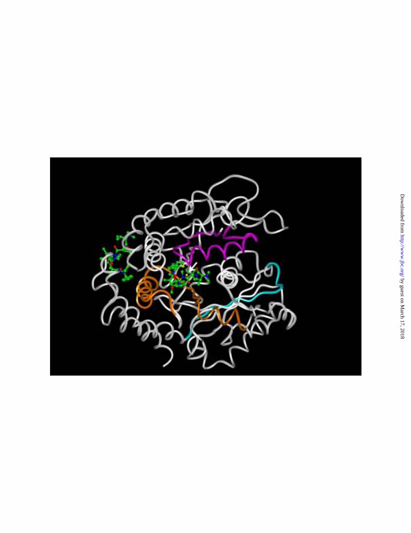
Fig. 4. Potential binding sites for dolastatin 10 on β-tubulin identified by global
scanning of the tubulin surface for areas of high hydrophobicity. A and B. A potential
binding site at the +-end of β-tubulin. Most likely areas for ligand binding are shown in
red, least likely in blue. The α-tubulin surface is shown in green. In panel A, the
exchangeable site GDP is shown as an atomic model (hydrogen atoms omitted) in black.
In panel B atomic models (hydrogen atoms omitted) of dolastatin 10 (light blue) and
paclitaxel (orange) are shown. C. A potential binding site on the “lateral” surface of
tubulin involved in interprotofilament contacts. Most likely areas for ligand binding are
shown in red, least likely in blue. The β-tubulin is on the right, α-tubulin on the left, with
the potential +-end binding site barely visualized on the far right. The optimal binding
position for dolastatin 10 is indicated by the atomic model (hydrogen atoms not shown)
of the peptide shown in light blue. Different color scales were used to generate the
surfaces shown in panels AB as compared with panel C. Overall, the lateral binding site
should have a higher probability of interacting with hydrophobic ligands, but the
predicted energetics for the binding of dolastatin 10 at the +-end site is almost three times
higher than for its binding at the lateral site.
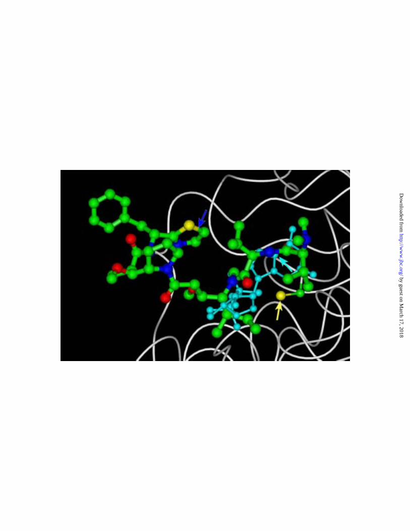
Fig. 5. Interrelationships of dolastatin 10, exchangeable site GDP, and the Cys-12
side chain at the proposed +-end binding site for dolastatin 10. The polypeptide
backbone of β-tubulin is shown in white, along with atomic models (hydrogen atoms not
shown) of GDP, dolastatin 10 and the Cys-12 side chain The atoms of GDP are shown in
light blue, while in dolastatin 10 and the Cys-12 side chain carbon atoms are shown in
by guest on March 17, 2018
http://ww
w.jbc.org/
Dow
nloaded from
39
green, sulfur in yellow, oxygen in red, and nitrogen in purplish-blue. The yellow arrow
indicates the Cys-12 sulfur atom, the light blue arrow the C-5 atom of GDP, and the
purplish-blue arrow the thiazole ring of dolastatin 10.
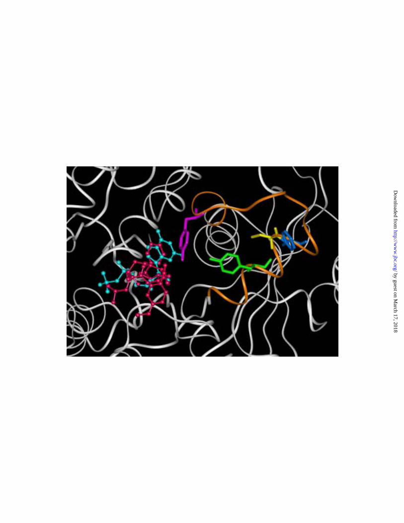
Fig. 6. Positions of three polypeptide chains of β-tubulin that have been photo
cross- linked to vinca domain drugs in relationship to the two potential dolastatin 10
binding sites shown in Fig. 4. The dolastatin 10 binding sites are indicated by atomic
models (hydrogen atoms not shown) of dolastatin 10 placed in the “lateral” binding site
(left side of diagram) and the +-end binding site (center of diagram), with the sulfur atom
shown in yellow, carbon atoms in green, oxygen in red, and nitrogen in purplish-blue.
Only the polypeptide backbone portion of β-tubulin is shown, except for the Cys-12 side
chain (arrow), with its carbon atom green and its sulfur atom yellow. The peptide
spanning residues 2-31 is shown in magenta, that spanning residues 175-213 in orange,
and that spanning residues 367-379 in light blue. The remainder of the β-tubulin peptide
backbone is shown in white.
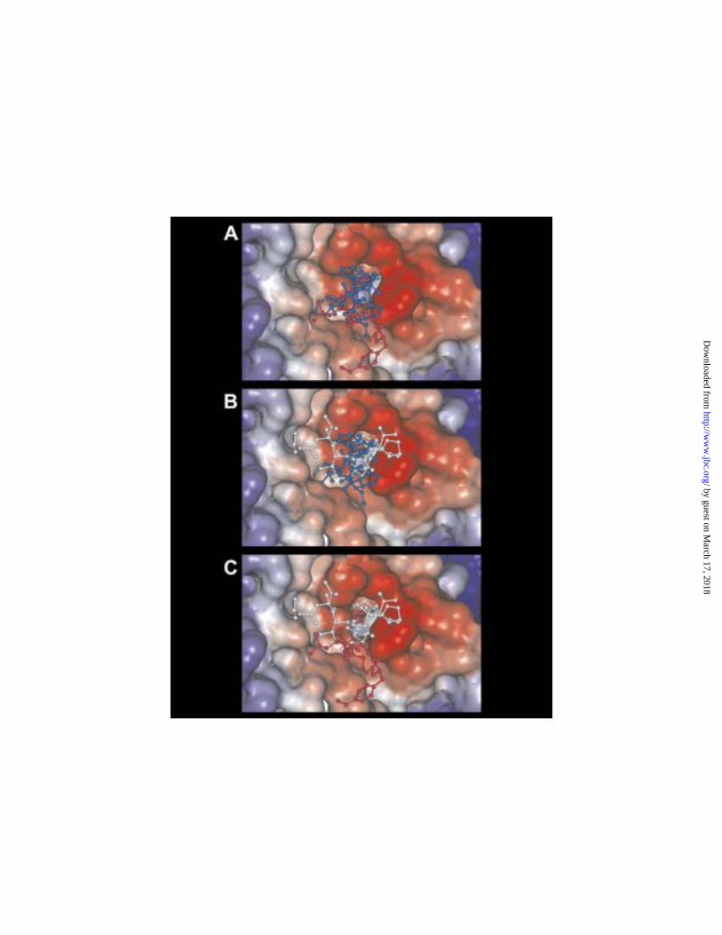
Fig. 7. Cryptophycin 1 and dolastatin 15 modeled into the +-end binding site, in
comparison with each other and with dolastatin 10. A. Best fit atomic models (hydrogen
atoms omitted) of dolastatin 10 (blue) and cryptophycin 1 (red) compared. B. Best fit
atomic models (hydrogen atoms omitted) of dolastatin 10 (blue) and dolastatin 15 (white)
compared. C. Best fit atomic models (hydrogen atoms omitted) of dolastatin 15 (white)
and cryptophycin 1 (red) compared.
Fig. 8. Best fit cryptophycin 1 binding site compared with elements of the
cryptophycin 52 binding site modeled by Barbier et al. (54). The polypeptide backbone
of β-tubulin is shown in white, except for the peptide spanning residues 204-225, which
by guest on March 17, 2018
http://ww
w.jbc.org/
Dow
nloaded from
40
is shown in orange. The exchangeable site GDP is shown in light blue, and cryptophycin
1, as modeled in Fig. 7, is shown in red. The peptide side chains of Tyr-208, Cys-211,
Phe-212, and Tyr-222 are shown in green, yellow, dark blue, and magenta, respectively.
by guest on March 17, 2018
http://ww
w.jbc.org/
Dow
nloaded from
Nguyen, George R. Pettit and Ernest HamelRuoli Bai, David G. Covell, George F. Taylor, John A. Kepler, Terry D. Copeland, Nga Y.
containing cysteine-12-tubulinβDirect photoaffinity labeling dolastatin 10 of the amino terminal peptide of
published online April 29, 2004J. Biol. Chem.
10.1074/jbc.M402110200Access the most updated version of this article at doi:
Alerts:
When a correction for this article is posted•
When this article is cited•
to choose from all of JBC's e-mail alertsClick here
by guest on March 17, 2018
http://ww
w.jbc.org/
Dow
nloaded from