
Revision - skinPT · 2018-04-23 · nodules (cellulite) and overweight. Many factors are involved...
13
1 m Product SILUSYNE ® Date February 2013 Revision 2 Technical Report
Transcript of Revision - skinPT · 2018-04-23 · nodules (cellulite) and overweight. Many factors are involved...

1
m
Product
SILUSYNE®
Date
February 2013
Revision
2
Technical Report
2
Contents
IMAGE AND FIGURE IMPORTANCE 3
ADIPOSE TISSUE AND FAT DEPOTS 4
FAT TISSUE VOLUME AND ADIPOCYTE DIFFERENTIATION 5
PGC-1α EFFECT ON THE SLIMMING PROCESS 6
SILUSYNE®, NEW MOLECULAR MECHANISM FOR THE PERSISTENT PROBLEM OF
CELLULLITE 7
IN VITRO EFFICACY
PGC-1α expression in human adipocytes 8
Effect on Lipid accumulation 9
IN VIVO EFFICACY
Instrumental evaluation of dermo-hypodermal junction 10
COSMETIC PROPERTIES 11
COSMETIC APPLICATIONS 11
TECHNICAL DATA
INCI name of the active ingredient 12
Presentation and preservatives 12
APPLICATION DATA
Processing 12
Incompatibilities 12
Solubility 12
Dosage 12
REFERENCES 13
3
Image and figure importance
Having the desired silhouette is nowadays a permanent goal for everyone. Our social image
has personal and professional implications, so everyone is willing to improve it and correct the
undesired physical features that can even influence our relationships.
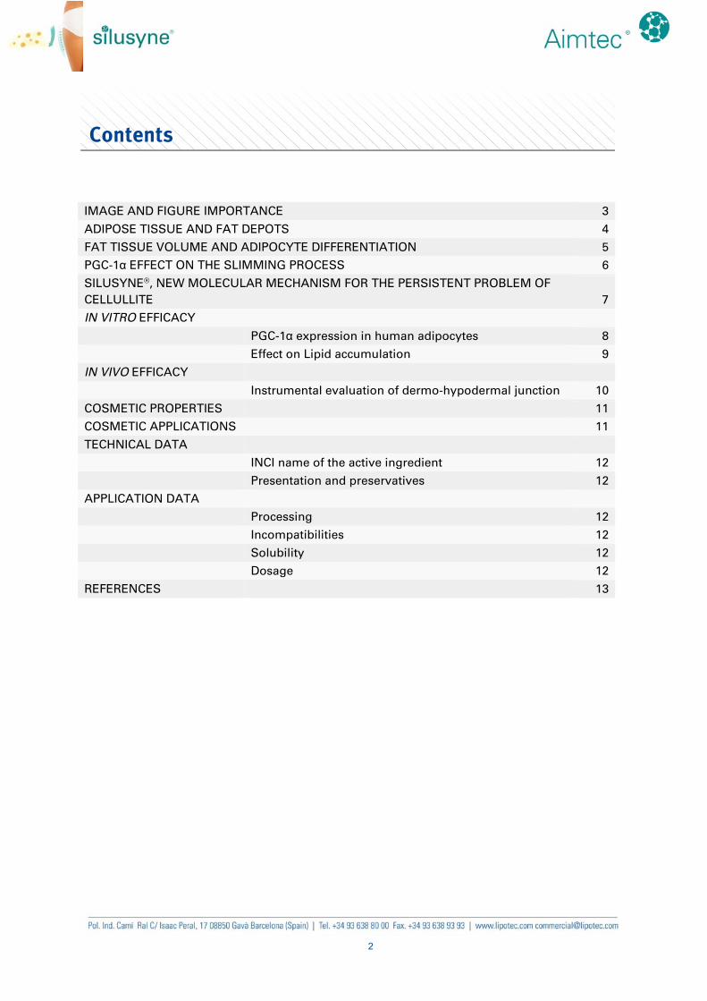
Cellulite is an aesthetical problem which
affects 90% of post-adolescent women
and a small percentage of men, who
consider it as an undesired skin disruption
to solve. Cellulite is the final step of
physiological changes that occur in the
subcutaneous fat layer, which lead to an
increase in the volume of fatty tissue and
the appearance of irregularities on the
skin surface, known as orange peel.
Moreover, overweight is an increasing
problem in modern society due to the lack
of exercise, extra caloric food intake and
long working days. Having an elevated
caloric food intake, results in an increase of
fat storage. This accumulation causes the
increase of the volume of the cells that keep
the extra lipids and, consequently, of their
surrounding tissue. As a result of this tissue
growth, the volume of the area increases
creating visible and non attractive fat
nodules (cellulite) and overweight.
Many factors are involved in the storage of
fat depots and the manifestation of cellulite
including age, genetics, gender, extra
caloric diet, lack of exercise, smoking, poor
blood circulation and an unhealthy lifestyle
in general. Thighs, buttocks and abdomen
are the areas where cellulite mainly
appears, but fat depots are also commonly
found in the stomach, breast and face and
neck in a smaller proportion. Although
cellulite is clearly linked to these extra fat
deposits, it is not exclusive of overweight
individuals; it can also be found in lean
women.
Overweight and cellulite are a problem
for most of the worldwide population.
People want to find an effective and
preferably non invasive solution that
allows them to reduce fat accumulation
and visible nodules, improve their
figure and feel better about themselves
without surgery or complex treatments.
4
Adipose tissue and fat depots
Adipose tissue is a specialised connective tissue mainly located beneath the skin. There are
two types of adipose tissue depending on its main function and their kind of adipocytes. In
early stages of human life, brown adipose tissue (BAT) is necessary despite its low percentage
(around 5%). However, white adipose tissue (WAT) is the prevalent type and the small
levels of brown tissue, present in youth, decrease with age.
Human BAT is characterised by a high
expression of mitochondrial genes and
polygonal adipocytes, which contain a great
number of mitochondria in the cytoplasm
and several small lipid droplets. The main
function of brown adipocytes is to dissipate
energy in the form of heat.
On the contrary, human WAT functions as
the major storage site for the lipids
incorporated by daily food intake.
Whenever the body requires energy for
cells to use, lipids contained in its cells are
burned. WAT contains round adipocytes
with a large single lipid droplet that are
responsible for storing lipids. It also has
macrophages, fibroblasts, leukocytes and
many collagen fibres that act as a support.
Preadipocyte cells are present in the fat
tissue as the precursors of adipocytes.
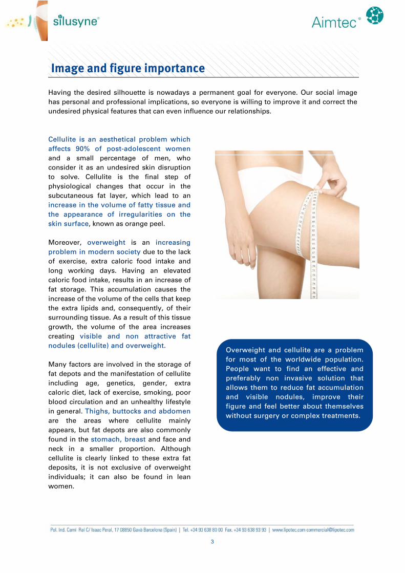
The main difference between white
preadipocytes and adipocytes is the
capacity of these last ones to store energy
in the form of triglycerides (and cholesterol
esters). White adipocytes contain a large
lipid droplet (80% of cell content), forcing
the nucleus and cytoplasm to remain in the
periphery of the cell (Fig. 1).
Due to several factors such as genetics or
high caloric intake, fat deposits of human
WAT can expand to such an extent that
push and distort the connective tissue.
This local volume increase can also induce
irregularities in the junction line between
dermis and hypodermis, increasing its
length and making the skin surface
appear uneven and with visible fat
nodules [1]. This extra lipid volume can
obstruct the lymphatic drainage system
making that waste materials like toxins or
proteins cannot be removed properly.
These materials create an immobile network, together with the collagen fibres, where fat cells
are trapped, leading to an inevitably increase of local volume and to the appearance of
unsightly irregularities on the skin surface (cellulite).
Fig. 1. Preadipocyte and mature adipocyte.
5
Fat tissue volume and adipocyte differentiation
The volume of WAT is a function of both adipocyte number and size, so its enlargement can
be caused either by an increase in the number of adipocytes or in lipid content.
Cellulite and overweight are related to a rise in the adipose depots, which is the result from
an imbalance between food intake and energy expenditure. Cellulite can appear as a
consequence of an extra local fat storage and it is known that overweight individuals produce
more white adipocytes per year and have a greater number of mature adipocytes than thin
individuals [2, 3]. For this reason, acting on the cycle of the cells that have the capacity to
store fat when they differentiate (white adipocytes) is one of the options to diminish fat
deposits and their consequences in the skin appearance (cellulite and irregularities).
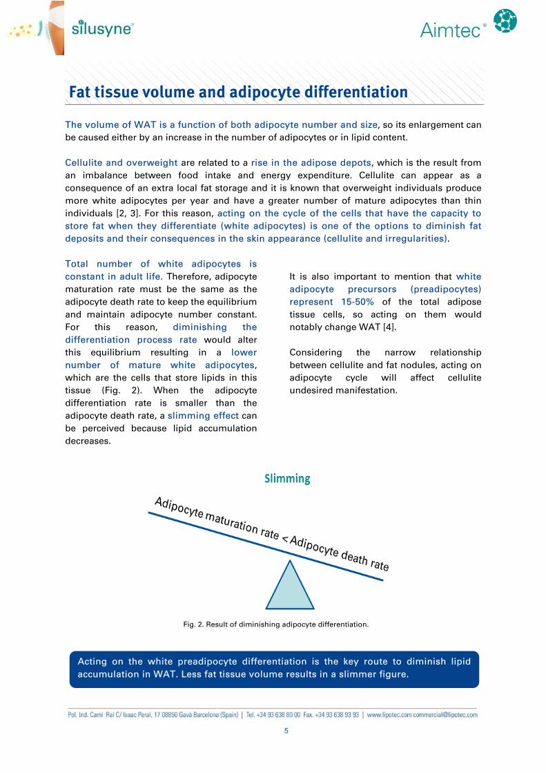
Total number of white adipocytes is
constant in adult life. Therefore, adipocyte
maturation rate must be the same as the
adipocyte death rate to keep the equilibrium
and maintain adipocyte number constant.
For this reason, diminishing the
differentiation process rate would alter
this equilibrium resulting in a lower
number of mature white adipocytes,
which are the cells that store lipids in this
tissue (Fig. 2). When the adipocyte
differentiation rate is smaller than the
adipocyte death rate, a slimming effect can
be perceived because lipid accumulation
decreases.
It is also important to mention that white
adipocyte precursors (preadipocytes)
represent 15-50% of the total adipose
tissue cells, so acting on them would
notably change WAT [4].
Considering the narrow relationship
between cellulite and fat nodules, acting on
adipocyte cycle will affect cellulite
undesired manifestation.
Acting on the white preadipocyte differentiation is the key route to diminish lipid
accumulation in WAT. Less fat tissue volume results in a slimmer figure.
Fig. 2. Result of diminishing adipocyte differentiation.
6
PGC-1α effect on the slimming process
The differentiation process from preadipocytes to mature adipocytes is a complex process
known as adipogenesis in which many factors and genes participate. Some genes need to be
expressed as they are distinctive of mature adipocytes while the typical genes of preadipocytes
need to be downregulated or almost inhibited in order to finally lead to the adipocyte
phenotype [5]. For this differentiation process to happen, transcriptional factors are required.
One of these key factors is Peroxisome proliferator-activated receptor-Gamma Coactivator 1
alpha (PGC-1α) due to its coactivation of a key receptor known as PPARγ.
PPARγ belongs to the Peroxisome
Proliferator-Activated Receptors (PPARs)
family, which is a group of nuclear
receptor proteins that functions as
transcriptional factors and regulates gene
expression in cellular differentiation among
other processes. This receptor forms
heterodimers with Retinoid X Receptors
which bind to specific regions on the DNA
of target genes and regulate their
expression. PPARγ is predominantly
expressed in the adipose tissue and it is
strictly necessary but not sufficient for
preadipocytes to differentiate.
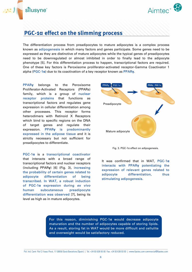
PGC-1α is a transcriptional coactivator
that interacts with a broad range of
transcriptional factors and nuclear receptors
(including PPARγ) [6] (Fig. 3), increasing
the probability of certain genes related to
adipocyte differentiation of being
transcribed. In WAT, a robust induction
of PGC-1α expression during ex vivo
human subcutaneous preadipocyte
differentiation was observed [7], being its
level as high as in mature adipocytes.
Fig. 3. PGC-1α effect on adipogenesis.
It was confirmed that in WAT, PGC-1α
interacts with PPARγ potentiating the
expression of relevant genes related to
adipocyte differentiation, thus
stimulating adipogenesis.
For this reason, diminishing PGC-1α would decrease adipocyte
maturation and the number of adipocytes capable of storing lipids.
As a result, storing fat in WAT would be more difficult and cellulite
and overweight would be satisfactory reduced.
Preadipocyte
Mature adipocyte
7
SILUSYNE®, new molecular mechanism for the persistent problem of cellulite
SILUSYNE® is a new ingredient for anti-cellulite and slimming products which contains a
hexapeptide in a novel delivery system.
SILUSYNE® contains a hexapeptide with natural amino acids which was identified by a
combinatorial chemistry approach from a library of 49,521,980 hexapeptides. The combinatorial
peptide library was screened using the reporter gene assay in a stably transfected cell line
where luciferase expression was controlled by PGC-1α promoter activity.
Lipotec developed a new specific delivery system in the submicron scale range with high
encapsulation efficiency, suitable for a wide range of active ingredients. It combines techniques
of microemulsion and microencapsulation and it allows improving the formulation of
SILUSYNE® peptide, in aqueous phases.
SILUSYNE® showed to diminish adipocyte
differentiation in white adipose tissue by
decreasing PGC-1α levels in vitro.
In vivo, SILUSYNE® proved to significantly
reduce the length of the dermo-
hypodermal junction line, which is related
to the formation of cellulite and skin
irregularities [1]. As a result, skin becomes
softer and flatter, making cellulite less
visible.
SILUSYNE® is the perfect ingredient
for anti-cellulite and slimming
cosmetic formulations.
8
In vitro efficacy
PGC-1α EXPRESSION IN HUMAN ADIPOCYTES
Efficacy of SILUSYNE® peptide was verified by measuring its effect in human subcutaneous
preadipocytes in culture.
Human subcutaneous preadipocytes were
incubated during 24 h in the Preadipocyte
Growth Medium (PGMTM-2), which was
used as the basal control (non-treated non-
differentiated cells). Differentiation was
induced by changing this medium to the
Preadipocyte Differentiation Medium (PDM-
2), which was used as a control for non-
treated differentiated cells. Afterwards, 25
or 100 μg/mL of SILUSYNE® peptide (Acetyl
Hexapeptide-39) were added during the
differentiation process and all samples
(including controls) were incubated at 37ºC
for 10 days.
Afterwards, cells were lysed, RNA was
extracted and reverse transcription was
carried out. The resulting cDNA was
analysed by quantitative RT-PCR (Fig. 4).
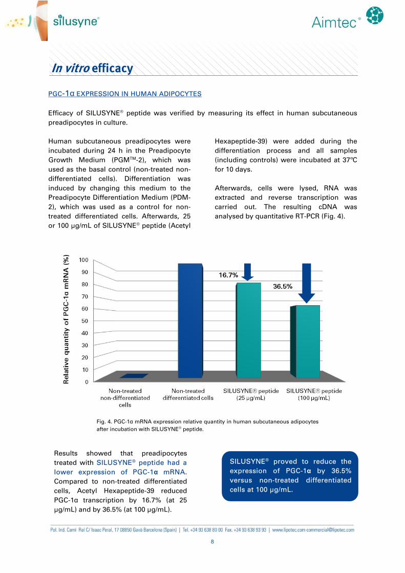
Fig. 4. PGC-1α mRNA expression relative quantity in human subcutaneous adipocytes
after incubation with SILUSYNE® peptide.
SILUSYNE® proved to reduce the
expression of PGC-1α by 36.5%
versus non-treated differentiated
cells at 100 μg/mL.
Results showed that preadipocytes
treated with SILUSYNE® peptide had a
lower expression of PGC-1α mRNA.
Compared to non-treated differentiated
cells, Acetyl Hexapeptide-39 reduced
PGC-1α transcription by 16.7% (at 25
μg/mL) and by 36.5% (at 100 μg/mL).
9
EFFECT ON LIPID ACCUMULATION
Human subcutaneous preadipocytes were
incubated during 24 h in the Preadipocyte
Growth Medium (PGMTM-2), which was
used as the basal control (non-treated non-
differentiated cells). Differentiation was
induced by changing this medium to the
Preadipocyte Differentiation Medium (PDM-
2) and incubating the cells for 10 days in the
presence of different treatments.
SILUSYNE® peptide was tested at two
different concentrations (25 and 100 μg/mL)
and caffeine (200 μg/mL) was also included
in the test. PDM-2 was used as a control for
non-treated differentiated cells.
After 10 days, the supernatants were
removed and wells were washed.
Afterwards, 5 μL of AdipoRedTM reagent
were added to each well and mixed.
The AdipoRedTM reagent is a hydrophilic
solution that turns into fluorescent in
hydrophobic environments. This facilitates
the detection of the levels of intracellular
lipid droplets accumulated during
preadipocyte differentiation, which become
stained. Fluorescence values were
quantified at 535 nm (excitation at 485 nm),
corrected with respect to basal fluorescence
and normalised with respect to the
fluorescence of the differentiation medium.
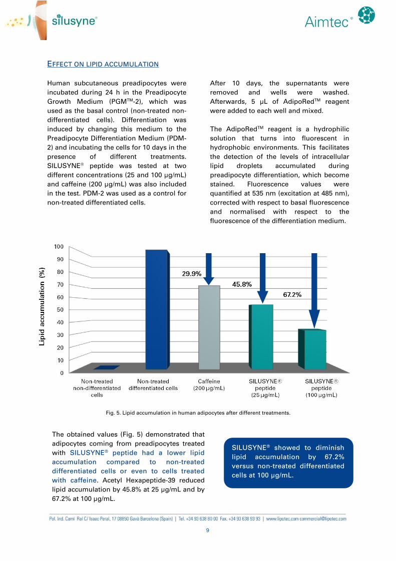
Fig. 5. Lipid accumulation in human adipocytes after different treatments.
SILUSYNE® showed to diminish
lipid accumulation by 67.2%
versus non-treated differentiated
cells at 100 μg/mL.
The obtained values (Fig. 5) demonstrated that
adipocytes coming from preadipocytes treated
with SILUSYNE® peptide had a lower lipid
accumulation compared to non-treated
differentiated cells or even to cells treated
with caffeine. Acetyl Hexapeptide-39 reduced
lipid accumulation by 45.8% at 25 μg/mL and by
67.2% at 100 μg/mL.
10
In vivo efficacy
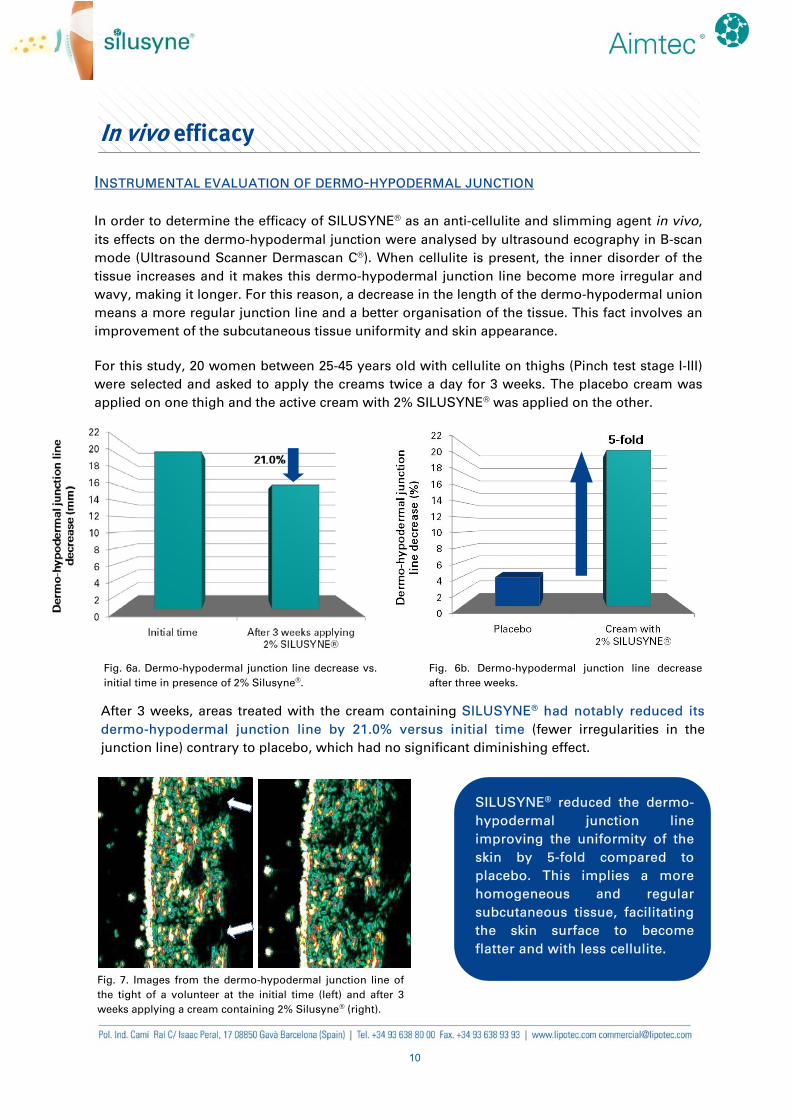
INSTRUMENTAL EVALUATION OF DERMO-HYPODERMAL JUNCTION
In order to determine the efficacy of SILUSYNE® as an anti-cellulite and slimming agent in vivo,
its effects on the dermo-hypodermal junction were analysed by ultrasound ecography in B-scan
mode (Ultrasound Scanner Dermascan C®). When cellulite is present, the inner disorder of the
tissue increases and it makes this dermo-hypodermal junction line become more irregular and
wavy, making it longer. For this reason, a decrease in the length of the dermo-hypodermal union
means a more regular junction line and a better organisation of the tissue. This fact involves an
improvement of the subcutaneous tissue uniformity and skin appearance.
For this study, 20 women between 25-45 years old with cellulite on thighs (Pinch test stage I-III)
were selected and asked to apply the creams twice a day for 3 weeks. The placebo cream was
applied on one thigh and the active cream with 2% SILUSYNE® was applied on the other.
Fig. 6a. Dermo-hypodermal junction line decrease vs.
initial time in presence of 2% Silusyne®.
Fig. 6b. Dermo-hypodermal junction line decrease
after three weeks.
SILUSYNE® reduced the dermo-
hypodermal junction line
improving the uniformity of the
skin by 5-fold compared to
placebo. This implies a more
homogeneous and regular
subcutaneous tissue, facilitating
the skin surface to become
flatter and with less cellulite.
After 3 weeks, areas treated with the cream containing SILUSYNE® had notably reduced its
dermo-hypodermal junction line by 21.0% versus initial time (fewer irregularities in the
junction line) contrary to placebo, which had no significant diminishing effect.
Fig. 7. Images from the dermo-hypodermal junction line of
the tight of a volunteer at the initial time (left) and after 3
weeks applying a cream containing 2% Silusyne® (right).
11
Cosmetic properties
SILUSYNE®:
is a novel ingredient containing a hexapeptide included in a special delivery system
ideal for anti-cellulite and slimming formulations.
diminishes preadipocyte differentiation in WAT by decreasing the expression of PGC-
1α, which is closely linked to adipogenesis. Its effects were demonstrated in vitro, where
it reduced PGC-1α by 36.5%.
reduces the lipid content in WAT, as it can be seen in vitro, where it diminished lipid
accumulation by 67.2%, obtaining even better reductions than caffeine.
improves skin uniformity by reducing the length of the dermo-hypodermal junction
line, related to cellulite and irregularities on the skin. Results of the in vivo study showed
that this junction was reduced by 21.0% versus initial time, being 5 times more
effective than the placebo.
Cosmetic applications
SILUSYNE® can be incorporated in many formulations for preventing and treating cellulite,
slimming purposes and as a slimming complement in hydrating, firming, remodelling,
tanning and sun care products.
It can also be used as ingredient in formulations designed for usual sportsmen and
sportswomen to enhance the slimming effect of the sport practise. Daily use products as body
milks can also incorporate this ingredient to produce an extra slimming effect.
12
Technical data
INCI NAME OF THE ACTIVE INGREDIENT
PRESENTATION AND PRESERVATIVES
Gel containing 0.05% of the peptide.
Application data
PROCESSING
SILUSYNE® can be added in the aqueous phase when formulating a gel. In case of preparing an
emulsion, it should be added once the emulsion is formed. In both cases, it should be provided
that the temperature is below 40ºC.
SILUSYNE® is stable at a pH range between 3.5 and 8.0.
INCOMPATIBILITIES
Not expected.
SOLUBILITY
Dispersible in water.
DOSAGE
A dosage of 2% of SILUSYNE® is recommended in final cosmetic formulations.
Active ingredient INCI name
SILUSYNE®
Isohexadecane, Sorbitan Sesquioleate, Acetyl Hexapeptide-39,
Starch Hydroxypropyltrimonium Chloride, Sodium Hyaluronate,
Potassium Cetyl Phosphate.
Code Product presentation Preservatives
PD205 SILUSYNE® Phenoxyethanol,
Sodium Benzoate
13
References
1. Quatresooz P, Xhauflaire-Uhoda E, Piérard-Franchimont C, et al. Cellulite histopathology
and related mechanobiology. International Journal of Cosmetic Science. 28: 207-210, 2006.
2. Spalding KL, Arner E, Westermark PO, et al. Dynamics of fat cell turnover in humans.
Nature. 453(5): 783-787, 2008.
3. Arner P, Spalding KL. Fat cell turnover in humans. Biochemical and Biophysical Research
Communications. 396: 101-104, 2010.
4. Tchkonia T, Morbeck DE, Zglinicki T., et al. Fat tissue, aging, and cellular senescence. Aging
Cell. 9: 667-684, 2010.
5. Gerhold DL, Liu F, Jiang G, et al. Gene expression profile of adipocyte differentiation and its
regulation by peroxisome proliferator-activated receptor-gamma agonists. Endocrinology.
143 (6): 2106-18, 2002.
6. Liang H, Ward WF. PGC-1α: a key regulator of energy metabolism. Adv Physiol Educ. 30:
145-151, 2006.
7. Semple RK, Crowley VC, Sewter CP, et al. Expression of the thermogenic nuclear hormone
receptor coactivator PGC-1α is reduced in the adipose tissue of morbidly obese subjects.
International Journal of Obesity. 28: 176-179, 2004.
Note: Graphs and photographs of efficacy tests are available for customer use provided that the final product contains
the same concentration of active as the formulations in our tests. Customers must request written permission for use of
the graphic material and/or ingredient tradenames to Lipotec. Customers are responsible for compliance with local and
international advertising regulations.
The specific situation of the trademark in each country may vary and we recommend that you contact us for updated
information.
Disclaimer:
While the claims and supporting data provided in this publication are believed to be reliable and they are presented free
and for guidance only, there are no warranties of any kind. All expressed and implied warranties are disclaimed. The
recipient is solely responsible for ensuring that products marketed to consumers comply with all relevant laws and
regulations. LIPOTEC is the exclusive holder of the both industrial and intellectual property rights identified herein.
Recipient of this publication agrees to indemnify and hold harmless each entity of the LIPOTEC organization for any and
all regulatory action arising from recipient’s use of any claims or information in this publication, including, but not
limited to, use in advertising and finished product label claims, and not present this publication as evidence of finished
product claim substantiation to any regulatory authority.


![Dark photon manifestation in the tripletlike QED processesMore information about DP (and dark matter) searches in astrophysical experiments can be found in Ref. [40]. In this work,](https://static.fdocument.org/doc/165x107/608adbeb5d3a293704137d36/dark-photon-manifestation-in-the-tripletlike-qed-processes-more-information-about.jpg)