REVIEW Open Access O-GlcNAc profiling: from proteins to ...
16
REVIEW Open Access O-GlcNAc profiling: from proteins to proteomes Junfeng Ma and Gerald W Hart * Abstract O-linked β-D-N-acetylglucosamine (O-GlcNAc) modification (O-GlcNAcylation) onto serine and threonine residues of proteins is an important post-translational modification (PTM), which is involved in many crucial biological processes including transcription, translation, proteasomal degradation, and signal transduction. Aberrant protein O-GlcNAcylation is directly linked to the pathological progression of chronic diseases including diabetes, cancer, and neurodegenerative disorders. Identification, site mapping, and quantification of O-GlcNAc proteins are a prerequisite to decipher their functions. In this review, we mainly focus on technological developments regarding O-GlcNAc protein profiling. Specifically, on one hand, we show how these techniques are being used for the comprehensive characterization of certain targeted proteins in which biologists are most interested. On the other hand, we present several newly developed approaches for O-GlcNAcomic profiling as well as how they provide us with a systems perspective to crosstalk amongst different PTMs and complicated biological events. Promising technical trends are also highlighted to evoke more efforts by diverse laboratories, which would further expand our understanding of the physiological and pathological roles of protein O-GlcNAcylation in chronic diseases. Keywords: O-GlcNAc, O-GlcNAcome, O-GlcNAcomics, Proteomics, Enrichment, Site mapping, Quantification, Mass spectrometry Background Cellular proteins are often decorated by multiple post- translational modifications (PTMs) including glycosylation, phosphorylation, methylation, acetylation, and ubiquity- lation (a detailed list of more than 400 different PTMs can be seen at http://www.uniprot.org/docs/ptmlist), which exert various biological functions in numerous processes. Among all the PTMs, glycosylation, which generally in- volves the covalent attachment of glycans to Ser/Thr/Asn residues, is predicted to occur in 80-90% of all extracellular and nucleocytoplasmic proteins and thus it is probably the most abundant and structurally diverse [1,2]. The classical glycosylation mainly occurs between Asn-linked (N-linked) or Ser/Thr-linked (‘mucin-type’ O-linked) oligosaccharides and cell surface and secreted proteins. However, O-linked β-D-N-acetylglucosamine modification (O-GlcNAcylation) is, 1) a monosaccharide modification onto hydroxyl groups of Ser/Thr residues, which is not elongated to complex sugar structures [3,4]; 2) almost exclusively on proteins localized in the nucleus, cytoplasm, and mitochondria [5]; 3) reversible and highly dynamic, which is controlled by two enzymes: O-GlcNAc transferase (OGT) (which cata- lyzes the addition of O-GlcNAc to Ser/Thr residues [6-8]) and β-D-N-acetylglucosaminidase (O-GlcNAcase) (which removes O-GlcNAc [9]); 4) interplayed with other PTMs (e.g., reciprocal/competitive with phosphorylation [10-12]); and 5) most common in metazoans. Since its discovery in the early 1980s [3,4], O-GlcNAcy- lation has been found to play key roles in many fundamen- tal biological processes including epigenetic regulation, transcription, translation, proteasomal degradation, sig- nal transduction, stress response, and homeostasis, thus O-GlcNAc regulates diverse physiological events like circa- dian rhythm, memory formation, and learning [13-16]. Of particular note, since the synthesis of UDP-GlcNAc, the substrate donor for OGT, is tightly regulated by multiple major metabolic pathways in cells (i.e., glucose metabo- lism, amino acid metabolism, fatty acid metabolism and nucleotide metabolism) via the hexosamine biosynthesis pathway [17], O-GlcNAc is a sensitive nutrient sensor which links cellular metabolism with versatile signaling pathways. Therefore, it is not surprising that aberrant pro- tein O-GlcNAcylation underlies the etiology and patho- logical progression of a number of chronic metabolic * Correspondence: [email protected] Department of Biological Chemistry, The Johns Hopkins University School of Medicine, 725 North Wolfe Street, Baltimore, MD 21205-2185, USA CLINICAL PROTEOMICS © 2014 Ma and Hart; licensee BioMed Central Ltd. This is an Open Access article distributed under the terms of the Creative Commons Attribution License (http://creativecommons.org/licenses/by/2.0), which permits unrestricted use, distribution, and reproduction in any medium, provided the original work is properly credited. The Creative Commons Public Domain Dedication waiver (http://creativecommons.org/publicdomain/zero/1.0/) applies to the data made available in this article, unless otherwise stated. Ma and Hart Clinical Proteomics 2014, 11:8 http://www.clinicalproteomicsjournal.com/content/11/1/8
Transcript of REVIEW Open Access O-GlcNAc profiling: from proteins to ...

CLINICALPROTEOMICS
Ma and Hart Clinical Proteomics 2014, 11:8http://www.clinicalproteomicsjournal.com/content/11/1/8
REVIEW Open Access
O-GlcNAc profiling: from proteins to proteomesJunfeng Ma and Gerald W Hart*
Abstract
O-linked β-D-N-acetylglucosamine (O-GlcNAc) modification (O-GlcNAcylation) onto serine and threonine residues ofproteins is an important post-translational modification (PTM), which is involved in many crucial biological processesincluding transcription, translation, proteasomal degradation, and signal transduction. Aberrant protein O-GlcNAcylationis directly linked to the pathological progression of chronic diseases including diabetes, cancer, and neurodegenerativedisorders. Identification, site mapping, and quantification of O-GlcNAc proteins are a prerequisite to decipher theirfunctions. In this review, we mainly focus on technological developments regarding O-GlcNAc protein profiling.Specifically, on one hand, we show how these techniques are being used for the comprehensive characterization ofcertain targeted proteins in which biologists are most interested. On the other hand, we present several newlydeveloped approaches for O-GlcNAcomic profiling as well as how they provide us with a systems perspective tocrosstalk amongst different PTMs and complicated biological events. Promising technical trends are also highlighted toevoke more efforts by diverse laboratories, which would further expand our understanding of the physiological andpathological roles of protein O-GlcNAcylation in chronic diseases.
Keywords: O-GlcNAc, O-GlcNAcome, O-GlcNAcomics, Proteomics, Enrichment, Site mapping, Quantification,Mass spectrometry
BackgroundCellular proteins are often decorated by multiple post-translational modifications (PTMs) including glycosylation,phosphorylation, methylation, acetylation, and ubiquity-lation (a detailed list of more than 400 different PTMs canbe seen at http://www.uniprot.org/docs/ptmlist), whichexert various biological functions in numerous processes.Among all the PTMs, glycosylation, which generally in-volves the covalent attachment of glycans to Ser/Thr/Asnresidues, is predicted to occur in 80-90% of all extracellularand nucleocytoplasmic proteins and thus it is probably themost abundant and structurally diverse [1,2]. The classicalglycosylation mainly occurs between Asn-linked (N-linked)or Ser/Thr-linked (‘mucin-type’ O-linked) oligosaccharidesand cell surface and secreted proteins. However, O-linkedβ-D-N-acetylglucosamine modification (O-GlcNAcylation)is, 1) a monosaccharide modification onto hydroxyl groupsof Ser/Thr residues, which is not elongated to complexsugar structures [3,4]; 2) almost exclusively on proteinslocalized in the nucleus, cytoplasm, and mitochondria [5];3) reversible and highly dynamic, which is controlled by
* Correspondence: [email protected] of Biological Chemistry, The Johns Hopkins University School ofMedicine, 725 North Wolfe Street, Baltimore, MD 21205-2185, USA
© 2014 Ma and Hart; licensee BioMed CentralCommons Attribution License (http://creativecreproduction in any medium, provided the orDedication waiver (http://creativecommons.orunless otherwise stated.
two enzymes: O-GlcNAc transferase (OGT) (which cata-lyzes the addition of O-GlcNAc to Ser/Thr residues [6-8])and β-D-N-acetylglucosaminidase (O-GlcNAcase) (whichremoves O-GlcNAc [9]); 4) interplayed with other PTMs(e.g., reciprocal/competitive with phosphorylation [10-12]);and 5) most common in metazoans.Since its discovery in the early 1980s [3,4], O-GlcNAcy-
lation has been found to play key roles in many fundamen-tal biological processes including epigenetic regulation,transcription, translation, proteasomal degradation, sig-nal transduction, stress response, and homeostasis, thusO-GlcNAc regulates diverse physiological events like circa-dian rhythm, memory formation, and learning [13-16]. Ofparticular note, since the synthesis of UDP-GlcNAc, thesubstrate donor for OGT, is tightly regulated by multiplemajor metabolic pathways in cells (i.e., glucose metabo-lism, amino acid metabolism, fatty acid metabolism andnucleotide metabolism) via the hexosamine biosynthesispathway [17], O-GlcNAc is a sensitive nutrient sensorwhich links cellular metabolism with versatile signalingpathways. Therefore, it is not surprising that aberrant pro-tein O-GlcNAcylation underlies the etiology and patho-logical progression of a number of chronic metabolic
Ltd. This is an Open Access article distributed under the terms of the Creativeommons.org/licenses/by/2.0), which permits unrestricted use, distribution, andiginal work is properly credited. The Creative Commons Public Domaing/publicdomain/zero/1.0/) applies to the data made available in this article,
Ma and Hart Clinical Proteomics 2014, 11:8 Page 2 of 16http://www.clinicalproteomicsjournal.com/content/11/1/8
diseases including diabetes [18,19], cancer [20-22], andneurodegenerative disorders [23,24].Even though numerous techniques have been developed
during the last several decades to study O-GlcNAc (seeexcellent reviews [25-30]), there is still a strong demandfor highly efficient tools, including methods for O-GlcNAcsite mapping and production of site-specific antibodies.The development of facile and robust approaches forthe assignment of the O-GlcNAc sites on proteins, aprerequisite for site-specific O-GlcNAc functional assays,would greatly facilitate probing the important roles of pro-tein O-GlcNAcylation in various cellular processes. In thisreview, we mainly cover two aspects, 1) describe the clas-sical and modern methods for the identification and sitemapping of targeted O-GlcNAc proteins from a historicview, which would be helpful to biologists working oncertain protein(s) and 2) delineate some newly developedenrichment and quantification techniques coupling withmass spectrometry (MS) for large-scale O-GlcNAc pro-filing from a proteomics view, which should offer asystems perspective for the function of O-GlcNAcylationon multiple proteins in physiology and diseases. More-over, a discussion on future technology development forO-GlcNAc protein profiling is provided.
Targeted O-GlcNAc protein profilingAs with other PTMs, O-GlcNAcylation of myriad pro-teins confers significant functions including alterationsin protein stability and enzymatic activity, translocation(e.g., from the cytosol to the nucleus), and regulation ofgene expression. Therefore, unambiguous identificationof the O-GlcNAcylation status of protein(s) is of primarypriority. Classical approaches, such as Western blottingand autoradiography, are still commonly used to confirmthe existence of O-GlcNAc on targeted proteins.Moreover, O-GlcNAcylation exerts diverse actions via a
site-specific manner. Akin to phosphorylation, O-GlcNA-cylation can occur on multiple Ser/Thr residues of pro-teins and O-GlcNAcylation on different sites often hasdistinct functional consequences. Therefore, the compre-hensive characterization of all modification sites on pro-teins is a prerequisite to elucidate their roles. Biologicalmass spectrometry, a relatively newly emerging technique,has gained popularity for O-GlcNAc site assignment inrecent years.
Classical biochemical assay for the identification of proteinO-GlcNAcylation
UDP-[3H]-galactose labelingTritiated UDP-Galactose (i.e., UDP-[3H]-galactose)-based‘hot labeling’ was used in the discovery of protein O-GlcNAcylation nearly 30 years ago [3,4] and is still a goldstandard for determining protein O-GlcNAcylation status.
In this approach, [3H]-galactose is added to the GlcNAcmoiety on target proteins by β1-4-galactosyltransferase(GalT), allowing for detection by autoradiography. An-other advantage is that by combining UDP-[3H]-galactoselabeling with β-elimination and subsequent analysis of thereleased disaccharide product, the presence of a singleGlcNAc residue can be confirmed. Of note, 1) proteinsshould be denatured for efficient incorporation of thegalactose residues; and 2) since tritium is not as sensitiveas other radiolabels, signals may take weeks to detectby autoradiography. Moreover, treatment with peptide:N-glycosidase F (PNGase F), a specific enzyme that re-moves nearly all N-linked glycans which may contain ter-minal GlcNAc residues, should be performed prior toUDP-[3H]-galactose labeling. In addition, nuclear/cyto-plasmic extraction might be helpful to reduce the po-tential contamination of proteins from the endoplasmicreticulum/Golgi apparatus, the intracellular machinery forthe synthesis of diverse types of glycans.
O-GlcNAc antibodiesThe advent of a number of antibodies including CTD110.6 [31], RL2 [32,33], and others [34-36] (see Table 1),which recognize the GlcNAc moiety on proteins, greatlyexpands the tools to probe O-GlcNAcylated proteins andenables Western blotting a facile approach for detectingprotein O-GlcNAcylation. In comparison to the classicalUDP-[3H]-galactose labeling approach, blotting withO-GlcNAc antibodies is a much more sensitive and con-venient tool. Notably, each of these antibodies is raised toa specific O-GlcNAc-dependent epitope and only recog-nizes a subset of O-GlcNAc–modified proteins (althoughCTD 110.6, which is relatively less dependent on theprotein structure, recognizes a wider range of O-GlcNA-cylated proteins). Due to the partial complementarity to-wards O-GlcNAc recognition between these antibodies,their combined use often benefits the detection of proteinO-GlcNAcylation status. Thus, multiple immunoblottinganalysis with several antibodies is recommended to de-termine whether the proteins of interest are modified byO-GlcNAc.It should be borne in mind that cross-reactivity between
O-GlcNAc and other sugars might occur when probingwith antibodies [37]. To avoid false positives, several proce-dures can be included, 1) treatment with PNGase F, and 2)O-GlcNAc competitive assay (i.e., preincubation of theantibody with 0.1-1 M free GlcNAc before blotting to com-pete the signal away), and 3) hexosaminidase treatment asa negative control to exclude the ‘mucin-type’ O-linked gly-cosylation. With the combination of upstream immuno-precipitation (IP) from complex samples, antibody-basedimmunoblotting remains the common practice for thedetection of protein O-GlcNAcylation. Moreover, by mea-suring the intensity (e.g., with densitometry) of the target
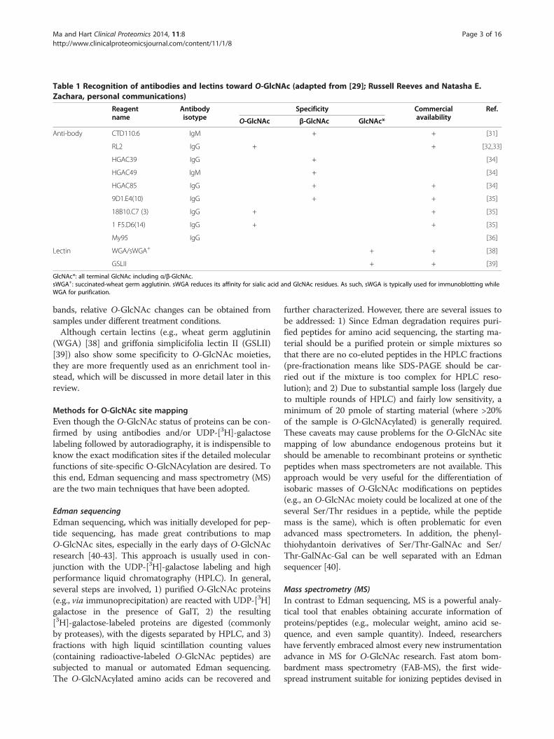
Table 1 Recognition of antibodies and lectins toward O-GlcNAc (adapted from [29]; Russell Reeves and Natasha E.Zachara, personal communications)
Reagentname
Antibodyisotype
Specificity Commercialavailability
Ref.
O-GlcNAc β-GlcNAc GlcNAc*
Anti-body CTD110.6 IgM + + [31]
RL2 IgG + + [32,33]
HGAC39 IgG + [34]
HGAC49 IgM + [34]
HGAC85 IgG + + [34]
9D1.E4(10) IgG + + [35]
18B10.C7 (3) IgG + + [35]
1 F5.D6(14) IgG + + [35]
My95 IgG [36]
Lectin WGA/sWGA+ + + [38]
GSLII + + [39]
GlcNAc*: all terminal GlcNAc including α/β-GlcNAc.sWGA+: succinated-wheat germ agglutinin. sWGA reduces its affinity for sialic acid and GlcNAc residues. As such, sWGA is typically used for immunoblotting whileWGA for purification.
Ma and Hart Clinical Proteomics 2014, 11:8 Page 3 of 16http://www.clinicalproteomicsjournal.com/content/11/1/8
bands, relative O-GlcNAc changes can be obtained fromsamples under different treatment conditions.Although certain lectins (e.g., wheat germ agglutinin
(WGA) [38] and griffonia simplicifolia lectin II (GSLII)[39]) also show some specificity to O-GlcNAc moieties,they are more frequently used as an enrichment tool in-stead, which will be discussed in more detail later in thisreview.
Methods for O-GlcNAc site mappingEven though the O-GlcNAc status of proteins can be con-firmed by using antibodies and/or UDP-[3H]-galactoselabeling followed by autoradiography, it is indispensible toknow the exact modification sites if the detailed molecularfunctions of site-specific O-GlcNAcylation are desired. Tothis end, Edman sequencing and mass spectrometry (MS)are the two main techniques that have been adopted.
Edman sequencingEdman sequencing, which was initially developed for pep-tide sequencing, has made great contributions to mapO-GlcNAc sites, especially in the early days of O-GlcNAcresearch [40-43]. This approach is usually used in con-junction with the UDP-[3H]-galactose labeling and highperformance liquid chromatography (HPLC). In general,several steps are involved, 1) purified O-GlcNAc proteins(e.g., via immunoprecipitation) are reacted with UDP-[3H]galactose in the presence of GalT, 2) the resulting[3H]-galactose-labeled proteins are digested (commonlyby proteases), with the digests separated by HPLC, and 3)fractions with high liquid scintillation counting values(containing radioactive-labeled O-GlcNAc peptides) aresubjected to manual or automated Edman sequencing.The O-GlcNAcylated amino acids can be recovered and
further characterized. However, there are several issues tobe addressed: 1) Since Edman degradation requires puri-fied peptides for amino acid sequencing, the starting ma-terial should be a purified protein or simple mixtures sothat there are no co-eluted peptides in the HPLC fractions(pre-fractionation means like SDS-PAGE should be car-ried out if the mixture is too complex for HPLC reso-lution); and 2) Due to substantial sample loss (largely dueto multiple rounds of HPLC) and fairly low sensitivity, aminimum of 20 pmole of starting material (where >20%of the sample is O-GlcNAcylated) is generally required.These caveats may cause problems for the O-GlcNAc sitemapping of low abundance endogenous proteins but itshould be amenable to recombinant proteins or syntheticpeptides when mass spectrometers are not available. Thisapproach would be very useful for the differentiation ofisobaric masses of O-GlcNAc modifications on peptides(e.g., an O-GlcNAc moiety could be localized at one of theseveral Ser/Thr residues in a peptide, while the peptidemass is the same), which is often problematic for evenadvanced mass spectrometers. In addition, the phenyl-thiohydantoin derivatives of Ser/Thr-GalNAc and Ser/Thr-GalNAc-Gal can be well separated with an Edmansequencer [40].
Mass spectrometry (MS)In contrast to Edman sequencing, MS is a powerful analy-tical tool that enables obtaining accurate information ofproteins/peptides (e.g., molecular weight, amino acid se-quence, and even sample quantity). Indeed, researchershave fervently embraced almost every new instrumentationadvance in MS for O-GlcNAc research. Fast atom bom-bardment mass spectrometry (FAB-MS), the first wide-spread instrument suitable for ionizing peptides devised in

Ma and Hart Clinical Proteomics 2014, 11:8 Page 4 of 16http://www.clinicalproteomicsjournal.com/content/11/1/8
the 1980s [44], was adopted for site mapping OGT-labeled synthetic peptides in early 1990s [45]. Shortlyafter FAB, the advent of electrospray ionization (ESI) [46]and matrix-assisted laser desorption-ionization (MALDI)[47], two ionization methods which are capable of directlyionizing involatile and labile biomolecules, have revo-lutionized the characterization of proteins/peptides. Incombination with new fragmentation techniques (e.g.,collision induced dissociation (CID), high-energy collisiondissociation (HCD) [48], and electron transfer dissociation(ETD) [49] and several mass analyzers (e.g., time of flight(TOF), ion tap, and Orbitrap), the ESI/MALDI-based bio-logical mass spectrometers provide tremendous impetusto the study of biomedical sciences, including the profilingof O-GlcNAcylated proteins. Moreover, the evolution ofmass spectrometry has helped analyzing O-GlcNAc in ahigh throughput way. Undoubtedly, these advanced massspectrometry techniques remain to be the cornerstonetools to date, due to the high sensitivity, selectivity, andthroughput.
Electrospray ionization-collision induced dissociation-tandemmass spectrometry (ESI-CID-MS/MS)Relative to matrix-assisted laser desorption ionization-timeof flight-tandem mass spectrometry (MALDI-TOF-MS/MS), ESI-CID-MS/MS has gained enormous popularityfor its almost perfect demonstration of characterizingmany types of PTMs on proteins/peptides. However, re-garding O-GlcNAc site mapping, limited success has beenachieved. For example, the electrospray ionization-col-lision induced dissociation-quadrupole time-of-flight tan-dem mass spectrometry (ESI-CID-Q-TOF-MS/MS) hasbeen used for direct identification of O-GlcNAc sites onsynthetic peptides [50] and from in-gel digests of over-expressed serum response factor [51]. A major challengefor direct detection by ESI-CID-MS/MS is that the glyco-sidic bond between O-GlcNAc and its peptide sequence ismore susceptible to breakage than that of the peptidebackbone during CID, where relatively high collisionenergy is often applied. Therefore, the O-GlcNAc group ispreferentially lost (producing an O-GlcNAc oxonium ion)prior to peptide fragmentation and thus, the exact modifi-cation sites can not be assigned. However, in some caseswhen large amounts of material are available, a very lowpercentage of fragment ions may still bear the O-GlcNAcmoiety and may be useful in identifying modification sites(as exemplified in Figure 1) [52,53].It is noteworthy that in comparison to conventional CID,
the newly developed HCD fragmentation can produce andmonitor the O-GlcNAc oxonium ion (+204.08) in a moreefficient way (Figure 1) [53]. Not only that, a series of frag-ments of the O-GlcNAc oxonium ion (i.e., m/z 186.07, m/z168.06, m/z 144.06, m/z 138.05, and m/z 126.05) can alsobe generated at pronounced high intensity. One striking
benefit of the O-GlcNAc oxonium ion and its fragments isthat they can serve as diagnostic ions for the presence ofO-GlcNAc on certain peptides, although it would be dif-ficult to accurately designate sites by using CID or HCDalone especially when there are more than one Ser/Thrresidues in the peptide sequence. Another feature is thatCID or HCD is adaptable to coupling with ETD (i.e., CID/ETD-MS/MS or HCD/ETD-MS/MS), enabling more reli-able identification and site mapping of O-GlcNAc peptidesvia the alternating scanning mode or CID/HCD-triggeredETD mode.One way to take advantage of the currently prevalent
CID/HCD-MS/MS is to convert the labile glycosidic bondto a CID/HCD-compatible bond which can withstand theCID or HCD fragmentation. For example, alkaline-inducedβ-elimination can convert the O-GlcNAcylated Ser or Thrto 2-aminopropenoic acid and 2-amino-2-butenoic acid,respectively [54], or further to sulfide derivatives in thepresence of reducing reagents (e.g., Dithiothreitol) [55].
Electrospray ionization-electron transfer dissociation-tandemmass spectrometry (ESI-ETD-MS/MS)A very recent breakthrough in MS is the invention of ETDfragmentation technique [49]. Different from CID, ETDinduces cleavage of the backbone N-Cα bond, generatingc- and z-ions for peptide sequencing. More importantly,ETD generally does not break the linkage between PTMsand their modified residues, thus CID-labile PTMs can bewell preserved during ETD (Figure 1), providing specificsite information [56]. Therefore, the ESI-ETD-MS/MSmethod has been increasingly adopted, largely facilitatingthe direct site assignment of O-GlcNAcylated proteins (asexemplified in [57-59]).Of note, a combination of several fragmentation ap-
proaches is very helpful for in-depth characterization sinceETD tends to perform better than CID or HCD on highercharge states (Z>2 positive charges) but yields a lowernumber of total identifications due to its relatively slowerscan rate and lower fragmentation efficiency [60]. Indeed,both CID/ETD-MS/MS [35,37,57,59] and HCD/ETD-MS/MS [53] have increased the confidence of O-GlcNAcylatedpeptide identification and site localization. In addition,pulsed Q dissociation (PQD) has also been coupled withETD for a two-stage tandem MS approach for O-GlcNAcpeptide analysis, facilitating the detection of such peptidesby PQD at low collision energy and the identification andsite localization by ETD [61]. By integrating with OScore[61], a scoring scheme which can discriminate O-GlcNAcpeptide spectra from those of naked peptides with >99%specificity, O-GlcNAc peptides in the low fmole rangewere detected and a 10-fold higher sensitivity than a singledata-dependent ETD-MS/MS experiment was achieved.Although site-specific detection for O-GlcNAc pep-
tides with ETD has gained great success, its utility for
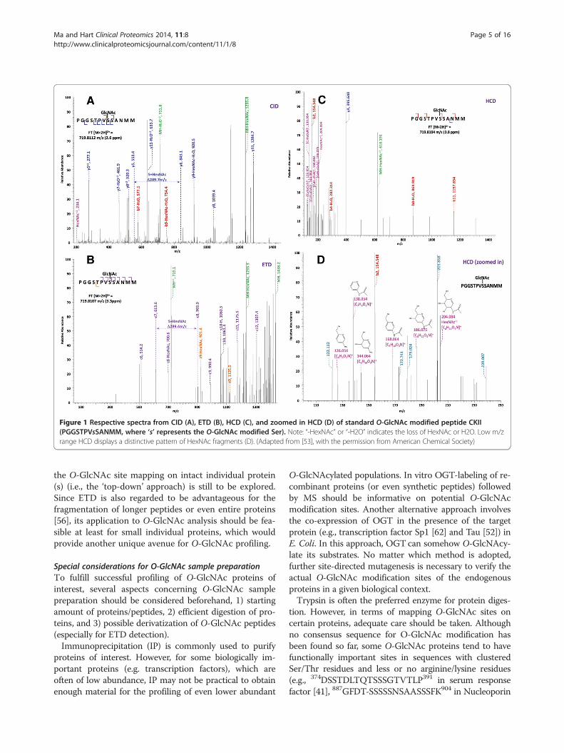
Figure 1 Respective spectra from CID (A), ETD (B), HCD (C), and zoomed in HCD (D) of standard O-GlcNAc modified peptide CKII(PGGSTPVsSANMM, where ‘s’ represents the O-GlcNAc modified Ser). Note: “-HexNAc” or “-H2O” indicates the loss of HexNAc or H2O. Low m/zrange HCD displays a distinctive pattern of HexNAc fragments (D). (Adapted from [53], with the permission from American Chemical Society)
Ma and Hart Clinical Proteomics 2014, 11:8 Page 5 of 16http://www.clinicalproteomicsjournal.com/content/11/1/8
the O-GlcNAc site mapping on intact individual protein(s) (i.e., the ‘top-down’ approach) is still to be explored.Since ETD is also regarded to be advantageous for thefragmentation of longer peptides or even entire proteins[56], its application to O-GlcNAc analysis should be fea-sible at least for small individual proteins, which wouldprovide another unique avenue for O-GlcNAc profiling.
Special considerations for O-GlcNAc sample preparationTo fulfill successful profiling of O-GlcNAc proteins ofinterest, several aspects concerning O-GlcNAc samplepreparation should be considered beforehand, 1) startingamount of proteins/peptides, 2) efficient digestion of pro-teins, and 3) possible derivatization of O-GlcNAc peptides(especially for ETD detection).Immunoprecipitation (IP) is commonly used to purify
proteins of interest. However, for some biologically im-portant proteins (e.g. transcription factors), which areoften of low abundance, IP may not be practical to obtainenough material for the profiling of even lower abundant
O-GlcNAcylated populations. In vitro OGT-labeling of re-combinant proteins (or even synthetic peptides) followedby MS should be informative on potential O-GlcNAcmodification sites. Another alternative approach involvesthe co-expression of OGT in the presence of the targetprotein (e.g., transcription factor Sp1 [62] and Tau [52]) inE. Coli. In this approach, OGT can somehow O-GlcNAcy-late its substrates. No matter which method is adopted,further site-directed mutagenesis is necessary to verify theactual O-GlcNAc modification sites of the endogenousproteins in a given biological context.Trypsin is often the preferred enzyme for protein diges-
tion. However, in terms of mapping O-GlcNAc sites oncertain proteins, adequate care should be taken. Althoughno consensus sequence for O-GlcNAc modification hasbeen found so far, some O-GlcNAc proteins tend to havefunctionally important sites in sequences with clusteredSer/Thr residues and less or no arginine/lysine residues(e.g., 374DSSTDLTQTSSSGTVTLP391 in serum responsefactor [41], 887GFDT-SSSSSNSAASSSFK904 in Nucleoporin
Ma and Hart Clinical Proteomics 2014, 11:8 Page 6 of 16http://www.clinicalproteomicsjournal.com/content/11/1/8
Nup153 [53], 48LSPP-SSSAASSYSFSDNLFTR68 in emerin[59], and tandem repeats of the consensus peptide sequence‘SPTSPS’ in the C-terminal domain of RNA Polymerase II[42]). In such cases, digestion with trypsin is not alwaysthe best choice. Instead, proteolysis with chemical clea-vage (e.g., with cyanogen bromide) and digestion withcomplimentary specificity enzymes (e.g., Asp-N, andGlu-C) could be beneficial for improved detection of cer-tain long peptides with few or no tryptic sites as wellas the assignment of their O-GlcNAc sites. Moreover,considering the limited success in the detection of low-charged peptides with ETD-MS/MS, derivatization tocertain residues (e.g., to the C-terminal carboxyl group[63]) may also be performed to impart more positivecharges and thus improved detection and site mapping ofO-GlcNAc peptides.
O-GlcNAc site predictionThere are a number of bioinformatic software tools avail-able for predicting modification sites for other PTMs [64].However, only a few have been developed for O-GlcNAcprediction, namely, YinOYang [65], OGlcNAcScan [66],and O-GlcNAcPRED [67] (Table 2). With the analysis ofan independent test dataset, O-GlcNAcPRED seems tohave better performance than the other two predictors,especially in terms of prediction sensitivity. Providingthe different computational models used, these toolsalso show certain complementarity in the prediction ofO-GlcNAc sites. Undoubtedly, they may provide someuseful reference for the modification status and potentialO-GlcNAc sites as well as the experimental detection ofO-GlcNAc on proteins of interest by MS. Further matur-ation of these tools (regarding the prediction accuracy andsensitivity) and the development of new ones wouldfacilitate research on O-GlcNAcylation and proteomicidentification.
O-GlcNAc stoichiometry of proteinsDetermination of O-GlcNAc stoichiometry of individualproteins provides additional information for understandingthe function and regulation of protein O-GlcNAcylation.However, the addition of O-GlcNAc (+203) does notusually alter the apparent molecular weight of a protein(unlike the classical N-linked and O-linked glycoproteins),as judged by methods such as SDS-PAGE. Moreover, thereare no changes in the charge status, leading to an
Table 2 Bioinformatic tools for O-GlcNAc site prediction
O-GlcNAc predictor Website
YinOYang http://www.cbs.dtu.dk/services/Y
dbOGAP (OGlcNAc Scan) http://cbsb.lombardi.georgetown
O-GlcNAcPRED (Not available
unchanged pI value for a protein (which is different fromphosphorylated proteins). Therefore, it is not feasible todistinguish the O-GlcNAc modified population from thenaked one by SDS-PAGE itself. Recently, the developmentof a mass-tagging strategy shows strength in quantifyingthe O-GlcNAcylation level on specific proteins [68-71].Basically, O-GlcNAcylated proteins are chemoenzyma-tically labeled using an UDP-ketogalactose analog and thenreacted with an aminooxy-functionalized PEG mass tag(e.g., 5kDa). By doing so, the O-GlcNAcylated species willmigrate differently than their native counterpart uponSDS-PAGE, which can be easily visualized by immunoblot-ting with antibodies against the protein of interest. Therelative O-GlcNAcylation level can thus be determined bycomparing the density of the modified species against thatof the total population. The striking feature of this ap-proach is that the O-GlcNAcylation state (e.g., mono-,di-, tri-) of proteins will be revealed if multiple bands as aladder can be observed. One potential caveat is that in-complete labeling caused by either enzymatic or chemicalreaction may also result in multiple bands. Therefore, fur-ther validation should be performed to confirm the multi-O-GlcNAcylation status to obtain accurate amount of eachpopulation for specific proteins.
Global O-GlcNAcomic profilingMS-based proteomics, a powerful technology referring tothe analysis of the expression, localization, PTMs, andinteractions of proteins expressed by a genome at a spe-cific time, has greatly changed our view about intricatemolecular networks [72-74]. By coupling high-resolutionseparation (mainly 2-D gel electrophoresis and HPLC)with unbiased isotopic labeling techniques, MS-basedproteomics is capable of providing comprehensive cha-racterization of thousands of proteins. Concomitantly,various enrichment methods toward specific PTMs haveemerged, largely advancing the qualitative and quantitativeanalysis of PTM-proteomes including the O-GlcNAcome.Furthermore, protein microarrays have also been used forO-GlcNAcomic profiling.
Gel-based O-GlcNAcomicsTraditionally, 2-D gel electrophoresis separated spotsare visualized with dyes, fluorophores, radioactivity, orantibody-based western blotting, enabling comparativeanalysis of proteins. The combined use of 2-D gel
Algorithm models (Ref.)
inOYang/ Artificial neuronal network [65]
.edu/hulab/OGAP.html Support vector machine [66]
yet) Support vector machine [67]

Ma and Hart Clinical Proteomics 2014, 11:8 Page 7 of 16http://www.clinicalproteomicsjournal.com/content/11/1/8
electrophoresis separation and MS detection, a core prote-omic tool in the 1990s, has been applied to O-GlcNAcanalysis in some studies [75-78]. Although certain successhas been achieved, several issues which are closely relatedto the 2-D gel separation technique itself should be ad-dressed [79], including 1) low efficiency in the analysis ofhydrophobic or extremely acidic/basic proteins; 2) obscu-rity of low abundance proteins; 3) low quantitative accu-racy due to the limited dynamic range; and 4) generalunavailability of O-GlcNAc modification site information.
Gel-free O-GlcNAcomicsIn comparison to 2-D gel electrophoresis, the gel-freeseparation approach (especially multiple dimensionalHPLC for peptides) has catapulted MS-based proteomics(including PTM-proteomics) to an unprecedented level.As with other PTMs, O-GlcNAc proteins are generallyregarded to be substoichiometric (e.g., less abundantthan phosphorylation), although one study has shownthat hundreds of O-GlcNAc peptides could be automa-tically identified from existing large-scale proteomic datasets with the recently developed software Oscore [61,80].Moreover, there is severe ion suppression for detectingO-GlcNAc modified peptides in the presence of nakedpeptides [26]. In addition, as aforementioned, no con-sensus O-GlcNAc motif has been found yet. All thehurdles make accurate O-GlcNAc site assignment achallenging task. As with other PTMs, selective enrich-ment for O-GlcNAc is indispensable, especially whencomplex biological samples are to be analyzed.According to the unique biochemical properties of
O-GlcNAc, an array of enrichment techniques has beendeveloped. With the aid of well-established quantifica-tion methods, large-scale O-GlcNAcomic profiling hasbegun to take off and contributed to a systems biologicalunderstanding of cells under physiological or patho-logical status.
Antibody based O-GlcNAc enrichmentHigh-affinity antibodies are generally the primary choiceto pull down proteins/peptides with certain PTM(s). Al-though pan-specific antibodies (e.g., CTD 110.6, RL2)work well for O-GlcNAc immunoblotting, they have ten-tative applications for enriching O-GlcNAc proteins dueto their relatively low affinity. By using CTD 110.6-con-jugated beads enrichment and MS, Wang et al. identi-fied 45 potentially O-GlcNAcylated proteins from COS7cells [81]. With the combination of SILAC (i.e., stableisotope labeling with amino acids in cell culture), an ap-parent increase in O-GlcNAcylation of >10 proteinswhile a decreased O-GlcNAcylation of nearly 20 proteinswas observed upon inhibition of glycogen synthasekinase-3 (GSK-3). With a similar approach, anotherstudy reported the identification of dozens of O-GlcNAc
proteins from COS7 cells [82]. Among them, a numberof proteins showed elevated levels of O-GlcNAcylationin response to heat stress.While the production of higher-affinity O-GlcNAc anti-
bodies seems extremely difficult, there has been a long-standing interest to develop novel antibodies over the years.The challenges of making O-GlcNAc antibodies mainly liein two aspects: 1) O-GlcNAc–modified epitopes are oftenself-antigens that are tolerated by the immune system and2) carbohydrate-protein interactions are relatively weak,which complicates antibody maturation [26,35]. Conti-nuous efforts, however, have been made to generate O-GlcNAc antibodies that can be applied for immunocapture.Recently, with three O-GlcNAc-specific monoclonal anti-bodies [35] for the enrichment of O-GlcNAc proteins fromHEK293 cell lysates, 83 O-GlcNAc sites were identifiedwith HCD/ETD-MS/MS [53].The combined usage of multiple antibodies and the
development of higher-affinity antibodies should furtherimprove the enrichment performance toward O-GlcNAc.One shortcoming with antibody-based O-GlcNAc proteinenrichment is that proteins interacting with O-GlcNAcy-lated ones would also be pulled down, leading to falsepositive identification. Independent techniques (e.g., im-munoblotting with CTD 110.6) should be used for con-firmation. By combining this approach with an advancedmass spectrometer (e.g., ETD-MS/MS), the accurate mo-dification sites on O-GlcNAcylated proteins can be identi-fied, which would be a definitive indicator for the proteinO-GlcNAcylation status.
Lectin based O-GlcNAc enrichmentDue to the binding interaction with the glycan structureon glycoconjugates, lectins serve as an important tool inglycoproteomics and glycomics [83]. However, only se-veral lectins have been used for O-GlcNAc research so far.Wheat germ agglutinin (WGA) is a lectin that recog-
nizes both terminal GlcNAc and sialic acid residues.Although succinylated WGA (sWGA) increases the spe-cificity to GlcNAc over sialic acid, its affinity towardGlcNAc is compromised as well [29]. Therefore, sWGAis mainly used for immunoblotting, although certainsuccess in the capture of O-GlcNAc proteins has beenshown in some cases. WGA, working as a dimer con-taining four carbohydrate binding sites, has high affinityinteractions with complex glycans via multi-point bind-ing [84]. Thus, it is not surprising that WGA shows amuch lower affinity for the monomeric O-GlcNAc. In-deed, O-GlcNAc interaction with WGA is quite weak,as demonstrated by the ~10 mM dissociation constantfor free GlcNAc to WGA [85]. In comparison to proteinenrichment, O-GlcNAc peptide enrichment has gainedmuch attention especially with the newly developedWGA-based lectin weak affinity chromatography (LWAC)
Ma and Hart Clinical Proteomics 2014, 11:8 Page 8 of 16http://www.clinicalproteomicsjournal.com/content/11/1/8
technique [86-90]. In LWAC, conjugated WGA is packedinto an adequately long column (e.g., 3 meters), which isthen coupled downstream to a low flow rate isobaricHPLC instrument. By doing so, compared to the unmo-dified peptides, O-GlcNAc peptides are retarded by thecolumn and recovered in later eluting fractions. The ap-plicability of this strategy was first demonstrated throughenrichment of 145 unique O-GlcNAc-modified peptidesfrom a postsynaptic density (PSD) preparation [86]. Bycombining this enrichment approach with ETD-MS/MS,Chalkley et al. identified 58 modification sites from mousePSD [87]. In a recent report, with the utilization of furtheroptimized LWAC enrichment and peptide separation(i.e., offline fractionation via basic reversed phase highperformance liquid chromatography (bRPLC)), 1750 O-GlcNAc sites were assigned to mouse brain synaptosomalproteins [89], greatly benefiting the future investigation ofbrain development and functions. In another study, withthe combination of LWAC and SILAC for the analysis ofthe nuclear fraction from embryonic stem cells (ESCs),the same group unambiguously found 142 O-GlcNAcmodification sites on 62 proteins, some of which areessential to maintain the ESC-specific expression profile[88]. Taken together, LWAC has shown reasonable affinitytowards clustered O-GlcNAc-bearing peptides as well assingly and doubly O-GlcNAc-modified peptides. The suc-cess of this technique has greatly expanded the O-GlcNAcprotein database. The more sophisticated applicationof such columns (e.g., improved collection of desiredfractions to decrease loss of O-GlcNAc peptides) may pro-mote a wider acceptance of this approach for the enrich-ment of O-GlcNAc peptides.Besides WGA, another lectin Ricinus communis ag-
glutinin I (RCA I) has been used for O-GlcNAc en-richment as well. However, different from WGA towardGlcNAc, RCA I can specifically recognize in vitro galac-tosylated GlcNAc. In this approach, GlcNAc-bearingpeptides are incubated with UDP-galactose in the pre-sence of GalT, with the resulting Galβ1-4-GlcNAc-pep-tides captured by conjugated RCA I. Compared to WGAfor GlcNAc, RCA1 for Galβ1-4-GlcNAc (i.e., LacNAc)shows a higher affinity (Ka=10μM). Although severalstudies have used this approach for the enrichment ofO-GlcNAc peptides from individual proteins [91-93] andimproved binding specificity proposed, its feasibility forlarge-scale application has yet to be evaluated.Collectively, lectins (especially WGA) are useful tools
for the enrichment of O-GlcNAc peptides. To improvethe binding specificity and capacity, PNGase F treat-ment is often required beforehand to remove N-linkedGlcNAc-terminating sugars on proteins/peptides. Otherlectins, which can improve the binding affinity to O-GlcNAc, are still worthy to be exploited for increasedenrichment efficiency.
Chemical derivatization-based O-GlcNAc enrichmentCompared with antibody- and lectin-based O-GlcNAcenrichment, chemical derivatization is a large categoryof indirect enrichment, which is often comprised ofthree steps: derivatization, capture, and release. Specifi-cally, the O-GlcNAc group is derivatized to add a handle(e.g., biotin) that can be readily captured onto beads(e.g., streptavidin-conjugated ones) and the releasedtagged O-GlcNAc peptides will then be subjected to MSdetection. To date, several chemical-derivatization tech-niques have been developed for O-GlcNAc enrichment.
Hydrazide chemistryHydrazide chemistry is a well-established method for N-glycoproteomic profiling [94]. Recently, an appropriatelymodified analog has been developed for O-GlcNAc en-richment [95]. In this approach, several steps are involved:1) a prolonged periodate oxidation is performed to con-vert the O-GlcNAc group to its dialdehyde derivative, 2)hydrazide resin is used to capture the oxidized O-GlcNAcpeptides, and 3) after proteolytic digestion, the resultingmodified peptides are released by hydroxylamine. Withthis enrichment procedure followed by MS/MS, severalO-GlcNAc sites were identified from Drosophila melano-gaster proteasome protein complex. To apply this tech-nique for large-scale O-GlcNAc site mapping, two issuesmay need to be further addressed, 1) to derivatize the lessactive O-GlcNAc moiety (largely due to the trans config-uration of the vicinal hydroxyls at positions C3 and C4),harsher conditions in periodate oxidation should be used,leading to undesired side reactions (e.g., oxidation of N-terminal Ser/Thr) and thus high background, and 2) moreefficient and specific release of tagged O-GlcNAc peptideswould be beneficial for the detection and site assignmentof O-GlcNAc peptides.
β-elimination Michael addition (BEMA)As aforementioned, O-GlcNAc can be removed from pro-teins/peptides by mild β-elimination, with Ser and Thrresidues converted into their dehydrated equivalents (i.e.,dehydroalanine and α-amino butyric acid, respectively)[54]. Based on this chemistry, a refined approach, namedβ-elimination Michael addition (BEMA), has been de-veloped to mark the site of O-GlcNAc modification. InBEMA, the α/β-unsaturated carbonyl is derivatized withnucleophilic reagents (e.g., DTT or biotinylated pentyla-mine/cystamine) and the resulting peptides can then beenriched by thiol-capture resin or streptavidin-conjugatedbeads. Since DTT is the preferred nucleophile, the β-elimination Michael addition of DTT has been termedBEMAD [55,96]. There are several striking features of thismethod, 1) in comparison to the initial labile glycosidicbonds in O-GlcNAc peptides, the final resulting sulfidederivatives are stable enough during fragmentation and
Ma and Hart Clinical Proteomics 2014, 11:8 Page 9 of 16http://www.clinicalproteomicsjournal.com/content/11/1/8
thus suitable for detection and site mapping by the mostprevalent CID-MS/MS without relying on ETD tech-nology and 2) quantitative O-GlcNAc site information canbe readily achieved by using isotopically labeled DTT (i.e.,D6-DTT and D10-DTT). Of special note is that, althoughphosphorylated peptides can also undergo BEMAD, fasterconversion to the BEMAD product under milder condi-tions is achieved for O-GlcNAc peptides due to the moreeasily eliminated O-glycosidic linkages [3,54,55,96-98],with less undesired side reactions. Therefore, optimizedBEMAD conditions and appropriate sample pre-treatment(e.g., with PNGase F) should be performed to avoid po-tential false positive identifications. In addition, distinctapproaches (e.g., HCD-MS/MS or ETD-MS/MS, and im-munoblotting) should be adopted for further validation.By using the BEMAD approach, several O-GlcNAc siteswere determined from the key contractile proteins, suchas actin and myosin heavy chains, in skeletal muscle [99].Recently, an adapted method involving β-elimination-based derivatization with a biotin-cystamine tag followedby streptavidin-conjugated beads has been developed[100]. By differential isotopic labeling with either lightbiotin-cystamine or deuterated heavy biotin-cystamine,the specificity of the enrichment approach can be in-creased. Several O-GlcNAc sites within the murine 20Sproteasome core complex were assigned.The combined use of BEMAD and other techniques
(e.g., chemoenzymatic labeling), which could furtherimprove the enrichment specificity, is also favorable forO-GlcNAc profiling.
Chemoenzymatic labelingChemoenzymatic labeling capitalizes on the merits of thetraditional GalT labeling and the advanced chemical de-rivatization techniques (especially the ketone-aminoxyprocess and bioorthogonal chemistry). Different from tra-ditional GalT labeling, unnatural galactose analogues withspecific chemical handles, which can facilitate the subse-quent capture procedure, are used in chemoenzymatic la-beling. An engineered mutant of GalT (GalTY289L), whichhas an enlarged binding pocket for the donor-substrate[101], is the best choice to selectively derivatize O-GlcNAcwith galactose analogues. To date, two major kinds of suchanalogues have been developed and used for chemo-enzymatic labeling, i.e, ketone-bearing UDP-galactose andazido-modified UDP-galactose (UDP-GalNAz).In one approach, GalTY289L is used to transfer the keto-
galactose onto O-GlcNAc proteins and a biotin-aminoxyreagent is then attached via the oxime formation (amino-xylation). The biotin-tagged derivatives are visualized bystreptavidin blotting [102] or subjected to streptavidin-conjugated beads enrichment followed by fluorescence[103] or MS detection [104,105]. By incorporating this ap-proach with isotopic dimethyl labeling and ETD-MS/MS,
comparative quantification of O-GlcNAc levels from twodifferent brain populations was performed [105].Another chemoenzymatic approach integrates UDP-
GalNAz-based GalTY289L labeling, copper (I)-catalyzedazide-alkyne cycloaddition (click chemistry), and streptavi-din-conjugated beads [106-110]. Since the biotin-strepta-vidin interaction is extraordinarily stable (Kd~= 10-15 Mfor the homo-tetramer streptavidin and 10-7-10-8 M formonomeric streptavidin), one elegant way is to introducea UV-cleavable linker to afford improved release efficiencyof tagged peptides from streptavidin-conjugated beads[107-109]. An additional advantage is that the releasedpeptides contain a basic aminomethyltriazoyl acetylgalac-tosamine moiety, enabling efficient ETD fragmentation.By using the combination of GalTY289L labeling, clickchemistry, UV-cleavage, and ETD-MS/MS, 141 O-GlcNAcsites were identified from component proteins in HeLamitotic spindles and midbodies [109] and 458 O-GlcNAcsites in 195 proteins from mouse cerebrocortical braintissue [108].Moreover, the chemoenzymatic labeling approach can
be readily coupled with BEMAD and CID-MS/MS forO-GlcNAc site mapping [111-114]. In one study, 35O-GlcNAc sites corresponding to 25 O-GlcNAcylatedproteins were identified from erythrocytes [114]. Inaddition, with the further integration of isobaric tag forrelative and absolute quantitation technique (iTRAQ),the relative occupancy ratio between normal and dia-betic erythrocytes was determined, revealing differentO-GlcNAcylation at individual sites on proteins underdistinct cellular conditions.
Metabolic labelingIn comparison to the enrichment methods mentionedabove, which are performed in vitro, metabolic labelingoffers an in vivo way to place a chemical handle ontoO-GlcNAc proteins. This approach is based on the uti-lization of N-azidoacetylglucosamine (GlcNAz), an analogto GlcNAc. Studies have shown that GlcNAz can be tole-rated by enzymes in the GlcNAc salvage pathway genera-ting UDP-GlcNAz, which can finally be accepted by OGTand transferred to substrate proteins in living cells [115].Therefore, by feeding cells with an appropriate amount ofperacetylated GlcNAz, proteins initially modified by O-GlcNAc will be substituted with GlcNAz. Peracetylationallows the compounds to enter the cells and endogenousdeacetylases rapidly remove the acetyl groups. GalNAzmay also be used for labeling O-GlcNAc modified pro-teins, because it is readily epimerized to GlcNAz [116].The GlcNAz-tagged proteins can be chemoselectivelyconjugated with a biotinylated phosphine reagent or a bio-tinylated alkyne reagent via Staudinger ligation [115,116]or click chemistry [117-120], respectively. After strepta-vidin-conjugated beads enrichment, tagged proteins are
Ma and Hart Clinical Proteomics 2014, 11:8 Page 10 of 16http://www.clinicalproteomicsjournal.com/content/11/1/8
then digested, with the digests identified by MS. Recently,with this method, 185 O-GlcNAc sites were assigned to 80proteins in HEK293 cells [120].In other studies, the alkynyl-modified GlcNAc analog
(GlcNAlk) has been exploited as a chemical reporterof O-GlcNAc modification in living cells [119,121]. Incombination with click chemistry (with an azide-biotinreagent), streptavidin-conjugated beads enrichment, pro-teolytic digestion and MS, 374 putative O-GlcNAc pro-teins were identified [121]. One feature of GlcNAlklabeling is that, while GlcNAz can be metabolically inter-converted to GalNAz [116,122], GlcNAlk does not, sug-gesting it may be a more specific metabolic reporter ofO-GlcNAc modification.Collectively, metabolic labeling has shown some ad-
vantages for facile enrichment of O-GlcNAc proteins.However, the major downside is that the cell’s enzymesprefer the natural substrate over the non-canonical ones,resulting in relatively low levels of tagging.
Quantification of O-GlcNAcylationGlobal quantitative analysis of the levels of proteins andtheir O-GlcNAc sites is key to a systematic understan-ding of the molecular function of O-GlcNAc proteins invarious biological processes. The traditional quantitationapproach, which relies on high-resolution protein sepa-ration by 2-D gels and mass spectrometry identificationof certain significantly altered spots, has been used forprobing changes of O-GlcNAc proteins from several celllines and tissues [75-78]. However, inherent drawbacksof the 2-D gel separation technique hinder its appli-cation for in-depth comparative analysis, as mentionedabove. In contrast, the integration of stable isotope label-ing with gel-free separation, specific enrichment, andmass spectrometry detection has been demonstrated tobe a very powerful tool to provide quantitative informa-tion about O-GlcNAc changes between samples in con-trol, diseases, and drug-perturbation conditions. Thereare mainly two ways: in vivo metabolic labeling andin vitro chemical reaction, to incorporate stable isotopesinto O-GlcNAc proteins/peptides for mass spectrometrybased quantification.
In vivo metabolic labeling-based O-GlcNAc quantificationAs an in vivo approach, stable isotope labeling with aminoacids in cell culture (SILAC) allows proteins to be labeledby growing cells in media containing isotopically labeledamino acids (e.g., 13C/15 N-arginine, 13C/15 N-lysine, 13C/2H-methionine). Due to the high quantification accuracy,SILAC has become a versatile tool for multiple proteomicapplications [123-125]. Wang et al. evaluated O-GlcNAcproteomic changes upon stimulation of cells by lithium, aselective inhibitor to glycogen synthase kinase-3 (GSK-3)which is extensively involved in many signaling pathways
[81]. By combining SILAC, CTD 110.6-bound beads en-richment, and LC-MS/MS, they identified 45 potentiallyO-GlcNAcylated proteins, 10 of which showed increasedO-GlcNAcylation while 19 showed decreased O-GlcNAcy-lation upon GSK-3 inhibition [81]. Their results indicatea complex interplay between phosphorylation and O-GlcNAcylation within signaling networks. With a similarapproach, Zachara et al. investigated the changes of O-GlcNAcylated proteins of cells in response to heat shock[82]. Amongst the proteins identified, some DNA-bindingproteins showed elevated levels of O-GlcNAcylation, sug-gesting a role for O-GlcNAc in regulating DNA damagesignaling or repair. In another study, by using a com-bination of SILAC, chemoenzymatic labeling-based en-richment, and LC-MS/MS, altered phosphorylation of keyproteins in cellular midbodies was revealed upon the over-expression of OGT [109], further illustrating the intricatecrosstalk between O-GlcNAcylation and phosphorylationof proteins in the regulation of cell division.Even though SILAC has been demonstrated to be a
powerful tool in quantitative proteomic studies for cul-tured cells, it is still not very practical for analyzing bio-logical samples that can not be grown in culture, such astissues or body fluids. The further development of SILACtechniques (i.e., tissues and even whole animal-targetedSILAC [126,127]) should further benefit related applica-tions including quantitative O-GlcNAc profiling.
In vitro labeling-based O-GlcNAc quantificationAs a non-biased approach, the in vitro labeling involvesincorporation of stable isotopic tags onto selective siteson proteins/peptides via chemical reactions. Isotopiclabeling can be introduced at the N-/C-terminus, onspecific amino acid residues (e.g. cysteine [128]), or atthe C-terminus of peptides during trypsin-catalyzed-18Olabeling of proteins [129]. Amongst those techniques,N-terminus-targeted labeling, especially the isobaric tagsfor relative and absolute quantitation (iTRAQ) [130] andisotope dimethyl labeling [131,132], has been adoptedfor O-GlcNAc quantification. In one study, iTRAQ wascoupled with the chemoenzymatic labeling enrichment andLC-MS/MS to investigate the extent of O-GlcNAcylationon human erythrocyte proteins from diabetic and normalindividuals [114]. Twenty-five O-GlcNAcylated erythrocyteproteins were identified with differential O-GlcNAcylationlevel between diabetic and normal erythrocytes, suggestinga potential regulatory role of O-GlcNAcylation on erythro-cyte proteins in response to glycemic status. In anotherstudy, isotope dimethyl labeling was used with chemoenzy-matic labeling enrichment and LC-MS/MS for probing thedynamics of O-GlcNAcylation in brain [105]. DifferentialO-GlcNAcylation on several proteins involved in theregulation of transcription and mRNA translocationwas revealed, suggesting important roles of protein O-
Ma and Hart Clinical Proteomics 2014, 11:8 Page 11 of 16http://www.clinicalproteomicsjournal.com/content/11/1/8
GlcNAcylation in mediating the communication betweenneurons. As a quite different approach, BEMAD canintroduce isotopic labels (i.e., deuterated DTT) onto ori-ginally O-GlcNAc modified Ser/Thr residues prior to thethio-affinity enrichment [96], allowing the evaluation ofsite-specific O-GlcNAc changes. Moreover, by normali-zing the level of site specific O-GlcNAc peptides to that ofthe corresponding proteins, relative site occupancy ratio(ROR) between different biological contexts can be ob-tained. By comparing iTRAQ based protein quantificationand isotopic DTT-mediated BEMAD-based O-GlcNAcpeptide quantification, the O-GlcNAc site occupancy onerythrocyte proteins from diabetic and normal individualswas determined [114]. Of note, certain proteins with sig-nificant O-GlcNAc site occupancy changes may serve as asensitive diagnostic tool for the early detection of diabetes.
Label-free quantification approachesThere has been increasing interest in the development oflabel-free mass-spectrometry quantification techniques,
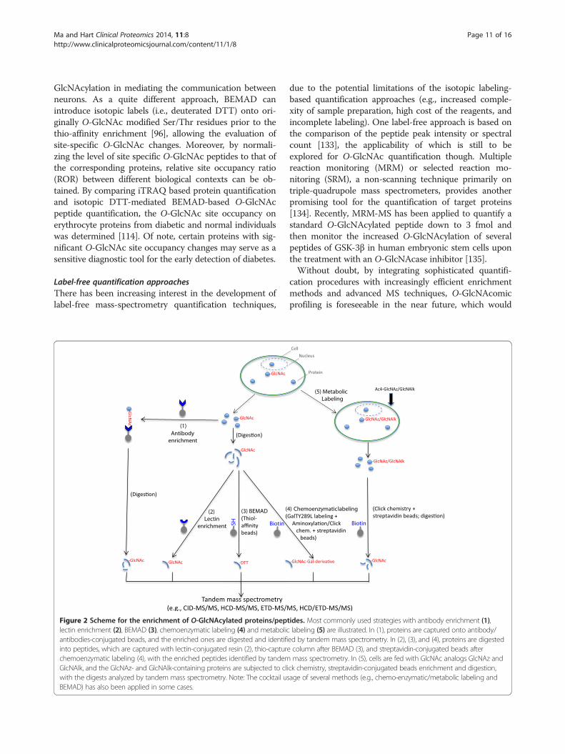
Figure 2 Scheme for the enrichment of O-GlcNAcylated proteins/peplectin enrichment (2), BEMAD (3), chemoenzymatic labeling (4) and metabolicantibodies-conjugated beads, and the enriched ones are digested and identifiinto peptides, which are captured with lectin-conjugated resin (2), thio-capturechemoenzymatic labeling (4), with the enriched peptides identified by tandemGlcNAlk, and the GlcNAz- and GlcNAlk-containing proteins are subjected to cliwith the digests analyzed by tandem mass spectrometry. Note: The cocktail usBEMAD) has also been applied in some cases.
due to the potential limitations of the isotopic labeling-based quantification approaches (e.g., increased comple-xity of sample preparation, high cost of the reagents, andincomplete labeling). One label-free approach is based onthe comparison of the peptide peak intensity or spectralcount [133], the applicability of which is still to beexplored for O-GlcNAc quantification though. Multiplereaction monitoring (MRM) or selected reaction mo-nitoring (SRM), a non-scanning technique primarily ontriple-quadrupole mass spectrometers, provides anotherpromising tool for the quantification of target proteins[134]. Recently, MRM-MS has been applied to quantify astandard O-GlcNAcylated peptide down to 3 fmol andthen monitor the increased O-GlcNAcylation of severalpeptides of GSK-3β in human embryonic stem cells uponthe treatment with an O-GlcNAcase inhibitor [135].Without doubt, by integrating sophisticated quantifi-
cation procedures with increasingly efficient enrichmentmethods and advanced MS techniques, O-GlcNAcomicprofiling is foreseeable in the near future, which would
tides. Most commonly used strategies with antibody enrichment (1),labeling (5) are illustrated. In (1), proteins are captured onto antibody/ed by tandem mass spectrometry. In (2), (3), and (4), proteins are digestedcolumn after BEMAD (3), and streptavidin-conjugated beads aftermass spectrometry. In (5), cells are fed with GlcNAc analogs GlcNAz and
ck chemistry, streptavidin-conjugated beads enrichment and digestion,age of several methods (e.g., chemo-enzymatic/metabolic labeling and
Ma and Hart Clinical Proteomics 2014, 11:8 Page 12 of 16http://www.clinicalproteomicsjournal.com/content/11/1/8
facilitate the in-depth elucidation of the important roles ofprotein O-GlcNAcylation in diverse biological contexts.
Protein microarray-based O-GlcNAcomicsDistinct from MS, protein microarray represents anotherhigh-throughput method for the analysis of PTMs such asphosphorylation and N-glycosylation [136]. Tarrant et al.used a protein array to screen for protein substrates ofO-GlcNAcylated and/or phosphorylated CKII [43]. Theirresults reveal that the substrate spectrum changes afterbinding to its interacting partner Pin1 and that thesubstrate selectivity of CKII is delicately modulated byO-GlcNAcylation and phosphorylation. To identify pro-tein kinases that are potentially O-GlcNAcylated, Dias andcoworkers used a functional human protein array con-taining 152 kinases as a substrate for OGT in vitro.Intriguingly, they identified 42 kinases that are O-GlcNA-cylated in vitro (~39% of all the kinases analyzed) [137],suggesting that a number of protein kinases may be regu-lated by O-GlcNAcylation and this regulation may furthercomplicate the already intricate relationship betweenO-GlcNAcylation and phosphorylation. Indeed, recentstudies have shown that a number of important kinases(including CKII [43], CaMKIV [138], PKC [139], Akt[140], IκB kinase [141], among others) are regulated by O-GlcNAcylation. With the further optimization and im-provement of related techniques, protein microarrays willstill be a valuable technology for O-GlcNAcomic studies.
Conclusions and perspectivesOver the first two decades since its discovery, O-GlcNAcy-lation was determined to be on ~500 proteins [142]. Withthe introduction of new enrichment techniques and ad-vanced mass spectrometers, the number of O-GlcNAcy-lated proteins has been increased to >4000 (a detailed list isbeing compiled). More importantly, numerous O-GlcNAcsites have also been mapped, which not only significantlyfacilitate deciphering the crucial roles of O-GlcNAc on in-dividual proteins in various biological processes, but alsoprovide us a much deeper insight on how this modificationclosely interplays with many other PTMs (especially phos-phorylation) in complex molecular networks.However, we are still in the early stage of O-GlcNAc pro-
filing, compared to the rapidly maturing characterizationof other PTMs (e.g., phosphorylation, N-glycosylation,lysine acetylation, and ubiquitination) for which a handfulof highly efficient and robust tools are available. Althoughmany enrichment methods have been developed for O-GlcNAc proteins/peptides (Figure 2), they are still far frombeing applicable routinely to the analysis of samples,especially for complex ones when large-scale comparativeO-GlcNAcomic profiling is desired. Moreover, the newlydesigned mass spectrometers (especially the ETD-equippedones) are not widely available to most labs, which hampers
the site-oriented O-GlcNAc functional assays. In addition,there are limited software and algorithms specifically de-signed for O-GlcNAc site prediction as well as mass spec-trometry data mining.Considering the extremely important roles O-GlcNAc
plays, the complete repertoire of O-GlcNAcylated proteinsas well as their specific sites must be defined. To this end,several aspects about improving O-GlcNAc profiling areanticipated. 1) Refinement of current enrichment tech-niques and development of novel ones should still be atopic of intense interest. 2) How to make full use of thecapacity and improve the performance of mass spectro-meters for O-GlcNAc detection remains to be addressed.The combination of different fragmentation modes (e.g.,HCD plus ETD) would be a powerful tool for improvedO-GlcNAc identification and site mapping. Moreover, thepotential for ETD in applications, such as multiple reactionmonitoring (MRM) for O-GlcNAc peptides and top-downcharacterization for O-GlcNAc proteins, should be ex-plored. 3) Quantitative proteomic techniques should befurther adopted in more O-GlcNAc studies. 4) Designingnovel bioinformatic tools for O-GlcNAc research will beanother goal in the future. 5) The development of a largenumber of site-specific antibodies, as are now available forprotein phosphorylation, will be critical to the rapid ad-vancement of this field by biologists. Taken together, aswith other PTMs, technology integration will hasten thematuration of diverse methods for O-GlcNAc profiling.We are sure that the technology-driven O-GlcNAcomicswill boom soon, which would profoundly contribute to theelucidation of crucial functions of protein O-GlcNAcyla-tion in versatile physiological and pathological conditionsand to a systems perspective of molecular mechanisms inbiological networks.
AbbreviationsO-GlcNAc: O-linked β-D-N-acetylglucosamine; O-GlcNAcylation: O-linkedβ-D-N-acetylglucosamine addition; UDP-GlcNAc: Uridine diphospho-N-acetylgluco-samine; OGT: O-GlcNAc transferase; O-GlcNAcase: β-N-acetyl-glucosaminidase; PTM: Post-translational modification; PNGase F: Peptide:N-glycosidase F; GalT: β1-4-galactosyltransferase; SDS-PAGE: SDS-polyacrylamide gel electrophoresis; HPLC: High performance liquidchromatography; MS: Mass spectrometry; CID: Collision induceddissociation; HCD: High-energy collision dissociation; ETD: Electron transferdissociation; WGA: Wheat germ agglutinin; BEMAD: Beta elimination/Michael addition with dithiothreitol; SILAC: Stable isotope labeling ofamino acids in cell culture; iTRAQ: Isobaric tag for relative and absolutequantitation.
Competing interestsOriginal research in the author’s laboratory is supported by NIHR01CA42486, R01DK61671, N01-HV-00240, P01HL107153, and the Patrick C.Walsh Prostate Cancer Research Fund. Dr. Hart receives a share of royaltyreceived by the university on sales of the CTD 110.6 antibody, which aremanaged by JHU. The authors have no other relevant affiliations or financialinvolvement with any organization or entity with a financial interest in orfinancial conflict with the subject matter or materials discussed in themanuscript apart from those disclosed.
Ma and Hart Clinical Proteomics 2014, 11:8 Page 13 of 16http://www.clinicalproteomicsjournal.com/content/11/1/8
Authors' contributionsJM drafted the manuscript. Both authors edited and approved the finalmanuscript.
AcknowledgmentsThe authors especially thank Dr. Kaoru Sakabe for critical reading andsuggestions of this manuscript and the whole Hart laboratory. They wouldalso like to acknowledge Dr. Natasha E. Zachara for insightful discussions.
Received: 30 September 2013 Accepted: 1 February 2014Published: 5 March 2014
References1. Varki A, Freeze HH, Gagneux P: Evolution of Glycan Diversity. In Essentials
of Glycobiology. 2nd edition. Edited by Varki A, Cummings RD, Esko JD,Freeze HH, Stanley P, Bertozzi CR, Hart GW, Etzler ME. Cold Spring HarborNY, USA: Cold Spring Harbor Laboratory Press; 2009.
2. Hart GW, Copeland RJ: Glycomics hits the big time. Cell 2010, 143:672–676.3. Torres CR, Hart GW: Topography and polypeptide distribution of terminal
N-acetylglucosamine residues on the surfaces of intact lymphocytes.Evidence for O-linked GlcNAc. J Biol Chem 1984, 259:3308–3317.
4. Holt GD, Hart GW: The subcellular distribution of terminal N-acetylglucosamine moieties. Localization of a novel protein-saccharidelinkage, O-linked GlcNAc. J Biol Chem 1986, 261:8049–8057.
5. Hu Y, Suarez J, Fricovsky E, Wang H, Scott BT, Trauger SA, Han W, Hu Y,Oyeleye MO, Dillmann WH: Increased enzymatic O-GlcNAcylation of mito-chondrial proteins impairs mitochondrial function in cardiac myocytesexposed to high glucose. J Biol Chem 2009, 284:547–555.
6. Haltiwanger RS, Holt GD, Hart GW: Enzymatic addition of O-GlcNAc to nu-clear and cytoplasmic proteins. Identification of a uridine diphospho-N-acetylglucosamine:peptide beta-N-acetyl-glucosaminyltransferase. J BiolChem 1990, 265:2563–2568.
7. Jacobsen SE, Binkowski KA, Olszewski NE: SPINDLY, a tetratricopeptiderepeat protein involved in gibberellin signal transduction in Arabidopsis.Proc Natl Acad Sci USA 1996, 93:9292–9296.
8. Hanover JA, Yu S, Lubas WB, Shin SH, Ragano-Caracciola M, Kochran J, LoveDC: Mitochondrial and nucleocytoplasmic isoforms of O-linked GlcNActransferase encoded by a single mammalian gene. Arch Biochem Biophys2003, 409:287–297.
9. Dong DL, Hart GW: Purification and characterization of an O-GlcNAc selectiveN-acetyl-β-D-glucosaminidase from rat spleen cytosol. J Biol Chem 1994,269:19321–19330.
10. Hu P, Shimoji S, Hart GW: Site-specific interplay between O-GlcNAcylationand phosphorylation in cellular regulation. FEBS Lett 2010, 584:2526–2538.
11. Butkinaree C, Park K, Hart GW: O-linked N-acetylglucosamine (O-GlcNAc):extensive crosstalk with phosphorylation to regulate signaling and tran-scription in response to nutrients and stress. Biochem Biophys Acta 2010,2010(1800):96–106.
12. Zeidan Q, Hart GW: The intersections between O-GlcNAcylation andphosphorylation: implications for multiple signaling pathways. J Cell Sci2010, 123:13–22.
13. Hart GW, Slawson C, Ramirez-Correa GA, Lagerlof O: Cross talk between GlcNA-cylation and phosphorylation: roles in signaling, transcription, and chronicdisease. Annu Rev Biochem 2011, 80:825–858.
14. Bond MB, Hanover JA: O-GlcNAc Cycling: a link between metabolism andchronic disease. Annu Rev Nutr 2013, 33:13.1–13.25.
15. Ruan HB, Singh JP, Li MD, Wu J, Yang X: Cracking the O-GlcNAc code in me-tabolism. Trends Endocrinol Metab 2013, 24:301–309.
16. Groves JA, Lee A, Yildirir G, Zachara NE: Dynamic O-GlcNAcylation and itsroles in the cellular stress response and homeostasis. Cell StressChaperone 2013, 18:535–558.
17. Marshall S, Bacote V, Traxinger RR: Discovery of a metabolic pathwaymediating glucose-induced desensitization of the glucose transport sys-tem. Role of hexosamine biosynthesis in the induction of insulin resist-ance. J Biol Chem 1991, 266:4706–4712.
18. Issad T, Masson E, Pagesy P: O-GlcNAc modification, insulin signaling anddiabetic complications. Diabetes Metab 2010, 36:423–435.
19. Ma J, Hart GW: Protein O-GlcNAcylation in diabetes and diabetic compli-cations. Expert Rev Proteomics 2013, 10:365–380.
20. Slawson C, Hart GW: O-GlcNAc signalling: implications for cancer cellbiology. Nat Rev Cancer 2011, 11:678–684.
21. Ma Z, Vosseller K: O-GlcNAc in cancer biology. Amino Acids 2013, 45:719–733. doi:10.1007/s00726-013-1543-8.
22. Fardini Y, Dehennaut V, Lefebvre T, et al: O-GlcNAcylation: a new cancerhallmark? Front Endocrinol 2013. doi:10.3389/fendo.2013.00099. http://www.frontiersin.org/Journal/10.3389/fendo.2013.00099/full.
23. Lazarus BD, Love DC, Hanover JA: O-GlcNAc cycling: implications forNeurodegenerative disorders. Int J Biochem Cell Biol 2009, 41:2134–2146.
24. Gong C, Liu F, Iqbal K: O-GlcNAc cycling modulates neurodegeneration.Proc Natl Acad Sci USA 2012, 109:17319–17320.
25. Whelan SA, Hart GW: Identification of O-GlcNAc sites on proteins. MethodsEnzymol 2006, 415:113–133.
26. Wang Z, Hart GW: Glycomic approaches to study GlcNAcylation: proteinidentification, site-mapping, and site-specific O-GlcNAc quantitation. ClinProteomics 2008, 4:5–13.
27. Chalkley RJ, Wells L, Vosseller K: O-GlcNAc proteomics: mass spectrometricanalysis of O-GlcNAc modifications on proteins. In Protein MassSpectrometry (edited by Julian Whitelegge) 2009, 52:353–374.
28. Zachara NE: Detecting the "O-GlcNAc-ome"; detection, purification, andanalysis of O-GlcNAc modified proteins. Methods Mol Biol 2009, 534:251–279.
29. Zachara NE, Vosseller K, Hart GW: Detection and analysis of proteinsmodified by O-Linked N-Acetylglucosamine. Curr Protoc Protein Sci 2011,66:12.8.1–12.8.33. http://onlinelibrary.wiley.com/doi/10.1002/0471140864.ps1208s66/abstract;jsessionid=1342F2ABD48579AA0ED64C41034F0502.f03t03.
30. Copeand RJ, Han G, Hart GW: O-GlcNAcomics-Revealing roles of O-GlcNAcylation in disease mechanisms and development of potential diag-nostics. Proteomics Clin Appl 2013, 7:597–606. doi:10.1002/prca.201300001.
31. Comer FI, Vosseller K, Wells L, Accavitti MA, Hart GW: Characterization of amouse monoclonl antibody specific for O-linked N-acetylglucosamine.Anal Biochem 2001, 293:169–177.
32. Snow CM, Senior A, Gerace L: Monoclonal antibodies identify a group ofnuclear pore complex glycoproteins. J Cell Biol 1987, 104:1143–1156.
33. Holt GD, Snow CM, Senior A, Haltiwanger RS, Gerace L, Hart GW: Nuclearpore complex glycoproteins contain cytoplasmically disposed O-linkedN-acetylglucosamine. J Cell Biol 1987, 104:1157–1164.
34. Turner JR, Tartakoff AM, Greenspan NS: Cytologic assessment of nuclearand cytoplasmic O-linked N–acetylglucosamine distribution by usinganti-streptococcal monoclonal antibodies. Proc Natl Acad Sci USA 1990,87:5608–5612.
35. Teo CF, Ingale S, Wolfert M, Elsayed GA, Nöt LG, Chatham JC, Wells L, BoonsGJ: Glycopeptide-specific mono-clonal antibodies suggest new roles forO-GlcNAc. Nat Chem Biol 2010, 6:338–343.
36. Matsuoka Y, Shibata S, Yasuhara N, Yoneda Y: Identification of Ewing’s sarcomagene product as a glycoprotein using a monoclonal antibody that recognizesan immunodeterminant containing O-linked N-acetylglucosamine moiety.Hybrid Hybridomics 2002, 21:233–236.
37. Isono T: O-GlcNAc-specific antibody CTD110.6 cross-reacts with N-GlcNAc2-modified proteins induced under glucose deprivation. PLoS ONE 2011, 6(4):e18959.
38. Kelly WG, Hart GW: Glycosylation of chromosomal proteins: localization ofO-linked N-acetylglucosamine in Drosophila chromatin. Cell 1989, 57:243–251.
39. Nakamura-Tsuruta S, Kominami J, Kamei M, Koyama Y, Suzuki T, Isemura M,Hirabayashi J: Analysis by frontal affinity chromatography ofoligosaccharide specificity of GlcNAc-binding lectins, Griffonia simplicifo-lia lectin-II (GSL-II) and Boletopsis leucomelas lectin (BLL). J Biochem 2006,140:285–291.
40. Zachara NE, Gooley AA: Identification of glycosylation sites in mucinpeptides by Edman degradation. Methods Mol Biol 2000, 125:121–128.
41. Reason AJ, Morris HR, Panico M, Marais R, Treisman RH, Haltiwanger RS, HartGW, Kelly WG, Dell A: Localization of O-GlcNAc modification on the serumresponse transcription factor. J Biol Chem 1992, 267:16911–16921.
42. Kelly WG, Dahmus ME, Hart GW: RNA polymerase II is a glycoprotein.Modification of the COOH-terminal domain by O-GlcNAc. J Biol Chem1993, 268:10416–10424.
43. Tarrant MK, Rho HS, Xie Z, Jiang YL, Gross C, Culhane JC, Yan G, Qian J,Ichikawa Y, Matsuoka T, Zachara N, Etzkorn FA, Hart GW, Jeong JS,Blackshaw S, Zhu H, Cole PA: Regulation of CK2 by Phosphorylation andO-GlcNAcylation revealed by Semisynthesis. Nature Chem Biol 2012,8:262–269.
Ma and Hart Clinical Proteomics 2014, 11:8 Page 14 of 16http://www.clinicalproteomicsjournal.com/content/11/1/8
44. Barber M, Bordoli RS, Sedgewick RD, Tyler AN: Fast atom bombardment ofsolids (F.A.B.): a new ion source for mass spectrometry. J Chem Soc ChemCommun 1981, 7:325–327.
45. Reason AJ, Blench IP, Haltiwanger RS, Hart GW, Morris HR, Panico M, Dell A:High-sensitivity FAB-MS strategies for O-GlcNAc characterization.Glycobiology 1991, 1:585–594.
46. Fenn JB, Mann M, Meng CK, Wong SF, Whitehouse CM: Electrosprayionization for mass spectrometry of large biomolecules. Science 1989,246:64–71.
47. Karas M, Hillenkamp F: Laser desorption ionization of proteins withmolecular masses exceeding 10,000 daltons. Anal Chem 1988, 60:2299–2301.
48. Olsen JV, Macek B, Lange O, Makarov A, Horning S, Mann M: Higher-energyC-trap dissociation for peptide modification analysis. Nat Methods 2007,4:709–712.
49. Syka JE, Coon JJ, Schroeder MJ, Shabanowitz J, Hunt DF: Peptide andprotein sequence analysis by electron transfer dissociation massspectrometry. Proc Natl Acad Sci USA 2004, 101:9528–9533.
50. Chalkley RJ, Burlingame AL: Identification of GlcNAcylation sites ofpeptides and alpha-crystallin using Q-TOF mass spectrometry. J Am SocMass Spectrom 2001, 12:1106–1113.
51. Chalkley RJ, Burlingame AL: Identification of novel sites of O-N-Acetylglucosamine modification of serum response factor usingquadrupole time-of-flight mass spectrometry. Mol Cell Proteomics 2003,2:182–190.
52. Yuzwa SA, Yadav AK, Skorobogatko Y, Clark T, Vosseller K, Vocadlo DJ:Mapping O-GlcNAc modification sites on Tau and generation of a site-specific O-GlcNAc Tau antibody. Amino Acids 2011, 40:857–868.
53. Zhao P, Viner R, Teo CF, Boons GJ, Horn D, Wells L: Combining high-energyC-trap dissociation and electron transfer dissociation for protein O-GlcNAcmodification site assignment. J Proteome Res 2011, 10:4088–4104.
54. Greis KD, Hayes BK, Comer FI, Kirk M, Barnes S, Lowary TL, Hart GW:Selective detection and site-analysis of O-GlcNAc-modified glycopep-tides by beta-elimination and tandem electrospray mass spectrometry.Anal Biochem 1996, 234:38–49.
55. Wells L, Vosseller K, Cole RN, Cronshaw JM, Matunis MJ, Hart GW:Mapping sites of O-GlcNAc modification using affinity tags for serineand threonine post-translational modifications. Mol Cell Proteomics2002, 1:791–804.
56. Mikesh LM, Ueberheide B, Chi A, Coon JJ, Syka JE, Shabanowitz J, Hunt DF:The utility of ETD mass spectrometry in proteomic analysis. BiochimBioohys Acta 2006, 1764:1811–1822.
57. Klein AL, Berkaw MN, Buse MG, Ball LE: O-linked N-acetylglucosaminemodification of insulin receptor substrate-1 occurs in close proximity tomultiple SH2 domain binding motifs. Mol Cell Proteomics 2009, 8:2733–2745.
58. Myers SA, Daou S, Affar el B, Burlingame A: Electron transfer dissociation(ETD): the mass spectrometric breakthrough essential for O-GlcNAc pro-tein site assignments-a study of the O-GlcNAcylated protein host cellfactor C1. Proteomics 2013, 13:982–991.
59. Berk JM, Maitra S, Dawdy AW, Shabanowitz J, Hunt DF, Wilson KL:O-GlcNAc regulates emerin binding to BAF in a chromatin- and laminB-enriched 'niche'. J Biol Chem 2013, 288:30192–30209. doi:10.1074/jbc.M113.503060.
60. Guthals A, Bandeira N: Peptide identification by tandem massspectrometry with alternate fragmentation modes. Mol Cell Proteomics2012, 11:550–557.
61. Hahne H, Kuster B: A novel two-stage tandem mass spectrometry approachand scoring scheme for the identification of O-GlcNAc modified peptides.J Am Soc Mass Spectrom 2011, 22:931–942.
62. Lim KH, Ha CH, Chang HI: Production of O-GlcNAc modified recombinantproteins in Escherichia coli. J Microbiol Biotech 2002, 12:306–311.
63. Frey BL, Ladror DT, Sondalle SB, Krusemark CJ, Jue AL, Coon JJ, Smith LM:Chemical derivatization of peptide carboxyl groups for highly efficientelectron transfer dissociation. J Am Soc Mass Spectrom 2013, 24:1710–1721.doi:10.1007/s13361-013-0701-2.
64. Kamath KS, Vasavada MS, Srivastava S: Proteomic databases and tools todecipher post-translational modifications. J Proteomics 2011, 75:127–44.
65. Wang J, Torii M, Liu H, Hart GW, Hu ZZ: dbOGAP—an integratedbioinformatics resource for protein O-GlcNAcylation. BMC Bioinforma2011, 12:91.
66. Gupta R, Brunak S: Prediction of glycosylation across the humanproteome and the correlation to protein function. Pac Symp Biocomput2002, 7:310–322.
67. Jia C, Liu T, Wang Z: O-GlcNAcPRED: A sensitive predictor to captureprotein O-GlcNAcylation sites. Mol BioSyst 2013, 9:2909–2913. doi:10.1039/C3MB-70326F.
68. Rexach JE, Rogers CJ, Yu SH, Tao J, Sun YE, Hsieh-Wilson LC: Quantificationof O-glycosylation stoichiometry and dynamics using resolvable masstags. Nature Chem Biol 2010, 6:645–651.
69. Rexach JE, Clark PM, Mason DE, Neve RL, Peters EC, Hsieh-Wilson LC: DynamicO-GlcNAc modification regulates CREB-mediated gene expression andmemory formation. Nature Chem Biol 2012, 8:253–261.
70. Yi W, Clark PM, Mason DE, Keenan MC, Hill C, Goddard WA III, Peters EC,Driggers EM, Hsieh-Wilson LC: Phosphofructokinase 1 glycosylationregulates cell growth and metabolism. Science 2012, 337:975–980.
71. Ramakrishnan P, Clark PM, Mason DE, Peters EC, Hsieh-Wilson LC, Baltimore D:Activation of the transcriptional function of the NF-κB Protein c-Rel by O-GlcNAc Glycosylation. Sci Signal 2013, 6:ra75. http://stke.sciencemag.org/cgi/content/abstract/sigtrans;6/290/ra75.
72. Yates JR, Ruse CI, Nakorchevsky A: Proteomics by mass spectrometry:approaches, advances, and applications. Annu Rev Biomed Eng 2009,11:49–79.
73. Cox J, Mann M: Quantitative, high-resolution proteomics for data-drivensystems biology. Annu Rev Biochem 2011, 80:273–299.
74. Bensimon A, Heck AJ, Aebersold R: Mass spectrometry-based proteomicsand network biology. Annu Rev Biochem 2012, 81:379–405.
75. Park J, Kwon H, Kang Y, Kim Y: Proteomic analysis of O-GlcNAc modificationsderived from streptozotocin and glucosamine induced beta-cell apoptosis.J Biochem Mol Biol 2007, 40:1058–1068.
76. Graham DR, Mitsak MJ, Elliott ST, Chen D, Whelan SA, Hart GW, Van Eyk JE:Two-dimensional gel-based approaches for the assessment of N-Linked andO-GlcNAc glycosylation in human and simian immunodeficiency viruses.Proteomics 2008, 8:4919–4930.
77. Drougat L, Olivier-Van Stichelen S, Mortuaire M, Foulquier F, Lacoste AS,Michalski JC, Lefebvre T, Vercoutter-Edouart AS: Characterization ofO-GlcNAc cycling and proteomic identification of differentiallyO-GlcNAcylated proteins during G1/S transition. Biochim Bioohys Acta2012, 1820:1839–1848.
78. Champattanachai V, Netsirisawan P, Chaiyawat P, Phueaouan T,Charoenwattanasatien R, Chokchaichamnankit D, Punyarit P, Srisomsap C,Svasti J: Proteomic analysis and abrogated expression of O-GlcNAcylatedproteins associated with primary breast cancer. Proteomics 2013, 13:2088–2099.
79. Rabilloud T, Chevallet M, Luche S, Lelong C: Two-dimensional gelelectrophoresis in proteomics: past, present and future. J Proteomics2010, 73:2064–2077.
80. Hahne H, Moghaddas-Gholami A, Kuster B: Discovery of O-GlcNAc-modifiedproteins in published large-scale proteome data. Mol Cell Proteomics 2012,11:843–850.
81. Wang Z, Pandey A, Hart GW: Dynamic interplay between O-linkedN-acetylglucosaminylation and glycogen synthase kinase-3-dependentphosphorylation. Mol Cell Proteomics 2007, 6:1365–1379.
82. Zachara NE, Molina H, Wong KY, Pandey A, Hart GW: The dynamic stress-induced “O-GlcNAc-ome” highlights functions for O-GlcNAc in regulatingDNA damage/repair and other cellular pathways. Amino Acids 2011,40:793–808.
83. Hirabayashi J: Lectin-based structural glycomics: Glycoproteomics andglycan profiling. Glycoconjugate J 2004, 21:35–40.
84. Wright CS: Crystal structure of a wheat germ agglutinin/glycophorin-sialoglycopeptide receptor complex. Structural basis for cooper-ativelectin-cell binding. J Biol Chem 1992, 267:14345–14352.
85. Leickt L, Bergstrom M, Zopf D, Ohlson S: Bioaffinity chromatography in the10 mM range of Kd. Anal Biochem 1997, 253:135–136.
86. Vosseller K, Trinidad JC, Chalkley RJ, Specht CG, Thalhammer A, Lynn AJ,Snedecor JO, Guan S, Medzihradszky KF, Maltby DA, Schoepfer R,Burlingame AL: O-Linked N-acetylglucosamine proteomics of postsynap-tic density preparations using lectin weak affinity chromatography andmass spectrometry. Mol Cell Proteomics 2006, 5:923–934.
87. Chalkley RJ, Thalhammer A, Schoepfer R, Burlingame AL: Identification ofprotein O-GlcNAcylation sites using electron transfer dissociation mass spec-trometry on native peptides. Proc Natl Acad Sci USA 2009, 106:8894–8899.
Ma and Hart Clinical Proteomics 2014, 11:8 Page 15 of 16http://www.clinicalproteomicsjournal.com/content/11/1/8
88. Myers SA, Panning B, Burlingame AL: Polycomb repressive complex2 is necessary for the normal site-specific O-GlcNAc distribution inmouse embryonic stem cells. Proc Natl Acad Sci USA 2011,108:9490–9495.
89. Trinidad JC, Barkan DT, Gulledge BF, Thalhammer A, Sali A, Schoepfer R,Burlingame AL: Global identification and characterization of bothO-GlcNAcylation and phosphorylation at the murine synapse.Mol Cell Proteomics 2012, 11:215–229.
90. Nagel AK, Schilling M, Comte-Walters S, Berkaw MN, Ball LE: Identifica-tion of O-linked N-acetylglucosamine (O-GlcNAc)-modified osteoblastproteins by electron transfer dissociation tandem mass spectrometryreveals proteins critical for bone formation. Mol Cell Proteomics 2013,12:945–55.
91. Hayes BK, Greis KD, Hart GW: Specific isolation of O-linkedN-acetylglucosamine glycopeptides from complex mixtures. AnalBiochem 1995, 228:115–122.
92. Haynes PA, Aebersold R: Simultaneous detection and identification ofO-GlcNAc-modified glycoproteins using liquid chromatography-tandemmass spectrometry. Anal Chem 2000, 72:5402–5410.
93. Kim YC, Udeshi ND, Balsbaugh JL, Shabanowitz J, Hunt DF, Olszewski NE:O-GlcNAcylation of the Plum pox virus capsid protein catalyzed bySECRET AGENT: characterization of O-GlcNAc sites by electron transferdissociation mass spectrometry. Amino Acid 2011, 40:869–876.
94. Klement E, Lipinszki Z, Kupihár Z, Udvardy A, Medzihradszky KF: Enrichmentof O-GlcNAc modified proteins by the periodate oxidation-hydrazideresin capture approach. J Proteome Res 2010, 9:2200–2206.
95. Zhang H, Li XJ, Martin DB, Aebersold R: Identification andquantification of N-linked glycoproteins using hydrazide chemistry,stable isotope labeling and mass spectrometry. Nat Biotechnol 2003,21:660–666.
96. Vosseller K, Hansen KC, Chalkley RJ, Trinidad JC, Wells L, Hart GW,Burlingame AL: Quantitative analysis of both protein expression andserine/threonine post-translational modifications through stable iso-tope labeling with dithiothreitol. Proteomics 2005, 5:388–398.
97. Meyer HE, Hoffmann-Posorske E, Heilmeyer LM Jr: Determination andlocation of phosphoserine in proteins and peptides by conversion toS-ethylcysteine. Methods Enzymol 1991, 201:169–185.
98. Czeszak X, Ricart G, Tetaert D, Michalski JC, Lemoine J: Identification ofsubstituted sites on MUC5AC mucin motif peptides after enzymatic O-glycosylation combining beta-elimination and fixed-charge derivatization.Rapid Commun Mass Spectrom 2002, 16:27–34.
99. Hédou J, Bastide B, Page A, Michalski JC, Morelle W:Mapping of O-linked beta-N-acetylglucosamine modification sites in key contractile proteins of rat skel-etal muscle. Proteomics 2009, 9:2139–2148.
100. Overath T, Kuckelkorn U, Henklein P, Strehl B, Bonar D, Kloss A, Siele D,Kloetzel PM, Janek K: Mapping of O-GlcNAc sites of 20S proteasome sub-units and Hsp90 by a novel biotin-cystamine tag. Mol Cell Proteomics2012, 11:467–477.
101. Ramakrishnan B, Qasba PK: Structure-based design of beta1,4-galactosyltransferase I (beta 4Gal-T1) with equally efficientN-acetylgalactosaminyltransferase activity: point mutation broadensbeta 4Gal-T1 donor specificity. J Biol Chem 2002, 277:20833–20839.
102. Khidekel N, Arndt S, Lamarre-Vincent N, Lippert A, Poulin-Kerstien KG,Ramakrishnan B, Qasba PK, Hsieh-Wilson LC: A chemoenzymatic approachtoward the rapid and sensitive detection of O-GlcNAc posttranslationalmodifications. J Am Chem Soc 2003, 125:16162–16163.
103. Clark PM, Dweck JF, Mason DE, Hart CR, Buck SB, Peters EC, Agnew BJ,Hsieh-Wilson LC: Direct in-gel fluorescence detection and cellular im-aging of O-GlcNAc-modified proteins. J Am Chem Soc 2008, 130:11576–11577.
104. Khidekel N, Ficarro SB, Peters EC, Hsieh-Wilson LC: Exploring the O-GlcNAcproteome: direct identification of O-GlcNAc-modified proteins from thebrain. Proc Natl Acad Sci USA 2004, 101:13132–13137.
105. Khidekel N, Ficarro SB, Clark PM, Bryan MC, Swaney DL, Rexach JE, Sun YE,Coon JJ, Peters EC, Hsieh-Wilson LC: Probing the dynamics of O-GlcNAcglycosylation in the brain using quantitative proteomics. Nat Chem Biol2007, 3:339–348.
106. Dehennaut V, Slomianny MC, Page A, Vercoutter-Edouart AS, Jessus C,Michalski JC, Vilain JP, Bodart JF, Lefebvre T: Identification of structural andfunctional O-linked N-acetylglucosamine-bearing proteins in Xenopuslaevis oocyte. Mol Cell Proteomics 2008, 7:2229–2245.
107. Wang Z, Udeshi ND, O’Malley M, Shabanowitz J, Hunt DF, Hart GW:Enrichment and site mapping of O-linked N-acetylglucosamine by acombination of chemical/enzymatic tagging, photochemical cleavage,and electron transfer dissociation mass spectrometry. Mol Cell Proteomics2010, 9:153–160.
108. Alfaro JF, Gong CX, Monroe ME, Aldrich JT, Clauss TR, Purvine SO, Wang Z,Camp DG II, Shabanowitz J, Stanley P, Hart GW, Hunt DF, Yang F, Smith RD:Tandem mass spectrometry identifies many mouse brain O-GlcNAcylatedproteins including EGF domain-specific O-GlcNAc transferase targets.Proc Natl Acad Sci USA 2012, 109:7280–7285.
109. Wang Z, Udeshi ND, Slawson C, Compton PD, Sakabe K, Cheung WD,Shabanowitz J, Hunt DF, Hart GW: Extensive crosstalk betweenO-GlcNAcylation and phosphorylation regulates cytokinesis. Sci Signal2010, 3:ra2. http://www.ncbi.nlm.nih.gov/pubmed/20068230.
110. Parker BL, Gupta P, Cordwell SJ, Larsen MR, Palmisano G: Purificationand identification of O-GlcNAc-modified peptides using phosphate-based alkyne CLICK chemistry in combination with titanium dioxidechromatography and mass spectrometry. J Proteome Res 2011,10:1449–1458.
111. Zeidan Q, Wang Z, De Maio A, Hart GW: O-GlcNAc cycling enzymesassociate with the translational machinery and modify core ribosomalproteins. Mol Biol Cell 2010, 21:1922–1936.
112. Sakabe K, Wang Z, Hart GW: β-N-acetylglucosamine (O-GlcNAc) is part ofthe histone code. Proc Natl Acad Sci USA 2010, 107:19915–19920.
113. Ramirez-Correa GA, Jin W, Wang Z, Zhong X, Gao WD, Dias WB, Vecoli C,Hart GW, Murphy AM: O-linked GlcNAc modification of cardiacmyofilament proteins: a novel regulator of myocardial contractilefunction. Circ Res 2008, 103:1354–1358.
114. Wang Z, Park K, Comer F, Hsieh-Wilson LC, Saudek CD, Hart GW:Site-specific GlcNAcylation of human erythrocyte proteins: potentialbiomarker(s) for diabetes. Diabetes 2009, 58:309–317.
115. Vocadlo DJ, Hang HC, Kim EJ, Hanover JA, Bertozzi CR: A chemicalapproach for identifying O-GlcNAc-modified proteins in cells. Proc NatlAcad Sci USA 2003, 100:9116–9121.
116. Boyce M, Carrico IS, Ganguli AS, Seok-Ho Yu SH, Hangauer MJ, Hubbard SC,Kohler JJ, Bertozzi CR: Metabolic cross-talk allows labeling of O-linkedβ-N-acetylglucosamine-modified proteins via theN-acetylgalactosaminesalvage pathway. Proc Natl Acad Sci USA 2011, 108:3141–3146.
117. Sprung R, Nandi A, Chen Y, Kim SC, Barma D, Falck JR, Zhao Y: Tagging-via-substrate strategy for probing O-GlcNAc modified proteins. J ProteomeRes 2005, 4:950–957.
118. Nandi A, Sprung R, Barma DK, Zhao Y, Kim SC, Falck JR, Zhao Y: Globalidentification of O-GlcNAc-modified proteins. Anal Chem 2006, 78:452–458.
119. Gurcel C, Vercoutter-Edouart AS, Fonbonne C, Mortuaire M, Salvador A,Michalski JC, Lemoine J: Identification of new O-GlcNAc modified proteinsusing a click-chemistry-based tagging. Anal Bioanal Chem 2008, 390:2089–2097.
120. Hahne H, Sobotzki N, Nyberg T, Helm D, Borodkin VS, van Aalten DM,Agnew B, Kuster B: Proteome wide purification and identification ofO-GlcNAc-modified proteins using click chemistry and mass spectrom-etry. J Proteome Res 2013, 12:927–936.
121. Zaro BW, Yang YY, Hang HC, Pratt MR: Chemical reporters forfluorescent detection and identification of O-GlcNAc-modified pro-teins reveal glycosylation of the ubiquitin ligase NEDD4-1. Proc NatlAcad Sci USA 2011, 108:8146–8151.
122. Banerjee PS, Ostapchuk P, Hearing P, Carrico I: Chemoselective attachmentof small molecule effector functionality to human adenovirusesfacilitates gene delivery to cancer cells. J Am Chem Soc 2010, 132:13615–13617.
123. Ong SE, Blagoev B, Kratchmarova I, Kristensen DB, Steen H, Pandey A, MannM: Stable isotope labeling by amino acids in cell culture, SILAC, as asimple and accurate approach to expression proteomics. Mol CellProteomics 2002, 1:376–386.
124. Ong SE, Mann M: A practical recipe for stable isotope labeling by aminoacids in cell culture (SILAC). Nat Protoc 2007, 1:2650–2660.
125. Harsha HC, Molina H, Pandey A: Quantitative proteomics using stableisotope labeling with amino acids in cell culture. Nat Protoc 2008, 3:505–516.
126. Krüger M, Moser M, Ussar S, Thievessen I, Luber CA, Forner F, Schmidt S,Zanivan S, Fässler R, Mann M: SILAC mouse for quantitative proteomics
Ma and Hart Clinical Proteomics 2014, 11:8 Page 16 of 16http://www.clinicalproteomicsjournal.com/content/11/1/8
uncovers kindlin-3 as an essential factor for red blood cell function. Cell2008, 134:353–64.
127. Geiger T, Cox J, Ostasiewicz P, Wisniewski JR, Mann M: Super-SILAC mix forquantitative proteomics of human tumor tissue. Nat Methods 2010,7:383–385.
128. Gygi SP, Rist B, Gerber SA, Turecek F, Gelb MH, Aebersold R: Quantitativeanalysis of complex protein mixtures using isotope-coded affinity tags.Nat Biotechnol 1999, 17:994–999.
129. Fenselau C, Yao X: 18O2-labeling in quantitative proteomic strategies: astatus report. J Proteome Res 2009, 8:2140–2143.
130. Ross PL, Huang YN, Marchese J, Williamson B, Parker K, Hattan S, KhainovskiN, Pillai S, Dey S, Daniels S, Purkayastha S, Juhasz P, Martin S, Bartlet-JonesM, He F, Jacobson A, Pappin DJ: Multiplexed protein quantitation inSaccharomyces cerevisiae using amine-reactive isobaric tagging re-agents. Mol Cell Proteomics 2004, 3:1154–1169.
131. Hsu JL, Huang SY, Chow NH, Chen SH: Stable-isotope dimethyl labelingfor quantitative proteomics. Anal Chem 2003, 75:6843–6852.
132. Boersema PJ, Raijmakers R, Lemeer S, Mohammed S, Heck AJ: Multiplexpeptide stable isotope dimethyl labeling for quantitative proteomics. NatProtoc 2009, 4:484–494.
133. Zhu W, Smith JW, Huang CM:Mass spectrometrybased label-free quantitativeproteomics. J Biomed Biotechnol 2010. Article ID 840518, doi:10.1155/2010/840518. http://www.hindawi.com/journals/bmri/2010/840518/.
134. Lange V, Picotti P, Domon B, Aebersold R: Selected reaction monitoringfor quantitative proteomics: a tutorial. Mol Syst Biol 2008, 4:222.
135. Maury JJ, Ng D, Bi X, Bardor M, Choo A: Multiple reaction monitoring massspectrometry for the discovery and quantification of O-GlcNAc-ModifiedProteins. Anal Chem 2014, 86:395–402.
136. Stoevesandt O, Taussig MJ, He M: Protein microarrays: high-throughputtools for proteomics. Expert Rev Proteomics 2009, 6:145–157.
137. Dias WB, Cheung WD, Hart GW: O-GlcNAcylation of kinases. Biochem BiophysRes Commun 2012, 422:224–228.
138. Dias WB, Cheung WD, Wang Z, Hart GW: Regulation of calcium/calmodulin-dependent kinase IV by O-GlcNAc modification. J Biol Chem2009, 284:21327–21337.
139. Robles-Flores M, Melendez L, Garcia W, Mendoza-Hernández G, Lam TT,Castañeda-Patlán C, González-Aguilar H: Posttranslational modifications onprotein kinase c isozymes. Effects of epinephrine and phorbol esters.Biochim Biophys Acta 2008, 1783:695–712.
140. Kang ES, Han D, Park J, Kwak TK, Oh MA, Lee SA, Choi S, Park ZY, Kim Y, Lee JW:O-GlcNAc modulation at Akt1 Ser473 correlates with apoptosis of murinepancreatic beta cells. Exp Cell Res 2008, 314:2238–2248.
141. Kawauchi K, Araki K, Tobiume K, Tanaka N: Loss of p53 enhances catalyticactivity of IKK-beta through O-linked beta-N-acetyl glucosamine modifi-cation. Proc Natl Acad Sci USA 2009, 106:3431–3436.
142. Hart GW, Housley MP, Slawson C: Cycling of O-linked β-N-acetyl-gluco-samine on nucleocytoplasmic proteins. Nature 2007, 446:1017–1022.
doi:10.1186/1559-0275-11-8Cite this article as: Ma and Hart: O-GlcNAc profiling: from proteins toproteomes. Clinical Proteomics 2014 11:8.
Submit your next manuscript to BioMed Centraland take full advantage of:
• Convenient online submission
• Thorough peer review
• No space constraints or color figure charges
• Immediate publication on acceptance
• Inclusion in PubMed, CAS, Scopus and Google Scholar
• Research which is freely available for redistribution
Submit your manuscript at www.biomedcentral.com/submit