Chromatin-wide and transcriptome profiling integration ...
15
RESEARCH Open Access Chromatin-wide and transcriptome profiling integration uncovers p38α MAPK as a global regulator of skeletal muscle differentiation Jessica Segalés 1 , Abul B. M. M. K. Islam 2 , Roshan Kumar 3 , Qi-Cai Liu 4 , Pedro Sousa-Victor 1,7 , F. Jeffrey Dilworth 4 , Esteban Ballestar 5 , Eusebio Perdiguero 1* and Pura Muñoz-Cánoves 1,6* Abstract Background: Extracellular stimuli induce gene expression responses through intracellular signaling mediators. The p38 signaling pathway is a paradigm of the mitogen-activated protein kinase (MAPK) family that, although originally identified as stress-response mediator, contributes to establishing stem cell differentiation fates. p38α is central for induction of the differentiation fate of the skeletal muscle stem cells (satellite cells) through not fully characterized mechanisms. Methods: To investigate the global gene transcription program regulated by p38α during satellite cell differentiation (myogenesis), and to specifically address whether this regulation occurs through direct action of p38α on gene promoters, we performed a combination of microarray gene expression and genome-wide binding analyses. For experimental robustness, two myogenic cellular systems with genetic and chemical loss of p38α function were used: (1) satellite cells derived from mice with muscle-specific deletion of p38α, and (2) the C2C12 murine myoblast cell line cultured in the absence or presence of the p38α/β inhibitor SB203580. Analyses were performed at cell proliferation and early differentiation stages. Results: We show that p38α binds to a large set of active promoters during the transition of myoblasts from proliferation to differentiation stages. p38α-bound promoters are enriched with binding motifs for several transcription factors, with Sp1, Tcf3/E47, Lef1, FoxO4, MyoD, and NFATc standing out in all experimental conditions. p38α association with chromatin correlates very well with high levels of transcription, in agreement with its classical function as an activator of myogenic differentiation. Interestingly, p38α also associates with genes repressed at the onset of differentiation, thus highlighting the relevance of p38-dependent chromatin regulation for transcriptional activation and repression during myogenesis. Conclusions: These results uncover p38α association and function on chromatin at novel classes of target genes during skeletal muscle cell differentiation. This is consistent with this MAPK isoform being a transcriptional regulator. * Correspondence: [email protected]; [email protected] 1 Department of Experimental and Health Sciences, Pompeu Fabra University (UPF), CIBER on Neurodegenerative diseases (CIBERNED), Barcelona, Spain Full list of author information is available at the end of the article © 2016 Segalés et al. Open Access This article is distributed under the terms of the Creative Commons Attribution 4.0 International License (http://creativecommons.org/licenses/by/4.0/), which permits unrestricted use, distribution, and reproduction in any medium, provided you give appropriate credit to the original author(s) and the source, provide a link to the Creative Commons license, and indicate if changes were made. The Creative Commons Public Domain Dedication waiver (http://creativecommons.org/publicdomain/zero/1.0/) applies to the data made available in this article, unless otherwise stated. Segalés et al. Skeletal Muscle (2016) 6:9 DOI 10.1186/s13395-016-0074-x
Transcript of Chromatin-wide and transcriptome profiling integration ...
RESEARCH Open Access
Chromatin-wide and transcriptomeprofiling integration uncovers p38α MAPKas a global regulator of skeletal muscledifferentiationJessica Segalés1, Abul B. M. M. K. Islam2, Roshan Kumar3, Qi-Cai Liu4, Pedro Sousa-Victor1,7, F. Jeffrey Dilworth4,Esteban Ballestar5, Eusebio Perdiguero1* and Pura Muñoz-Cánoves1,6*
Abstract
Background: Extracellular stimuli induce gene expression responses through intracellular signaling mediators. Thep38 signaling pathway is a paradigm of the mitogen-activated protein kinase (MAPK) family that, although originallyidentified as stress-response mediator, contributes to establishing stem cell differentiation fates. p38α is central forinduction of the differentiation fate of the skeletal muscle stem cells (satellite cells) through not fully characterizedmechanisms.
Methods: To investigate the global gene transcription program regulated by p38α during satellite cell differentiation(myogenesis), and to specifically address whether this regulation occurs through direct action of p38α on genepromoters, we performed a combination of microarray gene expression and genome-wide binding analyses. Forexperimental robustness, two myogenic cellular systems with genetic and chemical loss of p38α function were used:(1) satellite cells derived from mice with muscle-specific deletion of p38α, and (2) the C2C12 murine myoblast cell linecultured in the absence or presence of the p38α/β inhibitor SB203580. Analyses were performed at cell proliferationand early differentiation stages.
Results: We show that p38α binds to a large set of active promoters during the transition of myoblasts fromproliferation to differentiation stages. p38α-bound promoters are enriched with binding motifs for several transcriptionfactors, with Sp1, Tcf3/E47, Lef1, FoxO4, MyoD, and NFATc standing out in all experimental conditions. p38α associationwith chromatin correlates very well with high levels of transcription, in agreement with its classical function as anactivator of myogenic differentiation. Interestingly, p38α also associates with genes repressed at the onset ofdifferentiation, thus highlighting the relevance of p38-dependent chromatin regulation for transcriptional activationand repression during myogenesis.
Conclusions: These results uncover p38α association and function on chromatin at novel classes of target genesduring skeletal muscle cell differentiation. This is consistent with this MAPK isoform being a transcriptional regulator.
* Correspondence: [email protected]; [email protected] of Experimental and Health Sciences, Pompeu Fabra University(UPF), CIBER on Neurodegenerative diseases (CIBERNED), Barcelona, SpainFull list of author information is available at the end of the article
© 2016 Segalés et al. Open Access This article is distributed under the terms of the Creative Commons Attribution 4.0International License (http://creativecommons.org/licenses/by/4.0/), which permits unrestricted use, distribution, andreproduction in any medium, provided you give appropriate credit to the original author(s) and the source, provide a link tothe Creative Commons license, and indicate if changes were made. The Creative Commons Public Domain Dedication waiver(http://creativecommons.org/publicdomain/zero/1.0/) applies to the data made available in this article, unless otherwise stated.
Segalés et al. Skeletal Muscle (2016) 6:9 DOI 10.1186/s13395-016-0074-x
BackgroundCellular signaling is essential for the cells’ ability to re-spond to the environment by integrating external cuesto intracellular mediators and effectors. Activation ofmitogen-activated protein kinases (MAPKs) constitutesa paradigm of intracellular signaling. p38, a subgroup ofthe MAPKs, was initially identified as a transducer ofthe response to inflammatory and environmental stressconditions. There are four p38 MAPKs in mammals:MAPK14 (p38α), MAPK11 (p38β), MAPK12 (p38γ), andMAPK13 (p38δ) [1, 2]. Activation of these MAPKs hasalso been associated with the differentiation capacity ofseveral stem cell types. In particular, p38 plays an inte-gral role in the fate decision of stem cells of the skeletalmuscle lineage [3, 4]. Muscle stem cells (also called sat-ellite cells), established early during development, aremarked by the expression of the paired-box transcriptionfactor Pax7, and have as principal mission sustainingskeletal muscle regeneration [5, 6]. When stimulated byan injury or disease, these normally quiescent stem cellsare activated, begin to proliferate as myoblasts and, sub-sequently, they either exit the cell cycle, differentiate andfuse to form new fibers (or repair damaged ones), orself-renew to replenish the satellite cell pool. In vitrostudies using cellular models (satellite cell-derived pri-mary myoblasts or myoblast cell lines) that recapitulatethe myogenic stages of the in vivo regeneration process,in combination with the chemical inhibitor of p38α/p38β SB203580, have shown an active participation ofthe p38 MAPK pathway in each stage, with a principalfunction as a regulator of the myoblast proliferation-to-differentiation transition, by inducing cell cycle with-drawal and expression of muscle differentiation-specificgenes [3, 4, 7–9].Consistent with their kinase activity, several transcrip-
tion factors can be directly phosphorylated by p38α/βMAPKs, including E47, the dimerization partner of themaster myogenic regulatory factors (MRFs) of the MyoDfamily, and MEF2, a transcription factor cooperatingwith the MRFs in myogenic gene transcription; thesephosphorylation events have a profound effect on geneexpression as they modulate the activity of MyoD-E47and MEF2 on muscle-specific promoters [3, 10–14]. Fur-thermore, by phosphorylating the chromatin-associatedprotein BAF60c, p38α/β kinases contribute to the assem-bly of the myogenic transcriptosome on the chromatin ofmuscle loci by promoting the recruitment of SWI/SNFchromatin remodeling complex [15–17] and ASH2L-containing mixed-lineage leukemia (MLL) methyltransfer-ase complex [18, 19]. Through phosphorylation, p38α alsorecruits SNF2-related CBP activator protein (SRCAP) sub-unit p18Hamlet to muscle loci, which is in turn required forH2A.Z accumulation and transcriptional activation [20].p38α-mediated phosphorylation of Ezh2, the enzymatic
subunit of polycomb repressor complex 2 (PRC2) also reg-ulates the expression of Pax7, thus controlling the deci-sion of satellite cells to proliferate or differentiate [21]. Bycontrast, p38γ represses MyoD transcriptional activity bydirect phosphorylation, via association with the H3K9methyltransferase KMT1A, thereby also influencing thismyogenic decision [22]. Thus, the p38 MAPK pathwaycan either activate or repress gene expression in satellitecells, depending on the engagement of specific p38 iso-forms. Notably, in vitro studies using satellite cells lackingindividual p38 family members showed that the four p38isoforms are not completely redundant during myogenesis[7, 23, 24], and uncovered a predominant role of the p38αisoform in myogenic differentiation [7, 23, 24], with p38γsignaling contributing to proliferation by preventing pre-mature differentiation [22], whereas p38β and p38δ ap-peared rather dispensable for these processes [7, 23].Because muscle regeneration in vivo can proceed quite ef-fectively in the absence of the p38γ isoform [23], but is de-fective in the absence of p38α [25], p38α is indeedacknowledged as the master kinase for reprogramminggene expression during the proliferation-to-differentiationswitch of satellite cells in vitro and in vivo.Studies in yeast have shown that the p38 MAPK
homolog, Hog1, activates transcription in response toosmotic stress by acting directly at chromatin [26, 27].Likewise, mammalian p38 MAPK can bind to somestress-responsive loci upon exposure to several types ofstresses, emphasizing that the association of the MAPKwith target promoters can stimulate gene expression[28]. In skeletal myoblasts, p38α has also been found tobind certain muscle-specific genes such as myogenin,muscle creatine kinase, and myosin heavy chain duringthe process of myogenic differentiation [15, 21]. How-ever, whether p38α can regulate gene transcription glo-bally during myogenesis via direct action on genepromoters is unknown. Here, we assessed the genome-wide enrichment of the p38α MAPK isoform, coupled tothe p38α-dependent global gene expression program,during the switch of myogenic cells from proliferation-to-differentiation stages. Our findings enlarge the pictureof p38α MAPK as an important coordinator of myogenicgene expression through direct association to chromatin.
MethodsCell cultureThe C2C12 myoblast cell line was cultured at 37 °C in5 % CO2 in Dulbecco’s Modified Eagle’s Medium(DMEM; Invitrogen) with 10 % fetal bovine serum (FBS)and supplemented with antibiotics (100 U/ml penicillinand 100 μg/ml streptomycin (growth medium (GM)).Satellite cells were obtained from p38α-deficient and
WT mouse muscles, as described in [7] and their
Segalés et al. Skeletal Muscle (2016) 6:9 Page 2 of 15
myoblast progeny was maintained on collagen-coateddishes in Ham’s F10 medium supplemented with 20 %FBS, 100 U/ml penicillin, 100 μg/ml streptomycin, and5 ng/ml bFGF (GM). These cells are referred to as satel-lite cells throughout the study. Experiments were per-formed by plating cells on Matrigel™ (BD Biosciences)Basement Membrane Matrix-coated dishes. To inducedifferentiation in both primary and C2C12 myoblasts,GM was replaced by differentiation medium (DM)(DMEM supplemented with 2 % horse serum, 2 mM L-glutamine, 100 U/ml penicillin, and 100 μg/ml strepto-mycin) at myoblast subconfluence. For p38α/β MAPKinhibition studies, SB203580 was added directly to DMat a final concentration of 5 μM.
Western blottingWestern blotting was performed as described previously[7]. Antibodies used were phospho-p38 (Thr180/Tyr182)(Cell Signaling 9211) and Tubulin (Sigma T-6199).
Immunofluorescence in C2C12 myoblastsC2C12 cells were fixed in PFA 4 % for 10 min at roomtemperature, washed with PBS, and permeabilized with0.5 % Triton X-100 in PBS during 10 min. Cells were in-cubated with blocking solution (10 % goat serum, 10 %BSA in PBS) for 1 h at room temperature and then over-night at 4 °C with p38α antibody (Santa Cruz Biotech-nology sc-535) diluted 1/50 in blocking solution. Afterthree washes in PBS-Tween 0.025 %, cells were furtherincubated with goat anti-rabbit conjugated with Alexa-Fluor 488 (Invitrogen; 1/250) for 1 h. DAPI (1/1000) wasused to label DNA, and the slides were mounted withMowiol. Imaging was carried out with TCS-SPE confocalmicroscope (Leica) using LAS AF software.
RNA isolation, reverse transcription (RT), and quantitativePCR (qPCR)Total RNA from proliferating and differentiating myo-blasts was isolated with TriPure Isolation Reagent andquantified with Nanodrop. M-MLV reverse transcriptase(Promega) was used to synthesize cDNAs from the totalRNA (1 μg) following the manufacturer’s recommenda-tions. RT-qPCR reactions were performed with SYBRGreen in 384-well plates using the Roche LC-480 cycler(Roche Applied Science). The mRNA expression oftarget genes was normalized to L7 expression, and thedata are represented as the mean ± SD of three inde-pendent experiments. Primer sequences are listed inAdditional file 1: Table S2.
Chromatin immunoprecipitation (ChIP) assayChIP assays were performed from approximately 5*106
C2C12 cells per experiment. Briefly, cells were cross-
linked with 1 % formaldehyde for 10 min at roomtemperature, and the crosslinking was then quenchedwith 0.125 M glycine for 5 min. Cross-linked cells werewashed twice with cold PBS, resuspended in 600 μl ofSDS lysis buffer (1 % SDS, 10 mM EDTA, 50 mM Tris-HCl, pH 8.1, plus protease inhibitors) and incubated onice for 10 min. Cell lysate was sonicated to shear chro-matin to an average length of 200–500 bp using a Diage-node Bioruptor. Sonicated samples were centrifuged at13,000 rpm for 10 min and the chromatin concentrationof the supernatant was quantified. Seventy microgram ofchromatin were diluted 10-fold in ChIP dilution buffer(0.01 % SDS, 1.1 % Triton X-100, 1.2 mM EDTA,167 mM NaCl, 16.7 mM Tris-HCl, pH 8.1, plus proteaseinhibitors) and immunoprecipitated overnight at 4 °Cwith 3 μg of p38α antibody (Cell Signaling 9218) and1 μg of MyoD and Mef2 antibodies (Santa Cruz Biotech-nology sc-304 and sc-13917, respectively) or control IgGantibody (Cell Signaling 2729). Twenty microliter ofMagna ChIP Protein A + G Magnetic Beads (Millipore)were added and incubated for 1 h at 4 °C. Beads weresuccessively washed with 1 ml of different buffers: low-salt immune complex wash buffer (0.1 % SDS, 1 % Tri-ton X-100, 2 mM EDTA, 20 mM Tris-HCl, pH 8.1,150 mM NaCl), high-salt immune complex wash buffer(0.1 % SDS, 1 % Triton X-100, 2 mM EDTA, 20 mMTris-HCl, pH 8.1, 500 mM NaCl), LiCl immune complexwash buffer (0.25 M LiCl, 1 % NP40, 1 % deoxycholate,1 mM EDTA, 10 mM Tris-HCl, pH 8.1), and TE (1 mMEDTA, 10 mM Tris-HCl, pH 8.0). To elute the immuno-complexes, beads were incubated twice in elution buffer(1 % SDS, 100 mM NaHCO3) for 15 min at roomtemperature. The crosslinking was reverted by furtherincubating the DNA at 65 °C overnight. DNA wasextracted with phenol/chloroform, ethanol-precipitatedand resuspended in 80 μl of H2O for further analysis.Bound fraction and input were analyzed by PCR andqPCR using specific primer sets for each of the analyzedpromoters (see Additional file 1: Table S2). Relative re-cruitment is calculated as the amount of amplified DNAnormalized to input and relative to values from controlIgG immunoprecipitation, which were set as 1 in eachcalculation.
Microarray analysisRNA samples were collected from C2C12 cells (proliferat-ing and differentiated for 24 h ± the p38α/β inhibitorSB203580) and primary myoblasts from WT and p38α-deficient mice in proliferation conditions and at 24 h ofdifferentiation. Transcriptome analysis was performedusing the 44K Whole-Mouse Genome (Agilent), and theRNA labeling and hybridization process was performedfollowing the manufacturer’s instructions. Microarray ana-lysis was performed with three samples per condition; for
Segalés et al. Skeletal Muscle (2016) 6:9 Page 3 of 15
the primary myoblasts, each sample was a pool of threeindependent cultures. Moreover, and to avoid bias labelingartifacts, dye swap experiments were performed. Fluores-cent images were obtained with an Agilent G2565BAscanner. Microarray images were quantified using Gene-Pix software. Only spots with signal intensities twice abovethe local background, not saturated, and not flagged byGenePix were considered reliable and used for subsequentanalysis. Extracted intensities were subtracted from thelocal background, and the log2 ratios were normalized inan intensity-dependent fashion by global LOWESS. Fornormalization and differential expression analysis, we usedBioconductor [29] (www.bioconductor.org) packageLIMMA [27] for Agilent two-color microarray data. Fordifferential expression analysis, we filtered for genes thatshow at least Log2FC 0.25 (≥+0.25 for upregulationand ≤−0.25 for downregulation).
ChIP-on-chip analysisChromatin immunoprecipitation was performed in quadru-plicate from independent C2C12 cultures of each condition.Each sample was cross-linked, and immunoprecipitationwas performed as described above using p38α-specific anti-bodies (Cell Signaling). To avoid the high variability de-tected with p38α antibody, immunoprecipitated and inputDNA were pooled, labeled by ligation-mediated PCR withCy5 and Cy3 fluorescent dyes, respectively. Pools of labeledDNA were hybridized to a 244K Mouse Promoter Chip(Agilent) following the manufacturer’s instructions. Thearray was scanned and analyzed with GenePix software asexplained above. Genome-wide location data were normal-ized, and the ratio of immunoprecipitated to input DNAwas determined for each probe. For each array, a confidencevalue (P value) was calculated for each probe using an errormodel as explained before [27]. Briefly, probes that scoredsignificantly above background were then combined intoprobe sets of three probes if they passed the following filter:the center probe in the probe set has a single point P value<0.001, one of the flanking probes has a single pointP value <0.01, and the three probes combined have aP value <0.001. For binding peaks that typically span overtranscribed regions and thus span over several probes, thefollowing filter was used: the center probe in the probe sethas a single point P value <0.005, the three probescombined have a P value <0.0001 and one of the flankingprobes has a combined P value <0.001. Bound probe setsthat overlapped were collapsed into bound regions. Foreach bound region, the closest ORF was then assignedand the maximum ChIP enrichment identified (seeAdditional file 2: Table S1).
Bioinformatic and statistical analysisGene ontology analysis was performed using DAVID func-tional annotation clustering [30]. Gene set enrichment
analysis (GSEA) was performed using the default parame-ters [31]. Venn diagrams were generated using The BEGUgent tool (http://bioinformatics.psb.ugent.be/webtools/Venn/). Volcano plots of gene expression and statisticalanalysis data were performed using Graphpad Prism soft-ware. Quantitative data displayed as histograms areexpressed as means ± standard error of the mean (rep-resented as error bars). Results from each group wereaveraged and used to calculate descriptive statistics.Mann–Whitney U test (independent samples) was usedfor pairwise comparisons among groups at each timepoint. Statistical significance was set at a P value <0.05.
MiceMice carrying the floxed p38α, allele were generated byBoehringer Ingelheim Pharmaceuticals Inc. (Ridgefield,USA). Floxed p38α mice were crossed to More-CREknock-in mice to obtain p38α knockout mice. All animalexperiments and isolation of satellite cells from wild typeand knockout mice [7] were approved by the ethicscommittee of the Barcelona Biomedical Research Park(PRBB) and by the Catalan Government and used sex-,age- and weight matched littermate animals.
ResultsConsequences of genetic and chemical p38α inhibition onthe gene expression program of differentiating myoblastsBecause p38α controls the transition of myoblasts fromproliferating to differentiating stages, we aimed to studyspecifically the changes in gene expression occurringduring this transition (Fig. 1a). To increase the robust-ness of the experimental strategy, we used distinct myo-genic cellular models and p38α-inhibiting alternatives:satellite cells obtained from muscle of wild type (WT)mice or mice with deletion in p38α [7], and the C2C12myoblast cell line, cultured in the absence or presence ofthe p38α/β inhibitor SB203580 (SB) (Fig. 1a). Becausethe p38β isoform is not (or lowly) expressed in C2C12myoblasts (Additional file 3: Figure S1), the inhibitoryaction of SB203580 on these cells will likely be attribut-able to p38α. Genome-wide microarrays of both types ofcells (with/without genomic or chemical p38α-inhibitoryconditions) in proliferation (growth medium (GM)) andearly differentiation (24 h in differentiation medium(DM)) states were performed.Through these analyses, we identified a large number
of genes regulated by p38α (Fig. 1b; Additional file 2:Table S1), being this regulation particularly marked atthe early 24 h myoblast differentiation stage. Gene ontol-ogy (GO) analysis using DAVID functional annotationclustering of the GO terms showed a downregulation inexpression of muscle-differentiation-specific genes forsatellite cells lacking p38α, whereas genes involved inproliferation and cell-cycle progression were upregulated
Segalés et al. Skeletal Muscle (2016) 6:9 Page 4 of 15
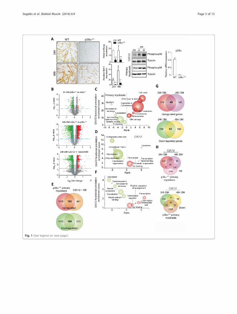
Fig. 1 (See legend on next page.)
Segalés et al. Skeletal Muscle (2016) 6:9 Page 5 of 15
(Fig. 1c). p38α signaling in satellite cells also ap-peared to regulate multiple genes and transcriptionfactors that had not been previously related to myo-genesis, for example, genes encoding signaling pro-teins and genes involved in proteolysis or DNAmetabolism and damage. In contrast, SB treatmentof C2C12 cells caused downregulation of genes re-lated to secretory pathways and cytoskeletonorganization, whereas the expression of genes relatedto transcription, chromatin organization, or ionbinding was upregulated (Fig. 1d). Of note, the cell-cycle-related genes that were found upregulated inp38α-deficient satellite cells after 24 h in DMremained unchanged in SB-treated C2C12 cells atthe same time point; however, SB-treated C2C12cells showed upregulation of these proliferation-related genes at a later time point in DM (i.e., 48 h),according to the recently published data [8], suggest-ing a delay in the cell-cycle exit of C2C12 immortal-ized myoblasts, compared to primary satellite cells,in differentiation-promoting conditions. Of the p38α-regulated genes in both experimental conditions,only 102 and 169 genes were commonly up- anddownregulated, respectively, in SB-treated C2C12myoblasts and p38α-deficient satellite cells (Fig. 1e).Thus, the stringency imposed by the use of distinctmuscle cell models and p38-inhibiting strategiesallowed the identification of a robust list of bona fidegenes dependent on p38α at the onset of myogenicdifferentiation (Additional file 2: Table S1). GO ana-lysis of genes commonly regulated in SB-treatedC2C12 cells, and p38α-deficient satellite cells at 24 hDM showed that the expression of muscledifferentiation-specific genes is commonly downregu-lated in both types of cells in the absence of p38αsignaling (Fig. 1f ). In contrast, genes commonly up-regulated in both conditions were related to the tran-scription and cell motion processes (Fig. 1f ).
A further comparison of the expression arrays fromC2C12 myoblasts in DM for 24 and 48 h (in the absenceor presence of SB) revealed a dramatic difference in bothtime points: only 20 % of the genes upregulated by the48 h DM + SB treatment were also upregulated at theearlier DM + SB 24 h time point (Fig. 1g); likewise, only30 % of the downregulated genes at the 48 h DM + SBtreatment were common with the 24 h DM + SB condi-tions. Thus, p38α regulates the muscle differentiationgene program in C2C12 myoblasts with distinct kinetics(i.e., p38α-dependent early and late myogenic genes)(Fig. 1g). Interestingly, comparison of the 24 and 48 hDM + SB C2C12 cells with the 24 h DM p38α-deficientsatellite cells demonstrated that, in addition to cell-cycleexit, the kinetics of the p38α-dependent proliferation-to-differentiation gene expression transition program differbetween both cell types; indeed, this transition was fasterin satellite cells, as shown by the similar number of com-mon p38α-regulated genes in the 24 h-DM satellite-celltime point and in C2C12 cells at both time points(Fig. 1h). It is probable that the faster differentiation kin-etics might be related to the more rapid and pronouncedactivation of p38 MAPK signaling in satellite cells thanC2C12 cells in the GM to DM transit conditions(Fig. 1a).We next performed qPCR analysis of p38α-deficient
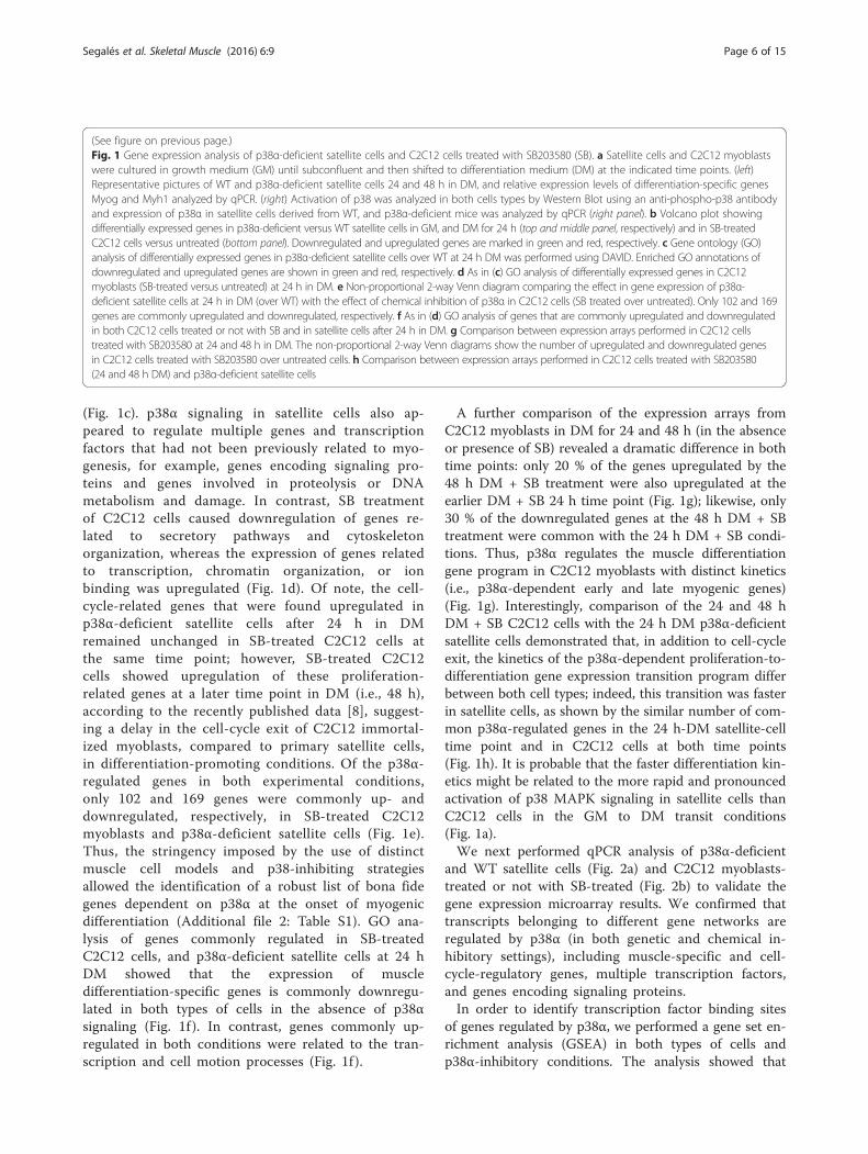
and WT satellite cells (Fig. 2a) and C2C12 myoblasts-treated or not with SB-treated (Fig. 2b) to validate thegene expression microarray results. We confirmed thattranscripts belonging to different gene networks areregulated by p38α (in both genetic and chemical in-hibitory settings), including muscle-specific and cell-cycle-regulatory genes, multiple transcription factors,and genes encoding signaling proteins.In order to identify transcription factor binding sites
of genes regulated by p38α, we performed a gene set en-richment analysis (GSEA) in both types of cells andp38α-inhibitory conditions. The analysis showed that
(See figure on previous page.)Fig. 1 Gene expression analysis of p38α-deficient satellite cells and C2C12 cells treated with SB203580 (SB). a Satellite cells and C2C12 myoblastswere cultured in growth medium (GM) until subconfluent and then shifted to differentiation medium (DM) at the indicated time points. (left)Representative pictures of WT and p38α-deficient satellite cells 24 and 48 h in DM, and relative expression levels of differentiation-specific genesMyog and Myh1 analyzed by qPCR. (right) Activation of p38 was analyzed in both cells types by Western Blot using an anti-phospho-p38 antibodyand expression of p38α in satellite cells derived from WT, and p38α-deficient mice was analyzed by qPCR (right panel). b Volcano plot showingdifferentially expressed genes in p38α-deficient versus WT satellite cells in GM, and DM for 24 h (top and middle panel, respectively) and in SB-treatedC2C12 cells versus untreated (bottom panel). Downregulated and upregulated genes are marked in green and red, respectively. c Gene ontology (GO)analysis of differentially expressed genes in p38α-deficient satellite cells over WT at 24 h DM was performed using DAVID. Enriched GO annotations ofdownregulated and upregulated genes are shown in green and red, respectively. d As in (c) GO analysis of differentially expressed genes in C2C12myoblasts (SB-treated versus untreated) at 24 h in DM. e Non-proportional 2-way Venn diagram comparing the effect in gene expression of p38α-deficient satellite cells at 24 h in DM (over WT) with the effect of chemical inhibition of p38α in C2C12 cells (SB treated over untreated). Only 102 and 169genes are commonly upregulated and downregulated, respectively. f As in (d) GO analysis of genes that are commonly upregulated and downregulatedin both C2C12 cells treated or not with SB and in satellite cells after 24 h in DM. g Comparison between expression arrays performed in C2C12 cellstreated with SB203580 at 24 and 48 h in DM. The non-proportional 2-way Venn diagrams show the number of upregulated and downregulated genesin C2C12 cells treated with SB203580 over untreated cells. h Comparison between expression arrays performed in C2C12 cells treated with SB203580(24 and 48 h DM) and p38α-deficient satellite cells
Segalés et al. Skeletal Muscle (2016) 6:9 Page 6 of 15
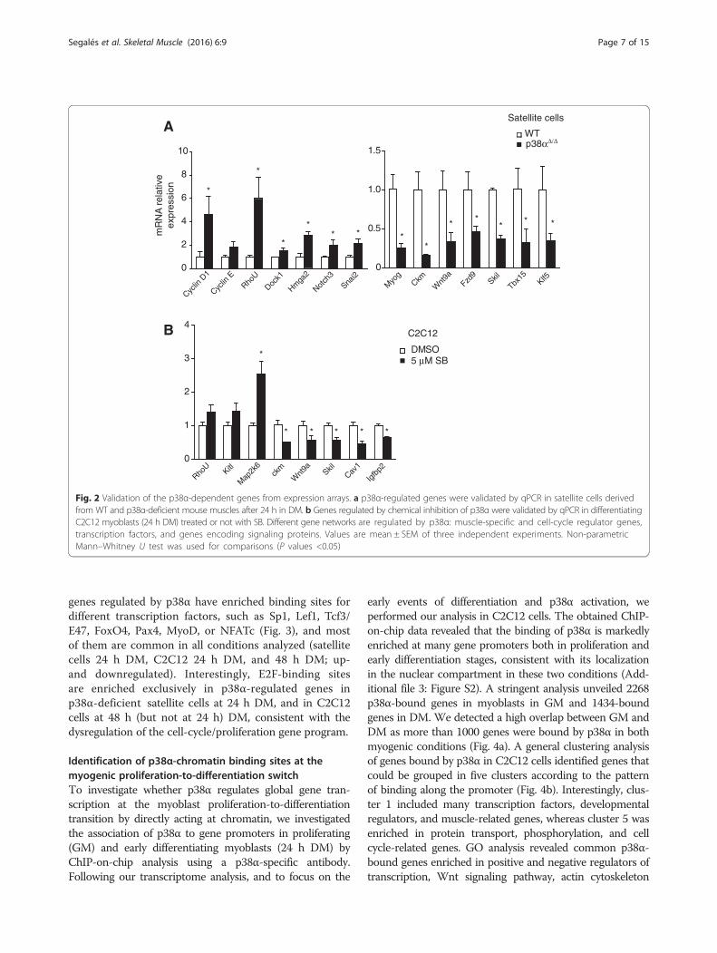
genes regulated by p38α have enriched binding sites fordifferent transcription factors, such as Sp1, Lef1, Tcf3/E47, FoxO4, Pax4, MyoD, or NFATc (Fig. 3), and mostof them are common in all conditions analyzed (satellitecells 24 h DM, C2C12 24 h DM, and 48 h DM; up-and downregulated). Interestingly, E2F-binding sitesare enriched exclusively in p38α-regulated genes inp38α-deficient satellite cells at 24 h DM, and in C2C12cells at 48 h (but not at 24 h) DM, consistent with thedysregulation of the cell-cycle/proliferation gene program.
Identification of p38α-chromatin binding sites at themyogenic proliferation-to-differentiation switchTo investigate whether p38α regulates global gene tran-scription at the myoblast proliferation-to-differentiationtransition by directly acting at chromatin, we investigatedthe association of p38α to gene promoters in proliferating(GM) and early differentiating myoblasts (24 h DM) byChIP-on-chip analysis using a p38α-specific antibody.Following our transcriptome analysis, and to focus on the
early events of differentiation and p38α activation, weperformed our analysis in C2C12 cells. The obtained ChIP-on-chip data revealed that the binding of p38α is markedlyenriched at many gene promoters both in proliferation andearly differentiation stages, consistent with its localizationin the nuclear compartment in these two conditions (Add-itional file 3: Figure S2). A stringent analysis unveiled 2268p38α-bound genes in myoblasts in GM and 1434-boundgenes in DM. We detected a high overlap between GM andDM as more than 1000 genes were bound by p38α in bothmyogenic conditions (Fig. 4a). A general clustering analysisof genes bound by p38α in C2C12 cells identified genes thatcould be grouped in five clusters according to the patternof binding along the promoter (Fig. 4b). Interestingly, clus-ter 1 included many transcription factors, developmentalregulators, and muscle-related genes, whereas cluster 5 wasenriched in protein transport, phosphorylation, and cellcycle-related genes. GO analysis revealed common p38α-bound genes enriched in positive and negative regulators oftranscription, Wnt signaling pathway, actin cytoskeleton
Cyclin
D1
Cyclin
ERho
U
Dock1
Hmga
2
Notch
3Sna
i20
2
4
6
8
10
WTA
Myo
gCkm
Wnt
9aFzd
9Skil
Tbx15
Klf50
0.5
1.0
1.5
BDMSO
RhoU
Kitl
Map
2k6
ckm
Wnt
9a SkilCav
1
Igfb
p20
1
2
3
4
mR
NA
rel
ativ
eex
pres
sion
5 μM SB
p38αΔ/Δ
*
*
*
** * *
*
* ** * *
*
* * * * *
Satellite cells
C2C12
Fig. 2 Validation of the p38α-dependent genes from expression arrays. a p38α-regulated genes were validated by qPCR in satellite cells derivedfrom WT and p38α-deficient mouse muscles after 24 h in DM. b Genes regulated by chemical inhibition of p38α were validated by qPCR in differentiatingC2C12 myoblasts (24 h DM) treated or not with SB. Different gene networks are regulated by p38α: muscle-specific and cell-cycle regulator genes,transcription factors, and genes encoding signaling proteins. Values are mean ± SEM of three independent experiments. Non-parametricMann–Whitney U test was used for comparisons (P values <0.05)
Segalés et al. Skeletal Muscle (2016) 6:9 Page 7 of 15
organization, and serine/threonine and protein kinase-related genes; genes bound only in proliferative conditionswere enriched in Golgi apparatus genes, positive regulatorsof transcription and further enriched in Wnt signalingmolecules. Interestingly, genes bound only in differ-entiating conditions were enriched in glycoproteinsand membrane proteins, and in general, but negativeregulators of transcription (positive general tran-scriptional regulators were not enriched) (Fig. 4c).Moreover, GSEA analysis of transcription factorbinding sites in the p38α-bound genes identifiedenriched binding sites for nearly the same transcrip-tion factors found in p38α-regulated genes obtainedin the expression microarrays, for example: Sp1,Lef1, Tcf3/E47, FoxO4, Pax4, MyoD, or Nfatc
(Fig. 4d). Since some of these transcription factorshave been described as phosphorylation substrates ofp38 MAPK in different model systems, includingSp1 [32, 33] Tcf3/E47 [12], NFATC4 [34], or E2F4[35], and the pattern of p38α recruitment to genes ismostly restricted to proximal promoter regions, it istempting to propose that p38α will likely regulatethe myoblast proliferation-to-differentiation transi-tion mainly through these transcription factors.Of note, genome-wide binding analysis of p38α in experi-
mental conditions of myogenic differentiation, where p38α isexpressed but its kinase activity is inhibited (C2C12 cells inDM + SB), revealed that the kinase activity was not requiredfor p38α recruitment to chromatin; on the contrary, at thegenome level, there was a generally increased occupancy
Factor Genes p-value Factor Genes p-value Factor Genes p-valueSp1 338 5.32E-88 Sp1 104 1.46E-28 E2f (10) 15 5.81E-13Lef1 (2) 264 8.29E-82 Lef1 (2) 75 2.74E-22 E2f (Tfbp1) 14 1.33E-11Tcf3 (E12) 258 4.42E-57 Foxo4 73 5.87E-20 Lef1 (2) 27 9.20E-11Foxo4 230 3.99E-56 81 6.16E-20 E2f4 12 2.29E-09Maz:Myc 236 5.79E-52 Srebf1 31 7.92E-16 Sp1 38 3.61E-08Nfatc 206 3.01E-48 Nfatc 62 2.03E-15 Tcf3 (E12) 34 5.51E-08Myc (2) 140 1.99E-43 Pax4 47 1.68E-13 Pax4 22 4.59E-07E2f (13) 65 6.89E-40 Ap4 51 3.29E-13 Maz:Myc 30 7.78E-07E2f (Tfbp1) 64 1.8E-38 Maz:Myc 64 6.17E-13 Nfil3 10 8.32E-07E2f (Tfbp2) 64 1.8E-38 Foxf2 37 3.82E-12 Sox9 11 1.75E-06Pax4 136 2.55E-30 Myod 37 4.47E-12 Nfatc 26 2.26E-06Foxf2 107 1.27E-27 Tcf8 33 2.37E-11 Atf2 8 3.07E-06Ets2 115 4.64E-26 Myc 38 2.66E-11 Pou2f1 9 8.84E-06Foxa1 90 8.16E-25 Ets2 38 1.11E-10 Foxo4 26 9.92E-06Ddit3 (Chop) 50 1.03E-24 Atf3 25 7.25E-10 Foxf2 16 1.34E-05Ap4 134 1.47E-22 Zfp161 17 7.56E-10 Ets2 17 2.75E-05Sox9 59 1.1E-21 Foxa1 29 1.50E-09 Gata1 8 3.00E-05Ap1 107 8.36E-21 E4f1 27 2.22E-09 Cdc5 8 4.09E-05CEBPa 50 1.03E-24 Ipf1 17 2.72E-09 Pax2 8 4.32E-05MyoD 94 2.93E-20 Runx1 17 2.88E-09 Pcbp1 8 4.57E-05p53 43 1.01E-17 Creb1 17 4.29E-09 Ipf1 8 4.57E-05
Factor Genes p-value Factor Genes p-value Factor Genes p-valueAp4 (3) 231 4.38E-98 Tcf3 147 7.9E-45 Ap4 (2) 58 1.1E-24
276 1.44E-85 Maz:Myc 130 7.2E-38 Sp1 76 5.4E-22Foxo4 216 1.67E-61 Sp1 149 1.3E-37 Nfatc 58 4.2E-20Sp1 262 7.34E-61 Nfatc 108 3.6E-31 Maz:Myc 62 5.2E-19Mef2 (6) 124 6.84E-60 Ap1 (3) 80 1.8E-29 Lef1 (2) 57 1.2E-18Maz:Myc 220 1.34E-56 Ap4 (2) 93 4.9E-29 Tcf3 63 9E-18Myod (3) 137 3.61E-56 Foxo4 103 3.1E-25 Pax4 45 9.1E-18Lef1 (2) 196 5.56E-52 Pax4 79 1E-24 Srf 20 4.7E-16Nfatc 184 3.32E-47 Lef1 (2) 96 1E-22 Ap1 39 1.6E-15Meis1 113 8.85E-43 Ets2 67 2.4E-21 Mef2 (2) 29 4.5E-12Pax4 138 9.93E-40 Myod 58 6.2E-19 Tead1 22 6.2E-12Nf1 99 1.11E-37 Meis1 50 7.9E-16 Tcf8 28 4.5E-10Ap1 113 2.13E-30 Mef2a 45 2.2E-15 Evi1 15 3.9E-10Vsx1 92 1.05E-28 Foxf2 48 6.7E-13 Foxo4 43 1.1E-09Tead1 58 4.87E-25 Tcf8 44 8E-13 Foxf2 27 1.8E-09Myc 97 6.1E-24 Myc 51 9.9E-13 Ets2 29 3.6E-09Tcf8 47 8.74E-24 Srf 24 1.1E-12 Nf1 23 6.6E-09Tbp 43 3.19E-21 Atf1 23 3.1E-12 Ap4 13 1.8E-08Ap4 42 1.1E-19 Nf1 40 9.8E-12 Meis1 24 1.8E-08Srf 38 1.33E-18 Err1 49 1.7E-11 Myod 25 3.6E-08
Enriched transcription factor binding sitesUp Primaries 24h Up C2C12 24h + SB Up C2C12 48h + SB
Down Primaries 24h Down C2C12 24h + SB Down C2C12 48h + SB
Tcf3 (E12) (2)
Tcf3 (E12) (2)
Fig. 3 Table showing transcription factor binding sites of p38α-regulated genes. Analyses of DNA-binding elements present in promoters wereperformed using GSEA. Red numbers indicate number of times that the factor appeared in the analysis with different DNA-binding elements, thehighest P value element is shown. Unknown binding elements have been omitted
Segalés et al. Skeletal Muscle (2016) 6:9 Page 8 of 15

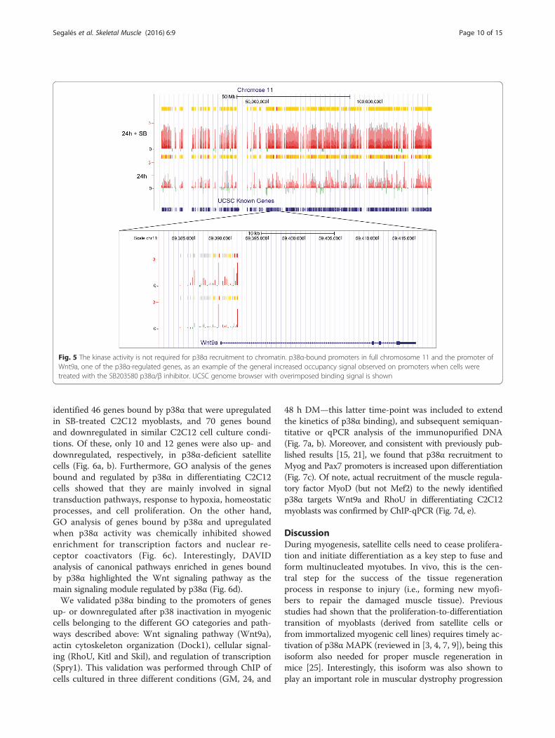
signal on promoters when cells were treated with the p38α/βinhibitor, as exemplified by full chromosome 11 and the pro-moter of Wnt9a, one of the p38α-regulated genes (Fig. 5).This indicates that recruitment of p38α to certain gene pro-moters is independent of its kinase activity, although theunderlying reason is currently unknown.
Identification of genes bound and regulated by p38αduring early myogenic differentiationTo complete the identification of the genes bound andregulated by p38α at the myogenic proliferation-to-differentiation switch, we crossed the two data sets: theexpression microarray and the ChIP-on-chip data. We
Fig. 4 ChIP-on-chip analysis for p38α in myogenic cells. Chromatin immunoprecipitation (ChIP) was performed in C2C12 myoblasts (under differentculture conditions) using a p38α-specific antibody. a Non-proportional 2-way Venn diagram showing the number of promoters bound by p38α in thetwo experimental conditions: proliferation (GM) and 24 h of differentiation (24 h DM). b Clustering analysis of genes bound by p38α in GM and 24 h DM.Genes were grouped in five clusters according to the pattern of binding along the promoter. Cluster 1 is enriched in transcription factors, developmentalregulators, and muscle-related genes. Cluster 2 is enriched in genes involved in bone development, developmental regulators, and transcription factorsand cluster 3 includes miscellaneous genes. Cluster 4 is enriched in sensory perception and olfactory receptors whereas cluster 5 is enriched in proteintransport, phosphorylation, and cell cycle-related genes. c Functional annotation analysis of the Gene ontology (GO) of bound genes in GM and 24 h DMwas performed using DAVID. The top annotation clusters are shown according to their enrichment score. Names are based on enriched GO annotations.d Table showing transcription factor binding sites of p38α-bound genes. Analyses of DNA-binding elements present in promoters were performed usingGSEA. The highest P value element is shown. Unknown binding elements have been omitted
Segalés et al. Skeletal Muscle (2016) 6:9 Page 9 of 15
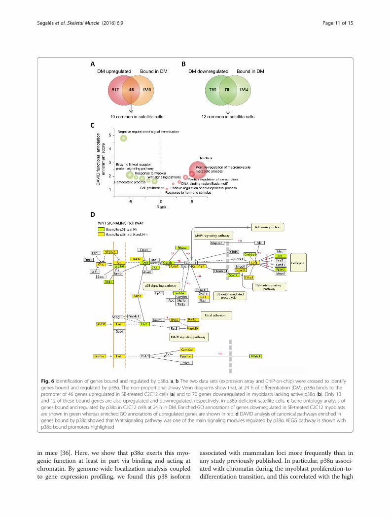
identified 46 genes bound by p38α that were upregulatedin SB-treated C2C12 myoblasts, and 70 genes boundand downregulated in similar C2C12 cell culture condi-tions. Of these, only 10 and 12 genes were also up- anddownregulated, respectively, in p38α-deficient satellitecells (Fig. 6a, b). Furthermore, GO analysis of the genesbound and regulated by p38α in differentiating C2C12cells showed that they are mainly involved in signaltransduction pathways, response to hypoxia, homeostaticprocesses, and cell proliferation. On the other hand,GO analysis of genes bound by p38α and upregulatedwhen p38α activity was chemically inhibited showedenrichment for transcription factors and nuclear re-ceptor coactivators (Fig. 6c). Interestingly, DAVIDanalysis of canonical pathways enriched in genes boundby p38α highlighted the Wnt signaling pathway as themain signaling module regulated by p38α (Fig. 6d).We validated p38α binding to the promoters of genes
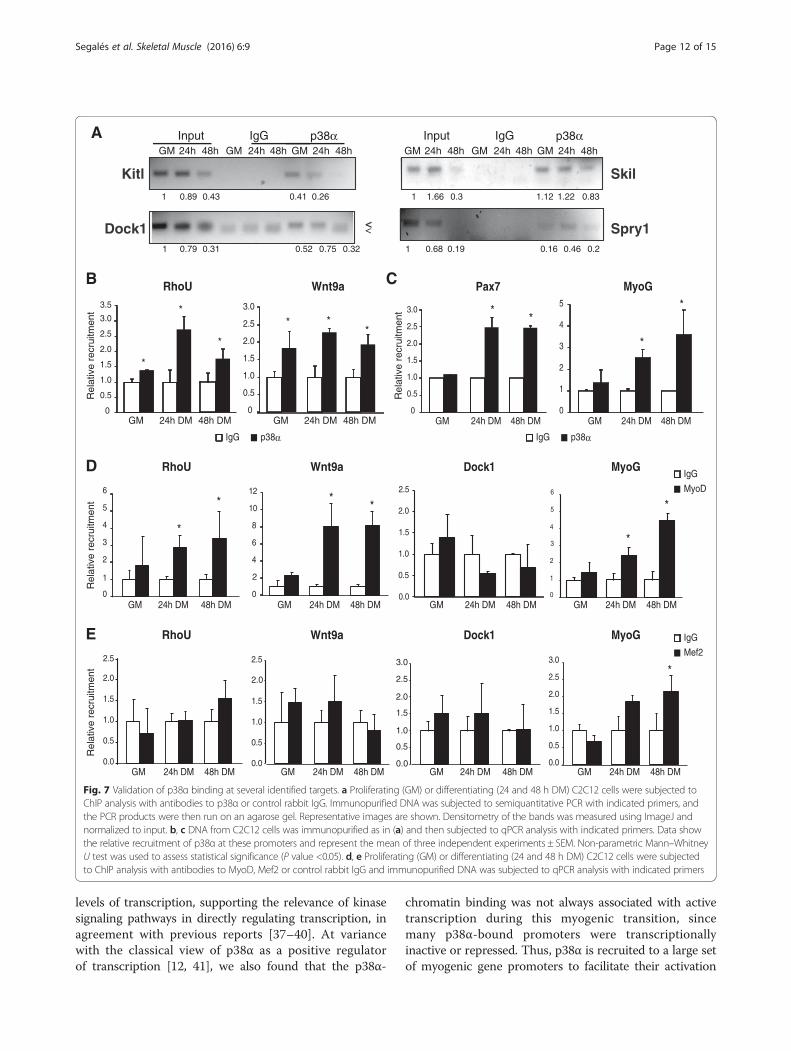
up- or downregulated after p38 inactivation in myogeniccells belonging to the different GO categories and path-ways described above: Wnt signaling pathway (Wnt9a),actin cytoskeleton organization (Dock1), cellular signal-ing (RhoU, Kitl and Skil), and regulation of transcription(Spry1). This validation was performed through ChIP ofcells cultured in three different conditions (GM, 24, and
48 h DM—this latter time-point was included to extendthe kinetics of p38α binding), and subsequent semiquan-titative or qPCR analysis of the immunopurified DNA(Fig. 7a, b). Moreover, and consistent with previously pub-lished results [15, 21], we found that p38α recruitment toMyog and Pax7 promoters is increased upon differentiation(Fig. 7c). Of note, actual recruitment of the muscle regula-tory factor MyoD (but not Mef2) to the newly identifiedp38α targets Wnt9a and RhoU in differentiating C2C12myoblasts was confirmed by ChIP-qPCR (Fig. 7d, e).
DiscussionDuring myogenesis, satellite cells need to cease prolifera-tion and initiate differentiation as a key step to fuse andform multinucleated myotubes. In vivo, this is the cen-tral step for the success of the tissue regenerationprocess in response to injury (i.e., forming new myofi-bers to repair the damaged muscle tissue). Previousstudies had shown that the proliferation-to-differentiationtransition of myoblasts (derived from satellite cells orfrom immortalized myogenic cell lines) requires timely ac-tivation of p38α MAPK (reviewed in [3, 4, 7, 9]), being thisisoform also needed for proper muscle regeneration inmice [25]. Interestingly, this isoform was also shown toplay an important role in muscular dystrophy progression
Fig. 5 The kinase activity is not required for p38α recruitment to chromatin. p38α-bound promoters in full chromosome 11 and the promoter ofWnt9a, one of the p38α-regulated genes, as an example of the general increased occupancy signal observed on promoters when cells weretreated with the SB203580 p38α/β inhibitor. UCSC genome browser with overimposed binding signal is shown
Segalés et al. Skeletal Muscle (2016) 6:9 Page 10 of 15
in mice [36]. Here, we show that p38α exerts this myo-genic function at least in part via binding and acting atchromatin. By genome-wide localization analysis coupledto gene expression profiling, we found this p38 isoform
associated with mammalian loci more frequently than inany study previously published. In particular, p38α associ-ated with chromatin during the myoblast proliferation-to-differentiation transition, and this correlated with the high
Fig. 6 Identification of genes bound and regulated by p38α. a, b The two data sets (expression array and ChIP-on-chip) were crossed to identifygenes bound and regulated by p38α. The non-proportional 2-way Venn diagrams show that, at 24 h of differentiation (DM), p38α binds to thepromoter of 46 genes upregulated in SB-treated C2C12 cells (a) and to 70 genes downregulated in myoblasts lacking active p38α (b). Only 10and 12 of these bound genes are also upregulated and downregulated, respectively, in p38α-deficient satellite cells. c Gene ontology analysis ofgenes bound and regulated by p38α in C2C12 cells at 24 h in DM. Enriched GO annotations of genes downregulated in SB-treated C2C12 myoblastsare shown in green whereas enriched GO annotations of upregulated genes are shown in red. d DAVID analysis of canonical pathways enriched ingenes bound by p38α showed that Wnt signaling pathway was one of the main signaling modules regulated by p38α. KEGG pathway is shown withp38α-bound promoters highlighted
Segalés et al. Skeletal Muscle (2016) 6:9 Page 11 of 15
levels of transcription, supporting the relevance of kinasesignaling pathways in directly regulating transcription, inagreement with previous reports [37–40]. At variancewith the classical view of p38α as a positive regulatorof transcription [12, 41], we also found that the p38α-
chromatin binding was not always associated with activetranscription during this myogenic transition, sincemany p38α-bound promoters were transcriptionallyinactive or repressed. Thus, p38α is recruited to a large setof myogenic gene promoters to facilitate their activation
A
B
Rel
ativ
e re
crui
tmen
t
Rel
ativ
e re
crui
tmen
t0
0.5
1.0
2.0
3.0
1.5
2.5
0
0.5
1.0
2.0
3.0
1.5
2.5
3.5
* **
*
*
*
GM 24h DM 48h DM GM 24h DM 48h DM
IgG p38α
Wnt9aRhoU
*
Skil
Dock1
Kitl
Spry1
GM GM GM24h 48h 24h 48h 24h 48hInput p38αIgG
GM GM GM24h 48h 24h 48h 24h 48hInput p38αIgG
1 0.79 0.31 0.52 0.75 0.32
1 1.66 0.3 1.12 1.22 0.831 0.89 0.43 0.41 0.26
1 0.68 0.19 0.16 0.46 0.2
C
<<
GM 24h DM 48h DM GM 24h DM 48h DM
Pax7 MyoG
0
0.5
1.0
2.0
3.0
1.5
2.5
0
1
2
4
3
5* *
*
IgG p38α
D Wnt9aRhoU Dock1 MyoG
Rel
ativ
e re
crui
tmen
t
0
2
4
6
8
10
12
GM 24h DM 48h DM
* *
IgG
MyoD
E Wnt9aRhoU Dock1 MyoG
Rel
ativ
e re
crui
tmen
t
IgG
Mef2
4
6
5
3
2
1
0
*
*
GM 24h DM 48h DM GM 24h DM 48h DM GM 24h DM 48h DM0.0
0.5
1.0
1.5
2.0
2.5
0
1
2
3
4
5
6
*
*
0.0
0.5
1.0
1.5
2.0
2.5
0.0
0.5
1.0
1.5
2.0
2.5
GM 24h DM 48h DMGM 24h DM 48h DM0.0
0.5
1.0
1.5
2.0
2.5
3.0
GM 24h DM 48h DM0.0
0.5
1.0
1.5
2.0
2.5
3.0*
GM 24h DM 48h DM
Fig. 7 Validation of p38α binding at several identified targets. a Proliferating (GM) or differentiating (24 and 48 h DM) C2C12 cells were subjected toChIP analysis with antibodies to p38α or control rabbit IgG. Immunopurified DNA was subjected to semiquantitative PCR with indicated primers, andthe PCR products were then run on an agarose gel. Representative images are shown. Densitometry of the bands was measured using ImageJ andnormalized to input. b, c DNA from C2C12 cells was immunopurified as in (a) and then subjected to qPCR analysis with indicated primers. Data showthe relative recruitment of p38α at these promoters and represent the mean of three independent experiments ± SEM. Non-parametric Mann–WhitneyU test was used to assess statistical significance (P value <0.05). d, e Proliferating (GM) or differentiating (24 and 48 h DM) C2C12 cells were subjectedto ChIP analysis with antibodies to MyoD, Mef2 or control rabbit IgG and immunopurified DNA was subjected to qPCR analysis with indicated primers
Segalés et al. Skeletal Muscle (2016) 6:9 Page 12 of 15
or repression, hence pointing to more complex regulatorymechanisms than previously anticipated.How is p38α recruited to muscle loci is not known,
but it likely involves interaction with chromatin-regulatory and/or transcription factors, as demonstratedfor several stress-induced genes [42]. Of interest, p38α-bound promoters are enriched with binding motifs forseveral transcription factors, principally Sp1, Tcf3/E47,Lef1, FoxO4, MyoD, and NFATc, which are known to bephosphorylation substrates of p38 MAPK. In skeletalmyoblasts, in particular, p38α associates with MyoD andE47 [12, 15] transcription factors, and specific phosphor-ylation of E47 promotes E47/MyoD dimerization, bind-ing to muscle-specific loci and initiation of geneexpression [12]. Likewise, phosphorylation of MEF2 byp38 facilitates both recruitment of MyoD and expressionof late myogenic genes, in a feed-forward mechanism[11], and of ASH2L-MLL methyltransferase complex[18, 19]. p38α also phosphorylates Baf60c allowingSWI/SNF-mediated chromatin remodeling at muscle-specific loci [15–17]. Interestingly, p38α was also shownto represses Pax7 gene expression via direct Ezh2 phos-phorylation, as an indispensable event for the transition ofproliferating myoblasts into their differentiating state [21].Thus, p38α may help both silence proliferation-associatedgenes and activate differentiation-promoting genes at thistransition, by directly targeting negative and positive epi-genetic and/or transcriptional regulators. Because the p38MAPK homolog Hog1 targets the RNA Pol II machineryand induces chromatin remodeling at stress-responsiveloci in yeast [43], it is possible that p38α regulates Pol II-dependent gene transcription in mammalian myogenesisin a similar way. It is worth noting that p38 was alsofound on a subset of genes independently of its kin-ase activity. Our work adds to several recent studiesof protein kinases that bind and act on chromatin:extracellular signal-regulated kinase (ERK), cJun N-terminal kinase (JNK), the MSK, AMPK, and Dyrk1A [37,38, 44–46]. This study also complements the previouslydescribed association of p38/Hog1 to osmostress genes tomediate adaptation to extracellular changes in mammalsand yeast [26, 27, 42].Of interest, we observed slower muscle differentiation
kinetics in C2C12 myoblasts compared to satellite cellsin culture; although the underlying causes for this differ-ence are not clear, they may relate to the distinct natureof both cell types (freshly isolated cells versus immortal-ized cells) and the different velocity of p38α activation inboth cell types (see Fig. 1a). Whether the expression ofthe identified p38α-regulated late genes depends onthe early ones, according to the proposed temporalregulation of muscle gene expression through a MyoD-mediated feed-forward circuit involving p38 MAPK [11],is not yet known.
Finally, although the nature of p38α-regulated genesencompasses several categories, DAVID analysis of canon-ical pathways enriched in genes bound by p38α showedthe Wnt pathway as the main signaling module regulatedby p38α. Consistent with this, MyoD was found to berecruited to the newly identified p38α target Wnt9a, inaddition to muscle-specific gene promoters, as expected.This is worth highlighting based on the reported relevantrole of canonical and non-canonical Wnt signaling indistinct satellite cell functions in vivo (expansion, switch-ing from proliferation to differentiation or cell motility)[47–49]. Taken together, this study increases our under-standing on how p38 MAPK regulates gene expressionduring myogenesis, and greatly enlarges the number andnature of target genes downstream of p38α activation.Notably, through this analysis, we have become aware ofnew categories of genes whose repression depends onp38α both in proliferating and differentiating conditions,expanding, yet complicating, the function of p38α as anegative regulator of muscle gene transcription. It is pos-sible that comparable principles of chromatin binding andactivity might be relevant for other kinases.
ConclusionsCollectively, through this study, we demonstrate associ-ation and action of p38α MAPK throughout chromatinuncovering new classes of target genes during the transi-tion of myoblasts from proliferation to differentiation.The recruitment of p38α to large sets of gene promotersappears to facilitate their activation or repression duringthis process. This is consistent with this MAPK isoformbeing a transcriptional regulator.
Additional files
Additional file 1: Table S2. List of primers used in the study. (XLSX 12 kb)
Additional file 2: Table S1. List of genes regulated and bound byp38α. (XLSX 686 kb)
Additional file 3: Supplementary Figures 1 and 2 and legends.Figure S1. Relative expression of p38α and p38β in C2C12 cells. Theexpression of p38α and p38β was measured by qPCR in differentiatingC2C12 cells. Values are mean ± SEM of three independent set of samples.Figure S2. Nuclear p38α remains constant in proliferating anddifferentiating myoblasts. Expression of p38α in C2C12 cells in GM and24 h in DM: p38α (green) and DAPI (blue). Nuclear to cytoplasmfluorescence ratio was calculated using integrated density values inImageJ software with DAPI as nuclear boundary. Scale bar = 25 μm.(PDF 7595 kb)
AbbreviationsBAF60c: BRG1/BRM-associated factor 60 variant c; ChIP: chromatinimmunoprecipitation; DAPI: 4′,6-diamidino-2-phenylindole;DM: differentiation medium; Dock1: dedicator of cytokinesis; FoxO4: forkheadbox protein O4; GM: growth medium; GO: gene ontology; GSEA: gene setenrichment analysis; Lef1: lymphoid enhancer-binding factor 1;MAPK: mitogen-activated protein kinase; MEF2: myocyte enhancer factor 2;MRFs: myogenic regulatory factors; NFATc: nuclear factor of activated T cells;Pax4: paired box gene 4; RT-qPCR: reverse transcriptase quantitative
Segalés et al. Skeletal Muscle (2016) 6:9 Page 13 of 15
polymerase chain reaction; Spry1: protein sprouty homolog 1;Tcf3: transcription factor 3 (E2A immunoglobulin enhancer-binding factorsE12/E47); Wnt9a: wingless-type MMTV integration site family, member 9A;WT: wild type.
Competing interestsThe authors declared that they have no competing interests.
Authors’ contributionsJS designed and performed experiments, analyzed data, and interpreted resultsand wrote the manuscript. ABMMK performed bioinformatic analysis and editedthe manuscript. RK performed bioinformatic analysis and edited the manuscript.QL performed experiments. PS-V performed experiments. FJD performedexperiments, analyzed data, and interpreted results. EB performed ChIPexperiments and edited the manuscript. EP designed and performedexperiments, analyzed data, and interpreted results and wrote the manuscript.PMC conceived the project, designed experiments, interpreted results, andwrote the manuscript. All authors read and approved the final manuscript.
AcknowledgementsWe thank Drs. V. Ruiz-Bonilla, M. Jardí, and M. Raya, for their help and contributionto this study. The CRG/UPF Genomic Unit for excellent support. Theauthors acknowledge funding from MINECO, Spain (SAF2012-38547,PLE2009-0124, SAF2015-67369-R, "María de Maeztu" Programme forUnits of Excellence in R&D MDM-2014-0370), AFM, MDA, DDP-Netherlands, E-RARE, Fundació Marató TV3 and EU-FP7 (Myoage, Optistemand Endostem). JS is recipient of a Juan de la Cierva postdoctoral fellowship.
Author details1Department of Experimental and Health Sciences, Pompeu Fabra University(UPF), CIBER on Neurodegenerative diseases (CIBERNED), Barcelona, Spain.2Department of Genetic Engineering and Biotechnology, University of Dhaka,Dhaka 1000, Bangladesh. 3Wyss Institute for Biologically Inspired Engineering,Harvard University, Boston, MA 02115, USA. 4Sprott Center for Stem CellResearch, Ottawa Hospital Research Institute, Ottawa, ON K1H 8L6, Canada.5Chromatin and Disease Group, Cancer Epigenetics and Biology Programme(PEBC), Bellvitge Biomedical Research Institute (IDIBELL), Barcelona, Spain.6Institució Catalana de Recerca i Estudis Avançats (ICREA), Barcelona, Spain.7Present address: Buck Institute for Research on Aging, Novato, CA, USA.
Received: 11 September 2015 Accepted: 5 January 2016
References1. Cuenda A, Rousseau S. p38 MAP-kinases pathway regulation, function and
role in human diseases. Biochim Biophys Acta. 2007;1773(8):1358–75.doi:10.1016/j.bbamcr.2007.03.010.
2. Cuadrado A, Nebreda AR. Mechanisms and functions of p38 MAPKsignalling. Biochem J. 2010;429(3):403–17. doi:10.1042/BJ20100323.
3. Lluis F, Perdiguero E, Nebreda AR, Munoz-Canoves P. Regulation of skeletalmuscle gene expression by p38 MAP kinases. Trends Cell Biol.2006;16(1):36–44. doi:10.1016/j.tcb.2005.11.002.
4. Keren A, Tamir Y, Bengal E. The p38 MAPK signaling pathway: a majorregulator of skeletal muscle development. Mol Cell Endocrinol.2006;252(1-2):224–30. doi:10.1016/j.mce.2006.03.017.
5. Mahmoudi S, Brunet A. Aging and reprogramming: a two-way street.Curr Opin Cell Biol. 2012;24(6):744–56. doi:10.1016/j.ceb.2012.10.004.
6. Neves J, Demaria M, Campisi J, Jasper H. Of flies, mice, and men:evolutionarily conserved tissue damage responses and aging. Dev Cell.2015;32(1):9–18. doi:10.1016/j.devcel.2014.11.028.
7. Perdiguero E, Ruiz-Bonilla V, Gresh L, Hui L, Ballestar E, Sousa-Victor P, et al.Genetic analysis of p38 MAP kinases in myogenesis: fundamental role ofp38alpha in abrogating myoblast proliferation. EMBO J. 2007;26(5):1245–56.
8. Mourikis P, Tajbakhsh S. Distinct contextual roles for Notch signalling in skeletalmuscle stem cells. BMC Dev Biol. 2014;14:2. doi:10.1186/1471-213X-14-2.
9. Vahidi Ferdousi L, Rocheteau P, Chayot R, Montagne B, Chaker Z, Flamant P, et al.More efficient repair of DNA double-strand breaks in skeletal muscle stem cellscompared to their committed progeny. Stem Cell Res. 2014;13(3 Pt A):492–507.doi:10.1016/j.scr.2014.08.005.
10. Zhao M, New L, Kravchenko VV, Kato Y, Gram H, Di Padova F, et al.Regulation of the MEF2 family of transcription factors by p38. Mol CellBiol. 1999;19(1):21–30.
11. Penn BH, Bergstrom DA, Dilworth FJ, Bengal E, Tapscott SJ. A MyoD-generated feed-forward circuit temporally patterns gene expression duringskeletal muscle differentiation. Genes Dev. 2004;18(19):2348–53.
12. Lluis F, Ballestar E, Suelves M, Esteller M, Munoz-Canoves P. E47phosphorylation by p38 MAPK promotes MyoD/E47 association andmuscle-specific gene transcription. EMBO J. 2005;24(5):974–84.doi:10.1038/sj.emboj.7600528.
13. Perdiguero E, Sousa-Victor P, Ballestar E, Munoz-Canoves P. Epigeneticregulation of myogenesis. Epigenetics. 2009;4(8):541–50.
14. Fulle S, Di Donna S, Puglielli C, Pietrangelo T, Beccafico S, Bellomo R,et al. Age-dependent imbalance of the antioxidative system in humansatellite cells. Exp Gerontol. 2005;40(3):189–97. doi:10.1016/j.exger.2004.11.006.
15. Simone C, Forcales SV, Hill DA, Imbalzano AN, Latella L, Puri PL. p38pathway targets SWI-SNF chromatin-remodeling complex to muscle-specificloci. Nat Genet. 2004;36(7):738–43. doi:10.1038/ng1378ng1378.
16. Serra C, Palacios D, Mozzetta C, Forcales SV, Morantte I, Ripani M, et al.Functional interdependence at the chromatin level between the MKK6/p38and IGF1/PI3K/AKT pathways during muscle differentiation. Mol Cell.2007;28(2):200–13. doi:10.1016/j.molcel.2007.08.021.
17. Forcales SV, Albini S, Giordani L, Malecova B, Cignolo L, Chernov A, et al.Signal-dependent incorporation of MyoD-BAF60c into Brg1-based SWI/SNFchromatin-remodelling complex. EMBO J. 2012;31(2):301–16.doi:10.1038/emboj.2011.391.
18. Rampalli S, Li L, Mak E, Ge K, Brand M, Tapscott SJ, et al. p38 MAPK signalingregulates recruitment of Ash2L-containing methyltransferase complexes tospecific genes during differentiation. Nat Struct Mol Biol. 2007;14(12):1150–6.doi:10.1038/nsmb1316.
19. Brack AS, Bildsoe H, Hughes SM. Evidence that satellite cell decrementcontributes to preferential decline in nuclear number from large fibresduring murine age-related muscle atrophy. J Cell Sci.2005;118(Pt 20):4813–21. doi:10.1242/jcs.02602.
20. Cuadrado A, Corrado N, Perdiguero E, Lafarga V, Munoz-Canoves P, NebredaAR. Essential role of p18(Hamlet)/SRCAP-mediated histone H2A.Z chromatinincorporation in muscle differentiation. EMBO J. 2010.doi:10.1038/emboj.2010.85.
21. Palacios D, Mozzetta C, Consalvi S, Caretti G, Saccone V, Proserpio V, et al.TNF/p38alpha/polycomb signaling to Pax7 locus in satellite cells linksinflammation to the epigenetic control of muscle regeneration. Cell StemCell. 2010;7(4):455–69. doi:10.1016/j.stem.2010.08.013.
22. Gillespie MA, Le Grand F, Scime A, Kuang S, von Maltzahn J, Seale V, et al.p38-{gamma}-dependent gene silencing restricts entry into the myogenicdifferentiation program. J Cell Biol. 2009;187(7):991–1005.doi:10.1083/jcb.200907037.
23. Ruiz-Bonilla V, Perdiguero E, Gresh L, Serrano AL, Zamora M, Sousa-Victor P,et al. Efficient adult skeletal muscle regeneration in mice deficient in p38beta,p38gamma and p38delta MAP kinases. Cell Cycle. 2008;7(14):2208–14.
24. Wang H, Xu Q, Xiao F, Jiang Y, Wu Z. Involvement of the p38 mitogen-activatedprotein kinase alpha, beta, and gamma isoforms in myogenic differentiation.Mol Biol Cell. 2008;19(4):1519–28. doi:10.1091/mbc.E07-08-0817.
25. Brien P, Pugazhendhi D, Woodhouse S, Oxley D, Pell JM. p38alpha MAPK regulatesadult muscle stem cell fate by restricting progenitor proliferation during postnatalgrowth and repair. Stem Cells. 2013;31(8):1597–610. doi:10.1002/stem.1399.
26. De Nadal E, Zapater M, Alepuz PM, Sumoy L, Mas G, Posas F. The MAPKHog1 recruits Rpd3 histone deacetylase to activate osmoresponsive genes.Nature. 2004;427(6972):370–4. doi:10.1038/nature02258.
27. Pokholok DK, Zeitlinger J, Hannett NM, Reynolds DB, Young RA. Activatedsignal transduction kinases frequently occupy target genes. Science.2006;31(5786):533–6. doi:10.1126/science.1127677.
28. Ferreiro I, Barragan M, Gubern A, Ballestar E, Joaquin M, Posas F. The p38 SAPKis recruited to chromatin via its interaction with transcription factors. J BiolChem. 2010;285(41):31819–28. doi:10.1074/jbc.M110.155846.
29. Gentleman RC, Carey VJ, Bates DM, Bolstad B, Dettling M, Dudoit S, et al.Bioconductor: open software development for computational biology andbioinformatics. Genome Biol. 2004;5(10):R80. doi:10.1186/gb-2004-5-10-r80.
30. da Huang W, Sherman BT, Lempicki RA. Systematic and integrative analysisof large gene lists using DAVID bioinformatics resources. Nat Protoc.2009;4(1):44–57. doi:10.1038/nprot.2008.211.
Segalés et al. Skeletal Muscle (2016) 6:9 Page 14 of 15
31. Subramanian A, Tamayo P, Mootha VK, Mukherjee S, Ebert BL, Gillette MA,et al. Gene set enrichment analysis: a knowledge-based approach forinterpreting genome-wide expression profiles. Proc Natl Acad Sci U S A.2005;102(43):15545–50. doi:10.1073/pnas.0506580102.
32. D’Addario M, Arora PD, McCulloch CA. Role of p38 in stress activation ofSp1. Gene. 2006;379:51–61. doi:10.1016/j.gene.2006.04.012.
33. Xu K, Shu HK. EGFR activation results in enhanced cyclooxygenase-2expression through p38 mitogen-activated protein kinase-dependentactivation of the Sp1/Sp3 transcription factors in human gliomas. CancerRes. 2007;67(13):6121–9. doi:10.1158/0008-5472.CAN-07-0141.
34. Yang TT, Xiong Q, Enslen H, Davis RJ, Chow CW. Phosphorylation of NFATc4by p38 mitogen-activated protein kinases. Mol Cell Biol. 2002;22(11):3892–904.
35. Morillo SM, Abanto EP, Roman MJ, Frade JM. Nerve growth factor-induced cell cycle reentry in newborn neurons is triggered byp38MAPK-dependent E2F4 phosphorylation. Mol Cell Biol. 2012;32(14):2722–37.doi:10.1128/MCB.00239-12.
36. Wissing ER, Boyer JG, Kwong JQ, Sargent MA, Karch J, McNally EM, et al.P38alpha MAPK underlies muscular dystrophy and myofiber death througha Bax-dependent mechanism. Hum Mol Genet. 2014;23(20):5452–63.doi:10.1093/hmg/ddu270.
37. Tiwari VK, Stadler MB, Wirbelauer C, Paro R, Schubeler D, Beisel C. Achromatin-modifying function of JNK during stem cell differentiation.Nat Genet. 2011;44(1):94–100. doi:10.1038/ng.1036.
38. Vicent GP, Ballare C, Nacht AS, Clausell J, Subtil-Rodriguez A, Quiles I, et al.Induction of progesterone target genes requires activation of Erk and Mskkinases and phosphorylation of histone H3. Mol Cell. 2006;24(3):367–81.doi:10.1016/j.molcel.2006.10.011.
39. Bungard D, Fuerth BJ, Zeng PY, Faubert B, Maas NL, Viollet B, et al. Signalingkinase AMPK activates stress-promoted transcription via histone H2Bphosphorylation. Science. 2010;329(5996):1201–5. doi:10.1126/science.1191241.
40. Di Vona C, Bezdan D, Islam AB, Salichs E, Lopez-Bigas N, Ossowski S,et al. Chromatin-wide profiling of DYRK1A reveals a role as a gene-specific RNA polymerase II CTD kinase. Mol Cell. 2015;57(3):506–20.doi:10.1016/j.molcel.2014.12.026.
41. Putker M, Madl T, Vos HR, de Ruiter H, Visscher M, van den Berg MC, et al.Redox-dependent control of FOXO/DAF-16 by transportin-1. Mol Cell.2013;49(4):730–42. doi:10.1016/j.molcel.2012.12.014.
42. Rezza A, Sennett R, Rendl M. Adult stem cell niches: cellular andmolecular components. Curr Top Dev Biol. 2014;107:333–72.doi:10.1016/B978-0-12-416022-4.00012-3.
43. Nadal-Ribelles M, Conde N, Flores O, Gonzalez-Vallinas J, Eyras E, Orozco M,et al. Hog1 bypasses stress-mediated down-regulation of transcription byRNA polymerase II redistribution and chromatin remodeling. Genome Biol.2012;13(11):R106. doi:10.1186/gb-2012-13-11-r106.
44. Aurora AB, Olson EN. Immune modulation of stem cells and regeneration.Cell Stem Cell. 2014;15(1):14–25. doi:10.1016/j.stem.2014.06.009.
45. Murphy MM, Keefe AC, Lawson JA, Flygare SD, Yandell M, Kardon G.Transiently active Wnt/beta-catenin signaling is not required but must besilenced for stem cell function during muscle regeneration. Stem Cell Rep.2014;3(3):475–88. doi:10.1016/j.stemcr.2014.06.019.
46. Lawrence MC, McGlynn K, Shao C, Duan L, Naziruddin B, Levy MF, et al.Chromatin-bound mitogen-activated protein kinases transmit dynamicsignals in transcription complexes in beta-cells. Proc Natl Acad Sci U S A.2008;105(36):13315–20. doi:10.1073/pnas.0806465105.
47. Le Grand F, Jones AE, Seale V, Scime A, Rudnicki MA. Wnt7a activates theplanar cell polarity pathway to drive the symmetric expansion of satellitestem cells. Cell Stem Cell. 2009;4(6):535–47. doi:10.1016/j.stem.2009.03.013.
48. Tanaka S, Terada K, Nohno T. Canonical Wnt signaling is involved inswitching from cell proliferation to myogenic differentiation of mousemyoblast cells. J Mol Signal. 2011;6:12. doi:10.1186/1750-2187-6-12.
49. Bentzinger CF, von Maltzahn J, Dumont NA, Stark DA, Wang YX, NhanK, et al. Wnt7a stimulates myogenic stem cell motility and engraftmentresulting in improved muscle strength. J Cell Biol. 2014;205(1):97–111.doi:10.1083/jcb.201310035.
• We accept pre-submission inquiries
• Our selector tool helps you to find the most relevant journal
• We provide round the clock customer support
• Convenient online submission
• Thorough peer review
• Inclusion in PubMed and all major indexing services
• Maximum visibility for your research
Submit your manuscript atwww.biomedcentral.com/submit
Submit your next manuscript to BioMed Central and we will help you at every step:
Segalés et al. Skeletal Muscle (2016) 6:9 Page 15 of 15


![Spontaneous and Bleomycin-Induced gH2AX … › pdf › ABB_2014061016004839.pdfchromosomes or a mandatory feature of chromatin condensation during mitosis [33]. It has been reported](https://static.fdocument.org/doc/165x107/5f02e4ed7e708231d40689c7/spontaneous-and-bleomycin-induced-gh2ax-a-pdf-a-abb-chromosomes-or-a-mandatory.jpg)