Regulation of the activity of L-aspartate β-decarboxylase by a novel allosteric mechanism
9
BIOCHEMISTRY Regulation of the Activity of L-Aspartate P-Decarboxylase by a Novel Allosteric Mechanism* Suresh S. Tate and Alton Meister ABSTRACT: Studies on the regulation of L-aspartate &decarboxylase of Alcaligenes faecalis were carried out under conditions in which aspartate is decarboxy- lated to a-alanine at a constant rate. Addition of a-keto acid increases the V,,, value for decarboxylation about sixfold at 37”, but the K, values for aspartate in the presence and absence of added a-keto acid are about the same (about 6 X M). When the enzyme is incubated with aspartate (in absence of added a-keto acid) there is an initial rapid decarboxylation associated with a burst of pyruvate formation followed by slower but linear decarboxylation. There is no net increase in pyruvate concentration during the latter phase. Studies in which lactate and malate dehydrogenases and re- duced diphosphopyridine nucleotide were added in- dicate that pyruvate is formed by decarboxylation and hydrolysis of the ketimine Schiff base intermediate and that pyruvate is essential for continued decarboxylase activity. A constant rate of decarboxylation results from A spartate 8-decarboxylase is markedly activated by pyruvate, a-ketoglutarate, and a number of other a-keto acids (Meister et al., 1951 ; Cattaneo-Lacombe et al., 1958; Crawford, 1958; Wilson, 1963; Novo- grodsky and Meister, 1964a; Chibata et al., 1967, 1968). This activation has been explained by studies on the aspartate 8-decarboxylase of Alcaligenes faecalis in which it was shown that the enzyme is inactivated by incubation with L-aspartate (and a number of other L-amino acids) and that such inactivation can be prevented by simultaneous addition of an a-keto acid (Novogrodsky et al., 1963 ; Novogrodsky and Meister, 1964a). The enzyme was shown to catalyze transami- nation between aspartate (or other L-amino acids) and pyruvate (or a-ketoglutarate) ; transamination of the enzyme with L-aspartate (or other L-amino acids) converts the enzyme-bound pyridoxal 5’-phosphate to enzyme-bound pyridoxamine 5’-phosphate. This form of the enzyme, inactive in the decarboxylation of aspartate, reverts to the pyridoxal 5’-phosphate form on transamination with a-keto acids. Thus, although the rate of the transamination reaction catalyzed by the enzyme is quantitatively low as compared to the rate of decarboxylation, it is of considerable sig- nificance since it can destroy (or regenerate) the enzyme-bound cofactor required for decarboxylation. ~~ * From the Department of Biochemistry, Cornel1 University Medical College, New York, New York. Receiced December 18, 1968. Supported in part by the National Institutes of Health, U. S. Public HealthService, and the Nationalscience Foundation. 1660 an equilibrium between enzyme-bound alanine-keti- mine and enzyme-bound pyridoxamine 5’-phosphate and pyruvate. The enzyme can bind close to 1 mole of a-keto acid/minimal catalytic unit ; studies with the inhibitory aspartate analog /3-cyanoalanine (Ki = 2.7 X M) indicate that the a-keto acid binding site is different from that for aspartate. The apoenzyme can also bind pyruvate effectively. The activity of the enzyme is increased by adding very low concentrations of pyruvate and decreased by removal of pyruvate; thus, the enzyme would be subject to highly sensitive regulation in an intracellular environment containing other enzymes capable of increasing or decreasing the concentrations of pyruvate, a-ketoglutarate, and other a-keto acids. This allosteric regulatory mechanism is novel in that the a-keto acid effector is produced by the enzyme from the substrate; the effector has no effect on substrate affinity, and reacts with the inactive enzyme-bound coenzyme to yield active coenzyme. Under certain conditions (e.g., in the presence of a high concentration of acetate buffer), pyridoxamine 5’-phosphate dissociates from the enzyme to yield the inactive apoenzyme, which can be reactivated by adding pyridoxal 5’-phosphate. In this report we describe additional studies on the regulation of this enzyme using conditions under which the enzyme catalyzes the decarboxylation of aspartate to a-alanine at a constant rate. It has been found that when the enzyme is incubated with aspartate (in the absence of added a-keto acid) there is an initial period of rapid decarboxylase activity accompanied by a “burst” of pyruvate formation; this is followed by decarboxylation at a much lower but constant rate. There is no net increase in pyruvate concentration during the period of slow decarboxylation. Evidence has been obtained that pyruvate is formed by decar- boxylation and hydrolysis of the ketimine Schiff base intermediate, and that it is crucially necessary for con- tinued decarboxylase activity. A constant rate of de- carboxylation is maintained by virtue of an equilibrium between the enzyme-bound alanine-ketimine inter- mediate and enzyme-bound pyridoxamine 5’-phosphate and pyruvate. The enzyme can bind with high affinity close to 1 mole of a-keto acid/catalytic site; the site for a-keto acid is different from that for aspartate. The activity of the enzyme is decreased by removal of pyru- vate and increased by addition of pyruvate or other a-keto acids. This regulatory mechanism, although allo- steric according to the original definition of this term, is TATE AND MEISTER
Transcript of Regulation of the activity of L-aspartate β-decarboxylase by a novel allosteric mechanism
B I O C H E M I S T R Y
Regulation of the Activity of L-Aspartate P-Decarboxylase by a Novel Allosteric Mechanism*
Suresh S. Tate and Alton Meister
ABSTRACT: Studies on the regulation of L-aspartate &decarboxylase of Alcaligenes faecalis were carried out under conditions in which aspartate is decarboxy- lated to a-alanine a t a constant rate. Addition of a-keto acid increases the V,,, value for decarboxylation about sixfold at 37”, but the K , values for aspartate in the presence and absence of added a-keto acid are about the same (about 6 X M). When the enzyme is incubated with aspartate (in absence of added a-keto acid) there is a n initial rapid decarboxylation associated with a burst of pyruvate formation followed by slower but linear decarboxylation. There is n o net increase in pyruvate concentration during the latter phase. Studies in which lactate and malate dehydrogenases and re- duced diphosphopyridine nucleotide were added in- dicate that pyruvate is formed by decarboxylation and hydrolysis of the ketimine Schiff base intermediate and that pyruvate is essential for continued decarboxylase activity. A constant rate of decarboxylation results from
A spartate 8-decarboxylase is markedly activated by pyruvate, a-ketoglutarate, and a number of other a-keto acids (Meister et al., 1951 ; Cattaneo-Lacombe et al., 1958; Crawford, 1958; Wilson, 1963; Novo- grodsky and Meister, 1964a; Chibata et al., 1967, 1968). This activation has been explained by studies on the aspartate 8-decarboxylase of Alcaligenes faecalis in which it was shown that the enzyme is inactivated by incubation with L-aspartate (and a number of other L-amino acids) and that such inactivation can be prevented by simultaneous addition of an a-keto acid (Novogrodsky et al., 1963 ; Novogrodsky and Meister, 1964a). The enzyme was shown to catalyze transami- nation between aspartate (or other L-amino acids) and pyruvate (or a-ketoglutarate) ; transamination of the enzyme with L-aspartate (or other L-amino acids) converts the enzyme-bound pyridoxal 5’-phosphate to enzyme-bound pyridoxamine 5’-phosphate. This form of the enzyme, inactive in the decarboxylation of aspartate, reverts to the pyridoxal 5’-phosphate form on transamination with a-keto acids. Thus, although the rate of the transamination reaction catalyzed by the enzyme is quantitatively low as compared to the rate of decarboxylation, it is of considerable sig- nificance since it can destroy (or regenerate) the enzyme-bound cofactor required for decarboxylation. ~~
* From the Department of Biochemistry, Cornel1 University Medical College, New York, New York. Receiced December 18, 1968. Supported in part by the National Institutes of Health, U. S. Public HealthService, and the Nationalscience Foundation. 1660
a n equilibrium between enzyme-bound alanine-keti- mine and enzyme-bound pyridoxamine 5’-phosphate and pyruvate. The enzyme can bind close t o 1 mole of a-keto acid/minimal catalytic unit ; studies with the inhibitory aspartate analog /3-cyanoalanine (Ki = 2.7 X M) indicate that the a-keto acid binding site is different from that for aspartate. The apoenzyme can also bind pyruvate effectively. The activity of the enzyme is increased by adding very low concentrations of pyruvate and decreased by removal of pyruvate; thus, the enzyme would be subject t o highly sensitive regulation in an intracellular environment containing other enzymes capable of increasing or decreasing the concentrations of pyruvate, a-ketoglutarate, and other a-keto acids. This allosteric regulatory mechanism is novel in that the a-keto acid effector is produced by the enzyme from the substrate; the effector has n o effect on substrate affinity, and reacts with the inactive enzyme-bound coenzyme t o yield active coenzyme.
Under certain conditions (e.g., in the presence of a high concentration of acetate buffer), pyridoxamine 5’-phosphate dissociates from the enzyme to yield the inactive apoenzyme, which can be reactivated by adding pyridoxal 5’-phosphate.
In this report we describe additional studies on the regulation of this enzyme using conditions under which the enzyme catalyzes the decarboxylation of aspartate to a-alanine a t a constant rate. It has been found that when the enzyme is incubated with aspartate (in the absence of added a-keto acid) there is an initial period of rapid decarboxylase activity accompanied by a “burst” of pyruvate formation; this is followed by decarboxylation at a much lower but constant rate. There is n o net increase in pyruvate concentration during the period of slow decarboxylation. Evidence has been obtained that pyruvate is formed by decar- boxylation and hydrolysis of the ketimine Schiff base intermediate, and that it is crucially necessary for con- tinued decarboxylase activity. A constant rate of de- carboxylation is maintained by virtue of an equilibrium between the enzyme-bound alanine-ketimine inter- mediate and enzyme-bound pyridoxamine 5’-phosphate and pyruvate. The enzyme can bind with high affinity close to 1 mole of a-keto acid/catalytic site; the site for a-keto acid is different from that for aspartate. The activity of the enzyme is decreased by removal of pyru- vate and increased by addition of pyruvate or other a-keto acids. This regulatory mechanism, although allo- steric according t o the original definition of this term, is
T A T E A N D M E I S T E R
V O L . 8, N O . 4, A P R I L 1 9 6 9
novel in that the a-keto acid effector is produced by the action of the enzyme on the substrate. The effector does not affect the affinity of the enzyme for substrate, but participates in a reaction with the inactive form of the coenzyme which can lead to a n increase in the amount of enzyme-bound coenzyme available for combination with substrate.
Experimental Section
Materials L-Aspartate &decarboxylase was isolated from A.
faecalis (strain N) as described previously (Tate and Meister, 1968).
L-Aspartic acid-4-I4C and DL-aspartic acid-1 - 4C were obtained from New England Nuclear Corp. Sodium p ~ r u v a t e - l - ' ~ C was purchased from Cal- biochem. Lactate dehydrogenase (rabbit muscle), malate dehydrogenase (pig heart), and DPNH were obtained from Sigma Chemical Co. Prior t o use, solutions (1 mg/ml) of the dehydrogenases were dialyzed against 1000 volumes of 0.01 M potassium phosphate buffer (pH 6.8) for 18 hr. P-Cyano-L- alanine and Y-cyano-L-a-aminobutyric acid were generously provided by Dr. Charlotte Ressler.
Methods The decarboxylation of L-aspartate was measured
at 37" as described earlier (Tate and Meister, 1969) in 1 ml of assay solution containing 0.2 M sodium ace- tate buffer (pH 5.5), 1 mM sodium a-ketoglutarate (or sodium pyruvate), and 15 mM ~-aspar ta te -4- '~C. The reaction was initiated by addition of enzyme; aliquots (0.1 ml) of the reaction mixture were pipetted, a t various intervals, into liquid scintillation bottles which contained 0.1 ml of 0.5 N HCl. This procedure im- mediately stops the enzymatic reaction and liberates dissolved I4CO2. After 10 min, 10 ml of liquid scin- tillation medium (Bray, 1960) was added and the radioactivity of the remaining ~-aspartate-4- 4C was determined. In studies in which aspartate-l-14C was used, labeled alanine formation was determined as follows. Aliquots (20 ~ 1 ) of the reaction mixtures were applied to columns of Dowex 1 (acetate) prepared in Pasteur pipets. Alanine-l-14C was eluted with 1 ml of water and 0.5 ml of the eluate was mixed with 10 ml of liquid scintillation medium and the radioactivity was determined.
Transamination between pyruvate-1- 14C and L- aspartate was determined by adding the enzyme to a reaction mixture containing 0.2 M sodium acetate buffer (pH 5.5), 1 mM sodium pyruvate-l-14C and 15 mM L-aspartate. After incubation, aliquots of the reaction mixture were placed on columns of Dowex 1 (acetate) prepared in Pasteur pipets. The labeled alanine was eluted and counted as described above.
The formation of pyruvate-1- 14C from aspartate- 1-' 4C was determined as follows. Aliquots of the reac- tion mixtures (1 ml) were mixed with 2 ml of a n ice- cold solution of 2,4-dinitrophenylhydrazine (0.2y0 in 2 N HCl). (A control experiment in which enzyme was
omitted was carried out simultaneously.) Carrier sodium pyruvate (0.5 pmole) was added to the solution and the 2,4-dinitrophenylhydrazone was extracted into ethyl acetate and then into 1 N sodium carbonate as described by El Hawary and Thompson (1953). The ethyl acetate extracts were washed with 2 ml of 1 N HCl. The sodium carbonate fractions were washed with 2 ml of ethyl acetate and then acidified at 0" with cold 1 N HC1. The 2,4-dinitrophenylhydrazones were finally extracted into ethyl acetate and the extracts were evaporated to 0.2 ml and applied to a strip of Whatman No. 1 paper. Ascending chromatography was carried out with a solvent consisting of l-butanol- ethanol-0.5 N ammonium hydroxide (7 : 1 :2, v/v). The area of the paper corresponding to the 2,4-dinitro- phenylhydrazone of pyruvic acid was cut out and eluted with 0.5 ml of methanol. The eluate was mixed with 10 ml of liquid scintillation solution and the radio- activity was then determined.
The binding of pyruvate by the enzyme was measured by the gel filtration technique of Hummel and Dreyer (1962) and Fairclough and Fruton (1966). A water- jacketed column of Sephadex G-25 (1 X 29 cm) was equilibrated at 37" with 0.05 M sodium acetate buffer (pH 5.5) containing from 0.5 X to 4.0 X M
sodium pyruvate-l-14C. The enzyme (1-1.5 mg/0.2 ml of buffer) was applied to the column and elution begun after allowing 10 min for equilibration. Fractions of 1.1 ml each were collected and analyzed for protein (by absorbance measurements a t 280 mp) and pyruvate (by determinations of radioactivity). The bound pyruvate was determined from the area under the pyru- vate peak and from that above the trough; these values agreed within 10% of each other and the data given here represent an average of the two values.
Results
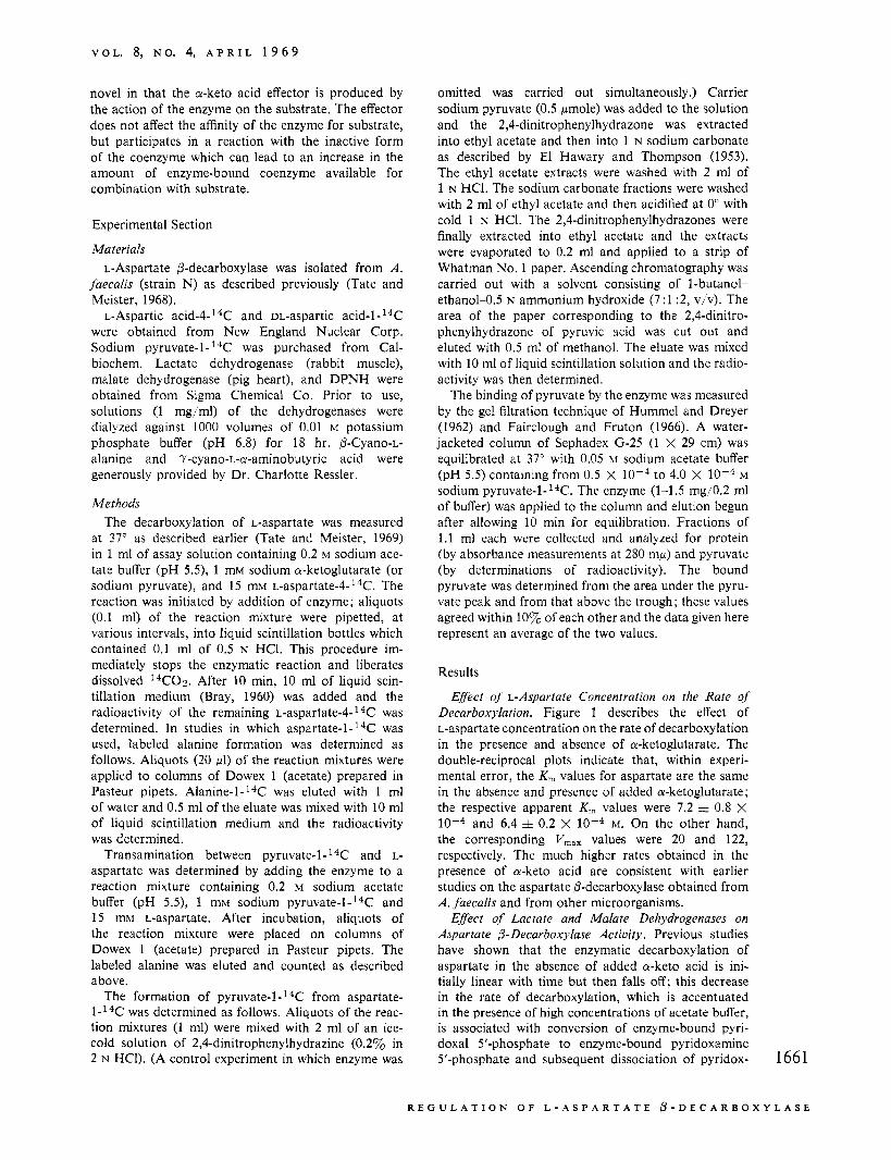
Effect of L-Aspartate Concentration on the Rate of Decarboxylation. Figure 1 describes the effect of L-aspartate concentration on the rate of decarboxylation in the presence and absence of a-ketoglutarate. The double-reciprocal plots indicate that, within experi- mental error, the K, values for aspartate are the same in the absence and presence of added a-ketoglutarate ; the respective apparent K, values were 7.2 =!z 0.8 X
and 6.4 & 0.2 X lo-* M. On the other hand, the corresponding V,,, values were 20 and 122, respectively. The much higher rates obtained in the presence of a-keto acid are consistent with earlier studies on the aspartate P-decarboxylase obtained from A. jhecalis and from other microorganisms.
Effect of Lactate and Malate Dehydrogenases on Aspartate p- Decarboxylase Activity. Previous studies have shown that the enzymatic decarboxylation of aspartate in the absence of added a-keto acid is ini- tially linear with time but then falls off; this decrease in the rate of decarboxylation, which is accentuated in the presence of high concentrations of acetate buffer, is associated with conversion of enzyme-bound pyri- doxal 5'-phosphate t o enzyme-bound pyridoxamine 5'-phosphate and subsequent dissociation of pyridox- 166 1
R E G U L A T I O N O F L - A S P A R T A T E 0 - D E C A R B O X Y L A S E
B I O C H E M I S T R Y
7 I I J I I I I I I I J I I I I
'2 4 6 8 IO 12 14 [Aspartate, rnM]
FIGURE 1 : Effect of L-aspartate concentration on the rate of decarboxylation in the presence and absence of a-keto- glutarate. Curve 1: a-ketoglutarate (1 mM) present; curve 2: a-ketoglutarate absent. The reaction mixtures contained enzyme (20 pg'ml), sodium acetate buffer (pH 5.5; 0.2 M), sodium a-ketoglutarate (curve 1 ; 1 mM), and L-aspartate as indicated; 37'. The inset gives the corresponding double- reciprocal plots of the data.
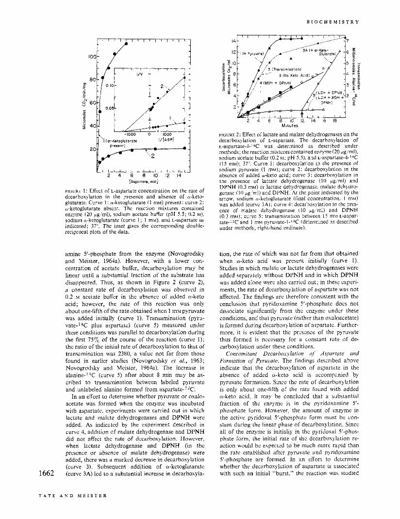
amine 5'-phosphate from the enzyme (Novogrodsky and Meister, 1964a). However, with a lower con- centration of acetate buffer, decarboxylation may be linear until a substantial fraction of the substrate has disappeared. Thus, as shown in Figure 2 (curve 2) , a constant rate of decarboxylation was observed in 0.2 M acetate buffer in the absence of added a-keto acid; however, the rate of this reaction was only about one-fifth of the rate obtained when 1 mM pyruvate was added initially (curve 1). Transamination (pyru- vate-I4C plus aspartate) (curve 5) measured under these conditions was parallel to decarboxylation during the first 757" of the course of the reaction (curve 1); the ratio of the initial rate of decarboxylation to that of transamination was 2380, a value not far from those found in earlier studies (Novogrodsky et af., 1963; Novogrodsky and Meister, 1964a). The increase in alanine-I4C (curve 5) after about 8 min may be as- cribed to transamination between labeled pyruvate and unlabeled alanine formed from aspartate- 2C.
In an effort to determine whether pyruvate or oxalo- acetate was formed when the enzyme was incubated with aspartate, experiments were carried out in which lactate and malate dehydrogenases and DPNH were added. As indicated by the experiment described in curve 4, addition of malate dehydrogenase and DPNH did not affect the rate of decarboxylation. However, when lactate dehydrogenase and DPNH (in the presence or absence of malate dehydrogenase) were added, there was a marked decrease in decarboxylation (curve 3). Subsequent addition of a-ketoglutarate (curve 3A) led to a substantial increase in decarboxyla- 1662
1
8
6
4
2
2 4 6 8 IO I2 14 16 18 Minutes
FIGURE 2: Effect of lactate and malate dehydrogenases on the decarboxylation of L-aspartate. The decarboxylation of ~-aspartate-4-14C was determined as described under methods; the reaction mixtures contained enzyme (20 pg/ml), sodium acetate buffer (0.2 M ; pH 5.5); and ~-aspartate-4-l~C (15 mM); 37". Curve 1: decarboxylation in the presence of sodium pyruvate (1 mM); curve 2: decarboxylation in the absence of added a-keto acid; curve 3: decarboxylation in the presence of lactate dehydrogenase (10 pg/ml) and DPNH (0.3 mM) or lactate dehydrogenase, malate dehydro- genase (10 pg,/ml) and DPNH. At the point indicated by the arrow, sodium a-ketoglutarate (final concentration, 1 mM) was added (curve 3A); curve 4: decarboxylation in the pres- ence of malate dehydrogenase (10 pg,'ml) and DPNH (0.3 mM); curve 5 : transamination between 15 mM L-aspar- tate-12C and 1 mM pyruvate-l-'4C (determined as described under methods; right-hand ordinate).
tion, the rate of which was not far from that obtained when a-keto acid was present initially (curve 1). Studies in which malate or lactate dehydrogenases were added separately without DPNH and in which DPNH was added alone were also carried out; in these experi- ments, the rate of decarboxylation of aspartate was not affected. The findings are therefore consistent with the conclusion that pyridoxamine 5'-phosphate does not dissociate significantly from the enzyme under these conditions, and that pyruvate (rather than oxaloacetate) is formed during decarboxylation of aspartate. Further- more, it is evident that the presence of the pyruvate thus formed is necessary for a constant rate of de- carboxylation under these conditions.
Concomitant Decarboxylation of Aspartate and Formation of Pyrucate. The findings described above indicate that the decarboxylation of aspartate in the absence of added a-keto acid is accompanied by pyruvate formation. Since the rate of decarboxylation is only about one-fifth of the rate found with added a-keto acid, it may be concluded that a substantial fraction of the enzyme is in the pyridoxamine 5' - phosphate form. However, the amount of enzyme in the active pyridoxal 5'-phosphate form must be con- stant during the linear phase of decarboxylation. Since all of the enzyme is initially in the pyridoxal 5'-phos- phate form, the initial rate of the decarboxylation re- action would be expected to be much more rapid than the rate established after pyruvate and pyridoxamine S'-phosphate are formed. In an effort to determine whether the decarboxylation of aspartate is associated with such an initial "burst," the reaction was studied
T A T E A N D M E I S T E R
V O L . 8, N O . 4, A P R I L 1 9 6 9
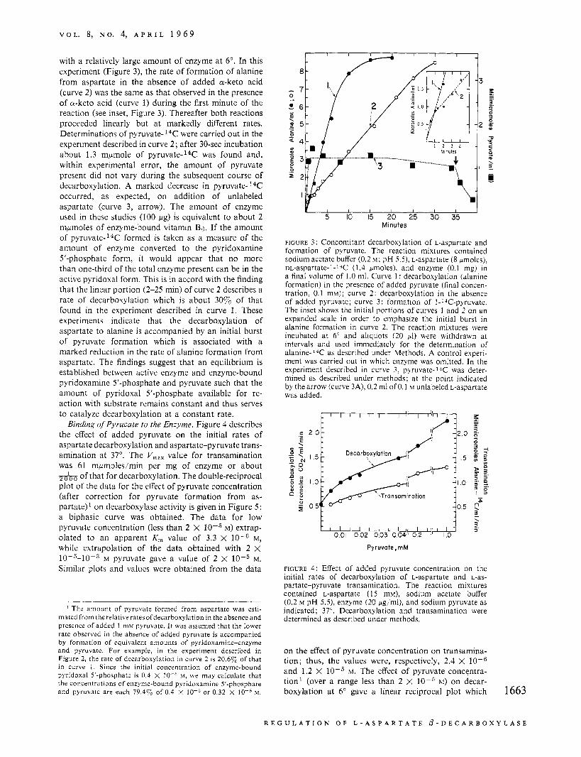
with a relatively large amount of enzyme at 6". In this experiment (Figure 3), the rate of formation of alanine from aspartate in the absence of added a-keto acid (curve 2 ) was the same as that observed in the presence of a-keto acid (curve 1) during the first minute of the reaction (see inset, Figure 3). Thereafter both reactions proceeded linearly but a t markedly different rates. Determinations of pyruvate- 14C: were carried out in the experiment described in curve 2 ; after 30-sec incubation about 1.3 mpmole of pyruvate-14C was found and, within experimental error, the amount of pyruvate present did not vary during the subsequent course of decarboxylation. A marked decrease in p y r ~ v a t e - ~ ~ C occurred, as expected, on addition of unlabeled aspartate (curve 3, arrow). The amount of enzyme used in these studies (100 pg) is equivalent to about 2 mpmoles of enzyme-bound vitamin B6. If the amount of p y r ~ v a t e - ' ~ C formed is taken as a measure of the amount of enzyme converted to the pyridoxamine 5'-phosphate form, it would appear that no more than one-third of the total enzyme present can be in the active pyridoxal form. This is in accord with the finding that the linear portion (2-25 min) of curve 2 describes a rate of decarboxylation which is about 30y0 of that found in the experiment described in curve 1. These experiments indicate that the decarboxylation of aspartate to alanine is accompanied by an initial burst of pyruvate formation which is associated with a marked reduction in the rate of alanine formation from aspartate. The findings suggest that an equilibrium is established between active enzyme and enzyme-bound pyridoxamine 5'-phosphate and pyruvate such that the amount of pyridoxal 5'-phosphate available for re- action with substrate remains constant and thus serves to catalyze decarboxylation at a constant rate. Binding of Pyrucate to the Enzyme. Figure 4 describes
the effect of added pyruvate on the initial rates of aspartate decarboxylation and aspartate-pyruvate trans- amination at 37". The V,,, value for transamination was 61 mpmoles/min per mg of enzyme or about _--_ to of that for decarboxylation. The double-reciprocal plot of the data for the effect of pyruvate concentration (after correction for pyruvate formation from as- partate)' on decarboxylase activity is given in Figure 5 ; a biphasic curve was obtained. The data for low pyruvate concentration (less than 2 X IO-5 M) extrap- olated to an apparent K, value of 3.3 X M, while extrapolation of the data obtained with 2 X 10⁻⁵-10-3 M pyruvate gave a value of 2 x 10⁻⁵ M.
Similar plots and values were obtained from the data
The amount of pyruvate formed from aspartate was esti- matedfrom therelativeratesofdecarboxylation in the absence and presence of added 1 mM pyruvate. It was assumed that the lower rate observed in the absence of added pyruvate is accompanied by formation of equivalent amounts of pyridoxamine-enzyme and pyruvate. For example, in the experiment described in Figure 2 , the rate of decarboxylation i n curve 2 is 20.67, of that in curve 1 . Since the initial concentration of enzyme-bound pyridoxal 5'-phosphate is 0.4 X M, we may calculate that the concentrations of enzyme-bound pyridoxamine 5'-phosphate and pyruvate are each 79.47, of 0.4 X or 0.32 x hi.
1 I I I I c P 1
Y I I I I I I I
5 IO 15 20 25 30 35 Minutes
FIGURE 3 : Concomitant decarboxylation of L-aspartate and formation of pyruvate. The reaction mixtures contained sodium acetate buffer (0.2 M ; pH 5.5). L-aspartate (8 pmoles), m-aspartate-l-l*C (1.4 pmoles), and enzyme (0.1 mg) i n a final volume of 1.0 ml. Curve 1 : decarboxylation (alanine formation) in the presence of added pyruvate (final concen- tration, 0.1 mM); curve 2: decarboxylation in the absence of added pyruvate; curve 3 : formation of l-*C-pyruvate. The inset shows the initial portions of curves 1 and 2 on an expanded scale in order to emphasize the initial burst in alanine formation in curve 2. The reaction mixtures were incubated at 6" and aliquots (20 p1) were withdrawn at intervals and used immediately for the determination of alanine-14C as described under Methods. A control experi- ment was carried out in which enzyme was omitted. In the experiment described in curve 3, pyruvate-l*C was deter- mined as described under methods; at the point indicated by the arrow (curve 3A), 0.2 ml of 0.1 M unlabeled L-aspartate was added.
c I
0 5 &Transominat ion
FIGURE 4: Effect of added pyruvate concentration on the initial rates of decarboxylation of L-aspartate and L-as- partate-pyruvate transamination. The reaction mixtures contained L-aspartate (15 mM), sodium acetate buffer (0.2 M pH 5 . 9 , enzyme (20 pg!ml), and sodium pyruvate as indicated; 37". Decarboxylation and transamination were determined as described under methods.
on the effect of pyruvate concentration on transamina- tion; thus, the values were, respectively, 2.4 X and 1.2 X 10⁻⁵ M. The effect of pyruvate concentra- tion' (over a range less than 2 x 10⁻⁵ M) on decar- boxylation at 6" gave a linear reciprocal plot which 1663
R E G U L A T I O N O F L - A S P A R T A T E @ - D E C A R B O X Y L A S E
B I O C H E M I S T R Y
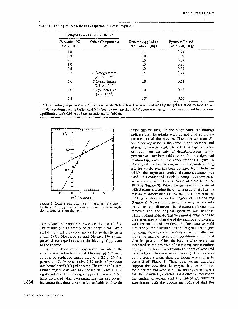
TABLE I : Binding of Pyruvate to L-Aspartate 0-Decarboxylase."
Composition of Column Buffer
P y r ~ v a t e - ~ ~ C Other Components Enzyme Applied to Pyruvate Bound (M x 104) (M) the Column (mg) (moles/50,000 g)
4.0 1.4 0.93 2.5 1 .o 0.90 2.5 1.5 0.88 2.0 1 .o 0.81 0.5 1 .o 0.39 2.5 a-Ketoglutarate 1.5 0.49
2.0 @-Cyanoalanine 1.0 0.74
2.0 @-Cyanoalanine 1 .o 0.62
2.5 1 ,3a 0.61
(2.5 x 10⁻⁴)
(2.5 x 10⁻⁴)
(5 x 10⁻⁴)
a The binding of p y r ~ v a t e - l - ' ~ C to L-aspartate /3-decarboxylase was measured by the gel filtration method at 37" in 0.05 M sodium acetate buffer (pH 5.5) (see the text, methods). Apoenzyme ($20,~ = 19s) was applied to a column equilibrated with 0.05 M sodium acetate buffer (pH 6).
I I I I 1 111 I I I I 1 I I I I 1 I I I I I I I I I I I I I J
-0.5 0 0.5 1.0 1.5
107 [PYRUVATE]
FIGURE 5 : Double-reciprocal plot of the data (of Figure 4) for the effect of pyruvate concentration on the decarboxyla- tion of aspartate (see the text).
extrapolated to an apparent K , value of 2.4 X loe6 M. The relatively high affinity of the enzyme for a-keto acid demonstrated by these and earlier studies (Meister et a/., 1951; Novogrodsky and Meister, 1964a) sug- gested direct experiments on the binding of pyruvate to the enzyme.
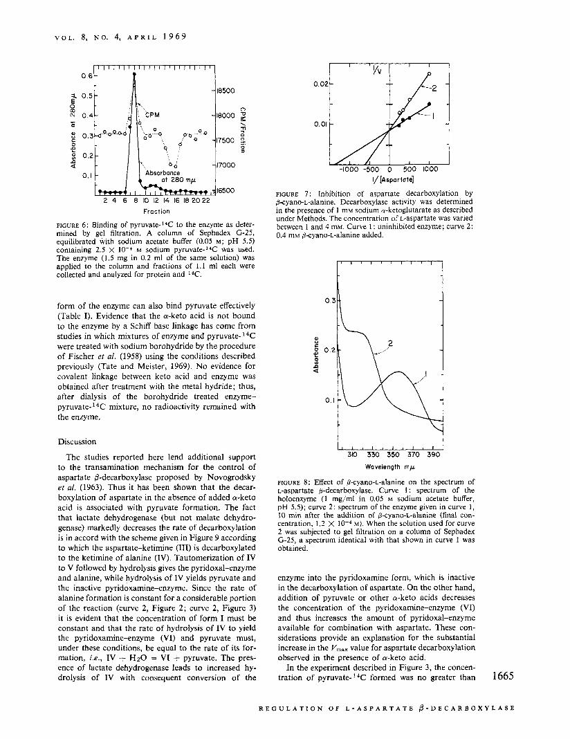
Figure 6 describes an experiment in which the enzyme was subjected to gel filtration at 37" on a column of Sephadex equilibrated with 2.5 x M p y r ~ v a t e - ' ~ C . In this study, 0.88 mole of pyruvate was bound per 50,000 g of enzyme. The results of several similar experiments are summarized in Table I. It is significant that the binding of pyruvate was substan- tially decreased when a-ketoglutarate was also present indicating that these a-keto acids probably bind to the 1664
same enzyme sites. On the other hand, the findings indicate that the a-keto acids do not bind at the as- partate site of the enzyme. Thus, the apparent K , value for aspartate is the same in the presence and absence of a-keto acid. The effect of aspartate con- centration on the rate of decarboxylation in the presence of 1 mM keto acid does not follow a sigmoidal relationship, even at low concentrations (Figure 1). Direct evidence that the enzyme has a separate binding site for a-keto acid has been obtained from studies in which the aspartate analog P-cyano-L-alanine was used. This compound is strictly competitive toward L- aspartate and exhibits a Ki value of close to 2.7 X
M (Figure 7). When the enzyme was incubated with @-cyano-L-alanine there was a prompt shift in the maximum absorbance at 358 mp to a spectrum ex- hibiting a shoulder in the region of 310-320 mp (Figure 8). When this form of the enzyme was sub- jected to gel filtration the @-cyano-L-alanine was removed and the original spectrum was restored. These findings indicate that 0-cyano-L-alanine binds to the L-aspartate binding site of the enzyme and interacts with enzyme-bound pyridoxal 5'-phosphate to yield a relatively stable ketimine on the enzyme. The higher homolog, 7-cyano-L-a-aminobutyric acid, neither in- hibits the enzyme under these conditions nor does it alter its spectrum. When the binding of pyruvate was measured in the presence of saturating concentrations of P-cyano-L-alanine, a substantial amount of keto acid became bound to the enzyme (Table I). The spectrum of the enzyme under these conditions was similar to curve 2 of Figure 8. These observations therefore support the view that the enzyme has separate sites for aspartate and keto acid. The findings also suggest that the vitamin Bg cofactor is not directly involved in the binding of a-keto acid and indeed gel filtration experiments with the apoenzyme indicated that this
T A T E A N D M E I S T E R
V O L . 8, N O . 4, A P R I L 1 9 6 9
i E 0 m N .I-
o 0 0 f! n a u)
Fraction
FIGURE 6: Binding of pyruvate-14C to the enzyme as deter- mined by gel filtration. A column of Sephadex (3-25, equilibrated with sodium acetate buffer (0.05 M ; pH 5.5) containing 2.5 X 10⁻⁴ M sodium pyruvate-14C was used. The enzyme (1.5 mg in 0.2 ml of the same solution) was applied to the column and fractions of 1 . 1 ml each were collected and analyzed for protein and I4C.
0.02 -
0.01 -
-1000 -500 0 500 1000 I/ [Aspa r tote]
FIGURE 7 : Inhibition of aspartate decarboxylation by 0-cyano-L-alanine. Decarboxylase activity was determined in the presence of 1 mM sodium a-ketoglutarate as described under Methods. The concentration of L-aspartate was varied between 1 and 4 mM. Curve 1: uninhibited enzyme; curve 2: 0.4 mM 0-cyano-L-alanine added.
form of the enzyme can also bind pyruvate effectively (Table I). Evidence that the a-keto acid is not bound to the enzyme by a Schiff base linkage has come from studies in which mixtures of enzyme and pyruvate-14C were treated with sodium borohydride by the procedure of Fischer et al. (1958) using the conditions described previously (Tate and Meister, 1969). N o evidence for covalent linkage between keto acid and enzyme was obtained after treatment with the metal hydride; thus, after dialysis of the borohydride treated enzyme- pyruvate- 14C mixture, n o radioactivity remained with the enzyme.
t Discussion
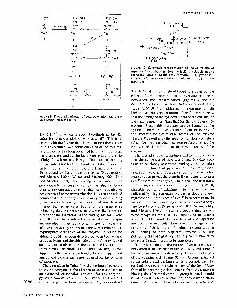
The studies reported here lend additional support to the transamination mechanism for the control of aspartate 0-decarboxylase proposed by Novogrodsky et al. (1963). Thus it has been shown that the decar- boxylation of aspartate in the absence of added a-keto acid is associated with pyruvate formation. The fact that lactate dehydrogenase (but not malate dehydro- genase) markedly decreases the rate of decarboxylation is in accord with the scheme given in Figure 9 according to which the aspartate-ketimine (111) is decarboxylated to the ketimine of alanine (IV). Tautomerization of IV to V followed by hydrolysis gives the pyridoxal-enzyme and alanine, while hydrolysis of IV yields pyruvate and the inactive pyridoxamine-enzyme. Since the rate of alanine formation is constant for a considerable portion of the reaction (curve 2, Figure 2; curve 2, Figure 3) it is evident that the concentration of form I must be constant and that the rate of hydrolysis of IV to yield the pyridoxamine-enzyme (VI) and pyruvate must, under these conditions, be equal t o the rate of its for- mation, i .e., IV + H2O = VI + pyruvate. The pres- ence of lactate dehydrogenase leads to increased hy- drolysis of IV with consequent conversion of the
u I I 111111111111
310 330 350 370 390 Wavelength mp
FIGURE 8: Effect of 0-cyano-L-alanine on the spectrum of L-aspartate @-decarboxylase. Curve 1 : spectrum of the holoenzyme (1 mg/ml in 0.05 M sodium acetate buffer, pH 5.5); curve 2: spectrum of the enzyme given in curve 1 , 10 min after the addition of 6-cyano-L-alanine (final con- centration, 1.2 X M). When the solution used for curve 2 was subjected to gel filtration on a column of Sephadex (3-25, a spectrum identical with that shown in curve 1 was obtained.
enzyme into the pyridoxamine form, which is inactive in the decarboxylation of aspartate. On the other hand, addition of pyruvate or other a-keto acids decreases the concentration of the pyridoxamine-enzyme (VI) and thus increases the amount of pyridoxal-enzyme available for combination with aspartate. These con- siderations provide an explanation for the substantial increase in the Y,,, value for aspartate decarboxylation observed in the presence of a-keto acid.
In the experiment described in Figure 3, the concen- tration of pyruvate-14C formed was no greater than 1665
R E G U L A T I O N O F L - A S P A R T A T E 0 - D E C A R B O X Y L A S E
B I O C H E M I S T R Y
1 y s - N ~ ?$ p C O 0 - 1 C ~ N - C H C O O - t I II 7 Il l
ENZ
ASPARTATE
f 'ITE
EN2
FIGURE 9: Proposed pathways of decarboxylation and pyru- vate formation (see the text).
1.5 X M, which is about two-thirds of the K, value for pyruvate (2.4 X M, a t 6"). This is in accord with the finding that the rate of decarboxylation in this experiment was about one-third of the maximal rate. Evidence has been presented here that the enzyme has a separate binding site for a-keto acid and that its affinity for a-keto acid is high. The maximal binding of pyruvate is not far from 1 mole/50,000 g of enzyme; earlier studies indicate that close to 1 mole of vitamin BG is bound by this amount of enzyme (Novogrodsky and Meister, 1964a; Wilson and Meister, 1966; Tate and Meister, 1969). The binding of pyruvate to the /3-cyano-L-alanine-enzyme complex is slightly lower than to the untreated enzyme; this may be related to occurrence of some transamination between the added amino acid and the enzyme or possibly to some binding of /3-cyano-L-alanine to the a-keto acid site. It is of interest that pyruvate is bound by the apoenzyme indicating that the presence of vitamin BG is not re- quired for the formation of the binding site for a-keto acid. It would be of interest to learn whether the apo- enzyme also has an intact binding site for aspartate. We have previously shown that the N-methylpyridoxal 5'-phosphate derivative of the enzyme, in which no aldimine bond has been detected between the e-amino group of lysine and the aldehyde group of the pyridoxal analog, can catalyze both the decarboxylase and the transaminase reactions (Tate and Meister, 1969). Apparently then, a covalent bond between the pyridoxal analog and the enzyme is not required for the binding of substrate.
The data given in Table I on the binding of pyruvate to the holoenzyme in the absence of aspartate lead to an estimated dissociation constant for the enzyme- pyruvate complex of about 8 X M . This value is substantially higher than the apparent K,, values (about 1666
a-KETO ACID S I T E 4
0 coo- l'/ R-C
PYRIDOXAL-P S ITE
FIGURE 10: Schematic representation of the active site of aspartate pdecarboxylase (see the text); the double arrows represent types of Schiff base formation: ( I ) pyridoxal- enzyme, (2) pyridoxamine-keto acid, and ( 3 ) pyridoxal- aspartate.
3 X M) for pyruvate obtained in studies on the effects of low concentrations of pyruvate on decar- boxylation and transamination (Figures 4 and 5 ) ; on the other hand, it is closer to the extrapolated K , value (2 X 10⁻⁵ M) obtained in experiments with higher pyruvate concentrations. The findings suggest that the affinity of the pyridoxal form of the enzyme for pyruvate is much less than that for the pyridoxamine- enzyme. Presumably pyruvate can be bound by the pyridoxal form, the pyridoxamine form, or by any of the intermediate Schiff base forms of the enzyme (Figure 9) as well as by the apoenzyme. Thus, the values of K, , for pyruvate obtained here probably reflect the resultant of the affinities of the several forms of the enzyme.
The present and earlier findings lead to the conclusion that the active site of aspartate P-decarboxylase con- tains three closely associated binding sites, i . e . , sites for the attachment of pyridoxal 5'-phosphate, aspar- tate, and a-keto acid. These must be situated in such a manner as to permit the vitamin Bcj cofactor to form a Schiff base with the enzyme, a-keto acid, and aspartate. In the diagrammatic representation given in Figure 10, plausible points of attachment to the enzyme are indicated by single arrows; the darker double arrows represent the three types of Schiff base formation. In view of the broad specificity of aspartate P-decarboxy- lase fora-keto acids (Meister el al., 1951 ; Novogrodsky and Meister, 1964a), it seems probable that the en- zyme recognizes the COCOO- moiety of the a-keto acids. The likelihood that a-keto acid and aspartate are bound in relatively close proximity suggests the possibility of designing a bifunctional reagent capable of attaching to both respective enzyme sites. The possibility that aspartate can form a Schiff base with pyruvate directly must also be considered.
It is evident that in the course of aspartate decar- boxylation in the absence of added a-keto acid some of the pyruvate formed by decarboxylation and hydrolysis of the ketimine (111; Figure 9) must become attached to the a-keto acid binding site. It is possible that the residual three-carbon chain moiety of the Schiff base formed by decarboxylation detaches from the aspartate binding site after the P-carboxyl group is lost. I t would be of interest to learn whether the three-carbon chain moiety of this Schiff base attaches to the a-keto acid
T A T E A N D M E I S T E R
V O L . 8, N O . 4, A P R I L 1 9 6 9
binding site ; thus, the subsequent transformation of IV t o VI might provide a mechanism for the formation of enzyme-bound pyruvate without the intermediate formation of free pyruvate. Transformation o f 1V-t V -+ I at this site would lead to alanine formation. The affinity of the enzyme for L-alanine and other ~ - a - amino acids is relatively low as compared with L-
aspartate (Novogrodsky and Meister, 1964a). It would be of interest to learn whether the ability of the enzyme to interact with a variety of L-amino acids is related to the low specificity of the a-keto acid site. This site might then be able to combine with both a-keto acids and a-amino acids other than L-aspartate and closely related derivatives (such as P-cyano-L- alanine and L-cysteine sulfinite; Soda et al., 1964). It may be pertinent t o note that the glutamate- aspartate transaminases exhibit higher affinities for a-keto acids than for a-amino acids (Fasella, 1968).
Chibata et al. (1968) have recently concluded that the activation of the aspartate P-decarboxylase of Pseudo- monas dacunhae by a-ketoglutarate is not due to a transamination mechanism but rather to an “allosteric” one. Although these workers concluded that the mech- anism proposed by Novogrodsky et a/. (1963) could not explain the observation that a-ketoglutarate was more effective as an activator than pyridoxal 5’-phosphate, it is evident that under conditions in which there is rela- tively little tendency for pyridoxamine 5’-phosphate to dissociate from the enzyme, a-keto acid will activate more than pyridoxal 5’-phosphate. Indeed in the orig- inal studies of Novogrodsky and Meister (1964a) and in the present work, a-ketoglutarate produced more activation than did pyridoxal 5’-phosphate. Acti- vation by pyridoxal 5’-phosphate can be observed only under conditions in which pyridoxamine 5’-phosphate dissociates from the enzyme. It is of interest that Chibata et ai. (1968) observed that the Pseudomonas enzyme catalyzes transamination between a-ketoglu- tarate and L-aspartate and that the ratio of the rates of decarboxylation and transamination was about the same as is found for the enzyme from A . faecalis. However, these investigators reported that the curve describing the relationship between decarboxylase activity (in the absence of added a-keto acid) and aspartate concentra- tion was sigmoidal; they also reported K , values of 0.1 and 5.5 X M for aspartate in the absence and presence, respectively, of a-ketoglutarate. These values are quite different from those obtained in the present study (7.2 X and 6.4 X M, respectively), which were derived from curves that are not sigmoidal. It may be pertinent to note that Chibata et al. (1968) added large amounts of bovine serum albumin to their reaction mixtures. Bovine serum albumin has been found to bind such compounds such as pyruvate, aspartate, and citrate and the presence of these com- pounds in commercially available samples of this protein has been reported (Hanson and Ballard, 1968). Further work is needed to determine whether the differ- ences between the data reported for the Pseudomonas enzyme and the enzyme used in the present work are related to methodological factors or to significant species differences.
The present studies indicate that the isolated A . faecalis enzyme does not decarboxylate aspartate a t a maximal rate. The rate of decarboxylation is very sensitive to the concentration of a-keto acid; thus, it is increased about sixfold in the presence of 4 X 10⁻⁵ M
pyruvate a t 37” and about fourfold at 6”. Furthermore, activity depends crucially upon the presence of pyru- vate; removal of pyruvate results in a marked decrease of decarboxylase activity. Thus, the activity of aspartate P-decarboxylase would be expected to be subject to highly sensitive regulation in an intracellular environ- ment containing other enzymes capable of increasing or decreasing the concentrations of pyruvate, a-keto- glutarate, and other a-keto acids.
The mechanism of regulation of this enzyme is novel in that the a-keto acid effector is produced by the enzyme’s action on the substrate. The effector acts to increase the amount of active enzyme-bound cofactor. The a-keto acid does not alter the affinity of the enzyme for substrate, but it participates in a chemical reaction with the enzyme-bound coenzyme. The enzyme is composed of subunits, which have been demonstrated by ultracentrifugal and electron microscope studies (Bowers et al., 1968). Although we have not specifically sought to determine whether a-keto acids induce a conformational change in the enzyme, it is not necessary to postulate a conformational change to explain the effect of a-keto acids. The a-keto acid activators are not close chemical analogs of the substrate and they are bound at a separate site; in these respects aspartate @-decarboxylase fulfills both the original definition of an allosteric enzyme (Monod et a/., 1963) and a more recent one (Stadtman, 1966). Nevertheless, the enzyme differs substantially in its control phenomena from many other enzymes that have been designated as allosteric. The present findings do not exclude the possibility that the activity of this enzyme may be influenced by other phenomena which may be allosteric in a more conventional sense. For example, the pro- tective effect of 4’-deoxypyridoxine 5’-phosphate on aspartate &decarboxylase indicates that the binding of this vitamin BG analog to some enzyme sites exerts an effect on the binding of coenzyme at other sites (Novo- grodsky and Meister, 1964b).
The ability of the enzyme t o catalyze both decar- boxylation and transamination raises the question as t o whether, in the course of evolution, the enzyme arose as a modification of a transaminase. On the other hand, it may also be speculated that the reverse situation occurred. Thus, it is evident that the decarboxylation pathway involves formation of an alanine-ketimine intermediate (IV, Figure 9), which is readily susceptible to both tautomerization and hydrolysis. Since the latter event leads to inactivation of the enzyme, development of a binding site on the enzyme for a-keto acid may have been favored since it offers a mechanism for preventing inactivation and at the same time for controlling enzyme activity.
Acknowledgment The authors are indebted to Dr. Daniel Wellner for
his valuable and stimulating discussions of this research. 1667
R E G U L A T I O N O F L - A S P A R T A T E f i - D E C A R B O X Y L A S E
B I O C H E M I S T R Y
We thank Dr. Charlotte Ressler for generous gifts of 0-cyano-L-alanine and 7-cyano-L-a-aminobutyric acid.
References
Bowers, W. F., Czubaroff, V., and Haschemeyer, R. H . (1968), 156th National Meeting of the American Chemical Society, Atlantic City, N. J., Sept, Abstract 177.
Bray, G. A. (1960). Anal. Biochem. I, 279. CattanCo-Lacombe, J., Senez, J. C., and Beaumont, P.
(1958), Biochim. Biophys. Acta 30, 458. Chibata, I. , Kakimoto, T., Kato, J., Shibatani, T.,
and Nishimura, N. (1967), Biochem. Biophys. Res. Commun. 26, 662.
Chibata, J. , Kakimoto, T., Kato, J., Shibatani, T., and Nishimura, N. (1968), Biochem. Biophys. Res. Commun. 32, 375.
Crawford, L. V. (1958), Biochem. J . 68, 221. El Hawary, M. F. S., and Thompson, R. H. S. (1953),
Biochem. J . 53, 340. Fairclough, G. F., and Fruton, J. S. (1966), Biochem-
istry 5, 673. Fasella, P. (1968), Pyridoxal Catalysis; Enzymes and
Model Systems, New York, N. Y., Interscience, pp 1-31.
Fischer, E. H., Kent, A. B., Snyder, E. R., and Krebs,
Hanson, R. W., and Ballard, F. J. (1968), J . Lipid
Hummel, J. P., and Dreyer, W. J. (1962), Biochim.
Meister, A., Sober, H . A,, and Tice, S. V. (1951),
Monod, J., Wyman, J., and Jacob, F. (1963), J . Mol.
Novogrodsky, A., and Meister, A. (1964a), J . Biol.
Novogrodsky, A., and Meister, A. (1964b), Biochim.
Novogrodsky, A., Nishimura, J. S., and Meister, A.
Soda, K., Novogrodsky, A., and Meister, A. (1964),
Stadtman, E. R. (1966), Aduan. Enzymol. 28, 41. Tate, S. S., and Meister, A. (1968), Biochemistry 7,
Tate, S . S., and Meister, A. (1969), Biochemistry, 8,
Wilson, E. M. (1963), Biochim. Biophys. Acta 67, 345. Wilson, E. M., and Meister, A. (1966), Biochemistry
E. G. (1958), J . Am. Chem. SOC. 80, 2906.
Res. 9, 667.
Biophys. Acta 63, 530.
J . Biol. Chem. 289, 577, 591.
Bid . 6, 306.
Ciiem. 239, 879.
Biophys. Acta 85, 170.
(1963), J . Biol. Chem. 238, PC1903.
Biochemistry 3, 1450.
3240.
1056.
5, 1166.
1668
T A T E A N D M E I S T E R