Recombinant Schizophyllum commune α glucuronidase...
53
Recombinant Schizophyllum commune α-glucuronidase expression and production in the methylotrophic yeast Pichia pastoris Master of Science Thesis HORMOZ NOORMOHAMMADIAN Department of Chemical and Biological Engineering CHALMERS UNIVERSITY OF TECHNOLOGY Gothenburg, Sweden 2012
Transcript of Recombinant Schizophyllum commune α glucuronidase...
Recombinant Schizophyllum commune α-glucuronidase
expression and production in the methylotrophic yeast
Pichia pastoris
Master of Science Thesis
HORMOZ NOORMOHAMMADIAN
Department of Chemical and Biological Engineering
CHALMERS UNIVERSITY OF TECHNOLOGY
Gothenburg, Sweden 2012
i
THESIS FOR THE DEGREE OF MASTER OF SCIENCE in BIOTECNOLOGY
Recombinant Schizophyllum commune α-glucuronidase expression and production in
the methylotrophic yeast Pichia pastoris
HORMOZ NOORMOHAMMADIAN
Under supervision of
Prof. Lisbeth Olsson
Dr. George Anasontzis
&
Hampus Sunner
Industrial Biotechnology
Department of Chemical and Biological engineering
CHALMERS UNIVERSITY OF TECHNOLOGY
Gothenburg, Sweden 2012

ii
Recombinant Schizophyllum commune α-glucuronidase expression and production in
the methylotrophic yeast Pichia pastoris
HORMOZ NOORMOHAMMADIAN
© HORMOZ NOORMOHAMMADIAN
Industrial Biotechnology
Department Chemical and Biological Engineering
Chalmers University of Technology
SE-41296 Gothenburg
Sweden
Cover illustration: Schematic representation of the project from gene optimization,
expression cassette construction and fermentation.
Gothenburg, Sweden 2012
iii
To my family……
‘‘There can be no progress without head-on confrontation’’
(Christopher Hitchens)
iv
Recombinant Schizophyllum commune α-glucuronidase expression and production in the
methylotrophic yeast Pichia pastoris
HORMOZ NOORMOHAMMADIAN
Industrial biotechnology
Department Chemical and Biological Engineering
Chalmers University of Technology
Göteborg, Sweden
Abstract
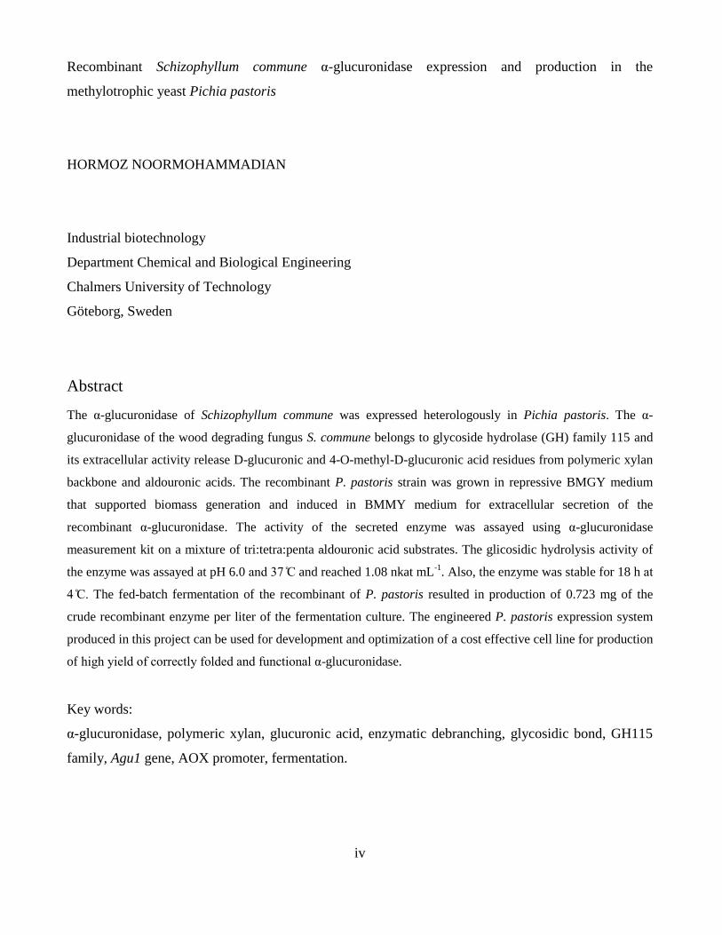
The α-glucuronidase of Schizophyllum commune was expressed heterologously in Pichia pastoris. The α-
glucuronidase of the wood degrading fungus S. commune belongs to glycoside hydrolase (GH) family 115 and
its extracellular activity release D-glucuronic and 4-O-methyl-D-glucuronic acid residues from polymeric xylan
backbone and aldouronic acids. The recombinant P. pastoris strain was grown in repressive BMGY medium
that supported biomass generation and induced in BMMY medium for extracellular secretion of the
recombinant α-glucuronidase. The activity of the secreted enzyme was assayed using α-glucuronidase
measurement kit on a mixture of tri:tetra:penta aldouronic acid substrates. The glicosidic hydrolysis activity of
the enzyme was assayed at pH 6.0 an and reached 1.08 nkat mL-1
. Also, the enzyme was stable for 18 h at
The fed-batch fermentation of the recombinant of P. pastoris resulted in production of 0.723 mg of the
crude recombinant enzyme per liter of the fermentation culture. The engineered P. pastoris expression system
produced in this project can be used for development and optimization of a cost effective cell line for production
of high yiel of correctly fol e an functional α-glucuronidase.
Key words:
α-glucuronidase, polymeric xylan, glucuronic acid, enzymatic debranching, glycosidic bond, GH115
family, Agu1 gene, AOX promoter, fermentation.
vi
1. Table of Contents 1. Introduction ....................................................................................................................... 1
2. Background ........................................................................................................................ 3
2.1 Hemicellulose ............................................................................................................. 3
2.2 Application of Xylan films ......................................................................................... 3
2.3 Structural modification of xylan ................................................................................. 4
2.3.1 Chemical modification ........................................................................................ 4
2.3.2 Enzymatic modification ....................................................................................... 5
2.4 Properties of α-Glucuronidase .................................................................................... 6
2.4.1 The GH6 family α-glucuronidases .................................................................... 6
2.4.2 The GH115 family α-glucuronidase .................................................................... 7
2.5 P. pastoris expression strains ...................................................................................... 9
2.5.1 AOX pathway ...................................................................................................... 9
2.5.2 Effects of the Mut+ and Mut
S Phenotypes ......................................................... 10
2.5.3 Expression vector .............................................................................................. 11
2.5.4 The signal sequence ........................................................................................... 11
2.5.5 Culture condition for expression ....................................................................... 12
3. Materials and Methods .................................................................................................... 13
3.1 Strains and vectors .................................................................................................... 13
3.2 Media ........................................................................................................................ 14
3.2.1 Lysogeny Broth (LB medium) .......................................................................... 14
3.2.2 Yeast Extract Peptone Dextrose (YPD medium) .............................................. 14
3.2.3 Yeast Extract Pepton Dextrose with Sorbitol (YPDS medium) ........................ 15
3.2.4 Buffered Glycerol-complex Medium (BMGY medium) .................................. 15
3.2.5 Buffered Methanol-complex Medium (BMMY medium)................................. 15
3.2.6 Fermentation basal salts medium ...................................................................... 16

vii
3.2.7 PTM1 trace salts ................................................................................................. 17
3.3 Transformation to E. coli .......................................................................................... 17
3.4 Plasmid extraction ..................................................................................................... 17
3.5 Construction of the expression cassette .................................................................... 18
3.5.1 Enzymatic restriction ......................................................................................... 18
3.5.2 Ligation .............................................................................................................. 18
3.6 P. pastoris transformation ......................................................................................... 18
3.6.1 Transformation by electroporation .................................................................... 18
3.6.2 Screening for positive transformants ................................................................. 18
3.6.3 Glycerol stock .................................................................................................... 19
3.7 Fermenter preparation ............................................................................................... 19
3.7.1 Glycerol Batch phase ......................................................................................... 19
3.7.2 Glycerol fed-batch phase ................................................................................... 19
3.7.3 Methanol fed-batch phase .................................................................................. 19
3.8 Biochemical analyses ................................................................................................ 20
3.8.1 Bradford assay ................................................................................................... 20
3.8.2 α-glucuronidase assay ........................................................................................ 20
4. Results ............................................................................................................................. 21
4.1 Construction of expression cassette .......................................................................... 21
4.1.1 Propagation of the Agu1 gene ............................................................................ 21
4.1.2 Isolation of pPI Zα ........................................................................................ 21
4.1.3 Isolation of Agu1 from pUC57 .......................................................................... 21
4.1.4 Cloning of Agu1 into pPI Zα ......................................................................... 23
4.2 Integration of the expression cassette into P. pastoris SMD1168H ......................... 26
4.2.1 Linearization of the pPI Zα construct............................................................ 26
4.2.2 Transformation of the linearize pPI Zα construct into P. pastoris .............. 26
4.2.3 Screening for positive transformants ................................................................. 27
4.3 Small scale cultivation of Gluc ................................................................................. 28

viii
4.4 Large-scale production of a-glucuronidase ............................................................... 30
4.4.1 Batch phase ........................................................................................................ 30
4.4.2 Glycerol fed-batch phase ................................................................................... 30
4.4.3 Methanol induction phase .................................................................................. 30
5. Discussion ........................................................................................................................ 33
6. Future works .................................................................................................................... 37
7. Conclusion ....................................................................................................................... 37
8. Acknowledgment ............................................................................................................. 38
9. References ....................................................................................................................... 39
10. Appendix. ..................................................................................................................... 43
10.1 Preparing 1 % agarose gel for DNA analysis ........................................................ 43
10.2 Extraction of plasmid DNA from E. coli DH5α ................................................... 44
10.3 Fermentation of P. pastoris ................................................................................... 45
1
1. Introduction
Lignocellulosic biomass is of great interest for production of novel materials. Lignocellulosic
raw materials possess two prominent properties; renewability and biodegradability that make
them desirable over fossil resources, which are not guaranteed resources, for long term
utilization. A major portion of fossil resources is used for production of synthetic polymers
specially packaging materials. Massive consumption of the plastic packaging materials
contributes to waste accumulation in the environment, thus alternative raw materials should
be biodegradable as a desired property of novel packaging material. Apparently, renewable
lignocellulosic feedstock is a promising alternative resource for production of novel materials
with engineered properties and less environmental impacts due to their inherent
biodegradability property ( Gatenholm, et al., 2004).
The major components of the plant cell wall are cellulose, hemicelluloses, and lignin which
altogether form 70% of the biomass (Brink & Vries, 2011). Xylan is the most frequent
hemicellulose among hardwood species, and after cellulose it is the most second copious
polysaccharide available in nature (Saxenaa, et al., 2011). While cellulose is a
homopolysacchari e forme by β (1-4) glycosidic bonds between glucose units,
hemicelluloses are categorized into subgroups, such as xylan, xyloglucan, glucuronoxylan,
arabinoxylan, mannan and glucomannan, based on the sugars existing in the backbone of the
polysaccharide (Brink & Vries, 2011). Process bottlenecks for utilization of xylose
(monomer unit of xylan) as a fermentable sugar for biofuel applications (Olsson & Hahn-
Hägerdal, 1993) are important factors that make xylan available as a precursor biomaterial.
Previous studies have illustrated that the mechanical properties of xylan are adversely
affected by moisture (Dammström, et al., 2005) due to the glucuronic acid substituents
present on the backbone of xylan (Viikari, et al., 1994). There is a direct correlation between
mechanical properties of the xylan film and the amount of glucuronic acid residues attached
to the backbone of xylan that can adversely affect the water transmission rate, which is an
important property for many packaging film applications (Saxenaa, et al., 2011). Gatenholm,
et al. (2004), demonstrated that oxygen permeability of glucuronoxylan film is reduced by
increasing solubility.
2
The challenge of water absorption, which is caused by glucuronic side groups, could be
overcome by removing them from the polymeric xylan backbone. Although glucuronic side
groups of xylan could be decreased in the alkaline process in kraft pulping (Viikari, et al.,
1994), but during the cooking xylan chains are partly degraded and short chains of xylan
form, which is not desirable. The idea of releasing glucuronic acid residues and producing
intact xylan chains motivated this project for the development of environmentally benign
processes, as well as new products from forest industry. One way to overcome this challenge
is through the enzymatic cleavage of the glucuronic acid residues off the polymeric xylan
backbone. Tenkanen & Siika-aho (2000) found that α-glucuronidase from Schizophyllum
commune is capable of debranching glucuronic acid residues of polymeric glucuronoxylan of
softwood.
In the present work we want to heterologously express Agu1 gene, which enco es α-
glucuronidase from S. commune, in methylotrophic yeast P. pastoris. This α-glucuronidase is
active on polymeric glucuronoxylan. Initially we will construct the expression vector for the
transformation of the P. pastoris cells. Then the potential transformants will be screened for
the expression levels of active α-glucuronidase. Finally, the fermentation of the selected P.
pastoris transformant will be scaled up to produce high enzyme titers.
3
2. Background
2.1 Hemicellulose
Hemicelluloses are branched heteropolysaccharides containing hexoses (D-glucose, D-
mannose, D-galactose) and pentoses (D-xylose, and L-arabinose) on their backbone. The
backbone of hemicelluloses is usually decorated with D-galactose, D-xylose, L-arabinose and
D-glucuronic acid side groups (Brink & Vries, 2011). Hemicelluloses (like cellulose) are
called glycans due to O-glycosidic linkages of their monomers. The dominant glycan in the
primary cell wall of the higher plants is xyloglucan which consists of covalently β-1,4-linked
D-glucose units with D-xylose residues glycosidically bonded to the O-6 position of the
glucose monomer. The secondary cell wall, which prevails in the lignocellulosic biomass,
differs depending on the origin of plant. The secondary cell wall of hardwood contains
glucuronoxylan, whereas arabinoxylan is dominating in cereal plants. Despite hardwood and
cereal plants, the major hemicellulosic component in softwood is galactoglucomannans
(Buchanan, et al., 2000; De Vries & Visser, 2001). The main monosaccharide in the
backbone of the D-xylan is β-D-xylopyranosyl which are covalently (1→ )-linked together.
Depending on the source of xylan, some of the monomeric units carry (1→ )-linked α-L-
arabinofuranosyl, an (1→2)-linked 4-O-methyl-α-D-glucopyranosyl uranic acid (glucuronic
acid) substituents which are glycosidically attached to the backbone of the polymeric xylan.
Furthermore, feruloyl, acetyl and p-coumaroyl residues are the other categories of sugar
moieties bonded with the D-xylan backbone via ester linkage (Castanare, et al., 1995). The
composition of hemicelluloses occurring in the plant cell wall varies significantly between
different plant species. For example, a comparison between hard wood and cereal xylans
shows that D-glucuronic acid residues are the dominating branches linke to xylan’s
backbone in hardwood, while in cereal xylans the major side group on the polymer backbone
is L-arabinose residue (Brink & Vries, 2011).
2.2 Application of Xylan films
Synthetic plastic materials are, nowadays, manufactured from fossil resources. Among all
applications for such materials, the majority (more than 40%) of plastics are consumed for
packaging purposes. Short shelf life of the packaging plastics and long post consumption
residence of these materials in the environment contribute to drastic growth of landfills and
rising of greenhouse gases after burning for the extraction of their thermal content. As
mentioned before glucuronoxylan is one of the major components of wood and also available
in great amounts in agriculture waste. Statistics revealed that, the annual biosynthesis of
4
hemicellulose is 60 billion tons. Not only commercial food application of glucuronoxylan are
limited (e.g. food emulsifier and sweetening agent) ( Gatenholm, et al., 2004), but also its
non-food applications are very restricted such as, adhesive and thickener to plastics, pulp and
paper industry etc. (Dasilva, et al., 2012). Availability and biodegradability of
glucuronoxylan make it an alternative candidate biomaterial for packaging applications
( Gatenholm, et al., 2004).
Materials for food packaging applications need the right mechanical and chemical properties
so that the quality of the food does not change over the storage period. Strong oxygen barrier
properties are obligatory for packaging materials to prohibit oxygen from damaging the food.
Another important property these materials should possess is water resistance. Furthermore
such materials should not release poisonous components into the food. A desirable property
for packaging materials is film forming ability, and glucuronoxylan film could be casted from
aqueous solution ( Gatenholm, et al., 2004). In addition to the availability of glucuronoxylan,
its good mechanical properties also contribute to make it an alternative packaging material.
With regards to all the advantages of xylan, it has drawbacks to be argued here.
Glucuronoxylan is an amorphous polymer in its natural state, even though its structure can
accommodate water, it is not water soluble. Side groups substituted on the backbone of xylan
such as glucuronic acid as well as free spaces in the molecular lattice of glucuronoxylan are
responsible for water absorption. The consequence of an increased content of water in
glucuronoxylan film is higher oxygen permeability ( Gatenholm, et al., 2004) which is not a
desirable property.
2.3 Structural modification of xylan
The term structural modification refers to removal of the substituted side groups on the
backbone of glucuronoxylan. This modification can take place chemically as well as
biologically. Debranching of xylan could improve the mechanical film properties by reducing
water absorption and oxygen permeability.
2.3.1 Chemical modification
Since xylan has been proposed recently as an alternative precursor biomaterial for food
packaging (Gatenholm, et al., 2004; Saxenaa et al., 2010), there was no known industrial
process to modify the structure of glucuronoxylan itself for film production. So far, the only
possible chemical processes for modification of glucuronoxylan are the kraft pulping and the
isolation of hemicellulose from intact wood in which both processes use high concentrations
5
of alkali. In the kraft pulping process the acetyl side groups are totally cleaved and the
amount of glucuronic acids is reduced but the number of arabinose substitutes is not reduced
significantly. The reduced number of the side groups on xylan varies after the isolation,
depending on the alkaline conditions used. In addition to cleavage of side groups on the
backbone of xylan, the above chemical processes reduce the length of xylan polymeric chain
(degree of polymerization) that is not desirable (Viikari, et al., 1994)
2.3.2 Enzymatic modification
So far, enzymatic treatment of lignocellulosic biomass has been applied in industry to
degrade the polysaccharides into their basic sugars (Brink & Vries, 2011). However,
accessory enzymes perform hydrolysis of the side residues on polysaccharides. Accessory
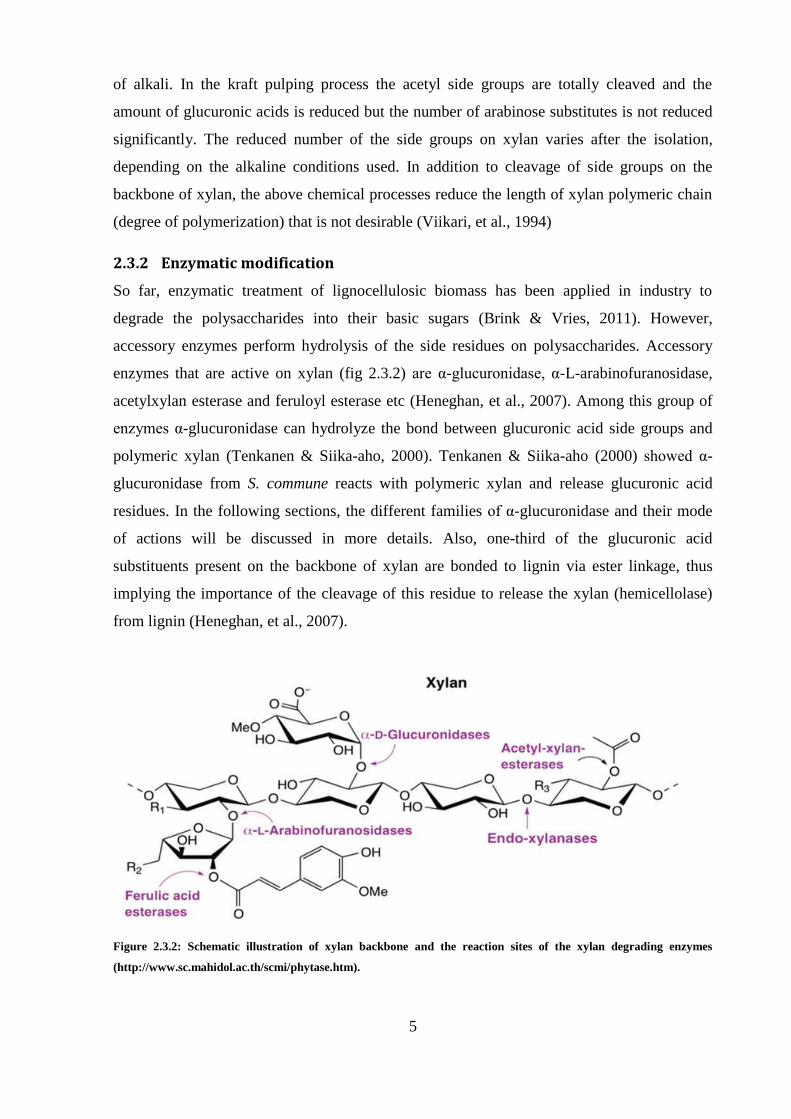
enzymes that are active on xylan (fig 2.3.2) are α-glucuroni ase, α-L-arabinofuranosidase,
acetylxylan esterase and feruloyl esterase etc (Heneghan, et al., 2007). Among this group of
enzymes α-glucuronidase can hydrolyze the bond between glucuronic acid side groups and
polymeric xylan (Tenkanen & Siika-aho, 2000). Tenkanen & Siika-aho (2000) showe α-
glucuronidase from S. commune reacts with polymeric xylan and release glucuronic acid
residues. In the following sections, the different families of α-glucuronidase and their mode
of actions will be discussed in more details. Also, one-third of the glucuronic acid
substituents present on the backbone of xylan are bonded to lignin via ester linkage, thus
implying the importance of the cleavage of this residue to release the xylan (hemicellolase)
from lignin (Heneghan, et al., 2007).
Figure 2.3.2: Schematic illustration of xylan backbone and the reaction sites of the xylan degrading enzymes
(http://www.sc.mahidol.ac.th/scmi/phytase.htm).
6
2.4 Properties of α-Glucuronidase
Alpha-glucuronidase is a glycoside hydrolase. Glycoside hydrolases facilitate the hydrolysis
of the glycosidic bonds of the polysaccharides like cellulose and hemicellulose. There are
two families of α-glucuronidases, GH67 and GH115. Members of the GH6 family of α-
glucuronidases are active on short oligoxylans. These enzymes hy rolyze the α-1,2
glycosidic linkage between 4-O-glucuronic acid residues substituted at the non-reducing end
of oligoxylan (Nurizzo, et al., 2002). Alpha-glucuronidases, in which belong to GH115
family, are active on the polymeric xylan ( Ryabova , et al., 2009). The recent family requires
the two adjacent xylose residues of the substituted residue, to be available for the enzyme
binding (Kolenova, et al., 2010). Kolenova and colleagues 2010 illustrated that the GH115
family α-glucuronidases are inverting enzymes that use single displacement mechanism to
hydrolyze the substrate, which formerly was shown by Nurizzo and colleagues 2002 & 2003,
to be the GH6 family α-glucuronidase mode of the action.
2.4.1 The GH67 family α-glucuronidases
The structure of Pseudomonas cellulosa GH67 α-glucuronidase has been determined by
(Nurizzo, et al., 2002). They have heterologously expressed the respective gene in E. coli and
the molecular mass of the recombinant α-glucuronidase was determined by SDS-PAGE at
77kDa, compatible with the expected molecular mass of the protein from the translated
sequence of the gene. Nevertheless, gel filtration revealed that the enzyme molecular mass
was 150 kDa in solution, indicating that the recombinant α-glucuronidase exists as a dimer.
The recombinant α-glucuronidase from P. cellulosa is a dimer, in which monomers
composed of three domains. The catalytic center of this enzyme is a (β/α)8 barrel located in
the central domain. Nurizzo and colleagues also commented on the evolutionary divergence
of α-glucuronidase. They found that the catalytic center of α-glucuronidase is conserved
among glycoside hydrolases that are involved in the decomposition of di, oligo and
polysaccharides. The search for triggering structural similarities of the GH6 α-glucuronidase
with other proteins by DALI (distance alignment matrix method) showed that the top seven
hits were glycosi e hy rolases with the central (α/β)8 cavity domain in common but in the
evolution these enzymes diversified in terms of the stereochemistry and the substrate
specificity.
7
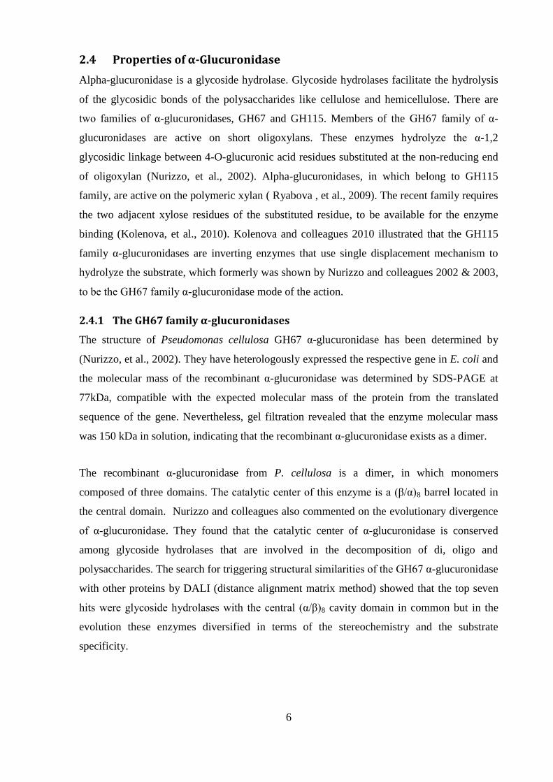
Figure 2.4.1: Single displacement mechanism of hydrolysis of the α1,2-glycosidic bond between uronic acid and xylan
moiety by the family GH67 α-glucuronidase (Nurizzo, et al., 2002).
Two catalytic substituents of the enzyme are required for this mechanism: the first is an acid
for the protonation of the glycosidic oxygen bridge, which facilitates the departure of the
glucuronic acid residue and the second is a base in order to assist the activation of a water
molecule prior to the nucleophilic attack of the anomeric carbon C1 (Nurizzo, et al., 2002).
Three carboxylate groups participate in enzymatic cleavage of the glycosidic bridge: Glu297,
a proton donor residue which acts as an acid in the catalysis and two residues, Asp365 or
Glu393, which are suggested to act as a base contribution to the nucleophilic attack at the α-
conformation of the anomeric carbon C1, which results in the inversion of the anomeric
carbon to β-conformation (Nurizzo, et al., 2003).
2.4.2 The GH115 family α-glucuronidase
The purifie α-glucuronidase from S. commune is a member of GH115 family with molecular
mass of 125 kDa and the isoelectric point of 3.6, determined by SDS-PAGE and IEF gels
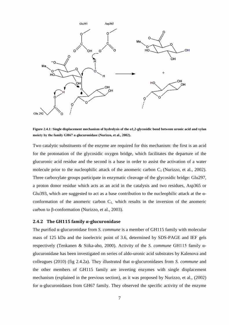
respectively (Tenkanen & Siika-aho, 2000). Activity of the S. commune GH115 family α-
glucuronidase has been investigated on series of aldo-uronic acid substrates by Kalenova and
colleagues (2010) (fig 2.4.2a). They illustrate that α-glucuronidases from S. commune and
the other members of GH115 family are inverting enzymes with single displacement
mechanism (explained in the previous section), as it was proposed by Nurizzo, et al., (2002)
for α-glucuronidases from GH67 family. They observed the specific activity of the enzyme
8
increased by length of the backbone xylan, for aldotetraouronic acid, aldopentaouronic acid
and aldohexaouronic acid. These results imply that the two adjacent residues of the
methylglucuronic acid substituted residues have an important role in recognition and binding
of the enzyme to the substrate (fig 2.4.2b). Apart from the catalytic center, which is
conserved between glycoside hydrolyzes, topology of the binding site explains the
evolutionary gap between them (Nurizzo, et al., 2002). Kolenova and colleagues (2010)
assumed the substrate binding site of GH115 α-glucuronidases is a cleft that can
accommodate a few neighbor xylose residues in addition to the substituted residue with
glucuronic acid. Whereas, situation of the catalytic apparatus of the GH6 α-glucuronidase in
the deep pocket topology, demands for substituation of the glucuronic acid residue on the
non-reducing end of the substrate. These explain the selection of substrate by both the
GH115 and GH6 α-glucuronidases (Kolenova, et al., 2010).
Figure 2.4.2a: Substrates used by Kolenova and colleagues (2010) for determining the activity of the S.
commune GH115 α-gucurnidase (Kolenova, et al., 2010): A) MeGlc2AXyl2, B) MeGlcA3Xyl3 C)
MeGlcA2Xyl3 D) MeGlcA3Xyl4 E) MeGlcA3Xyl5 F) Reduced MeGlcA3Xyl3-xylitol.
9
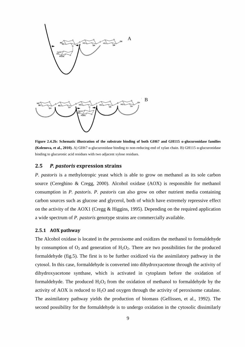
Figure 2.4.2b: Schematic illustration of the substrate binding of both GH67 and GH115 α-glucuronidase families
(Kolenova, et al., 2010). A) GH6 α-glucuronidase binding to non-reducing end of xylan chain. B) GH115 α-glucuronidase
binding to glucuronic acid residues with two adjacent xylose residues.
2.5 P. pastoris expression strains
P. pastoris is a methylotropic yeast which is able to grow on methanol as its sole carbon
source (Cereghino & Cregg, 2000). Alcohol oxidase (AOX) is responsible for methanol
consumption in P. pastoris. P. pastoris can also grow on other nutrient media containing
carbon sources such as glucose and glycerol, both of which have extremely repressive effect
on the activity of the AOX1 (Cregg & Higgins, 1995). Depending on the required application
a wide spectrum of P. pastoris genotype strains are commercially available.
2.5.1 AOX pathway
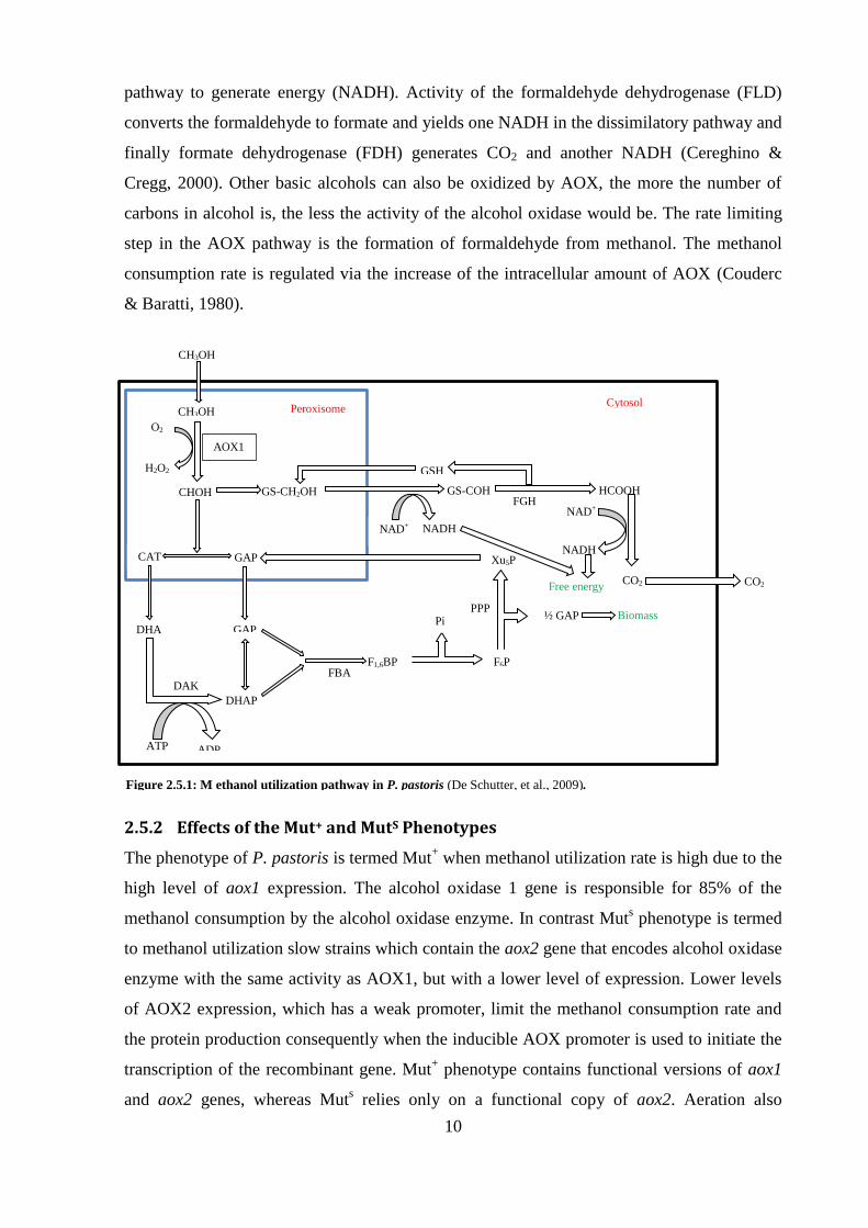
The Alcohol oxidase is located in the peroxisome and oxidizes the methanol to formaldehyde
by consumption of O2 and generation of H2O2. There are two possibilities for the produced
formaldehyde (fig.5). The first is to be further oxidized via the assimilatory pathway in the
cytosol. In this case, formaldehyde is converted into dihydroxyacetone through the activity of
dihydroxyacetone synthase, which is activated in cytoplasm before the oxidation of
formaldehyde. The produced H2O2 from the oxidation of methanol to formaldehyde by the
activity of AOX is reduced to H2O and oxygen through the activity of peroxisome catalase.
The assimilatory pathway yields the production of biomass (Gellissen, et al., 1992). The
second possibility for the formaldehyde is to undergo oxidation in the cytosolic dissimilarly
A
B
10
pathway to generate energy (NADH). Activity of the formaldehyde dehydrogenase (FLD)
converts the formaldehyde to formate and yields one NADH in the dissimilatory pathway and
finally formate dehydrogenase (FDH) generates CO2 and another NADH (Cereghino &
Cregg, 2000). Other basic alcohols can also be oxidized by AOX, the more the number of
carbons in alcohol is, the less the activity of the alcohol oxidase would be. The rate limiting
step in the AOX pathway is the formation of formaldehyde from methanol. The methanol
consumption rate is regulated via the increase of the intracellular amount of AOX (Couderc
& Baratti, 1980).
2.5.2 Effects of the Mut+ and MutS Phenotypes
The phenotype of P. pastoris is termed Mut+ when methanol utilization rate is high due to the
high level of aox1 expression. The alcohol oxidase 1 gene is responsible for 85% of the
methanol consumption by the alcohol oxidase enzyme. In contrast Muts phenotype is termed
to methanol utilization slow strains which contain the aox2 gene that encodes alcohol oxidase
enzyme with the same activity as AOX1, but with a lower level of expression. Lower levels
of AOX2 expression, which has a weak promoter, limit the methanol consumption rate and
the protein production consequently when the inducible AOX promoter is used to initiate the
transcription of the recombinant gene. Mut+ phenotype contains functional versions of aox1
and aox2 genes, whereas Muts relies only on a functional copy of aox2. Aeration also
CH3OH
AOX1
O2
H2O2
CH3OH
CHOH
CAT GAP
GAP DHA
DHAP
DAK
ADP ATP
FBA F1,6BP
Pi
Xu5P
PPP
F6P
½ GAP Biomass
GS-CH2OH
NAD+
GS-COH
NADH
HCOOH FGH
GSH
NAD+
NADH
Free energy CO2 CO2
Cytosol Peroxisome
Figure 2.5.1: M ethanol utilization pathway in P. pastoris (De Schutter, et al., 2009).

11
influences the level of recombinant protein production because AOX has a poor affinity for
oxygen and cell compensates by expressing a high level of aox1 which has the same
promoter as the recombinant gene (Daly & Hearn, 2005). In order to determine the culture
conditions it is essential to determine the Mut+/Mus
s phenotypes. Typical methanol
concentrations in the medium used for Mut+ are 0.5-1.0% v/v whereas, higher concentrations
of methanol than 0.3% v/v potentially results in poisoning of the Muts constructs. In large
scale industrial fermenters Mut+
strains require a large methanol supply throughout the
induction phase which might be hazardous due to the flammability of methanol. An
alternative solution for reducing the explosion risk could be to use Muts strains due to their
lower methanol demand. Muts
constructs are able to grow on mannitol and sorbitol, and
consume less methanol to keep the induction. The slow growth rate and protein production
rate of Muts
phenotypes can be exploited to produce proteins with slow folding rate
(Romanos, et al., 1995).
2.5.3 Expression vector
Among all commercial vectors that are used for expression of recombinant protein in P.
pastoris, pPI Zα was selected to transfer the agu1 gene into P. pastoris The pPI Zα
expression cassette contains a copy of α-mating factor (α- MF) signal sequence before the
multicloning site, which leads to secretion of the recombinant protein. This vector also
carries the 5´ AOX1 promoter The pPI Zα expression vector contains a zeocin resistance
gene that allows selection for positive transformants that are resistant to zeocin. The short
length of the pPI Zα ( 6kb) allows for efficient transformation an contributes to prepare
stable expression strains compared to larger expression vectors developed for transformation
into the P. pastoris (Daly & Hearn, 2005).
2.5.4 The signal sequence
The alpha–mating factor pre-pro leader sequence (α-MF) consists of two regions, namely the
pre and pro sequences. The pre-sequence contains a 19 amino acid signal peptide and the pro-
sequence consists of a 60 amino acids. Upon translation, the pro-protein is translocated into
the endoplasmic reticulum, while the signal peptidase cleaves the signal peptide sequence off
the protein. For further processing the pro-protein is carried to Golgi where kex2 protease
removes the pro-sequence prior to secretion of the mature protein into the extracellular media
(Brake, et al., 1984).
12
2.5.5 Culture condition for expression
Before beginning the fermentation, there are some issues regarding the culture conditions of
P. pastoris to take into account. The first issue is the scale of the culture. Shake flasks are
mostly used in the early stage of the cell culture to perform screening and to obtain relevant
data for scaling the culture up. Limited aeration and methanol supply are the main restriction
integrated with the shaken flask expression system, especially when Mut+
phenotype is
utilized. While using Mut+ transformants for the culture of P. pastoris, the concentration of
methanol is an important parameter for optimizing the expression levels of the enzyme. The
normal range of methanol concentration, reported for P. pastoris cell lines, lies between 0.5-
1.0%. The accumulation of methanol can repress the protein production, which is a negative
effect. This implies the requirement of the optimal methanol concentration assessment, which
tolerates among different family members of P. pastoris and the type of the synthesized
protein (Romanos, et al., 1995). The pH and ingredients of the medium have a significant
effect on the level of the expression of the recombinant protein. The extent of proteolysis of
the secreted protein will reduce if the medium is buffered to pH values from 3 to 6
(Sreekrishna, et al., 1997; Cregg, et al., 1993). There are other ways to diminish the
proteolysis such as supplementing the medium with peptone and yeast extract, or addition of
1 % casamino acids. Addition of ammonium ions has also been recommended, in the form of
ammonium sulfate. Tsujikawa and colleagues (1996), observed 10 fold reduction of
proteolysis by supplementing medium IM medium with ammonium ions. Proteolysis is found
to amplify over the induction period when the amount of viable cells in the culture is
reduced. The way to engineer around this problem is to exchange the culture medium with
the fresh medium and recover the product; this method reduces the proteolysis in the lab
scale semi continuous cultures. The fermenter can be rebooted as many times as needed with
only one start up (Sreekrishna, et al., 1997).
13
3. Materials and Methods
3.1 Strains and vectors
The codon optimized Agu1 gene (2925bp) in pUC57 vector was provided by NZYtech (Lda,
Lisbon, Portugal). pPI Zα plasmi (carried in E. coli TOP10 strain) was used for
construction of the expression cassette and was purchased from Invitrogen (Life
Technologies, NY, USA). E.coli DH5α was use for vector construction an maintenance
The P. pastoris SMD1168H strain which was used was for expression was purchased from
Invitrogen (Life Technologies, NY, USA).
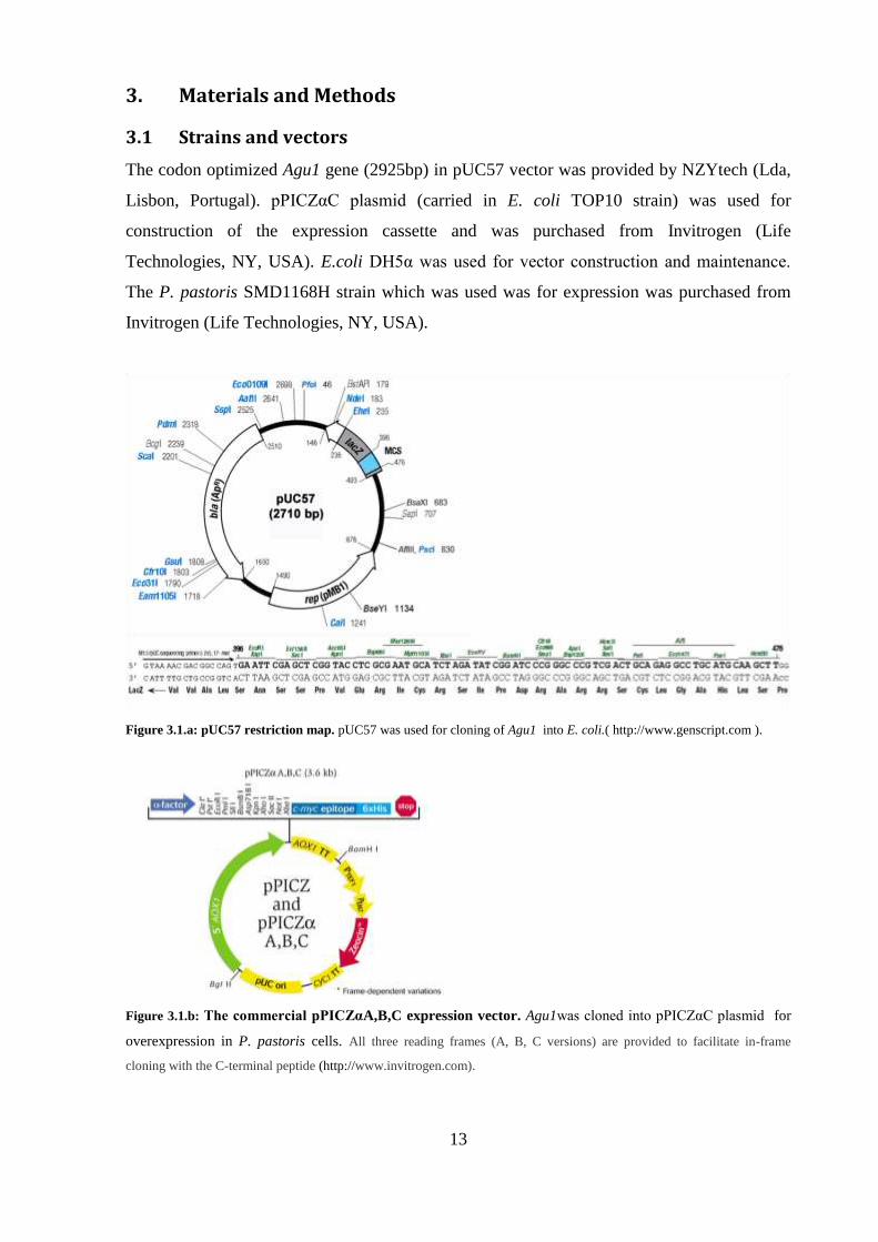
Figure 3.1.a: pUC57 restriction map. pUC57 was used for cloning of Agu1 into E. coli.( http://www.genscript.com ).
Figure 3.1.b: The commercial pPICZαA,B,C expression vector. Agu1was clone into pPI Zα plasmi for
overexpression in P. pastoris cells. All three reading frames (A, B, C versions) are provided to facilitate in-frame
cloning with the C-terminal peptide (http://www.invitrogen.com).
14
3.2 Media
All media were prepared according to the Invitrogen manuals (Cat. no. V195-20, Cat. no.
K1710-01 and P. pastoris Fermentation Process Guidelines) and were sterilized by autoclave
on liquid cycle at 15 psi and 1 1 for min, unless otherwise stated.
3.2.1 Lysogeny Broth (LB medium)
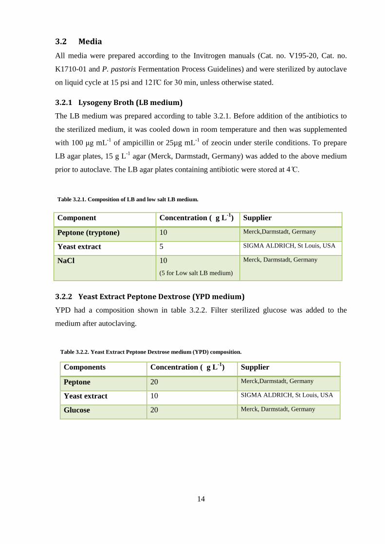
The LB medium was prepared according to table 3.2.1. Before addition of the antibiotics to
the sterilized medium, it was cooled down in room temperature and then was supplemented
with 100 μg mL-1
of ampicillin or 25µg mL-1
of zeocin under sterile conditions. To prepare
LB agar plates, 15 g L-1
agar (Merck, Darmstadt, Germany) was added to the above medium
prior to autoclave. The LB agar plates containing antibiotic were store at
3.2.2 Yeast Extract Peptone Dextrose (YPD medium)
YPD had a composition shown in table 3.2.2. Filter sterilized glucose was added to the
medium after autoclaving.
Table 3.2.1. Composition of LB and low salt LB medium.
Component Concentration ( g L-1
) Supplier
Peptone (tryptone) 10 Merck,Darmstadt, Germany
Yeast extract 5 SIGMA ALDRICH, St Louis, USA
NaCl 10
(5 for Low salt LB medium)
Merck, Darmstadt, Germany
Components Concentration ( g L-1
) Supplier
Peptone 20 Merck,Darmstadt, Germany
Yeast extract 10 SIGMA ALDRICH, St Louis, USA
Glucose 20 Merck, Darmstadt, Germany
Table 3.2.2. Yeast Extract Peptone Dextrose medium (YPD) composition.
15
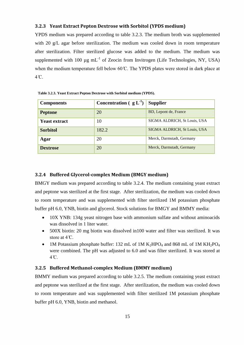
3.2.3 Yeast Extract Pepton Dextrose with Sorbitol (YPDS medium)
YPDS medium was prepared according to table 3.2.3. The medium broth was supplemented
with 20 g/L agar before sterilization. The medium was cooled down in room temperature
after sterilization. Filter sterilized glucose was added to the medium. The medium was
supplemented with 100 µg mL-1
of Zeocin from Invitrogen (Life Technologies, NY, USA)
when the me ium temperature fell below 6 The PD plates were store in ark place at
3.2.4 Buffered Glycerol-complex Medium (BMGY medium)
BMGY medium was prepared according to table 3.2.4. The medium containing yeast extract
and peptone was sterilized at the first stage. After sterilization, the medium was cooled down
to room temperature and was supplemented with filter sterilized 1M potassium phosphate
buffer pH 6.0, YNB, biotin and glycerol. Stock solutions for BMGY and BMMY media:
10X YNB: 134g yeast nitrogen base with ammonium sulfate and without aminoacids
was dissolved in 1 liter water.
500X biotin: 20 mg biotin was dissolved in100 water and filter was sterilized. It was
store at
1M Potassium phosphate buffer: 132 mL of 1M K2HPO4 and 868 mL of 1M KH2PO4
were combined. The pH was adjusted to 6.0 and was filter sterilized. It was stored at
3.2.5 Buffered Methanol-complex Medium (BMMY medium)
BMMY medium was prepared according to table 3.2.5. The medium containing yeast extract
and peptone was sterilized at the first stage. After sterilization, the medium was cooled down
to room temperature and was supplemented with filter sterilized 1M potassium phosphate
buffer pH 6.0, YNB, biotin and methanol.
Components Concentration ( g L-1
) Supplier
Peptone 20 BD, Lepont de, France
Yeast extract 10 SIGMA ALDRICH, St Louis, USA
Sorbitol 182.2 SIGMA ALDRICH, St Louis, USA
Agar 20 Merck, Darmstadt, Germany
Dextrose 20 Merck, Darmstadt, Germany
Table 3.2.3. Yeast Extract Pepton Dextrose with Sorbitol medium (YPDS).

16
3.2.6 Fermentation basal salts medium
Six liters of fermentation basal salt was prepared according to table 3.2.6 and was sterilized
in a 30 liters fermenter from infors HT Company (Basel, Switzerland). After the medium
cooled down to room temperature, its pH was adjusted with 28% ammonium hydroxide to
pH 5.
Components Concentration ( g L-1
) Supplier
Yeast extract 10 Merck, Darmstadt, Germany
Peptone 20 SIGMA ALDRICH, St Louis, USA
YNB 13.4 Alfa Aesor, Karlsruhe, Germany
Biotin 4 × 10⁻⁴
SIGMA ALDRICH, St Louis, USA
Glycerol 10 Merck, Hohenbrunn, Germany
Potassium phosphate buffer 100 mL 1M Merck, Darmstadt, Germany
Components Concentration( g L-1
) Supplier
Yeast extract 10 Merck, Darmstadt, Germany
Peptone 20 SIGMA ALDRICH, St Louis, USA
YNB 13.4 Alfa Aesor, Karlsruhe, Germany
Biotin 4 × 10⁻⁴
SIGMA ALDRICH, St Louis, USA
Methanol 5
Potassium phosphate buffer 100 mL1M Merck, Darmstadt, Germany
Table 3.2.4. Composition of buffered Glycerol-complex Medium (BMGY).
Table 3.2.5. Composition of Buffered Methanol-complex Medium (BMMY).
Components Concentration ( g L-1
)
Phosphoric acid 85% 26.7 (mL L-1
)
Calcium sulfate 0.93
Potassium sulfate 18.2
Magnesium sulfate-7 H2O 14.9
Potassium hydroxide 4.13
Glycerol 40.0
Table 3.2.6. Composition of fermentation basal salts.
17
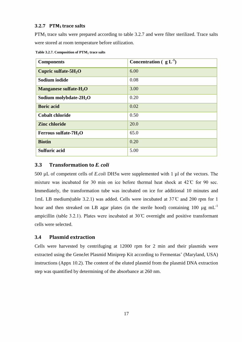
3.2.7 PTM1 trace salts
PTM1 trace salts were prepared according to table 3.2.7 and were filter sterilized. Trace salts
were stored at room temperature before utilization.
3.3 Transformation to E. coli
500 µL of competent cells of E.coli DH5α were supplemented with 1 µl of the vectors The
mixture was incubate for min on ice before thermal heat shock at for sec
Immediately, the transformation tube was incubated on ice for additional 10 minutes and
1mL LB medium(table 3.2.1) was added. Cells were incubated at an rpm for 1
hour and then streaked on LB agar plates (in the sterile hood) containing 100 µg mL-1
ampicillin (table 1) Plates were incubate at C overnight and positive transformant
cells were selected.
3.4 Plasmid extraction
Cells were harvested by centrifuging at 12000 rpm for 2 min and their plasmids were
extracted using the GeneJet Plasmid Miniprep Kit according to Fermentas’ (Maryland, USA)
instructions (Appx 10.2). The content of the eluted plasmid from the plasmid DNA extraction
step was quantified by determining of the absorbance at 260 nm.
Components Concentration ( g L-1
)
Cupric sulfate-5H2O 6.00
Sodium iodide 0.08
Manganese sulfate-H2O 3.00
Sodium molybdate-2H2O 0.20
Boric acid 0.02
Cobalt chloride 0.50
Zinc chloride 20.0
Ferrous sulfate-7H2O 65.0
Biotin 0.20
Sulfuric acid 5.00
Table 3.2.7. Composition of PTM1 trace salts
18
3.5 Construction of the expression cassette
3.5.1 Enzymatic restriction
All restriction enzymes were provided by Fermentas (Maryland, USA). Plasmid and DNA
fragments restriction were performed according to respective manufacturer’s protocols.
Digests were purified using Illustra GFX PCR DNA and gel band Purifiation Kit GE
Healthcare (Buckinghamshire, UK).
3.5.2 Ligation
Digested DNA fragments were mixed with the linearized vector in the presence of T4 DNA
ligase from Fermentas (Maryland, USA). The ligation reaction set up was according the
manufacturer’s instructions. The ligation mixtures were incubated overnight at room
temperature. Ligation products were used for transformation into E. coli DH5α cells to test
the integrity and amplification of the construct.
3.6 P. pastoris transformation
3.6.1 Transformation by electroporation
P.pastoris SMD1168H was grown overnight, in shake flasks, in YPD medium (table 3.2.2) at
and 170 rpm in a rotary shaker. Cells were harvested for electroporation according to
the Invitrogen protocol (Cat. no.V195-20, version F) and resuspended in 1 mL of 1 M ice
cold sorbitol. The linearized vectors were mixed with the resuspended cells and then
transferred to an electroporation cuvette, which was then incubated on ice for 5 min. The
BIO-RAD electroporation device’s parameters were adjusted to P. pastoris set up. Cells were
pulsed and then 1 mL of 1 M ice cold sorbitol was added to the electroporation cuvette.
Contents of the cuvette were transferred to a 15 mL Falcon tube and incubated at
without shaking for 2 hours. Finally the cells were streaked out on YPDS (table 3.2.3) agar
plates containing 100 µg mL-1
of zeocin an incubate at for 72 hours.
3.6.2 Screening for positive transformants
Zeocin resistant colonies were selected and grown overnight, in shake flasks, in YPD
medium (table 3.2.2) containing 100 µg mL-1
of zeocin at 30 C. Cells were harvested at
12000 rpm and their genomic DNA were isolated by phenol chloroform method and
amplified by PCR through AOX forward and reverse primers to verify the recombination of
the expression cassette into the SMD1168H strains genome. Then the PCR products were ran
on gel electrophoresis.
19
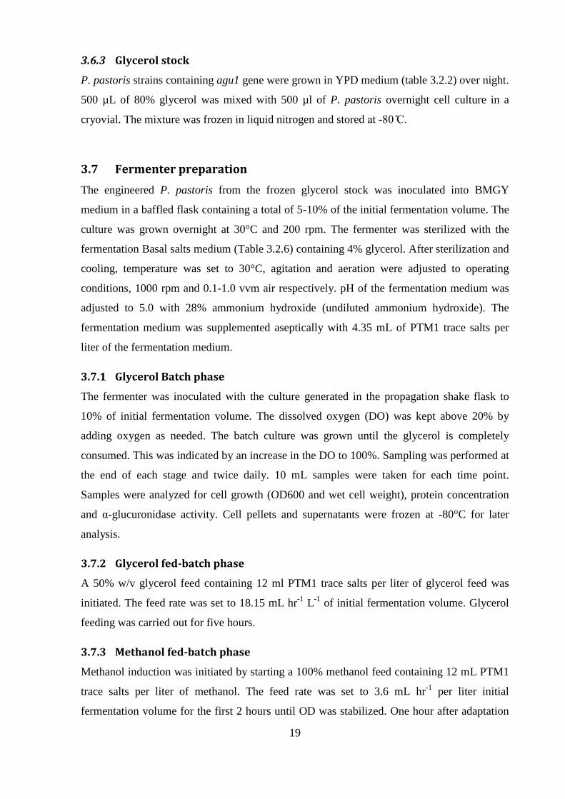
3.6.3 Glycerol stock
P. pastoris strains containing agu1 gene were grown in YPD medium (table 3.2.2) over night.
500 µL of 80% glycerol was mixed with 500 µl of P. pastoris overnight cell culture in a
cryovial. The mixture was frozen in liquid nitrogen and stored at -
3.7 Fermenter preparation
The engineered P. pastoris from the frozen glycerol stock was inoculated into BMGY
medium in a baffled flask containing a total of 5-10% of the initial fermentation volume. The
culture was grown overnight at 30°C and 200 rpm. The fermenter was sterilized with the
fermentation Basal salts medium (Table 3.2.6) containing 4% glycerol. After sterilization and
cooling, temperature was set to 30°C, agitation and aeration were adjusted to operating
conditions, 1000 rpm and 0.1-1.0 vvm air respectively. pH of the fermentation medium was
adjusted to 5.0 with 28% ammonium hydroxide (undiluted ammonium hydroxide). The
fermentation medium was supplemented aseptically with 4.35 mL of PTM1 trace salts per
liter of the fermentation medium.
3.7.1 Glycerol Batch phase
The fermenter was inoculated with the culture generated in the propagation shake flask to
10% of initial fermentation volume. The dissolved oxygen (DO) was kept above 20% by
adding oxygen as needed. The batch culture was grown until the glycerol is completely
consumed. This was indicated by an increase in the DO to 100%. Sampling was performed at
the end of each stage and twice daily. 10 mL samples were taken for each time point.
Samples were analyzed for cell growth (OD600 and wet cell weight), protein concentration
and α-glucuronidase activity. Cell pellets and supernatants were frozen at -80°C for later
analysis.
3.7.2 Glycerol fed-batch phase
A 50% w/v glycerol feed containing 12 ml PTM1 trace salts per liter of glycerol feed was
initiated. The feed rate was set to 18.15 mL hr-1
L-1
of initial fermentation volume. Glycerol
feeding was carried out for five hours.
3.7.3 Methanol fed-batch phase
Methanol induction was initiated by starting a 100% methanol feed containing 12 mL PTM1
trace salts per liter of methanol. The feed rate was set to 3.6 mL hr-1
per liter initial
fermentation volume for the first 2 hours until OD was stabilized. One hour after adaptation
20
of the culture to methanol utilization, the feed rate is doubled to7.3 mL-1
hr-1
per liter of initial
fermentation volume. After, two hours at the 7.3 mL hr-1
per liter of initial fermentation
volume, the methanol feed rate was increased to10 mL hr-1
per liter of initial fermentation
volume. This feed rate was maintained throughout the rest of the fermentation. The entire
methanol fed-batch phase lasted 84 hours.
3.8 Biochemical analyses
3.8.1 Bradford assay
Quantification of protein was performed with the Bradford reagent supplied by Bio-Rad
(California, USA) in a microplate format according to the manufacturer’s instructions. A
standard curve of bovine serum albumin in duplicate concentrations from 0.05 µg mL-1
to 0.5
µg mL-1
was prepared.
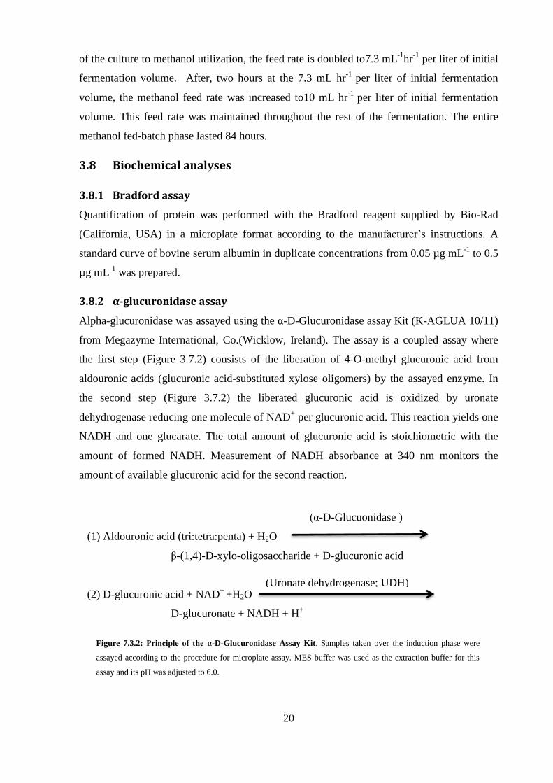
3.8.2 α-glucuronidase assay
Alpha-glucuronidase was assayed using the α-D-Glucuronidase assay Kit (K-AGLUA 10/11)
from Megazyme International, Co.(Wicklow, Ireland). The assay is a coupled assay where
the first step (Figure 3.7.2) consists of the liberation of 4-O-methyl glucuronic acid from
aldouronic acids (glucuronic acid-substituted xylose oligomers) by the assayed enzyme. In
the second step (Figure 3.7.2) the liberated glucuronic acid is oxidized by uronate
dehydrogenase reducing one molecule of NAD+ per glucuronic acid. This reaction yields one
NADH and one glucarate. The total amount of glucuronic acid is stoichiometric with the
amount of formed NADH. Measurement of NADH absorbance at 340 nm monitors the
amount of available glucuronic acid for the second reaction.
(1) Aldouronic acid (tri:tetra:penta) + H2O
β-(1,4)-D-xylo-oligosaccharide + D-glucuronic acid
(2) D-glucuronic acid + NAD+
+H2O
D-glucuronate + NADH + H+
(α-D-Glucuonidase )
(Uronate dehydrogenase; UDH)
Figure 7.3.2: Principle of the α-D-Glucuronidase Assay Kit. Samples taken over the induction phase were
assayed according to the procedure for microplate assay. MES buffer was used as the extraction buffer for this
assay and its pH was adjusted to 6.0.
21
4. Results
The constructed strain was checked in a set of experiments to determine the integration of the
expression cassette and to investigate its influences on the pro uction of α-glucuronidase. To
investigate the recombination of Agu1 gene into the expression vector, the constructed vector
was analyzed by restriction digestion and DNA sequencing. Also integration of the
expression cassette into SMD1168H P. pastoris transformants’ genome was confirmed by
isolation of the genomic DNA and PCR. In or er to screen the best α-glucuronidase
producing strain, the transformants were grown in BMGY and BMMY media and the activity
of the secrete α-glucuronidase was assayed. The candidate strain was further evaluated by
cultivation in a shake flask and fermentation in a 30 liter fermenter to investigate the effect of
the genetic modifications on the protein production yield and to assay the activity of α-
glucuronidase.
4.1 Construction of expression cassette
4.1.1 Propagation of the Agu1 gene
The synthetic Agu1 was codon optimized for expression in P. pastoris. It was cloned between
XbaI and ClaI restriction sites into EcoRV region of pUC57 donor vector. The vector carries
the ampicillin resistant gene that enables the selection of positive transformants on the LB
agar plates containing 100 µg mL-1
ampicillin (table 3.2.1). Efficient transformation into E.
coli DH5α was checked by overnight growth of ampicillin resistant colonies in LB medium
(table 3.2.1) supplemented with 100 µg mL-1
ampicillin. Cells were harvested and their
plasmids were isolated using the GeneJet Plasmid Miniprep Kit according to manufacturer’s
instructions (Appx 10.2). The quantity of the eluted plasmid was 219 ng µL-1
, determined by
absorbance at 260 nm (table 4.1.2).
4.1.2 Isolation of pPICZαC
pPI Zα was isolated from from E. coli TOPO strain grown in LB medium supplemented
with 25 µg mL-1
of zeocin. Concentration of the purified pPI Zα vector was determined 74
ng µL-1
by measurement of absorbance at 260nm (table 4.1.2).
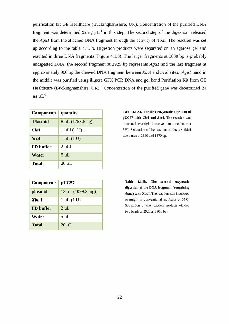
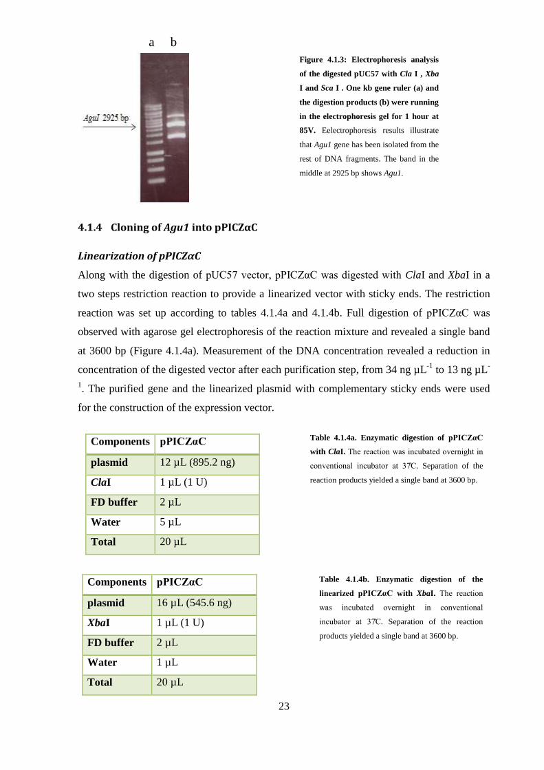
4.1.3 Isolation of Agu1 from pUC57
Agu1 was isolated from pUC57 by enzymatic digestion with XbaI, ClaI and ScaI, in a two
step restriction. Initially, plasmid was digested with ScaI and ClaI overnight at (table
4.1.3a). This digestion resulted in two separate bands, at 3830 bp and 1870 bp. The larger
band that contained Agu1 was purified using illustra GFX PCR DNA and gel band
22
purification kit GE Healthcare (Buckinghamshire, UK). Concentration of the purified DNA
fragment was determined 92 ng µL-1
in this step. The second step of the digestion, released
the Agu1 from the attached DNA fragment through the activity of XbaI. The reaction was set
up according to the table 4.1.3b. Digestion products were separated on an agarose gel and
resulted in three DNA fragments (Figure 4.1.3). The larger fragments at 3830 bp is probably
undigested DNA, the second fragment at 2925 bp represents Agu1 and the last fragment at
approximately 900 bp the cleaved DNA fragment between XbaI and ScaI sites. Agu1 band in
the middle was purified using illustra GFX PCR DNA and gel band Purifiation Kit from GE
Healthcare (Buckinghamshire, UK). Concentration of the purified gene was determined 24
ng µL-1
.
Components quantity
Plasmid 8 µL (1753.6 ng)
ClaI 1 µLl (1 U)
ScaI 1 µL (1 U)
FD buffer 2 µLl
Water 8 µL
Total 20 µL
Table 4.1.3a. The first enzymatic digestion of
pUC57 with ClaI and ScaI. The reaction was
incubate overnight in conventional incubator at
eparation of the reaction pro ucts yiel e
two bands at 3830 and 1870 bp.
Components pUC57
plasmid 12 µL (1099.2 ng)
Xba I 1 µL (1 U)
FD buffer 2 µL
Water 5 µL
Total 20 µL
Table 4.1.3b. The second enzymatic
digestion of the DNA fragment (containing
Agu1) with XbaI. The reaction was incubated
overnight in conventional incubator at C.
Separation of the reaction products yielded
two bands at 2925 and 905 bp.
23
4.1.4 Cloning of Agu1 into pPICZαC
Linearization of pPICZαC
Along with the digestion of pU 5 vector, pPI Zα was igeste with ClaI and XbaI in a
two steps restriction reaction to provide a linearized vector with sticky ends. The restriction
reaction was set up according to tables 4.1.4a and 4.1.4b. Full digestion of pPI Zα was
observed with agarose gel electrophoresis of the reaction mixture and revealed a single band
at 3600 bp (Figure 4.1.4a). Measurement of the DNA concentration revealed a reduction in
concentration of the digested vector after each purification step, from 34 ng µL-1
to 13 ng µL-
1. The purified gene and the linearized plasmid with complementary sticky ends were used
for the construction of the expression vector.
Figure 4.1.3: Electrophoresis analysis
of the digested pUC57 with Cla I , Xba
I and Sca I . One kb gene ruler (a) and
the digestion products (b) were running
in the electrophoresis gel for 1 hour at
85V. Eelectrophoresis results illustrate
that Agu1 gene has been isolated from the
rest of DNA fragments. The band in the
middle at 2925 bp shows Agu1.
Components pPICZαC
plasmid 12 µL (895.2 ng)
ClaI 1 µL (1 U)
FD buffer 2 µL
Water 5 µL
Total 20 µL
Table 4.1.4a. Enzymatic digestion of pPICZαC
with ClaI. The reaction was incubate overnight in
conventional incubator at . Separation of the
reaction products yielded a single band at 3600 bp.
Components pPICZαC
plasmid 16 µL (545.6 ng)
XbaI 1 µL (1 U)
FD buffer 2 µL
Water 1 µL
Total 20 µL
Table 4.1.4b. Enzymatic digestion of the
linearized pPICZαC with XbaI. The reaction
was incubate overnight in conventional
incubator at . Separation of the reaction
products yielded a single band at 3600 bp.
a b

24
Ligation of Agu1 into pPICZαC
The cleaved Agu1 was combine with the linearize pPI Zα vector in the presence of T
DNA ligase to clone the gene into the pPI Zα vector by the function of T4 DNA ligase.
The amount of the vector and the insert required for 1:1, 1:3 and 1:5 vector: insert ratios was
calculated according to the concentrations of the purified vector and insert presented in tables
4.1.3c and 4.1.4c. The ligation reaction was set up (table 4.1.4c) according to the
manufacturer’s instructions The reaction was incubated in room temperature overnight and
the recombinant expression vectors were transformed into E.coli DH5α
Figure 4.1.4a: Electrophoresis analysis of the
digested pPICZαC with Cla I and Xba I. One
kb gene ruler (a) and digestion product (b)
were running in the electrophoresis gel for 1
hour at 85V. Electrophoresis results illustrate
that pPI Zα has been fully digested and the
band at 3600 bp corresponds to pPI Zα .
Components Quantity (1:1) Quantity (1:3) Quantity (1:5)
Digested plasmid 1.5µL (19 ng) 2.0µL (26 ng) 1.0 µL (13 ng)
Insert 2.5 µL (60 ng) 5.5 µL (133 ng) 4.5 µL (108 ng)
T4 ligation buffer 10X 1.0 µL 1.0 µL 1.0 µL
T4 Ligase 0.7 µL (3.5 U) 0.7 µL (3.5 U) 0.7 µL (3.5 U)
MilliQ-Water Fill to 10 µL Fill to 10 µL Fill 10 µL
Table 4.1.4c. Ligation of the insert into the expression vector. The reaction was incubated overnight in room
temperature.
a b

25
Evaluation of transformation of the expression vector into E. coli DH5α
After the transformation of the ligation products into E. coli DH5α, selection of the positive
transformants was carried out on low salt LB agar plates containing 25 µg mL-1
zeocin.
Zeocin resistant colonies were selected and grown overnight in low salt liquid LB medium
supplemented with 25 µg mL-1
zeocin. Cells were harvested and the plasmids of the cells
were extracted. Finally, content of the eluted plasmid from the plasmid DNA extraction step
was quantified by measurement of absorbance at 260 nm (table 4.1.4d). Purified DNA
plasmi s were sent for sequencing to verify the integration of the insert into pPI Zα Two
separate diagnostic restrictions were also performed on the sample plasmids. The first
reaction was performed with XbaI and ClaI with the same set up as the cleavage of the Agu1
gene from the pUC57 vector. In the second reaction, the recombinant plasmids were digested
with SpeI, a single cutter enzyme, which cleaves inside the gene region (table 4.1.4e).
Diagnostic digestion of the purified plasmid with SpeI showed that 9 of the colonies out of 12
gave rise to a single band at 6600 bp (figure 4.1.4b), which implies the insert has been cloned
into the vector. In addition, two of the samples, which were diagnosed positive recombination
of the insert, were sent for sequencing. The sequencing results also confirmed integration of
the recombinant gene in to the pPI Zα plasmi Eventually one of the expression cassettes,
which had full alignment with the sequence of Agu1, was used for transformation into P.
pastoris SMD1168H.
Concentration
Expression cassette 101 ng µL-1
Table 4.1.4d. Concentration of the recombinant expression vector purified from zeocin resistant colony.
Components Expression cassette
Plasmid 8 µl (809 ng)
SpeI 1 µl (1 U)
FD buffer 2 µl
Water 9 µl
Total 20 µl
Table 4.1.4e. Enzymatic digestion of the
expression vector with SpeI. The reaction
was incubate overnight in conventional
incubator at . Separation of the
reaction products by electrophoresis
yielded a single band at 6600 bp.

26
4.2 Integration of the expression cassette into P. pastoris SMD1168H
4.2.1 Linearization of the pPICZαC construct
pPI Zα plasmi containing α-glucuronidase gene was linearized with MssI (Pme I) (table
4.2.1). The linearization reaction took place for 12 hours at The concentration of the
purified expression vector isolated from E.coli DH5α was 1 ng µL-1
.
4.2.2 Transformation of the linearized pPICZαC construct into P. pastoris
P. pastoris SMD1168H cells were grown overnight in YPD medium (table 3.2.2) and
harvested at OD600 2.73 by centrifuging at 5000 rpm at C. Harvested cells were
resuspended in 1 ml of 1M ice cold sorbitol. Then 5.5 µg of the linearized acceptor vectors
were mixed with 80 µl of the resuspended cells and added into an electroporation cuvette.
The mixture was incubated on ice for 5 min. The BIO-RAD electroporation device
Figure 4.1.4b: Electrophoresis analysis of
the digested expression vectors isolated
from potential positive DH5α colonies with
SpeI. The undigested expression vector (a),
the digestion product (b) and 1 kb gene
ruler were running in the electrophoresis
gel for 1 hour at 85V. Spe I cuts inside Agu1
region therefore we expect a single band at
6600 bp. Nine out of twelve colonies
contained the expression vector. Undigested
cassettes corresponding to each digested
cassette have been loaded to their left hand
well as control.
Components pPICZαC
Plasmid 9.5 µl ( 950 ng)
Pme I 1 µl ( 1 U)
FD buffer 2 µl
Water 6.5 µl
Total 20 µl
Table 4.2.1. Enzymatic digestion of the
expression vector with PmeI. The reaction
was incubated overnight in conventional
incubator at . Separation of the reaction
products yielded a single band at 6600 bp. The
final concentration of the linearized vector after
purification reached 0.55 µg µL-1.
a b c

27
parameters were adjusted to P. pastoris set ups. After pulsing the cells, 1 ml of 1M ice cold
sorbitol was added to the electroporation cuvette. Contents of the cuvette were transferred to
a 15 ml falcon tube and incubated at without shaking for hours inally the cells were
streaked on YPDS agar plates containing 100 µg mL-1
of zeocin until colonies were formed.
Transformation yielded 5 positive colonies.
4.2.3 Screening for positive transformants
This section acknowledges the results from the evaluation of the potential transformants
grown in BMGY (table 3.2.4) and BMMY (table. 8.2.5) media. This was performed in order
to know whether an to which content the cells are able to pro uce an secrete α-
glucuronidase into extracellular media.
In order to screen the positive transformants, they were inoculated into 10 ml of BMGY
medium in a 50 ml Falcon tube. Cell cultures were incubate on a shaker at an 5
rpm for 28 hours. When the culture concentration reached an OD600 =3, cells were harvested
by centrifuging at room temperature and 5000 rpm for 5 min. The cell pellet was resuspended
in 35 ml of BMMY medium until OD600 =1 to induce expression. Cell cultures have been
supplemented with 100% methanol, to 0.5% of their final volume every 24 hours, to maintain
induction. After 96 hours of induction with methanol, cells were harvested by centrifuging at
8000 rpm for 5 min. Cells and supernatant of each cell culture were stored at - for
further analysis.
Genomic DNA of the zeocin resistant transformants, namely strains GluA, GluB, GluC,
GluD, GluE, were isolated and amplified by PCR using AOX1 forward and reverse primers.
Analysis of the PCR reaction by electrophoresis (fig 4.2.3) showed two bands, one between
3500 and 3000 bp and the other between 2500 and 2000 bp, in which the fragment between
3500 and 3000 bp confirms the heterologous integration of Agu1into P. pastoris genome.
Figure 4.2.3 Electrophoresis analysis of the PCR
amplified region of the genomic DNA of the selected
transformants by AOX primers. The amplified Agu1
gene is represented by a band between 3000 and 3500
bp. The second band between 2000 and 2500 bp shows
chromosomal AOX1 of P. pastoris. Positive
transformants from left to right on the gel were named
GluA, GluB, GluC, GluD and GluE and for further
characterization assaye by α-glucuronidase assay kit.
a
4000 bp
2000 bp
28
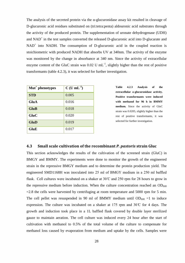
The analysis of the secreted protein via the α-glucuronidase assay kit resulted in cleavage of
D-glucuronic acid residues substituted on (tri:tetra:penta) aldouronic acid substrates through
the activity of the produced protein. The supplementation of uronate dehydrogenase (UDH)
and NAD+ in the test samples converted the released D-glucuronic acid into D-glucarate and
NAD+ into NADH. The consumption of D-glucuronic acid in the coupled reaction is
stoichiometric with produced NADH that absorbs UV at 340nm. The activity of the enzyme
was monitored by the change in absorbance at 340 nm. Since the activity of extracellular
enzyme content of the GluC strain was 0.02 U mL-1
, slightly higher than the rest of positive
transformants (table 4.2.3), it was selected for further investigation.
4.3 Small scale cultivation of the recombinant P. pastoris strain Gluc
This section acknowledges the results of the cultivation of the screened strain (GluC) in
BMGY and BMMY. The experiments were done to monitor the growth of the engineered
strain in the repressive BMGY medium and to determine the protein production yield. The
engineered SMD1168H was inoculated into 5 ml of G me ium in a 5 ml baffle
flask ell cultures were incubate on a shaker at an 5 rpm for hours to grow in
the repressive medium before induction. When the culture concentration reached an OD600
=2.8 the cells were harvested by centrifuging at room temperature and 5000 rpm for 5 min.
The cell pellet was resuspended in 90 ml of BMMY medium until OD600 =1 to induce
expression. The culture was incubated on a shaker at 1 5 rpm an for 4 days. The
growth and induction took place in a 1L baffled flask covered by double layer sterilized
gauze to maintain aeration. The cell culture was induced every 24 hour after the start of
cultivation with methanol to 0.5% of the total volume of the culture to compensate for
methanol loss caused by evaporation from medium and uptake by the cells. Samples were
Mut+ phenotypes C (U mL
-1)
STD 0.005
GluA 0.016
GluB 0.018
GluC 0.020
GluD 0.019
GluE 0.017
Table 4.2.3 Analysis of the
extracellular α-glucuronidase activity.
Positive transformants were induced
with methanol for 96 h in BMMY
medium. Since the activity of GluC
strain was 0.0205, slightly higher than the
rest of positive transformants, it was
selected for further investigation.

29
taken in following time points (hours): 24 h, 48 h, 72 h and 96 h. Samples were centrifuged at
16000 rcf and room temperature for 3 min. The supernatant and cells pellet were separated
and frozen in liquid nitrogen and stored at - for further analysis
The extracellular α-glucuronidase activity of the samples resulted in an increased absorbance
of NADH. The maximum activity of the GluC strain was observed at 96 h and it was found
to be 0.02 U mL-1
. Compared to the corresponding standard enzyme (supplied by the kit),
which had a maximum activity of 0.005 U mL-1
(Table 4.2.3), α-glucuronidase produced by
P. pastoris GluC strain had a 3.7–fold higher activity. The measured activity in the shake
flask cultivation is comparable with the previously measured 0.020 U mL-1
α-glucuronidase
activity of this strain during the screening phase.
Figure 4.3a: Biomass concentration
over time. The cell culture in BMGY
medium, which is a glycerol repressive
medium, was harvested at OD600 2.8
after 28 hours of the.
Figure 4.3b. Activity of the enzyme
over the induction phase. Samples
were taken every 24 hours and the
activity of the enzyme in each sample
(time point) was assay by α-
glucuronidase assay.
30
4.4 Large-scale production of α-glucuronidase
The main objective of the fermentation of P. pastoris GluC was to analyze the performance
of the strain under controlled conditions for the large scale production of recombinant α-
glucuronidase. The fermentation was performed in three phases; glycerol batch cultivation,
glycerol fed batch cultivation and methanol fed-batch cultivation.
4.4.1 Batch phase
The engineered GluC was grown in a 30 liter fermenter on Basal salts, glycerol and
ammonium hydroxide (table 3.2.6). The initial volume of the fermentation medium was 6
liters and were inoculated with 600 ml of P. pastoris GluC overnight culture grown up to
OD600 =1.03. The dissolved oxygen was relatively 100% before the culture starts to grow.
Then, the cells were grown for 25 hours at to generate enough biomass. At the end of
glycerol batch phase, cell density reached OD600 of 41.8 and wet cell weight 42 g L-1
were
achieved. Analysis of the sample at 25 hours by the α-glucuronidase and the Bradford assays
did not show any detectable trace of extracellular protein secretion. During the batch phase as
the cells grow, they will consume oxygen, decreasing dissolved oxygen (DO). The DO was
kept above 20% over the glycerol batch phase. After consumption of the available glycerol in
the medium, the dissolved oxygen raised to 100% before the initiation of the glycerol
feeding.
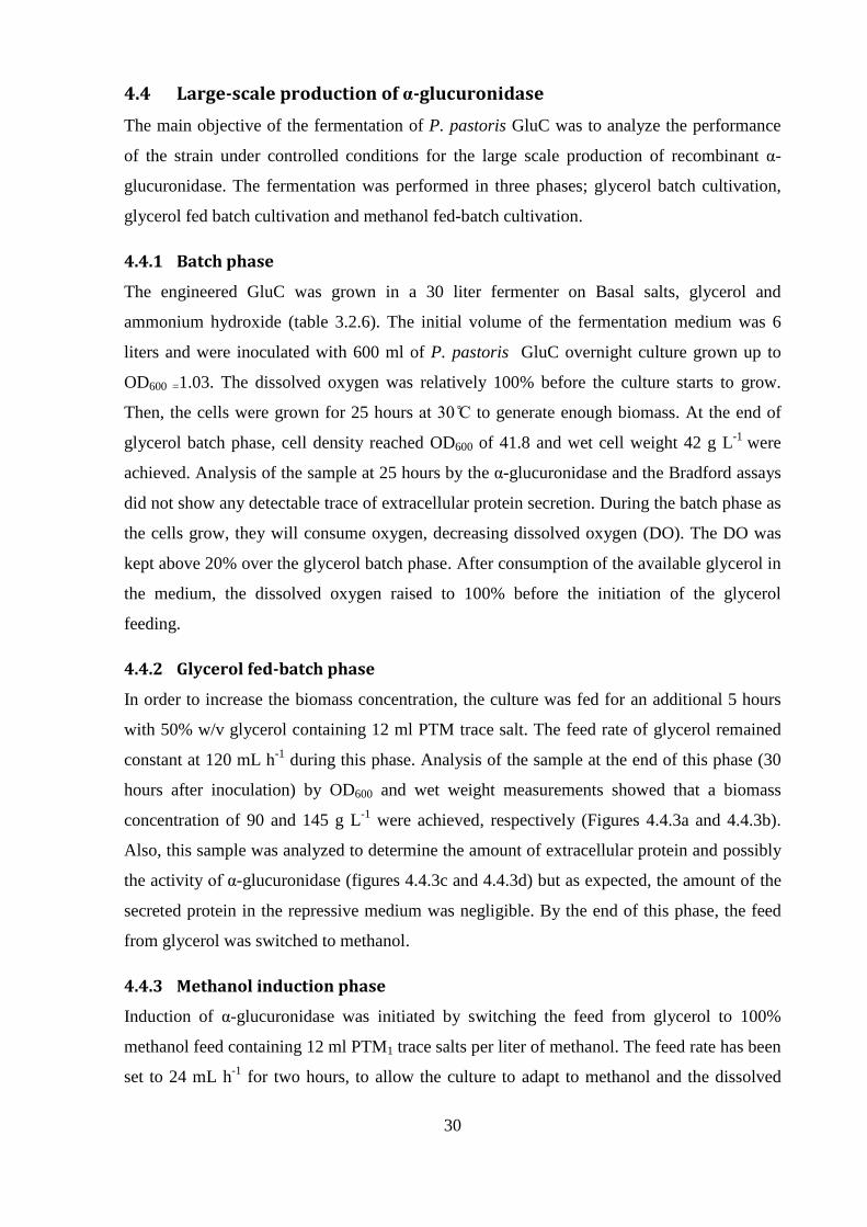
4.4.2 Glycerol fed-batch phase
In order to increase the biomass concentration, the culture was fed for an additional 5 hours
with 50% w/v glycerol containing 12 ml PTM trace salt. The feed rate of glycerol remained
constant at 120 mL h-1
during this phase. Analysis of the sample at the end of this phase (30
hours after inoculation) by OD600 and wet weight measurements showed that a biomass
concentration of 90 and 145 g L-1
were achieved, respectively (Figures 4.4.3a and 4.4.3b).
Also, this sample was analyzed to determine the amount of extracellular protein and possibly
the activity of α-glucuronidase (figures 4.4.3c and 4.4.3d) but as expected, the amount of the
secreted protein in the repressive medium was negligible. By the end of this phase, the feed
from glycerol was switched to methanol.
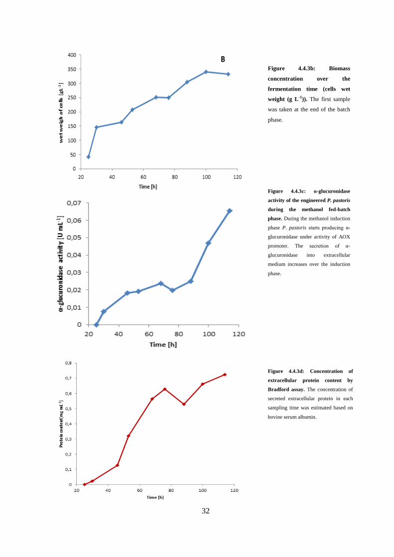
4.4.3 Methanol induction phase
Induction of α-glucuronidase was initiated by switching the feed from glycerol to 100%
methanol feed containing 12 ml PTM1 trace salts per liter of methanol. The feed rate has been
set to 24 mL h-1
for two hours, to allow the culture to adapt to methanol and the dissolved

31
oxygen to stabilize. The dissolved oxygen was fluctuating between 30% and 50% until the
end of fermentation. One hour after adaptation of the cells to methanol feed rate doubled to
50 mL h-1
for 2 hours. Finally, the feed rate was fixed to 72 mL h-1
throughout the remainder
of the induction phase (80 hours). Agitation and temperature were kept constant during the
fed-batch phases and set to 1 rpm an , respectively.
Sampling took place twice a day for analysis of growth (figures 4.4.3a-b) and protein
production (figures 4.4.3 c-d). The optical density of the fermentation culture and wet weight
exhibited an increasing pattern to the end of fermentation, from OD600 90 and 145 g L-1
to
OD600 140 and 332 g L-1
, respectively.
Samples were also analyze for extra cellular protein pro uction by ra for assay an α-
glucuroni ase assay for α-glucuronidase activity 18 hours after harvesting the cells. Analysis
showed that α-glucuronidase was secreted into medium after initiation of the methanol
induction phase at 46 h (16 hours after induction). The extracellular protein content of the
sample was determined 0.12 mg mL-1
by Bradford assay while its α-glucuronidase activity
was 0.018 U mL-1
. The amount of extracellular protein reached 0.72 mg mL-1
(figure 4.4.3d)
at the en of fermentation, however α-glucuronidase assay activity increases slowly until 88
h but the activity augments sharply from 0.024 to 0.065 U mL-1
, between 88 h to 114 h
(figure 4.4.3.c).
Figure 4.4.3.a Biomass concentration
over the fermentation time (OD600
absorbance). The first sample was taken
at the end of the batch phase.
32
Figure 4.4.3b: Biomass
concentration over the
fermentation time (cells wet
weight (g L-1)). The first sample
was taken at the end of the batch
phase.
Figure 4.4.3d: Concentration of
extracellular protein content by
Bradford assay. The concentration of
secreted extracellular protein in each
sampling time was estimated based on
bovine serum albumin.
Figure 4.4.3c: α-glucuronidase
activity of the engineered P. pastoris
during the methanol fed-batch
phase. During the methanol induction
phase P. pastoris starts pro ucing α-
glucuronidase under activity of AOX
promoter. The secretion of α-
glucuronidase into extracellular
medium increases over the induction
phase.
33
5. Discussion
In recent years many proteins has been produced recombinantly in P. pastoris. Basic features
that make methylotrophic P. pastoris a good choice for protein production are combination of
easy genetic manipulation, high specific growth rates comparable to E. coli and an eukaryotic
subcellular machinery for performing PTMs (Post Translational Modifications) on proteins
(Aoki, et al., 2003). P. pastoris SMD1168H was used in this study for the expression of α-
glucuronidase enzyme that has wild type methanol utilization phenotype (Mut+) and benefits
from aox1. SMD1168H is a protease (carboxypeptidase Y and protease B1) deficient strain
(pep4-) and it is proven that it reduces the degradation of foreign proteins effectively
(Cereghino & Cregg, 2000). Strains such as GS115, SMD1163 and SMD1165 are also Mut+
phenotypes of P.pastoris but KM71phenotype contains aox2 gene that slows down utilization
of methanol while MC100-3 phenotype does not contain even aox2 gene and cannot grow on
methanol and is known as Mut- phenotype. Except from SMD1168H strain, all of the
mentioned strains produce vacuole peptidase A. The amount of vacuole peptidases are
considerable in fermenter cultures with high cell density level, due to lysis of a small
percentage of cells (Daly & Hearn, 2005).
The amount of secreted protein into extracellular medium in fed-batch fermentation (figure
4.4.3d) of the engineered P. pastoris GluC supported our decision to use pPI Zα plasmi
for constructing expression vector. AOX1 promoter in the vector derived expression of Agu1
subcloned in the vector, in transcriptional level. The other important feature of AOX1
promoter is the ability to switching on or off the promoter by changing carbon sources.
ra for an α-glucuronidase assays’ results (figures 4.4.3d and 4.4.3e) clearly show that
switching between carbon sources from repressive glycerol to methanol at 30 hours after
inoculation, started extracellular protein production.
election of the signal sequence can be base on the protein’s native signal sequence such as
α-MF, PHO or SUC2 signal sequences (Li, et al., 2001). It is not possible to predetermine
whether using any of the mentioned signal sequences will lead to successful and efficient
secretion of the recombinant protein or not (Daly & Hearn, 2005). Many recombinant
expression studies has been performed in P. pastoris comparing effects of different signal
sequences that illustrate α-MF signal sequence has yielded higher secretion of the
recombinant protein (Daly & Hearn, 2005). Fermentation of the engineered SMD1168H
strain resulted in secretion of large amount of extracellular α-glucuronidase but so far no one
34
has attempted to overexpress Agu1in P. pastoris and secrete it by using other signal
sequences therefore there is no irect evi ence to know the secretion yiel of the α-
glucuronidase will improve or decrease by using the other signal sequences.
The synthetic Agu1 originate from S. commune which is a non-yeast organism and it should
be optimized by altering the codons bias and the GC bases content prior to expression in P.
pastoris. Codons that are available in S. commune may be rare in P. pastoris, therefore to
avoid limited translation by the amounts of available tRNA in P. pastoris, the gene should be
codon optimized (biased) for expression in P. pastoris (Romanos, et al., 1995). When
specific tRNAs are consumed, premature termination of the protein may happen (Leuking, et
al., 2000). Also if the gene has high content of AT bases, GC content of the gene should be
improved as well (Daly & Hearn, 2005).
We employed triple digestion of the pUC57 vector to isolate Agu1 with sticky ends. In the
first trial XbaI and ClaI restriction enzymes were used to isolate the gene from pUC57 vector.
After running the restriction products on electrophoresis gel instead of two separate bands,
one strong intense band between 2500 bp and 3000 bp appeared. The reason why we got a
single band was relatively similar molecular weights of the gene and the empty vector 2925
bp and 2710 bp, respectively. We decided to break pUC57 further down with ScaI to smaller
sizes for precise separation of the gene from the vector. ScaI converted empty pUC57 to two
separate bands with approximately sizes of 900bp and 1800 bp. By using this strategy a
single band at 2900bp corresponding to Agu1 gene appeared on the electrophoresis gel. Also,
pPI Zα vectors were digested with XbaI and ClaI to prepare an open frame for
construction of the expression vector.
During the construction of the expression vector, yield of the ligation reaction significantly
improved by increasing the incubation time from 4 hours to 18 hours. The first ligation
reaction trial was incubated 4 hours at room temperature. After transformation of the ligation
products to E. coli DH5α competent cells some ampicillin resistance colonies grown on LB
plates. Electrophoresis analysis of the digested plasmids that were isolated from positive
transformants revealed that the gene was not inserted in pUC57 vector and resistance of the
positive transformants was due to integration of the empty plasmids. The second ligation trial
was performed at room temperature and incubated for 18 hours, the results of the diagnostic
digestion by SpeI and sequencing confirmed cloning of the gene into pPI Zα SpeI is a
35
single cutter restriction enzyme that just cut inside the gene, precise band at 6600 bp was
observe , which is the expecte size of the gene an pPI Zα plasmi As a part of
confirmation tests, two of the expression vectors were subjected to sequencing that resulted
in full alignment of the sequenced expression vector with the sequence of the gene.
Low efficiency transformation of the expression vector into SMD1168H was improved by
increasing the linearized vector concentration. Previously purified vectors from
electrophoresis gel resulted in a very low concentration, approximately 0.07 µg µL-1
. The
required concentration of the linearized vector for electroporation is 1-2 µg µL-1
.
Concentration of the linearized vectors was improved by direct purification of the restriction
products to 0.55 µg µL-1
, instead of purification from electrophoresis gel. Another important
factor that influences the efficiency of the transformation to P. pastoris is the expression
vector size. Expression vectors with large size can result in less stable transformats (Daly &
Hearn, 2005), so, due to relatively large size of Agu1which was 2900 bp, it could have been
an additional reason for the low transformation efficiency of electroporation. Alternatively
spheroplast transformation method could have been used but the risk of contamination is high
and cell lysing enzyme reduces the cell viability (Daly & Hearn, 2005).
We precultured the inoculum in PD me ium at an 1 5 rpm to OD600 ≈1 for 24
hours before inoculation of the fermentation medium. After 25 hours of glycerol batch
culture we reached a wet cell weight of 42 g L-1
that was half the expected cellular titer of 90
g L -1
wet cell weight suggested by Invitrogen protocol. It could have been due to low cellular
concentration of the non-fully grown inoculum, OD600 ≈1 instead of 3-6. Further feeding
of the culture for 5 hour with glycerol resulted in 145 g L-1
wet cell weight, which is
relatively less than 180 g L-1
wet cell weight suggested by Invitrogen protocol. During the
methanol induction phase the overall 332 g L-1
of wet cellular weight was tittered. In
comparison to 350 g L-1
wet cell weight in the protocol, the difference between theoretical
and experimental titer of wet cell weight in the fermentation was reduced.
Methanol induction phase carried out at 30 accor ing to Invitrogen’s manual for P.
pastoris Fermentation Process Guidelines, but generally the protein synthesis rate is higher in
optimal temperature 25- 6 (Daly & Hearn, 2005; ). In uction of the protein pro uction at
6 enhances protein folding pathway and increases cell viability during (Aoki, et al., 2003).
Induction in lower temperatures provide more time for the nascent polypeptide chains to be
36
correctly folded specifically for proteins with the high content of disulfide bridges. In
addition lowering the induction temperature may reduce the proteolytic degradation of
extracellular enzymes (Li, et al., 2001) over the long induction phase and improve the protein
solubility (Aoki, et al., 2003; Chen , et al., 2000).
The time course of the recombinant pro uction of α-glucuronidase by engineered
SMD1168H was studied via ra for an α-glucuronidase assays to determine the yield of
the protein production and optimal length of the cultivation. A detectable trace of extra
cellular protein was observed at 46 h after inoculation of the fermentation medium by
Bradford assay and gradually increased until harvesting the fermentation culture at 114 hour
(fig 4.4.3c). Similar increasing pattern was observe by α-glucuronidase assay as can be seen
in fig 4.4.3d. The enzyme activity increased drastically after 88 h (58 h after induction) until
the end of induction phase. Tenkanen & Siika-aho, (2000) in their attept to produce α-
glucuronidase in S. commune, produced 4717 mg of extracellular protein after 188 hours of
cultivation in final volume of 17 L, whereas, we reached 5784 mg of extracellular protein
after 114 hours of the cultivation (86 hours of induction) of the modified P. pastoris to the
final volume of 12 L. They reported 16 nkat mL-1α-glucuronidase activity of the culture
filtrate concentrate (15 time concentrated to the final volume of 265 ml) purified by ion
exchange chromatography (DEAE Sepharose FF) The maximum α-glucuronidase activity
that we assayed from the culture filtrate was 1.08 nkat mL-1
. Lower activity of our enzyme
resulted from low concentration of the enzyme in the culture filtrate (8 L filtrate) that can be
improved by performing purification of the filtrate with ion exchange chromatography.
Low selectivity of GH115 α-glucuronidase to the aldouronic acids substrate with D-
glucuronic acid residues on the non-reducing end of xylan of the oligoxylans contributes to
the lower activity of the α-glucuronidase (Kolenova, et al., 2010). Aldouronic acids
(tri:tetra:penta) supplied by the kit lacking one adjacent residue due to terminal substitution
of glucuronic acid groups on the non-reducing end. Whereas, Tenkanen & Siika-aho, (2000)
had investigated the activity of GH115 α-glucuronidase from S. commune on a series of
polymeric substrates such as glucuronoxylan and acetyl glucuronoxylan from birch wood and
arabino-glucuronoxylan from spruce that could be another explanation of the higher activity
that they reached.
37
6. Future works
During the fermentation of the engineered strains identical process parameters suggested by
Invitrogen were applied. According to the literature, it is evident that reducing temperature
during methanol induction phase increases protein production. Feeding strategy of P. pastoris
in glycerol and methanol fed batch phases is another process bottleneck to improve. Optimal
feed rate increases protein production via increasing biomass generation yield and reducing
toxic effect of the accumulated glycerol or methanol. Therefore a set of designed experiments
can be suggested to find the optimal fermentation parameters.
The ionic interaction between enzyme and substrate seems to play a key role for the reaction.
The role of methyl group of aldouronic acid is not understood so far in the substrate binding
mechanism of GH115 α-glucuronidase. To draw a conclusion on the architecture of the
substrate binding site, substrate binding and catalysis mechanisms of the enzyme a crystal
structure of GH115 family coupled with substrate has to be solved.
7. Conclusion
Modification of SMD1168H P. pastoris parental strain for production of GH115 family α-
glucuronidase resulted in generation of new strain which is able to extracellular secretion of
this enzyme. Agu1 was codon optimized for overexpression in P. pastoris and used for
expression cassette construction in pPI Zα vector All candidate transformants produced
extracellular α-glucuronidase and were screened base on the highest α-glucuronidase activity.
Batch-fermentation of the screened strain in basal salts and later feeding with glycerol and
methanol yielded 732 mg extracellular protein per liter of fermentation filtrate with 0.062 U
mL-1
α-glucuroni ase activity This enzyme is the only known α-glucuronidase with catalytic
activity on polymeric xylan to cleave D-glucuronic acid residues. Development of P.
pastoris, which is able to grow on a cheap carbon source such as methanol is promising
economical process for pro uction of α-glucuronidase in larger scales. On the whole, the
results illustrate the capability of P. pastoris for heterologous production of biologically
active enzymes.
38
8. Acknowledgment
This thesis is dedicated to the friendship and memory of Kianoosh Asa. He was a chemical
engineering master student at Tehran University of Science and Technology who encouraged
me over a number of years. He lived his life well, acting upon his spiritual beliefs
conscientiously by assisting both friends and strangers in need. Kianooh's strength and faith
during the last year of his life gave me a new appreciation for the meaning and importance of
friendship. He faced his too early death bravely for freedom and democracy in Iran.
Kianoosh shot dead by revolutionary guards in peaceful demonstration after 2009
presidential elections in Iran and his funeral ceremony was held on his thesis defense date.
His memories always inspired me and kept me working whenever I wanted to give up.
I would like to express my deepest gratitude to my advisor, Prof. Lisbeth Olsson, for her
excellent guidance, caring, patience, and providing me with an excellent atmosphere for
doing research. My special thanks go to Dr. George Anasontzis and Hampus Sunner for
guiding my research for the past several months and helping me to develop my background
in molecular biology, biochemistry, and fermentation. Undoubtedly without their day and
night support this project could have not been successful. The support and encouragement of
all friends and colleagues in industrial biotechnology group have been indispensable.
39
9. References
Aoki, H., Ahsan, M. N. & Watabe, S., 2003. Heterologous expression in Pichia pastoris and
single-step purification of a cysteine proteinase from northern shrimp. Protein Expression
and Purification, Volume 31, p. 213–221.
Biely, P., Vries, R. d., Vrsanska, M. & Visser, J., 2000. Inverting charactor of α-
glucuronidase A from Aspergillus tubingensis. Biochimica et Biophysica , Volume 1474, pp.
360-364.
Brake, A. J. et al., 1984. Alpha-factor-directed synthesis and secretion of mature foreing
proteins in Saccharomyces cerevisiae. Proc Natl Sci USA, Volume 81(15), pp. 4642-4646.
Brink, J. v. d. & Vries, R. P. d., 2011. Fungal enzyme sets for plant polysaccharide
degradation. App Microbiol Biotechnol, Volume 91, pp. 1477-1492.
Buchanan, B., Gruissem, W. & Jones, R., 2000. Biochemistry and molecular biology of
plants 2 ed.. American Society of plant Biologists, Volume 2, pp. 61-75.
Castanare, A., Hay, A. J., Gordon, A. H. & McCrae, S. I., 1995. D-Xylan-degrading enzyme
system from the fungus Phanerochaete Chrysosporium: isolation and partial characterization
of an α-(4-o-methyl)-D-glucuronidase. Journal of Biotechnology, Volume 43, pp. 183-194.
Cereghino, J. L. & Cregg, J. M., 2000. Heterologous protein expression in methylotropic
yeast Pichia pastoris. FEMS Microbiology reviews, Volume 24, pp. 45-66.
Chen , Y. et al., 2000. Expression and characterization of glycosylated and catalytically
active recombinant human alpha-galactosidase a produced in Pichia pastoris. Protein
Expression nad Purification, Volume 20, pp. 472-484.
Chong, S.-L.et al., 2011. The α-glucuronnidase Agu1 from Schizophyllum commune is a
member of a novel glycoside hydrolase family(GH115). Appl Microbiol Biotechnol, Volume
90, pp. 1323-1332.
40
Couderc, R. & Baratti, J., 1980. Oxidation of methanol by the yeast, Pichia pastoris.
Purification and properties of the Alcohol Oxidase. Agri. Biol. Chem., Volume 44 (10), pp.
2279-2289.
Cregg, J. M. & Higgins, D. R., 1995. Production of foreign proteins in the yeast Pichia
pastoris. Canadian Journal of Botany, Volume 73(S1), pp. 981-897.
Cregg, J. M., Vedvick, T. S. & Raschke, w. C., 1993. Recent Advances in the Expression of
Foreign Genes in Pichia pastoris. Nature biotechnology, Volume 11, pp. 905-910.
Daly, R. & Hearn, M. T., 2005. Expression of heterologous proteins in Pichia pastoris: a
usefull experimental tool in protein engineering and production. journal of molecular
recognition, Volume 18, pp. 119-138.
Dammström, S., Salmen, L. & Gatenholm, . P., 2005. The effect of moisture on the
dynamical mechanical properties of bacterial. Polymer, Volume 46 , p. 10364–10371.
Dasilva, A. E. et al., 2012. Xylan, A Promising Hemicellulose for for Pharmaceutical Use. In:
C. J. R. Verbeek, ed. Products and Applications of Biopolymers. s.l.:Intech, pp. 61-84.
De Schutter, K. et al., 2009. Genome sequence of the recombinant protein production host
Pichia pastoris. Nature Biotechnology, Volume 27, pp. 561-566.
De Vries, R. P. & Visser, J., 2001. Aspergillus Enzymes Involved in Degradation of Plant
Cell Wall Polysaccharides. Microbiology and Molecular Biology reviews, Volume 65, pp.
497-522.
Gatenholm, p., Gröndahl, M. & Eriksson, L., 2004. Material Properties of Plasticized
Hardwood Xylans for Potential Application as Oxygen Barrier Films. Biomacromolecules,
Volume 5, pp. 1528-1535.
Gellissen, G. et al., 1992. Heterologous protein production in yeast. Antonie van
Leeuwenhoek, Volume 62, pp. 79-93.

41
Heneghan, M., McLoughlin , L., Murray, P. & Tuohy, M., 2007. Cloning, characterisation
and expression analysis of α-glucuronidase. Enzyme and Microbial Technology, Volume 41,
p. 677–682.
Kolenova, k., Ryabova, O., Vrsanska, m. & Biely, P., 2010. Inverting character of familt
Gh115 α-glucuronidases. FEBS Letters, Volume 584, pp. 4063-4068.
Leuking, a. et al., 2000. A system for dual protein expression in Pichia pastoris and
Escherichia coli. Protein Expression and Purification, Volume 20, pp. 372-378.
Li, P., Gao, X. & Renugopalakrishan, V., 2001. Glycosylated and phosphorylated proteins--
expression in yeast and oocytes of Xenopus: prospects and challenges--relevance to
expression of thermostable proteins.. Protein Expression and Purification, 22(3), pp. 368-
380.
Li, Z. et al., 2001. Low-temperature increases the yield of biologically active herring
antifreeze protein in Pichia pastoris. Protein Expression and Purification, Volume 21, pp.
438-445.
Nurizzo, D. et al., 2003. the α-Glucuronidase, GlcA67A, of Cellevibrio japonicus utilize the
carboxylate and methyl groups of aldobiouronic acid as important substrate recognition
determinants. The journal of biological chemistry , Volume 278, pp. 20286-20292.
Nurizzo, D., Nagy, T., Harry J., G. & Davies, G. J., 2002. The Structural Basis for Catalysis
and specificity of the Pseudomonas cellulosa α-glucuronidase, GlcA67A. Structure, Volume
10, pp. 547-556.
Olsson, L. & Hahn- Hägerdal, B., 1993. fermentative process of bacteria and yeasts in
lignocellulose hydrolysates. brocess biochem, Volume 28, pp. 249-257.
Romanos, M., Wellcome, G. & Beckenham, 1995. advances in the use of Pichia pastoris for
high-level gene expression. Current Opinion in Biotechnology, Volume 6, pp. 527-533.
42
Ryabova , O et al , A novel family of hemicellulolytic α-glucuronidase. FEBS Letters,
Volume 583, p. 1457–1462.
Saxenaa, A., Elderb, . T. J. & Ragauskas, . A. J., 2011. Moisture barrier properties of xylan
composite films. Carbohydrate Polymers, Volume 84 , p. 1371–1377.
Sreekrishna, K. et al., 1997. Strategies for optimal synthesis and secretion of heterologous
proteins in the methylotropic Pichia pastoris. Gene, Volume 190, pp. 55-62.
Tenkanen, M. & Siika-aho, M., 2000. An α-glucuronidase of Schizophyllum commune acting
on polymeric xylan. Journal of Biotechnology , Volume 78 , p. 149–161.
Tsujikawa, M., Okabayashi, K., Morita, M. & Tanabe, T., 1996. Secretion of a Variant of
Human Single-Chain Urokinase-Type Plasminogen Activator without an N-GlycosylatLon
Site in the Methylotrophic Yeast, Pichia pastoris and Characterization of the Secreted
Product. Yeast, Volume 12, pp. 541-553.
Viikari, L., Kantelinen, A., Buchert, J. & Plus, J., 1994. Enzymatic accessibility of xylans in
lignocellulosic materials. Appl Microbiol Biotechnol, Volume 41, pp. 124-129.
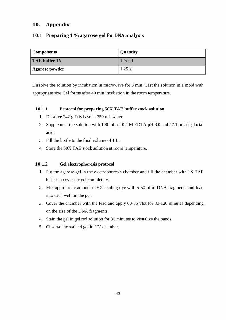
43
10. Appendix
10.1 Preparing 1 % agarose gel for DNA analysis
Components Quantity
TAE buffer 1X 125 ml
Agarose powder 1.25 g
Dissolve the solution by incubation in microwave for 3 min. Cast the solution in a mold with
appropriate size.Gel forms after 40 min incubation in the room temperature.
10.1.1 Protocol for preparing 50X TAE buffer stock solution
1. Dissolve 242 g Tris base in 750 mL water.
2. Supplement the solution with 100 mL of 0.5 M EDTA pH 8.0 and 57.1 mL of glacial
acid.
3. Fill the bottle to the final volume of 1 L.
4. Store the 50X TAE stock solution at room temperature.
10.1.2 Gel electrophoresis protocol
1. Put the agarose gel in the electrophoresis chamber and fill the chamber with 1X TAE
buffer to cover the gel completely.
2. Mix appropriate amount of 6X loading dye with 5-50 µl of DNA fragments and load
into each well on the gel.
3. Cover the chamber with the lead and apply 60-85 vlot for 30-120 minutes depending
on the size of the DNA fragments.
4. Stain the gel in gel red solution for 30 minutes to visualize the bands.
5. Observe the stained gel in UV chamber.

44
10.2 Extraction of plasmid DNA from E. coli DH5α
Figure 10.2 QuickprotocolTM – GeneJETTM Plasmid Miniprep Kit: Used for extraction and purification of
DNA/plasmids from E.coli (http://www.fermentas.com).
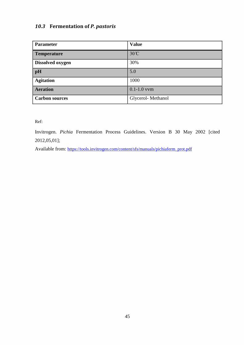
45
10.3 Fermentation of P. pastoris
Parameter Value
Temperature
Dissolved oxygen 30%
pH 5.0
Agitation 1000
Aeration 0.1-1.0 vvm
Carbon sources Glycerol- Methanol
Ref:
Invitrogen. Pichia Fermentation Process Guidelines. Version B 30 May 2002 [cited
2012,05,01];
Available from: https://tools.invitrogen.com/content/sfs/manuals/pichiaferm_prot.pdf