QM/MM simulations as an assay for carbapenemase activity ...
4
14736 | Chem. Commun., 2014, 50, 14736--14739 This journal is © The Royal Society of Chemistry 2014 Cite this: Chem. Commun., 2014, 50, 14736 QM/MM simulations as an assay for carbapenemase activity in class A b-lactamases† Ewa I. Chudyk, a Michael A. L. Limb, a Charlotte Jones, a James Spencer, b Marc W. van der Kamp* a and Adrian J. Mulholland* a Carbapenems, ‘last resort’ antibiotics for many bacterial infections, can now be broken down by several class A b-lactamases ( i.e. carbapene- mases). Here, carbapenemase activity is predicted through QM/MM dynamics simulations of acyl–enzyme deacylation, requiring only the 3D structure of the apo-enzyme. This may assist in anticipating resistance and future antibiotic design. Antibiotic (antimicrobial) resistance, particularly in Gram-negative bacteria such as Escherichia coli and Pseudomonas aeruginosa, 1 has emerged as an extremely serious and growing medical problem. It threatens modern medicine, not only due to the growing difficulty of treating bacterial infections, but also jeopardizing many thera- pies and surgical procedures. 2 Much of the developing resistance in such bacteria can be attributed to b-lactamases, enzymes which catalyse the breakdown of the essential b-lactam ring present in all classes of b-lactam antibiotics. 3 Carbapenems are a highly potent class of antibiotics: they have a broad spectrum of antibacterial activity and, in contrast to many other classes of antibiotic, have not been susceptible to resistance due to hydrolysis by b-lactamases. The characteristic broad-spectrum antibacterial activity exhibited by carbapenems means that they are currently used as effective treat- ments for otherwise incurable bacterial infections. 4 Alarmingly, however, a variety of carbapenem-hydrolysing b-lactamases have been reported 5 and are becoming increasingly common. These b-lactamases, known as carbapenemases, are able to efficiently hydrolyse the b-lactam ring in carbapenems, rendering this formerly highly potent class of antibiotics inactive and resulting in the re-emergence of potentially untreatable bacterial infections. 6 The most common enzymes with carbapenemase activity are b-lactamases of class A (exemplified by the SFC-1 and KPC-2 enzymes 7 ) and class D. Class B b-lactamases, such as NDM-1, are also increasingly prevalent carbapenemases. 8 Here, we focus on b-lactamases of class A due to the growing frequency with which carbapenem-hydrolysing enzymes are encountered in both clinical and environmental bacteria. Carbapenemase activity in these enzymes is due to efficient deacylation; the majority of class A enzymes are inhibited by carbapenems because a long-lived acyl– enzyme complex is formed. The first step of the class A b-lactamase hydrolysis mechanism is formation of an acyl–enzyme intermediate ( via nucleophilic attack by an active site serine), with coincident opening of the b-lactam ring. 9 Deacylation is then required to release the cleaved b-lactam and allow enzyme turnover. Deacylation is initiated by nucleophilic attack of a conserved water molecule (the deacylating water mole- cule, DW) on the ester carbon of the acyl–enzyme (Fig. 1). The water molecule is activated by proton abstraction by a glutamate residue situated in the active site acting as a base. 10 Carbapenem-inhibited class A b-lactamases are acylated readily but have a greatly reduced rate of deacylation, 11 resulting in a long- lived acyl–enzyme. In contrast, b-lactamases with carbapenemase activity deacylate efficiently (with a short-lived acyl–enzyme), confer- ring carbapenem resistance on bacteria that carry these enzymes. Despite multiple structural 12,13 and biochemical studies 14,15 on class A b-lactamases, the difference in carbapenemase activity exhibited by different b-lactamases (which have highly homologous active sites) is not well understood. Recently, we suggested that the orientation of the 6a-1R-hydroxyethyl group of carbapenems in the enzyme Fig. 1 The first step of carbapenem deacylation: the acyl–enzyme (A) reacts to form a tetrahedral intermediate (B) (numbering for the TEM-1 enzyme). 10 a Centre for Computational Chemistry, School of Chemistry, University of Bristol, Cantock’s Close, Bristol, BS8 1TS, UK. E-mail: [email protected], [email protected] b School of Cellular and Molecular Medicine, University of Bristol, University Walk, Bristol, BS8 1TD, UK † Electronic supplementary information (ESI) available: Full description of meth- ods, including PDB accession codes for all structures used (Table S1). Free energy surfaces for all reactions simulated (Fig. S2–S10). See DOI: 10.1039/c4cc06495j Received 18th August 2014, Accepted 2nd October 2014 DOI: 10.1039/c4cc06495j www.rsc.org/chemcomm ChemComm COMMUNICATION Open Access Article. Published on 02 October 2014. Downloaded on 3/20/2022 10:08:38 AM. This article is licensed under a Creative Commons Attribution 3.0 Unported Licence. View Article Online View Journal | View Issue
Transcript of QM/MM simulations as an assay for carbapenemase activity ...
14736 | Chem. Commun., 2014, 50, 14736--14739 This journal is©The Royal Society of Chemistry 2014
Cite this:Chem. Commun., 2014,
50, 14736
QM/MM simulations as an assay forcarbapenemase activity in class A b-lactamases†
Ewa I. Chudyk,a Michael A. L. Limb,a Charlotte Jones,a James Spencer,b
Marc W. van der Kamp*a and Adrian J. Mulholland*a
Carbapenems, ‘last resort’ antibiotics for many bacterial infections, can
now be broken down by several class A b-lactamases (i.e. carbapene-
mases). Here, carbapenemase activity is predicted through QM/MM
dynamics simulations of acyl–enzyme deacylation, requiring only the
3D structure of the apo-enzyme. This may assist in anticipating resistance
and future antibiotic design.
Antibiotic (antimicrobial) resistance, particularly in Gram-negativebacteria such as Escherichia coli and Pseudomonas aeruginosa,1 hasemerged as an extremely serious and growing medical problem. Itthreatens modern medicine, not only due to the growing difficultyof treating bacterial infections, but also jeopardizing many thera-pies and surgical procedures.2 Much of the developing resistance insuch bacteria can be attributed to b-lactamases, enzymes whichcatalyse the breakdown of the essential b-lactam ring present in allclasses of b-lactam antibiotics.3 Carbapenems are a highly potentclass of antibiotics: they have a broad spectrum of antibacterialactivity and, in contrast to many other classes of antibiotic, have notbeen susceptible to resistance due to hydrolysis by b-lactamases.The characteristic broad-spectrum antibacterial activity exhibited bycarbapenems means that they are currently used as effective treat-ments for otherwise incurable bacterial infections.4
Alarmingly, however, a variety of carbapenem-hydrolysingb-lactamases have been reported5 and are becoming increasinglycommon. These b-lactamases, known as carbapenemases, are ableto efficiently hydrolyse the b-lactam ring in carbapenems, renderingthis formerly highly potent class of antibiotics inactive and resultingin the re-emergence of potentially untreatable bacterial infections.6
The most common enzymes with carbapenemase activity areb-lactamases of class A (exemplified by the SFC-1 and KPC-2 enzymes7)
and class D. Class B b-lactamases, such as NDM-1, are alsoincreasingly prevalent carbapenemases.8 Here, we focus onb-lactamases of class A due to the growing frequency with whichcarbapenem-hydrolysing enzymes are encountered in both clinicaland environmental bacteria. Carbapenemase activity in theseenzymes is due to efficient deacylation; the majority of class Aenzymes are inhibited by carbapenems because a long-lived acyl–enzyme complex is formed.
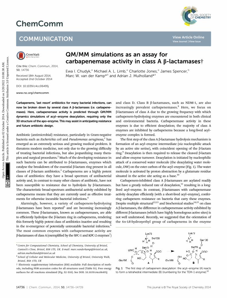
The first step of the class A b-lactamase hydrolysis mechanism isformation of an acyl–enzyme intermediate (via nucleophilic attackby an active site serine), with coincident opening of the b-lactamring.9 Deacylation is then required to release the cleaved b-lactamand allow enzyme turnover. Deacylation is initiated by nucleophilicattack of a conserved water molecule (the deacylating water mole-cule, DW) on the ester carbon of the acyl–enzyme (Fig. 1). The watermolecule is activated by proton abstraction by a glutamate residuesituated in the active site acting as a base.10
Carbapenem-inhibited class A b-lactamases are acylated readilybut have a greatly reduced rate of deacylation,11 resulting in a long-lived acyl–enzyme. In contrast, b-lactamases with carbapenemaseactivity deacylate efficiently (with a short-lived acyl–enzyme), confer-ring carbapenem resistance on bacteria that carry these enzymes.Despite multiple structural12,13 and biochemical studies14,15 on classA b-lactamases, the difference in carbapenemase activity exhibited bydifferent b-lactamases (which have highly homologous active sites) isnot well understood. Recently, we suggested that the orientation ofthe 6a-1R-hydroxyethyl group of carbapenems in the enzyme
Fig. 1 The first step of carbapenem deacylation: the acyl–enzyme (A) reactsto form a tetrahedral intermediate (B) (numbering for the TEM-1 enzyme).10
a Centre for Computational Chemistry, School of Chemistry, University of Bristol,
Cantock’s Close, Bristol, BS8 1TS, UK. E-mail: [email protected],
[email protected] School of Cellular and Molecular Medicine, University of Bristol, University Walk,
Bristol, BS8 1TD, UK
† Electronic supplementary information (ESI) available: Full description of meth-ods, including PDB accession codes for all structures used (Table S1). Free energysurfaces for all reactions simulated (Fig. S2–S10). See DOI: 10.1039/c4cc06495j
Received 18th August 2014,Accepted 2nd October 2014
DOI: 10.1039/c4cc06495j
www.rsc.org/chemcomm
ChemComm
COMMUNICATION
Ope
n A
cces
s A
rtic
le. P
ublis
hed
on 0
2 O
ctob
er 2
014.
Dow
nloa
ded
on 3
/20/
2022
10:
08:3
8 A
M.
Thi
s ar
ticle
is li
cens
ed u
nder
a C
reat
ive
Com
mon
s A
ttrib
utio
n 3.
0 U
npor
ted
Lic
ence
.
View Article OnlineView Journal | View Issue
This journal is©The Royal Society of Chemistry 2014 Chem. Commun., 2014, 50, 14736--14739 | 14737
active site may have an important influence on the deacylationrate (based on crystal structures and molecular dynamics (MD)simulations of the carbapenemase SFC-1).7 We proposed that,in carbapenemases, a conformation of the 6a-1R-hydroxyethylthat does not interfere with the nucleophilic attack of the DW isfavored, which facilitates deacylation. Further subtle structuraldifferences were identified that may affect the rate of deacylation,supporting previous findings.14,16,17 Here we use simulations toinvestigate class A b-lactamases, with the aim of identifyingdeterminants of carbapenemase activity, and ultimately of applyingthese principles to assess the ability of uncharacterized enzymes tohydrolyse carbapenems. The need to recognize new and emergingresistance threats is growing in importance as new enzymescontinue to be identified and new sequences emerge fromlarge-scale sequencing projects.
We employ hybrid quantum mechanics/molecular mechanics(QM/MM) umbrella sampling MD calculations to investigatecarbapenem hydrolysis in class A b-lactamases.18 Because thedifference between carbapenemases and carbapenem-inhibitedenzymes is in the deacylation step, and tetrahedral intermediate(TI) formation is likely to have the highest barrier in thisreaction,11 we model the first step of deacylation only (Fig. 1).Simulations were performed with the semi-empirical SCC-DFTBQM method19 in the AMBER12 simulation package,20 using theff12SB MM force-field for the protein, the TIP4P-Ew water modeland the General AMBER Force Field (GAFF) for the part ofmeropenem not in the QM region. The QM region (Fig. S1, ESI†)consisted of the common carbapenem scaffold (omitting the3-[5-(dimethylcarbamoyl) pyrrolidin-2-y]-sulfanyl R-group whenmodelling meropenem, see Fig. 1), DW and the Ser70 andGlu166 sidechains from Cb. The protocol can be summarizedas follows (see ESI† for details):
(1) Generate and solvate the acyl–enzyme complex (AC) startingstructure. In absence of an acyl–enzyme crystal structure, dockmeropenem into the protein in the conformation observed crystallo-graphically in the SFC-1 acyl–enzyme (PDB ID: 4EV4).
(2) Heat the system to 300 K and equilibrate at 300 K and1 atm in QM/MM MD (see ESI†).
(3) 300 ps of unrestrained QM/MM MD of the acyl–enzyme.(4) Select a starting structure for reaction modelling. Previous
work7 indicated that three distinct conformations of the 6a-1R-hydroxyethyl group could be present in the acyl–enzymecomplex. A suitable structure for reaction modelling must satisfytwo criteria: (i) it contains the predominant conformation of the6a-1R-hydroxyethyl observed over the course of 300 ps unrestrainedMD simulation and (ii) it contains the DW correctly positionedfor nucleophilic attack of the electrophilic carbon in the ligand(indicated by a distance of less than 3.5 Å). If both cannot besatisfied, steps 1–3 are repeated with the 6a-1R-hydroxyethylconformation as in the structure with PDB ID: 4EUZ.7
(5) Perform QM/MM umbrella sampling MD along two reactioncoordinates: rx = d(Oe1Glu166–H2DW) � d(H2DW–ODW) (proton trans-fer from DW to Glu166) and ry = d(CCMer–ODW) (nucleophilic attackof DW on the carbonyl carbon) to simulate the deacylation reaction.Perform 20 ps of MD for each simulation window. We repeated thisstep three times to test convergence.
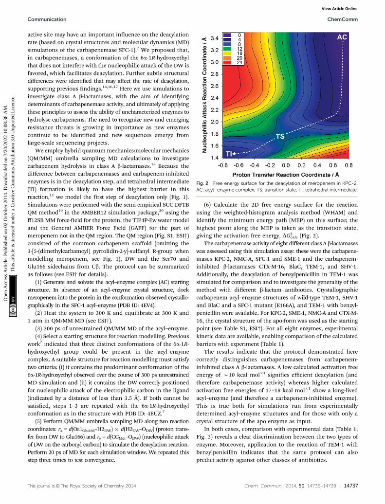
(6) Calculate the 2D free energy surface for the reactionusing the weighted-histogram analysis method (WHAM) andidentify the minimum energy path (MEP) on this surface; thehighest point along the MEP is taken as the transition state,giving the activation free energy, DG‡
calc (Fig. 2).The carbapenemase activity of eight different class A b-lactamases
was assessed using this simulation assay: these were the carbapene-mases KPC-2, NMC-A, SFC-1 and SME-1 and the carbapenem-inhibited b-lactamases CTX-M-16, BlaC, TEM-1, and SHV-1.Additionally, the deacylation of benzylpenicillin in TEM-1 wassimulated for comparison and to investigate the generality of themethod with different b-lactam antibiotics. Crystallographiccarbapenem acyl–enzyme structures of wild-type TEM-1, SHV-1and BlaC and a SFC-1 mutant (E166A), and TEM-1 with benzyl-penicillin were available. For KPC-2, SME-1, NMC-A and CTX-M-16, the crystal structure of the apo-form was used as the startingpoint (see Table S1, ESI†). For all eight enzymes, experimentalkinetic data are available, enabling comparison of the calculatedbarriers with experiment (Table 1).
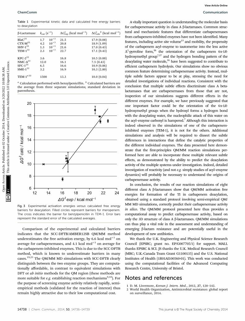
The results indicate that the protocol demonstrated herecorrectly distinguishes carbapenemases from carbapenem-inhibited class A b-lactamases. A low calculated activation freeenergy of B10 kcal mol�1 signifies efficient deacylation (andtherefore carbapenemase activity) whereas higher calculatedactivation free energies of 17–18 kcal mol�1 show a long-livedacyl–enzyme (and therefore a carbapenem-inhibited enzyme).This is true both for simulations run from experimentallydetermined acyl–enzyme structures and for those with only acrystal structure of the apo enzyme as input.
In both cases, comparison with experimental data (Table 1;Fig. 3) reveals a clear discrimination between the two types ofenzyme. Moreover, application to the reaction of TEM-1 withbenzylpenicillin indicates that the same protocol can alsopredict activity against other classes of antibiotics.
Fig. 2 Free energy surface for the deacylation of meropenem in KPC-2.AC: acyl–enzyme complex; TS: transition state; TI: tetrahedral intermediate.
Communication ChemComm
Ope
n A
cces
s A
rtic
le. P
ublis
hed
on 0
2 O
ctob
er 2
014.
Dow
nloa
ded
on 3
/20/
2022
10:
08:3
8 A
M.
Thi
s ar
ticle
is li
cens
ed u
nder
a C
reat
ive
Com
mon
s A
ttrib
utio
n 3.
0 U
npor
ted
Lic
ence
.View Article Online
14738 | Chem. Commun., 2014, 50, 14736--14739 This journal is©The Royal Society of Chemistry 2014
Comparison of the experimental and calculated barriersindicates that the SCC-DFTB/AMBER12SB QM/MM methodunderestimates the free activation energy, by 6.6 kcal mol�1 onaverage for carbapenemases, and 4.1 kcal mol�1 on average forthe carbapenem-inhibited enzymes. This is due to the SCC-DFTBmethod, which is known to underestimate barriers in manycases.19,25 The QM/MM MD simulations with SCC-DFTB clearlydistinguish between the two enzyme groups. They are computa-tionally affordable, in contrast to equivalent simulations withDFT or ab initio methods for the QM region (these methods aremore suitable for e.g. establishing reaction mechanisms9,10). Forthe purpose of screening enzyme activity relatively rapidly, semi-empirical methods (validated for the reaction of interest) thusremain highly attractive due to their low computational cost.
A vitally important question is understanding the molecular basisfor carbapenemase activity in class A b-lactamases. Common struc-tural and mechanistic features that differentiate carbapenemasesfrom carbapenem-inhibited enzymes have not been identified. Manyfeatures, including active site volume13 and mobility, the propensityof the carbapenem acyl–enzyme to tautomerise into the less activeD1-pyrroline form,26 the orientation of the carbapenem 6a-1R-hydroxymethyl group7,27 and the hydrogen bonding pattern of thedeacylating water molecule,16 have been suggested to contribute toefficient carbapenem hydrolysis. Our simulations show no obviouscommon feature determining carbapenemase activity. Instead, mul-tiple subtle factors appear to be at play, stressing the need fordetailed investigations of individual reactions. Consistent with theconclusion that multiple subtle effects discriminate class A beta-lactamases that are carbapenemases from those that are not,inspection of our simulations suggests different effects in thedifferent enzymes. For example, we have previously suggested thatone important factor could be the orientation of the 6a-1R-hydroxymethyl group: when the hydroxyl forms a hydrogen bondwith the deacylating water, the nucleophilic attack of this water onthe acyl–enzyme carbonyl is hampered.7 Although this interaction isindeed observed in the simulations of one of the carbapenem-inhibited enzymes (TEM-1), it is not for the others. Additionalsimulations and analysis will be required to dissect the subtledifferences in interactions that define the catalytic properties ofthe different individual enzymes. The data presented here demon-strate that the first-principles QM/MM reaction simulations per-formed here are able to incorporate these multiple relevant subtleeffects, as demonstrated by the ability to predict the deacylationactivity of the multiple systems under investigation. Indeed, detailedinvestigation of reactivity (and not e.g. simply studies of acyl–enzymedynamics) will probably be necessary to understand the origins ofcarbapenemase activity.
In conclusion, the results of our reaction simulations of eightdifferent class A b-lactamases show that QM/MM activation freeenergies for formation of the TI in carbapenem deacylation,obtained using a standard protocol involving semi-empirical QM/MM MD simulations, correctly predict their carbapenemase activityin silico. The QM/MM protocol presented here thus provides acomputational assay to predict carbapenemase activity, based ononly the 3D structure of class A b-lactamases. QM/MM simulationscan thus play a vital role in the assessment and understanding ofemerging b-lactam resistance and are potentially useful in thedevelopment of new antibiotics.
We thank the U.K. Engineering and Physical Science ResearchCouncil (EPSRC; grant no. EP/G007705/1) for support. MALLthanks EPSRC & SCI. JS thanks the U.K. Medical Research Council(MRC; U.K.-Canada Team Grant G1100135) and the U.S. NationalInstitutes of Health (1R01AI100560-01). This work was conductedusing the computational facilities of the Advanced ComputingResearch Centre, University of Bristol.
Notes and references1 D. M. Livermore, Korean J. Intern. Med., 2012, 27, 128–142.2 World Health Organization, Antimicrobial resistance: global report
on surveillance, 2014.
Table 1 Experimental kinetic data and calculated free energy barriersto deacylation
b-Lactamase kcat (s�1) DG‡exp (kcal mol�1) DG‡
calcb (kcal mol�1)
BlaC21 1.7 � 10�3 21.5 17.9 (0.08)CTX-M22 4.2 � 10�3 20.8 18.9 (1.09)SHV-122 1.3 � 10�3 21.6 17.0 (0.43)TEM-122 2.3 � 10�3 22.7 17.1 (0.43)
KPC-222 3.6 16.8 10.5 (0.88)NMC-A23 12.0 16.1 7.5 (0.43)SFC-124 6.5 16.6 10.9 (0.86)SME-122 3.2 16.9 10.3 (2.80)
TEM-1a 22 1500 13.3 10.0 (0.04)
a Calculation performed with benzylpenicillin. b Calculated barriers arethe average from three separate simulations; standard deviation inparenthesis.
Fig. 3 Experimental activation energies versus calculated free energybarriers for deacylation. Filled circles represent barriers for meropenem.The cross indicates the barrier for benzylpenicillin in TEM-1. Error barsrepresent the standard error of the calculated averages.
ChemComm Communication
Ope
n A
cces
s A
rtic
le. P
ublis
hed
on 0
2 O
ctob
er 2
014.
Dow
nloa
ded
on 3
/20/
2022
10:
08:3
8 A
M.
Thi
s ar
ticle
is li
cens
ed u
nder
a C
reat
ive
Com
mon
s A
ttrib
utio
n 3.
0 U
npor
ted
Lic
ence
.View Article Online
This journal is©The Royal Society of Chemistry 2014 Chem. Commun., 2014, 50, 14736--14739 | 14739
3 J. F. Fisher, S. O. Meroueh and S. Mobashery, Chem. Rev., 2005, 105,395–424.
4 D. P. Nicolau, Expert Opin. Pharmacother., 2008, 9, 23–37; K. M. Papp-Wallace, A. Endimiani, M. A. Taracila and R. A. Bonomo, Antimicrob.Agents Chemother., 2011, 55, 4943–4960.
5 H. Yigit, A. M. Queenan, G. J. Anderson, A. Domenech-Sanchez,J. W. Biddle, C. D. Steward, S. Alberti, K. Bush and F. C. Tenover,Antimicrob. Agents Chemother., 2001, 45, 1151–1161; I. Henriques,A. Moura, A. Alves, M. J. Saavedra and A. Correia, Antimicrob. AgentsChemother., 2004, 48, 2321–2323.
6 P. Nordmann, G. Cuzon and T. Naas, Lancet Infect. Dis., 2009, 9,228–236.
7 F. Fonseca, E. I. Chudyk, M. W. van der Kamp, A. Correia, A. J.Mulholland and J. Spencer, J. Am. Chem. Soc., 2012, 134, 18275–18285.
8 T. R. Walsh, Int. J. Antimicrob. Agents, 2010, 36(suppl 3), S8–S14.9 J. C. Hermann, J. Pradon, J. N. Harvey and A. J. Mulholland, J. Phys.
Chem. A, 2009, 113, 11984–11994.10 J. C. Hermann, L. Ridder, H.-D. Holtje and A. J. Mulholland, Org.
Biomol. Chem., 2006, 4, 206–210.11 L. Mourey, K. Miyashita, P. Swaren, A. Bulychev, J. P. Samama and
S. Mobashery, J. Am. Chem. Soc., 1998, 120, 9382–9383.12 P. Swaren, L. Maveyraud, X. Raquet, S. Cabantous, C. Duez, J. D. Pedelacq,
S. Mariotte-Boyer, L. Mourey, R. Labia, M. H. Nicolas-Chanoine,P. Nordmann, J. M. Frere and J. P. Samama, J. Biol. Chem., 1998, 273,26714–26721; W. Sougakoff, G. L’Hermite, L. Pernot, T. Naas, V. Guillet,P. Nordmann, V. Jarlier and J. Delettre, Acta Crystallogr., Sect. D: Biol.Crystallogr., 2002, 58, 267–274; S. Petrella, N. Ziental-Gelus, C. Mayer,M. Renard, V. Jarlier and W. Sougakoff, Antimicrob. Agents Chemother.,2008, 52, 3725–3736.
13 W. Ke, C. R. Bethel, J. M. Thomson, R. A. Bonomo and F. van denAkker, Biochemistry, 2007, 46, 5732–5740.
14 G. Zafaralla and S. Mobashery, J. Am. Chem. Soc., 1992, 114, 1505–1506.15 H. Frase, Q. Shi, S. A. Testero, S. Mobashery and S. B. Vakulenko, J. Biol.
Chem., 2009, 284, 29509–29513; F. K. Majiduddin and T. Palzkill,Antimicrob. Agents Chemother., 2003, 47, 1062–1067; F. K. Majiduddinand T. Palzkill, Antimicrob. Agents Chemother., 2005, 49, 3421–3427;K. M. Papp-Wallace, M. Taracila, J. M. Hornick, A. M. Hujer,K. M. Hujer, A. M. Distler, A. Endimiani and R. A. Bonomo, Antimicrob.Agents Chemother., 2010, 54, 2867–2877; K. M. Papp-Wallace, M. Taracila,
C. J. Wallace, K. M. Hujer, C. R. Bethel, J. M. Hornick and R. A. Bonomo,Protein Sci., 2010, 19, 1714–1727.
16 M. Nukaga, C. R. Bethel, J. M. Thomson, A. M. Hujer, A. Distler,V. E. Anderson, J. R. Knox and R. A. Bonomo, J. Am. Chem. Soc., 2008,130, 12656–12662.
17 L. Maveyraud, L. Mourey, L. P. Kotra, J. D. Pedelacq, V. Guillet,S. Mobashery and J. P. Samama, J. Am. Chem. Soc., 1998, 120,9748–9752; C. R. Bethel, M. Taracila, T. Shyr, J. M. Thomson,A. M. Distler, K. M. Hujer, A. M. Hujer, A. Endimiani, K. Papp-Wallace, R. Bonnet and R. A. Bonomo, Antimicrob. Agents Chemother.,2011, 55, 3465–3475.
18 M. W. van der Kamp and A. J. Mulholland, Biochemistry, 2013, 52,2708–2728.
19 M. Elstner, T. Frauenheim, E. Kaxiras, G. Seifert and S. Suhai, Phys.Status Solidi B, 2000, 217, 357–376; M. Gaus, Q. Cui and M. Elstner,Wiley Interdiscip. Rev.: Comput. Mol. Sci., 2014, 4, 49–61.
20 D. A. Case, T. A. Darden, T. E. Cheatham III, C. L. Simmerling, J. Wang,R. E. Duke, R. Luo, R. C. Walker, W. Zhang, K. M. Merz, B. Roberts,S. Hayik, A. Roitberg, G. Seabra, J. Swails, A. W. Gotz, I. Kolossvary,K. F. Wong, F. Paesani, J. Vanicek, R. M. Wolf, J. Liu, X. Wu, S. R. Brozell,T. Steinbrecher, H. Gohlke, Q. Cai, X. Ye, J. Wang, M.-J. Hsieh, G. Cui,D. R. Roe, D. H. Mathews, M. G. Seetin, R. Salomon-Ferrer, C. Sagui,V. Babin, T. Luchko, S. Gusarov, A. Kovalenko and P. A. Kollman, AMBER12, University of California, San Francisco, 2012.
21 J.-E. Hugonnet, L. W. Tremblay, H. I. Boshoff, C. E. Barry III andJ. S. Blanchard, Science, 2009, 323, 1215–1218.
22 A. M. Queenan, W. Shang, R. Flamm and K. Bush, Antimicrob. AgentsChemother., 2010, 54, 565–569.
23 M. S. Boyer, N. M. H. Chanoine and R. Labia, FEMS Microbiol. Lett.,1996, 143, 29–33.
24 F. Fonseca, A. C. Sarmento, I. Henriques, B. Samyn, J. van Beeumen,P. Domingues, M. R. Domingues, M. J. Saavedra and A. Correia,Antimicrob. Agents Chemother., 2007, 51, 4512–4514.
25 H. L. Woodcock, M. Hodoscek and B. R. Brooks, J. Phys. Chem. A,2007, 111, 5720–5728.
26 M. Kelp and P. R. Carey, Biochemistry, 2008, 47, 11830–11837.27 C. A. Smith, H. Frase, M. Toth, M. Kumarasiri, K. Wiafe, J. Munoz,
S. Mobashery and S. B. Vakulenko, J. Am. Chem. Soc., 2012, 134,19512–19515.
Communication ChemComm
Ope
n A
cces
s A
rtic
le. P
ublis
hed
on 0
2 O
ctob
er 2
014.
Dow
nloa
ded
on 3
/20/
2022
10:
08:3
8 A
M.
Thi
s ar
ticle
is li
cens
ed u
nder
a C
reat
ive
Com
mon
s A
ttrib
utio
n 3.
0 U
npor
ted
Lic
ence
.View Article Online