
Products for Flow Cytometry - Biotium
12
Products for Flow Cytometry CF® Dyes for Flow Cytometer Laser Lines 2 Primary Antibodies and Bioconjugates 3 Antibody Labeling Kits 4 Cellular Assays 6 Cell viability assays 6 Cell proliferation and cell cycle 7 Apoptosis and necrosis 8 Mitochondrial membrane potential dyes Flow Cytometry Accessories 10 Spectral Flow Cytometry 11 www.biotium.com 10
Transcript of Products for Flow Cytometry - Biotium
Products for Flow Cytometry
CF® Dyes for Flow Cytometer Laser Lines 2
Primary Antibodies and Bioconjugates 3
Antibody Labeling Kits 4
Cellular Assays 6
Cell viability assays 6
Cell proliferation and cell cycle 7
Apoptosis and necrosis 8
Mitochondrial membrane potential dyes
Flow Cytometry Accessories 10
Spectral Flow Cytometry 11
www.biotium.com
10
2 • www.biotium.com
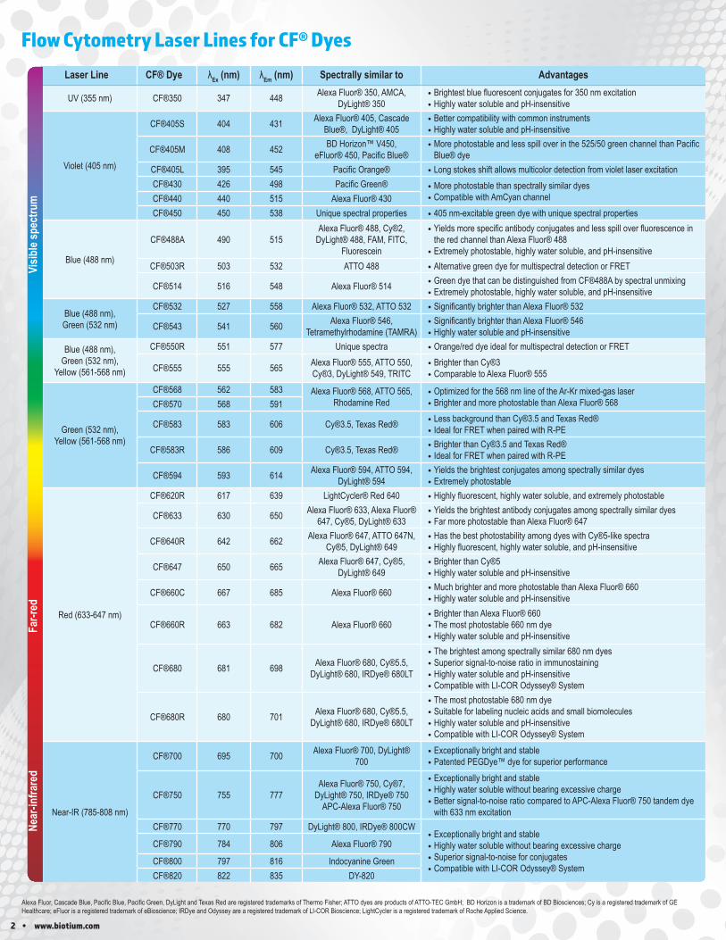
Laser Line CF® Dye λEx (nm) λEm (nm) Spectrally similar to Advantages
UV (355 nm) CF®350 347 448 Alexa Fluor® 350, AMCA, DyLight® 350
• Brightest blue fluorescent conjugates for 350 nm excitation • Highly water soluble and pH-insensitive
Violet (405 nm)
CF®405S 404 431 Alexa Fluor® 405, Cascade Blue®, DyLight® 405
• Better compatibility with common instruments • Highly water soluble and pH-insensitive
CF®405M 408 452 BD Horizon™ V450, eFluor® 450, Pacific Blue®
• More photostable and less spill over in the 525/50 green channel than Pacific Blue® dye
CF®405L 395 545 Pacific Orange® • Long stokes shift allows multicolor detection from violet laser excitationCF®430 426 498 Pacific Green® • More photostable than spectrally similar dyes
• Compatible with AmCyan channelCF®440 440 515 Alexa Fluor® 430CF®450 450 538 Unique spectral properties • 405 nm-excitable green dye with unique spectral properties
Blue (488 nm)
CF®488A 490 515Alexa Fluor® 488, Cy®2,
DyLight® 488, FAM, FITC, Fluorescein
• Yields more specific antibody conjugates and less spill over fluorescence in the red channel than Alexa Fluor® 488
• Extremely photostable, highly water soluble, and pH-insensitiveCF®503R 503 532 ATTO 488 • Alternative green dye for multispectral detection or FRET
CF®514 516 548 Alexa Fluor® 514 • Green dye that can be distinguished from CF®488A by spectral unmixing • Extremely photostable, highly water soluble, and pH-insensitive
Blue (488 nm),Green (532 nm)
CF®532 527 558 Alexa Fluor® 532, ATTO 532 • Significantly brighter than Alexa Fluor® 532
CF®543 541 560 Alexa Fluor® 546, Tetramethylrhodamine (TAMRA)
• Significantly brighter than Alexa Fluor® 546 • Highly water soluble and pH-insensitive
Blue (488 nm),Green (532 nm),
Yellow (561-568 nm)
CF®550R 551 577 Unique spectra • Orange/red dye ideal for multispectral detection or FRET
CF®555 555 565 Alexa Fluor® 555, ATTO 550, Cy®3, DyLight® 549, TRITC
• Brighter than Cy®3 • Comparable to Alexa Fluor® 555
Green (532 nm),Yellow (561-568 nm)
CF®568 562 583 Alexa Fluor® 568, ATTO 565, Rhodamine Red
• Optimized for the 568 nm line of the Ar-Kr mixed-gas laser • Brighter and more photostable than Alexa Fluor® 568CF®570 568 591
CF®583 583 606 Cy®3.5, Texas Red® • Less background than Cy®3.5 and Texas Red® • Ideal for FRET when paired with R-PE
CF®583R 586 609 Cy®3.5, Texas Red® • Brighter than Cy®3.5 and Texas Red® • Ideal for FRET when paired with R-PE
CF®594 593 614 Alexa Fluor® 594, ATTO 594, DyLight® 594
• Yields the brightest conjugates among spectrally similar dyes • Extremely photostable
Red (633-647 nm)
CF®620R 617 639 LightCycler® Red 640 • Highly fluorescent, highly water soluble, and extremely photostable
CF®633 630 650 Alexa Fluor® 633, Alexa Fluor® 647, Cy®5, DyLight® 633
• Yields the brightest antibody conjugates among spectrally similar dyes • Far more photostable than Alexa Fluor® 647
CF®640R 642 662 Alexa Fluor® 647, ATTO 647N, Cy®5, DyLight® 649
• Has the best photostability among dyes with Cy®5-like spectra • Highly fluorescent, highly water soluble, and pH-insensitive
CF®647 650 665 Alexa Fluor® 647, Cy®5, DyLight® 649
• Brighter than Cy®5 • Highly water soluble and pH-insensitive
CF®660C 667 685 Alexa Fluor® 660 • Much brighter and more photostable than Alexa Fluor® 660 • Highly water soluble and pH-insensitive
CF®660R 663 682 Alexa Fluor® 660 • Brighter than Alexa Fluor® 660 • The most photostable 660 nm dye • Highly water soluble and pH-insensitive
CF®680 681 698 Alexa Fluor® 680, Cy®5.5, DyLight® 680, IRDye® 680LT
• The brightest among spectrally similar 680 nm dyes • Superior signal-to-noise ratio in immunostaining • Highly water soluble and pH-insensitive • Compatible with LI-COR Odyssey® System
CF®680R 680 701 Alexa Fluor® 680, Cy®5.5, DyLight® 680, IRDye® 680LT
• The most photostable 680 nm dye • Suitable for labeling nucleic acids and small biomolecules • Highly water soluble and pH-insensitive • Compatible with LI-COR Odyssey® System
Near-IR (785-808 nm)
CF®700 695 700 Alexa Fluor® 700, DyLight® 700
• Exceptionally bright and stable • Patented PEGDye™ dye for superior performance
CF®750 755 777Alexa Fluor® 750, Cy®7,
DyLight® 750, IRDye® 750 APC-Alexa Fluor® 750
• Exceptionally bright and stable • Highly water soluble without bearing excessive charge • Better signal-to-noise ratio compared to APC-Alexa Fluor® 750 tandem dye with 633 nm excitation
CF®770 770 797 DyLight® 800, IRDye® 800CW • Exceptionally bright and stable • Highly water soluble without bearing excessive charge • Superior signal-to-noise for conjugates • Compatible with LI-COR Odyssey® System
CF®790 784 806 Alexa Fluor® 790
CF®800 797 816 Indocyanine GreenCF®820 822 835 DY-820
Alexa Fluor, Cascade Blue, Pacific Blue, Pacific Green, DyLight and Texas Red are registered trademarks of Thermo Fisher; ATTO dyes are products of ATTO-TEC GmbH; BD Horizon is a trademark of BD Biosciences; Cy is a registered trademark of GE Healthcare; eFluor is a registered trademark of eBioscience; IRDye and Odyssey are a registered trademark of LI-COR Bioscience; LightCycler is a registered trademark of Roche Applied Science.
Flow Cytometry Laser Lines for CF® DyesNe
ar-in
frare
dFa
r-red
Visib
le sp
ectru
m
www.biotium.com • 3
Other Bioconjugates Application
Annexin V Apoptosis (phosphatidylserine) detection (see p. 9)Dextrans & BSA Fluid phase endocytosis tracer
Streptavidin Detection of biotinylated probesBiotin Biotinylated probes, and biotin
Anti-Tag and Anti-Hapten Antibodies
Mouse Monoclonal Anti-GFPRabbit Anti-RFPRabbit Anti-GST
Rabbit Anti-HA TagMouse Monoclonal Anti-6X His Tag
Rabbit Anti-Myc TagRabbit Anti-V5 Tag
Rabbit Anti-FLAG TagMouse Monoclonal Anti-Biotin
Mouse Monoclonal Anti-Fluorescein
Format Concentration Size
CF® dye conjugates (13 colors) 0.1 mg/mL 100 or 500 uLBiotin, HRP, or AP conjugates 0.1 mg/mL 100 or 500 uL
R-PE, APC, or PerCP conjugates 0.1 mg/mL 250 uLPurified, with BSA 0.2 mg/mL 100 or 500 uL
Purified, BSA-free (Mix-n-Stain™ Ready) 1 mg/mL 50 uL
Biotium offers a wide selection of primary antibodies and biomolecules conjugated to our bright and photostable CF® dyes, as well as R-phycoerythrin (R-PE), allophyocyanin (APC), PerCP, and biotin. Visit www.biotium.com for a full listing of fluorescent conjugates for flow cytometry.
Primary Antibodies and Other Bioconjugates
Primary Antibody Sizes and FormatsGrowing selection of over 1000 mAbs
• Comprehensive library of recombinant monoclonal mouse and rabbit antibodies
• Choice of 13 bright and photostable CF® dyes• Other fluorophores available including R-PE, APC, PerCP, HRP,
AP, or Biotin• Isotype control antibodies available conjugated to the same dyes
and labels• BSA-free purified antibodies ready to use for Mix-n-Stain™ labeling
Looking for another conjugation?If you are looking for a product not listed on our website, please contact [email protected] and let us know. We may be able to add it as a new product, or perform a custom conjugation for you.
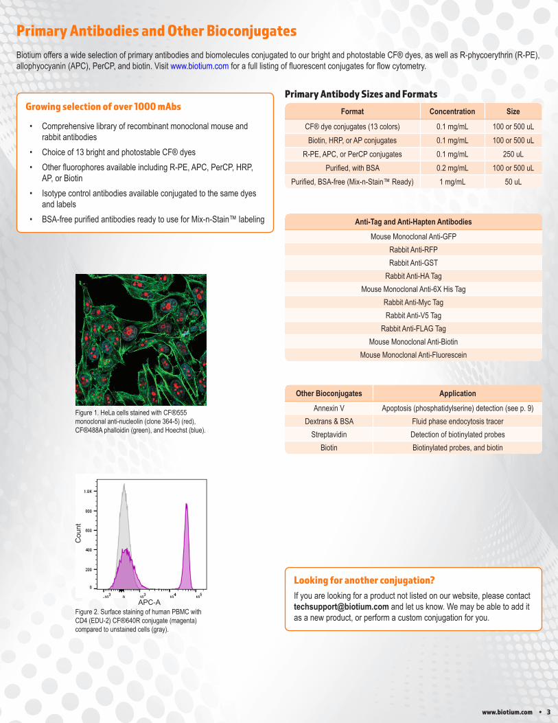
Figure 1. HeLa cells stained with CF®555 monoclonal anti-nucleolin (clone 364-5) (red), CF®488A phalloidin (green), and Hoechst (blue).
Figure 2. Surface staining of human PBMC with CD4 (EDU-2) CF®640R conjugate (magenta) compared to unstained cells (gray).
Cou
nt
APC-A
4 • www.biotium.com
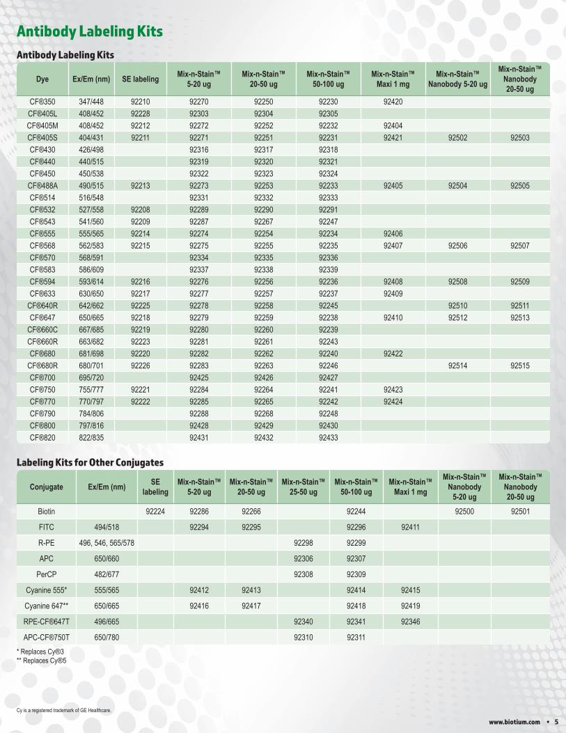
Antibody Labeling Kits
CF dyes and Mix-n-Stain antibody labeling technology are covered by granted and pending U.S. and international patents. We welcome inquiries about licensing the use of our dyes, trademarks or technologies. Please submit inquiries by e-mail to [email protected].
Mix-n-Stain™ antibody labeling kits dramatically simplify the process of preparing fluorescently-labeled antibodies. Simply mix your antibody with the reaction buffer and desired fluorophore provided with the kit. The resulting conjugates offer comparable performance to commercially available pre-labeled antibodies (Fig. 1). Because the labeling is covalent, the conjugates are stable for long-term storage, and ideal for multi-color imaging and multiplex flow cytometry.
Mix-n-Stain™ kits feature Biotium’s CF® dyes, which have advantages in brightness and photostability compared to other fluorescent dyes. A variety of other labels are also available.
We also offer Mix-n-Stain™ Nanobody Labeling Kits which allows rapid and optimal labeling of single-chain nanobodies with CF® dyes or biotin without a purification step.
Mix-n-Stain™ kits are also available in a maxi size for labeling up to 1 mg of IgG antibody with CF® dyes or other dyes. See the tables on page 5 for a complete listing of available Mix-n-Stain™ Antibody Labeling Kits.
Mix-n-Stain™ Antibody Labeling KitsMix-n-Stain™ Advantages
• Rapid and efficient labeling with minimal hands-on time• Available with 29 CF® dyes, R-PE, APC, PerCP, or tandem dyes• Other label options: Biotin & FITC, HRP, AP, and GOx• 100% yield with no purification required• Labeling tolerates BSA, gelatin, ascites fluid, and Tris• Kits available for labeling as little as 1 ug and up to 1 mg of IgG
antibody• Labeling kits for nanobodies also available
CF® Dye SE Protein Labeling kits allow you to label your antibody of interest with one of our many bright and photostable CF® Dyes or biotin. The difference is that while the Mix-n-Stain™ kits come optimized for fast and consistent labeling of small amounts of antibody, the SE Protein Labeling Kits are designed for 3 labelings of up to 1 mg protein with removal of free dye after labeling. The kits also allows the user to adjust the amount of dye and protein to optimize the degree of labeling. See the tables on page 5 for a complete listing of available SE protein labeling kits.
CF® Dye SE Protein Labeling Kits
CF® Dye SE Protein Labeling Kit Features
• Kits that allow more flexibility for labeling optimization• Choice of 19 CF® dye colors or biotin• Includes reagents for 3 x 1 mg labeling reactions with purification• Includes instructions for determining the degree of labeling (DOL)
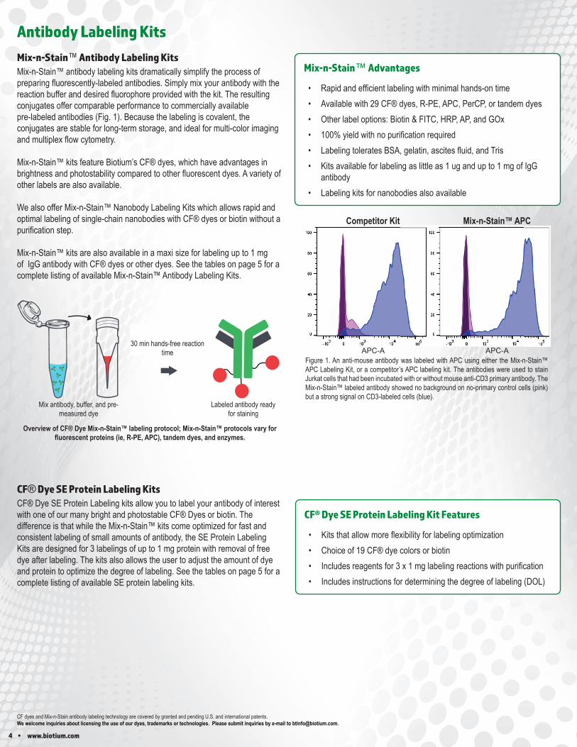
Figure 1. An anti-mouse antibody was labeled with APC using either the Mix-n-Stain™ APC Labeling Kit, or a competitor’s APC labeling kit. The antibodies were used to stain Jurkat cells that had been incubated with or without mouse anti-CD3 primary antibody. The Mix-n-Stain™ labeled antibody showed no background on no-primary control cells (pink) but a strong signal on CD3-labeled cells (blue).
Competitor Kit Mix-n-Stain™ APC
APC-A APC-A
30 minhands-free
reaction time
Mix antibody, buffer, and pre-measured dye Labeled antibody ready for staining
Mix antibody, buffer, and pre-measured dye
Labeled antibody ready for staining
30 min hands-free reaction time
Overview of CF® Dye Mix-n-Stain™ labeling protocol; Mix-n-Stain™ protocols vary for fluorescent proteins (ie, R-PE, APC), tandem dyes, and enzymes.
www.biotium.com • 5
Conjugate Ex/Em (nm) SE labeling
Mix-n-Stain™ 5-20 ug
Mix-n-Stain™ 20-50 ug
Mix-n-Stain™ 25-50 ug
Mix-n-Stain™ 50-100 ug
Mix-n-Stain™ Maxi 1 mg
Mix-n-Stain™ Nanobody
5-20 ug
Mix-n-Stain™ Nanobody20-50 ug
Biotin 92224 92286 92266 92244 92500 92501
FITC 494/518 92294 92295 92296 92411
R-PE 496, 546, 565/578 92298 92299
APC 650/660 92306 92307
PerCP 482/677 92308 92309
Cyanine 555* 555/565 92412 92413 92414 92415
Cyanine 647** 650/665 92416 92417 92418 92419
RPE-CF®647T 496/665 92340 92341 92346
APC-CF®750T 650/780 92310 92311
Dye Ex/Em (nm) SE labeling Mix-n-Stain™ 5-20 ug
Mix-n-Stain™ 20-50 ug
Mix-n-Stain™ 50-100 ug
Mix-n-Stain™ Maxi 1 mg
Mix-n-Stain™ Nanobody 5-20 ug
Mix-n-Stain™ Nanobody 20-50 ug
CF®350 347/448 92210 92270 92250 92230 92420CF®405L 408/452 92228 92303 92304 92305CF®405M 408/452 92212 92272 92252 92232 92404CF®405S 404/431 92211 92271 92251 92231 92421 92502 92503CF®430 426/498 92316 92317 92318CF®440 440/515 92319 92320 92321CF®450 450/538 92322 92323 92324
CF®488A 490/515 92213 92273 92253 92233 92405 92504 92505CF®514 516/548 92331 92332 92333CF®532 527/558 92208 92289 92290 92291CF®543 541/560 92209 92287 92267 92247CF®555 555/565 92214 92274 92254 92234 92406CF®568 562/583 92215 92275 92255 92235 92407 92506 92507CF®570 568/591 92334 92335 92336CF®583 586/609 92337 92338 92339CF®594 593/614 92216 92276 92256 92236 92408 92508 92509CF®633 630/650 92217 92277 92257 92237 92409
CF®640R 642/662 92225 92278 92258 92245 92510 92511CF®647 650/665 92218 92279 92259 92238 92410 92512 92513
CF®660C 667/685 92219 92280 92260 92239CF®660R 663/682 92223 92281 92261 92243CF®680 681/698 92220 92282 92262 92240 92422
CF®680R 680/701 92226 92283 92263 92246 92514 92515CF®700 695/720 92425 92426 92427CF®750 755/777 92221 92284 92264 92241 92423CF®770 770/797 92222 92285 92265 92242 92424CF®790 784/806 92288 92268 92248CF®800 797/816 92428 92429 92430CF®820 822/835 92431 92432 92433
Antibody Labeling Kits
Antibody Labeling Kits
Labeling Kits for Other Conjugates
* Replaces Cy®3** Replaces Cy®5
Cy is a registered trademark of GE Healthcare.
6 • www.biotium.com
Cat. No. Product Unit Size
30002-T Viability/Cytotoxicity Assay Kit for Animal Live & Dead Cells
150 assays30002 300 assays
30026 Calcein AM Cell Viability Assay Kit 1000 assays
30027 Viability/Cytotoxicity Assay kit for Bacteria Live & Dead Cells 100-1000 assays
32001 Bacterial Viability and Gram Stain Kit 200 assays
Product Ex/Em (nm)
Cat. No.50 Rxns
Cat. No. 200 Rxns
Live-or-Dye™ 350/448 347/448 32002-T 32002Live-or-Dye™ 375/600* 375/595 32014-T 32014Live-or-Dye™ 405/452 408/452 32003-T 32003Live-or-Dye™ 405/545 395/545 32009-T 32009Live-or-Dye™ 510/550* 516/549 32012-T 32012Live-or-Dye™ 488/515 490/515 32004-T 32004Live-or-Dye™ 568/583 562/583 32005-T 32005Live-or-Dye™ 594/614 593/614 32006-T 32006Live-or-Dye™ 615/740* 674/813 32015-T 32015Live-or-Dye™ 640/662 642/662 32007-T 32007Live-or-Dye™ 665/685* 667/685 32103-T 32013Live-or-Dye™ 750/777 755/777 32008-T 32008Live-or-Dye™ 787/808 783/803 32011-T 32011
Live-or-Dye NucFix™ Red 520/593** 32010-T 32010
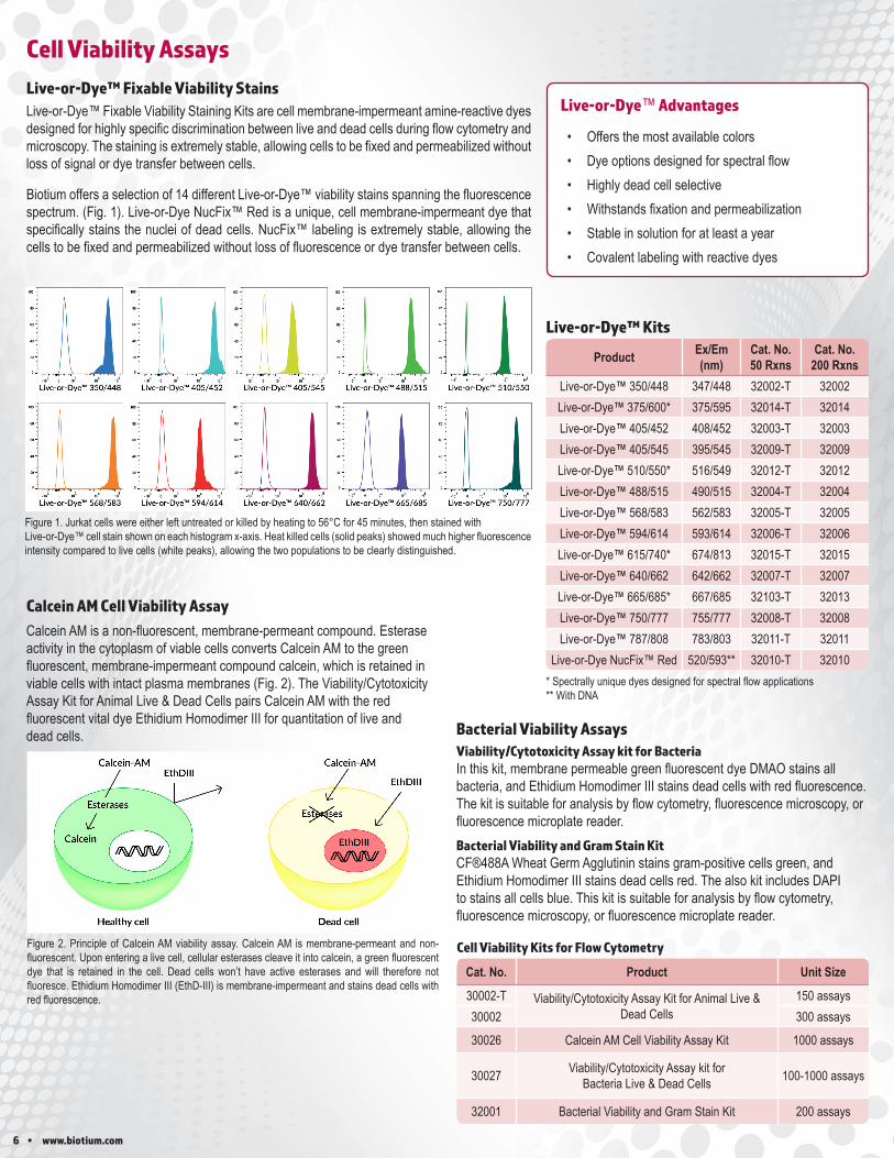
Live-or-Dye™ Fixable Viability Staining Kits are cell membrane-impermeant amine-reactive dyes designed for highly specific discrimination between live and dead cells during flow cytometry and microscopy. The staining is extremely stable, allowing cells to be fixed and permeabilized without loss of signal or dye transfer between cells.
Biotium offers a selection of 14 different Live-or-Dye™ viability stains spanning the fluorescence spectrum. (Fig. 1). Live-or-Dye NucFix™ Red is a unique, cell membrane-impermeant dye that specifically stains the nuclei of dead cells. NucFix™ labeling is extremely stable, allowing the cells to be fixed and permeabilized without loss of fluorescence or dye transfer between cells.
Live-or-Dye™ Fixable Viability Stains
Cell Viability Assays
Figure 2. Principle of Calcein AM viability assay. Calcein AM is membrane-permeant and non-fluorescent. Upon entering a live cell, cellular esterases cleave it into calcein, a green fluorescent dye that is retained in the cell. Dead cells won’t have active esterases and will therefore not fluoresce. Ethidium Homodimer III (EthD-III) is membrane-impermeant and stains dead cells with red fluorescence.
Calcein AM is a non-fluorescent, membrane-permeant compound. Esterase activity in the cytoplasm of viable cells converts Calcein AM to the green fluorescent, membrane-impermeant compound calcein, which is retained in viable cells with intact plasma membranes (Fig. 2). The Viability/Cytotoxicity Assay Kit for Animal Live & Dead Cells pairs Calcein AM with the red fluorescent vital dye Ethidium Homodimer III for quantitation of live and dead cells.
Calcein AM Cell Viability Assay
Cell Viability Kits for Flow Cytometry
Viability/Cytotoxicity Assay kit for BacteriaIn this kit, membrane permeable green fluorescent dye DMAO stains all bacteria, and Ethidium Homodimer III stains dead cells with red fluorescence. The kit is suitable for analysis by flow cytometry, fluorescence microscopy, or fluorescence microplate reader.Bacterial Viability and Gram Stain KitCF®488A Wheat Germ Agglutinin stains gram-positive cells green, and Ethidium Homodimer III stains dead cells red. The also kit includes DAPI to stains all cells blue. This kit is suitable for analysis by flow cytometry, fluorescence microscopy, or fluorescence microplate reader.
Bacterial Viability Assays
Figure 1. Jurkat cells were either left untreated or killed by heating to 56°C for 45 minutes, then stained with Live-or-Dye™ cell stain shown on each histogram x-axis. Heat killed cells (solid peaks) showed much higher fluorescence intensity compared to live cells (white peaks), allowing the two populations to be clearly distinguished.
Live-or-Dye™ Kits
* Spectrally unique dyes designed for spectral flow applications** With DNA
Live-or-Dye™ Advantages
• Offers the most available colors• Dye options designed for spectral flow• Highly dead cell selective• Withstands fixation and permeabilization• Stable in solution for at least a year• Covalent labeling with reactive dyes
www.biotium.com • 7
Cat. No. Product Unit Size
40060-1RedDotTM1 Far-Red Nuclear Stain, 200X in Water
1 mL40060 250 uL
40060-T 25 uL40016 Propidium Iodide (PI) 100 mg40017 Propidium Iodide (PI), 1 mg/mL in water 10 mL40037
7-AAD1 mg
40084 1 mL40085
NucSpot® Far-Red, 1000X in DMSO0.5 mL
40085-T 50 uL
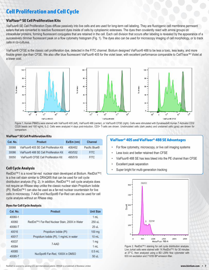
ViaFluor® SE Cell Proliferation Dyes diffuse passively into live cells and are used for long-term cell labeling. They are fluorogenic cell membrane permeant esters that are converted to reactive fluorescent dyes inside of cells by cytoplasmic esterases. The dyes then covalently react with amine groups on intracellular proteins, forming fluorescent conjugates that are retained in the cell. Each cell division that occurs after labeling is revealed by the appearance of a successively dimmer fluorescent peak on a flow cytometry histogram (Fig. 1). The dyes also can be used for microscopy imaging of cell morphology, or to track cells in co-cultures.
ViaFluor® CFSE is the classic cell proliferation dye, detected in the FITC channel. Biotium designed ViaFluor® 488 to be less a toxic, less leaky, and more fixable green dye than CFSE. We also offer blue fluorescent ViaFluor® 405 for the violet laser, with excellent performance comparable to CellTrace™ Violet at a lower cost.
Cell Proliferation and Cell Cycle
ViaFluor® 405 and ViaFluor® 488 SE Advantages
• For flow cytometry, microscopy, or live cell imaging systems• Less toxic and better retained than CFSE• ViaFluor® 488 SE has less bleed into the PE channel than CFSE• Excellent peak separation• Super bright for multi-generation tracking
ViaFluor® SE Cell Proliferation Kits
RedDot1 is covered by pending U.S. and international patents. DRAQ5 is a trademark of Biostatus Limited.
Dyes for Cell Cycle Analysis
RedDot™1 is a novel far-red nuclear stain developed at Biotium. RedDot™1 is a live cell stain similar to DRAQ®5 that can be used for cell cycle distribution analysis (Fig. 2). In addition, RedDot™1 cell cycle analysis does not require an RNase step unlike the classic nuclear stain Propidium Iodide (PI). RedDot™1 can also be used as a far-red nuclear counterstain for live cells in microscopy. 7-AAD and NucSpot® Far-Red can also be used for cell cycle analysis without an RNase step.
Cell Cycle Analysis
Cat. No. Product Ex/Em (nm) Channel
30068 ViaFluor® 405 SE Cell Proliferation Kit 408/452 Pacific Blue®30086 ViaFluor® 488 SE Cell Proliferation Kit 493/532 FITC30050 ViaFluor® CFSE Cell Proliferation Kit 495/519 FITC
ViaFluor® SE Cell Proliferation Kits
Figure 1. Human PBMCs were stained with ViaFluor® 405 (left), ViaFluor® 488 (center), or ViaFluor® CFSE (right). Cells were stimulated with Dynabeads® Human T-Activator CD3/CD28 beads and 100 ng/mL IL-2. Cells were analyzed 4 days post-induction. CD3+ T-cells are shown. Unstimulated cells (dark peaks) and unstained cells (gray) are shown for comparison.
Figure 2. RedDot™1 staining for cell cycle distribution analysis. Live Jurkat cells were stained with 1X RedDot™1 for 30 minutes at 37°C, then analyzed using a BD LSRII flow cytometer with 633 nm excitation and 710/50 BP emission filter.
8 • www.biotium.com
Cat. No. Product Ex/Em
(nm) Unit Size
10402 NucView® 488 Caspase-3 Substrate, 1 mM in DMSO
500/530
100 uL
10403 NucView® 488 Caspase-3 Substrate, 1 mM in PBS 100 uL
30029 NucView® 488 Caspase-3 Assay Kit for Live Cells 100 assays
10405 NucView® 405 Caspase-3 Substrate, 1 mM in DMSO429/469
100 uL
10407 NucView® 405 Caspase-3 Substrate, 1 mM in PBS 100 uL
10406 NucView® 530 Caspase-3 Substrate, 1 mM in DMSO528/563
100 uL
10408 NucView® 530 Caspase-3 Substrate, 1 mM in PBS 100 uL
30062 NucView® 488 and MitoView™ 633 Apoptosis Kit 500/530, 622/648 100 assays
30072 NucView® 488 and RedDot™2 Apoptosis & Necrosis Kit
500/530, 665/695 100 assays
30067 Dual Apoptosis Assay with NucView® 488 Caspase-3 Substrate and CF®594 Annexin V
500/530, 593/614 50 assays
30073 Dual Apoptosis Assay with NucView® 488 Caspase-3 Substrate and CF®640R Annexin V
500/530, 642/662 50 assays
30030 Dual Apoptosis Assay with NucView® 488 Caspase-3 Substrate and Texas Red®-Annexin V
500/530, 594/615 50 assays
NucView® Advantages
• Rapid, no-wash, endpoint or real-time assays• Non-toxic, allowing multi-day experiments• For flow cytometry, microscopy, or live cell imaging systems• Dual detection of caspase activity and nuclear morphology• Formaldehyde-fixable• Published in over 200 scientific papers and validated in more than
100 cell lines
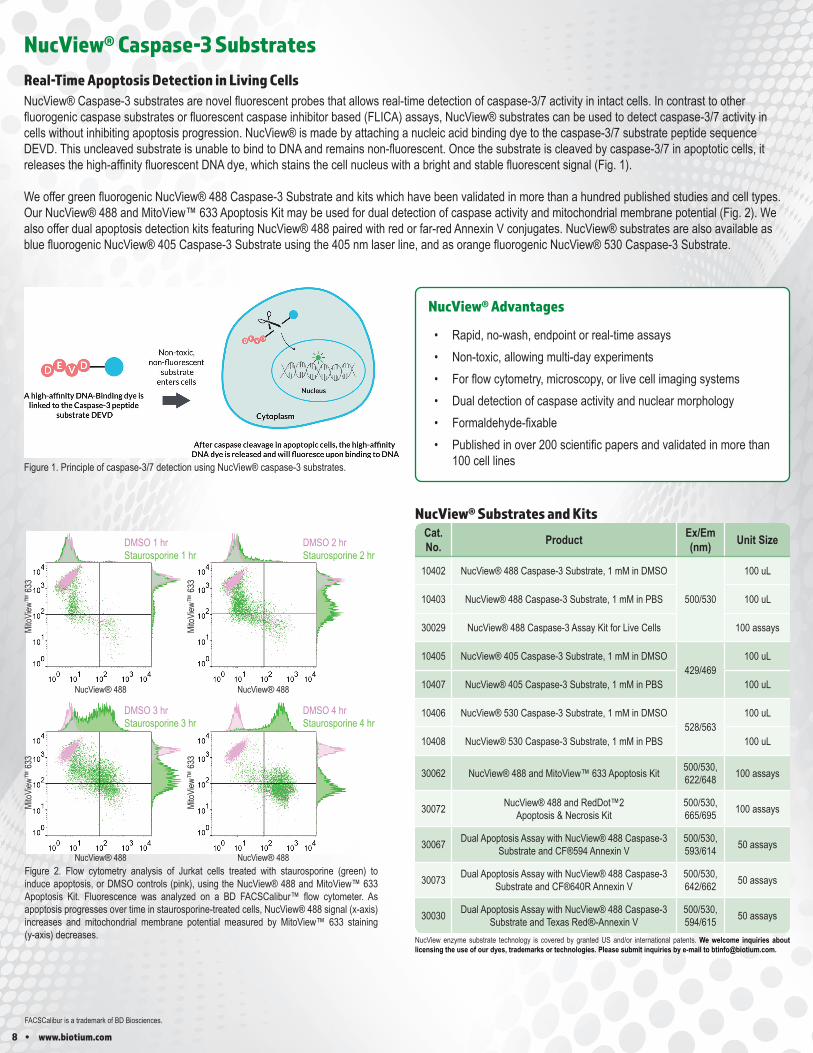
NucView® Caspase-3 Substrates
Figure 1. Principle of caspase-3/7 detection using NucView® caspase-3 substrates.
NucView® Caspase-3 substrates are novel fluorescent probes that allows real-time detection of caspase-3/7 activity in intact cells. In contrast to other fluorogenic caspase substrates or fluorescent caspase inhibitor based (FLICA) assays, NucView® substrates can be used to detect caspase-3/7 activity in cells without inhibiting apoptosis progression. NucView® is made by attaching a nucleic acid binding dye to the caspase-3/7 substrate peptide sequence DEVD. This uncleaved substrate is unable to bind to DNA and remains non-fluorescent. Once the substrate is cleaved by caspase-3/7 in apoptotic cells, it releases the high-affinity fluorescent DNA dye, which stains the cell nucleus with a bright and stable fluorescent signal (Fig. 1).
We offer green fluorogenic NucView® 488 Caspase-3 Substrate and kits which have been validated in more than a hundred published studies and cell types. Our NucView® 488 and MitoView™ 633 Apoptosis Kit may be used for dual detection of caspase activity and mitochondrial membrane potential (Fig. 2). We also offer dual apoptosis detection kits featuring NucView® 488 paired with red or far-red Annexin V conjugates. NucView® substrates are also available as blue fluorogenic NucView® 405 Caspase-3 Substrate using the 405 nm laser line, and as orange fluorogenic NucView® 530 Caspase-3 Substrate.
Real-Time Apoptosis Detection in Living Cells
NucView® Substrates and Kits
NucView enzyme substrate technology is covered by granted US and/or international patents. We welcome inquiries about licensing the use of our dyes, trademarks or technologies. Please submit inquiries by e-mail to [email protected].
Figure 2. Flow cytometry analysis of Jurkat cells treated with staurosporine (green) to induce apoptosis, or DMSO controls (pink), using the NucView® 488 and MitoView™ 633 Apoptosis Kit. Fluorescence was analyzed on a BD FACSCalibur™ flow cytometer. As apoptosis progresses over time in staurosporine-treated cells, NucView® 488 signal (x-axis) increases and mitochondrial membrane potential measured by MitoView™ 633 staining (y-axis) decreases.
DMSO 1 hrStaurosporine 1 hr
DMSO 2 hrStaurosporine 2 hr
DMSO 3 hrStaurosporine 3 hr
DMSO 4 hrStaurosporine 4 hr
NucView® 488
MitoV
iew™
633
NucView® 488
MitoV
iew™
633
NucView® 488
MitoV
iew™
633
NucView® 488
MitoV
iew™
633
FACSCalibur is a trademark of BD Biosciences.
www.biotium.com • 9
Cat. No. Product Unit Size
30065 Apoptosis & Necrosis Quantitation Kit Plus 50 assays
30066 Apoptotic, Necrotic, & Healthy Cells Quantitation Kit Plus 50 assays
30060 CF®488A Annexin V and 7-AAD Apoptosis Kit 100 assays
30061 CF®488A Annexin V and PI Apoptosis Kit 100 assays
30067 Dual Apoptosis Assay with NucView® 488 and CF®594 Annexin V 50 assays
30072 NucView® 488 & RedDot™ 2 Apoptosis & Necrosis Kit 100 assays
30073 Dual Apoptosis Assay with NucView® 488 and CF®640R Annexin V 50 assays
Cat. No. Conjugate Ex/Em (nm) Unit Size
29012 Annexin V, CF®350, 50 ug/mL347/448
0.5 mL
29012R-5ug Annexin V, CF®350, Azide-Free, Lyophilized 5 ug
29009 Annexin V, CF®405M, 50 ug/mL408/452
0.5 mL
29009-5ug Annexin V, CF®405M, Azide-Free, Lyophilized 5 ug
29083 Annexin V, CF®450, 50 ug/mL450/538
0.5 mL
29083R-5ug Annexin V, CF®450, Azide-Free, Lyophilized 5 ug
29005 Annexin V, CF®488A, 50 ug/mL490/515
0.5 mL
29005R-5ug Annexin V, CF®488A, Azide-Free, Lyophilized 5 ug
29004 Annexin V, CF®555, 50 ug/mL 555/565
0.5 mL
29004R-5ug Annexin V, CF®555, Azide-Free, Lyophilized 5 ug
29010 Annexin V, CF®568, 50 ug/mL562/583
0.5 mL
29010R-5ug Annexin V, CF®568, Azide-Free, Lyophilized 5 ug
29085 Annexin V, CF®583R, 50 ug/mL586/609
500 uL
29085R-5ug Annexin V, CF®583R, Azide-Free, Lyophilized 5 ug
29011 Annexin V, CF®594, 50 ug/mL 593/614
0.5 mL
29011R-5ug Annexin V, CF®594, Azide-Free, Lyophilized 5 ug
29008 Annexin V, CF®633, 50 ug/mL630/650
0.5 mL
29008R-5ug Annexin V, CF®633, Azide-Free, Lyophilized 5 ug
29014 Annexin V, CF®640R, 50 ug/mL642/662
0.5 mL
29014R-5ug Annexin V, CF®640R, Azide-Free, Lyophilized 5 ug
29003 Annexin V, CF®647, 50 ug/mL650/665
0.5 mL
29003R-5ug Annexin V, CF®647, Azide-Free, Lyophilized 5 ug
29069 Annexin V, CF®660R, 50 ug/mL663/682
0.5 mL
29069R-5ug Annexin V, CF®660R, Azide-Free, Lyophilized 5 ug
29007 Annexin V, CF®680, Azide-Free, Lyophilized 681/698 25 ug
29070 Annexin V, CF®680R, Azide-Free, Lyophilized 680/701 25 ug
29082 Annexin V, CF®700, Azide-Free, Lyophilized 699/737 25 ug
29006 Annexin V, CF®750, Azide-Free, Lyophilized 755/777 25 ug
29046 Annexin V, CF®770, Azide-Free, Lyophilized 770/797 25 ug
29047 Annexin V, CF®790, Azide-Free, Lyophilized 784/806 25 ug
29078 Annexin V, CF®800, Azide-Free, Lyophilized 797/816 25 ug
29001 Annexin V, FITC, 50 ug/mL 490/525 0.5 mL
29045-100 uLAnnexin V, R-PE 496, 546,
565/57820 assays
29045-500 uL 100 assays
29057-100 uLAnnexin V, APC 633, 640/660
20 assays
29057-500 uL 100 assays
29013 Annexin V, Biotin, 50 ug/mL N/A 0.5 mL
99902 5X Annexin V Binding Buffer N/A 15 mL
Apoptosis and Necrosis Assays
Annexin V Conjugates
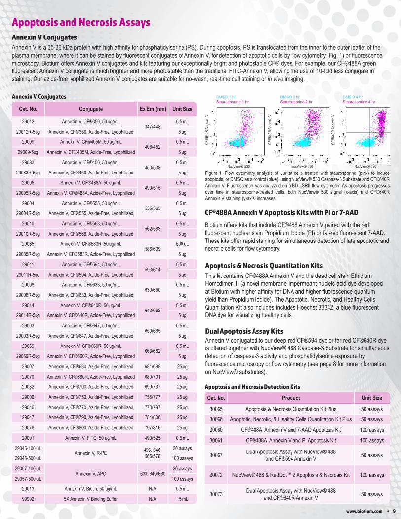
Annexin V is a 35-36 kDa protein with high affinity for phosphatidylserine (PS). During apoptosis, PS is translocated from the inner to the outer leaflet of the plasma membrane, where it can be stained by fluorescent conjugates of Annexin V, for detection of apoptotic cells by flow cytometry (Fig. 1) or fluorescence microscopy. Biotium offers Annexin V conjugates and kits featuring our exceptionally bright and photostable CF® dyes. For example, our CF®488A green fluorescent Annexin V conjugate is much brighter and more photostable than the traditional FITC-Annexin V, allowing the use of 10-fold less conjugate in staining. Our azide-free lyophilized Annexin V conjugates are suitable for no-wash, real-time cell staining or in vivo imaging.
Annexin V Conjugates
CF®488A Annexin V Apoptosis Kits with PI or 7-AAD
Biotium offers kits that include CF®488 Annexin V paired with the red fluorescent nuclear stain Propidium Iodide (PI) or far-red fluorescent 7-AAD. These kits offer rapid staining for simultaneous detection of late apoptotic and necrotic cells for flow cytometry.
Apoptosis and Necrosis Detection Kits
Apoptosis & Necrosis Quantitation KitsThis kit contains CF®488A Annexin V and the dead cell stain Ethidium Homodimer III (a novel membrane-impermeant nucleic acid dye developed at Biotium with higher affinity for DNA and higher fluorescence quantum yield than Propidium Iodide). The Apoptotic, Necrotic, and Healthy Cells Quantitation Kit also includes includes Hoechst 33342, a blue fluorescent DNA dye for visualizing healthy cells.
Dual Apoptosis Assay KitsAnnexin V conjugated to our deep-red CF®594 dye or far-red CF®640R dye is offered together with NucView® 488 Caspase-3 Substrate for simultaneous detection of caspase-3 activity and phosphatidylserine exposure by fluorescence microscopy or flow cytometry (see page 8 for more information on NucView® substrates).
Figure 1. Flow cytometry analysis of Jurkat cells treated with staurosporine (pink) to induce apoptosis, or DMSO as a control (blue), using NucView® 530 Caspase-3 Substrate and CF®640R Annexin V. Fluorescence was analyzed on a BD LSRII flow cytometer. As apoptosis progresses over time in staurosporine-treated cells, both NucView® 530 signal (x-axis) and CF®640R Annexin V staining (y-axis) increases.
DMSO 1 hrStaurosporine 1 hr
DMSO 2 hrStaurosporine 2 hr
DMSO 4 hrStaurosporine 4 hr
NucView® 530
CF®
640R
Ann
exin
V
CF®
640R
Ann
exin
V
CF®
640R
Ann
exin
V
NucView® 530 NucView® 530
10 • www.biotium.com
Cat. No. Product Unit Size
23006 Flow Cytometry Fixation/Permeabilization Kit 50 tests
22015 Fixation Buffer 100 mL
22016 Permeabilization Buffer 100 mL
22017 Permeabilization and Blocking Buffer (5X) 100 mL
22020 10X Phosphate-Buffered Saline (PBS) 4 L
22010 10% Fish Gelatin Blocking Buffer 100 mL
22011 Fish Gelatin Powder 2 x 50 g
22013 Bovine Serum Albumin, Fraction V 50 g
22014 30% Bovine Serum Albumin Solution 100 mL
22002 Tween®-20 50 mL
22003 Mini Cell Scrapers Pack of 200
Cat. No. Product Ex/Em (nm) Potential-dependent?
30001 JC-1 Mitochondrial Membrane Potential Detection Kit
510/527; 585/590* Ratiometric*70014 JC-1, Iodide Salt
70011 JC-1, Chloride Salt
70016 TMRE549/574 Yes
70005 TMRE, 2 mM in DMSO
70017 TMRM 548/573 Yes
70010 Rhodamine 123 505/534 Yes
Cat. No. Product Ex/Em (nm) Potential-dependent?
70070 MitoView™ 405 398/440 Partial3
70054 MitoView™ Green 490/523 No70055 MitoView™ 633 622/6481 Yes70075 MitoView™ 650 644/670 Partial3
70068 MitoView™ 720 720/7582 Partial3
Membrane Potential Dyes and Other Accessories
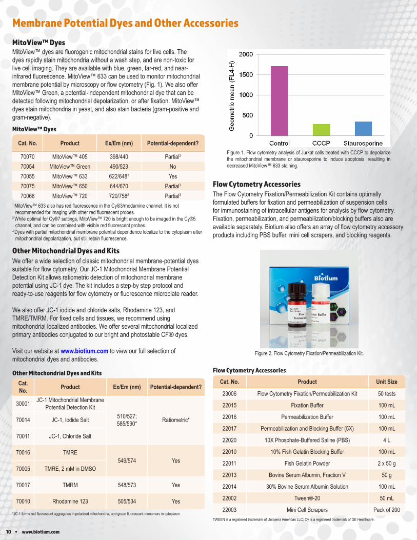
Figure 1. Flow cytometry analysis of Jurkat cells treated with CCCP to depolarize the mitochondrial membrane or staurosporine to induce apoptosis, resulting in decreased MitoView™ 633 staining.
1 MitoView™ 633 also has red fluorescence in the Cy®3/rhodamine channel. It is not recommended for imaging with other red fluorescent probes.
2 While optimal for Cy®7 settings, MitoView™ 720 is bright enough to be imaged in the Cy®5 channel, and can be combined with visible red fluorescent probes.
3 Dyes with partial mitochondrial membrane potential dependence localize to the cytoplasm after mitochondrial depolarization, but still retain fluorescence.
MitoView™ Dyes
MitoView™ DyesMitoView™ dyes are fluorogenic mitochondrial stains for live cells. The dyes rapidly stain mitochondria without a wash step, and are non-toxic for live cell imaging. They are available with blue, green, far-red, and near-infrared fluorescence. MitoView™ 633 can be used to monitor mitochondrial membrane potential by microscopy or flow cytometry (Fig. 1). We also offer MitoView™ Green, a potential-independent mitochondrial dye that can be detected following mitochondrial depolarization, or after fixation. MitoView™ dyes stain mitochondria in yeast, and also stain bacteria (gram-positive and gram-negative).
The Flow Cytometry Fixation/Permeabilization Kit contains optimally formulated buffers for fixation and permeabilization of suspension cells for immunostaining of intracellular antigens for analysis by flow cytometry. Fixation, permeabilization, and permeabilization/blocking buffers also are available separately. Biotium also offers an array of flow cytometry accessory products including PBS buffer, mini cell scrapers, and blocking reagents.
Flow Cytometry Accessories
TWEEN is a registered trademark of Uniqema Americas LLC; Cy is a registered trademark of GE Healthcare.
Flow Cytometry Accessories
Other Mitochondrial Dyes and KitsWe offer a wide selection of classic mitochondrial membrane-potential dyes suitable for flow cytometry. Our JC-1 Mitochondrial Membrane Potential Detection Kit allows ratiometric detection of mitochondrial membrane potential using JC-1 dye. The kit includes a step-by step protocol and ready-to-use reagents for flow cytometry or fluorescence microplate reader.
We also offer JC-1 iodide and chloride salts, Rhodamine 123, and TMRE/TMRM. For fixed cells and tissues, we recommend using mitochondrial localized antibodies. We offer several mitochondrial localized primary antibodies conjugated to our bright and photostable CF® dyes.
Visit our website at www.biotium.com to view our full selection of mitochondrial dyes and antibodies.
Other Mitochondrial Dyes and Kits
*JC-1 forms red fluorescent aggregates in polarized mitochondria, and green fluorescent monomers in cytoplasm
Figure 2. Flow Cytometry Fixation/Permeabilization Kit.
www.biotium.com • 11
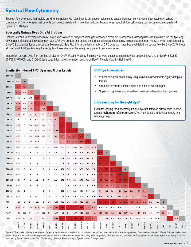
Figure 1. The Similarity Index is a measure of spectral similarity on a scale from 0 to 1. Values close to 0 indicate that the full spectrum signatures of the two dyes are very different from each other, and values close to 1 indicate that the signatures are very similar to each other. When designing multiplex panels, it is important to choose unique fluorophores with minimal spectral similarity. Data was provided by Cytek® Biosciences from CD4 labeling of human PBMCs using a Cytek® Aurora flow cytometer.
Similarity Index of CF® Dyes and Other LabelsCF®405L 1
CF®405M 0.13 1
CF®430 0.7 0.52 1
CF®450 0.53 0.12 0.51 1
CF®488A 0.01 0 0.02 0.78 1
CF®514 0.34 0.04 0.24 0.78 0.57 1
CF®532 0.5 0.04 0.29 0.69 0.34 0.89 1
CF®543 0.19 0.01 0.09 0.17 0.05 0.21 0.47 1
CF®555 0 0 0 0.05 0.02 0.09 0.3 0.96 1
CF®568 0.06 0 0.03 0.05 0.01 0.05 0.27 0.95 0.97 1
CF®570 0.02 0 0.01 0.04 0.01 0.06 0.27 0.9 0.95 0.98 1
CF®583 0.07 0 0.02 0.04 0.01 0.05 0.2 0.56 0.64 0.72 0.84 1
CF®594 0.06 0 0.02 0.04 0 0.03 0.14 0.4 0.5 0.52 0.65 0.9 1
CF®633 0.02 0 0.01 0.01 0 0.01 0.02 0.03 0.05 0.05 0.09 0.15 0.16 1
CF®640R 0.01 0 0 0 0 0 0.01 0.02 0.03 0.03 0.05 0.08 0.09 0.98 1
CF®647 0 0 0 0 0 0 0 0.01 0.02 0.02 0.04 0.06 0.08 0.95 0.98 1
CF®660C 0.01 0 0 0 0 0 0 0.01 0.02 0.02 0.03 0.05 0.07 0.85 0.86 0.92 1
CF®660R 0.01 0 0 0 0 0 0.01 0.01 0.02 0.02 0.05 0.07 0.1 0.86 0.87 0.92 0.99 1
CF®680 0.01 0 0 0 0 0 0 0.01 0.01 0.01 0.02 0.03 0.05 0.74 0.7 0.74 0.91 0.91 1
CF®680R 0.01 0 0 0.01 0 0.01 0.01 0.01 0.02 0.02 0.04 0.06 0.08 0.73 0.68 0.72 0.88 0.89 0.99 1
CF®700 0.01 0 0 0 0 0 0 0 0.01 0.01 0.01 0.02 0.03 0.6 0.54 0.53 0.66 0.67 0.86 0.88 1
CF®750 0.01 0 0 0 0 0 0 0.01 0.01 0.01 0.01 0.01 0.01 0.2 0.2 0.15 0.23 0.3 0.31 0.34 0.44 1
CF®770 0.01 0 0 0.01 0 0 0 0.01 0.01 0.01 0.01 0.01 0.01 0.16 0.15 0.12 0.18 0.25 0.26 0.3 0.38 0.95 1
APC 0.03 0 0.01 0.01 0 0.01 0.02 0.05 0.07 0.08 0.13 0.21 0.23 0.97 0.94 0.9 0.75 0.77 0.6 0.6 0.48 0.15 0.11 1
PE 0.16 0.01 0.07 0.19 0.05 0.24 0.53 0.97 0.91 0.88 0.83 0.49 0.33 0.03 0.01 0.01 0.01 0.01 0.01 0.01 0 0.01 0.01 0.04 1
PerCP 0.03 0.01 0.01 0.03 0.01 0.06 0.07 0.05 0.08 0.07 0.14 0.22 0.26 0.27 0.23 0.23 0.21 0.25 0.15 0.19 0.09 0.04 0.03 0.34 0.06 1
Cy®3 0.04 0.04 0.05 0.1 0.04 0.13 0.34 0.95 0.99 0.96 0.96 0.7 0.56 0.1 0.06 0.06 0.05 0.06 0.03 0.05 0.02 0.01 0.01 0.14 0.91 0.18 1
CF®40
5L
CF®40
5M
CF®43
0
CF®45
0
CF®48
8A
CF®51
4
CF®53
2
CF®54
3
CF®55
5
CF®56
8
CF®57
0
CF®58
3
CF®59
4
CF®63
3
CF®64
0R
CF®64
7
CF®66
0C
CF®66
0R
CF®68
0
CF®68
0R
CF®70
0
CF®75
0
CF®77
0
APC
PE
PerCP
Cy®3
Spectral Flow CytometrySpectral flow cytometry is a rapidly growing technology with significantly enhanced multiplexing capabilities over conventional flow cytometry. Where conventional flow cytometer instruments can detect panels with more than a dozen fluorophores, spectral flow cytometers can accommodate panels with upwards of 40 dyes.
Biotium is proud to develop spectrally unique dyes aimed at filling emission gaps between available fluorophores, allowing users to maximize the multiplexing advantages of spectral flow cytometry. Our CF® dye product line boasts the largest selection of spectrally unique fluorophores, many of which are licensed by Cytek® Biosciences for use in spectral flow panels. See Fig. 1 for a similarity matrix of CF® dyes that have been validated in spectral flow by Cytek®. With our Mix-n-Stain CF® Dye Antibody Labeling Kits, these dyes can be easily conjugated to your antibodies.
In addition, several dyes from our line of Live-or-Dye™ Fixable Viability Staining Kits were designed specifically for spectral flow: Live-or-Dye™ 510/550, 665/685, 375/600, and 615/740 (see page 6 for more information on Live-or-Dye™ Fixable Viability Staining Kits).
Spectrally Unique Dyes Only At Biotium
CF® Dye Advantages
• Widest selection of spectrally unique dyes to accomodate highly complex panels
• Greatest coverage across visible and near-IR wavelengths
• Superior brightness and signal-to-noise over alternative fluorophores
Still searching for the right dye?
If you are looking for a spectrally unique dye not listed on our website, please contact [email protected]. We may be able to develop a new dye to fit your needs.
v1.1
About UsAt Biotium, we are dedicated to developing cutting-edge fluorescent solutions for life science research. Our efforts have resulted in a growing number of unique and industry-leading fluorescence-based technologies for a wide array of biological research applications. Our products are available in the U.S. through our website, and worldwide through our extensive network of domestic and international distributors.
We license our technologies to a number of international biotechnology companies, and collaborate with academic laboratories to develop tools for the constantly evolving research community. We welcome inquiries about licensing the use of our dyes, technologies, or trademarks; email us at [email protected].
Biotium implements a Quality System, certified by QAS according to Standard QAS ISO 9001:2015.
www.biotium.comPhone: 800-304-5357Biotium, Inc.46117 Landing ParkwayFremont, CA 94538 USA


![Glucose Metabolism and AMPK Signaling Regulate ...bionmr.unl.edu/files/publications/128.pdf(Molecular Probes). Flow cytometry was performed as de-scribed previously [10, 30, 39, 40].](https://static.fdocument.org/doc/165x107/5f481d3d3b482616b93519a1/glucose-metabolism-and-ampk-signaling-regulate-molecular-probes-flow-cytometry.jpg)