PLOS ONE_ Humanization and Characterization of an Anti-Human TNF-α Murine Monoclonal Antibody
14
8/16/13 PLOS ONE: Humanization and Characterization of an Anti-Human TNF-α Murine Monoclonal Antibody www.plosone.org/article/info%3Adoi%2F10.1371%2Fjournal.pone.0016373 1/14 About the Authors Metrics Comments Related Content Hide Figures ADVERTISEMENT plos.org create account sign in advanced search Search Articles For Authors About Us Humanization and Characterization of an Anti-Human TNF- α Murine Monoclonal Antibody 4,236 VIEWS 2 CITATIONS 17 SAVES OPEN ACCESS PEER-REVIEWED RESEARCH ARTICLE Wei-Chun Chiu, Ya-Ping Lai, Min-Yuan Chou Abstract Introduction Results Discussion Materials and Methods Supporting Information Acknowledgments Author Contributions References Reader Comments (0) Figures Abstract A murine monoclonal antibody, m357, showing the highly neutralizing activities for human tumor necrosis factor (TNF-α) was chosen to be humanized by a variable domain resurfacing approach. The non-conserved surface residues in the framework regions of both the heavy and light chain variable regions were identified via a molecular modeling of m357 built by computer- assisted homology modeling. By replacing these critical surface residues with the human counterparts, a humanized version, h357, was generated. The humanized h357 IgG was then stably expressed in a mammalian cell line and the purified antibody maintained the high antigen binding affinity as compared with the parental m357 based on a soluble TNF-α neutralization bioassay. Furthermore, h357 IgG possesses the ability to mediate antibody- dependent cell-mediated cytotoxicity and complement dependent cytotoxicity upon binding to cells bearing the transmembrane form of TNF-α. In a mouse model of collagen antibody- induced arthritis, h357 IgG significantly inhibited disease progression by intra-peritoneal injection of 50 µg/mouse once-daily for 9 consecutive days. These results provided a basis for the development of h357 IgG as therapeutic use. Citation: Chiu W-C, Lai Y-P, Chou M-Y (2011) Humanization and Characterization of an Anti- Human TNF-α Murine Monoclonal Antibody. PLoS ONE 6(1): e16373. doi:10.1371/journal.pone.0016373 Editor: Wei-Chun Chin, University of California, Merced, United States of America Received: August 21, 2010; Accepted: December 20, 2010; Published: January 31, 2011 Copyright: © 2011 Chiu et al. This is an open-access article distributed under the terms of the Creative Commons Attribution License, which permits unrestricted use, distribution, and reproduction in any medium, provided the original author and source are credited. Funding: This work was funded by the Ministry of Economic Affairs, grant number 9356E71100(http://www.moeaidb.gov.tw/). The funders had no role in study design, data collection and analysis, decision to publish, or preparation of the manuscript. 1 1 Download Print Share Article Humanization and Characterization of an Anti-Human TNF-α Murine Monoclonal Antibody Wei-Chun Chiu, Ya-Ping Lai, Min-Yuan Chou
-
Upload
deepak1133 -
Category
Documents
-
view
22 -
download
0
description
plos
Transcript of PLOS ONE_ Humanization and Characterization of an Anti-Human TNF-α Murine Monoclonal Antibody
8/16/13 PLOS ONE: Humanization and Characterization of an Anti-Human TNF-α Murine Monoclonal Antibody
www.plosone.org/article/info%3Adoi%2F10.1371%2Fjournal.pone.0016373 1/14
About the Authors Metrics Comments Related Content
Hide Figures
ADVERTISEMENT
plos.org create account sign in
advanced search
Search
Articles For Authors About Us
Humanization and Characterization of an Anti-Human TNF-α Murine Monoclonal Antibody
4,236
VIEWS
2
CITATIONS
17
SAVES
OPEN ACCESS PEER-REVIEWED
RESEARCH ARTICLE
Wei-Chun Chiu, Ya-Ping Lai, Min-Yuan Chou
Abstract
Introduction
Results
Discussion
Materials and Methods
Supporting Information
Acknowledgments
Author Contributions
References
Reader Comments (0)
Figures
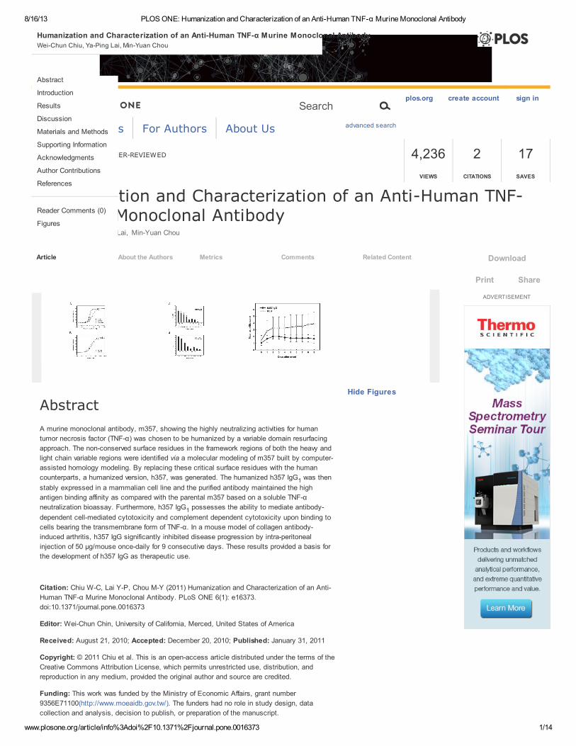
Abstract
A murine monoclonal antibody, m357, showing the highly neutralizing activities for human
tumor necrosis factor (TNF-α) was chosen to be humanized by a variable domain resurfacing
approach. The non-conserved surface residues in the framework regions of both the heavy and
light chain variable regions were identified via a molecular modeling of m357 built by computer-
assisted homology modeling. By replacing these critical surface residues with the human
counterparts, a humanized version, h357, was generated. The humanized h357 IgG was then
stably expressed in a mammalian cell line and the purified antibody maintained the high
antigen binding affinity as compared with the parental m357 based on a soluble TNF-α
neutralization bioassay. Furthermore, h357 IgG possesses the ability to mediate antibody-
dependent cell-mediated cytotoxicity and complement dependent cytotoxicity upon binding to
cells bearing the transmembrane form of TNF-α. In a mouse model of collagen antibody-
induced arthritis, h357 IgG significantly inhibited disease progression by intra-peritoneal
injection of 50 µg/mouse once-daily for 9 consecutive days. These results provided a basis for
the development of h357 IgG as therapeutic use.
Citation: Chiu W-C, Lai Y-P, Chou M-Y (2011) Humanization and Characterization of an Anti-
Human TNF-α Murine Monoclonal Antibody. PLoS ONE 6(1): e16373.
doi:10.1371/journal.pone.0016373
Editor: Wei-Chun Chin, University of California, Merced, United States of America
Received: August 21, 2010; Accepted: December 20, 2010; Published: January 31, 2011
Copyright: © 2011 Chiu et al. This is an open-access article distributed under the terms of the
Creative Commons Attribution License, which permits unrestricted use, distribution, and
reproduction in any medium, provided the original author and source are credited.
Funding: This work was funded by the Ministry of Economic Affairs, grant number
9356E71100(http://www.moeaidb.gov.tw/). The funders had no role in study design, data
collection and analysis, decision to publish, or preparation of the manuscript.
1
1
Download
Print Share
Article
Humanization and Characterization of an Anti-Human TNF-α Murine Monoclonal Antibody
Wei-Chun Chiu, Ya-Ping Lai, Min-Yuan Chou
8/16/13 PLOS ONE: Humanization and Characterization of an Anti-Human TNF-α Murine Monoclonal Antibody
www.plosone.org/article/info%3Adoi%2F10.1371%2Fjournal.pone.0016373 2/14
Competing interests: The authors have declared that no competing interests exist.
Introduction
Tumor necrosis factor (TNF-α) is a pro-inflammatory cytokine produced primarily by cells of the
immune system, including macrophages and monocytes. TNF-α is present as a homotrimeric
protein in which each subunit is initially translated as a 26 kDa transmembrane precursor
protein. After being cleaved at a site proximal to the transmembrane domain of TNF-α by TNF-α
converting enzyme, a soluble trimeric form of TNF-α is released and exerts its activity by
binding to two structurally distinct type I and type II TNF receptors (TNFRI and TNFRII) on
effector cells. The transmembrane form of TNF-α is also known as its unique biologic functions,
such as cytotoxic activity and polyclonal B cell activation, in a cell-to-cell contact manner [1].
TNF-α has been proved to have certain effects on autoimmune processes and has become a
key therapeutic target for many autoimmune diseases [2]. So far, some anti-TNF-α agents, like
etanercept, adalimumab and infliximab were approved by the Food and Drug Administration,
and all have the capability to neutralize soluble form of TNF-α effectively as a major
pharmacological mechanism of action. However, the binding effects of these antagonists on the
transmembrane form of TNF-α are different, which may cause different results on clinical
diseases [3]. For instance, etanercept is not clinically effective for the pathogenesis of
granulomatous diseases, in which the transmembrane form of TNF-α may play a critical role[1].
Therefore, whether or not anti-TNF-α agents can bind to the transmembrane form of TNF-α is
prerequisite to trigger antibody dependent cell mediated cytotoxicity (ADCC), complement
dependent cytotoxicity (CDC), apoptotic and outside-to-inside signaling mechanisms.
The major impediment of the murine monoclonal antibody in clinical practice is that it may elicit
human anti-murine antibody (HAMA) response in patients [4], [5], [6]. Hence, to improve the
efficiency in clinical use, genetic engineering technology has been employed to replace the
murine content with the amino acid residues of human counterparts, and to reduce the
possibility of inducing immunogenicity in patients. An ideal antibody humanization should be
capable of maintaining the specificity and affinity toward the antigen and reduces the
immunogenicity as much as possible. So far, many approaches have been used for antibody
humanization, such as chimeric antibodies, which consists of murine antigen-binding variable
regions fused genetically to human antibody constant regions, is the earliest attempt to reduce
immunogenicity [7]. However, chimeric antibodies still generate undesirable anti-variable region
response [8]. Complementarity determining region (CDR)-grafting is another approach involving
the transfer of the CDRs from a rodent antibody to the Fv frameworks (FRs) of a human
antibody [9]. Unfortunately, the interface changes between CDRs and new FRs may largely
disturb the binding to the antigen. The initial CDR-grafted antibodies tend to lose the parental
binding affinity, and therefore require additional work for back-mutation of several murine
framework amino acids, which are regarded to be crucial for CDRs loop conformation [10].
Humanization via variable domain resurfacing is another approach that can maintain the
specificity and binding affinity of parental antibody, which can reduce the immunogenicity of
antibodies through the replacement of surface exposed residues in the murine FRs with those
usually found in human antibodies [11], [12], [13], [14], [15]. Although the current molecular-
biology techniques can render this approach more straightforward in practice, it is still difficult
in determining the critical residues exposed in solvent on the surface, especially when requiring
a reliable computer model of the antibody [14].
This study is in an attempt to humanize a murine anti-human TNF-α monoclonal antibody,
m357, raised by immunization of mice with recombinant human TNF-α. In order to keep the
parental binding affinity, we resurfaced m357 in the variable regions of both the heavy and light
chains via computer-aided molecular modeling followed by functional characterization of the
humanized antibody. The results indicated that the humanized antibody, h357, retained in
vitrobioactivities similar to the parental antibody. Moreover, the humanized h357 IgG is capable
of binding to the cells bearing the transmembrane TNF-α and triggering ADCC with effector
cells and CDC. The humanized 357 IgG at a dose level of 50 µg/mouse/day significantly
inhibited disease progression in a mouse model of collagen antibody-induced arthritis when
compared with saline treated mice. These results provided a basis for the development of h357
IgG as therapeutic use.
Results
Molecular modeling of m357 variable fragments
The murine monoclonal antibody derived from the hybridoma cell line, 357-101-4 (ECACC No.
92030603), is of therapeutic interest for its reported strong neutralizing biological activity
against human TNF-α [16] The cDNAs encoding the heavy chain variable region (V ) and lightH
8/16/13 PLOS ONE: Humanization and Characterization of an Anti-Human TNF-α Murine Monoclonal Antibody
www.plosone.org/article/info%3Adoi%2F10.1371%2Fjournal.pone.0016373 3/14
Download:
chain variable region (V ) of the murine anti-human TNF-α monoclonal antibody (designated as
m357) were first obtained by RT-PCR (Figure S1). The three-dimensional structure of the
deduced amino acid sequences of the V and V fragments of m357 was constructed
individually by homology modeling (program MODELLER) as described under “Materials and
Methods”. Afterward, molecular models of the V and V domains of m357 were created using
the crystal structures of the V domain of the anti-breast tumour antibody SM3 (PDB code
1SM3; 87% sequence identity) [17] and the V domain of the anti-Thermus aquaticus DNA
polymerase I antibody (PDB code 1AY1; 85% sequence identity) [18], respectively. The final
refined structures of the V and V domains of m357 were obtained through Discovery Studio
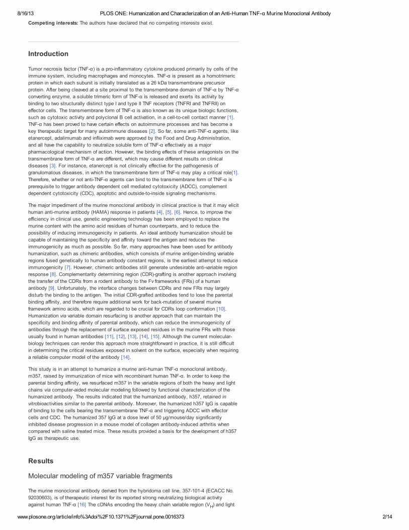
modeling 2.1 program as shown in Figure 1.
Figure 1. Molecular model of the m357 variable regions.
The three-dimensional structure of m357 was generated by homology modeling using
the crystal structures of the anti-breast tumor antibody SM3 (PDB code 1SM3) and the
anti-Thermus aquaticus DNA polymerase I antibody (PDB code 1AY1) for the V and
V domains, respectively. The six residues which are preferably mutated to “human”
amino acid are indicated by stick structures (green). Four residues in the V framework
and two residues in the V framework were humanized. Valine 59, which is within about
5 Å of the CDR2 in light chain and may affect the antigen binding, is retained for
preserving the antigen binding affinity and shown as stick structure (orange). The CDR
loops are shown in red color.
doi:10.1371/journal.pone.0016373.g001
Humanization of m357 variable fragments
Humanization by variable domain resurfacing was first proposed by Padlan in 1991, in which
the potential antigenic sites in the framework regions of an antibody were eliminated without
affecting the antigen binding affinity [11]. This method is based on the hypothesis that the
human anti-mouse antibody (HAMA) response to the variable region derives from the surface
residues only and has adopted and modified by other researchers [14], [15], [19]. In this study,
we performed the variable domain resurfacing of m357 with the following three steps: first, the
molecular models of the V and V domains of m357 were constructed individually; second, we
used AREAIMOL program to calculate the solvent accessible residues for identifying the non-
human like framework surface residues, and finally we mutated these surface residues to the
human counterparts according the results of the sequence alignment of human framework. In
order to replace the non-human like framework surface residues of the murine m357 on the
variable domain, a set of highly homologous surface residues from a human target sequence
were selected. We searched the IMGT database (http://imgt.cines.fr/) to identify the human
V and V sequence pairs, which are most homologous to the corresponding variable regions of
m357, while eliminating the sequences of phage-display or humanized antibodies from our
search results. The most identical surface residues found from human sequences were the
variable regions of PPS4 (V : 76% and V : 73% in sequence identity in the FRs, respectively)
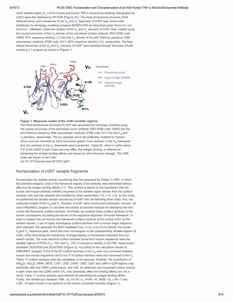
(Genbank DQ187629 and DQ187550) (Figure 2). According to the calculation results of
AREAIMOL program, 8 out of the 20 surface residues in the V were non-conserved between
human and mouse sequences and 9 out of 16 surface residues were non-conserved in the V .
These 17 surface residues were the candidates to be replaced. However, the substitution of
H52 Q, H52 S, H54N, H61E, L31F, L55S, L90W, L92D, L93Y and L96R in CDR regions may
potentially alter the CDR's conformation, and V59, an additional non-conserved surface residue
in light chain near the CDR2 within 5 Å, may potentially affect the binding affinity too. As a
result, these 11 murine residues were retained for preserving the antigen binding affinity.
Finally, the remaining 6 residues: H3K→Q, H11W→L, H19K→R, H42E→G, L10I→T and
L18K→R were chosen to be replaced to the human conserved residues (Figure 1).
L
H L
H L
H
L
H L
PowerPoint slidePPT
larger image (328KB)PNG
original image(437KB)
TIFF
H
L
H
L
H L
H L
H L
H
L
B C
8/16/13 PLOS ONE: Humanization and Characterization of an Anti-Human TNF-α Murine Monoclonal Antibody
www.plosone.org/article/info%3Adoi%2F10.1371%2Fjournal.pone.0016373 4/14
Download:
Download:
Figure 2. Amino acid sequence alignment of the V (A) and V (B) domains of
m357 and PPS4.
The sequences of PPS4 Fv which were the most homologous to m357 Fv used as the
acceptor of human surface residues for m357 humanization is shown as PPS4V and
PPS4V for comparison. CDR residues were within brackets. Conserved surface
residues were marked with clear boxes, and non-conserved surface residues were
marked with shadowed boxes. Amino acid sequences are numbered according to the
Kabat's convention[33].
doi:10.1371/journal.pone.0016373.g002
Construction and expression of the humanized 357 IgG
The amino acid sequences of the humanized V and V of m357, comprising the signal peptide
at the N-terminus, were in-frame fused to the human IgG γ1 heavy chain and kappa light chain
constant regions, respectively. For the expression of an intact humanized 357 (h357)
IgG molecule, two different mammalian expression vectors, pSecTag2/Hygro and pcDNA3.3,
were used to adopt the humanized heavy chain and light chain of h357 IgG, respectively. After
co-transfection and antibiotic selection, one of the clones showing the highest expression level
was chosen for further studies. The expression level of the recombinant h357 IgG was ~14
mg/L. Both m357 and h357 IgGs were purified by protein A chromatography individually and the
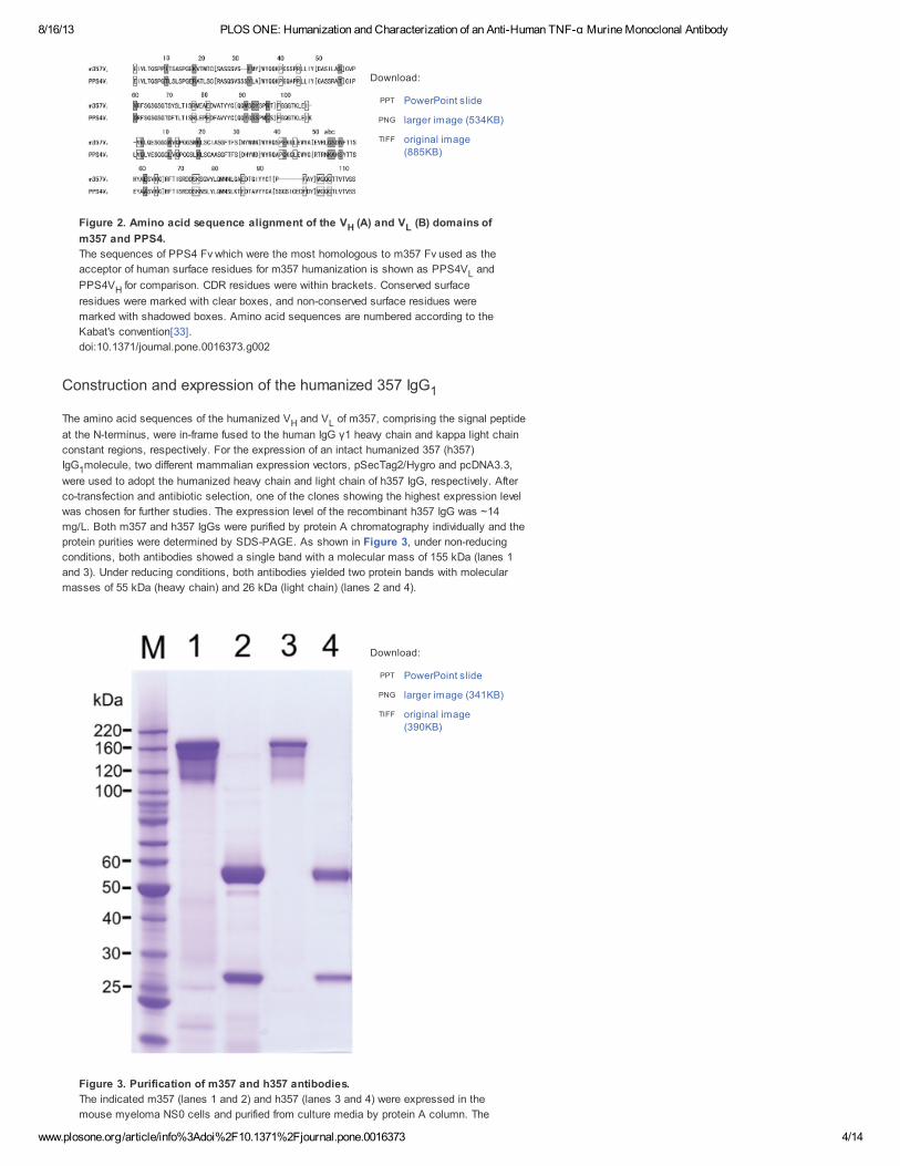
protein purities were determined by SDS-PAGE. As shown in Figure 3, under non-reducing
conditions, both antibodies showed a single band with a molecular mass of 155 kDa (lanes 1
and 3). Under reducing conditions, both antibodies yielded two protein bands with molecular
masses of 55 kDa (heavy chain) and 26 kDa (light chain) (lanes 2 and 4).
Figure 3. Purification of m357 and h357 antibodies.
The indicated m357 (lanes 1 and 2) and h357 (lanes 3 and 4) were expressed in the
mouse myeloma NS0 cells and purified from culture media by protein A column. The
PowerPoint slidePPT
larger image (534KB)PNG
original image(885KB)
TIFF
H L
L
H
1
H L
1
PowerPoint slidePPT
larger image (341KB)PNG
original image(390KB)
TIFF
8/16/13 PLOS ONE: Humanization and Characterization of an Anti-Human TNF-α Murine Monoclonal Antibody
www.plosone.org/article/info%3Adoi%2F10.1371%2Fjournal.pone.0016373 5/14
Download:
samples were electrophoresed on a 4~12% SDS/Bis-Tris polyacrylamide gel with
MOPS buffer under non-reducing conditions (lanes 1 and 3) and reducing conditions
(lanes 2 and 4). Gel was stained with Coommassie blue. M, molecular mass standards.
doi:10.1371/journal.pone.0016373.g003
Neutralization of TNF-α-mediated cellular cytotoxicity by m357 andh357
To examine the functional activity of the anti-TNF-α antibodies, the ability of the antibody to
inhibit soluble TNF-α activity was performed. TNF-α causes cell cytotoxicity to murine L929
cells. Both m357 and h357 IgGs were evaluated in L929 assays by co-incubation of antibodies
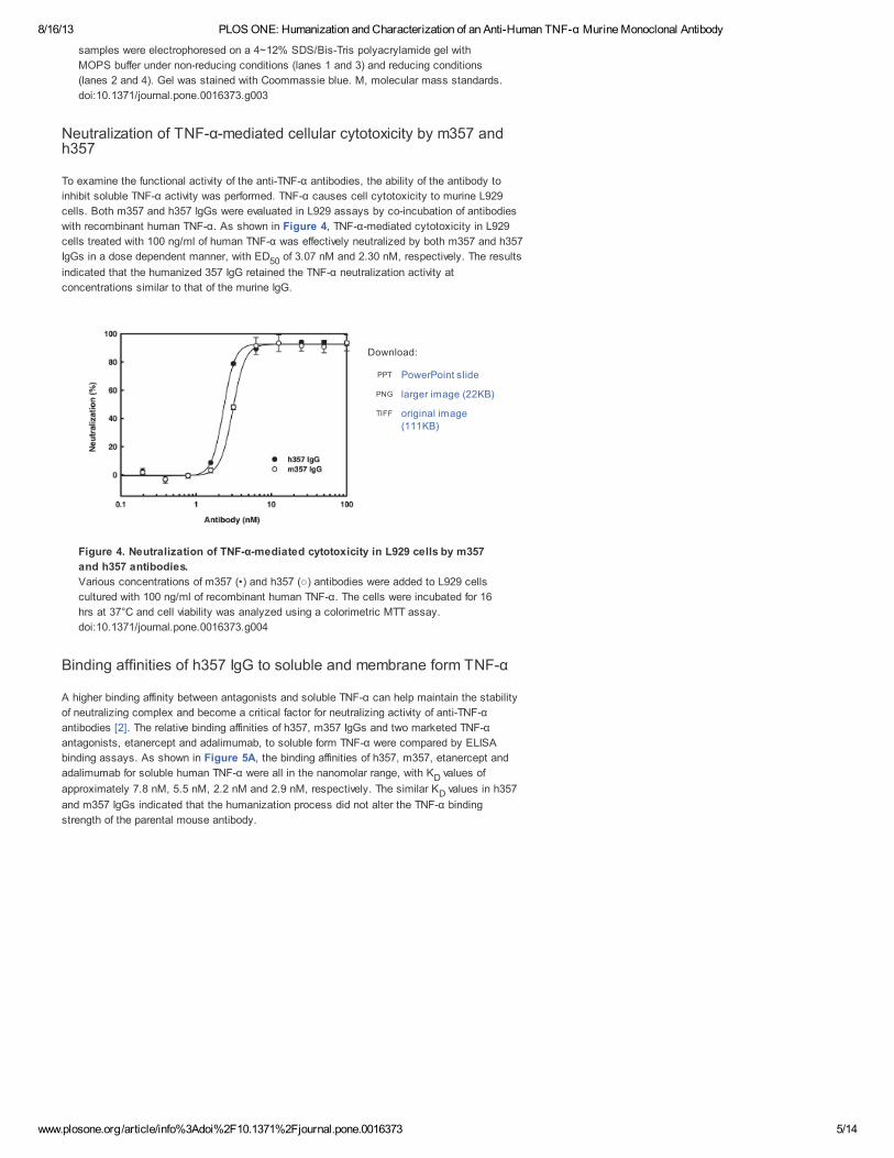
with recombinant human TNF-α. As shown in Figure 4, TNF-α-mediated cytotoxicity in L929
cells treated with 100 ng/ml of human TNF-α was effectively neutralized by both m357 and h357
IgGs in a dose dependent manner, with ED of 3.07 nM and 2.30 nM, respectively. The results
indicated that the humanized 357 IgG retained the TNF-α neutralization activity at
concentrations similar to that of the murine IgG.
Figure 4. Neutralization of TNF-α-mediated cytotoxicity in L929 cells by m357
and h357 antibodies.
Various concentrations of m357 (•) and h357 (○) antibodies were added to L929 cells
cultured with 100 ng/ml of recombinant human TNF-α. The cells were incubated for 16
hrs at 37°C and cell viability was analyzed using a colorimetric MTT assay.
doi:10.1371/journal.pone.0016373.g004
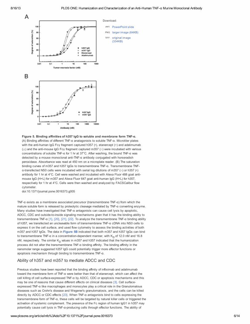
Binding affinities of h357 IgG to soluble and membrane form TNF-α
A higher binding affinity between antagonists and soluble TNF-α can help maintain the stability
of neutralizing complex and become a critical factor for neutralizing activity of anti-TNF-α
antibodies [2]. The relative binding affinities of h357, m357 IgGs and two marketed TNF-α
antagonists, etanercept and adalimumab, to soluble form TNF-α were compared by ELISA
binding assays. As shown in Figure 5A, the binding affinities of h357, m357, etanercept and
adalimumab for soluble human TNF-α were all in the nanomolar range, with K values of
approximately 7.8 nM, 5.5 nM, 2.2 nM and 2.9 nM, respectively. The similar K values in h357
and m357 IgGs indicated that the humanization process did not alter the TNF-α binding
strength of the parental mouse antibody.
50
PowerPoint slidePPT
larger image (22KB)PNG
original image(111KB)
TIFF
D
D
8/16/13 PLOS ONE: Humanization and Characterization of an Anti-Human TNF-α Murine Monoclonal Antibody
www.plosone.org/article/info%3Adoi%2F10.1371%2Fjournal.pone.0016373 6/14
Download:
Figure 5. Binding affinities of h357 IgG to soluble and membrane form TNF-α.
(A) Binding affinities of different TNF-α anatagonists to soluble TNF-α. Microtiter plates
with the anti-human IgG Fcγ fragment captured h357 (•), etanercept (▾) and adalimumab
(△) and the anti-mouse IgG Fcγ fragment captured m357 (○) were incubated with various
concentrations of soluble TNF-α for 1 hr at 37°C. After washing, the bound TNF-α was
detected by a mouse monoclonal anti-TNF-α antibody conjugated with horseradish
peroxidase. Absorbance was read at 450 nm on a microplate reader. (B) The saturation
binding curves of m357 and h357 IgGs to transmembrane TNF-α. Transmembrane TNF-
α-transfected NS0 cells were incubated with serial log dilutions of m357 (○) or h357 (•)
antibody for 1 hr at 4°C. Cell were washed and incubated with Alexa Fluor 488 goat anti-
mouse IgG (H+L) for m357 and Alexa Fluor 647 goat anti-human IgG (H+L) for h357,
respectively for 1 hr at 4°C. Cells were then washed and analyzed by FACSCalibur flow
cytometer.
doi:10.1371/journal.pone.0016373.g005
TNF-α exists as a membrane associated precursor (transmembrane TNF-α) from which the
mature soluble form is released by proteolytic cleavage mediated by TNF-α converting enzyme.
Many studies have investigated that TNF-α antagonists can cause cell lysis by apoptotic,
ADCC, CDC and outside-to-inside signaling mechanisms given that it has the binding ability to
transmembrane TNF-α [1], [20], [21], [22]. To analyze the transmembrane TNF-α binding ability
of h357, we transfected an uncleavable form of transmembrane TNF-α cDNA into NS0 cells to
express it on the cell surface, and used flow cytometry to assess the binding activities of both
m357 and h357 IgGs. The data in Figure 5B indicated that both m357 and h357 IgGs can bind
to transmembrane TNF-α in a concentration-dependent manner, with K of 12.0 nM and 16.8
nM, respectively. The similar K values in m357 and h357 indicated that the humanization
process did not alter the transmembrane TNF-α binding affinity. The binding affinity in the
nanomolar range suggested h357 IgG could potentially trigger more effector functions or
apoptosis mechanism through binding to transmembrane TNF-α.
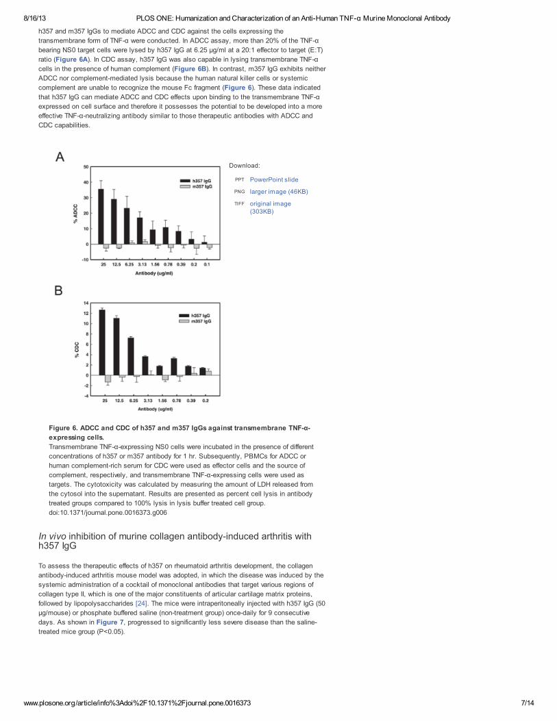
Ability of h357 and m357 to mediate ADCC and CDC
Previous studies have been reported that the binding affinity of infliximab and adalimumab
toward the membrane form of TNF-α were better than that of etanercept, which can affect the
cell killing of cell surface-expressed TNF-α by ADCC, CDC or apoptosis mechanisms and this
may be one of reasons that cause different effects on clinical diseases [3]. Cell surface-
expressed TNF-α like macrophages and monocytes play a critical role in the Granulomatous
diseases such as Crohn's disease and Wegener's granulomatosis, and the cells can be killed
directly by ADCC or CDC effects [23]. When TNF-α antagonists bind to cells expressing the
transmembrane form of TNF-α, these cells will be targeted by natural killer cells or triggered the
activation of systemic complement. The presence of the Fc region of human IgG1 in h357 may
potentially cause cell lysis in TNF-α-producing cells through effector functions. The ability of
PowerPoint slidePPT
larger image (64KB)PNG
original image(334KB)
TIFF
D
D
8/16/13 PLOS ONE: Humanization and Characterization of an Anti-Human TNF-α Murine Monoclonal Antibody
www.plosone.org/article/info%3Adoi%2F10.1371%2Fjournal.pone.0016373 7/14
Download:
h357 and m357 IgGs to mediate ADCC and CDC against the cells expressing the
transmembrane form of TNF-α were conducted. In ADCC assay, more than 20% of the TNF-α
bearing NS0 target cells were lysed by h357 IgG at 6.25 µg/ml at a 20:1 effector to target (E:T)
ratio (Figure 6A). In CDC assay, h357 IgG was also capable in lysing transmembrane TNF-α
cells in the presence of human complement (Figure 6B). In contrast, m357 IgG exhibits neither
ADCC nor complement-mediated lysis because the human natural killer cells or systemic
complement are unable to recognize the mouse Fc fragment (Figure 6). These data indicated
that h357 IgG can mediate ADCC and CDC effects upon binding to the transmembrane TNF-α
expressed on cell surface and therefore it possesses the potential to be developed into a more
effective TNF-α-neutralizing antibody similar to those therapeutic antibodies with ADCC and
CDC capabilities.
Figure 6. ADCC and CDC of h357 and m357 IgGs against transmembrane TNF-α-
expressing cells.
Transmembrane TNF-α-expressing NS0 cells were incubated in the presence of different
concentrations of h357 or m357 antibody for 1 hr. Subsequently, PBMCs for ADCC or
human complement-rich serum for CDC were used as effector cells and the source of
complement, respectively, and transmembrane TNF-α-expressing cells were used as
targets. The cytotoxicity was calculated by measuring the amount of LDH released from
the cytosol into the supernatant. Results are presented as percent cell lysis in antibody
treated groups compared to 100% lysis in lysis buffer treated cell group.
doi:10.1371/journal.pone.0016373.g006
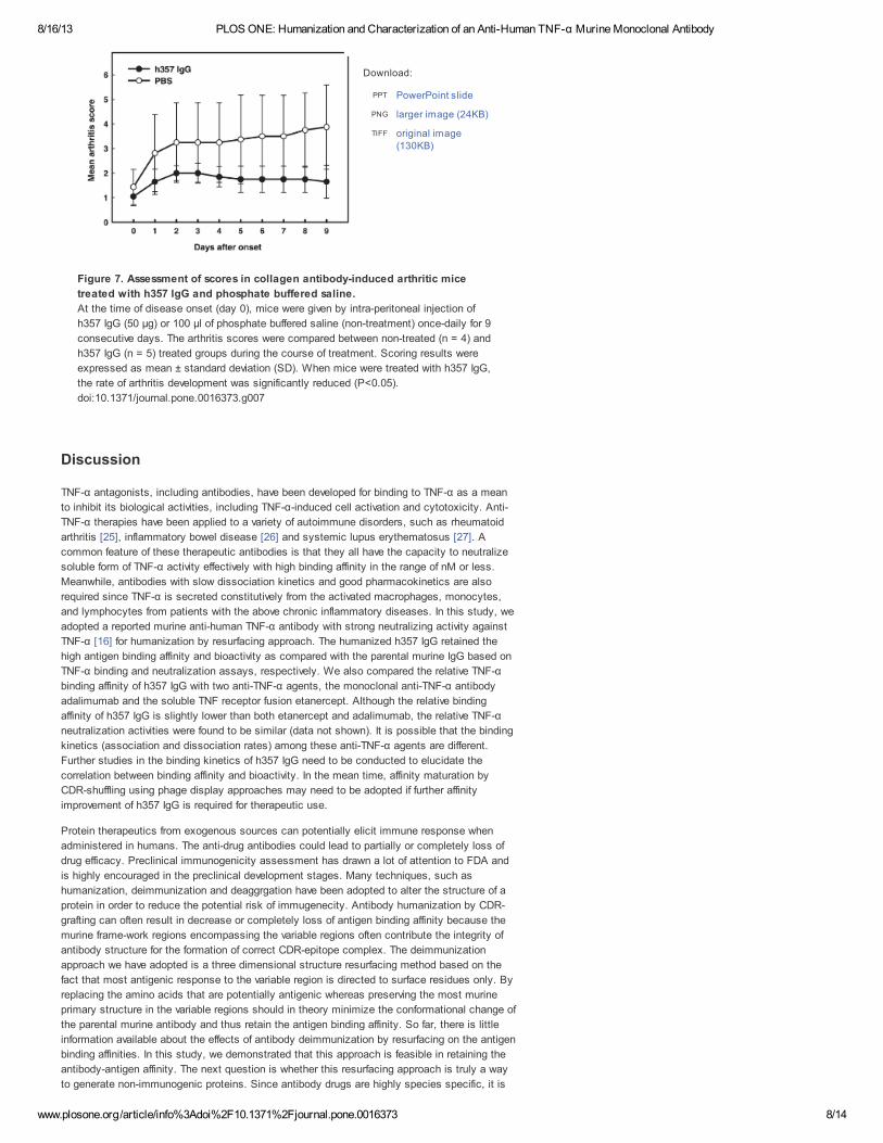
In vivo inhibition of murine collagen antibody-induced arthritis withh357 IgG
To assess the therapeutic effects of h357 on rheumatoid arthritis development, the collagen
antibody-induced arthritis mouse model was adopted, in which the disease was induced by the
systemic administration of a cocktail of monoclonal antibodies that target various regions of
collagen type II, which is one of the major constituents of articular cartilage matrix proteins,
followed by lipopolysaccharides [24]. The mice were intraperitoneally injected with h357 IgG (50
µg/mouse) or phosphate buffered saline (non-treatment group) once-daily for 9 consecutive
days. As shown in Figure 7, progressed to significantly less severe disease than the saline-
treated mice group (P<0.05).
PowerPoint slidePPT
larger image (46KB)PNG
original image(303KB)
TIFF
8/16/13 PLOS ONE: Humanization and Characterization of an Anti-Human TNF-α Murine Monoclonal Antibody
www.plosone.org/article/info%3Adoi%2F10.1371%2Fjournal.pone.0016373 8/14
Download:
Figure 7. Assessment of scores in collagen antibody-induced arthritic mice
treated with h357 IgG and phosphate buffered saline.
At the time of disease onset (day 0), mice were given by intra-peritoneal injection of
h357 IgG (50 µg) or 100 µl of phosphate buffered saline (non-treatment) once-daily for 9
consecutive days. The arthritis scores were compared between non-treated (n = 4) and
h357 IgG (n = 5) treated groups during the course of treatment. Scoring results were
expressed as mean ± standard deviation (SD). When mice were treated with h357 IgG,
the rate of arthritis development was significantly reduced (P<0.05).
doi:10.1371/journal.pone.0016373.g007
Discussion
TNF-α antagonists, including antibodies, have been developed for binding to TNF-α as a mean
to inhibit its biological activities, including TNF-α-induced cell activation and cytotoxicity. Anti-
TNF-α therapies have been applied to a variety of autoimmune disorders, such as rheumatoid
arthritis [25], inflammatory bowel disease [26] and systemic lupus erythematosus [27]. A
common feature of these therapeutic antibodies is that they all have the capacity to neutralize
soluble form of TNF-α activity effectively with high binding affinity in the range of nM or less.
Meanwhile, antibodies with slow dissociation kinetics and good pharmacokinetics are also
required since TNF-α is secreted constitutively from the activated macrophages, monocytes,
and lymphocytes from patients with the above chronic inflammatory diseases. In this study, we
adopted a reported murine anti-human TNF-α antibody with strong neutralizing activity against
TNF-α [16] for humanization by resurfacing approach. The humanized h357 IgG retained the
high antigen binding affinity and bioactivity as compared with the parental murine IgG based on
TNF-α binding and neutralization assays, respectively. We also compared the relative TNF-α
binding affinity of h357 IgG with two anti-TNF-α agents, the monoclonal anti-TNF-α antibody
adalimumab and the soluble TNF receptor fusion etanercept. Although the relative binding
affinity of h357 IgG is slightly lower than both etanercept and adalimumab, the relative TNF-α
neutralization activities were found to be similar (data not shown). It is possible that the binding
kinetics (association and dissociation rates) among these anti-TNF-α agents are different.
Further studies in the binding kinetics of h357 IgG need to be conducted to elucidate the
correlation between binding affinity and bioactivity. In the mean time, affinity maturation by
CDR-shuffling using phage display approaches may need to be adopted if further affinity
improvement of h357 IgG is required for therapeutic use.
Protein therapeutics from exogenous sources can potentially elicit immune response when
administered in humans. The anti-drug antibodies could lead to partially or completely loss of
drug efficacy. Preclinical immunogenicity assessment has drawn a lot of attention to FDA and
is highly encouraged in the preclinical development stages. Many techniques, such as
humanization, deimmunization and deaggrgation have been adopted to alter the structure of a
protein in order to reduce the potential risk of immugenecity. Antibody humanization by CDR-
grafting can often result in decrease or completely loss of antigen binding affinity because the
murine frame-work regions encompassing the variable regions often contribute the integrity of
antibody structure for the formation of correct CDR-epitope complex. The deimmunization
approach we have adopted is a three dimensional structure resurfacing method based on the
fact that most antigenic response to the variable region is directed to surface residues only. By
replacing the amino acids that are potentially antigenic whereas preserving the most murine
primary structure in the variable regions should in theory minimize the conformational change of
the parental murine antibody and thus retain the antigen binding affinity. So far, there is little
information available about the effects of antibody deimmunization by resurfacing on the antigen
binding affinities. In this study, we demonstrated that this approach is feasible in retaining the
antibody-antigen affinity. The next question is whether this resurfacing approach is truly a way
to generate non-immunogenic proteins. Since antibody drugs are highly species specific, it is
PowerPoint slidePPT
larger image (24KB)PNG
original image(130KB)
TIFF
8/16/13 PLOS ONE: Humanization and Characterization of an Anti-Human TNF-α Murine Monoclonal Antibody
www.plosone.org/article/info%3Adoi%2F10.1371%2Fjournal.pone.0016373 9/14
difficult to assess the immune response in humans when conducting preclinical animal
experiments even with non-human primates. The adopted three dimensional structure
resurfacing method currently we used is distinct from the in silico primary structure-based
algorisms which mainly focused on MHC class II peptide binding predictions. Meanwhile,
recently developed T-cell based procedures are showing promise for predicting potential
immunogenicity with some biologics including monoclonal antibodies. Further experimental
work focusing on both in silico and T-cell assays of the h357 IgG will be assessed to further
minimize the immunogenicity issues for drug development.
Materials and Methods
Ethics Statement
Human whole blood was collected from healthy donors at Hsinchui Blood Center, Taiwan, under
a protocol approved by the Institutional Review Board of Industrial Technology Research
Institute (IRB number 9901008) and reviewed by the Joint Institutional Review Board
(http://www.jirb.org.tw/), case number 10-S-004, a committee for allowing all blood centers in
Taiwan to provided blood for research purposes from those donors with written consent. All
animal work was reviewed and approved by the Institutional Animal Care and Use Committee of
Industrial Technology Research Institute (approval number ITRI-IACUC-2010-030R).
Computer modeling of m357 for resurfacing
The homology modeling process of m357 was performed using Discovery Studio Modeling 2.1
(Accelrys, Inc., San Diego, CA). Two separate BLASTP searches were performed for V and
V of m357. For identifying the homology of m357 protein sequences, sequences were
analyzed by searching over the Protein Data Bank (PDB)
(http://www.rcsb.org/pdb/home/home.do). The three-dimensional (3D) structure of the m357 Fv
fragment was constructed by homology modeling based on the structures of the V domain of
the murine anti-breast tumor antibody Fab fragment SM-3[PDB entry: 1SM3] [17] and the
V domain structure of an anti-thermus aquaticus DNA polymerase I monoclonal antibody Fab
fragment [PDB entry: 1AY1] [18]. The final 3D model was generated by MODELLER
module[28], which implements an automated approach to comparative protein structure
modeling by satisfaction of spatial restraints. It performed automatically protein homology
modeling and loop modeling for m357. Model building of the CDR loops were performed by
selecting template structures from the PDB database with the highest sequence identity using
Model antibody Loops module and were refined using Loop Refinement module in order to
minimize steric clashes and ensure correct bond lengths and angles. Then, this whole model
could be further refined by CHARMm [29] with Accelrys CHARMm forcefield in Discovery
Studio Modeling 2.1. The structure was energy-minimized in two steps, first by 5000 steps of
restrained steepest descent minimization, and followed by another 5000 steps of conjugated
gradient minimization while the alpha carbons of the framework were held fixed in position. The
solvent-accessible surface areas of m357 residues were calculated on the three-dimensional
model by the AREAIMOL program [30]. The residues which relative accessibility was greater
than 30% were defined as being accessible.
Construction, expression and purification of humanized anti-humanTNF-α IgG
The total RNA of around 10 cells of the hybridoma 357-101-4 (ECACC No. 92030603) which
secrete anti-human TNF-α IgG (m357) was isolated and the first-strand cDNA was generated
using oligo (dT) primers with Superscript III reverse transcriptase (Invitrogen, San Diego, CA).
The cDNA fragments encoding the heavy chain and light chain variable regions of m357 were
obtained by PCR with the Heavy Primers (Cat. No. 27-1586-01, GE Healthcare) and the Light
Primer Mix (Cat. No. 27-1583-01, GE Healthcare), respectively. The cDNA fragments encoding
the humanized heavy chain and light chain variable regions of m357 were obtained by over-
lapping PCR with primers covering the humanized regions. For the construction of the heavy
chain of the humanized 357 (h357) antibody, the cDNA construct consisting the murine Ig
kappa-chain V-J2-C signal peptide derived from the mammalian expression vector
pSecTag2/Hygro (Invitrogen, San Diego, CA), the V region of h357, and the human IgG1
constant region (CH1, hinge, CH2 and CH3) which derived from the cloning vector pFUSE-CHIg-
hG1 (Invivogen, San Diego, CA) were obtained by over-lapping PCR, followed by sub-cloning
into the vector pSecTag2/Hygro (Invitrogen, San Diego, CA) at Nhe I and Not I sites. For the
construction of the light chain of the humanized 357 (h357), the cDNA construct consisting the
above murine Ig kappa-chain V-J2-C signal peptide, the V region of h357, and the human
kappa light chain constant region which derived from the cloning vector pFUSE2-CLIg-hk
(Invivogen, San Diego, CA) were obtained by over-lapping PCR, followed by sub-cloning into the
mammalian expression vectors pcDNA3.3-TOPO TA (Invitrogen, San Diego, CA). Plasmids
containing the heavy and light chain cDNAs of h357 IgG were co-transfected into mouse
L
H
H
L
7
H
L
1
8/16/13 PLOS ONE: Humanization and Characterization of an Anti-Human TNF-α Murine Monoclonal Antibody
www.plosone.org/article/info%3Adoi%2F10.1371%2Fjournal.pone.0016373 10/14
myeloma NS0 cells (European Collection of Animal Cell cultures, Salisbury, Wiltshire, UK)
using Effectene (Qiagen). After selection with hygromycin B (400 µg/ml) and G418 (800 µg/ml)
for 4 weeks, culture media from several stable clones were screened by ELISA with human
TNF-α as coating and anti-human IgG Fcγ-HRP as detection reagents. An h357 IgG high-
expressing clone was picked and cultured in shaker flask at an initial seeding density of
5×10 cells/ml in a chemically-defined medium HyQNS0 (Hyclone) without serum addition.
Media were harvested 5 days later and h357 IgG was purified from the supernatant by Protein
A (GE Healthcare) chromatography.
Anti-TNF-α neutralization potency assay
Neutralizing activities of m357 and h357 against human TNF-α were measured on the murine
fibroblast L929 cells (ATCC Cat. No. CCL-1) treated with actinomycin D according to the
method described previously[31]. Briefly, L929 cells were seeded in triplicate at 3×10 cells/well
into a 96-well plate and cultured in RPMI 1640 medium supplemented with 10% (v/v) fetal
bovine serum for 16 hours. Then, several dilutions of the antibodies were prepared in medium
containing actinomycin D (2 µg/ml) and TNF-α (100 ng/ml) and incubated at 37°C for 16 hours.
After the supernatant were removed, 3-4,5-dimethylthiazol-2-yl-2,5-diphenyltetrazoliumbromide
(MTT) (5 mg/ml) (Sigma-Aldrich) was added and incubated in 37°C for 4 hours. SDS solution
(10%) was then added to the well. After 24 hours of incubation at room temperature, color in
each well was recorded by colorimeter at 570 nm. Blank control (culture alone), TNF-α control
(TNF-α alone), and antibody control (antibody alone) were also designed in the experiment. The
ED value was calculated by complex sigmoid non-linear regression analysis using SigmaPlot
software (Systat software, Inc. Richmond, CA).
Stable expression of transmembrane TNF-α on NS0 cells
A mutant form of transmembrane TNF-α, which is resistant to TNF-α converting enzyme-
mediated cleavage, was generated by site-directed mutagenesis as described previously [32].
In this uncleavable form of transmembrane TNF-α, the amino acids +1 through +12 of the native
transmembrane TNF-α were deleted. An uncleavable form of transmembrane TNF-α gene was
cloned into pSecTag2/Hygro mammalian expression vector (Invitrogen, San Diego, CA) and
transfected into mouse myeloma NS0 cells by Effectene for expressing transmembrane TNF-α
on cell surface.
Antibody binding studies
The binding activities of h357 IgG, m357 IgG, etanercept and adalimumab to soluble TNF-α
were determined by ELISA. For capturing h357 IgG, etanercept (Wyeth) and adalimumab
(Abbott), the microtiter plate was coated with 2 µg/ml of goat anti-hunam IgG Fcγ fragment
(Jackson ImmunoResearch Laboratories, Inc., West Grove, PA) in PBS overnight at 4°C. For
capturing m357 IgG, the microtiter plate was coated with goat anti-mouse IgG Fcγ fragment
(Jackson ImmunoResearch Laboratories, Inc., West Grove, PA). After blocking the wells with
StartingBlock™ blocking buffer (Thermo Scientific), 2 µg/ml each of the TNF-α antagonists, as
mentioned above, were added and incubated for 1 hr at 37°C. After washing, varing
concentrations of human recombinant TNF-α (eBioscience, Inc., San Diego, CA) in PBS were
incubated for 1 hr at 37°C. After washing, the bound soluble TNF-α was detected by incubation
with horseradish peroxidase-conjuagted mouse monoclonal anti-TNF-α antibody (clone F6C5,
Abcam, Cambridge, MA) for 1 hr at 37°C and using 3,3′,5,5′,-tetramethylbenzidine as substrate.
Absorbance was read at 450 nm on a microplate reader. The dissociation constant (K ) of each
antagonist was calculated as the concentration of TNF-α required to achieve half-saturation of
the total binding sites (maximum absorbance).
Saturation binding assay of m357 and h357 IgGs to transmembraneTNF-α
NS0 cells stably expressing the transmembrane TNF-α were incubated with serial log dilutions
of m357 and h357 IgGs for 1 hr at 4°C in phosphate buffered saline (PBS) containing 2% fetal
bovine serum (fluorescence-activated cell sorting [FACS] buffer). Cells were washed with a
FACS buffer for 3 times and were then stained with Alexa Fluor 488 conjugated goat anti-
mouse IgG (H+L) (Invitrogen, San Diego, CA) for m357 IgG and Alexa Fluor 647 conjugated
goat anti-human IgG (H+L) (Invitrogen, San Diego, CA) for h357 IgG, respectively for 1 hr at
4°C. Fluorescence intensities were measured using a FACSCalibur flow cytometer (Becton
Dickinson, San Jose, CA).
ADCC and CDC assays
Peripheral blood mononuclear cells (PBMCs) were prepared by Ficoll-Hypaque density gradient
centrifugation. ADCC and CDC activities of h357 and m357 IgGs were measured by LDH
Cytotoxicity Detection Kit (Clontech), which measures lactate dehydrogenase (LDH) activity
released from the cytosol of damaged cells. Briefly, NS0 cells stably expressing the
1
5
1
5
50
D
8/16/13 PLOS ONE: Humanization and Characterization of an Anti-Human TNF-α Murine Monoclonal Antibody
www.plosone.org/article/info%3Adoi%2F10.1371%2Fjournal.pone.0016373 11/14
transmembrane TNF-α were incubated with different concentration of h357 or m357 IgG for 1 hr
in assay medium (DMEM +1% FBS) in a 5% CO incubator at 37°C, followed by the addition of
either human peripheral blood mononuclear cells (PBMC) as effector cells (effector to target
ratio = 20:1 for ADCC assay) or human complement-rich serum (Quidel) (1.25% vol/vol, for CDC
assay). After an additional incubation for 16 hrs for ADCC assay and 5 hrs for CDC assay at
37°C, respectively, 100 µl of supernatant from each well were transferred into a clean flat-
bottom 96-well plate. LDH substrate (100 µl) was added to each well and incubated for 30 min
at room temperature protected from light. The absorbance of the samples was measure at 490
nm with an ELISA reader. Maximum LDH release was determined by incubating cells with lysis
buffer. Percentage of specific lysis was calculated according to the following formula: %
cytotoxicity = [experimental release – spontaneous release]/[maximum release – spontaneous
release]×100.
In vivo inhibition of murine collagen antibody-induced arthritis withh357 IgG
For collagen antibody-induced arthritis experiments, 8- to10-week old male DBA/1J mice were
purchased from Jackson laboratories (Bar Harbor, ME). Mice were maintained under a climate
controlled environment in a 12-h light/dark cycle. Arthritic mice were induced by intra-peritoneal
(i.p.) injection of 3 mg/mouse of type II collagen specific antibodies (ModiQuest Research).
Mice were further boosted with 25 µg lipopolysaccharides (LPS) (Sigma) by i.p. injection on
day 6. Clinical arthritis scores were evaluated using a scale of 0–2 for each paw for a total
score of 8. Paws were assigned a clinical score based on the index (ModiQuest Research
scoring method): 0 = normal; 0.25 = one or two swollen toes; 0.5 = three and four swollen toes;
0.75 = slightly swollen footpad or ankle; 1 = swollen footpad or ankle; 1.25 = one or two
swollen toes and swollen footpad or ankle; 2.0 = swollen toes and swollen footpad and swollen
ankle. At the time of disease onset, mice were administered by i.p. injection of 50 µg per does
for 9 consecutive days. Scoring results were expressed as mean ± standard deviation (SD).
Statistical differences between the control (phosphate buffered saline) and experimental groups
were analyzed by Mann-Whitney U test for the severity of arthritis.
Supporting Information
2
Figure_S1.eps
RT-PCR results showing the variable fragments and schematic representation of
the strategy to assemble the heavy and light chains. (A) Amplification of the variable
fragment cDNAs from the mouse hybridoma 357-101-4 secreting m357 IgG by RT-PCR
with the Heavy Primers (Cat. No. 27-1586-01, GE Healthcare) and the Light Primer Mix
(Cat. No. 27-1583-01, GE Healthcare), respectively. Lane M: Molecular weight marker.
Lane 1: V , ~340 bp. Lane 2: V , ~325 bp. (B) Assembly of the cDNAs encoding the openH L
figshare download
8/16/13 PLOS ONE: Humanization and Characterization of an Anti-Human TNF-α Murine Monoclonal Antibody
www.plosone.org/article/info%3Adoi%2F10.1371%2Fjournal.pone.0016373 12/14
1.
CrossRef PubMed/NCBI Google Scholar
2.
CrossRef PubMed/NCBI Google Scholar
3.
CrossRef PubMed/NCBI Google Scholar
4.
CrossRef PubMed/NCBI Google Scholar
5.
CrossRef PubMed/NCBI Google Scholar
6.
Figure S1.
RT-PCR results showing the variable fragments and schematic representation of the
strategy to assemble the heavy and light chains. (A) Amplification of the variable fragment
cDNAs from the mouse hybridoma 357-101-4 secreting m357 IgG by RT-PCR with the Heavy
Primers (Cat. No. 27-1586-01, GE Healthcare) and the Light Primer Mix (Cat. No. 27-1583-01,
GE Healthcare), respectively. Lane M: Molecular weight marker. Lane 1: V , ~340 bp. Lane 2:
V , ~325 bp. (B) Assembly of the cDNAs encoding the open reading frames of the heavy and
light chains of the humanized 357 (h357) IgG1 by over-lapping PCR, respectively. The cDNA
construct consisting the murine signal peptide (SP) derived from the cloning vector
pSecTag2/Hygro, the V region of h357, and the human IgG1 constant region (CH1, hinge, CH2
and CH3) derived from the cloning vector pFUSE-CHIg-hG1 were obtained by over-lapping PCR,
followed by sub-cloning into the vector pSecTag2/Hygro at Nhe I and Not I sites. The cDNA
construct consisting the above signal peptide, the V region of h357, and the human kappa
light chain constant region derived from the cloning vector pFUSE2-CLIg-hk were obtained by
over-lapping PCR, followed by sub-cloning into the mammalian expression vectors pcDNA3.3-
TOPO TA (Invitrogen, San Diego, CA). The locations of the primers and the restriction sites are
shown in the diagram. SP, murine Ig kappa-chain V-J2-C signal peptide.
doi:10.1371/journal.pone.0016373.s001
(EPS)
Acknowledgments
We are grateful to Ting-Shou Chen and Jiun-Lin Guo for their computer technical assistance
and Shu-Hui Chang for her technical contributions in animal experiments.
Author Contributions
Conceived and designed the experiments: MYC WCC. Performed the experiments: WCC YPL
MYC. Analyzed the data: WCC YPL MYC. Contributed reagents/materials/analysis tools:
WCC YPL MYC. Wrote the paper: WCC YPL MYC.
References
Mitoma H, Horiuchi T, Tsukamoto H, Tamimoto Y, Kimoto Y, et al. (2008) Mechanisms
for cytotoxic effects of anti-tumor necrosis factor agents on transmembrane tumor
necrosis factor alpha-expressing cells: comparison among infliximab, etanercept, and
adalimumab. Arthritis Rheum 58: 1248–1257. doi: 10.1002/art.23447.
Feldmann M (2001) Pathogenesis of arthritis: recent research progress. Nat Immunol 2:
771–773.
Taylor PC (2010) Pharmacology of TNF blockade in rheumatoid arthritis and other
chronic inflammatory diseases. Curr Opin Pharmacol 10: 1–8.
doi:10.1016/j.coph.2010.01.005.
Sandhu JS (1992) Protein engineering of antibodies. Crit Rev Biotechnol 12: 437–462.
doi: 10.3109/07388559209114235.
Owens RJ, Young RJ (1994) The genetic engineering of monoclonal antibodies. J
Immunol Methods 168: 149–165. doi: 10.1016/0022-1759(94)90051-5.
Schroff RW, Foon KA, Beatty SM, Oldham RK, Morgan AC Jr (1985) Human anti-
murine immunoglobulin responses in patients receiving monoclonal antibody therapy.
reading frames of the heavy and light chains of the humanized 357 (h357) IgG1 by over-
lapping PCR, respectively. The cDNA construct consisting the murine signal peptide (SP)
derived from the cloning vector pSecTag2/Hygro, the V region of h357, and the human
IgG1 constant region (CH1, hinge, CH2 and CH3) derived from the cloning vector pFUSE-
CHIg-hG1 were obtained by over-lapping PCR, followed by sub-cloning into the vector
pSecTag2/Hygro at Nhe I and Not I sites. The cDNA construct consisting the above signal
peptide, the V region of h357, and the human kappa light chain constant region derived
from the cloning vector pFUSE2-CLIg-hk were obtained by over-lapping PCR, followed by
sub-cloning into the mammalian expression vectors pcDNA3.3-TOPO TA (Invitrogen, San
Diego, CA). The locations of the primers and the restriction sites are shown in the
diagram. SP, murine Ig kappa-chain V-J2-C signal peptide.
H L
H
L
H
L
H
L
8/16/13 PLOS ONE: Humanization and Characterization of an Anti-Human TNF-α Murine Monoclonal Antibody
www.plosone.org/article/info%3Adoi%2F10.1371%2Fjournal.pone.0016373 13/14
CrossRef PubMed/NCBI Google Scholar
7.
CrossRef PubMed/NCBI Google Scholar
8.
CrossRef PubMed/NCBI Google Scholar
9.
CrossRef PubMed/NCBI Google Scholar
10.
CrossRef PubMed/NCBI Google Scholar
11.
CrossRef PubMed/NCBI Google Scholar
12.
CrossRef PubMed/NCBI Google Scholar
13.
CrossRef PubMed/NCBI Google Scholar
14.
CrossRef PubMed/NCBI Google Scholar
15.
CrossRef PubMed/NCBI Google Scholar
16.
CrossRef PubMed/NCBI Google Scholar
17.
CrossRef PubMed/NCBI Google Scholar
18.
CrossRef PubMed/NCBI Google Scholar
19.
CrossRef PubMed/NCBI Google Scholar
20.
CrossRef PubMed/NCBI Google Scholar
21.
Cancer Res 45: 879–885.
Morrison SL, Johnson MJ, Herzenberg LA, Oi VT (1984) Chimeric human antibody
molecules: mouse antigen-binding domains with human constant region domains. Proc
Natl Acad Sci U S A 81: 6851–6855. doi: 10.1073/pnas.81.21.6851.
Bruggemann M, Winter G, Waldmann H, Neuberger MS (1989) The immunogenicity of
chimeric antibodies. J Exp Med 170: 2153–2157. doi: 10.1084/jem.170.6.2153.
Verhoeyen M, Milstein C, Winter G (1988) Reshaping human antibodies: grafting an
antilysozyme activity. Science 239: 1534–1536. doi: 10.1126/science.2451287.
Queen C, Schneider WP, Selick HE, Payne PW, Landolfi NF, et al. (1989) A
humanized antibody that binds to the interleukin 2 receptor. Proc Natl Acad Sci U S A
86: 10029–10033. doi: 10.1073/pnas.86.24.10029.
Padlan EA (1991) A possible procedure for reducing the immunogenicity of antibody
variable domains while preserving their ligand-binding properties. Mol Immunol 28: 489–
498. doi: 10.1016/0161-5890(91)90163-e.
Roguska MA, Pedersen JT, Keddy CA, Henry AH, Searle SJ, et al. (1994)
Humanization of murine monoclonal antibodies through variable domain resurfacing.
Proc Natl Acad Sci U S A 91: 969–973. doi: 10.1073/pnas.91.3.969.
Zhang W, Feng J, Li Y, Guo N, Shen B (2005) Humanization of an anti-human TNF-
alpha antibody by variable region resurfacing with the aid of molecular modeling. Mol
Immunol 42: 1445–1451. doi: 10.1016/j.molimm.2005.01.015.
Fontayne A, Vanhoorelbeke K, Pareyn I, Van Rompaey I, Meiring M, et al. (2006)
Rational humanization of the powerful antithrombotic anti-GPIbalpha antibody: 6B4.
Thromb Haemost 96: 671–684.
Staelens S, Desmet J, Ngo TH, Vauterin S, Pareyn I, et al. (2006) Humanization by
variable domain resurfacing and grafting on a human IgG4, using a new approach for
determination of non-human like surface accessible framework residues based on
homology modelling of variable domains. Mol Immunol 43: 1243–1257.
doi:10.1016/j.molimm.2005.07.018.
Meager A, Parti S, Leung H, Peil E, Mahon B (1987) Preparation and characterization
of monoclonal antibodies directed against antigenic determinants of recombinant
human tumour necrosis factor (rTNF). Hybridoma 6: 305–311.
doi:10.1089/hyb.1987.6.305.
Dokurno P, Bates PA, Band HA, Stewart LM, Lally JM, et al. (1998) Crystal structure
at 1.95 A resolution of the breast tumour-specific antibody SM3 complexed with its
peptide epitope reveals novel hypervariable loop recognition. J Mol Biol 284: 713–728.
doi: 10.1006/jmbi.1998.2209.
Murali R, Helmer-Citterich M, Sharkey DJ, Scalice ER, Daiss JL, et al. (1998)
Structural studies on an inhibitory antibody against Thermus aquaticus DNA
polymerase suggest mode of inhibition. Protein Eng 11: 79–86.
doi:10.1093/protein/11.2.79.
O'Connor SJ, Meng YG, Rezaie AR, Presta LG (1998) Humanization of an antibody
against human protein C and calcium-dependence involving framework residues. Protein
Eng 11: 321–328. doi: 10.1093/protein/11.4.321.
Caron G, Delneste Y, Aubry JP, Magistrelli G, Herbault N, et al. (1999) Human NK
cells constitutively express membrane TNF-alpha (mTNFalpha) and present
mTNFalpha-dependent cytotoxic activity. Eur J Immunol 29: 3588–3595.
doi:10.1002/(sici)1521-4141(199911)29:11<3588::aid-immu3588>3.0.co;2-o.
Scallon BJ, Moore MA, Trinh H, Knight DM, Ghrayeb J (1995) Chimeric anti-TNF-alpha
monoclonal antibody cA2 binds recombinant transmembrane TNF-alpha and activates
8/16/13 PLOS ONE: Humanization and Characterization of an Anti-Human TNF-α Murine Monoclonal Antibody
www.plosone.org/article/info%3Adoi%2F10.1371%2Fjournal.pone.0016373 14/14
Publications
PLOS Biology
PLOS Medicine
PLOS Computational Biology
PLOS Currents
PLOS Genetics
PLOS Pathogens
PLOS ONE
PLOS Neglected Tropical Diseases
plos.org
Blogs
Collections
Send us feedback
CrossRef PubMed/NCBI Google Scholar
22.
CrossRef PubMed/NCBI Google Scholar
23.
CrossRef PubMed/NCBI Google Scholar
24.
CrossRef PubMed/NCBI Google Scholar
25.
CrossRef PubMed/NCBI Google Scholar
26.
CrossRef PubMed/NCBI Google Scholar
27.
CrossRef PubMed/NCBI Google Scholar
28.
CrossRef PubMed/NCBI Google Scholar
29.
CrossRef PubMed/NCBI Google Scholar
30.
CrossRef PubMed/NCBI Google Scholar
31.
CrossRef PubMed/NCBI Google Scholar
32.
CrossRef PubMed/NCBI Google Scholar
33.
immune effector functions. Cytokine 7: 251–259. doi: 10.1006/cyto.1995.0029.
Arora T, Padaki R, Liu L, Hamburger AE, Ellison AR, et al. (2009) Differences in binding
and effector functions between classes of TNF antagonists. Cytokine 45: 124–131.
doi:10.1016/j.cyto.2008.11.008.
Beenhouwer D, Wallis R, Broder M, Furst DE (2004) Mechanisms of action of tumor
necrosis factor antagonist and granulomatous infections. J Rheumatol 31: 1888–1892.
Khachigian LM (2006) Collagen antibody-induced arthritis. Nat Protoc 1: 2512–2516.
doi: 10.1038/nprot.2006.393.
Feldmann M (2002) Development of anti-TNF therapy for rheumatoid arthritis. Nat Rev
Immunol 2: 364–371. doi: 10.1038/nri802.
Sandborn WJ, Feagan BG, Stoinov S, Honiball PJ, Rutgeerts P, et al. (2007)
Certolizumab pegol for the treatment of Crohn's disease. N Engl J Med 357: 228–238.
doi: 10.1056/nejmoa067594.
Aringer M, Smolen JS (2008) Efficacy and safety of TNF-blocker therapy in systemic
lupus erythematosus. Expert Opin Drug Saf 7: 411–419.
doi:10.1517/14740338.7.4.411.
Sali A, Potterton L, Yuan F, van Vlijmen H, Karplus M (1995) Evaluation of comparative
protein modeling by MODELLER. Proteins 23: 318–326. doi: 10.1002/prot.340230306.
Brooks BR, Bruccoleri RE, Olafson BD, States DJ, Swaminathan S, Karplus M (1983)
CHARMM: A program for macromolecular energy, minimization, and dynamics
calculations. J Comp Chem 4: 187–217. doi: 10.1002/jcc.540040211.
Anonymous (1994) The CCP4 suite: programs for protein crystallography. Acta
Crystallogr D Biol Crystallogr 50: 760–763. doi: 10.1107/s0907444994003112.
Matthews N, Neale ML (1987) Cytotoxicity assays for tumor necrosis factor and
lymphotoxin. Lymphokines and Interferons, A Practical Approach 221–225.
Perez C, Albert I, DeFay K, Zachariades N, Gooding L, et al. (1990) A nonsecretable
cell surface mutant of tumor necrosis factor (TNF) kills by cell-to-cell contact. Cell 63:
251–258. doi: 10.1016/0092-8674(90)90158-b.
Kabat EA, Wu TT, Reid-Miller M, Perry HM (1991) Sequences of Immunological
Interest. Bethesda, MD: US Department of Health and Human Services, Public Health
Service, National Institutes of Health.
Ambra 2.7.5 Managed Colocation provided by Internet Systems Consortium.
Privacy Policy | Terms of Use | Advertise | Media Inquiries