Benjamin J. P. Jones, MIT Talk at Pheno2015, Pittsburgh, PA 1.
μCT imaging of hard and soft tissue Flexible 3D X-ray imaging with ZEISS/Xradia 510 Versa
μ-VIS X-ray Imaging Centre Philipp Schneider 22-May-2014
Images: Sharif Ahmed/UoS
Micro-computed tomography (μCT)
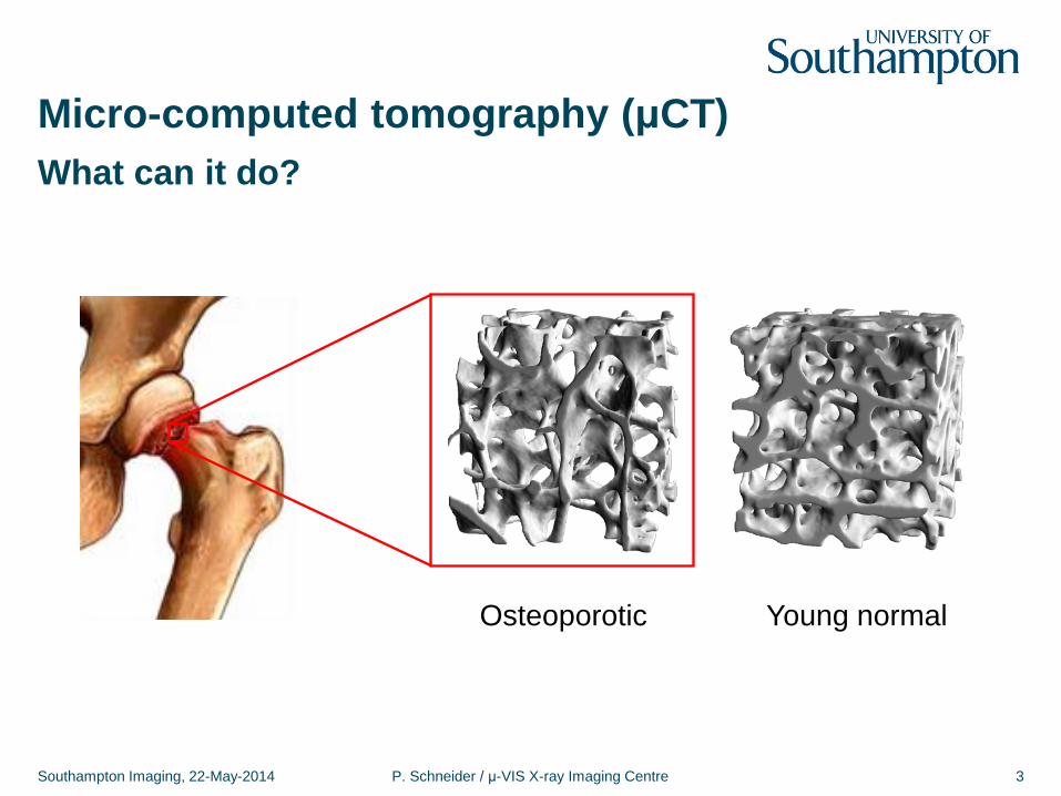
Micro-computed tomography (μCT)
Southampton Imaging, 22-May-2014 P. Schneider / μ-VIS X-ray Imaging Centre 3
What can it do?
Osteoporotic Young normal
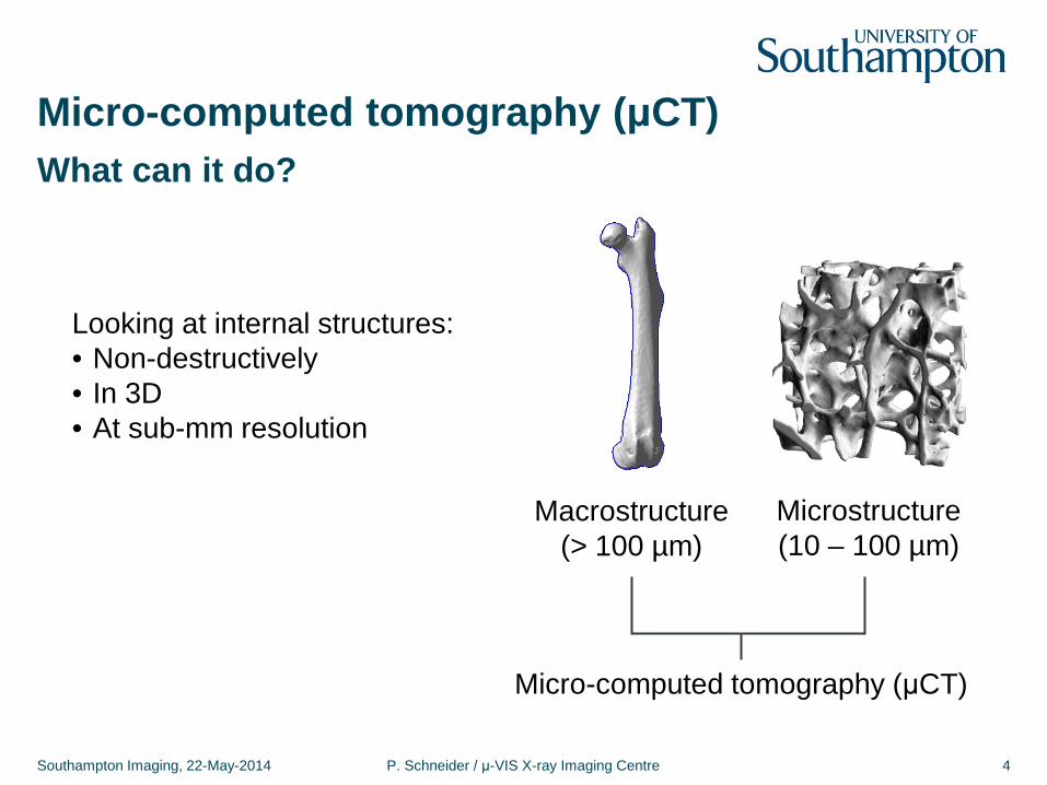
Micro-computed tomography (μCT)
Southampton Imaging, 22-May-2014 P. Schneider / μ-VIS X-ray Imaging Centre 4
What can it do?
Micro-computed tomography (μCT)
Microstructure (10 – 100 µm)
Macrostructure (> 100 µm)
Looking at internal structures: • Non-destructively • In 3D • At sub-mm resolution
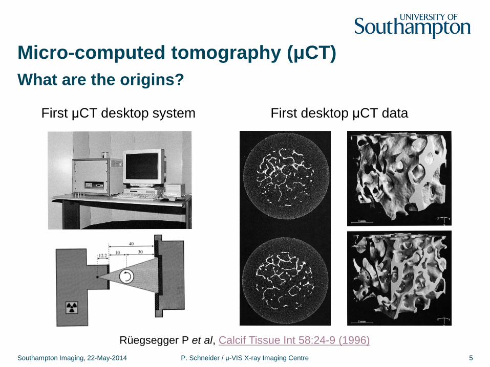
Micro-computed tomography (μCT)
Southampton Imaging, 22-May-2014 P. Schneider / μ-VIS X-ray Imaging Centre 5
What are the origins?
Rüegsegger P et al, Calcif Tissue Int 58:24-9 (1996)
First μCT desktop system First desktop μCT data
Micro-computed tomography (μCT)
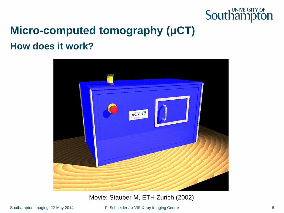
Southampton Imaging, 22-May-2014 P. Schneider / μ-VIS X-ray Imaging Centre 6
How does it work?
Movie: Stauber M, ETH Zurich (2002)
Micro-computed tomography (μCT)
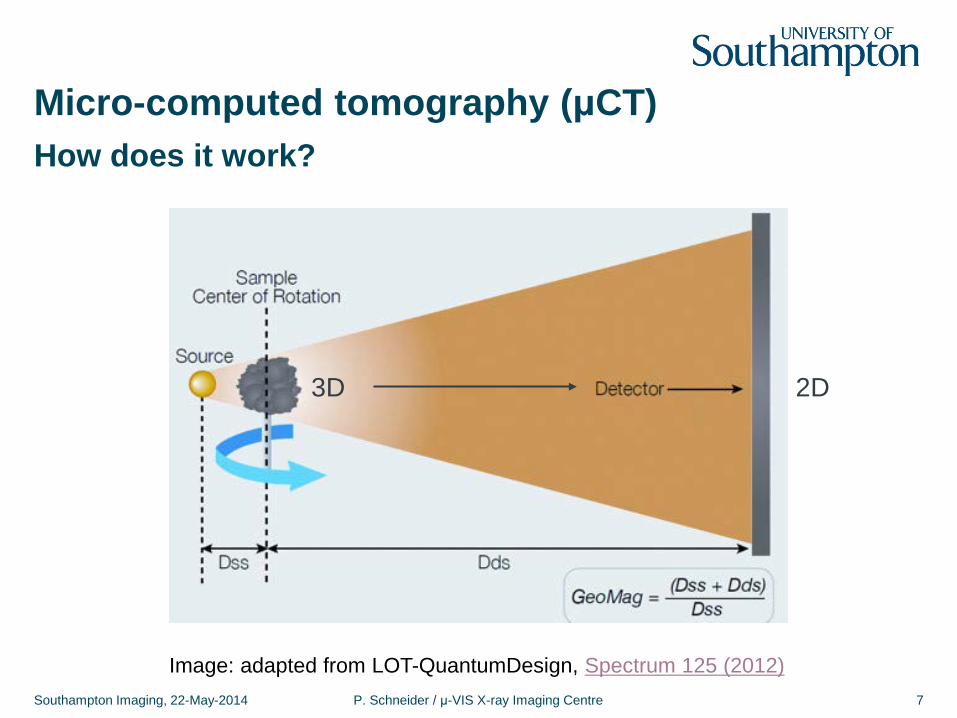
Southampton Imaging, 22-May-2014 P. Schneider / μ-VIS X-ray Imaging Centre 7
How does it work?
Image: adapted from LOT-QuantumDesign, Spectrum 125 (2012)
2D 3D
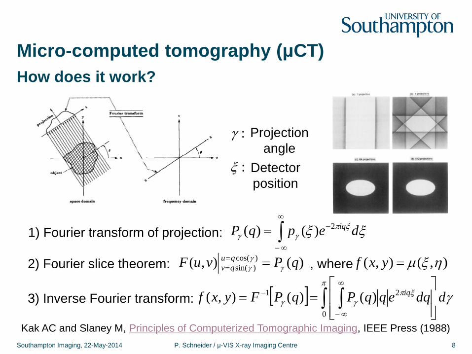
Micro-computed tomography (μCT)
Southampton Imaging, 22-May-2014 P. Schneider / μ-VIS X-ray Imaging Centre 8
How does it work?
1) Fourier transform of projection: ∫∞
∞−
−= ξξ ξπγγ depqP iq2)()(
2) Fourier slice theorem: )(),( )cos()sin( qPvuF qu
qv γγγ ==
= , where ),(),( ηξµ=yxf
3) Inverse Fourier transform: [ ] γπ
ξπγγ ddqeqqPqPFyxf iq∫ ∫
==
∞
∞−
−
0
21 )()(),(
Projection angle
:ξ
:γ
Detector position
Kak AC and Slaney M, Principles of Computerized Tomographic Imaging, IEEE Press (1988)
Micro-computed tomography (μCT)
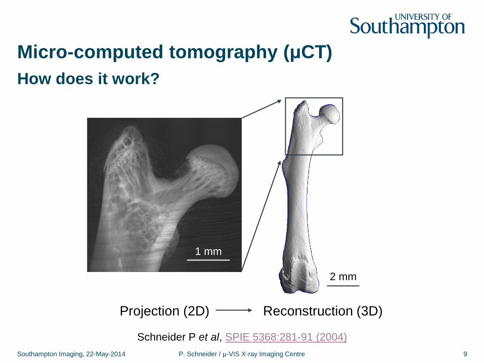
Southampton Imaging, 22-May-2014 P. Schneider / μ-VIS X-ray Imaging Centre 9
How does it work?
2 mm
1 mm
Projection (2D) Reconstruction (3D)
Schneider P et al, SPIE 5368:281-91 (2004)
Micro-computed tomography (μCT)
Southampton Imaging, 22-May-2014 P. Schneider / μ-VIS X-ray Imaging Centre 10
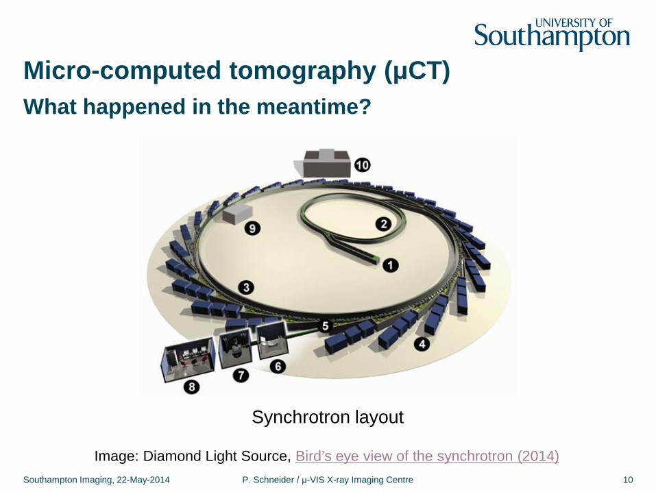
What happened in the meantime?
Image: Diamond Light Source, Bird’s eye view of the synchrotron (2014)
Synchrotron layout
Micro-computed tomography (μCT)
Southampton Imaging, 22-May-2014 P. Schneider / μ-VIS X-ray Imaging Centre 11
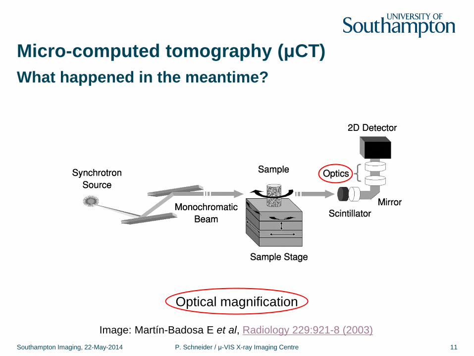
What happened in the meantime?
Image: Martín-Badosa E et al, Radiology 229:921-8 (2003)
Optical magnification
Micro-computed tomography (μCT)
Southampton Imaging, 22-May-2014 P. Schneider / μ-VIS X-ray Imaging Centre 12
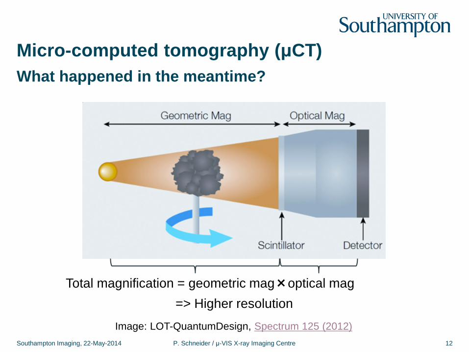
What happened in the meantime?
Image: LOT-QuantumDesign, Spectrum 125 (2012)
=> Higher resolution Total magnification = geometric mag×optical mag
Micro-computed tomography (μCT)
Southampton Imaging, 22-May-2014 P. Schneider / μ-VIS X-ray Imaging Centre 13
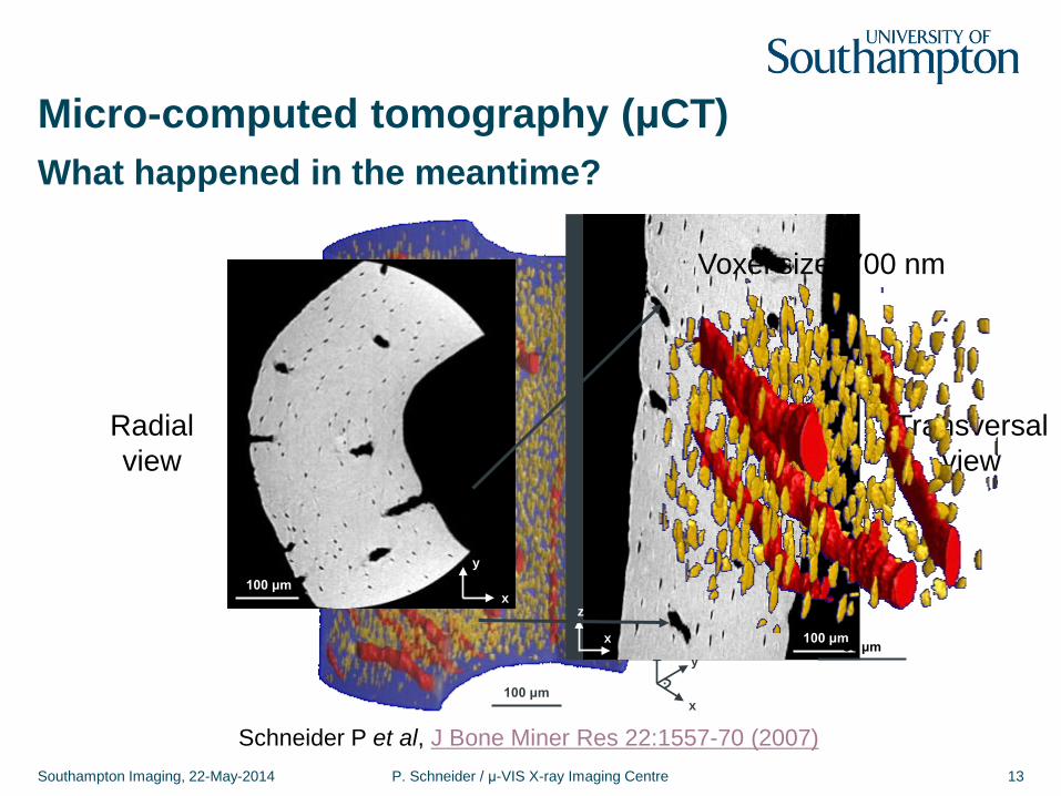
What happened in the meantime?
Schneider P et al, J Bone Miner Res 22:1557-70 (2007)
100 μm
z
x
y .
x
z
100 μm
x
y 100 μm
Radial view
Transversal view
50 μm
Voxel size: 700 nm
1
Micro-computed tomography (μCT)
Southampton Imaging, 22-May-2014 P. Schneider / μ-VIS X-ray Imaging Centre 14
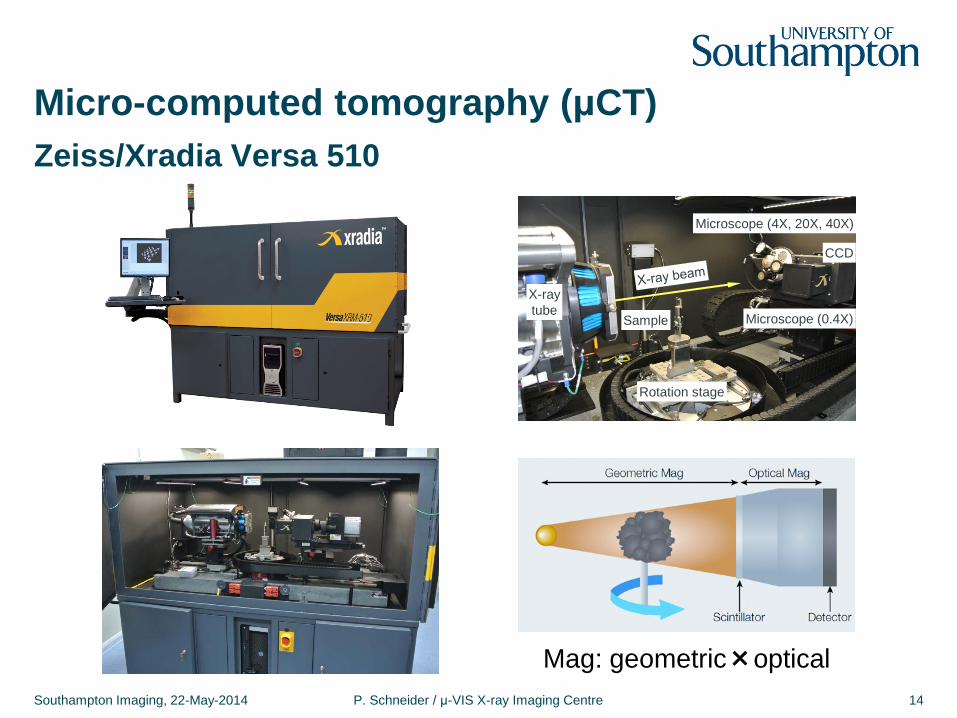
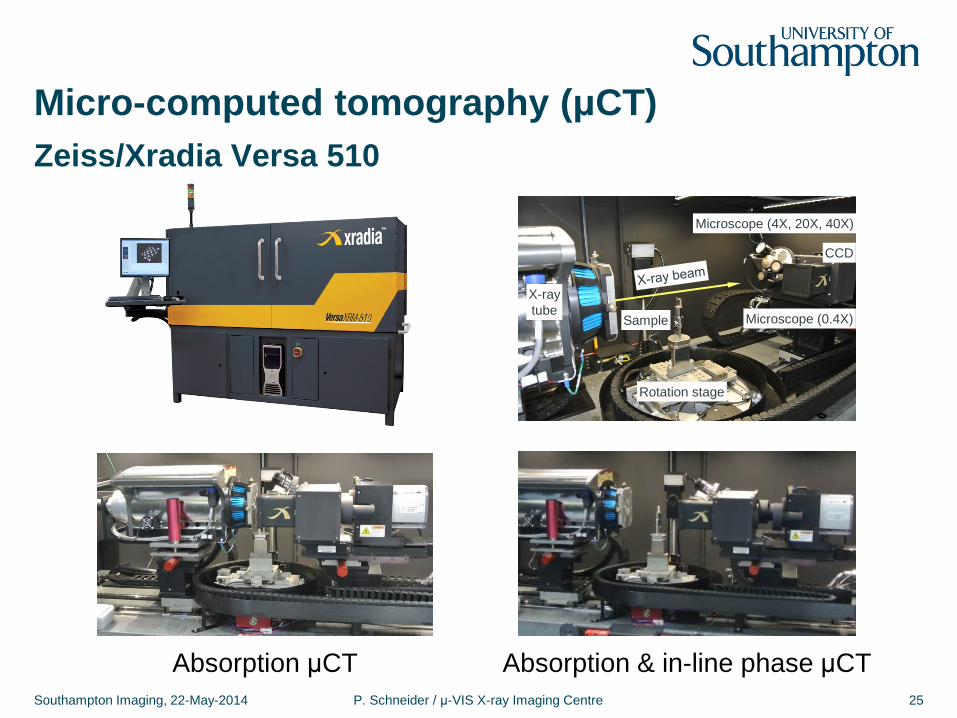
Zeiss/Xradia Versa 510
Mag: geometric×optical
X-ray tube
Sample
Rotation stage
Microscope (4X, 20X, 40X)
Microscope (0.4X)
CCD
1
Micro-computed tomography (μCT)
Southampton Imaging, 22-May-2014 P. Schneider / μ-VIS X-ray Imaging Centre 15
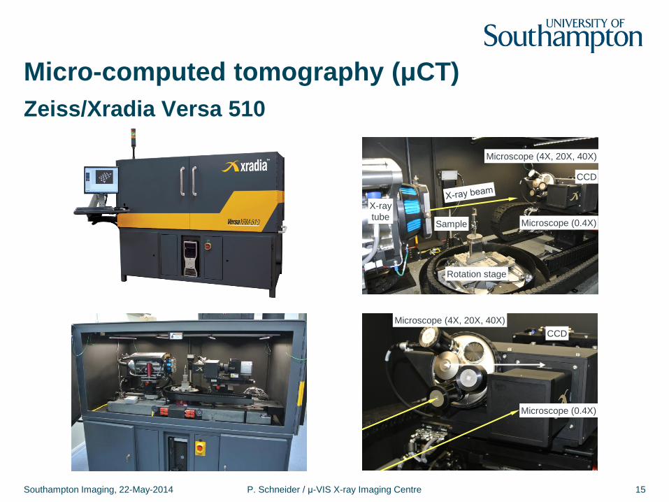
Zeiss/Xradia Versa 510
X-ray tube
Sample
Rotation stage
Microscope (4X, 20X, 40X)
Microscope (0.4X)
CCD
Microscope (4X, 20X, 40X)
Microscope (0.4X)
CCD
Micro-computed tomography (μCT)
Southampton Imaging, 22-May-2014 P. Schneider / μ-VIS X-ray Imaging Centre 16
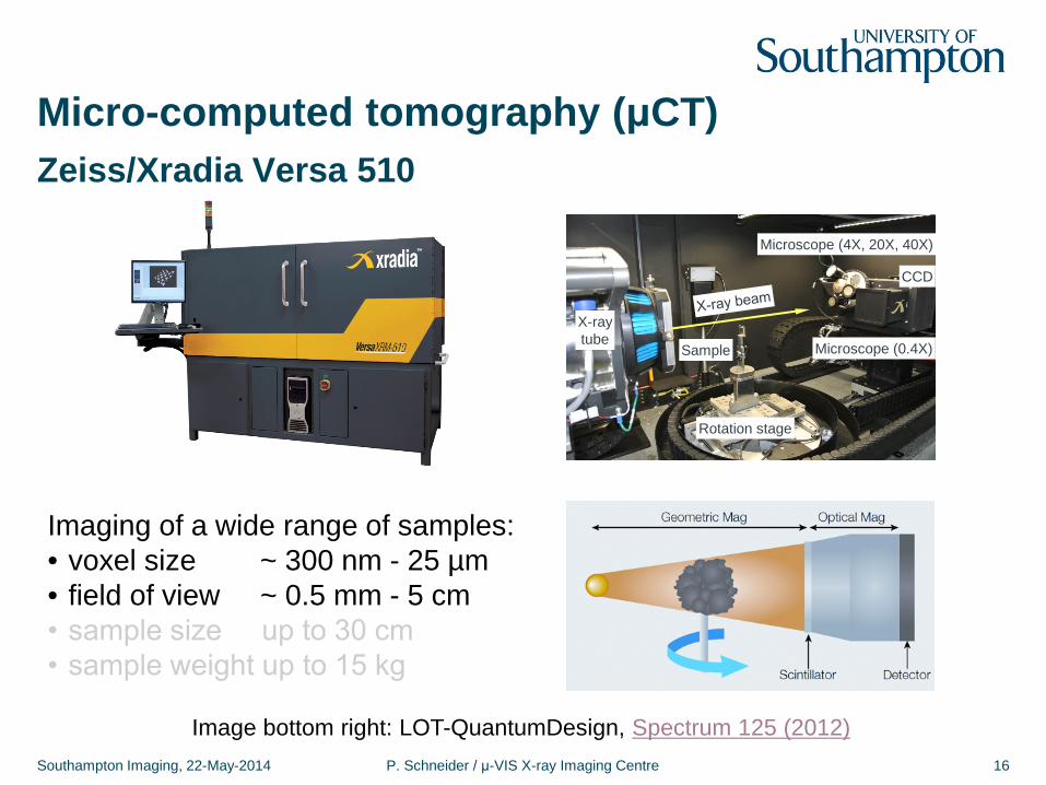
Zeiss/Xradia Versa 510
Imaging of a wide range of samples: • voxel size ~ 300 nm - 25 µm • field of view ~ 0.5 mm - 5 cm
1
Image bottom right: LOT-QuantumDesign, Spectrum 125 (2012)
X-ray tube
Sample
Rotation stage
Microscope (4X, 20X, 40X)
Microscope (0.4X)
CCD
Micro-computed tomography (μCT)
Southampton Imaging, 22-May-2014 P. Schneider / μ-VIS X-ray Imaging Centre 17
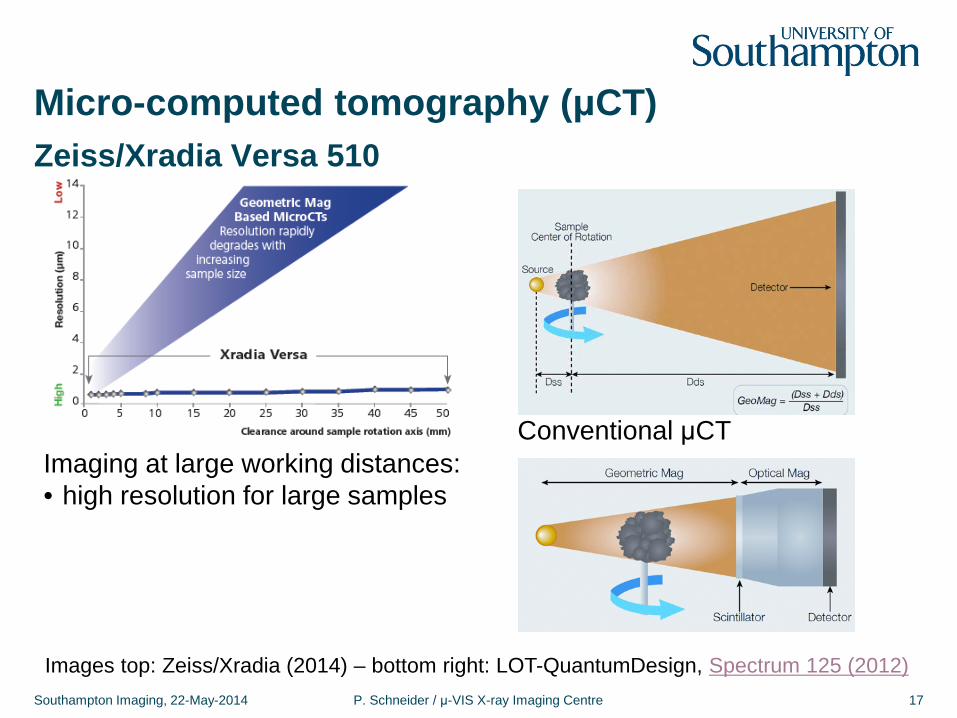
Zeiss/Xradia Versa 510
Imaging at large working distances: • high resolution for large samples
Images top: Zeiss/Xradia (2014) – bottom right: LOT-QuantumDesign, Spectrum 125 (2012)
Conventional μCT
Micro-computed tomography (μCT)
Southampton Imaging, 22-May-2014 P. Schneider / μ-VIS X-ray Imaging Centre 18
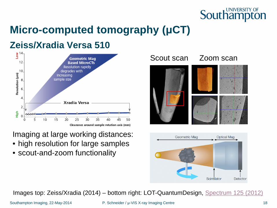
Zeiss/Xradia Versa 510
Imaging at large working distances: • high resolution for large samples • scout-and-zoom functionality
Scout scan Zoom scan
Images top: Zeiss/Xradia (2014) – bottom right: LOT-QuantumDesign, Spectrum 125 (2012)
Micro-computed tomography (μCT)
Southampton Imaging, 22-May-2014 P. Schneider / μ-VIS X-ray Imaging Centre 19
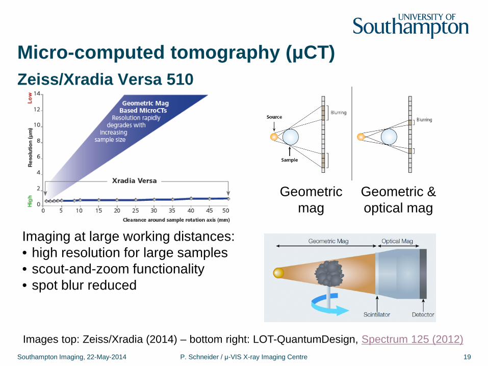
Zeiss/Xradia Versa 510
Imaging at large working distances: • high resolution for large samples • scout-and-zoom functionality • spot blur reduced
Geometric mag
Geometric & optical mag
Images top: Zeiss/Xradia (2014) – bottom right: LOT-QuantumDesign, Spectrum 125 (2012)
Micro-computed tomography (μCT)
Southampton Imaging, 22-May-2014 P. Schneider / μ-VIS X-ray Imaging Centre 20
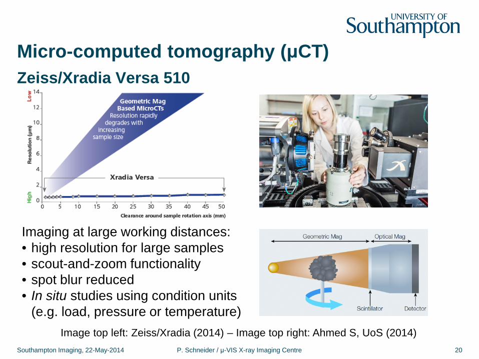
Zeiss/Xradia Versa 510
Imaging at large working distances: • high resolution for large samples • scout-and-zoom functionality • spot blur reduced • In situ studies using condition units
(e.g. load, pressure or temperature) Image top left: Zeiss/Xradia (2014) – Image top right: Ahmed S, UoS (2014)
Micro-computed tomography (μCT)
Southampton Imaging, 22-May-2014 P. Schneider / μ-VIS X-ray Imaging Centre 21
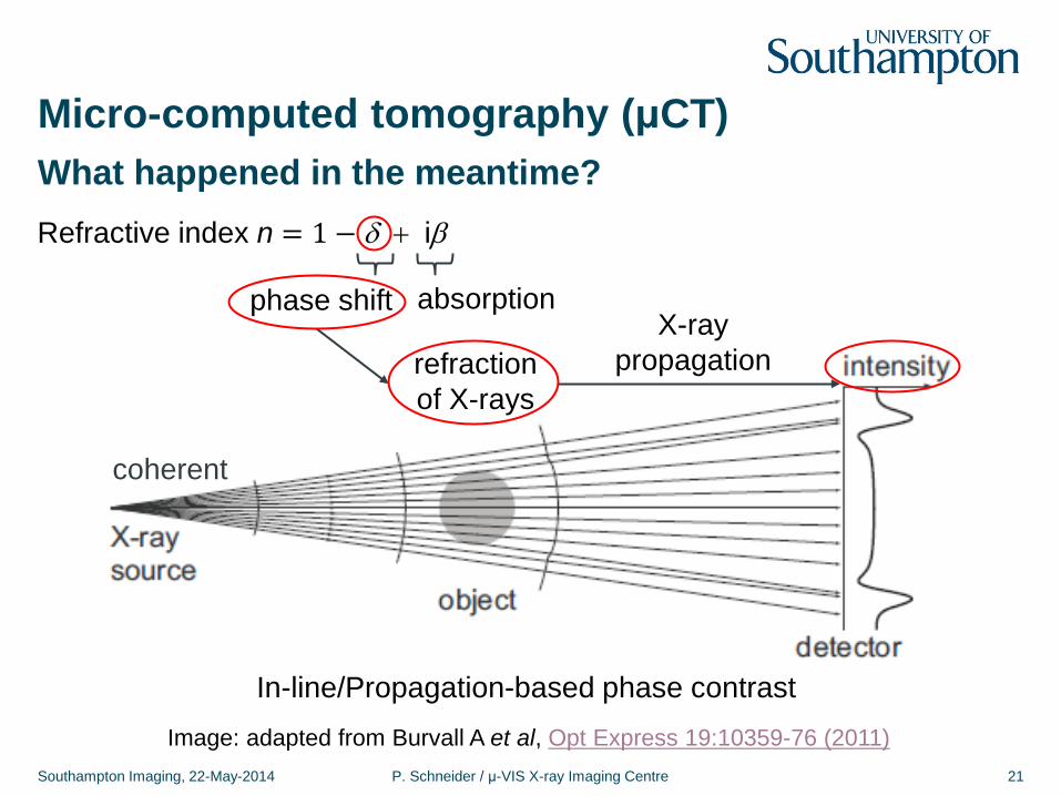
What happened in the meantime?
Image: adapted from Burvall A et al, Opt Express 19:10359-76 (2011)
In-line/Propagation-based phase contrast
coherent
refraction of X-rays
Refractive index n = 1 − δ + iβ
phase shift absorption X-ray
propagation
Micro-computed tomography (μCT)
Southampton Imaging, 22-May-2014 P. Schneider / μ-VIS X-ray Imaging Centre 22
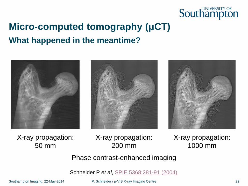
What happened in the meantime?
Schneider P et al, SPIE 5368:281-91 (2004)
X-ray propagation: 50 mm
X-ray propagation: 200 mm
X-ray propagation: 1000 mm
Phase contrast-enhanced imaging
Micro-computed tomography (μCT)
Southampton Imaging, 22-May-2014 P. Schneider / μ-VIS X-ray Imaging Centre 23
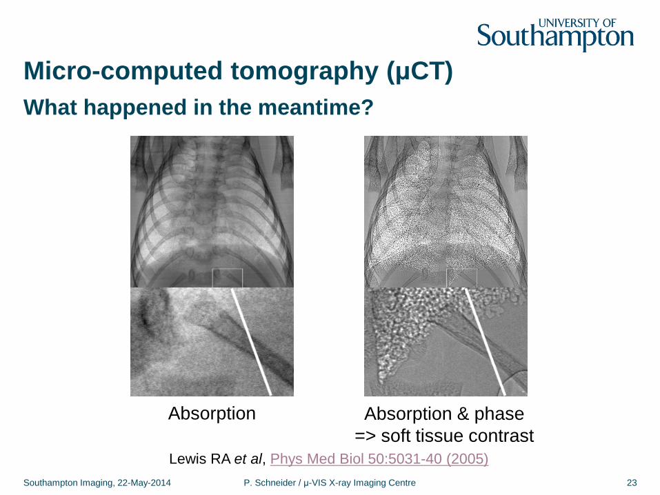
What happened in the meantime?
Lewis RA et al, Phys Med Biol 50:5031-40 (2005)
Absorption Absorption & phase => soft tissue contrast
Micro-computed tomography (μCT)
Southampton Imaging, 22-May-2014 P. Schneider / μ-VIS X-ray Imaging Centre 24
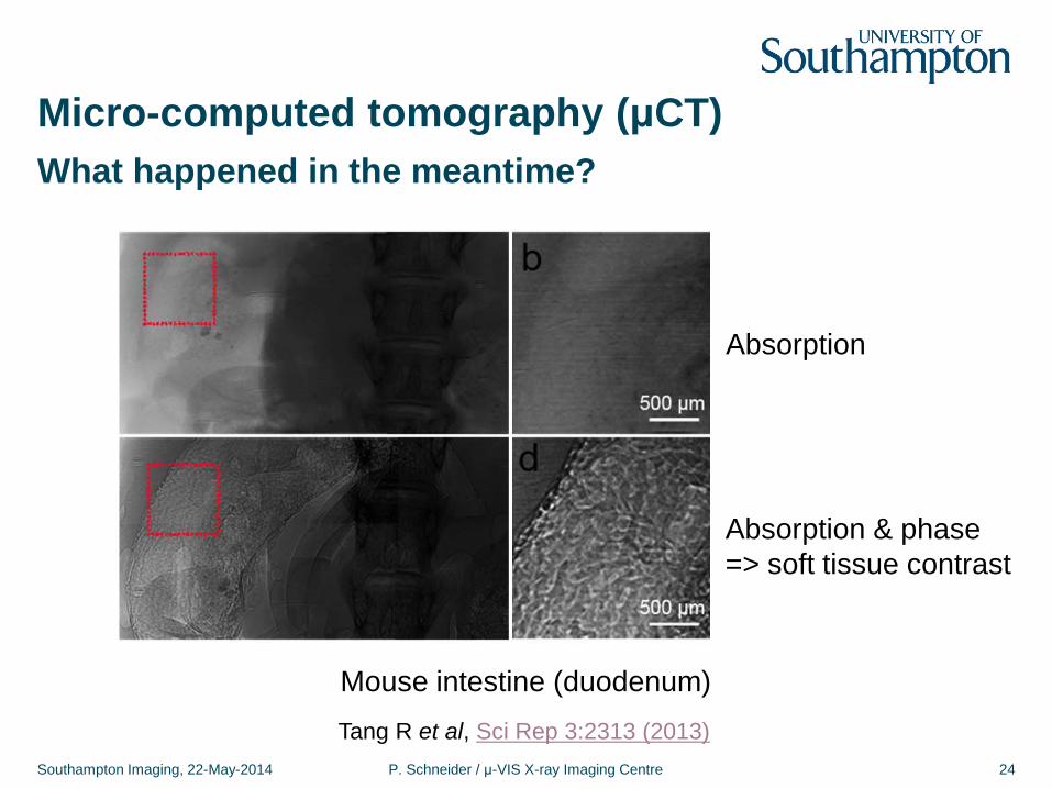
What happened in the meantime?
Tang R et al, Sci Rep 3:2313 (2013)
Absorption
Absorption & phase => soft tissue contrast
Mouse intestine (duodenum)
Micro-computed tomography (μCT)
Southampton Imaging, 22-May-2014 P. Schneider / μ-VIS X-ray Imaging Centre 25
Zeiss/Xradia Versa 510
1
Absorption μCT Absorption & in-line phase μCT
X-ray tube
Sample
Rotation stage
Microscope (4X, 20X, 40X)
Microscope (0.4X)
CCD

1
Outlook
Southampton Imaging, 22-May-2014 P. Schneider / μ-VIS X-ray Imaging Centre 26
Happy scanning!
We are looking forward to your research using μCT!
X-ray tube
Sample
Rotation stage
Microscope (4X, 20X, 40X)
Microscope (0.4X)
CCD