PDF - Journal of Neuroinflammation
11
RESEARCH Open Access Absence of IL-1β positively affects neurological outcome, lesion development and axonal plasticity after spinal cord injury Francesco Boato 1,2 , Karen Rosenberger 3 , Sofie Nelissen 1 , Lies Geboes 1 , Eva M Peters 4,5 , Robert Nitsch 6 and Sven Hendrix 1* Abstract Precise crosstalk between the nervous and immune systems is important for neuroprotection and axon plasticity after injury. Recently, we demonstrated that IL-1β acts as a potent inducer of neurite outgrowth from organotypic brain slices in vitro, suggesting a potential function of IL-1β in axonal plasticity. Here, we have investigated the effects of IL-1β on axon plasticity during glial scar formation and on functional recovery in a mouse model of spinal cord compression injury (SCI). We used an IL-1β deficiency model (IL-1βKO mice) and administered recombinant IL-1β. In contrast to our hypothesis, the histological analysis revealed a significantly increased lesion width and a reduced number of corticospinal tract fibers caudal to the lesion center after local application of recombinant IL-1β. Consistently, the treatment significantly worsened the neurological outcome after SCI in mice compared with PBS controls. In contrast, the absence of IL-1β in IL-1βKO mice significantly improved recovery from SCI compared with wildtype mice. Histological analysis revealed a smaller lesion size, reduced lesion width and greatly decreased astrogliosis in the white matter, while the number of corticospinal tract fibers increased significantly 5 mm caudal to the lesion in IL-1βKO mice relative to controls. Our study for the first time characterizes the detrimental effects of IL-1β not only on lesion development (in terms of size and glia activation), but also on the plasticity of central nervous system axons after injury. Keywords: IL-1β, Corticospinal tract, Glial scar, Spinal cord compression injury Introduction IL-1β is a 17 kDa protein and is one of the most exten- sively studied proinflammatory cytokines. IL-1β is al- most undetectable in the undamaged central nervous system (CNS), but its expression increases several fold after injury (including neurotoxic stimuli, ischemia and trauma) [1]. Microglia are the principal cells expressing IL-1β, but many other resident cells (including astro- cytes and neurons) or invading cells are also able to pro- duce the cytokine [2]. IL-1β mainly acts by activating the immune response, fostering the production of in- flammatory mediators and other cytokines. Interestingly, when applied on healthy neurons, IL-1β does not cause damage or death; however, it induces a number of cellular reactions such as changes in intracellular cal- cium concentrations and ionic conductances [3,4]. In an organotypic model we recently demonstrated that IL-1β is a potent inducer of neurite outgrowth from brain slices [5], suggesting a potential function of IL-1β in axonal plasticity. This is in contrast to its well-studied contribution to both acute neuronal loss and chronic neurodegeneration [1]. Despite the intensive research on IL-1β in recent decades, its role in the CNS remains far from fully understood. The in vitro effects of IL-1β are very heterogeneous. IL-1β supports survival of dorsal root ganglion neurons [6] and pyramidal neurons in vitro [7], but it also induces depolarization, increases the spike fre- quency and enhances vulnerability of hippocampal neu- rons induced by N-methyl-D-aspartate receptor-mediated increase of intracellular calcium [4]. IL-1β also has strong effects on astrocytes, promoting activation, proliferation and production of neurotoxic mediators, as well as survival * Correspondence: [email protected] 1 Department of Morphology & BIOMED Institute, Campus Diepenbeek, Hasselt University, Agoralaan Gebouw C, Diepenbeek BE 3590, Belgium Full list of author information is available at the end of the article JOURNAL OF NEUROINFLAMMATION © 2013 Boato et al.; licensee BioMed Central Ltd. This is an Open Access article distributed under the terms of the Creative Commons Attribution License (http://creativecommons.org/licenses/by/2.0), which permits unrestricted use, distribution, and reproduction in any medium, provided the original work is properly cited. Boato et al. Journal of Neuroinflammation 2013, 10:6 http://www.jneuroinflammation.com/content/10/1/6
Transcript of PDF - Journal of Neuroinflammation
RESEARCH Open Access
Absence of IL-1β positively affects neurologicaloutcome, lesion development and axonalplasticity after spinal cord injuryFrancesco Boato1,2, Karen Rosenberger3, Sofie Nelissen1, Lies Geboes1, Eva M Peters4,5, Robert Nitsch6
and Sven Hendrix1*
Abstract
Precise crosstalk between the nervous and immune systems is important for neuroprotection and axon plasticityafter injury. Recently, we demonstrated that IL-1β acts as a potent inducer of neurite outgrowth from organotypicbrain slices in vitro, suggesting a potential function of IL-1β in axonal plasticity. Here, we have investigated theeffects of IL-1β on axon plasticity during glial scar formation and on functional recovery in a mouse model of spinalcord compression injury (SCI). We used an IL-1β deficiency model (IL-1βKO mice) and administered recombinantIL-1β. In contrast to our hypothesis, the histological analysis revealed a significantly increased lesion width and areduced number of corticospinal tract fibers caudal to the lesion center after local application of recombinant IL-1β.Consistently, the treatment significantly worsened the neurological outcome after SCI in mice compared with PBScontrols. In contrast, the absence of IL-1β in IL-1βKO mice significantly improved recovery from SCI compared withwildtype mice. Histological analysis revealed a smaller lesion size, reduced lesion width and greatly decreasedastrogliosis in the white matter, while the number of corticospinal tract fibers increased significantly 5 mm caudalto the lesion in IL-1βKO mice relative to controls. Our study for the first time characterizes the detrimental effects ofIL-1β not only on lesion development (in terms of size and glia activation), but also on the plasticity of centralnervous system axons after injury.
Keywords: IL-1β, Corticospinal tract, Glial scar, Spinal cord compression injury
IntroductionIL-1β is a 17 kDa protein and is one of the most exten-sively studied proinflammatory cytokines. IL-1β is al-most undetectable in the undamaged central nervoussystem (CNS), but its expression increases several foldafter injury (including neurotoxic stimuli, ischemia andtrauma) [1]. Microglia are the principal cells expressingIL-1β, but many other resident cells (including astro-cytes and neurons) or invading cells are also able to pro-duce the cytokine [2]. IL-1β mainly acts by activatingthe immune response, fostering the production of in-flammatory mediators and other cytokines. Interestingly,when applied on healthy neurons, IL-1β does not causedamage or death; however, it induces a number of
cellular reactions such as changes in intracellular cal-cium concentrations and ionic conductances [3,4]. In anorganotypic model we recently demonstrated that IL-1βis a potent inducer of neurite outgrowth from brainslices [5], suggesting a potential function of IL-1β inaxonal plasticity. This is in contrast to its well-studiedcontribution to both acute neuronal loss and chronicneurodegeneration [1]. Despite the intensive research onIL-1β in recent decades, its role in the CNS remains farfrom fully understood. The in vitro effects of IL-1β arevery heterogeneous. IL-1β supports survival of dorsal rootganglion neurons [6] and pyramidal neurons in vitro [7],but it also induces depolarization, increases the spike fre-quency and enhances vulnerability of hippocampal neu-rons induced by N-methyl-D-aspartate receptor-mediatedincrease of intracellular calcium [4]. IL-1β also has strongeffects on astrocytes, promoting activation, proliferationand production of neurotoxic mediators, as well as survival
* Correspondence: [email protected] of Morphology & BIOMED Institute, Campus Diepenbeek,Hasselt University, Agoralaan Gebouw C, Diepenbeek BE 3590, BelgiumFull list of author information is available at the end of the article
JOURNAL OF NEUROINFLAMMATION
© 2013 Boato et al.; licensee BioMed Central Ltd. This is an Open Access article distributed under the terms of the CreativeCommons Attribution License (http://creativecommons.org/licenses/by/2.0), which permits unrestricted use, distribution, andreproduction in any medium, provided the original work is properly cited.
Boato et al. Journal of Neuroinflammation 2013, 10:6http://www.jneuroinflammation.com/content/10/1/6
promoting factors [8]. Studies from our group showed ei-ther no effects of IL-1β on neurotrophin-induced out-growth from dorsal root ganglion neurons [9] and spinalcord explants or a beneficial effect on axonal growth frombrain slices in vitro [5]. The in vivo role of IL-1β is also notclear. Intracerebroventricular administration of IL-1β dur-ing ischemic damage after permanent middle cerebral ar-tery occlusion results in a highly enhanced infarct volume(~92%) [10], similar to the effect of systemic [11] or local[12] administration of the cytokine in mice with middlecerebral artery occlusion. Moreover, IL-1β leads to exacer-bated cell death and neurodegeneration in other experi-mental models of CNS trauma [13,14]. In contrast, there isevidence that IL-1β is necessary for proper remyelinationof the CNS following death of mature oligodendrocytessince IL-1β−/− nerve fibers are unable to remyelinate prop-erly after cuprizone-induced demyelination [15]. Finally,IL-1β contributes to sensory nerve regeneration in vivo fol-lowing sciatic nerve injury [16,17].In this study, we have investigated the effects of
increased local levels of IL-1β compared with IL-1β ab-sence (in IL-1βKO mice) after compression of the spinalcord [18]. In contrast to our in vitro-based hypothesis,but in line with the results on IL-1βKO mice reported ina recently published study from the Shioda laboratory[19], we here demonstrate for the first time detrimentaleffects of IL-1β on lesion development, in terms of le-sion size and glial activation, and on the plasticity ofCNS axons in vivo after injury.
Materials and methodsSpinal cord compression injuryAll experiments with C57BL/6 wildtype (WT) mice andhomozygous mice deficient in IL-1β [20] (IL-1βKO)(females, 8 to 12 weeks old) were performed in accord-ance with the German guidelines on the use of laboratoryanimals. Spinal cord injury, corticospinal tract (CST)tracing and subsequent analysis were carried out followinga standardized protocol [18,21]. Briefly, C57BL/6 miceand IL-1β-deficient mice underwent a dorsal laminectomyat thoracic level T8, and the compression of the spinalcord was induced with a modified SPI Correx Tension/Compression Gage (Penn Tool, Maplewood, NJ, USA) at10 cN for 3 seconds. For recombinant IL-1β (rIL-1β) andPBS application, a piece of Gelfoam (Pharmacia & Upjohn,Erlangen, Germany) soaked in 5 μl solution with PBSalone or with 1 or 20 μg rIL-1β was placed directly on topof the injured spinal cord and in contact with the perfo-rated dura before suturing the muscles. Important to notein these experiments is that when recombinant cytokinewas applied, a Gelfoam patch was in direct contact withthe injured spinal cord, and this led to a lower score incontrol mice compared with the WT mice in the knock-out experiments. The rIL-1β in vivo dosage was based on
in vitro results coming from our group [5] demonstratingthat rIL-1β increases axonal outgrowth when applied in ahigh therapeutic dosage in a well-established organotypicslice culture model [22-25]. The effective dosage in thatstudy (500 ng rIL-1β in 500 μl medium) was substantiallyhigher than the concentrations found in vivo after spinalcord injury (300 pg/ml in spinal cord (1 cm) homogenate6 hours after injury). In the first experiment we thereforeapplied a high therapeutic dosage of 20 μg rIL-1β in Gel-foam, also taking into account that the dispersion of thecytokine is higher in vivo than in vitro, that the time of ob-servation is longer (2 days for in vitro experiments and14 days for in vivo experiments) and that the lesion vol-ume in the spinal cord is much bigger than a 350 μm thickslice of the enthorinal cortex. Furthermore, to distinguishbetween local and systemic effects on functional recovery,a 100 μl solution of PBS alone or with 1 μg rIL-1β wasalso applied systemically by intraperitoneal injection im-mediately after injury.
Behavioral analysisThe spinal cord compression injury (SCI) mice weretested over 14 days for functional recovery with the BassoMouse Scale (BMS) [26], which is a locomotor rating scaleranging from 0 to 9 (0 = complete hind limb paralysis; 9 =normal locomotion). In BMS testing, mice are scoredaccording to the mobility of the hind limbs for a period of4 minutes in an open field by two investigators carefullyblinded to experimental groups. Furthermore, since sub-scores for each parameter of the BMS can be used tomeasure individual locomotor features [26] and since cor-rect foot placing correlates with proper CST function[27,28], stepping performance and correct paw positioningwere evaluated as previously described [18]. The analysisof the stepping emphasized whether plantar stepping waspresent in <50% or in >50% of the steps (scores 0 and 1,respectively). For the scoring of paw positioning, weassessed whether the paws were rotated at both initialcontact and lift-off (score 0), parallel at initial contact butrotated at lift-off (score 1), or parallel at both initial con-tact and lift-off (score 2). For both stepping performanceand paw positioning, the score for each animal was thenrepresented as a percentage, taking as 100% a score of, re-spectively, 1 and 2. For the BMS, stepping performanceand paw positioning analysis we used the mean of the leftand right hindlimb scores for each animal. Data shownrepresent mean values for all the animals of each experi-mental group ± standard error of the mean and were ana-lyzed using a two-way analysis of variance as describedpreviously [26].
Corticospinal tract tracing and analysisFor biotinylated dextran amine (BDA, 10%; Invitrogen,Darmstadt, Germany) tracing, a small hole was drilled
Boato et al. Journal of Neuroinflammation 2013, 10:6 Page 2 of 11http://www.jneuroinflammation.com/content/10/1/6
into the skull directly after SCI (after suturing the backmuscles) and a Hamilton syringe was inserted into themotor cortex to apply 2 μl of 10% solution of the antero-grade tracer. At the end of the observation period, CSTfibers were visualized by diaminobenzidine staining onparaformaldehyde-fixed longitudinal cryosections (20 μm)of the spinal cord. BDA-labeled nerve fibers of the corti-cospinal tract were quantified at defined distances caudalto the lesion center in the complete microscopic fieldalong the dorso-ventral axis at a total magnification of ×400. Counted fibers are shown as a percentage of the totalnumber of labeled fibers within a standardized 20 μmwide area of the dorso-ventral diameter of the CST at levelC4. Fibers were counted on five to seven adjacent sectionswith a clearly recognizable lesion and CST end (presentingretraction bulbs), and only the axons fulfilling the criteriaoutlined by Steward and colleagues [29] were included inthe analysis. Data represent mean values ± standard errorof the mean and were analyzed using the Mann–WhitneyU test.
Evaluation of lesion size, lesion width and degree ofgliosisLongitudinal spinal cord cryosections, 20 μm thick(obtained from transcardially perfused animals at theend of behavioral analysis period), were preincubatedin PBS with 10% normal goat serum containing 0.5%Triton X-100 for 60 minutes at room temperature.Incubation with primary antibodies was carried outovernight at 4°C. Secondary antibodies were applied for1 hour at room temperature. For measurement of thelesion size and gliosis, three to six sections per animal,the central part of the spinal cord included, were analyzed.
The lesion size and lesion width were evaluated by stain-ing against ionized calcium binding adaptor molecule 1(Iba1) using rabbit polyclonal antibodies (Wako Chemi-cals, Neuss, Germany). Alexa Fluor 488 goat anti-rabbitantibodies (Invitrogen) were applied as secondary anti-body. The Iba1-positive area was quantified using imageanalysis software (ImageJ open source software; Na-tional Institutes of Health, Bethesda, MD, USA) andaveraged for each animal after the analysis of three tosix sections. The lesion size was evaluated on sagittalsections and represents the mean area in the center ofthe spinal cord (corresponding to the lesion center, in-cluding the CST end), while the lesion width representsthe total number of sagittal sections presenting the le-sion, multiplied by the thickness of each section. Iba1intensity was calculated for the same area and wasfound not to differ significantly between the experimen-tal groups (data not shown). While the use of an Iba1-positive area to measure the lesion size is not a classical
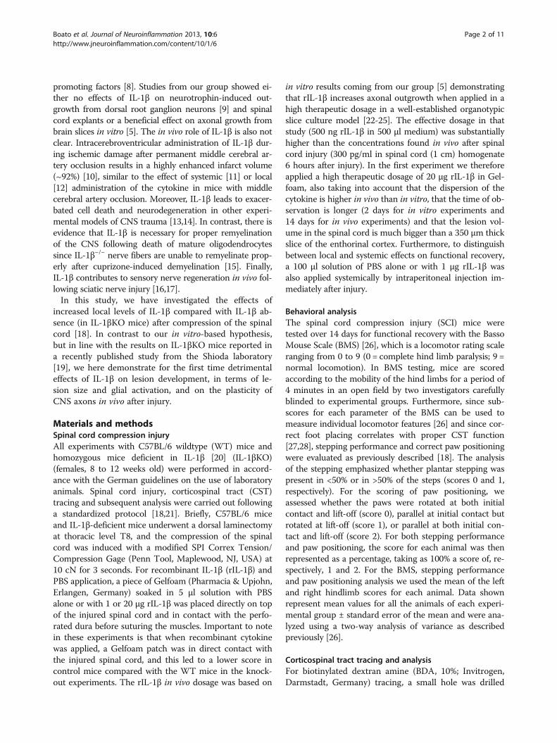
Figure 1 Dose-dependent mortality after spinal cordcompression injury. The mortality rate in mice with localadministration of recombinant IL-1β (rIL-1β) in Gelfoam directly afterspinal cord compression injury (SCI) was 100% in mice treated with20 μg rIL-1β, and was significantly higher compared with micetreated with 1 μg rIL-1β or only with PBS. P <0.0001 using thelog-rank test.
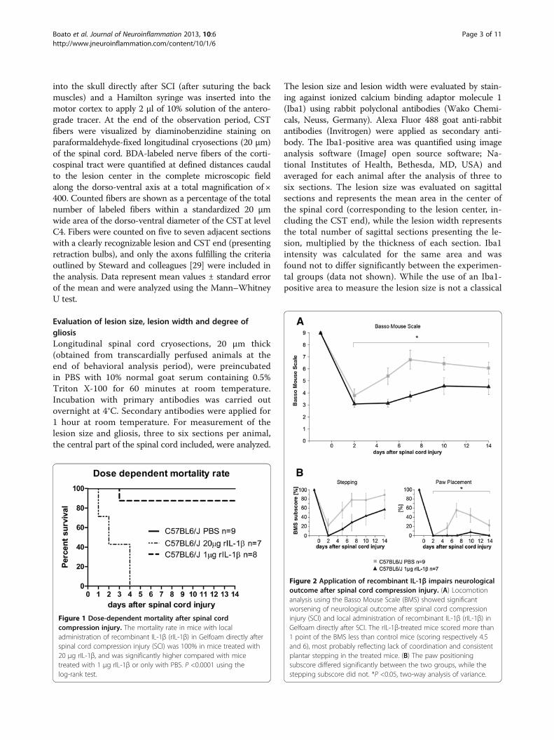
Figure 2 Application of recombinant IL-1β impairs neurologicaloutcome after spinal cord compression injury. (A) Locomotionanalysis using the Basso Mouse Scale (BMS) showed significantworsening of neurological outcome after spinal cord compressioninjury (SCI) and local administration of recombinant IL-1β (rIL-1β) inGelfoam directly after SCI. The rIL-1β-treated mice scored more than1 point of the BMS less than control mice (scoring respectively 4.5and 6), most probably reflecting lack of coordination and consistentplantar stepping in the treated mice. (B) The paw positioningsubscore differed significantly between the two groups, while thestepping subscore did not. *P <0.05, two-way analysis of variance.
Boato et al. Journal of Neuroinflammation 2013, 10:6 Page 3 of 11http://www.jneuroinflammation.com/content/10/1/6
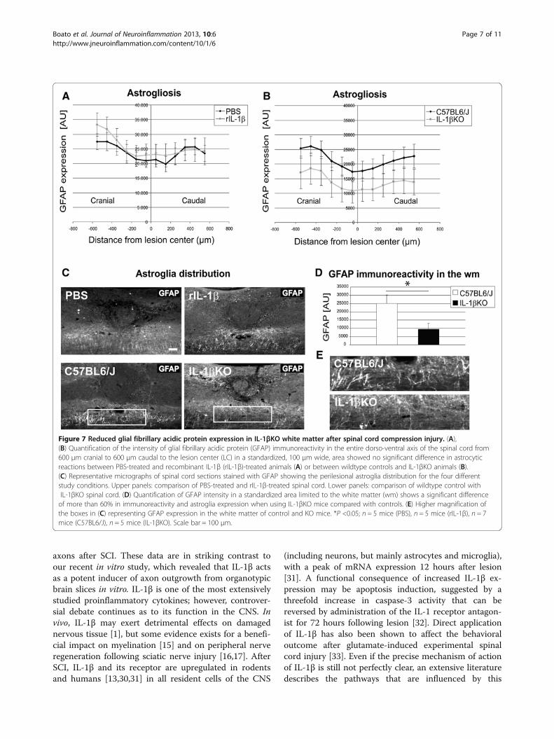
procedure, in this study we could not use traditionalglial fibrillary acidic protein (GFAP) immunoreactivitymeasures for this goal (as often found in the literatureand as performed in previous work from our laboratory[18]); this was due to the reduced GFAP immunoreac-tivity in the IL-1βKO mice, which did not allow forcomprehensive analysis. Iba1 immunoreactivity in thelesion, however, correlated perfectly well with the lesionsize found using bright-field microscopy, with the addedadvantage that Iba1 immunoreactivity was much easierto distinguish from the background. Astrogliosis wasevaluated using staining against GFAP with mouse mAb(Sigma-Aldrich, St Louis, MO, USA). Alexa Fluor 568goat anti-mouse (Invitrogen) was used as secondaryantibody. Quantification of GFAP expression was per-formed as previously described [18,30] by means of in-tensity analysis using ImageJ software within an area100 μm wide along the dorso-ventral axis of the spinalcord, extending from 600 μm cranial to 600 μm caudalto the lesion center. Additionally, the total intensity ofGFAP staining in the white matter in the same area wascalculated for each animal in the rostro-caudal axis.Data represent the mean ± standard error of the meanand were analyzed using the Mann–Whitney-U test.
ResultsIn a first experimental approach we applied a high dos-age of rIL-1β to mice that underwent spinal cord injury.To our knowledge this is the first study to investigate
the in vivo effect of rIL-1β on axonal plasticity in theCNS, so the choice of the rIL-1β in vivo dosage was basedon in vitro results from our group. Perilesional administra-tion of 20 μg rIL-1β in Gelfoam immediately after SCIresulted in 100% mortality, with all mice (n = 7) dyingwithin the first 4 days after the operation (Figure 1). Werefrained from repeating the experiment to perform nec-ropsies to diagnose the cause of death for obvious ethicalreasons. Based on these results, we used a drasticallyreduced amount of cytokine (1 μg in 5 μl PBS), which sub-stantially reduced the mortality (only one out of eightoperated mice died 3 days after injury). The application of1 μg rIL-1β significantly impaired functional performancecompared with mice treated with PBS alone (Figure 2A).At the end of the investigation period (14 days after in-jury) the two experimental groups varied by more thanone point on the BMS (from about 4.5 for rIL-1β-treatedmice to 6 for PBS-treated mice), mainly reflecting a sub-stantial difference in coordination and consistency of plan-tar stepping during walking. To rule out the possibility
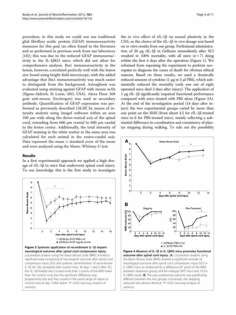
Figure 3 Systemic application of recombinant IL-1β impairsneurological outcome after spinal cord compression injury.Locomotion analysis using the Basso Mouse Scale (BMS) showed asignificant early worsening of neurological outcome after spinal cordcompression injury (SCI) and systemic administration of recombinantIL-1β (rIL-1β) compared with control mice. At days 1 and 2 after SCI,the rIL-1β-treated mice scored more than 2 points of the BMS lowerthan the control mice, but the significant difference wasprogressively lost and they scored in the same range of values ascontrol mice at day 7 after lesion. *P <0.05, two-way analysis ofvariance.
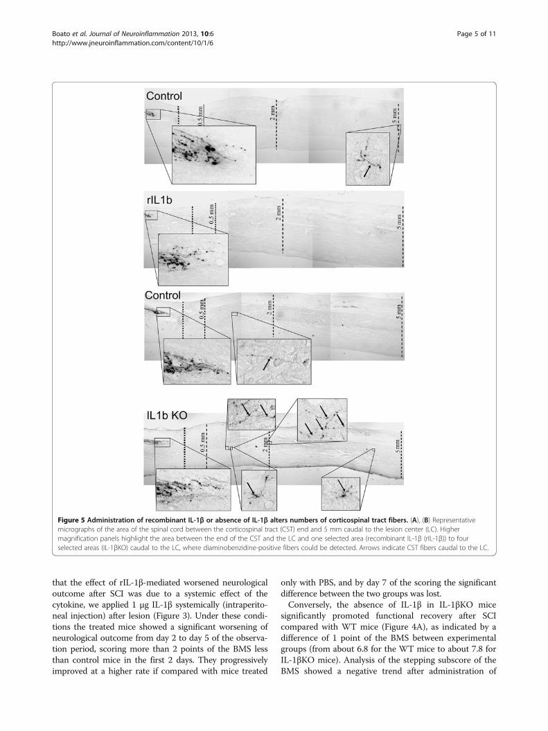
Figure 4 Absence of IL-1β in IL-1βKO mice promotes functionaloutcome after spinal cord injury. (A) Locomotion analysis usingthe Basso Mouse Scale (BMS) showed a significant increase ofneurological outcome after spinal cord compression injury (SCI) inIL-1βKO mice, as evidenced by a difference of1 point of the BMSbetween treatment groups (6.8 for wildtype (WT) mice and 7.8 forIL-1βKO mice). (B) The paw positioning subscore was significantlydifferent between the two groups. Conversely, the steppingsubscore was almost identical. *P <0.05, two-way analysis ofvariance.
Boato et al. Journal of Neuroinflammation 2013, 10:6 Page 4 of 11http://www.jneuroinflammation.com/content/10/1/6
that the effect of rIL-1β-mediated worsened neurologicaloutcome after SCI was due to a systemic effect of thecytokine, we applied 1 μg IL-1β systemically (intraperito-neal injection) after lesion (Figure 3). Under these condi-tions the treated mice showed a significant worsening ofneurological outcome from day 2 to day 5 of the observa-tion period, scoring more than 2 points of the BMS lessthan control mice in the first 2 days. They progressivelyimproved at a higher rate if compared with mice treated
only with PBS, and by day 7 of the scoring the significantdifference between the two groups was lost.Conversely, the absence of IL-1β in IL-1βKO mice
significantly promoted functional recovery after SCIcompared with WT mice (Figure 4A), as indicated by adifference of 1 point of the BMS between experimentalgroups (from about 6.8 for the WT mice to about 7.8 forIL-1βKO mice). Analysis of the stepping subscore of theBMS showed a negative trend after administration of
Figure 5 Administration of recombinant IL-1β or absence of IL-1β alters numbers of corticospinal tract fibers. (A), (B) Representativemicrographs of the area of the spinal cord between the corticospinal tract (CST) end and 5 mm caudal to the lesion center (LC). Highermagnification panels highlight the area between the end of the CST and the LC and one selected area (recombinant IL-1β (rIL-1β)) to fourselected areas (IL-1βKO) caudal to the LC, where diaminobenzidine-positive fibers could be detected. Arrows indicate CST fibers caudal to the LC.
Boato et al. Journal of Neuroinflammation 2013, 10:6 Page 5 of 11http://www.jneuroinflammation.com/content/10/1/6
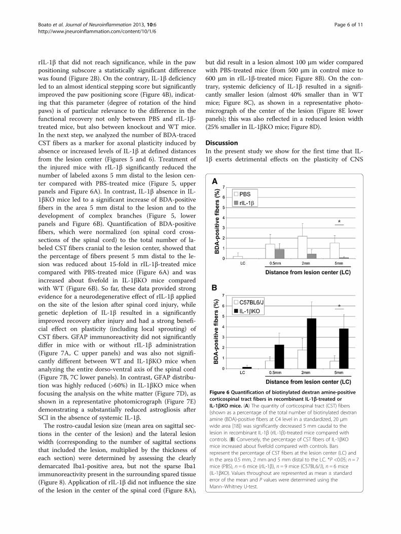
rIL-1β that did not reach significance, while in the pawpositioning subscore a statistically significant differencewas found (Figure 2B). On the contrary, IL-1β deficiencyled to an almost identical stepping score but significantlyimproved the paw positioning score (Figure 4B), indicat-ing that this parameter (degree of rotation of the hindpaws) is of particular relevance to the difference in thefunctional recovery not only between PBS and rIL-1β-treated mice, but also between knockout and WT mice.In the next step, we analyzed the number of BDA-tracedCST fibers as a marker for axonal plasticity induced byabsence or increased levels of IL-1β at defined distancesfrom the lesion center (Figures 5 and 6). Treatment ofthe injured mice with rIL-1β significantly reduced thenumber of labeled axons 5 mm distal to the lesion cen-ter compared with PBS-treated mice (Figure 5, upperpanels and Figure 6A). In contrast, IL-1β absence in IL-1βKO mice led to a significant increase of BDA-positivefibers in the area 5 mm distal to the lesion and to thedevelopment of complex branches (Figure 5, lowerpanels and Figure 6B). Quantification of BDA-positivefibers, which were normalized (on spinal cord cross-sections of the spinal cord) to the total number of la-beled CST fibers cranial to the lesion center, showed thatthe percentage of fibers present 5 mm distal to the le-sion was reduced about 15-fold in rIL-1β-treated micecompared with PBS-treated mice (Figure 6A) and wasincreased about fivefold in IL-1βKO mice comparedwith WT (Figure 6B). So far, these data provided strongevidence for a neurodegenerative effect of rIL-1β appliedon the site of the lesion after spinal cord injury, whilegenetic depletion of IL-1β resulted in a significantlyimproved recovery after injury and had a strong benefi-cial effect on plasticity (including local sprouting) ofCST fibers. GFAP immunoreactivity did not significantlydiffer in mice with or without rIL-1β administration(Figure 7A, C upper panels) and was also not signifi-cantly different between WT and IL-1βKO mice whenanalyzing the entire dorso-ventral axis of the spinal cord(Figure 7B, 7C lower panels). In contrast, GFAP distribu-tion was highly reduced (>60%) in IL-1βKO mice whenfocusing the analysis on the white matter (Figure 7D), asshown in a representative photomicrograph (Figure 7E)demonstrating a substantially reduced astrogliosis afterSCI in the absence of systemic IL-1β.The rostro-caudal lesion size (mean area on sagittal sec-
tions in the center of the lesion) and the lateral lesionwidth (corresponding to the number of sagittal sectionsthat included the lesion, multiplied by the thickness ofeach section) were determined by assessing the clearlydemarcated Iba1-positive area, but not the sparse Iba1immunoreactivity present in the surrounding spared tissue(Figure 8). Application of rIL-1β did not influence the sizeof the lesion in the center of the spinal cord (Figure 8A),
but did result in a lesion almost 100 μm wider comparedwith PBS-treated mice (from 500 μm in control mice to600 μm in rIL-1β-treated mice; Figure 8B). On the con-trary, systemic deficiency of IL-1β resulted in a signifi-cantly smaller lesion (almost 40% smaller than in WTmice; Figure 8C), as shown in a representative photo-micrograph of the center of the lesion (Figure 8E lowerpanels); this was also reflected in a reduced lesion width(25% smaller in IL-1βKO mice; Figure 8D).
DiscussionIn the present study we show for the first time that IL-1β exerts detrimental effects on the plasticity of CNS
Figure 6 Quantification of biotinylated dextran amine-positivecorticospinal tract fibers in recombinant IL-1β-treated orIL-1βKO mice. (A) The quantity of corticospinal tract (CST) fibers(shown as a percentage of the total number of biotinylated dextranamine (BDA)-positive fibers at C4 level in a standardized, 20 μmwide area [18]) was significantly decreased 5 mm caudal to thelesion in recombinant IL-1β (rIL-1β)-treated mice compared withcontrols. (B) Conversely, the percentage of CST fibers of IL-1βKOmice increased about fivefold compared with controls. Barsrepresent the percentage of CST fibers at the lesion center (LC) andin the area 0.5 mm, 2 mm and 5 mm distal to the LC. *P <0.05; n = 7mice (PBS), n = 6 mice (rIL-1β), n = 9 mice (C57BL6/J), n = 6 mice(IL-1βKO). Values throughout are represented as mean ± standarderror of the mean and P values were determined using theMann–Whitney U-test.
Boato et al. Journal of Neuroinflammation 2013, 10:6 Page 6 of 11http://www.jneuroinflammation.com/content/10/1/6
axons after SCI. These data are in striking contrast toour recent in vitro study, which revealed that IL-1β actsas a potent inducer of axon outgrowth from organotypicbrain slices in vitro. IL-1β is one of the most extensivelystudied proinflammatory cytokines; however, controver-sial debate continues as to its function in the CNS. Invivo, IL-1β may exert detrimental effects on damagednervous tissue [1], but some evidence exists for a benefi-cial impact on myelination [15] and on peripheral nerveregeneration following sciatic nerve injury [16,17]. AfterSCI, IL-1β and its receptor are upregulated in rodentsand humans [13,30,31] in all resident cells of the CNS
(including neurons, but mainly astrocytes and microglia),with a peak of mRNA expression 12 hours after lesion[31]. A functional consequence of increased IL-1β ex-pression may be apoptosis induction, suggested by athreefold increase in caspase-3 activity that can bereversed by administration of the IL-1 receptor antagon-ist for 72 hours following lesion [32]. Direct applicationof IL-1β has also been shown to affect the behavioraloutcome after glutamate-induced experimental spinalcord injury [33]. Even if the precise mechanism of actionof IL-1β is still not perfectly clear, an extensive literaturedescribes the pathways that are influenced by this
Figure 7 Reduced glial fibrillary acidic protein expression in IL-1βKO white matter after spinal cord compression injury. (A),(B) Quantification of the intensity of glial fibrillary acidic protein (GFAP) immunoreactivity in the entire dorso-ventral axis of the spinal cord from600 μm cranial to 600 μm caudal to the lesion center (LC) in a standardized, 100 μm wide, area showed no significant difference in astrocyticreactions between PBS-treated and recombinant IL-1β (rIL-1β)-treated animals (A) or between wildtype controls and IL-1βKO animals (B).(C) Representative micrographs of spinal cord sections stained with GFAP showing the perilesional astroglia distribution for the four differentstudy conditions. Upper panels: comparison of PBS-treated and rIL-1β-treated spinal cord. Lower panels: comparison of wildtype control withIL-1βKO spinal cord. (D) Quantification of GFAP intensity in a standardized area limited to the white matter (wm) shows a significant differenceof more than 60% in immunoreactivity and astroglia expression when using IL-1βKO mice compared with controls. (E) Higher magnification ofthe boxes in (C) representing GFAP expression in the white matter of control and KO mice. *P <0.05; n = 5 mice (PBS), n = 5 mice (rIL-1β), n = 7mice (C57BL6/J), n = 5 mice (IL-1βKO). Scale bar = 100 μm.
Boato et al. Journal of Neuroinflammation 2013, 10:6 Page 7 of 11http://www.jneuroinflammation.com/content/10/1/6
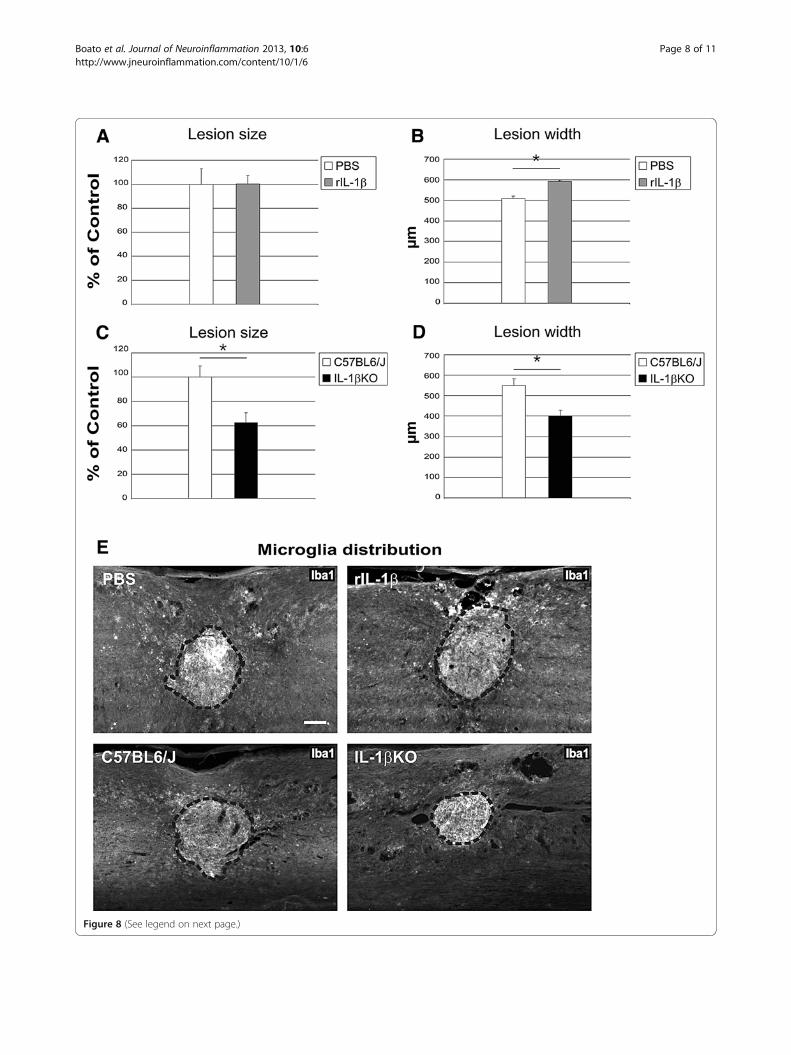
Figure 8 (See legend on next page.)
Boato et al. Journal of Neuroinflammation 2013, 10:6 Page 8 of 11http://www.jneuroinflammation.com/content/10/1/6
cytokine. Of particular interest is the observation thatIL-1β injections stimulate macrophage activation andmyelin clearance in spinal cord white matter, while anabsence leads to an increased number of intact myelinsheaths [34]. Furthermore, IL-1β application abrogatesneurotrophin-induced neuronal cell survival in vitro[35,36]. Moreover, administration of IL-1β intrathecallyactivates p38 mitogen-activated protein kinase, and leadsto high levels of inducible nitric oxide synthase andrelease of nitric oxide [37]. However, none of these stud-ies investigated the influence of IL-1β on CNS plasticity.The present study demonstrates in vivo that the sum ofall these negative effects in the CNS appears to abrogatepotential IL-1-dependent axon elongation expected fromour recent in vitro study [5].Here, we provide the first in vivo evidence for a sub-
stantial IL-1β effect on plasticity and lesion develop-ment. We analyzed the effect of locally applied rIL-1βand its constitutive deficiency on functional recovery,CST fibers and astrogliosis in a mouse model of SCI,which mimics the most common type of spinal cord in-jury in humans [38]. Based on previous in vitro findingsdemonstrating that a high therapeutic dosage of rIL-1βincreases axonal outgrowth in an organotypic slice cul-ture model [5] we administered perilesionally a highdose of rIL-1β after SCI (20 μg rIL-1β in Gelfoam). Un-fortunately, this local application was lethal. However, alower, nonlethal dosage of rIL-1β (1 μg rIL-1β in Gel-foam) led to a significantly impaired functional recoveryaccording to the BMS. We used a mild lesion to ensurethat any possible negative effect of the application of thecytokine could be revealed. This treatment also resultedin a highly reduced number of BDA-positive CST fiberscaudal to the lesion compared with PBS-treated mice.Consistently, the analysis of the injured spinal cords ofIL-1β-deficient mice revealed a close to fivefold increasein the number of CST fibers caudal to the lesion com-pared with WT mice.These mice also displayed a significantly improved
neurological outcome. Based on morphological criteria[29], BDA-positive fibers counted caudal to the lesionappeared to present newly formed fibers, but due to thenature of the lesion we cannot exclude a small percentageof (undetected) sparing. Counted fibers could thus be a
mixture of newly established fibers derived from the siteof the lesion and of those sprouting from uninjured fibers.It is reasonable to assume that multiple spinal motor
systems are positively affected by the absence of IL-1β(and the consecutively reduced astrogliosis as discussedhereafter). The improvement in paw placement as an in-dicator of CST function [27,28] and increased numbersof anterogradely labeled CST nerve fibers in the IL-1β-deficient injured spinal cord support the concept thatenhanced CST plasticity may significant contribute to theimproved clinical outcome demonstrated in our study.Substantial differences between the in vivo and in vitro
model may explain why IL-1β stimulates neurite out-growth in vitro [5] but has a negative impact on axonplasticity in the present in vivo study. The in vitro studywas performed using organotypic slice cultures frompostnatal brains as previously described [22-25]. Acutebrain slices as used in our study, should be considered amodel for the early, highly acute phase of CNS traumasince they are acutely excised from of the living brain;most neurons are axotomized, the blood–brain barrier isheavily damaged, high levels of neuronal death appear,and astrocytes as well as many immune cells are acti-vated [39,40]. Contrastingly, in the in vivo model usedhere, the SCI is followed by at least three different in-flammatory phases (acute, subacute and chronic) thatare characterized by dramatic differences, for example,in terms of cytokine levels as well as immune cell activa-tion and migration patterns [41]. The in vivo effects ofIL-1β on functional parameters become clearly detect-able about 2 weeks after the highly acute phase. IL-1βmay therefore exert direct and/or indirect effects mainlyin the subacute or early chronic phase. In a landmarkpaper, the Kapfhammer group demonstrated that neuriteoutgrowth can only be reliably studied in embryonic andpostnatal brain slices until 4 days after birth [42]. Brainslice experiments are therefore performed with embryonicor early postnatal brains. In later developmental phases,neurite outgrowth from slices is substantially reduced orabsent. A further difference between the models is the ab-sence of systemic neuroendocrinological influences in thebrain slice model. The contradicting results in our brainslice study in vitro [5] and the present in vivo study maythus be due to differences in the CNS region (cortex
(See figure on previous page.)Figure 8 Application of recombinant IL-1β and its deficiency influence lesion size after spinal cord compression injury. (A) to (D)Quantification of the lesion size and lesion width based on a clearly distinguishable Iba1-positive area. Lesion size measurement in the centralsections of recombinant IL-1β (rIL-1β)-treated mice indicated no difference compared with controls (A), while the lesion width was about 20%greater (B). Both the lesion size (C) and the lesion width (D) were reduced by 40% and 25%, respectively, in IL-1βKO mice compared withcontrols. (E) Representative micrographs of Iba1 immunoreactive microglia distribution around the compression injury site in spinal cord sections.Upper panels: comparison of PBS-treated and IL-1β-treated spinal cord. Lower panels: comparison of wildtype control with IL-1βKO spinal cord.Dashed line, area of the lesion. Iba1 intensity did not differ significantly between groups (data not shown). *P <0.05; n = 5 mice (PBS), n = 5 mice(rIL-1β), n = 7 mice (C57BL6/J), n = 5 mice (IL-1βKO). Scale bar = 100 μm.
Boato et al. Journal of Neuroinflammation 2013, 10:6 Page 9 of 11http://www.jneuroinflammation.com/content/10/1/6
versus spinal cord), the inflammatory phase, the develop-mental stage (postnatal versus adult) or the presence orabsence of systemic neuroendocrinological influences.In the present study we also show significantly reduced
astrogliosis in the perilesional white matter in IL-1βKOmice. This is in line with the role of IL-1β as an astroglialgrowth factor in the mammalian brain (promoting prolif-eration) [43] and with the IL-1β-dependent astrocyte acti-vation following CNS injury, demonstrated in a murinecorticectomy model [44], leading to exacerbated astroglio-sis. However, in contrast to these studies, which describeearly stages after brain injury, we here demonstrate thatreduced astrogliosis in the absence of IL-1β is still present2 weeks after injury. Consistently, we also find a greatlyreduced lesion size (and consequently more spared spinalcord tissue).While the results of the present study are encouraging
to further investigate IL-1β modulation for spinal cordrepair, they should be interpreted with care keeping inmind the complexity of the plethora of potential mechan-isms as outlined in the introduction. Further studiesare needed to elucidate the intricate network of IL-1βmechanisms of action after SCI, which is likely to bemultifaceted and not limited to demyelination, cell deathand cytotoxic neuroinflammation.
ConclusionOur results show that IL-1β has a strong negative effecton axon plasticity, lesion development and gliosis afterSCI, associated with substantially impaired functional out-come. In particular, mice with constitutive absence of IL-1β revealed a close-to-opposite effect compared with micetreated with rIL-1β, since IL-1βKO mice were character-ized by a smaller lesion size, less astrogliosis and a greaternumber of labeled CST fibers caudal to the lesion, whichresulted in a significantly improved neurological outcome.These data provide a strong basis for further studies to beconducted with the goal of developing therapeutic strat-egies targeting the IL-1β pathways in spinal cord recoveryafter injury.
AbbreviationsBDA: Biotinylated dextran amine; BMS: Basso Mouse Scale; CNS: Centralnervous system; CST: Corticospinal tract; GFAP: Glial fibrillary acidic protein;Iba1: Ionized calcium binding adaptor molecule 1; IL: Interleukin; IL-1βKO: IL-1β deficiency; mAb: monoclonal antibody; PBS: Phosphate-buffered saline;rIL-1β: Recombinant IL-1β; SCI: Spinal cord compression injury; WT: Wildtype.
Competing interestsThe authors declare that they have no competing interests.
Authors’ contributionsFB calculated the mortality rate. FB, KR, SN, LG and SH performed thesurgeries, tracing, treated the animals, performed the neurological evaluationand statistics. FB and KR did fibers analysis. FB and EMP did the stainings andanalysis of gliosis. SH and FB wrote the manuscript. RN, SH and FB plannedthe experiments. All authors read and approved the final manuscript.
AcknowledgementsThe authors wish to thank Doreen Lüdecke and Julia König for excellenttechnical assistance and Katherine S. Matho for editing the text. This studywas supported in part by grants from Investitionsbank Berlin and theDeutsche Forschungsgemeinschaft (SPP1394) to SH and from FondsWetenschappelijk Onderzoek – Vlaanderen to SH (G.0834.11N, G.0389.12) andLG (1.5.056.12N).
Author details1Department of Morphology & BIOMED Institute, Campus Diepenbeek,Hasselt University, Agoralaan Gebouw C, Diepenbeek BE 3590, Belgium.2Present Address: Université Pierre et Marie Curie, Institut de la Vision, 17 rueMoreau, Paris 75012, France. 3Department of Neurology, CharitéUniversitätsmedizin, Charitéplatz 1, Berlin D-10117, Germany.4Psychoneuroimmunology, University-Medicine Charité, Charité Center 12 forInternal Medicine and Dermatology, Berlin D-10117, Germany. 5Departmentof Psychosomatic Medicine, Justus-Liebig-University, Klinikstrasse 32, D-35392,Gießen, Germany. 6Institute of Microanatomy and Neurobiology, UniversityMedical Center, Johannes Gutenberg University, Langenbeckstrasse 1, 55131,Mainz, Germany.
Received: 6 June 2012 Accepted: 7 December 2012Published: 14 January 2013
References1. Allan SM, Tyrrell PJ, Rothwell NJ: Interleukin-1 and neuronal injury. Nat Rev
Immunol 2005, 5:629–640.2. Bauer J, Berkenbosch F, Van Dam AM, Dijkstra CD: Demonstration of
interleukin-1 beta in Lewis rat brain during experimental allergicencephalomyelitis by immunocytochemistry at the light andultrastructural level. J Neuroimmunol 1993, 48:13–21.
3. Desson SE, Ferguson AV: Interleukin 1β modulates rat subfornical organneurons as a result of activation of a non-selective cationic conductance.J Physiol 2003, 550(Pt 1):113–122.
4. Viviani B, Bartesaghi S, Gardoni F, Vezzani A, Behrens MM, Bartfai T, BinagliaM, Corsini E, Di Luca M, Galli CL, Marinovich M: Interleukin-1β enhancesNMDA receptor-mediated intracellular calcium increase throughactivation of the Src family of kinases. J Neurosci 2003, 23:8692–8700.
5. Boato F, Hechler D, Rosenberger K, Lüdecke D, Peters EM, Nitsch R, HendrixS: Interleukin-1 beta and neurotrophin-3 synergistically promote neuritegrowth in vitro. J Neuroinflammation 2011, 8:183.
6. Edoff K, Jerregard H: Effects of IL-1β, IL-6 or LIF on rat sensory neuronsco-cultured with fibroblast-like cells. J Neurosci Res 2002, 67:255–263.
7. Zeise ML, Madamba S, Siggins GR: Interleukin-1 beta increases synapticinhibition in rat hippocampal pyramidal neurons in vitro. Regul Pept1992, 39:1–7.
8. John GR, Lee SC, Song X, Rivieccio M, Brosnan CF: IL-1-regulated responsesin astrocytes: relevance to injury and recovery. Glia 2005, 49:161–176.
9. Gölz G, Uhlmann L, Lüdecke D, Markgraf N, Nitsch R, Hendrix S: Thecytokine/neurotrophin axis in peripheral axon outgrowth. Eur J Neurosci2006, 24:2721–2730.
10. Loddick SA, Rothwell NJ: Neuroprotective effects of human recombinantinterleukin-1 receptor antagonist in focal cerebral ischaemia in the rat.J Cereb Blood Flow Metab 1996, 16:932–940.
11. McColl BW, Rothwell NJ, Allan SM: Systemic inflammatory stimuluspotentiates the acute phase and CXC chemokine responses toexperimental stroke and exacerbates brain damage via interleukin-1-and neutrophil-dependent mechanisms. J Neurosci 2007,27:4403–4412.
12. Yamasaki Y, Matsuura N, Shozuhara H, Onodera H, Itoyama Y, Kogure K:Interleukin-1 as a pathogenetic mediator of ischemic brain damage inrats. Stroke 1995, 26:676–680. discussion 681.
13. Wang CX, Olschowka JA, Wrathall JR: Increase of interleukin-1β mRNA andprotein in the spinal cord following experimental traumatic injury in therat. Brain Res 1997, 759:190–196.
14. Wang XJ, Kong KM, Qi WL, Ye WL, Song PS: Interleukin-1 beta induction ofneuron apoptosis depends on p38 mitogen-activated protein kinaseactivity after spinal cord injury. Acta Pharmacol Sin 2005, 26:934–942.
15. Mason JL, Suzuki K, Chaplin DD, Matsushima GK: Interleukin-1β promotesrepair of the CNS. J Neurosci 2001, 21:7046–7052.
Boato et al. Journal of Neuroinflammation 2013, 10:6 Page 10 of 11http://www.jneuroinflammation.com/content/10/1/6
16. Temporin K, Tanaka H, Kuroda Y, Okada K, Yachi K, Moritomo H, Murase T,Yoshikawa H: IL-1β promotes neurite outgrowth by deactivating RhoAvia p38 MAPK pathway. Biochem Biophys Res Commun 2008, 365:375–380.
17. Temporin K, Tanaka H, Kuroda Y, Okada K, Yachi K, Moritomo H, Murase T,Yoshikawa H: Interleukin-1 beta promotes sensory nerve regenerationafter sciatic nerve injury. Neurosci Lett 2008, 440:130–133.
18. Boato F, Hendrix S, Huelsenbeck SC, Hofmann F, Grosse G, Djalali S,Klimaschewski L, Auer M, Just I, Ahnert-Hilger G, Höltje M: C3 peptideenhances recovery from spinal cord injury by improved regenerativegrowth of descending fiber tracts. J Cell Sci 2010, 123(Pt 10):1652–1662.
19. Sato A, Ohtaki H, Tsumuraya T, Song D, Ohara K, Asano M, Iwakura Y,Atsumi T, Shioda S: Interleukin-1 participates in the classical andalternative activation of microglia/macrophages after spinal cord injury.J Neuroinflammation 2012, 9:65.
20. Shornick LP, De Togni P, Mariathasan S, Goellner J, Strauss-Schoenberger J,Karr RW, Ferguson TA, Chaplin DD: Mice deficient in IL-1β manifestimpaired contact hypersensitivity to trinitrochlorobenzone. J Exp Med1996, 183:1427–1436.
21. Loske P, Boato F, Hendrix S, Piepgras J, Just I, Ahnert-Hilger G, Höltje M:Minimal essential length of Clostridium botulinum C3 peptides toenhance neuronal regenerative growth and connectivity in a non-enzymatic mode. J Neurochem 2012, 120:1084–1096.
22. Hechler D, Boato F, Nitsch R, Hendrix S: Differential regulation of axonoutgrowth and reinnervation by neurotrophin-3 and neurotrophin-4 inthe hippocampal formation. Exp Brain Res 2010, 205:215–221.
23. Höltje M, Djalali S, Hofmann F, Münster-Wandowski A, Hendrix S, Boato F,Dreger SC, Grosse G, Henneberger C, Grantyn R, Just I, Ahnert-Hilger G: A29-amino acid fragment of Clostridium botulinum C3 protein enhancesneuronal outgrowth, connectivity, and reinnervation. FASEB J 2009,23:1115–1126.
24. Schmitt KR, Boato F, Diestel A, Hechler D, Kruglov A, Berger F, Hendrix S:Hypothermia-induced neurite outgrowth is mediated by tumor necrosisfactor-alpha. Brain Pathol 2010, 20:771–779.
25. Schmitt KR, Kern C, Lange PE, Berger F, Abdul-Khaliq H, Hendrix S: S100Bmodulates IL-6 release and cytotoxicity from hypothermic brain cellsand inhibits hypothermia-induced axonal outgrowth. Neurosci Res 2007,59:68–73.
26. Basso DM, Fisher LC, Anderson AJ, Jakeman LB, McTigue DM, Popovich PG:Basso Mouse Scale for locomotion detects differences in recovery afterspinal cord injury in five common mouse strains. J Neurotrauma 2006,23(5):635–659.
27. De Ryck M, Van Reempts J, Duytschaever H, Van Deuren B, Clincke G:Neocortical localization of tactile/proprioceptive limb placing reactionsin the rat. Brain Res 1992, 573:44–60.
28. Metz GA, Whishaw IQ: Cortical and subcortical lesions impair skilledwalking in the ladder rung walking test: a new task to evaluate fore-and hindlimb stepping, placing, and co-ordination. J Neurosci Methods2002, 115:169–179.
29. Steward O, Zheng B, Tessier-Lavigne M: False resurrections: distinguishingregenerated from spared axons in the injured central nervous system.J Comp Neurol 2003, 459:1–8.
30. Wang X, Budel S, Baughman K, Gould G, Song KH, Strittmatter SM:Ibuprofen enhances recovery from spinal cord injury by limitingtissue loss and stimulating axonal growth. J Neurotrauma 2009,26:81–95.
31. Pineau I, Lacroix S: Proinflammatory cytokine synthesis in the injuredmouse spinal cord: multiphasic expression pattern and identification ofthe cell types involved. J Comp Neurol 2007, 500:267–285.
32. Nesic O, Xu GY, McAdoo D, High KW, Hulsebosch C, Perez-Pol R: IL-1receptor antagonist prevents apoptosis and caspase-3 activation afterspinal cord injury. J Neurotrauma 2001, 18:947–956.
33. Liu S, Xu GY, Johnson KM, Echetebu C, Ye ZS, Hulsebosch CE, McAdoo DJ:Regulation of interleukin-1β by the interleukin-1 receptor antagonist inthe glutamate-injured spinal cord: endogenous neuroprotection.Brain Res 2008, 1231:63–74.
34. Perrin FE, Lacroix S, Avilés-Trigueros M, David S: Involvement of monocytechemoattractant protein-1, macrophage inflammatory protein-1α andinterleukin-1β in Wallerian degeneration. Brain 2005, 128(Pt 4):854–866.
35. Soiampornkul R, Tong L, Thangnipon W, Balazs R, Cotman CW: Interleukin-1β interferes with signal transduction induced by neurotrophin-3 incortical neurons. Brain Res 2008, 1188:189–197.
36. Tong L, Balazs R, Soiampornkul R, Thangnipon W, Cotman CW: Interleukin-1beta impairs brain derived neurotrophic factor-induced signaltransduction. Neurobiol Aging 2008, 29:1380–1393.
37. Sung CS, Wong CS: Cellular mechanisms of neuroinflammatory pain: therole of interleukin-1beta. Acta Anaesthesiol Taiwan 2007, 45:103–109.
38. Sekhon LH, Fehlings MG: Epidemiology, demographics, andpathophysiology of acute spinal cord injury. Spine (Phila Pa 1976) 2001,26(24 Suppl):S2–S12.
39. Eyupoglu IY, Savaskan NE, Bräuer AU, Nitsch R, Heimrich B: Identification ofneuronal cell death in a model of degeneration in the hippocampus.Brain Res Brain Res Protoc 2003, 11:1–8.
40. Wolf SA, Fisher J, Bechmann I, Steiner B, Kwidzinski E, Nitsch R:Neuroprotection by T-cells depends on their subtype and activationstate. J Neuroimmunol 2002, 133:72–80.
41. Vidal PM, Lemmens E, Geboes L, Vangansewinkel T, Nelissen S, Hendrix S:Late blocking of peripheral TNF-α is ineffective after spinal cord injury inmice. Immunobiology 2012, doi:10.1016/j.cytogfr.2012.08.008.
42. Prang P, Del Turco D, Kapfhammer JP: Regeneration of entorhinal fibers inmouse slice cultures is age dependent and can be stimulated by NT-4,GDNF, and modulators of G-proteins and protein kinase C. Exp Neurol2001, 169:135–147.
43. Giulian D, Young DG, Woodward J, Brown DC, Lachman LB: Interleukin-1 isan astroglial growth factor in the developing brain. J Neurosci 1988,8:709–714.
44. Herx LM, Yong VW: Interleukin-1 beta is required for the early evolutionof reactive astrogliosis following CNS lesion. J Neuropathol Exp Neurol2001, 60:961–971.
doi:10.1186/1742-2094-10-6Cite this article as: Boato et al.: Absence of IL-1β positively affectsneurological outcome, lesion development and axonal plasticity afterspinal cord injury. Journal of Neuroinflammation 2013 10:6.
Submit your next manuscript to BioMed Centraland take full advantage of:
• Convenient online submission
• Thorough peer review
• No space constraints or color figure charges
• Immediate publication on acceptance
• Inclusion in PubMed, CAS, Scopus and Google Scholar
• Research which is freely available for redistribution
Submit your manuscript at www.biomedcentral.com/submit
Boato et al. Journal of Neuroinflammation 2013, 10:6 Page 11 of 11http://www.jneuroinflammation.com/content/10/1/6


![Elovanoids counteract oligomeric β-amyloid-induced …cognition (Alzheimer’s disease) and sight (age-related macular de-generation [AMD]). How neuroinflammation can be counteracted](https://static.fdocument.org/doc/165x107/5f2eb83dff582622624e3d80/elovanoids-counteract-oligomeric-amyloid-induced-cognition-alzheimeras-disease.jpg)