Atorvastatin inhibits proliferation and promotes apoptosis ...
Gaojian et al. Cell Death Discovery (2020) 6:97
https://doi.org/10.1038/s41420-020-00333-8 Cell Death Discovery
ART ICLE Open Ac ce s s
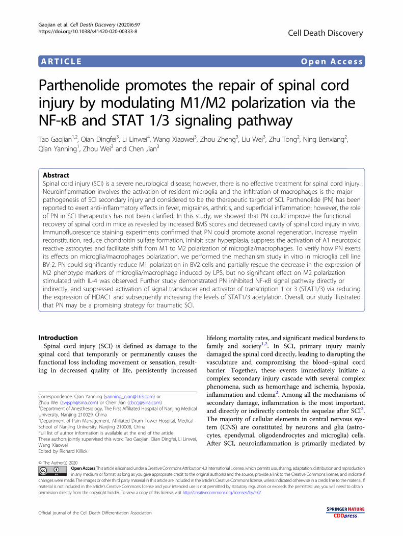
Parthenolide promotes the repair of spinal cordinjury by modulating M1/M2 polarization via theNF-κB and STAT 1/3 signaling pathwayTao Gaojian1,2, Qian Dingfei3, Li Linwei4, Wang Xiaowei3, Zhou Zheng3, Liu Wei3, Zhu Tong2, Ning Benxiang2,Qian Yanning1, Zhou Wei3 and Chen Jian3
AbstractSpinal cord injury (SCI) is a severe neurological disease; however, there is no effective treatment for spinal cord injury.Neuroinflammation involves the activation of resident microglia and the infiltration of macrophages is the majorpathogenesis of SCI secondary injury and considered to be the therapeutic target of SCI. Parthenolide (PN) has beenreported to exert anti-inflammatory effects in fever, migraines, arthritis, and superficial inflammation; however, the roleof PN in SCI therapeutics has not been clarified. In this study, we showed that PN could improve the functionalrecovery of spinal cord in mice as revealed by increased BMS scores and decreased cavity of spinal cord injury in vivo.Immunofluorescence staining experiments confirmed that PN could promote axonal regeneration, increase myelinreconstitution, reduce chondroitin sulfate formation, inhibit scar hyperplasia, suppress the activation of A1 neurotoxicreactive astrocytes and facilitate shift from M1 to M2 polarization of microglia/macrophages. To verify how PN exertsits effects on microglia/macrophages polarization, we performed the mechanism study in vitro in microglia cell lineBV-2. PN could significantly reduce M1 polarization in BV2 cells and partially rescue the decrease in the expression ofM2 phenotype markers of microglia/macrophage induced by LPS, but no significant effect on M2 polarizationstimulated with IL-4 was observed. Further study demonstrated PN inhibited NF-κB signal pathway directly orindirectly, and suppressed activation of signal transducer and activator of transcription 1 or 3 (STAT1/3) via reducingthe expression of HDAC1 and subsequently increasing the levels of STAT1/3 acetylation. Overall, our study illustratedthat PN may be a promising strategy for traumatic SCI.
IntroductionSpinal cord injury (SCI) is defined as damage to the
spinal cord that temporarily or permanently causes thefunctional loss including movement or sensation, result-ing in decreased quality of life, persistently increased
lifelong mortality rates, and significant medical burdens tofamily and society1,2. In SCI, primary injury mainlydamaged the spinal cord directly, leading to disrupting thevasculature and compromising the blood–spinal cordbarrier. Together, these events immediately initiate acomplex secondary injury cascade with several complexphenomena, such as hemorrhage and ischemia, hypoxia,inflammation and edema2. Among all the mechanisms ofsecondary damage, inflammation is the most important,and directly or indirectly controls the sequelae after SCI3.The majority of cellular elements in central nervous sys-tem (CNS) are constituted by neurons and glia (astro-cytes, ependymal, oligodendrocytes and microglia) cells.After SCI, neuroinflammation is primarily mediated by
© The Author(s) 2020OpenAccessThis article is licensedunder aCreativeCommonsAttribution 4.0 International License,whichpermits use, sharing, adaptation, distribution and reproductionin any medium or format, as long as you give appropriate credit to the original author(s) and the source, provide a link to the Creative Commons license, and indicate if
changesweremade. The images or other third partymaterial in this article are included in the article’s Creative Commons license, unless indicated otherwise in a credit line to thematerial. Ifmaterial is not included in the article’s Creative Commons license and your intended use is not permitted by statutory regulation or exceeds the permitted use, you will need to obtainpermission directly from the copyright holder. To view a copy of this license, visit http://creativecommons.org/licenses/by/4.0/.
Correspondence: Qian Yanning ([email protected]) orZhou Wei ([email protected]) or Chen Jian ([email protected])1Department of Anesthesiology, The First Affiliated Hospital of Nanjing MedicalUniversity, Nanjing 210029, China2Department of Pain Management, Affiliated Drum Tower Hospital, MedicalSchool of Nanjing University, Nanjing 210008, ChinaFull list of author information is available at the end of the articleThese authors jointly supervised this work: Tao Gaojian, Qian Dingfei, Li Linwei,Wang XiaoweiEdited by Richard Killick
Official journal of the Cell Death Differentiation Association
1234
5678
90():,;
1234
5678
90():,;
1234567890():,;
1234
5678
90():,;
microglia cells, which are the main components of theinnate immune system and regarded as resident macro-phages in the CNS4–6. Due to the destruction ofblood–spinal cord barrier (BBB) and blood vessel injury,peripheral macrophages, neutrophils and lymphocytes arerecruited in damaged tissues under the influence of localinflammatory factors, chemokines, and vasoactive pep-tides with the synergy of firstly microglia activation2,4.Because microglia and peripheral macrophages havemyelomonocytic origin, therefore peripheral macrophagesadopt many of the markers and behaviors of microglia7,8.These similarities make it difficult to distinguish betweenthem in spinal cord injury foci.Under the stimulus of the lesion microenvironment of
spinal cord, microglia/macrophages polarize into twophenotypes: a classically activated pro-inflammatory(M1) phenotype and an alternatively activated anti-inflammatory (M2) phenotype9. The ratio of M1 to M2maintains the homeostasis of the local microenvironment.M1 phenotype, is associated with the production ofproinflammatory cytokines such as tumor necrosis factor-α (TNF-α), interleukin-1 beta (IL-1β), interleukin-6 (IL-6), glutamate, superoxide, nitric oxide (NO), reactiveoxygen species (ROS), chemokines, and proteases3,4,10.These inflammatory mediators initiate cascades of neu-rotoxic responses in secondary phase of SCI and cancooperate to contribute to the preponderance of damage(apoptosis and necrosis) to endothelia, neurons, axonsand oligodendrocytes, and finally phagocytosis. In con-trast, M2 phenotype is symbolized by the release of anti-inflammatory cytokines (IL-4, IL-10, IL-13), transforminggrowth factor beta (TGF-β), and several neurotrophicfactors, such as ciliary neurotrophic factor (CNTF),insulin-like growth factor (IGF), epidermal growth factor(EGF), and nerve growth factor (NGF), all of whichantagonize the pro-inflammatory responses and facilitateneuroregeneration, particularly axonal extension, afterCNS injury3,4,10. Therefore, Inhibiting M1 or promotingM2 polarization may be a promising treatment strategy tofacilitate functional recovery after SCI. Additionally, M1phenotype of microglia/macrophages are quickly acti-vated in the first few days and continue to be activated28 days after injury; however, within 7 days, transientactivation of a small number of M2 macrophages/microglia are observed11,12. This is a critical time forspinal cord injury repair with one month of the injury, andto be clinically effective, targeting M1/M2 polarizationbalance needs depend on the time window.Parthenolide (PN), as a sesquiterpene lactone used for
therapy of inflammation, via inhibiting canonical NF-κB(p65/p50) activation followed by direct interaction of PNwith inhibitor of nuclear factor kappa-B kinase (IKK) βand subsequently suppressing IKK signalsome13,14, as wellas an inductor of HDAC1 degradation by proteasome
activity15 has been reported to have anti-cancer prop-erty16,17. Furthermore, PN has also been considered to beinvolved in the regulation of signal transducer and acti-vator of transcriptions (STATs) signaling pathway18,19,but the mechanism is still unclear. Both the NF-κB andSTATs signals are considered as important pathways inregulating microglia/macrophages polarization. However,the ability of PN to regulate M1/M2 polarization and itsneuroprotective effect in SCI remain unknown. This studyaimed to investigate whether PN could regulate M1/M2polarization and promote functional recovery of miceafter SCI as well as the underlying mechanisms.Inspiringly, we found that PN could improve functional
recovery in SCI mice by promoting axonal regeneration,increasing myelin reconstitution, reducing chondroitinsulfate proteoglycan (CSPG) formation, inhibiting scarhyperplasia, and suppressing the activation of A1 neuro-toxic reactive astrocytes. In addition, we also showed thatM2 polarization of microglia/macrophage was activatedand M1 polarization was suppressed by PN in vivo. Invitro, PN mainly inhibited the M1 polarization of BV2cells induced by LPS accompanied by a reversal of M2phenotype and had no effect on M2 polarization inducedby IL-4. Further study demonstrated that PN regulatedM1/M2 polarization balance mainly via inhibiting NF-κBsignal pathway directly or indirectly, and suppressingactivation of STAT1/3 via the reduced expressionof HDAC1 and the subsequently increasing levels ofSTAT1/3 acetylation. This study demonstrates that PNmay be a promising therapeutic drug to target microgliapolarization in SCI.
ResultsPN treatment improved functional recovery followingtraumatic SCITo test the neuroprotective efficacy of PN in vivo, we
performed motor function assessments as well as histo-logical injury study in mice with weight-induced spinalcontusion at T10 (Fig. 1a). Within 10min posttrauma, weintraperitoneally administered PN and then continuouslyinjected daily until 7 days considering the blood–brainbarrier (BBB) closing time after injury20. Motor functionin the SCI group and the PN group gradually improvedover the first week following SCI as evidenced by BMSscores. Mice treated with PN showed significantly betterperformance than the controls beginning at day 7 andmaintained this superiority over the remainder of thestudy (Fig. 1b). Coordination of forepaw–hindpawmovements decreased significantly immediately after SCIas determined by gait analysis, but animals treated withPN showed significantly faster gait recovery and improvedmotor coordination compared with SCI group animals(Fig. 1c, d). As shown by the gross morphology of theinjured spinal cords, the traumatic lesion area (brown
Gaojian et al. Cell Death Discovery (2020) 6:97 Page 2 of 16
Official journal of the Cell Death Differentiation Association
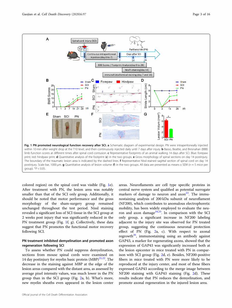
colored region) on the spinal cord was visible (Fig. 1e).After treatment with PN, the lesion area was notablysmaller than that of the SCI only group. Additionally, itshould be noted that motor performance and the grossmorphology of the sham-surgery group remainedunchanged throughout the test period. Nissl stainingrevealed a significant loss of SCI tissue in the SCI group at2 weeks post injury that was significantly reduced in thePN treatment group (Fig. 1f, g). Collectively, these datasuggest that PN promotes the functional motor recoveryfollowing SCI.
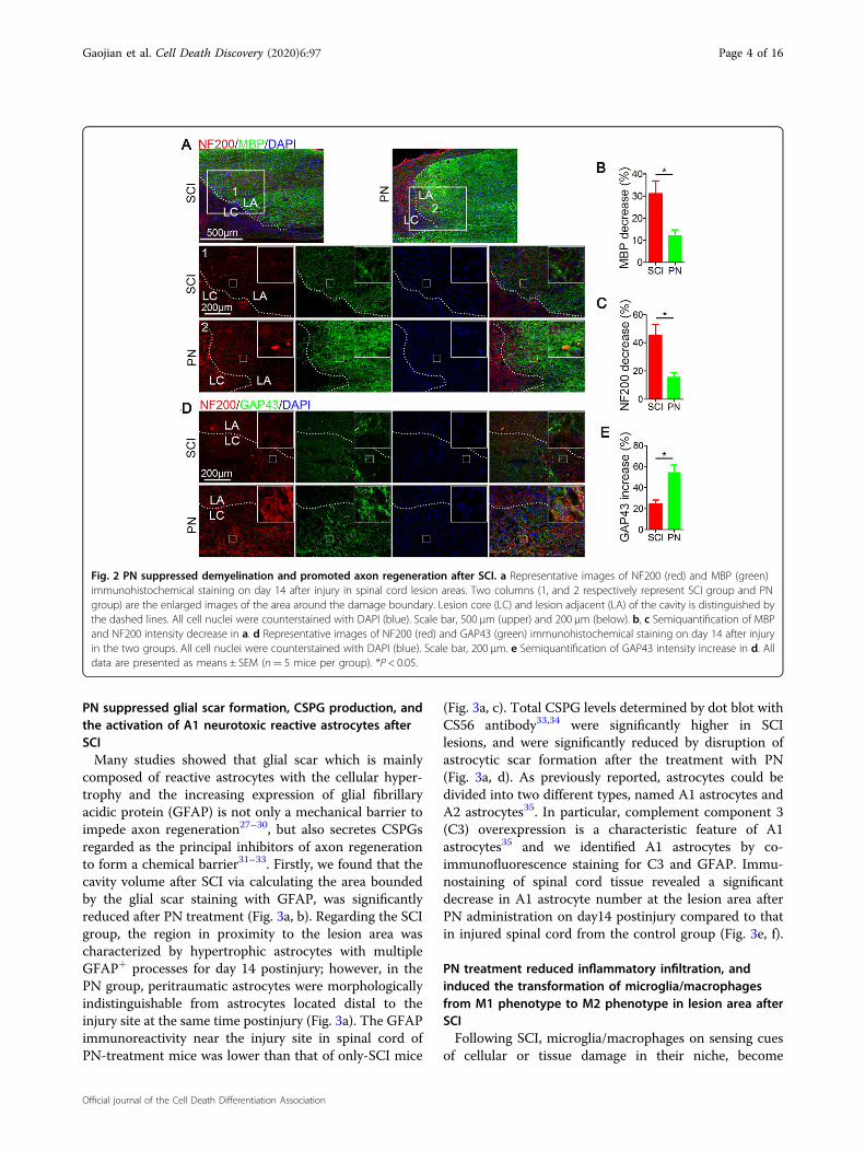
PN treatment inhibited demyelination and promoted axonregeneration following SCITo assess whether PN could suppress demyelination,
sections from mouse spinal cords were examined on14 day postinjury for myelin basic protein (MBP)21,22. Thedecrease in the staining against MBP at the edge of thelesion areas compared with the distant area, as assessed byaverage pixel intensity values, was much lower in the PNgroup than in the SCI group (Fig. 2a, b). What’s more,new myelin sheaths even appeared in the lesion center
areas. Neuroflaments are cell type specific proteins incentral nerve system and qualified as potential surrogatemarkers of damage to neuron and axon23. The immu-nostaining analysis of 200 kDa subunit of neuroflament(NF200), which contributes to anomalous electrophoreticmobility, has been widely employed to evaluate the neu-ron and axon damage24,25. In comparison with the SCIonly group, a significant increase in NF200 labelingadjacent to the injury site was observed for PN treatedgroup, suggesting the continuous neuronal protectioneffect of PN (Fig. 2a, c). With respect to axonalregrowth26, immunostaining using an antibody againstGAP43, a marker for regenerating axons, showed that theexpression of GAP43 was significantly increased both atthe lesion epicenter in mice treated with PN in compar-ison with SCI group (Fig. 2d, e). Besides, NF200-positivefibers in mice treated with PN were more likely to bereproduced at the injury center, and most of these fibersexpressed GAP43 according to the merge image betweenNF200 staining with GAP43 staining (Fig. 2d). Theseresults indicate that PN reduces the demyelination, andpromote axonal regeneration in the injured lesion area.
Fig. 1 PN promoted neurological function recovery after SCI. a Schematic diagram of experimental design. PN were intraperitoneally injectedwithin 10 min after weight drop at the T10 level, and then continuously injected daily until 7 days after injury. b Basso, Beattie, and Bresnahan (BBB)limb function scores at different times after spinal cord contusion. c Representative footprints of an animal walking 14 days after SCI. Blue: forepawprint; red: hindpaw print. d Quantitative analysis of the footprint (c) in the two groups. e Gross morphology of spinal sections on day 14 postinjury.The boundary of the traumatic lesion area is indicated by the dashed lines. f Representative Nissl-stained sagittal section of spinal cord on day 14postinjury. Scale bar, 1000 μm. g Quantitative analysis of lesion volume (f) in the two groups. All data are presented as means ± SEM (n= 5 mice pergroup). *P < 0.05.
Gaojian et al. Cell Death Discovery (2020) 6:97 Page 3 of 16
Official journal of the Cell Death Differentiation Association
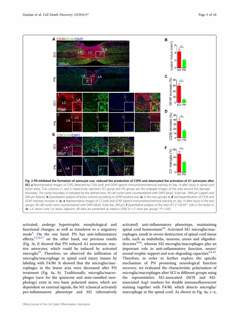
PN suppressed glial scar formation, CSPG production, andthe activation of A1 neurotoxic reactive astrocytes afterSCIMany studies showed that glial scar which is mainly
composed of reactive astrocytes with the cellular hyper-trophy and the increasing expression of glial fibrillaryacidic protein (GFAP) is not only a mechanical barrier toimpede axon regeneration27–30, but also secretes CSPGsregarded as the principal inhibitors of axon regenerationto form a chemical barrier31–33. Firstly, we found that thecavity volume after SCI via calculating the area boundedby the glial scar staining with GFAP, was significantlyreduced after PN treatment (Fig. 3a, b). Regarding the SCIgroup, the region in proximity to the lesion area wascharacterized by hypertrophic astrocytes with multipleGFAP+ processes for day 14 postinjury; however, in thePN group, peritraumatic astrocytes were morphologicallyindistinguishable from astrocytes located distal to theinjury site at the same time postinjury (Fig. 3a). The GFAPimmunoreactivity near the injury site in spinal cord ofPN-treatment mice was lower than that of only-SCI mice
(Fig. 3a, c). Total CSPG levels determined by dot blot withCS56 antibody33,34 were significantly higher in SCIlesions, and were significantly reduced by disruption ofastrocytic scar formation after the treatment with PN(Fig. 3a, d). As previously reported, astrocytes could bedivided into two different types, named A1 astrocytes andA2 astrocytes35. In particular, complement component 3(C3) overexpression is a characteristic feature of A1astrocytes35 and we identified A1 astrocytes by co-immunofluorescence staining for C3 and GFAP. Immu-nostaining of spinal cord tissue revealed a significantdecrease in A1 astrocyte number at the lesion area afterPN administration on day14 postinjury compared to thatin injured spinal cord from the control group (Fig. 3e, f).
PN treatment reduced inflammatory infiltration, andinduced the transformation of microglia/macrophagesfrom M1 phenotype to M2 phenotype in lesion area afterSCIFollowing SCI, microglia/macrophages on sensing cues
of cellular or tissue damage in their niche, become
Fig. 2 PN suppressed demyelination and promoted axon regeneration after SCI. a Representative images of NF200 (red) and MBP (green)immunohistochemical staining on day 14 after injury in spinal cord lesion areas. Two columns (1, and 2 respectively represent SCI group and PNgroup) are the enlarged images of the area around the damage boundary. Lesion core (LC) and lesion adjacent (LA) of the cavity is distinguished bythe dashed lines. All cell nuclei were counterstained with DAPI (blue). Scale bar, 500 μm (upper) and 200 μm (below). b, c Semiquantification of MBPand NF200 intensity decrease in a. d Representative images of NF200 (red) and GAP43 (green) immunohistochemical staining on day 14 after injuryin the two groups. All cell nuclei were counterstained with DAPI (blue). Scale bar, 200 μm. e Semiquantification of GAP43 intensity increase in d. Alldata are presented as means ± SEM (n= 5 mice per group). *P < 0.05.
Gaojian et al. Cell Death Discovery (2020) 6:97 Page 4 of 16
Official journal of the Cell Death Differentiation Association
activated, undergo hypertrophic morphological andfunctional changes, as well as transform to a migratorymode4. On the one hand, PN has anti-inflammatoryeffects;13,16,17 on the other hand, our previous results(Fig. 3e, f) showed that PN reduced A1 neurotoxic reac-tive astrocytes, which could be induced by activatedmicroglia35. Therefore, we observed the infiltration ofmicroglia/macrophage in spinal cord injury tissues bylabeling with F4/80. It showed that the microglia/mac-rophages in the lesion area were decreased after PNtreatment (Fig. 4a, b). Traditionally, microglia/macro-phages (save for the quiescent and state-ramified mor-phology) exist in two basic polarized states, which aredependent on external signals, the M1 (classical activated)pro-inflammatory phenotype and M2 (alternatively
activated) anti-inflammatory phenotype, maintainingspinal cord homeostasis36. Activated M1 microglia/mac-rophages, result in severe destruction of spinal cord tissuecells, such as endothelia, neurons, axons and oligoden-drocytes3,4,6, whereas M2 microglia/macrophages play animportant role in anti-inflammatory function, neuro/axonal-trophic support and scar-degrading capacities3,4,10.Therefore, in order to further explore the specificmechanism of PN promoting neurological functionrecovery, we evaluated the characteristic polarization ofmicroglia/macrophages after SCI in different groups usingthe representative M1-assocaited iNOS and M2-associated Arg1 markers for double immunofluorescentstaining together with F4/80, which detects microglia/macrophage in the spinal cord. As shown in Fig. 4a, c–e,
Fig. 3 PN inhibited the formation of astrocyte scar, reduced the production of CSPG and attenuated the activation of A1 astrocytes afterSCI. a Representative images of CSPG detected by CS56 (red) and GFAP (green) immunohistochemical staining on day 14 after injury in spinal cordlesion areas. Two columns (1, and 2 respectively represent SCI group and PN group) are the enlarged images of the area around the damageboundary. The cavity boundary is indicated by the dashed lines. All cell nuclei were counterstained with DAPI (blue). Scale bar, 1000 μm (upper) and200 μm (below). b Quantitative analysis of lesion volume according to GFAP-positive scar (a) in the two groups. c, d Semiquantification of CS56 andGFAP intensity increase in (a). e Representative images of C3 (red) and GFAP (green) immunohistochemical staining on day 14 after injury in the twogroups. All cell nuclei were counterstained with DAPI (blue). Scale bar, 200 μm. f Quantitative analysis of the ratio of C3+/GFAP+ cells in the lesion in(e). LA, lesion core; LA, lesion adjacent. All data are presented as means ± SEM (n= 5 mice per group). *P < 0.05.
Gaojian et al. Cell Death Discovery (2020) 6:97 Page 5 of 16
Official journal of the Cell Death Differentiation Association
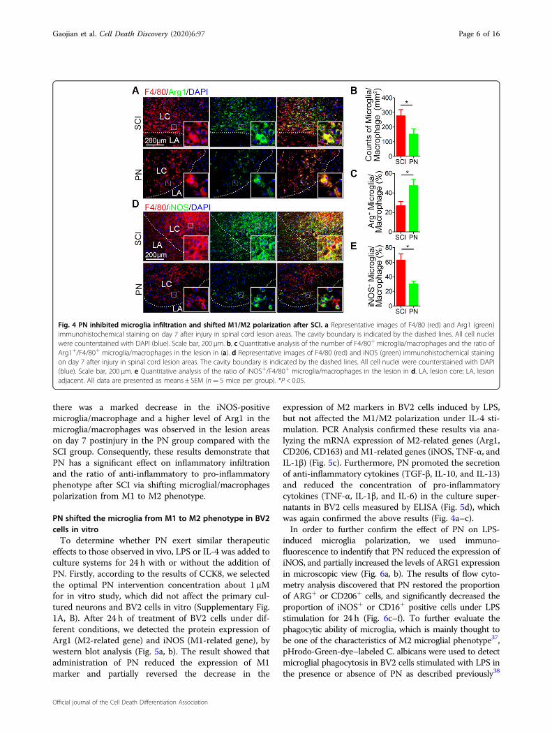
there was a marked decrease in the iNOS-positivemicroglia/macrophage and a higher level of Arg1 in themicroglia/macrophages was observed in the lesion areason day 7 postinjury in the PN group compared with theSCI group. Consequently, these results demonstrate thatPN has a significant effect on inflammatory infiltrationand the ratio of anti-inflammatory to pro-inflammatoryphenotype after SCI via shifting microglial/macrophagespolarization from M1 to M2 phenotype.
PN shifted the microglia from M1 to M2 phenotype in BV2cells in vitroTo determine whether PN exert similar therapeutic
effects to those observed in vivo, LPS or IL-4 was added toculture systems for 24 h with or without the addition ofPN. Firstly, according to the results of CCK8, we selectedthe optimal PN intervention concentration about 1 μMfor in vitro study, which did not affect the primary cul-tured neurons and BV2 cells in vitro (Supplementary Fig.1A, B). After 24 h of treatment of BV2 cells under dif-ferent conditions, we detected the protein expression ofArg1 (M2-related gene) and iNOS (M1-related gene), bywestern blot analysis (Fig. 5a, b). The result showed thatadministration of PN reduced the expression of M1marker and partially reversed the decrease in the
expression of M2 markers in BV2 cells induced by LPS,but not affected the M1/M2 polarization under IL-4 sti-mulation. PCR Analysis confirmed these results via ana-lyzing the mRNA expression of M2-related genes (Arg1,CD206, CD163) and M1-related genes (iNOS, TNF-α, andIL-1β) (Fig. 5c). Furthermore, PN promoted the secretionof anti-inflammatory cytokines (TGF-β, IL-10, and IL-13)and reduced the concentration of pro-inflammatorycytokines (TNF-α, IL-1β, and IL-6) in the culture super-natants in BV2 cells measured by ELISA (Fig. 5d), whichwas again confirmed the above results (Fig. 4a–c).In order to further confirm the effect of PN on LPS-
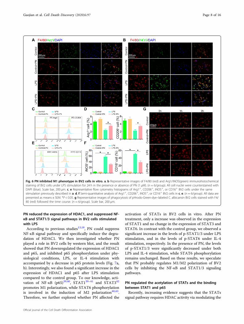
induced microglia polarization, we used immuno-fluorescence to indentify that PN reduced the expression ofiNOS, and partially increased the levels of ARG1 expressionin microscopic view (Fig. 6a, b). The results of flow cyto-metry analysis discovered that PN restored the proportionof ARG+ or CD206+ cells, and significantly decreased theproportion of iNOS+ or CD16+ positive cells under LPSstimulation for 24 h (Fig. 6c–f). To further evaluate thephagocytic ability of microglia, which is mainly thought tobe one of the characteristics of M2 microglial phenotype37,pHrodo-Green-dye–labeled C. albicans were used to detectmicroglial phagocytosis in BV2 cells stimulated with LPS inthe presence or absence of PN as described previously38
Fig. 4 PN inhibited microglia infiltration and shifted M1/M2 polarization after SCI. a Representative images of F4/80 (red) and Arg1 (green)immunohistochemical staining on day 7 after injury in spinal cord lesion areas. The cavity boundary is indicated by the dashed lines. All cell nucleiwere counterstained with DAPI (blue). Scale bar, 200 μm. b, c Quantitative analysis of the number of F4/80+ microglia/macrophages and the ratio ofArg1+/F4/80+ microglia/macrophages in the lesion in (a). d Representative images of F4/80 (red) and iNOS (green) immunohistochemical stainingon day 7 after injury in spinal cord lesion areas. The cavity boundary is indicated by the dashed lines. All cell nuclei were counterstained with DAPI(blue). Scale bar, 200 μm. e Quantitative analysis of the ratio of iNOS+/F4/80+ microglia/macrophages in the lesion in d. LA, lesion core; LA, lesionadjacent. All data are presented as means ± SEM (n= 5 mice per group). *P < 0.05.
Gaojian et al. Cell Death Discovery (2020) 6:97 Page 6 of 16
Official journal of the Cell Death Differentiation Association
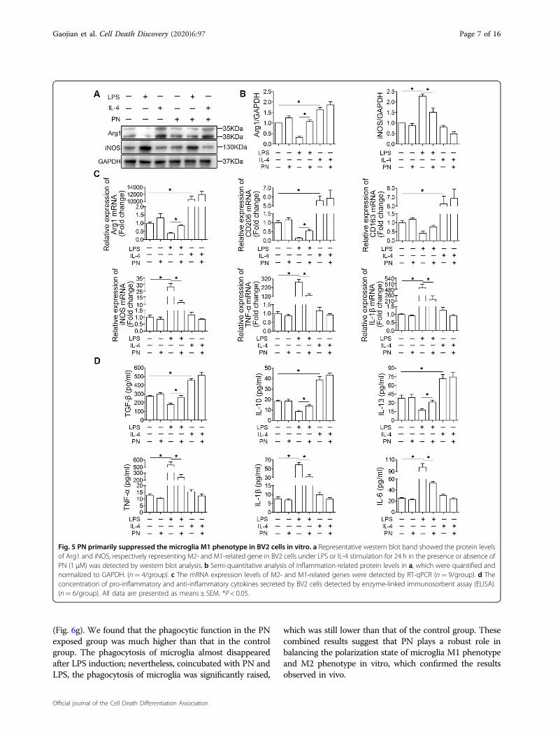
(Fig. 6g). We found that the phagocytic function in the PNexposed group was much higher than that in the controlgroup. The phagocytosis of microglia almost disappearedafter LPS induction; nevertheless, coincubated with PN andLPS, the phagocytosis of microglia was significantly raised,
which was still lower than that of the control group. Thesecombined results suggest that PN plays a robust role inbalancing the polarization state of microglia M1 phenotypeand M2 phenotype in vitro, which confirmed the resultsobserved in vivo.
Fig. 5 PN primarily suppressed the microglia M1 phenotype in BV2 cells in vitro. a Representative western blot band showed the protein levelsof Arg1 and iNOS, respectively representing M2- and M1-related gene in BV2 cells under LPS or IL-4 stimulation for 24 h in the presence or absence ofPN (1 μM) was detected by western blot analysis. b Semi-quantitative analysis of inflammation-related protein levels in a, which were quantified andnormalized to GAPDH. (n= 4/group). c The mRNA expression levels of M2- and M1-related genes were detected by RT-qPCR (n= 9/group). d Theconcentration of pro-inflammatory and anti-inflammatory cytokines secreted by BV2 cells detected by enzyme-linked immunosorbent assay (ELISA).(n= 6/group). All data are presented as means ± SEM. *P < 0.05.
Gaojian et al. Cell Death Discovery (2020) 6:97 Page 7 of 16
Official journal of the Cell Death Differentiation Association
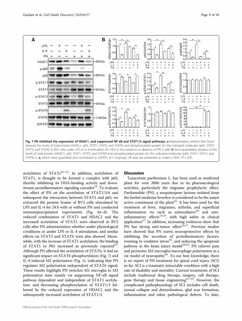
PN reduced the expression of HDAC1, and suppressed NF-κB and STAT1/3 signal pathways in BV2 cells stimulatedwith LPSAccording to previous studies13,18, PN could suppress
NF-κB signal pathway and specifically induce the degra-dation of HDAC1. We then investigated whether PNplayed a role in BV2 cells by western blot, and the resultshowed that PN downregulated the expression of HDAC1and p65, and inhibited p65 phosphorylation under phy-siological conditions, LPS, or IL-4 stimulation withaccompanied by a decrease in p65 protein levels (Fig. 7a,b). Interestingly, we also found a significant increase in theexpression of HDAC1 and p65 after LPS stimulationcompared to the control group. To our knowledge, acti-vation of NF-κB (p65)39,40, STAT139–41 and STAT342
promotes M1 polarization, while STAT6 phosphorylationis involved in the induction of M2 polarization40,43.Therefore, we further explored whether PN affected the
activation of STATs in BV2 cells in vitro. After PNtreatment, only a increase was observed in the expressionof STAT1 and no change in the expression of STAT3 andSTAT6. In contrast with the control group, we observed asignificant increase in the levels of p-STAT1/3 under LPSstimulation, and in the levels of p-STAT6 under IL-4stimulation, respectively. In the presence of PN, the levelsof p-STAT1/3 were significantly decreased under bothLPS and IL-4 stimulation, while STAT6 phosphorylationremains unchanged. Based on these results, we speculatethat PN probably regulates M1/M2 polarization of BV2cells by inhibiting the NF-κB and STAT1/3 signalingpathways.
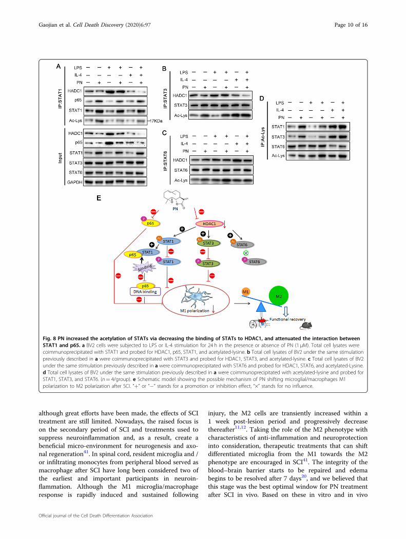
PN regulated the acetylation of STATs and the bindingbetween STAT1 and p65Recently, increasing evidence suggests that the STATs
signal pathway requires HDAC activity via modulating the
Fig. 6 PN inhibited M1 phenotype in BV2 cells in vitro. a, b Representative images of F4/80 (red) and Arg1/iNOS(green) immunohistochemicalstaining of BV2 cells under LPS stimulation for 24 h in the presence or absence of PN (1 μM). (n= 6/group). All cell nuclei were counterstained withDAPI (blue). Scale bar, 200 μm. c, e Representative flow cytometry histograms of Arg1+, CD206+, iNOS+, or CD16+ BV2 cells under the samestimulation previously described in a. d, f Semi-quantitative analysis of Arg1+, CD206+, iNOS+, or CD16+ BV2 cells in c, e. (n= 6/group). All data arepresented as means ± SEM. *P < 0.05. g Representative images of phagocytosis of pHrodo-Green-dye–labeled C. albicansin BV2 cells stained with F4/80 (red) followed the time course. (n= 6/group). Scale bar, 200 μm.
Gaojian et al. Cell Death Discovery (2020) 6:97 Page 8 of 16
Official journal of the Cell Death Differentiation Association
acetylation of STATs44–53. In addition, acetylation ofSTAT1, is thought to be formed a complex with p65,thereby inhibiting its DNA-binding activity and down-stream proinflammatory signaling cascades54. To evaluatethe effect of PN on the acetylation of STAT1/3/6 andsubsequent the interaction between STAT1 and p65, weextracted the protein lysates of BV2 cells stimulated byLPS and IL-4 for 24 h with or without PN and conductedimmunoprecipitation experiments (Fig. 8a–d). Thereduced combination of STAT1 and HDAC1 and theincreased acetylation of STAT1 were observed in BV2cells after PN administration whether under physiologicalconditions or under LPS or IL-4 stimulation, and similareffects on STAT3 and STAT6 were also showed. Mean-while, with the increase of STAT1 acetylation, the bindingof STAT1 to P65 increased as previously reported54.Although PN affected the acetylation of STAT6, it had nosignificant impact on STAT6 phosphorylation (Fig. 7) andIL-4 induced M2 polarization (Fig. 5), indicating that PNregulates M2 polarization independent of STAT6 signal.These results highlight PN switches M1 microglia to M2polarization state mainly via suppressing NF-κB signalpathway dependent and independent of STAT1 acetyla-tion, and decreasing phosphorylation of STAT1/3 fol-lowed by the reduced expression of HDAC1 and thesubsequently increased acetylation of STAT1/3.
DiscussionTanacetum parthenium L. has been used as medicinal
plant for over 2000 years due to its pharmacologicalactivities, particularly the migraine prophylactic effect.Parthenolide (PN), a sesquiterpene lactone isolated fromthe herbal medicine feverfew is considered to be the majoractive constituent of the plant55. It has been used for thetreatment of fever, migraines, arthritis, and superficialinflammation via such as antioxidative56 and anti-inflammatory effects14,18, with high safety in clinicalapplication57. In addition, increasing evidences show thatPN has strong anti-tumor effect16,17. Previous studieshave showed that PN exerts neuroprotective effects byinhibiting the secretion of proinflammatory factors,resisting to oxidative stress58, and reducing the apoptosispathway in the brain injury model58,59. PN relieves painand promotes M2 microglia/macrophage polarization in arat model of neuropathy60. To our best knowledge, thereis no report of PN treatment for spinal cord injury (SCI)so far. SCI is a traumatic intractable condition with a highrate of disability and mortality. Current treatments of SCIinclude traditional drug therapy, surgery, cell therapy,gene therapy and tissue engineering61–63. However, thecomplicated pathophysiology of SCI includes cell death,axonal collapse and demyelination, glial scar formation,inflammation and other pathological defects. To date,
Fig. 7 PN inhibited the expression of HDAC1, and suppressed NF-κB and STAT1/3 signal pathways. a Representative western blot bandshowed the levels of total protein (HDAC1, p65, STAT1, STAT3, and STAT6) and phosphorylated protein for the indicated molecules (p65, STAT1,STAT3, and STAT6) in BV2 cells under LPS or IL-4 stimulation for 24 h in the presence or absence of PN (1 μM). b Semi-quantitative analysis of thelevels of total protein (HDAC1, p65, STAT1, STAT3, and STAT6) and phosphorylated protein for the indicated molecules (p65, STAT1, STAT3, andSTAT6) in a, which were quantified and normalized to GAPDH. (n= 4/group). All data are presented as means ± SEM. *P < 0.05.
Gaojian et al. Cell Death Discovery (2020) 6:97 Page 9 of 16
Official journal of the Cell Death Differentiation Association
although great efforts have been made, the effects of SCItreatment are still limited. Nowadays, the raised focus ison the secondary period of SCI and treatments used tosuppress neuroinflammation and, as a result, create abeneficial micro-environment for neurogenesis and axo-nal regeneration41. In spinal cord, resident microglia and /or infiltrating monocytes from peripheral blood served asmacrophage after SCI have long been considered two ofthe earliest and important participants in neuroin-flammation. Although the M1 microglia/macrophageresponse is rapidly induced and sustained following
injury, the M2 cells are transiently increased within a1 week post-lesion period and progressively decreasethereafter11,12. Taking the role of the M2 phenotype withcharacteristics of anti-inflammation and neuroprotectioninto consideration, therapeutic treatments that can shiftdifferentiated microglia from the M1 towards the M2phenotype are encouraged in SCI41. The integrity of theblood–brain barrier starts to be repaired and edemabegins to be resolved after 7 days20, and we believed thatthis stage was the best optimal window for PN treatmentafter SCI in vivo. Based on these in vitro and in vivo
Fig. 8 PN increased the acetylation of STATs via decreasing the binding of STATs to HDAC1, and attenuated the interaction betweenSTAT1 and p65. a BV2 cells were subjected to LPS or IL-4 stimulation for 24 h in the presence or absence of PN (1 μM). Total cell lysates werecoimmunoprecipitated with STAT1 and probed for HDAC1, p65, STAT1, and acetylated-lysine. b Total cell lysates of BV2 under the same stimulationpreviously described in a were coimmunoprecipitated with STAT3 and probed for HDAC1, STAT3, and acetylated-lysine. c Total cell lysates of BV2under the same stimulation previously described in a were coimmunoprecipitated with STAT6 and probed for HDAC1, STAT6, and acetylated-Lysine.d Total cell lysates of BV2 under the same stimulation previously described in a were coimmunoprecipitated with acetylated-lysine and probed forSTAT1, STAT3, and STAT6. (n= 4/group). e Schematic model showing the possible mechanism of PN shifting microglial/macrophages M1polarization to M2 polarization after SCI. “+” or “−” stands for a promotion or inhibition effect, “×” stands for no influence.
Gaojian et al. Cell Death Discovery (2020) 6:97 Page 10 of 16
Official journal of the Cell Death Differentiation Association
results, we formally found PN for the first time to pro-mote axon regeneration and neurological functionrecovery by modulating the M1/M2 polarization state ofmicroglia/macrophage known as pro-inflammatory phe-notype and anti-inflammatory phenotype, respectively,which will provide a new drug option for the treatment ofspinal cord injury. Experimental evidence shows that M2microglia could reverse the detrimental effects of M1microglia, and promote remyelination64,65 through upre-gulation of Insulin-like growth factor (IGF)-166 ordecreasing production of typical toxic M1 molecules suchas TNF-α67. This indicates that an M1 to M2 switch inmicroglia/macrophage phenotype protects frominflammation-induced demyelination and improve neu-rological function after SCI65, which is consistent with thefinding in this study.Astrocytes, the most abundant resident cells in the
CNS, play a crucial role in SCI pathology through aphenotypic change known as reactive astrogliosis. In theearly stage of SCI, reactive astrocytes migrate to the lesionepicenter and seclude inflammatory cells, leading to tissuerepair and functional improvement68. However, astrocyticscars with the accumulation of CSPG produced byastrocytes31–33 have been shown to be the main impedi-ment to CNS axonal regeneration, resulting in limitedfunctional recovery in the chronic phase of SCI27–30. Ourresearch showed that PN could attenuate the hyperplasiaof astrocytic scar and inhibit the expression of CSPG,which was also a possible role for axon regeneration.Recently, reactive astrocytes were further classified intoA1 astrocytes and A2 astrocytes according to their func-tions induced by neuroinflammation and ischemia,respectively35. After central nervous system (CNS) injury,A1 astrocytes can secrete neurotoxins that induce rapiddeath of neurons and oligodendrocytes, whereas A2astrocytes promote neuronal survival and tissue repair bysecreting by several trophic factors32,35. As well asreleasing a potent neurotoxin, A1 astrocytes were less ableto promote the formation of new synapses, and caused adecrease in the excitatory function of CNS neurons35.Recently more and more researches indicate that A1astrocytes may encourage the development of neurode-generative diseases, such as Alzheimer’s disease (AD),Huntington’s disease, Parkinson’s disease, amyotrophiclateral sclerosis, and multiple sclerosis (MS), and block ofA1 astrocyte conversion by microglia is neuroprotec-tive35,69–71. The A2 astrocyte-related gene S100A10 isessential for cell proliferation, membrane repair, andinhibition of cell apoptosis35. Moreover, A2 astrocytespromote the expression of anti-inflammatory cytokineTGF-β, which participates in synaptogenesis and plays aneuroprotective role32. Our previous studies have shownthat after SCI, inflammatory infiltration activates a newtype of A1 neurotoxic reactive astrocytes, and inhibiting
their activation can promote the recovery of nerve func-tion72,73. Considering that type IL-1α, TNF, and C1q,known as typical proinflammatory cytokines producedfrom M1 phenotype of microglia40,74 were inducers of A1astrocytes, and they were secreted by activated microgliaunder LPS stimulation35, which is contributed to micro-glia M1 polarization40, we speculate that the activation ofM1 microglia is the key to the activation of A1 astrocytes.This study found that PN could inhibit the activation ofA1 astrocytes probably via switching microglia/macro-phage M1 phenotype to M2 phenotype, but othermechanisms cannot be ruled out, such as direct restraintof STAT3 activation in astrocytes, which plays a vital rolein differentiation of A1 astrocytes73.Previous studies13,18 reported that PN specifically
downregulates the expression of HDAC1 and can inhibitthe NF-κB signaling pathway, but the mechanism bywhich PN regulates microglia/macrophage M1/M2polarization has not been clearly elucidated. Our studyfound that PN significantly inhibited the LPS-induced NF-κB signaling pathway in BV2 cells, and its activation isbelieved to promote M1 polarization39,40. In addition, PNinhibited the expression of HDAC1 in BV2 cells asreported18, whether under physiological conditions orunder LPS or IL-4 induced condition. Here, it wasreported that restraining activity of HDAC1 facilitatesM1-to-M2 shift of microglia75,76. As far as we know,HDAC isoforms (HDACs) are divided into four cate-gories: class I HDACs (1, 2, 3 and 8), class II HDACs (4–7,9 and 10), class III sirtuins (1–7) and class IV (HDAC11).Moreover, Lcardi et al.45 believe that HDACs control theactivation of STATs, which are the main molecules thatmodulate M1/M2 polarization through their own deace-tylation. HDACs inhibitors, such as trichostatin A (TSA)or valproic acid (VPA) increase acetylation of STAT1correlating with inactivation of STAT148 possibly viacatalyzing dephosphorylation of STAT1, known as aphosphorylation-acetylation switch mechanism49. It hasbeen suggested that acetylated STAT1 interacts with NF-κB (p65) and consequently inhibits NF-κB signal path-way54. In this study, it was observed that increased acet-ylation of STAT1 was accompanied by its decreasedphosphorylation and improved the association with p65,via reducing the binding of HDAC1 to STAT1 after PNadministration, which suggests that PN plays an impor-tant role in LPS-induced STAT1 activation and indirectregulation of NF-κB signal by downregulating HDAC1expression. Treatment of diffuse large B-cell lymphoma(DLBCL) with the HDAC inhibitor panobinostat(LBH589) results in STAT3 hyperacetylation, depho-sphorylation and increased nuclear export, inhibiting thetranscription of STAT3-responsive anti-apoptotic genesand the survival of DLBCL cells44. Consistently, admin-istration of Trichostatin A (TSA) as inhibition of HDACs
Gaojian et al. Cell Death Discovery (2020) 6:97 Page 11 of 16
Official journal of the Cell Death Differentiation Association
is also shown to increase STAT3 acetylation while sup-pressing phosphorylation on Tyr705 and cell prolifera-tion50. And, we found that the effect of PN on STAT3activation similar to previous reports above44,50 wasconsistent with its regulation on STAT1. Yang et al.53
consider that TSA also leads to a increase in the acet-ylation of STAT6, and inhibits the activation ofSTAT6 signaling pathway during mouse dendritic celldifferentiation. Unlike STAT1/3, the biological con-sequence of STAT6 acetylation and the relationshipbetween its acetylation and phosphorylation are notknown at present. Although PN could increase the acet-ylation of STAT6, it did not affect the phosphorylation ofSTAT6, which may be related to the activation of its site-specific lysine acetylation by PN, considering that differ-ent acetylation sites phosphorylate STAT1/3 differently52.Thus, the acetylation sites in STAT6 and correspondingbiological functions need to explore further. In vitroresults suggested that PN could significantly reduced M1polarization in BV2 cells and partially rescued thedecrease in the expression of M2 phenotype markers ofmicroglia/macrophage induced by LPS, but no significanteffect on M2 polarization stimulated with IL-4 wasobserved. Combined with the above results, we speculatethat PN facilitates shift from M1 to M2 polarizationof microglia/macrophage during SCI by regulatingNF-κB signal directly or indirectly and the activation ofSTAT1/3.In summary, our study showed that PN could shift
microglia/macrophages from M1 to M2 phenotype, whichcan inhibit the activation of NF-κB and STAT1/3 signal-ing pathway. The present study provides the first evidencefor the critical role of the PN in promoting axonalregeneration, increasing myelin reconstitution, reducingCSPG formation, inhibiting scar hyperplasia, suppressingthe transformation of A1 astrocytes and consequently,improving functional behavioral recovery after acute SCIby regulating microglia/macrophages polarization.
Methods and materialsReagents and antibodiesThe Parthenolide (PN) was purchased from MCE
(Monmouth Junction, NJ, USA). The microglial activatorlipopolysaccharide (LPS) was purchased from Sigma-Aldrich (St. Louis, MO, USA). Recombinant mouse IL-4was obtained from Abcam (Cambridge, UK). Primaryantibodies used in this study included rabbit anti-INOS(Abcam), rabbit anti-Arginase (Santa Cruz, Dallas, Texas,USA), mouse anti-F4/80 (Abcam), mouse anti-Neurofilament (Abcam), mouse anti-chondroitin sulfate(Abcam), rabbit anti-GFAP (Abcam), rabbit anti-MBP(Cell Signaling Technology, Danvers, MA, USA), rabbitanti-NF-κB p65 (Cell Signaling Technology), rabbit anti-p-p65 (Cell Signaling Technology), rabbit anti-C3
(Abcam), rabbit anti-GAP43 (Abcam), mouse anti-STAT1 (Santa Cruz), mouse anti-p-STAT1 (SantaCruz), mouse anti-STAT3 (Cell Signaling Technology),mouse anti-p-STAT3 (Cell Signaling Technology), mouseanti-STAT6 (Santa Cruz), mouse anti-p-STAT6 (SantaCruz), rabbit anti-HDAC1 (Santa Cruz), and rabbit anti-acetyl Lysine (Santa Cruz). Alexa Fluor 488 and 594conjugated to goat anti-mouse IgG (H+L) or goat anti-rabbit IgG for immunofluorescence staining wereobtained from Jackson ImmunoResearch (West Grove,PA, USA). Horseradish peroxidase (HRP)-conjugated goatanti-rabbit IgG (H+L) and HRP-conjugated goat anti-mouse IgG (H+L) for immunoblotting were purchasedfrom Invitrogen (Carlsbad, CA, USA). Protein A/GMagnetic Beads for co-immunoprecipitation (co-IP) wereobtained from Pierce Biotechnology (Rockford, IL, USA).The DeadEnd™ Fluorometric TUNEL System was pur-chased from Promega (Madison, WI, USA). ELISA kits forTNF-α, IL-1β, IL-6, IL-13, IL-10 and TGF-β were pur-chased from Abcam. The antibodies used for flow cyto-metry were: PE-Cyanine7-labeled anti-mouse Arg1mAb (eBiosciences, San Diego, CA), PE-labeled anti-mouse CD206 mAb (eBiosciences), FITC-labeledanti-mouse CD16 mAb (eBiosciences), FITC-labeledanti-mouse INOS mAb (BD Biosciences, San Jose,CA, USA).
Preparation of the contusive SCI mouse modelAll animal procedures were conducted according to the
Guidelines for the Care and Use of Laboratory Animals(National Institutes of Health) and approved by the Ani-mal Care and Use Committee of Nanjing Medical Uni-versity. Briefly, 16 male C57BL/6J mice (18–35 g; 8 weeksold) were first deeply anesthetized by intraperitonealinjection of pentobarbital sodium (50 mg/kg of bodyweight). The hair on the back of the mice was shaved offand 75% alcohol was used to disinfect the skin. The ver-tebral column was carefully exposed, followed by excisionof the T9 lamina. The spine was stabilized by clampingthe T7 and T11 spinous processes. After the dorsal sur-face of the cord was fully exposed, a rod (1.3 mm in dia-meter; RWD Life Science Corp., C4p01-001, China) wasused to compress the spinal cord with a force of 50 kdynesand a dwell time of 10 s. After the moderate SCI modelwas created, the muscles and the skin were quicklysutured. The bladders of mice were manually and gentlymassaged three times a day to avoid retention of urineuntil the reflexive control of micturition was restored.Mice fulfilling these SCI models were randomly dividedinto two groups, a SCI-only group and a treatment group(n= 8/group). The SCI-only group served as the control(n= 8/group). PN was intraperitoneally injected at a doseof 2 mg/kg daily referred to previous reports77,78 for7 days in the treatment group for subsequent analysis.
Gaojian et al. Cell Death Discovery (2020) 6:97 Page 12 of 16
Official journal of the Cell Death Differentiation Association
Tissue processingMice were sacrificed with an overdose of pentobarbital
sodium (80 mg/kg, intraperitoneally) on day 3, 7, and 14post-injury. The hearts of mice were immediately per-fused by 10ml ice-cold physiological saline followed byice-cold physiological saline plus paraformaldehyde (4%,w/v). The incision of SCI was opened and segments ofspinal cord containing the lesion site were extracted.Spinal cord samples were then fixed in paraformaldehyde(4%, w/v) at 4 °C for 24 h, dehydrated in a sucrose-PBSgradient (20 and 30%, w/v). Finally, the samples wereembedded in OCT, and cut into serial longitudinal sec-tions of about 10 µm. All sections were stored at −80 °Cuntil processed for immunostaining.
Assessment of locomotor capacityThe motor function after SCI was quantified at 1, 3, 7,
14, 21, and 28 day after surgery using the Basso MouseScale (BMS) for locomotion. Scoring ranged from 0 forcomplete paraplegia to 9 for normal function.
Footprint analysisGait and motor coordination were assessed at 3, 14 and
28 day after surgery. The front and rear paws were paintedwith different colors of dye. The mouse was then placedon a piece of blotting paper surrounded by a cage toencourage it to walk in a straight line. The footprintpattern was digitized and a representative picture wasused to assess coordination.
BV2 cell culture and treatmentThe BV2 microglial cell line was purchased from the
Cell Bank of the Chinese Academy of Science (Shanghai,China). Briefly, the BV2 cells were cultured in Dulbecco’smodified Eagle’s medium (DMEM, Thermo Fisher Sci-entific, Waltham, MA, USA) supplied with 10% fetalbovine serum (FBS, Thermo Fisher Scientific), 100 units/ml penicillin, and 100 μg/ml streptomycin (Thermo FisherScientific) at 37 °C with 5% CO2. The complete DMEMmedium was changed once a day and cells were passedevery 3 days. When the BV2 cells grew to a confluence ofabout 80%, the medium was replaced with serum-freeDMEM, and incubated with LPS (100 ng/ml)/IL-4 (20 ng/ml) with or without PN (1 μM) according to CCK8 resultfor 24 h. Then the BV2 cells were used for furtherexperiments.
Cell viabilityPrimary neurons were prepared from WT C57/BL6
embryonic day 15 mice according to an established pro-tocol24. Viability of primary neurons and BV2 cells asevaluated with a Cell Counting Kit-8 (CCK-8) assay(Dojindo, Kumamoto, Japan) to examine the effect of PNon viability assessment. After 0, 6, 24, and 48 h of
incubation with different PN concentration, Wells wererinsed 3 times with PBS, and CCK8 solution (10 ml; 1:10dilution) in fresh culture medium was added to wells andincubated for 2 h at 37 °C. The optical absorbance of eachsample at 450 nm was measured using a microplate reader(ELx800; Bio-Tek, Winooski, VT, USA). Cell viability wasexpressed as the dehydrogenase activity in cells relative tothat in the PBS control group.
Western blot analysisProteins were extracted from cells and treated with
RIPA lysis and extraction buffer (KeyGen Biotechnology,Shanghai, China). Protein concentration was determinedusing the BCA method. Equal amounts of protein wereseparated by SDS-PAGE, transferred to PVDF membranes(EMD Millipore Corp., Burlington, MA), and incubatedovernight at 4 °C with primary antibodies followed byblocking with 5% bovine serum albumin (BSA, KeyGenBiotechnology). Membranes were then incubated for120min at room temperature with the secondary anti-body. Reacting bands were visualized using ECL reagent(Thermo Fisher Scientific), and the density of proteinbands was semiquantified using Image J (National Insti-tutes of Health, Bethesda, MD, USA).
Co-immunoprecipitation (CO-IP)After treatment, the cell samples were lysed in 1ml ice-
cold buffer A (20 mM TrisHCl, pH 7.4, 150mM NaCl, 1%Triton X-100, 0.5% sodium deoxycholate, 12 mM glycer-ophosphate, 10 mM sodium fluoride, 5 mM EGTA, 2 mMsodium vanadate, 1 mM PMSF, 2 mg/ml aprotinin, and2mg/ml leupeptin) for 30 min at 4 °C. Lysates were thencentrifuged at 14,000 rpm for 15min to collect thesupernatants. The supernatants were immunoprecipitatedwith the indicated antibodies overnight at 4 °C usingprotein A/G Magnetic Beads. The beads were rinsed threetimes with buffer A and the eluent proteins were sub-jected to 12% SDS-PAGE for separation and transferred topolyvinylidene difluoride (PVDF) membranes for immu-noblotting as described in western blot analysis.
Immunofluorescence stainingSpinal cord tissues or cultured BV2 cells were fixed with
precooled 4% paraformaldehyde for 20min at 4 °C, thenpermeabilized with 0.2% Triton X-100 for 20min, andblocked with 10% normal goat serum for 0.5 h. Fixedtissues and cells were then incubated at 4 °C overnightwith primary antibodies, followed by Alexa Fluor 488–and Alexa Flour 594–conjugated goat secondary antibody(Jackson ImmunoResearch) for 1 h at room temperature.After triple washing by PBS, nuclei were stained withDAPI (Thermo Fisher Scientific) and fluorescent imageswere acquired using an epifluorescence microscope(AxioVertA1 and ImagerA2). The lesion volume was
Gaojian et al. Cell Death Discovery (2020) 6:97 Page 13 of 16
Official journal of the Cell Death Differentiation Association
obtained by the sum of total lesion according to GFAP+
lesion cavity area multiplied by distance (about 200 μm)between the sections.
Phagocytosis assayPHrodo-Green-dye–labeled C. albicans (Invitrogen,
Carlsbad, CA, USA) were used to access the phagocytosisability of BV2 cells at the presence or absence of PN.Briefly, 2 × 10⁶ BV2 cells were seeded in each well of 24-well plates. After the cells adhere to the wall, the mediumwas replaced with serum-free DMEM, and incubated withLPS (100 ng/ml) with or without PN (1 μM) for 24 h.Then the supernatant was replaced by a suspension ofpHrodo-labeled C. albicans at a 1:5 ratio (progenitor:yeasts) in 1 ml serum-free DMEM for the indicateddurations. At indicated time points, the C. albicans sus-pension was removed and the cells were rinsed with PBSfor 3 times. Then the cells were fixed, permeabilized andblocked, and incubated with primary antibody of F4/80followed by Alexa Flour 594–conjugated goat secondaryantibody (Jackson). The mean green fluorescence inten-sity of engulfed bacteria was estimated by an epi-fluorescence microscope (AxioVertA1 and ImagerA2).
Flow cytometry (FCM) analysisAfter incubation with LPS (100 ng/ml)/IL-4 (20 ng/ml)
with or without PN (1 μM) for 24 h, the rate of BV2 cellstransition to different polarized phenotypes was measuredby flow cytometry. Briefly, the culture medium was dis-carded followed by three washes with PBS. BV2 cells weredigested using 0.25% trypsin to prepare a single-cell sus-pension. The suspension was then incubated with indi-cated antibodies at 4 °C under darkness for 30 min. Cellswere harvested by centrifugation at 1500 rpm for 5 min,and washed twice with PBS. Finally, the suspension wasanalyzed by flow cytometry (FACSCalibur, BD Bios-ciences, Franklin Lakes, NJ, USA).
Real-time reverse transcription polymerase chain reaction(RT-qPCR)Total RNA was extracted from BV2 cells after different
treatments using Trizol Reagent (Invitrogen) according tothe manufacturer’s instructions. The concentrations oftotal RNA were measured by a Biometra Optical Ther-mocycler (Analytik Jena, Goettingen, Germany). RNA(500 ng) was reverse transcribed into cDNA using theHigh Capacity cDNA Reverse Transcription Kit (ThermoFisher Scientific) according to the manufacturer’sinstruction. The primer sequences used for qPCRamplification were:TNF-α: 5′-CCCTCACACTCAGATCATCTTCT-3′ (forward)5′-GCTACGACGTGGGCTACAG-3′ (reverse).IL-1β: 5′-GCAACTGTTCCTGAACTCAACT-3′ (forward)5′-ATCTTTTGGGGTCCGTCAACT-3′ (reverse).
iNOS: 5′-CTCCTTCAAAGAGGCAAAAATA-3′ (forward)5′-CACTTCCTCCAGGATGTTGT-3′ (reverse).Arg-1: 5′-CCTGGAACTGAAAGGAAAG-3′ (forward)5′-TTGGCAGATATGCAGGGAGT-3′ (reverse).CD206: 5′-CTGCAGATGGGTGGGTTATT-3′ (forward)5′-GGCATTGATGCTGCTGTTATG-3′ (reverse).CD163: 5′-GCC ATA ACT GCA GGC ACA AA-3′
(forward)5′-GTT GGT CAG CCT CAG AGA CA-3′ (reverse).GAPDH: 5′-TGTGATGGGTGTGAACCACG-3′ (forward)5′-CAGTGAGCTTCCCGTTCACC-3′ (reverse).Quantitative real-time PCR was performed using SYBR
qRCR premix (Takara, Kyoto, Japan) according to themanufacturer’s instructions. Cycling conditions were aninitial denaturation at 95 °C for 30 s, followed by 40 cyclesat 95 °C for 5 s, 60 °C for 30 s, and 72 °C for 10 min. Targetgene expression was normalized to GAPDH expression(internal control) using the ΔΔCT method.
ELISAThe concentrations of TNF-α, IL-1β, IL-6, IL-13, IL-
10, TGF-β were measured in BV2 culture media afterindicated treatments using ELISA kits according to themanufacturers’ protocols. Briefly, the culture medium ofBV2 cells was collected and centrifuged at 2000 g for10 min to remove debris. 50 µl of all samples or standardwas added to appropriate wells followed by 50 µl of theAntibody Cocktail to each well. The samples wereincubated for 1 h at room temperature on a plate shakerat 400 rpm. Then all wells were washed three times with1×Wash Buffer PT followed by 100 µl/well of TMBsubstrate and incubated under darkness for 10 min.Finally, STOP Solution was added and absorbance wasread at 450 nm wavelengths using a microplate reader(ELx800, Bio-Tek, USA).
Nissl staining of spinal cord slicesThe cytosolic Nissl substance in spinal cord sections
was stained with cresol violet on the 28th day after sur-gery. In brief, the slices were washed with distilled waterand stained in a cresol violet solution for 10min. Afterrinsing with distilled water, the slices were differentiatedwith 95% ethanol, rinsed with xylene, and fixed withneutral balsam. Regions of traumatic injury were identi-fied by severe tissue destruction or staining loss. Six Nissl-stained sections by distance (about 200 μm) between thesections were selected to estimate the average number ofresidual anterior horn motor neurons and the propor-tional lesion size.
Statistical analysesResults are expressed as mean ± SEM of at least three
independent experiments. Means were compared by one-way ANOVA followed by Bonferroni’s post hoc tests for
Gaojian et al. Cell Death Discovery (2020) 6:97 Page 14 of 16
Official journal of the Cell Death Differentiation Association
multiple comparisons (SPSS 20; SPSS, Chicago, IL, USA).A p < 0.05 (two tailed) was regarded as significant forall tests.
AcknowledgementsThis study was supported by grants from the National Natural ScienceFoundation of China [81902211], the Medical Science and technologydevelopment Foundation from Nanjing Department of Health [YKK17060], theHigh Level Innovation Team [JX102GSP201727], and the Jiangsu NaturalScience Foundation [BK20191061].
Author details1Department of Anesthesiology, The First Affiliated Hospital of Nanjing MedicalUniversity, Nanjing 210029, China. 2Department of Pain Management, AffiliatedDrum Tower Hospital, Medical School of Nanjing University, Nanjing 210008,China. 3Department of Orthopedic, The First Affiliated Hospital of NanjingMedical University, Nanjing 210029, China. 4Department of Orthopedic, TheThird Affiliated Hospital of Soochow University, Changzhou 213003, China
Data availabilityThe data that support the findings of this study are available from thecorresponding author upon reasonable request.
Conflict of interestThe authors declare that they have no conflict of interest.
Publisher’s noteSpringer Nature remains neutral with regard to jurisdictional claims inpublished maps and institutional affiliations.
The online version of this article (https://doi.org/10.1038/s41420-020-00333-8)contains supplementary material, which is available to authorized users.
Received: 20 August 2020 Accepted: 11 September 2020
References1. McDonald, J. W. & Sadowsky, C. Spinal-cord injury. Lancet 359, 417–425 (2002).2. Ahuja, C. S. et al. Traumatic spinal cord injury. Nat. Rev. Dis. Prim. 3, 17018
(2017).3. Zhou, X., He, X. & Ren, Y. Function of microglia and macrophages in secondary
damage after spinal cord injury. Neural Regen. Res. 9, 1787–1795 (2014).4. Anwar, M. A., Al Shehabi, T. S. & Eid, A. H. Inflammogenesis of secondary spinal
cord injury. Front. Cell. Neurosci. 10, 98 (2016).5. Noristani, H. N. et al. RNA-Seq analysis of microglia reveals time-dependent
activation of specific genetic programs following spinal cord injury. Front. Mol.Neurosci. 10, 90 (2017).
6. David, S. & Kroner, A. Repertoire of microglial and macrophage responses afterspinal cord injury. Nat. Rev. Neurosci. 12, 388–399 (2011).
7. Bellver-Landete, V. et al. Microglia are an essential component of the neuro-protective scar that forms after spinal cord injury. Nat. Commun. 10, 518(2019).
8. Schmitz, G., Leuthauser-Jaschinski, K. & Orso, E. Are circulating monocytes asmicroglia orthologues appropriate biomarker targets for neuronal diseases?Cent. Nerv. Syst. Agents Med. Chem. 9, 307–330 (2009).
9. Lawrence, T. & Natoli, G. Transcriptional regulation of macrophage polar-ization: enabling diversity with identity. Nat. Rev. Immunol. 11, 750–761(2011).
10. Tang, Y. & Le, W. Differential roles of M1 and M2 microglia in neurodegen-erative diseases. Mol. Neurobiol. 53, 1181–1194 (2016).
11. Kigerl, K. A. et al. Identification of two distinct macrophage subsets withdivergent effects causing either neurotoxicity or regeneration in the injuredmouse spinal cord. J. Neurosci. 29, 13435–13444 (2009).
12. Kroner, A. et al. TNF and increased intracellular iron alter macrophage polar-ization to a detrimental M1 phenotype in the injured spinal cord. Neuron 83,1098–1116 (2014).
13. Kwok, B. H. et al. The anti-inflammatory natural product parthenolide from themedicinal herb Feverfew directly binds to and inhibits IkappaB kinase. Chem.Biol. 8, 759–766 (2001).
14. Hehner, S. P., Hofmann, T. G., Droge, W. & Schmitz, M. L. The antiinflammatorysesquiterpene lactone parthenolide inhibits NF-kappa B by targeting the Ikappa B kinase complex. J. Immunol. 163, 5617–5623 (1999).
15. Carlisi, D. et al. Parthenolide sensitizes hepatocellular carcinoma cells to TRAILby inducing the expression of death receptors through inhibition of STAT3activation. J. Cell. Physiol. 226, 1632–1641 (2011).
16. Zhao, X., Liu, X. & Su, L. Parthenolide induces apoptosis via TNFRSF10B andPMAIP1 pathways in human lung cancer cells. J. Exp. Clin. Cancer Res. 33, 3(2014).
17. Nakshatri, H. et al. NF-kappaB-dependent and -independent epigeneticmodulation using the novel anti-cancer agent DMAPT. Cell. Death. Dis. 6,e1608 (2015).
18. Gopal, Y. N., Arora, T. S. & Van Dyke, M. W. Parthenolide specifically depleteshistone deacetylase 1 protein and induces cell death through ataxia tel-angiectasia mutated. Chem. Biol. 14, 813–823 (2007).
19. Guzman, M. L. et al. The sesquiterpene lactone parthenolide induces apoptosisof human acute myelogenous leukemia stem and progenitor cells. Blood 105,4163–4169 (2005).
20. Figley, S. A. et al. Characterization of vascular disruption and blood-spinal cordbarrier permeability following traumatic spinal cord injury. J. Neurotrauma 31,541–552 (2014).
21. Hesp, Z. C. et al. Chronic oligodendrogenesis and remyelination after spinalcord injury in mice and rats. J. Neurosci. 35, 1274–1290 (2015).
22. Zhang, S. et al. Inhibition of epidermal growth factor receptor improvesmyelination and attenuates tissue damage of spinal cord injury. Cell Mol.Neurobiol. 36, 1169–1178 (2016).
23. Petzold, A. Neurofilament phosphoforms: surrogate markers for axonal injury,degeneration and loss. J. Neurol. Sci. 233, 183–198 (2005).
24. Chen, J. et al. GPCR kinase 2-interacting protein-1 protects against ischemia-reperfusion injury of the spinal cord by modulating ASK1/JNK/p38 signaling.FASEB J. 32, 6833–6847 (2018).
25. Liu, D. et al. Biodegradable spheres protect traumatically injured spinal cord byalleviating the glutamate-induced excitotoxicity. Adv. Mater. 30, e1706032(2018).
26. Hara, M. et al. Interaction of reactive astrocytes with type I collagen inducesastrocytic scar formation through the integrin-N-cadherin pathway after spinalcord injury. Nat. Med. 23, 818–828 (2017).
27. Stichel, C. C. & Muller, H. W. The CNS lesion scar: new vistas on an oldregeneration barrier. Cell. Tissue Res. 294, 1–9 (1998).
28. Fawcett, J. W. Overcoming inhibition in the damaged spinal cord. J. Neuro-trauma 23, 371–383 (2006).
29. Horner, P. J. & Gage, F. H. Regenerating the damaged central nervous system.Nature 407, 963–970 (2000).
30. Anderson, M. A. et al. Astrocyte scar formation aids central nervous systemaxon regeneration. Nature 532, 195–200 (2016).
31. Wu, D. et al. Expressing constitutively active Rheb in adult neurons after acomplete spinal cord injury enhances axonal regeneration beyond achondroitinase-treated glial scar. J. Neurosci. 35, 11068–11080 (2015).
32. Cregg, J. M. et al. Functional regeneration beyond the glial scar. Exp. Neurol.253, 197–207 (2014).
33. Silver, J. & Miller, J. H. Regeneration beyond the glial scar. Nat. Rev. Neurosci. 5,146–156 (2004).
34. Avnur, Z. & Geiger, B. Immunocytochemical localization of native chondroitin-sulfate in tissues and cultured cells using specific monoclonal antibody. Cell38, 811–822 (1984).
35. Liddelow, S. A. et al. Neurotoxic reactive astrocytes are induced by activatedmicroglia. Nature 541, 481–487 (2017).
36. Durafourt, B. A. et al. Comparison of polarization properties of human adultmicroglia and blood-derived macrophages. Glia 60, 717–727 (2012).
37. Ji, J. et al. Antagonizing peroxisome proliferator-activated receptor gammafacilitates M1-to-M2 shift of microglia by enhancing autophagy via the LKB1-AMPK signaling pathway. Aging Cell. 17, e12774 (2018).
38. Maneu, V. et al. Dectin-1 mediates in vitro phagocytosis of Candida albicansyeast cells by retinal microglia. FEMS Immunol. Med. Microbiol. 63, 148–150(2011).
39. Chen, S. et al. Valproic acid attenuates traumatic spinal cord injury-inducedinflammation via STAT1 and NF-kappaB pathway dependent of HDAC3. J.Neuroinflammation 15, 150 (2018).
Gaojian et al. Cell Death Discovery (2020) 6:97 Page 15 of 16
Official journal of the Cell Death Differentiation Association
40. Lan, X. et al. Modulators of microglial activation and polarization after intra-cerebral haemorrhage. Nat. Rev. Neurol. 13, 420–433 (2017).
41. Liu, W. et al. Exosome-shuttled miR-216a-5p from hypoxic preconditionedmesenchymal stem cells repair traumatic spinal cord injury by shiftingmicroglial M1/M2 polarization. J. Neuroinflammation 17, 47 (2020).
42. Fang, Y. Y. & Zhang, J. H. MFG-E8 alleviates oxygen-glucose deprivation-induced neuronal cell apoptosis by STAT3 regulating the selective polarizationof microglia. Int. J. Neurosci. 130, 1–10 (2020).
43. He, Y. et al. IL-4 switches microglia/macrophage M1/M2 polarization andalleviates neurological damage by modulating the JAK1/STAT6 pathway fol-lowing ICH. Neuroscience 437, 161–171 (2020).
44. Gupta, M. et al. Regulation of STAT3 by histone deacetylase-3 in diffuselarge B-cell lymphoma: implications for therapy. Leukemia 26, 1356–1364(2012).
45. Icardi, L., De Bosscher, K. & Tavernier, J. The HAT/HDAC interplay: multilevelcontrol of STAT signaling. Cytokine Growth Factor Rev. 23, 283–291 (2012).
46. Stronach, E. A. et al. HDAC4-regulated STAT1 activation mediates platinumresistance in ovarian cancer. Cancer Res. 71, 4412–4422 (2011).
47. Kramer, O. H. & Heinzel, T. Phosphorylation-acetylation switch in the regulationof STAT1 signaling. Mol. Cell. Endocrinol. 315, 40–48 (2010).
48. Guo, L. et al. Stat1 acetylation inhibits inducible nitric oxide synthaseexpression in interferon-gamma-treated RAW264.7 murine macrophages.Surgery 142, 156–162 (2007).
49. Kramer, O. H. et al. A phosphorylation-acetylation switch regulatesSTAT1 signaling. Genes Dev. 23, 223–235 (2009).
50. Minami, J. et al. Histone deacetylase 3 as a novel therapeutic target in multiplemyeloma. Leukemia 28, 680–689 (2014).
51. Yang, Q. et al. Cross talk between histone deacetylase 4 and STAT6 in thetranscriptional regulation of arginase 1 during mouse dendritic cell differ-entiation. Mol. Cell. Biol. 37, 63–75 (2017).
52. Zhuang, S. Regulation of STAT signaling by acetylation. Cell. Signal. 25,1924–1931 (2013).
53. Yang, Q. et al. Cross talk between histone deacetylase 4 and STAT6 in thetranscriptional regulation of arginase 1 during mouse dendritic cell differ-entiation. Mol. Cell. Biol. 35, 63–75 (2015).
54. Kumar, P. et al. Inhibition of HDAC enhances STAT acetylation, blocks NF-kappaB, and suppresses the renal inflammation and fibrosis in Npr1 haplotypemale mice.Am. J. Physiol. Ren. Physiol. 313, F781–F795 (2017).
55. Vegh, K. et al. Three newly identified lipophilic flavonoids in Tanacetumparthenium supercritical fluid extract penetrating the blood-brain barrier. J.Pharm. Biomed. Anal. 149, 488–493 (2018).
56. Mao, W. & Zhu, Z. Parthenolide inhibits hydrogen peroxideinduced osteoblastapoptosis. Mol. Med. Rep. 17, 8369–8376 (2018).
57. Ghantous, A., Sinjab, A., Herceg, Z. & Darwiche, N. Parthenolide: from plantshoots to cancer roots. Drug. Discov. Today 18, 894–905 (2013).
58. Wang, J. A. et al. Parthenolide ameliorates intracerebral hemorrhage-inducedbrain injury in rats. Phytother. Res. 34, 153–160 (2020).
59. Dong, L. et al. Parthenolide is neuroprotective in rat experimental strokemodel: downregulating NF-kappaB, phospho-p38MAPK, and caspase-1 andameliorating BBB permeability. Mediators. Inflamm. 2013, 370804 (2013).
60. Popiolek-Barczyk, K. et al. Parthenolide relieves pain and promotes M2microglia/macrophage polarization in rat model of neuropathy. Neural Plast.2015, 676473 (2015).
61. Fan, B. et al. Microenvironment imbalance of spinal cord injury. Cell. Transplant.27, 853–866 (2018).
62. Eckert, M. J. & Martin, M. J. Trauma: spinal cord injury. Surg. Clin. North. Am. 97,1031–1045 (2017).
63. Ahuja, C. S. et al. Traumatic spinal cord injury-repair and regeneration. Neu-rosurgery 80, S9–S22 (2017).
64. Yu, Z. et al. MSX3 switches microglia polarization and protects frominflammation-induced demyelination. J. Neurosci. 35, 6350–6365 (2015).
65. Wu, Q. et al. Riluzole improves functional recovery after acute spinal cordinjury in rats and may be associated with changes in spinal microglia/mac-rophages polarization. Neurosci. Lett. 723, 134829 (2020).
66. Butovsky, O. et al. Microglia activated by IL-4 or IFN-gamma differentiallyinduce neurogenesis and oligodendrogenesis from adult stem/progenitorcells. Mol. Cell. Neurosci. 31, 149–160 (2006).
67. Paintlia, A. S., Paintlia, M. K., Singh, I. & Singh, A. K. IL-4-induced peroxisomeproliferator-activated receptor gamma activation inhibits NF-kappaB transactivation in central nervous system (CNS) glial cells and protects oligoden-drocyte progenitors under neuroinflammatory disease conditions: implicationfor CNS-demyelinating diseases. J. Immunol. 176, 4385–4398 (2006).
68. Okada, S. et al. Conditional ablation of Stat3 or Socs3 discloses a dual role forreactive astrocytes after spinal cord injury. Nat. Med. 12, 829–834 (2006).
69. Yun, S. P. et al. Block of A1 astrocyte conversion by microglia is neuropro-tective in models of Parkinson’s disease. Nat. Med. 24, 931–938 (2018).
70. Xu, X. et al. MFG-E8 reverses microglial-induced neurotoxic astrocyte (A1) viaNF-kappaB and PI3K-Akt pathways. J. Cell. Physiol. 234, 904–914 (2018).
71. Reichenbach, N. et al. Inhibition of Stat3-mediated astrogliosis amelioratespathology in an Alzheimer’s disease model. EMBO. Mol. Med. 11, e9665 (2019).
72. Liu, W. et al. Exosomes derived from bone mesenchymal stem cells repairtraumatic spinal cord injury by suppressing the activation of A1 neurotoxicreactive astrocytes. J. Neurotrauma 36, 469–484 (2019).
73. Qian, D. F. et al. Blocking Notch signal pathway suppresses the activation ofneurotoxic A1 astrocytes after spinal cord injury. Cell. Cycle 18, 3010–3029(2019).
74. Zhou, X. et al. L-cysteine-derived H2S promotes microglia M2 polarization viaactivation of the AMPK pathway in hypoxia-ischemic neonatal mice. Front.Mol. Neurosci. 12, 58 (2019).
75. Ji, J. et al. The intra-nuclear SphK2-S1P axis facilitates M1-to-M2 shift ofmicroglia via suppressing HDAC1-mediated KLF4 deacetylation. Front.Immunol. 10, 1241 (2019).
76. Wang, J. et al. Long noncoding RNA H19 promotes neuroinflammation inischemic. Stroke Driv. Histone Deacetylase 1-Depend. M1 Microglial Polarization.Stroke 48, 2211–2221 (2017).
77. Rummel, C., Gerstberger, R., Roth, J. & Hubschle, T. Parthenolide attenuatesLPS-induced fever, circulating cytokines and markers of brain inflammation inrats. Cytokine 56, 739–748 (2011).
78. Barbier-Torres, L. et al. Histone deacetylase 4 promotes cholestatic liver injuryin the absence of prohibitin-1. Hepatology 62, 1237–1248 (2015).
Gaojian et al. Cell Death Discovery (2020) 6:97 Page 16 of 16
Official journal of the Cell Death Differentiation Association