Original Article Role of Wnt/β-catenin signaling pathway ... · ing pathway is involved in...
12
Int J Clin Exp Pathol 2017;10(2):2351-2362 www.ijcep.com /ISSN:1936-2625/IJCEP0045636 Original Article Role of Wnt/β-catenin signaling pathway in the repair of intestinal mucosa associated with crypt stem cell in a rat model of abdominal compartment syndrome Tong Lu 1* , Huayou Luo 1,2* , Jia Liu 3 , Cheng Chen 1 , Feng Wang 1 , Zongwen Shen 1 , Tonglei Liu 1 , Yujian Zeng 1,2 , Ting-Hua Wang 3# , Kun-Hua Wang 1,2# 1 Department of Gastrointestinal Surgery, First Affiliated Hospital, Kunming Medical University, Kunming , China; 2 Yunnan Institute of Digestive Disease; Engineering Technology Center for Diagnosis and Treatment of Digestive Diseases in Kunming, Kunming , China; 3 Animal Center, Institute of Neuroscience Kunming Medical University, Kunming, China. * Equal contributors. # Equal contributors. Received December 2, 2016; Accepted December 25, 2016; Epub February 1, 2017; Published February 15, 2017 Abstract: The Wnt/β-catenin signaling plays a vital role in the maintenance of intestinal crypt epithelial cell regen- eration, but its role in intestinal epithelial cell after injury is poorly understood. This study investigated the role of Wnt/β-catenin signaling on the regeneration of intestinal crypt stem cells in Abdominal Compartment Syndrome (ACS) rat model. Arterial blood was collected for biochemical analysis and then microscopic observation showed extensive ileal villus damage after prolonged exposure to Intra-abdominal pressure. Moreover, ACS injury increased the mRNA and protein expressions for Wnt-1, β-catenin, transcription factor 4 (TCF4), and c-Myc. Immunofluores- cence staining confirmed the increased proteins of β-catenin and c-Myc in the intestinal crypt stem cells according to morphology. Liver, kidney, heart and other organs were injured after ACS indicated by the blood biochemical analysis, and ACS causes the intestinal mucosal injury, which aggravates with time, and the Wnt/β-catenin signal- ing pathway is involved in intestinal epithelium regeneration, which is associated with crypt stem cells in early stage after ACS injury. Keywords: ACS, Wnt/β-catenin signaling pathway, intestinal crypt stem cell Introduction Abdominal compartment syndrome (ACS) is usually induced by intra-abdominal pressure (IAP) greater than 20 mmHg and results in sev- eral organs dysfunctions, failure, or death [1]. ACS is the most common cause of retroperito- neal bleeding, pancreatitis, intestinal obstruc- tion, tension ascites, peritonitis, and severe visceral edema because of fluid resuscitation. The affected systems include cardiovascular, respiratory, kidney, central nervous system, internal organs, and systemic accumulation [2]. The incidence of ACS in intensive care unit is 5%-12% [3], and the mortality rate of ACS is 63%~75% [4]. Therefore, timely and effective diagnosis and treatment of ACS are some of the important problems faced by clinicians. Studying the pathogenesis to elimination ACS is particularly important in achieving a good therapeutic effect [5]. Increased IAP, which usually induces multiple organ dysfunctions in several systems, can damage the function of the intestinal tract [6]. The intestinal mechani- cal barrier, which is composed of the mucosal epithelium, lamina propria and muscularis mucosa, as well as the intestinal epithelial cells, including absorptive cells, goblet cells, Pan’s cells, stem cells and endocrine cells, are very important in this process. Among these cells, stem cells located in the intestinal crypt region showed a strong capacity to maintain proliferation and differentiation for epithelium repair. Therefore, stem cells play an important role in the intestinal mucosal regeneration and repair [7]. Under the influence of physiological or pathological signals, intestinal stem cells could proliferate and differentiate into absorp- tive cells, goblet cells, Pan’s cells and endo- crine cells. However, the underlying mechanism is yet to be determined. Recent studies showed four main signaling pathways that maintain intestinal stem cells
Transcript of Original Article Role of Wnt/β-catenin signaling pathway ... · ing pathway is involved in...
Int J Clin Exp Pathol 2017;10(2):2351-2362www.ijcep.com /ISSN:1936-2625/IJCEP0045636
Original Article Role of Wnt/β-catenin signaling pathway in the repair of intestinal mucosa associated with crypt stem cell in a rat model of abdominal compartment syndrome
Tong Lu1*, Huayou Luo1,2*, Jia Liu3, Cheng Chen1, Feng Wang1, Zongwen Shen1, Tonglei Liu1, Yujian Zeng1,2, Ting-Hua Wang3#, Kun-Hua Wang1,2#
1Department of Gastrointestinal Surgery, First Affiliated Hospital, Kunming Medical University, Kunming , China; 2Yunnan Institute of Digestive Disease; Engineering Technology Center for Diagnosis and Treatment of Digestive Diseases in Kunming, Kunming , China; 3Animal Center, Institute of Neuroscience Kunming Medical University, Kunming, China. *Equal contributors. #Equal contributors.
Received December 2, 2016; Accepted December 25, 2016; Epub February 1, 2017; Published February 15, 2017
Abstract: The Wnt/β-catenin signaling plays a vital role in the maintenance of intestinal crypt epithelial cell regen-eration, but its role in intestinal epithelial cell after injury is poorly understood. This study investigated the role of Wnt/β-catenin signaling on the regeneration of intestinal crypt stem cells in Abdominal Compartment Syndrome (ACS) rat model. Arterial blood was collected for biochemical analysis and then microscopic observation showed extensive ileal villus damage after prolonged exposure to Intra-abdominal pressure. Moreover, ACS injury increased the mRNA and protein expressions for Wnt-1, β-catenin, transcription factor 4 (TCF4), and c-Myc. Immunofluores-cence staining confirmed the increased proteins of β-catenin and c-Myc in the intestinal crypt stem cells according to morphology. Liver, kidney, heart and other organs were injured after ACS indicated by the blood biochemical analysis, and ACS causes the intestinal mucosal injury, which aggravates with time, and the Wnt/β-catenin signal-ing pathway is involved in intestinal epithelium regeneration, which is associated with crypt stem cells in early stage after ACS injury.
Keywords: ACS, Wnt/β-catenin signaling pathway, intestinal crypt stem cell
Introduction
Abdominal compartment syndrome (ACS) is usually induced by intra-abdominal pressure (IAP) greater than 20 mmHg and results in sev-eral organs dysfunctions, failure, or death [1]. ACS is the most common cause of retroperito-neal bleeding, pancreatitis, intestinal obstruc-tion, tension ascites, peritonitis, and severe visceral edema because of fluid resuscitation. The affected systems include cardiovascular, respiratory, kidney, central nervous system, internal organs, and systemic accumulation [2]. The incidence of ACS in intensive care unit is 5%-12% [3], and the mortality rate of ACS is 63%~75% [4]. Therefore, timely and effective diagnosis and treatment of ACS are some of the important problems faced by clinicians. Studying the pathogenesis to elimination ACS is particularly important in achieving a good therapeutic effect [5]. Increased IAP, which usually induces multiple organ dysfunctions in
several systems, can damage the function of the intestinal tract [6]. The intestinal mechani-cal barrier, which is composed of the mucosal epithelium, lamina propria and muscularis mucosa, as well as the intestinal epithelial cells, including absorptive cells, goblet cells, Pan’s cells, stem cells and endocrine cells, are very important in this process. Among these cells, stem cells located in the intestinal crypt region showed a strong capacity to maintain proliferation and differentiation for epithelium repair. Therefore, stem cells play an important role in the intestinal mucosal regeneration and repair [7]. Under the influence of physiological or pathological signals, intestinal stem cells could proliferate and differentiate into absorp-tive cells, goblet cells, Pan’s cells and endo-crine cells. However, the underlying mechanism is yet to be determined.
Recent studies showed four main signaling pathways that maintain intestinal stem cells
Role of Wnt/β-catenin pathway in the rat model of ACS
2352 Int J Clin Exp Pathol 2017;10(2):2351-2362
and the cuff. The syringe with 50 ml transmit-ted nitrogen was linked to the infusion tube of the three-way tap.
Preparation of animal model
Before the experiment, the rats were fasting for 12 h and provided with water. The following two groups were created. (1) The ACS group (n=24) was anesthetized by intraperitoneal injection of 3% pentobarbital (1 ml•kg-1). The supine was fixed, and the chest, abdomen and right lower extremity skin were prepared by disinfection. Indwelling cannula needle (No. 16) was insert-ed into the left lower abdomen, and purse-string sutures and ligature-knotted needle were administered to prevent the needle from com-ing out. The 50 ml syringe was used to inject nitrogen at a rate of 20 ml/10 min-1 into the abdominal cavity of the rats by the three-way pipe until the IAP value was increased to 20 mmHg and maintained for 30 s. The three-way pipe was turned to combine the blood pres- sure with the animals’ abdominal pressure. The values of IAP were maintained and monitored until the scheduled time. (2) In the sham group (n=24), the needle was also removed after anesthetization, but the rats were not further injected with nitrogen. Lastly, the samples were harvested at the corresponding time.
Specimen collection
The ACS rats were subjected to IAP (20 mmHg) for 0.5, 2, 4, and 6 h. The rats were then fixed with supine position, and a cut was performed along the central line of skin to expose the chest. After the xiphoid was lifted with twee-zers, and the diaphragm was removed, the ribs and muscles alongside the chest were incised to expose the heart. Briefly, 150 ml of physio-logical saline was perfused into the ascending aorta from the left ventricle, and 2.5 cm of small intestine was removed along a 5 cm area from the ileocecal valve. Mesenteric tissue was removed, washed with saline, and fixed with 4% paraformaldehyde. Subsequently, about 12 cm of small intestinal tissue and the mesentery was removed and cleaned after dividing them into four sections. The parts were placed in a frozen storage tube with RNA enzyme following the order of 1, 2, 3, and 4, and were placed in the refrigerator at -80°C for preservation.
renewal and promote cell differentiation [8]. The most important pathway is the Wnt/β-catenin signaling pathway [9], and Wnt1, β-catenin, TCF4, and c-Myc are key molecules in the signaling pathway. Wnt/β-catenin signal-ing pathway is a signal transduction system, which plays an important regulation role in cell proliferation and differentiation. Previous data show that the activation of Wnt/β-catenin sig-naling promotes the growth of cancer stem-like cells [10-12]. The current study on the Wnt/β-catenin signal pathway in gastrointestinal tract mainly focuses on its role in cancer [10, 13, 14]. The Wnt/β-catenin signaling pathway is involved in the interaction of stem cells and the extracellular matrix in other models [15]. How- ever, the process on how Wnt/β-catenin signal-ing pathway promotes the proliferation and dif-ferentiation of intestinal crypt stem cells as well as the associated mechanism in the repair of intestinal mechanical barrier damage of ACS is poorly understood. In-depth study of Wnt/β-Catenin signaling pathway for ACS intestinal stem cell proliferation/differentiation and intes-tinal mechanical barrier repair as well as fur-ther exploration on the protective mechanism are necessary to provide a theoretical basis for the clinical treatment of ACS patients with intestinal mechanical barrier.
Materials and methods
Animals and groups
A total of 48 healthy adult male SD rats weigh-ing 250-300 g were provided by the Experi- mental Animal Center of Kunming Medical University. The rats were randomly divided into a sham group and an ACS group (IAP at 20 mmHg), with 24 rats in each group. The ACS groups was randomly divided into four sub-groups per the IAP duration (0.5, 2, 4, and 6 h), with six rats in each subgroups. The sham and ACS groups were compared.
Simple IAP meter production
A 40 cm long transfusion tube was cut off from the infusion tube. One end was retained with filter and connected with the intravenous infu-sion needle. The other end was linked with the three-way tap; the connecting pipe was pulled off between the table type sphygmomanometer
Role of Wnt/β-catenin pathway in the rat model of ACS
2353 Int J Clin Exp Pathol 2017;10(2):2351-2362
10 s, and 60°C for 20 s for 40 cycles. Lastly relative mRNA expression was calculated by 2-ΔΔ.
Western blot analysis
The nuclei of small intestine (about 200 mg) were extracted by the nuclear extraction kit (Solarbio, Cat#SN0020, CHN), and the nuclear proteins were extracted by protein lysis kit (RIPA). Another sample (100 mg) was used to extract the total protein from the tissue with RIPA. Briefly, 100 µg protein (nuclear protein 40 µg) was separated by sodium dodecyl sulfate polyacrylamide gel, electrophoresis and trans-ferred in the nitrocellulose membrane. The membrane was incubated with primary anti-body and secondary antibodies (goat anti-rab-bit/goat anti-mouse, 1:5000, Zhongshan Golden Bridge Biotechnology Co., Ltd. China). Known as anti-rabbit β-catenin/CTNNB (1:750, Yunnan Chien Technology, Affinity, DF6794), anti-rabbit TCF4 (1:750, Yunnan Chien Technology, Affinity, DF627), anti-rabbit c-Myc (1:1000, Yunnan Chien Technology, Affinity, AF0358), anti-rabbit Wnt1 (1:1000, Yunnan Chien Technology, Affinity, AF5315c), and anti-mouse β-actin (1:1000, Yunnan Chien Technology, Affinity, BF0198) were used as the internal reference of the total protein. Anti-rabbit LMNB1 (1:1000, GeneTex, LaminB1) is the internal reference of the nuclear protein. Quantitative analysis was performed with Image J (BIO RAD, ChemiDocTM XRS + Imaging System) software using the preferred gray values.
Immunofluorescence
After rinsing with 0.01 mol/L of PBS, the sec-tions fixed with 4% paraformaldehyde and sucrose gradient were dehydrated for 5 min, and the procedure was repeated thrice. Goat serum (5%) was then added to close non-spe-cific sites at room temperature for 1 h. Anti-rabbit β-catenin/CTNNB (1:100, Yunnan Chien Technology, Affinity, DF6794), anti-rabbit c-Myc (1:100, Yunnan Chien Technology, Affinity, AF0358), and anti-mouse Lgr5 (1:200, Yunnan Chien Technology, Origene, CF503155) were used to incubate the sections in the fridge at 4°C overnight (over 18 h). Next, the sections were rinsed five times with 0.01 mol/L of phos-phate-buffered saline with Tween (PBST) with each wash lasting for 5 min. The following steps
Blood biochemistry
After the serum was taken out of from -80°C, the levels of bilirubin, aminotransferase, creati-nine, urea nitrogen and prothrombin time were detected by automatic biochemical analyzer, and the changes of various indexes and organ function were analyzed.
Hematoxylin eosin (HE)-staining
After the specimens were fixed in 4% parafor-maldehyde for 4 h, 15% sucrose-paraformalde-hyde was used overnight until the specimens sunk. The tissue was then placed into 30% sucrose mixed with paraformaldehyde until sinking. The tissue was used as frozen medium for the frozen section (Leica, CM1860, Germany) with thickness of 6 µm. HE staining was performed as following: the sections were washed thrice with distilled water (with each wash lasting for 5 min) and then stained by hematoxylin for 5 min. After washing with dis-tilled water for 3 s, 1% hydrochloric acid-etha-nol was added for 3 s, and the sections were washed with distilled water for 5 min and stained by 0.5% eosin for 5 s. The sections were slightly washed with distilled water for 2 s, dehydrated using grade ethanol, and cleared with xylene twice (with each wash lasting for 2 min). Lastly, the sections were sealed with neu-tral gum and were observed under normal opti-cal microscope (Ningbo Sunny Instruments, CX40) and image acquisition.
Quantitative real-time transcription-poly-merase chain reaction (QRT-PCR)
After total RNA was extracted using Trizol in- testinal tissue kit (Life Technologies) and pre-cipitated with chloroform and isopropanol, the RNA was reverse transcribed into first strand cDNA using the reverse transcription kit (Thermo, #K1622 For 100 rxns). Genes for Wnt-1 (Rn01761722_m1), c-Myc (Rn00561- 507_m1), TCF4 (Rn00584481_m1), β-catenin (Ctnnb1) (Rn00584431_g1), and Actb (Rn00- 667869_m1) were amplified using mixed re- agents (probe and primer) (Applied Biosystems), whereas Actb (β-actin) was used as internal control. Reaction for QRT-PCR was performed by using the required instrument (BIO RAD, CFX96 TouchTM Thermal Cycler). The reaction system is as follows: initial deformation 95°C, one cycle for 5 min; 95°C for 10 s, annealing for
Role of Wnt/β-catenin pathway in the rat model of ACS
2354 Int J Clin Exp Pathol 2017;10(2):2351-2362
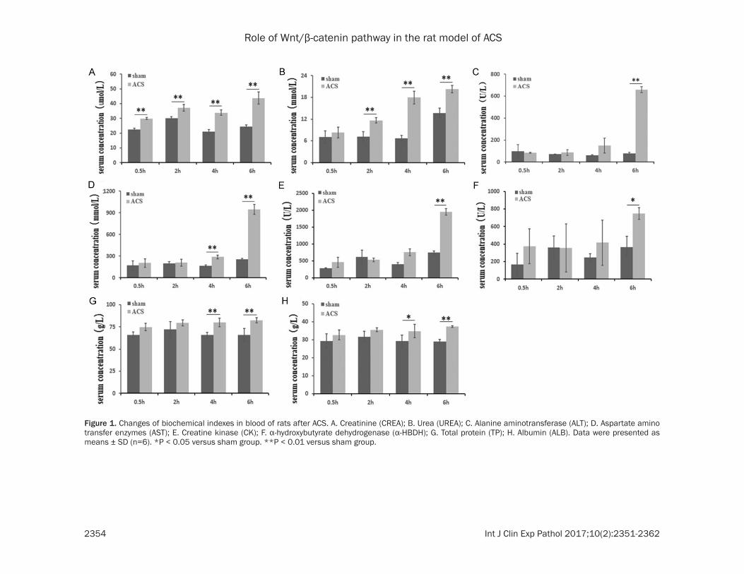
Figure 1. Changes of biochemical indexes in blood of rats after ACS. A. Creatinine (CREA); B. Urea (UREA); C. Alanine aminotransferase (ALT); D. Aspartate amino transfer enzymes (AST); E. Creatine kinase (CK); F. α-hydroxybutyrate dehydrogenase (α-HBDH); G. Total protein (TP); H. Albumin (ALB). Data were presented as means ± SD (n=6). *P < 0.05 versus sham group. **P < 0.01 versus sham group.
Role of Wnt/β-catenin pathway in the rat model of ACS
2355 Int J Clin Exp Pathol 2017;10(2):2351-2362
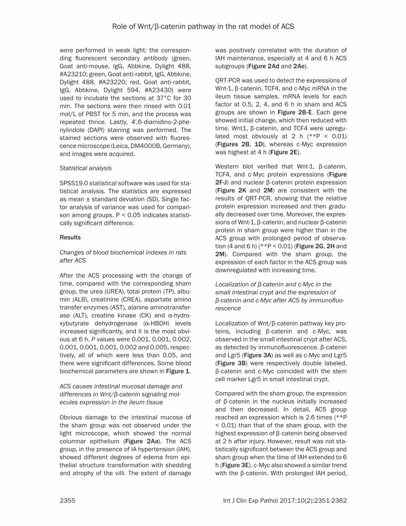
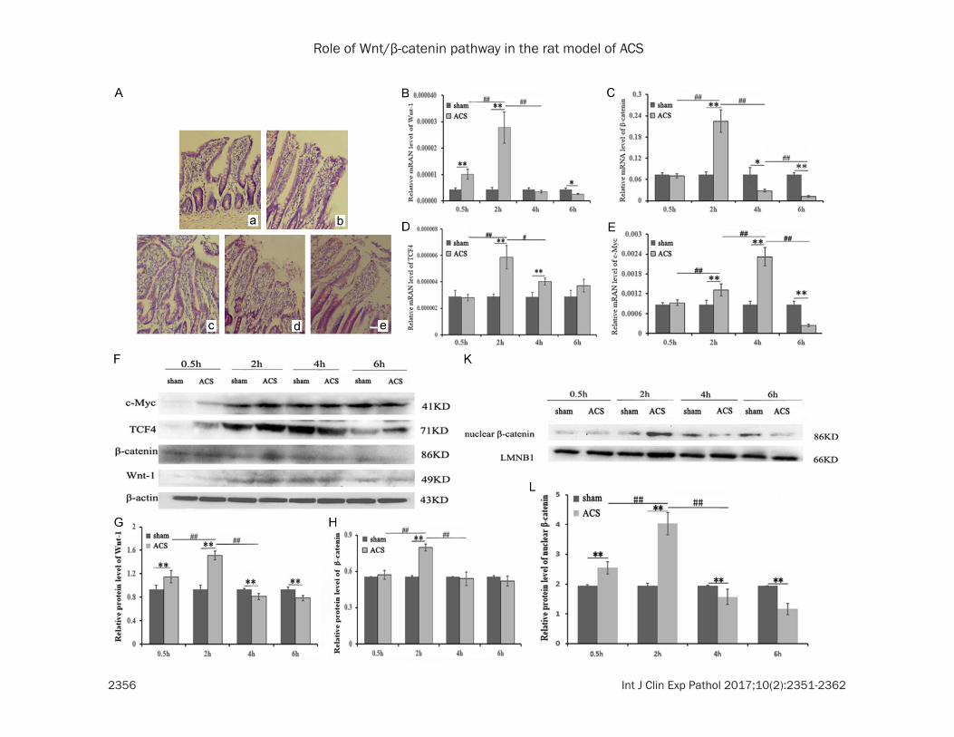
was positively correlated with the duration of IAH maintenance, especially at 4 and 6 h ACS subgroups (Figure 2Ad and 2Ae).
QRT-PCR was used to detect the expressions of Wnt-1, β-catenin, TCF4, and c-Myc mRNA in the ileum tissue samples. mRNA levels for each factor at 0.5, 2, 4, and 6 h in sham and ACS groups are shown in Figure 2B-E. Each gene showed initial change, which then reduced with time. Wnt1, β-catenin, and TCF4 were upregu-lated most obviously at 2 h (**P < 0.01) (Figures 2B, 1D), whereas c-Myc expression was highest at 4 h (Figure 2E).
Western blot verified that Wnt-1, β-catenin, TCF4, and c-Myc protein expressions (Figure 2F-J) and nuclear β-catenin protein expression (Figure 2K and 2M) are consistent with the results of QRT-PCR, showing that the relative protein expression increased and then gradu-ally decreased over time. Moreover, the expres-sions of Wnt-1, β-catenin, and nuclear β-catenin protein in sham group were higher than in the ACS group with prolonged period of observa-tion (4 and 6 h) (**P < 0.01) (Figure 2G, 2H and 2M). Compared with the sham group, the expression of each factor in the ACS group was downregulated with increasing time.
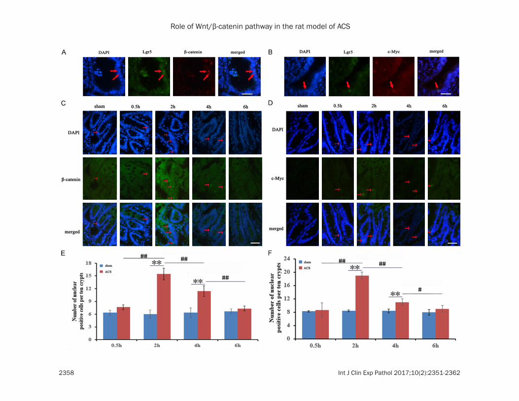
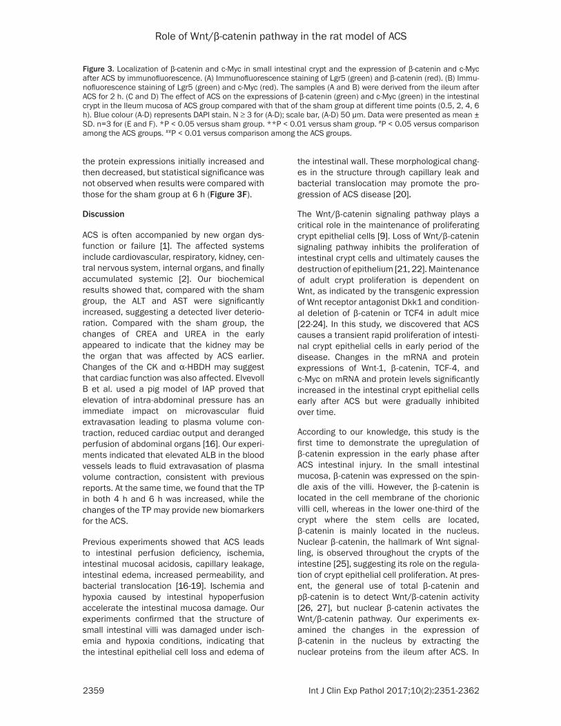
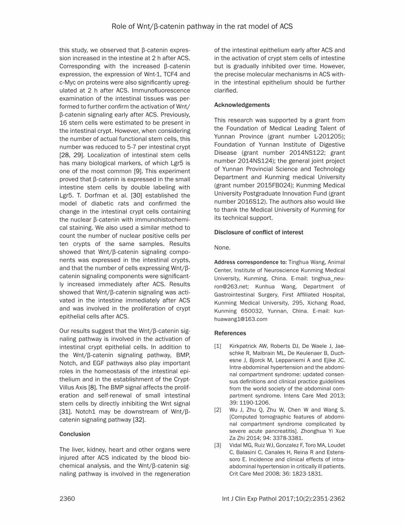
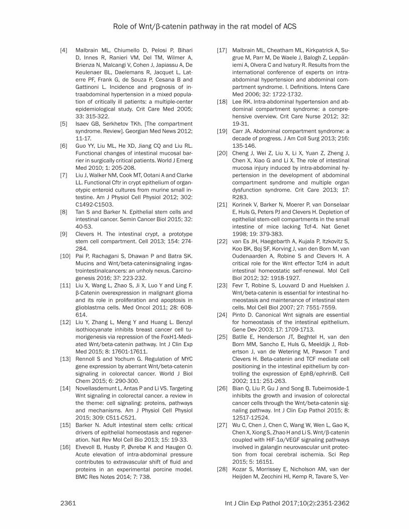
Localization of β-catenin and c-Myc in the small intestinal crypt and the expression of β-catenin and c-Myc after ACS by immunofluo-rescence
Localization of Wnt/β-catenin pathway key pro-teins, including β-catenin and c-Myc, was observed in the small intestinal crypt after ACS, as detected by immunofluorescence. β-catenin and Lgr5 (Figure 3A) as well as c-Myc and Lgr5 (Figure 3B) were respectively double labeled. β-catenin and c-Myc coincided with the stem cell marker Lgr5 in small intestinal crypt.
Compared with the sham group, the expression of β-catenin in the nucleus initially increased and then decreased. In detail, ACS group reached an expression which is 2.6 times (**P < 0.01) than that of the sham group, with the highest expression of β-catenin being observed at 2 h after injury. However, result was not sta-tistically significant between the ACS group and sham group when the time of IAH extended to 6 h (Figure 3E). c-Myc also showed a similar trend with the β-catenin. With prolonged IAH period,
were performed in weak light: the correspon- ding fluorescent secondary antibody (green, Goat anti-mouse, IgG, Abbkine, Dylight 488, #A23210; green, Goat anti-rabbit, IgG, Abbkine, Dylight 488, #A23220; red, Goat anti-rabbit, IgG, Abbkine, Dylight 594, #A23430) were used to incubate the sections at 37°C for 30 min. The sections were then rinsed with 0.01 mol/L of PBST for 5 min, and the process was repeated thrice. Lastly, 4’,6-diamidino-2-phe-nylindole (DAPI) staining was performed. The stained sections were observed with fluores-cence microscope (Leica, DM4000B, Germany), and images were acquired.
Statistical analysis
SPSS19.0 statistical software was used for sta-tistical analysis. The statistics are expressed as mean ± standard deviation (SD). Single fac-tor analysis of variance was used for compari-son among groups. P < 0.05 indicates statisti-cally significant difference.
Results
Changes of blood biochemical indexes in rats after ACS
After the ACS processing with the change of time, compared with the corresponding sham group, the urea (UREA), total protein (TP), albu-min (ALB), creatinine (CREA), aspartate amino transfer enzymes (AST), alanine aminotransfer-ase (ALT), creatine kinase (CK) and α-hydro- xybutyrate dehydrogenase (α-HBDH) levels increased significantly, and it is the most obvi-ous at 6 h. P values were 0.001, 0.001, 0.002, 0.001, 0.001, 0.001, 0.002 and 0.005, respec-tively, all of which were less than 0.05, and there were significant differences. Some blood biochemical parameters are shown in Figure 1.
ACS causes intestinal mucosal damage and differences in Wnt/β-catenin signaling mol-ecules expression in the ileum tissue
Obvious damage to the intestinal mucosa of the sham group was not observed under the light microscope, which showed the normal columnar epithelium (Figure 2Aa). The ACS group, in the presence of IA hypertension (IAH), showed different degrees of edema from epi-thelial structure transformation with shedding and atrophy of the villi. The extent of damage
Role of Wnt/β-catenin pathway in the rat model of ACS
2356 Int J Clin Exp Pathol 2017;10(2):2351-2362
Role of Wnt/β-catenin pathway in the rat model of ACS
2357 Int J Clin Exp Pathol 2017;10(2):2351-2362
Figure 2. ACS causes intestinal mucosal damage and differences in Wnt/β-catenin signaling molecules expression in the ileum tissue. Observation on the pathologi-cal changes of ileum mucosa was conducted by light microscope (A). (Aa) represents the sham group; (Ab-e) represent 0.5, 2, 4, and 6 h ACS group. N ≥ 3 for (A); scale bar, (A) 100 µm. (B-E) Representative mRNA expression of Wnt-1 (B), β-catenin (C), TCF-4 (D), and c-Myc (E) in the Ileum mucosa of ACS group compared with the sham group at different time points (0.5, 2, 4, and 6 h), as determined by QRT-PCR. The expression of each target gene was normalized to β-actin. (F) Protein expressions of Wnt-1, β-catenin, TCF4 and c-Myc in the Ileum mucosa of ACS group compared with the sham group at different time points (0.5, 2, 4, and 6 h), as measured by Western blot. β-actin was used as a loading control. (K) The nucleus of the ileum mucosa was extracted to detect the change in the nuclear β-catenin protein in ACS group compared with the sham group, as measured by Western blot. LMNB1 was used as loading control. (G-J, L) Quantification of band density was performed with Image Lab software. Data were presented as mean ± SD. n=6 for (B-E); n=3 for (G-J, L). *P < 0.05 versus sham group. **P < 0.01 versus sham group. #P < 0.05 versus comparison among the ACS groups. ##P < 0.01 versus comparison among the ACS groups.
Role of Wnt/β-catenin pathway in the rat model of ACS
2358 Int J Clin Exp Pathol 2017;10(2):2351-2362
Role of Wnt/β-catenin pathway in the rat model of ACS
2359 Int J Clin Exp Pathol 2017;10(2):2351-2362
the intestinal wall. These morphological chang-es in the structure through capillary leak and bacterial translocation may promote the pro-gression of ACS disease [20].
The Wnt/β-catenin signaling pathway plays a critical role in the maintenance of proliferating crypt epithelial cells [9]. Loss of Wnt/β-catenin signaling pathway inhibits the proliferation of intestinal crypt cells and ultimately causes the destruction of epithelium [21, 22]. Maintenance of adult crypt proliferation is dependent on Wnt, as indicated by the transgenic expression of Wnt receptor antagonist Dkk1 and condition-al deletion of β-catenin or TCF4 in adult mice [22-24]. In this study, we discovered that ACS causes a transient rapid proliferation of intesti-nal crypt epithelial cells in early period of the disease. Changes in the mRNA and protein expressions of Wnt-1, β-catenin, TCF-4, and c-Myc on mRNA and protein levels significantly increased in the intestinal crypt epithelial cells early after ACS but were gradually inhibited over time.
According to our knowledge, this study is the first time to demonstrate the upregulation of β-catenin expression in the early phase after ACS intestinal injury. In the small intestinal mucosa, β-catenin was expressed on the spin-dle axis of the villi. However, the β-catenin is located in the cell membrane of the chorionic villi cell, whereas in the lower one-third of the crypt where the stem cells are located, β-catenin is mainly located in the nucleus. Nuclear β-catenin, the hallmark of Wnt signal-ling, is observed throughout the crypts of the intestine [25], suggesting its role on the regula-tion of crypt epithelial cell proliferation. At pres-ent, the general use of total β-catenin and pβ-catenin is to detect Wnt/β-catenin activity [26, 27], but nuclear β-catenin activates the Wnt/β-catenin pathway. Our experiments ex- amined the changes in the expression of β-catenin in the nucleus by extracting the nuclear proteins from the ileum after ACS. In
the protein expressions initially increased and then decreased, but statistical significance was not observed when results were compared with those for the sham group at 6 h (Figure 3F).
Discussion
ACS is often accompanied by new organ dys-function or failure [1]. The affected systems include cardiovascular, respiratory, kidney, cen-tral nervous system, internal organs, and finally accumulated systemic [2]. Our biochemical results showed that, compared with the sham group, the ALT and AST were significantly increased, suggesting a detected liver deterio-ration. Compared with the sham group, the changes of CREA and UREA in the early appeared to indicate that the kidney may be the organ that was affected by ACS earlier. Changes of the CK and α-HBDH may suggest that cardiac function was also affected. Elvevoll B et al. used a pig model of IAP proved that elevation of intra-abdominal pressure has an immediate impact on microvascular fluid extravasation leading to plasma volume con-traction, reduced cardiac output and deranged perfusion of abdominal organs [16]. Our experi-ments indicated that elevated ALB in the blood vessels leads to fluid extravasation of plasma volume contraction, consistent with previous reports. At the same time, we found that the TP in both 4 h and 6 h was increased, while the changes of the TP may provide new biomarkers for the ACS.
Previous experiments showed that ACS leads to intestinal perfusion deficiency, ischemia, intestinal mucosal acidosis, capillary leakage, intestinal edema, increased permeability, and bacterial translocation [16-19]. Ischemia and hypoxia caused by intestinal hypoperfusion accelerate the intestinal mucosa damage. Our experiments confirmed that the structure of small intestinal villi was damaged under isch-emia and hypoxia conditions, indicating that the intestinal epithelial cell loss and edema of
Figure 3. Localization of β-catenin and c-Myc in small intestinal crypt and the expression of β-catenin and c-Myc after ACS by immunofluorescence. (A) Immunofluorescence staining of Lgr5 (green) and β-catenin (red). (B) Immu-nofluorescence staining of Lgr5 (green) and c-Myc (red). The samples (A and B) were derived from the ileum after ACS for 2 h. (C and D) The effect of ACS on the expressions of β-catenin (green) and c-Myc (green) in the intestinal crypt in the Ileum mucosa of ACS group compared with that of the sham group at different time points (0.5, 2, 4, 6 h). Blue colour (A-D) represents DAPI stain. N ≥ 3 for (A-D); scale bar, (A-D) 50 µm. Data were presented as mean ± SD. n=3 for (E and F). *P < 0.05 versus sham group. **P < 0.01 versus sham group. #P < 0.05 versus comparison among the ACS groups. ##P < 0.01 versus comparison among the ACS groups.
Role of Wnt/β-catenin pathway in the rat model of ACS
2360 Int J Clin Exp Pathol 2017;10(2):2351-2362
of the intestinal epithelium early after ACS and in the activation of crypt stem cells of intestine but is gradually inhibited over time. However, the precise molecular mechanisms in ACS with-in the intestinal epithelium should be further clarified.
Acknowledgements
This research was supported by a grant from the Foundation of Medical Leading Talent of Yunnan Province (grant number L-201205); Foundation of Yunnan Institute of Digestive Disease (grant number 2014NS122; grant number 2014NS124); the general joint project of Yunnan Provincial Science and Technology Department and Kunming medical University (grant number 2015FB024); Kunming Medical University Postgraduate Innovation Fund (grant number 2016S12). The authors also would like to thank the Medical University of Kunming for its technical support.
Disclosure of conflict of interest
None.
Address correspondence to: Tinghua Wang, Animal Center, Institute of Neuroscience Kunming Medical University, Kunming, China. E-mail: [email protected]; Kunhua Wang, Department of Gastrointestinal Surgery, First Affiliated Hospital, Kunming Medical University, 295, Xichang Road, Kunming 650032, Yunnan, China. E-mail: [email protected]
References
[1] Kirkpatrick AW, Roberts DJ, De Waele J, Jae-schke R, Malbrain ML, De Keulenaer B, Duch-esne J, Bjorck M, Leppaniemi A and Ejike JC. Intra-abdominal hypertension and the abdomi-nal compartment syndrome: updated consen-sus definitions and clinical practice guidelines from the world society of the abdominal com-partment syndrome. Intens Care Med 2013; 39: 1190-1206.
[2] Wu J, Zhu Q, Zhu W, Chen W and Wang S. [Computed tomographic features of abdomi-nal compartment syndrome complicated by severe acute pancreatitis]. Zhonghua Yi Xue Za Zhi 2014; 94: 3378-3381.
[3] Vidal MG, Ruiz WJ, Gonzalez F, Toro MA, Loudet C, Balasini C, Canales H, Reina R and Estens-soro E. Incidence and clinical effects of intra-abdominal hypertension in critically ill patients. Crit Care Med 2008; 36: 1823-1831.
this study, we observed that β-catenin expres-sion increased in the intestine at 2 h after ACS. Corresponding with the increased β-catenin expression, the expression of Wnt-1, TCF4 and c-Myc on proteins were also significantly upreg-ulated at 2 h after ACS. Immunofluorescence examination of the intestinal tissues was per-formed to further confirm the activation of Wnt/β-catenin signaling early after ACS. Previously, 16 stem cells were estimated to be present in the intestinal crypt. However, when considering the number of actual functional stem cells, this number was reduced to 5-7 per intestinal crypt [28, 29]. Localization of intestinal stem cells has many biological markers, of which Lgr5 is one of the most common [9]. This experiment proved that β-catenin is expressed in the small intestine stem cells by double labeling with Lgr5. T. Dorfman et al. [30] established the model of diabetic rats and confirmed the change in the intestinal crypt cells containing the nuclear β-catenin with immunohistochemi-cal staining. We also used a similar method to count the number of nuclear positive cells per ten crypts of the same samples. Results showed that Wnt/β-catenin signaling compo-nents was expressed in the intestinal crypts, and that the number of cells expressing Wnt/β-catenin signaling components were significant-ly increased immediately after ACS. Results showed that Wnt/β-catenin signaling was acti-vated in the intestine immediately after ACS and was involved in the proliferation of crypt epithelial cells after ACS.
Our results suggest that the Wnt/β-catenin sig-naling pathway is involved in the activation of intestinal crypt epithelial cells. In addition to the Wnt/β-catenin signaling pathway, BMP, Notch, and EGF pathways also play important roles in the homeostasis of the intestinal epi-thelium and in the establishment of the Crypt-Villus Axis [8]. The BMP signal affects the prolif-eration and self-renewal of small intestinal stem cells by directly inhibiting the Wnt signal [31]. Notch1 may be downstream of Wnt/β-catenin signaling pathway [32].
Conclusion
The liver, kidney, heart and other organs were injured after ACS indicated by the blood bio-chemical analysis, and the Wnt/β-catenin sig-naling pathway is involved in the regeneration
Role of Wnt/β-catenin pathway in the rat model of ACS
2361 Int J Clin Exp Pathol 2017;10(2):2351-2362
[17] Malbrain ML, Cheatham ML, Kirkpatrick A, Su-grue M, Parr M, De Waele J, Balogh Z, Leppän-iemi A, Olvera C and Ivatury R. Results from the international conference of experts on intra-abdominal hypertension and abdominal com-partment syndrome. I. Definitions. Intens Care Med 2006; 32: 1722-1732.
[18] Lee RK. Intra-abdominal hypertension and ab-dominal compartment syndrome: a compre-hensive overview. Crit Care Nurse 2012; 32: 19-31.
[19] Carr JA. Abdominal compartment syndrome: a decade of progress. J Am Coll Surg 2013; 216: 135-146.
[20] Cheng J, Wei Z, Liu X, Li X, Yuan Z, Zheng J, Chen X, Xiao G and Li X. The role of intestinal mucosa injury induced by intra-abdominal hy-pertension in the development of abdominal compartment syndrome and multiple organ dysfunction syndrome. Crit Care 2013; 17: R283.
[21] Korinek V, Barker N, Moerer P, van Donselaar E, Huls G, Peters PJ and Clevers H. Depletion of epithelial stem-cell compartments in the small intestine of mice lacking Tcf-4. Nat Genet 1998; 19: 379-383.
[22] van Es JH, Haegebarth A, Kujala P, Itzkovitz S, Koo BK, Boj SF, Korving J, van den Born M, van Oudenaarden A, Robine S and Clevers H. A critical role for the Wnt effector Tcf4 in adult intestinal homeostatic self-renewal. Mol Cell Biol 2012; 32: 1918-1927.
[23] Fevr T, Robine S, Louvard D and Huelsken J. Wnt/beta-catenin is essential for intestinal ho-meostasis and maintenance of intestinal stem cells. Mol Cell Biol 2007; 27: 7551-7559.
[24] Pinto D. Canonical Wnt signals are essential for homeostasis of the intestinal epithelium. Gene Dev 2003; 17: 1709-1713.
[25] Batlle E, Henderson JT, Beghtel H, van den Born MM, Sancho E, Huls G, Meeldijk J, Rob-ertson J, van de Wetering M, Pawson T and Clevers H. Beta-catenin and TCF mediate cell positioning in the intestinal epithelium by con-trolling the expression of EphB/ephrinB. Cell 2002; 111: 251-263.
[26] Bian Q, Liu P, Gu J and Song B. Tubeimoside-1 inhibits the growth and invasion of colorectal cancer cells through the Wnt/beta-catenin sig-naling pathway. Int J Clin Exp Pathol 2015; 8: 12517-12524.
[27] Wu C, Chen J, Chen C, Wang W, Wen L, Gao K, Chen X, Xiong S, Zhao H and Li S. Wnt/β-catenin coupled with HIF-1α/VEGF signaling pathways involved in galangin neurovascular unit protec-tion from focal cerebral ischemia. Sci Rep 2015; 5: 16151.
[28] Kozar S, Morrissey E, Nicholson AM, van der Heijden M, Zecchini HI, Kemp R, Tavare S, Ver-
[4] Malbrain ML, Chiumello D, Pelosi P, Bihari D, Innes R, Ranieri VM, Del TM, Wilmer A, Brienza N, Malcangi V, Cohen J, Japiassu A, De Keulenaer BL, Daelemans R, Jacquet L, Lat-erre PF, Frank G, de Souza P, Cesana B and Gattinoni L. Incidence and prognosis of in-traabdominal hypertension in a mixed popula-tion of critically ill patients: a multiple-center epidemiological study. Crit Care Med 2005; 33: 315-322.
[5] Isaev GB, Serkhetov TKh. [The compartment syndrome. Review]. Georgian Med News 2012; 11-17.
[6] Guo YY, Liu ML, He XD, Jiang CQ and Liu RL. Functional changes of intestinal mucosal bar-rier in surgically critical patients. World J Emerg Med 2010; 1: 205-208.
[7] Liu J, Walker NM, Cook MT, Ootani A and Clarke LL. Functional Cftr in crypt epithelium of organ-otypic enteroid cultures from murine small in-testine. Am J Physiol Cell Physiol 2012; 302: C1492-C1503.
[8] Tan S and Barker N. Epithelial stem cells and intestinal cancer. Semin Cancer Biol 2015; 32: 40-53.
[9] Clevers H. The intestinal crypt, a prototype stem cell compartment. Cell 2013; 154: 274-284.
[10] Pai P, Rachagani S, Dhawan P and Batra SK. Mucins and Wnt/beta-cateninsignaling ingas-trointestinalcancers: an unholy nexus. Carcino-genesis 2016; 37: 223-232.
[11] Liu X, Wang L, Zhao S, Ji X, Luo Y and Ling F. β-Catenin overexpression in malignant glioma and its role in proliferation and apoptosis in glioblastma cells. Med Oncol 2011; 28: 608-614.
[12] Liu Y, Zhang L, Meng Y and Huang L. Benzyl isothiocyanate inhibits breast cancer cell tu-morigenesis via repression of the FoxH1-Medi-ated Wnt/beta-catenin pathway. Int J Clin Exp Med 2015; 8: 17601-17611.
[13] Rennoll S and Yochum G. Regulation of MYC gene expression by aberrant Wnt/beta-catenin signaling in colorectal cancer. World J Biol Chem 2015; 6: 290-300.
[14] Novellasdemunt L, Antas P and Li VS. Targeting Wnt signaling in colorectal cancer. a review in the theme: cell signaling: proteins, pathways and mechanisms. Am J Physiol Cell Physiol 2015; 309: C511-C521.
[15] Barker N. Adult intestinal stem cells: critical drivers of epithelial homeostasis and regener-ation. Nat Rev Mol Cell Bio 2013; 15: 19-33.
[16] Elvevoll B, Husby P, Øvrebø K and Haugen O. Acute elevation of intra-abdominal pressure contributes to extravascular shift of fluid and proteins in an experimental porcine model. BMC Res Notes 2014; 7: 738.
Role of Wnt/β-catenin pathway in the rat model of ACS
2362 Int J Clin Exp Pathol 2017;10(2):2351-2362
[31] He XC, Zhang J, Tong WG, Tawfik O, Ross J, Sco-ville DH, Tian Q, Zeng X, He X, Wiedemann LM, Mishina Y and Li L. BMP signaling inhibits in-testinal stem cell self-renewal through sup-pression of Wnt-beta-catenin signaling. Nat Genet 2004; 36: 1117-1121.
[32] Wang R, Sun Q, Wang P, Liu M, Xiong S, Luo J, Huang H, Du Q, Geller DA and Cheng B. Notch and Wnt/beta-catenin signaling pathway play important roles in activating liver cancer stem cells. Oncotarget 2016; 7: 5754-5768.
meulen L and Winton DJ. Continuous clonal labeling reveals small numbers of functional stem cells in intestinal crypts and adenomas. Cell Stem Cell 2013; 13: 626-633.
[29] Vermeulen L, Morrissey E, van der Heijden M, Nicholson AM, Sottoriva A, Buczacki S, Kemp R, Tavare S and Winton DJ. Defining stem cell dynamics in models of intestinal tumor initia-tion. Science 2013; 342: 995-998.
[30] Dorfman T, Pollak Y, Sohotnik R, Coran AG, Be-jar J and Sukhotnik I. Enhanced intestinal epi-thelial cell proliferation in diabetic rats corre-lates with β-catenin accumulation. J Endocrinol 2015; 226: 135-143.