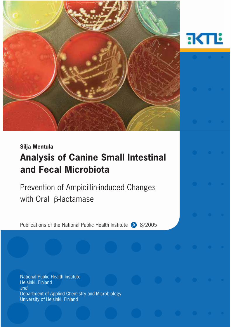
Analysis of canine small intestinal and fecal microbiota - Helda
86
Publications of the National Public Health Institute A 8/2005 National Public Health Institute Helsinki, Finland and Department of Applied Chemistry and Microbiology University of Helsinki, Finland Analysis of Canine Small Intestinal and Fecal Microbiota Prevention of Ampicillin-induced Changes with Oral β-lactamase Silja Mentula
Transcript of Analysis of canine small intestinal and fecal microbiota - Helda
Publications of the National Public Health Institute A 8/2005
National Public Health InstituteHelsinki, FinlandandDepartment of Applied Chemistry and MicrobiologyUniversity of Helsinki, Finland
Analysis of Canine Small Intestinal and Fecal Microbiota
Prevention of Ampicillin-induced Changes with Oral β-lactamase
Silja Mentula
Silja Mentula
ANALYSIS OF CANINE SMALL INTESTINAL AND FECAL MICROBIOTA
P R E V E N T I O N O F A M P I C I L L I N - I N D U C E D C H A N G E S W I T H O R A L β - L A C T A M A S E
A C A D E M I C D I S S E R T A T I O N
To be presented with the permission of the Faculty of Agriculture and Forestry, University of Helsinki, for public examination in Auditorium XII,
University Main Building,Unioninkatu 34, on June 9th, at 12 noon.
National Public Health Institute, Helsinki, Finland and
Department of Applied Chemistry and Microbiology, University of Helsinki, Finland
Helsinki 2004
S u p e r v i s e d b y
Professor Pentti Huovinen Antimicrobial Research Laboratory
Department of Bacterial and Inflammatory Diseases National Public Health Institute, Turku
Docent Eija Könönen Anaerobe Reference Laboratory
Department of Bacterial and Inflammatory Diseases National Public Health Institute, Helsinki
R e v i e w e d b y
Professor Charlotta Edlund Södertörns högskola, University College
andDepartment of Laboratory Medicine
Division of Clinical Bacteriology Karolinska Institutet Stockholm, Sweden
Docent Maria Saarela VTT Biotechnology, Foods and Processes
VTT, Espoo
O p p o n e n t
Professor Ian Poxton Division of Medical Microbiology
University of Edinburgh Medical SchoolEdinburgh, Scotland, UK

To my family
P u b l i c a t i o n s o f t h e N a t i o n a l P u b l i c H e a l t h I n s t i t u t e K T L A 8 / 2 0 0 5
Copyright National Public Health Institute
Julkaisija-Utgivare-Publisher
Kansanterveyslaitos (KTL)Mannerheimintie 166 00300 Helsinki Puh. vaihde (09) 474 41, telefax (09) 4744 8408
FolkhälsoinstitutetMannerheimvägen 166 00300 Helsingfors Tel. växel (09) 474 41, telefax (09) 4744 8408
National Public Health Institute Mannerheimintie 166 FIN-00300 Helsinki, Finland Telephone +358 9 474 41, telefax +358 9 4744 8408
ISBN 951-740-517-0 ISSN 0359-3584 ISBN 951-740-518-9 (pdf) ISSN 1458-6290 (pdf)
Edita Prima Oy Helsinki 2005
CONTENTS
ABBREVIATIONS .......................................................................................................7 ABSTRACT...................................................................................................................8 TIIVISTELMÄ ............................................................................................................10 INTRODUCTION .......................................................................................................14 REVIEW OF THE LITERATURE .............................................................................16
1. Gut microbiota in health and disease ...................................................................16 1.1. The importance of microbes .........................................................................16 1.2. The effects of intestinal microbiota ..............................................................17 1.3. Dysfunctions of the gut microbiota in humans .............................................18
2. Composition of the human gut microbiota ..........................................................19 2.1. General features ............................................................................................19 2.2. Species composition......................................................................................21 2.3. Upper gut microbiota ....................................................................................24 2.4. Opportunistic commensals............................................................................25
3. The digestive tract and intestinal microbiota of dog............................................27 4. Obtaining samples from the intestine ..................................................................29
4.1. Getting representative samples from the upper gut ......................................29 4.2. Jejunal fistula ................................................................................................30
5. Analysing the microbiota.....................................................................................31 5.1. Microscopic methods ....................................................................................31 5.2. Culture method..............................................................................................31 5.3. Molecular biological methods.......................................................................32 5.4. Molecular typing methods ............................................................................34 5.5. Indirect methods for analysing microbiota ...................................................35
6. Antimicrobial agents............................................................................................36 6.1. Classification of antimicrobial agents...........................................................36 6.2. Emergence of antimicrobial resistance .........................................................36 6.3. Mechanisms of resistance .............................................................................38 6.4. Adverse effects of antimicrobial agents........................................................38 6.5. Use of antimicrobials in humans and animals ..............................................40 6.6. β-Lactam antibiotics .....................................................................................41 6.7. β-Lactamases ................................................................................................42 6.8. Inhibition of β-lactam resistance ..................................................................43 6.9. Testing antimicrobial susceptibility..............................................................44
AIMS OF THE STUDY ..............................................................................................45 MATERIALS AND METHODS.................................................................................46
1. Study design.........................................................................................................46 2. Study animals and sample collection...................................................................47 3. Study location ......................................................................................................48 4. Bacterial culture ...................................................................................................48 5. Identification, susceptibility testing and PFGE typing of coliforms....................50 6. TGGE (temperature gradient gel electrophoresis) ...............................................52 7. PCR for detecting TEM gene...............................................................................52 8. Measurement of ampicillin concentration ...........................................................53 9. Statistical analysis................................................................................................54
RESULTS ....................................................................................................................55 1. Jejunal versus faecal cultivable microbiota (I) ....................................................55 2. Stability of the cultivable intestinal microbiota (I) ..............................................57
3. Effects of ampicillin and prevention of them by TRBL (II, III) ..........................57 3.1. Changes detected by culture (II, III) .............................................................58 3.2. Microbial shifts detected by TGGE (II)........................................................58 3.3. Ampicillin concentrations (II).......................................................................59 3.4. Change in TEM gene levels (II)....................................................................59
4. Antimicrobial resistance in coliforms (II, III, IV) ...............................................59 4.1. Emergence of resistance in coliforms (II, III, IV) ........................................59 4.2. Assessing the relatedness of coliforms (IV) .................................................62
DISCUSSION..............................................................................................................64 Characterisation of jejunal and faecal microbiota by culture ..................................64 Comparison of culture to molecular methods..........................................................66 Ampicillin-induced changes ....................................................................................66 Resistance within coliform populations...................................................................67 Prevention of ampicillin-induced changes by TRBL...............................................68
KEY FINDINGS AND CONCLUSIONS ...................................................................70 ACKNOWLEDGEMENTS.........................................................................................72 REFERENCES ............................................................................................................74
7
ABBREVIATIONS
CFU colony forming unit
DNA deoxyribonucleic acid
GALT gut-associated lymphoid tissue
IBD inflammatory bowel disease
i.v. intra venous
MAC microflora-associated characteristics
MRSA methicillin-resistant Staphylococcus aureus
NCCLS National Committee for Clinical Laboratory Standards
PCR polymerase chain reaction
PFGE pulsed-field gel electrophoresis
p.o. per oral
rRNA ribosomal ribonucleic acid
SIBO small intestinal bacterial overgrowth
TGGE temperature-gradient gel electrophoresis
TEM class of plasmid-mediated β-lactamases (first isolated from a patient
named Temoniera)
TRBL targeted recombinant β-lactamase
VRE vancomycin-resistant enterococci
8
Silja Mentula, Analysis of canine small intestinal and fecal microbiota – prevention of ampicillin-induced changes with oral β-lactamase Publications of the National Public Health Insitute, A8/2005, 136 PagesISBN 951-740-517-0; 951-740-518-9 (pdf-version) ISSN 0359-3584; 1458-6290 (pdf-version) http://www.ktl.fi/portal/suomi/julkaisut/julkaisusarjat/kansanterveyslaitoksen_julkaisuja_a/
ABSTRACT
Background: The intestinal microbiota affects many physiological and
immunological functions of the body and is closely connected to the homeostasis and
health status of an individual. Due to difficulties in obtaining samples from the
intestine and studying complex microbial populations the knowledge of the
composition, dynamics and metabolic potential of microbial populations in the upper
gut is limited. Antimicrobials may disrupt the balance of the microbiota by causing
quantitative and qualitative changes such as emergence and enrichment of antibiotic-
resistant populations, and decrease in colonisation resistance against pathogens. The
aims of this study were to analyse cultivable jejunal microbiota of a dog in detail and
compare it to faecal findings, and to evaluate the ability of oral β-lactamase
administration, targeted to degrade intestinal ampicillin residues, to prevent
ampicillin-induced changes in the intestinal microbiota.
Material and methods: Jejunal and faecal samples were obtained from healthy
laboratory beagles with permanent jejunal fistula randomised into 3 treatment groups
receiving ampicillin, ampicillin + β-lactamase or placebo. Samples were collected
before, during and after the treatment and analysed using bacterial culture and TGGE
(temperature-gradient gel electrophoresis) for total bacterial counts and composition
of the dominant microbiota. Susceptibility testing and PCR for detection of TEM gene
were the methods for monitoring resistance rates. In addition, jejunal and serum
ampicillin concentrations were determined.
Results: The relative quantities of predominant bacterial groups, proportion of
anaerobes, intra- and inter-individual species variety and stability of the microbiota
differed significantly between the jejunum and faeces. Oral β-lactamase inhibited the
effects of ampicillin, monitored as significant differences in resistance rates and
9
bacterial composition during treatment between the groups receiving ampicillin with
and without β-lactamase.
Conclusion: This study suggests that faeces with stable and rich microbiota does not
represent jejunal microbiota that is characterised as harbouring few individually
varying bacterial species at a time with fluctuating counts. Oral β-lactamase has
potential in inhibiting the adverse effects of β-lactam therapy.
Keywords: gastrointestinal tract, jejunum, microbiota, β-lactams, susceptibility
10
Silja Mentula, Analysis of canine small intestinal and fecal microbiota – prevention of ampicillin-induced changes with oral β-lactamase Kansanterveyslaitoksen julkaisuja, A8/2005, 136 sivuaISBN 951-740-517-0; 951-740-518-9 (pdf-versio) ISSN 0359-3584; 1458-6290 (pdf-versio) http://www.ktl.fi/portal/suomi/julkaisut/julkaisusarjat/kansanterveyslaitoksen_julkaisuja_a/
TIIVISTELMÄ
Taustaa: Suolistomikrobisto vaikuttaa moniin kehon fysiologisiin ja immunologisiin
toimintoihin ja siten myös kehon tasapainotilaan sekä yksilön terveyteen. Johtuen
vaikeuksista näytteiden saamisessa suoliston eri osista sekä monimuotoisten
mikrobipopulaatioden tutkimisen hankaluuksista, ymmärtämys suolistomikrobiston
koostumuksesta, dynamiikasta ja metaboliikasta on vaillinaista. Antibiootit voivat
häiritä mikrobiston tasapainoa aiheuttaen niin määrällisiä kuin laadullisia muutoksia
mikrobistossa kuten antibiootille vastustuskykyisten mikrobien ilmaantumista ja
rikastumista ja taudinaiheuttajamikrobien kolonisaatioresistenssin alenemista. Tämän
tutkimuksen tavoitteena oli analysoida koiran ohutsuolen mikrobisto
yksityiskohtaisesti viljelymenetelmää käyttäen ja verrata sitä ulosteen mikrobistoon.
Lisäksi tavoitteena oli arvioida suun kautta annettavan, suolistoon kulkeutuvien β-
laktaamiryhmän antibioottijäämien hajoittamiseen suunnitellun β-
laktamaasivalmisteen tehoa ampisilliinin suolistomikrobistossa aiheuttamien
muutosten ehkäisyssä.
Materiaalit ja menetelmät: Ohutsuolen sisältö ja ulostenäytteet kerättiin terveiltä
beagle-koirilta, joille oli tehty pysyvä fisteli (permanent jejunal fistula). Koirat oli
satunnaistettu kolmeen hoitoryhmään, jotka saivat joko ampisilliiniä, ampisilliiniä ja
β-laktamaasia tai lumevalmisteita. Näytteet kerättiin ennen antibioottikuuria, kuurin
aikana ja sen jälkeen ja analysoitiin käyttäen viljelyä ja TGGE-profilointia
(temperature-gradient gel electrophoresis) bakteerien kokonaismäärien ja
koostumuksen tutkimiseen. Antibioottiresistenssin esiintymistä seurattiin
herkkyysmäärityksin sekä TEM-geenin (plasmidi-välitteinen β-laktamaasigeeni)
esiintymistä PCR-määrityksin. Lisäksi seurattiin ohutsuolen sisällön sekä seerumin
ampisilliinipitoisuuksia.
11
Tulokset: Vallitsevien bakteeriryhmien suhteelliset osuudet, anaerobisten bakteerien
osuus, yksilöiden sisäinen sekä yksilöiden välinen lajivaihtelu ja mikrobiston
stabiilius erosivat merkittävästi ohutsuolen ja ulosteen mikrobistojen välillä. β-
laktamaasivalmiste ehkäisi ampisilliinin vaikutuksia: teho havaittiin merkittävinä
eroina resistenssin esiintymisessä ja mikrobiston koostumuksessa ampisilliinia ja
ampisilliiniä sekä β-laktamaasivalmistetta saaneiden ryhmien välillä.
Johtopäätökset: Tämä tutkimus osoittaa, että ulosteen melko stabiili ja runsas
mikrobisto ei edusta ohutsuolen rajoittunutta, yksilöittäin, ajallisesti, määrällisesti ja
laadullisesti vaihtelevaa mikrobistoa. β-laktamaasi on lupaava valmiste β-
laktaamiantibioottien aiheuttamien epäsuotuisten vaikutusten ehkäisyyn.
Avainsanat: suolistomikrobisto, ohutsuoli, β-laktaamit, mikrobilääkeherkkyys
12
LIST OF ORIGINAL PUBLICATIONS
This thesis is based on the following original articles referred to in the text by their
Roman numerals:
I Mentula S, Harmoinen J, Heikkilä M, Westermarck E, Rautio M, Huovinen P,
Könönen E. Comparison between the cultured small intestinal and fecal
microbiota in beagle dogs. Appl Environ Microbiol 2005, in press.
II Harmoinen J, Mentula S, Heikkilä M, van der Rest M, Rajala-Schultz PJ,
Donskey CJ, Frias R, Koski P, Wickstrand N, Jousimies-Somer H,
Westermarck E, Lindevall K. Orally administered targeted recombinant Beta-
lactamase prevents ampicillin-induced selective pressure on the gut
microbiota: a novel approach to reducing antimicrobial resistance. Antimicrob
Agents Chemother 2004;48:75-79.
III Mentula S, Harmoinen J, Koski P, Westermarck E, Rautio M, Huovinen P,
Könönen E. Inhibition of ampicillin-induced emergence of resistance in
intestinal coliforms by targeted recombinant beta-lactamase. Int J Antimicrob
Agents 2004;24:555-561.
IV Mentula S, Virtanen T, Kanervo-Nordström A, Harmoinen J, Rautio M,
Huovinen P, Könönen E. Relatedness of Escherichia coli strains with different
susceptibility patterns isolated from beagle dogs during ampicillin medication.
(submitted for publication)
These articles are reproduced with the kind permission of their copyright holders.
13
Author’s contribution
Publication I
Silja Mentula (SM) was responsible for the microbiological experimental work and
carried out identification of the strains, excluding 16S RNA gene sequencing.
Collected and interpreted the results and wrote the paper.
Publication II
SM was responsible for the culture-based experimental work, including the selection
of coliform isolates and collecting and modifying the susceptiblity data. Interpreted
the culture-based results and wrote the paper together with the corresponding author.
Publication III
Responsibilities in experimental work were similar to publication II. SM analysed the
resistance patterns, interpreted the results and wrote the paper.
Publication IV
SM was responsible for the microbiological experimental work, analysed and
compared the typing data to susceptibility data, interpreted the results and wrote the
paper.
14
INTRODUCTION
The intestinal microbiota is known to have a crucial impact on the health status of an
individual. Microbes influence functions such as nutrition, development and
maintenance of the immune system, and defence against pathogens. To understand the
relationship between the intestinal microbiota and health, and the metabolic potential
of the microbes, and to evaluate the effect of different diets, treatments or medical
conditions on the microbiota, fundamental knowledge of the intestinal microbial
populations is needed.
Because of the inaccessibility of the intestine, there are major difficulties in studying
the intestinal microbiota in its natural habitat and most studies of the intestinal
microbiota have been done using faecal samples. Due to low number of studies data
on the differences between the microbiota of different parts of the gut are limited. The
microbiota of the human upper gut and caecum has been reported to differ
significantly from that of the colon and faeces (Gorbach et al. 1967, Justesen et al.
1984, Simon & Gorbach 1986, Marteau et al. 2001). However, the recoverability of
small intestinal microbiota from faeces has not been fully analysed.
Bacterial composition, activities and concentrations can be severely distorted by
outside agents such as antimicrobials. Exposure of gut microbiota to antimicrobials
selects resistant bacteria that may enrich and spread within a given surrounding
(Livermore 2003, Münnich & Lübke-Becker 2004, Drazenovich et al. 2004).
Increasing rates of antimicrobial resistance following extensive use of antimicrobials
has led to a worldwide interest in finding ways to inhibit the emergence of resistance.
The present study was done in order to make a detailed analysis on the canine jejunal
microbiota and compare it with corresponding faecal microbiota in 22 beagle dogs
with permanent jejunal fistula. The other aim of the study was to analyse the effect of
i.v. ampicillin treatment on the intestinal microbiota, especially on the antimicrobial
resistance of coliforms, and to analyse whether the adverse effects of ampicillin could
be inhibited by simultaneous oral administration of β-lactamase, designed to degrade
antibiotic residues in the gut. The analyses were performed using culture method and
15
various molecular tools (TGGE, quantitative PCR, PFGE). The ultimate aim of the
study was to contribute to the better understanding of the composition and dynamics
of the gut microbiota, and to the development of means to prevent antimicrobial
resistance.
16
REVIEW OF THE LITERATURE
1. Gut microbiota in health and disease
1.1. The importance of microbes
Microorganisms colonise all mucosal surfaces forming a complex and dynamic
entities. The intestine harbours the vast majority of the microbes of the body, and
there the microbes also have the most prominent impact on our health. The gut
microbiota can be described to be in a state of unstable stability with reference to the
relatively stable composition of the host specific main bacterial groups and their
proportions within an individual, and to the simultaneous constant flow of new
bacterial clones co-existing and replacing the previous ones in a balanced and
seemingly homologous living system (McCartney et al. 1996, Franks et al. 1998,
Zoetendal et al. 1998, Falk et al. 1998). Gut microbiota can be regarded as a
metabolically active organ of the body, beneficial to both microbes and the host.
Compared to skin, the contracting gastrointestinal tract with villi and microvilli
comprises 100-fold surface area (2 m2 versus 150-200 m2, respectively) providing a
considerable amount of space for digestive and physiological processes (Van Dijk
1997, Waldeck 1990). The vital importance of the microbiota to the homeostasis of
the body is obvious since the imbalance of the microbiota may cause several different
disturbances of the gastrointestinal functions such as diarrhoea and impairs the health
status of an individual.
Microbiota has crucial role in many physiological/biochemical and immunological
functions of the body (Gordon & Pesti 1971, Berg 1996). The importance of the
microbiota has been analysed by comparisons of germ-free (sterile) and conventional
animals, most often mice, rats, or chickens. Individuals grown in a microbe-free
(sterile) environment lack many features of the immune system, require 30% more
calories to maintain body mass and die sooner than those grown in normal conditions
(Gordon & Pesti 1971, Wostmann et al. 1983, Tanaka & Ishikawa 2004, Bäckhed et
al. 2004). The features being altered by bacterial activity, differing between germfree
and conventional mice, are collectively known as microflora associated characteristics
(MACs) (Midtvedt 1989). In turn living microbes in the intestine are provided a niche
17
with abundance of nutrients and fermentable carbon sources originating from host
secretions, diet, cast off epithelial cells and mucin. Additionally, the conditions are
stable, warm and humid.
1.2. The effects of intestinal microbiota
To colonise different niches of the intestinal tract microbes have acquired distinct and
appropriate adaptation strategies. The commensal and symbiotic (indigenous)
microbes are those that are beneficial or at least not harmful to the host in contrast to
the harmful pathogenic (transient) species. In addition to the immunopotentiation
(maturation of the gut-associated lymphoid tissue, GALT) the commensal bacteria
play a significant role in the nutrition by degrading otherwise non-digestible food
compounds, producing vitamins and short-chain fatty acids, used as an energy source
by the colonic mucosa, conversion of urobilin to urobilinogen, conversion of
cholesterol to coprostanol, and in absorption of ions (Moore & Holdeman 1974,
Mackowiak 1982, Simon & Gorbach 1986). The gut-associated lymphoid tissue
(GALT) contains over 80% of all B-cells (antibody producing lymphocytes) of the
body and more lymphoid cells than the spleen, peripheral lymph nodes and blood
together (Brandtzaeg et al. 1989). The increasing frequency of atopic allergies has
been linked to overly hygienic living conditions, referred to as the hygiene hypothesis,
altering the colonisation patterns in infancy and leading to lack of tolerance to
harmless food proteins and antigens (Wold 1998). Indigenous bacteria also stimulate
vascularisation and development of intestinal villi, have an important role in the
metabolism of endogenous and exogenous compounds (detoxification), and in
prevention of colonisation and proliferation of pathogens and opportunistic microbes
(Hooper et al. 2002, Stappenbeck et al. 2002). Term colonisation resistance was
introduced by van der Waaij et al. (1971). It provides non-specific defence against
infections and includes various bacterial mechanisms such as competitive exclusion
(competition of living/adhesion space and nutrients) and production of substances
harmful to pathogens (antimicrobial agents, bacteriocins) as well as anatomical and
physiological features of the host (mucosa, secretions, gastrointestinal motility). Most
effects of bacteria against other bacteria are strictly local since e.g. bacteriocins are
degradable by host digestive enzymes (Guarner & Malagelada 2003).
18
1.3. Dysfunctions of the gut microbiota in humans
Clinical disturbances
Whether the causes for dysfunctions in the intestine are of physiological or exogenous
origin, they are often associated with gastrointestinal tract infections with altered
bacterial composition, bowel movements, and/or water and ion balance. The clinical
terms for gastrointestinal tract infections are gastroenteritis, diarrhoea, dysentery and
enterocolitis, each with distinctive set of clinical manifestations (Mims et al. 1998).
One of the most common type of chronic gut dysfunction, the inflammatory bowel
disease (IBD), has been linked to loss of tolerance to commensal bacteria. Intestinal
bacteria may be involved in maintaining the inflammation reaction in IBD (e.g.
ulcerative colitis, Crohn’s disease) since patients have increased mucosal secretion of
IgG antibodies against several commensal bacteria and have higher amounts of
bacteria attached to their epithelial surfaces compared with healthy people
(Macpherson et al. 1996, Swidsinski et al. 2002).
Another serious clinical condition possibly linked to disturbances in gut microbiota is
colon cancer. By production of potentially toxic or carcinogenic substances such as
amines, phenols and indols, intestinal bacteria are most likely one of the factors
modulating the risk of initiation of colon cancer (Smith & Macfarlane 1996,
Macfarlane & Macfarlane 1997). Increase in the incidence and growth rate of colonic
tumors have been shown to be associated to some Bacteroides and Clostridium
species while probiotic bifidobacteria bacteria prevent tumorigenesis in rats and mice
(Onoue et al. 1997, Horie et al. 1999, Singh et al. 1997). Similarly, the presence of
Bacteroides vulgatus and Bacteroides stercoris has been connected with a high risk of
colon cancer whereas Lactobacillus acidophilus, Lactobacillus S06 and Eubacterium
aerofaciens (currently Collinsella) associate with a low risk in humans (Moore &
Moore 1995). Short-chain fatty acids produced by bacterial fermentation have been
associated with reduction in the incidence of colon cancer and IBD (Wachtershauser
& Stein 2000, Blottiere et al. 2003, Miyauchi et al. 2004).
19
Small intestinal bacterial overgrowth
In small intestinal bacterial overgrowth (SIBO), also called the contaminated small
bowel syndrome, total bacterial counts exceed those of healthy subjects by 1-2 log
difference, usually crossing the limit of 105 cfu/g (Finegold & George 1989, Stotzer
et al. 1998, Corazza et al. 1990). Patients with SIBO may have no symptoms and no
predisposing factors, or may suffer from clinical manifestations such as malabsorption
of lipids, amino acids and carbohydrates, vitamin B12 deficiency, and disturbance in
water and electrolyte transport in enterocytes (Gracey 1983). The causes and
pathogenesis of the SIBO are poorly understood but several factors promoting
manifestations have been identified. They include pathological abnormalities of the
intestinal tract, stagnation of bowel contents, decreased gastric acidity, antimicrobial
therapy and impaired immune system (Finegold & George 1989, Gracey 1983).
SIBO, as well as increased permeability of the intestinal mucosal barrier and
deficiencies in host immune defences (e.g. mucin and secretory IgA production) are
thought to promote bacterial translocation, i.e. the crossing of bacteria through the
host epithelial mucosa (Berg 1999, Macpherson et al. 2000). Bacterial translocation
enables bacteria to transfer within the body causing local infections, sepsis, shock,
multisystem organ failure, or death of the host (Guarnier & Malagelada 2003).
Delayed small intestinal transit times, possibly promoting small intestinal overgrowth,
has been recorded in several conditions with high incidences of bacterial translocation
such as obstructive pancreatitis, hepatic failure, and portal vein obstruction (Moody et
al. 1995, Wang et al. 1994, Yi et al. 1999).
2. Composition of the human gut microbiota
2.1. General features
The mammal gut microbiota is composed of up to 1000 bacterial species with 30-40
species comprising the majority (99%) at a time, and with a particular individual
combination of predominant species and unique strains distinct from other individuals
(Savage et al. 1968, Savage 1977, Lee et al. 1971, Moore & Holdeman 1974, Xu &
Gordon 2003).
20
The stability of the microbiota is challenged by gastric, pancreatic, and biliary acid
secretions, peristaltic activity and continuous turnover of the epithelium and the
mucus layer. On the other hand, peristaltic movement of the gut induces the exposure
of different segments of the gut to the varied components of the intestinal contents
ensuring the continuous beneficial interaction of microbiota, dietary molecules,
epithelium and gut-associated immune system (Falk et al. 1998). The most numerous
bacteria in the gut are obligatory anaerobic species accompanied by smaller amounts
of aerobic/facultative bacteria with aerobes comprising only 1:100 - 1:1000 of the
total count in the intestinal lumen (Finegold et al. 1983, Berg 1996). However, the
proportion of aerobic species varies between the different regions of the intestinal
tract.
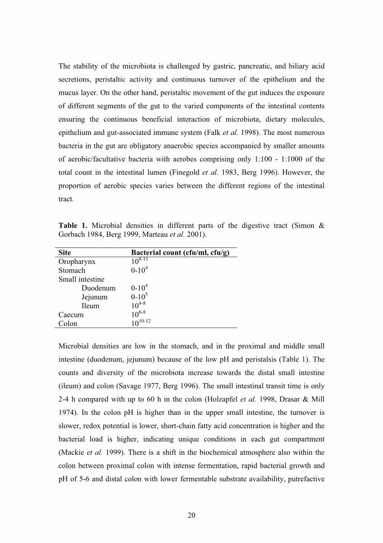
Table 1. Microbial densities in different parts of the digestive tract (Simon & Gorbach 1984, Berg 1999, Marteau et al. 2001).
Site Bacterial count (cfu/ml, cfu/g) Oropharynx 108-11
Stomach 0-104
Small intestine Duodenum 0-104
Jejunum 0-105
Ileum 104-8
Caecum 106-8
Colon 1010-12
Microbial densities are low in the stomach, and in the proximal and middle small
intestine (duodenum, jejunum) because of the low pH and peristalsis (Table 1). The
counts and diversity of the microbiota increase towards the distal small intestine
(ileum) and colon (Savage 1977, Berg 1996). The small intestinal transit time is only
2-4 h compared with up to 60 h in the colon (Holzapfel et al. 1998, Drasar & Mill
1974). In the colon pH is higher than in the upper small intestine, the turnover is
slower, redox potential is lower, short-chain fatty acid concentration is higher and the
bacterial load is higher, indicating unique conditions in each gut compartment
(Mackie et al. 1999). There is a shift in the biochemical atmosphere also within the
colon between proximal colon with intense fermentation, rapid bacterial growth and
pH of 5-6 and distal colon with lower fermentable substrate availability, putrefactive
21
processes, slow bacterial growth and neutral pH (Cummings & Macfarlane 1991,
Tannock 1999a, Guarner & Malagelada 2003).
The conditions in the lumen differ also from those proximal to the mucosa. The gut
can be divided crosswise into four different microhabitats: lumen, mucus layer, deep
mucus layer and mucosal epithelial cells (Berg 1996). These microhabitats provide
separate niches with unique properties. The predominant bacterial species associated
to the jejunal, ileal and colonic mucosa differ significantly from the respective jejunal
or ileal lumen and faecal bacterial community, and additionally bacterial counts
between separate mucosal sites vary considerably (Bhat et al. 1980, Zoetendal et al.
2002, Wang et al. 2003). The mucosa-associated bacteria in colon and rectum are
fairly similar in total counts and species composition (Poxton et al. 1997). The
analysis of the mucosal microbiota is complicated by the undefined effects on the
bacteria caused by the evacuation of the intestine preceding the biopsy collection and
the possible washing procedures at sampling. A very limited number of studies has
focused on the differences between the bacterial composition and activities in the
different gut compartments.
2.2. Species composition
The composition of the microbiota is known to be affected by several factors
concerning the host such as age, diet, medication, health status, microbes in the
environment and stress, as well as factors concerning the microbes such as growth
rate, nutritional and adherence characteristics and microbial interactions (Holzapfel et
al. 1998, Mitsuoka 1992). In addition, also host genetics have been reported to have
significant correlations between bacterial communities (van der Merwe et al. 1993,
Toivanen et al. 2001, Zoetendal et al. 2001). Once established, the microbiota remains
relatively constant over time in adults (Finegold et al. 1983). Community shifts within
the microbiota occur throughout the lifespan, especially in infants and in elderly
people (Mitsuoka 1992, Hopkins et al. 2001, Favier et al. 2002).
As the composition of the microbiota is influenced by external factors such as
acquisition of microbes in the diet, manipulating the microbiota from outside in order
to improve the health of the host has drawn much attention (Mackie et al. 1999). The
22
concept of bacterial therapy using microbial cocktails has been introduced and it has
shown to be a promising method to treat and prevent various respiratory and
gastrointestinal tract infections (Huovinen 2001).
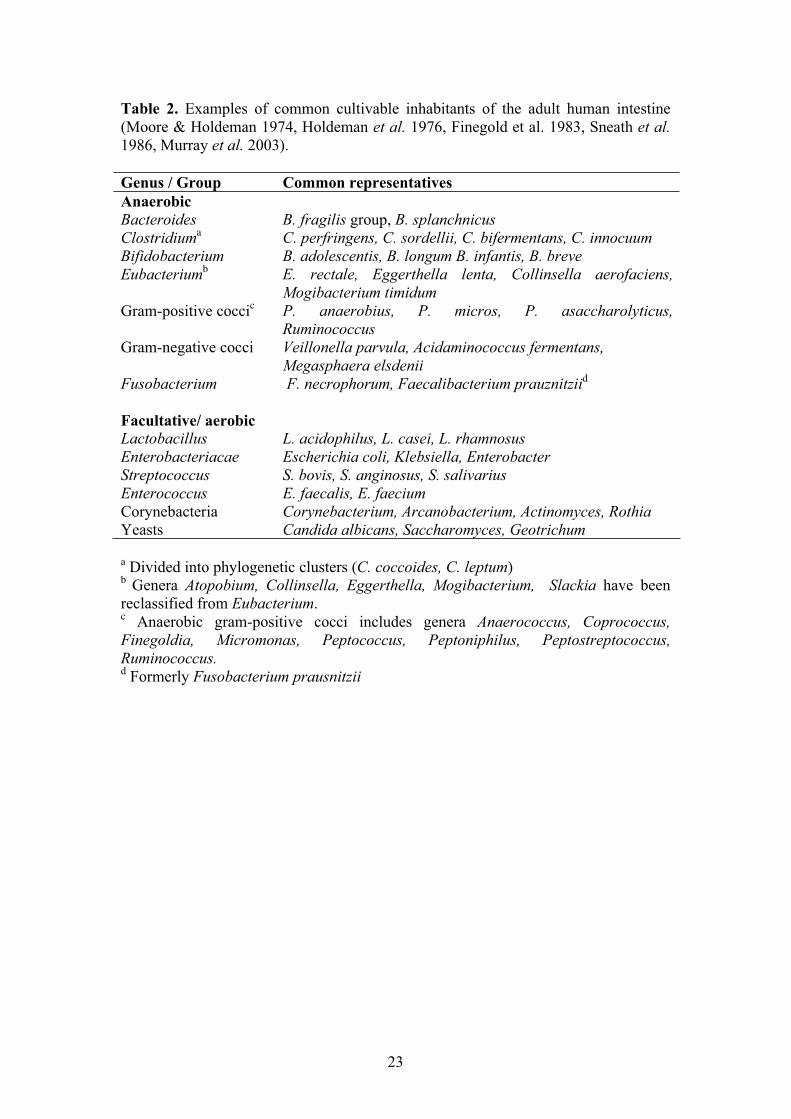
The cultivable dominating fecal bacteria belong to genera Bacteroides,
Bifidobacterium, Eubacterium and related genera, Clostridium, Peptococcus,
Peptostreptococcus and related genera, Fusobacterium and Ruminococcus, whereas
the subdominant family and genera comprise of facultative anaerobes (aerobes) such
as Enterobacteriacae, Streptococcus, Enterococcus, Lactobacillus and Proteus (Table
2) (Simon & Gorbach 1984, Tannock 1999c). Some of these genera, especially
Eubacterium, anaerobic gram-positive cocci and Clostridium have gone through
extensive taxonomic revisions on the basis of new molecular methods (DNA-DNA
hybridisation, 16S rRNA sequencing) that are replacing phenotypic methods as means
to classify microbes. Molecular methods have also provided new aspects to the
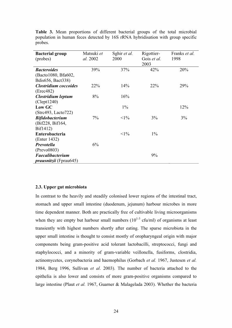
quantitative relations between different bacteria by determining proportions of group
specific rRNA of total rRNA of a microbiota (Table 3). Attempts to combine culture-
based data with molecular methods-based data has not yet provided us an accurate
picture of the numerically predominant active species in the gut. Based on the partial
sequencing of 16S rRNA, most of the bacteria found in the human colon fall into four
phylogenetic clusters consisting of (1) Bacteroides, (2) Bifidobacterium and other
gram-positive bacteria with high G+C content, (3) Clostridium coccoides and
relatives (Coprococcus, Eubacterium, Lachnospira, Ruminococcus), and (4)
Clostridium leptum and relatives, fusobacteria, and the Atopobium group (Wilson et
al. 1997).
23
Table 2. Examples of common cultivable inhabitants of the adult human intestine (Moore & Holdeman 1974, Holdeman et al. 1976, Finegold et al. 1983, Sneath et al.1986, Murray et al. 2003).
Genus / Group Common representatives Anaerobic Bacteroides B. fragilis group, B. splanchnicus Clostridiuma C. perfringens, C. sordellii, C. bifermentans, C. innocuum Bifidobacterium B. adolescentis, B. longum B. infantis, B. breve Eubacteriumb E. rectale, Eggerthella lenta, Collinsella aerofaciens,
Mogibacterium timidum Gram-positive coccic P. anaerobius, P. micros, P. asaccharolyticus,
Ruminococcus Gram-negative cocci Veillonella parvula, Acidaminococcus fermentans,
Megasphaera elsdenii Fusobacterium F. necrophorum, Faecalibacterium prauznitziid
Facultative/ aerobicLactobacillus L. acidophilus, L. casei, L. rhamnosus Enterobacteriacae Escherichia coli, Klebsiella, Enterobacter Streptococcus S. bovis, S. anginosus, S. salivarius Enterococcus E. faecalis, E. faecium Corynebacteria Corynebacterium, Arcanobacterium, Actinomyces, Rothia Yeasts Candida albicans, Saccharomyces, Geotrichum
a Divided into phylogenetic clusters (C. coccoides, C. leptum)b Genera Atopobium, Collinsella, Eggerthella, Mogibacterium, Slackia have been reclassified from Eubacterium.c Anaerobic gram-positive cocci includes genera Anaerococcus, Coprococcus, Finegoldia, Micromonas, Peptococcus, Peptoniphilus, Peptostreptococcus, Ruminococcus. d Formerly Fusobacterium prausnitzii
24
Table 3. Mean proportions of different bacterial groups of the total microbial population in human feces detected by 16S rRNA hybridisation with group specific probes.
Bacterial group (probes)
Matsuki et al. 2002
Sghir et al.2000
Rigottier-Gois et al.2003
Franks et al.1998
Bacteroides(Bacto1080, Bfa602, Bdis656, Bact338)
39% 37% 42% 20%
Clostridium coccoides(Erec482)
22% 14% 22% 29%
Clostridium leptum(Clept1240)
8% 16%
Low GC (Strc493, Lacto722)
1% 12%
Bifidobacterium(Bif228, Bif164, Bif1412)
7% <1% 3% 3%
Enterobacteria (Enter 1432)
<1% 1%
Prevotella (Prevo0803)
6%
Faecalibacterium prausnitzii (Fprau645)
9%
2.3. Upper gut microbiota
In contrast to the heavily and steadily colonised lower regions of the intestinal tract,
stomach and upper small intestine (duodenum, jejunum) harbour microbes in more
time dependent manner. Both are practically free of cultivable living microorganisms
when they are empty but harbour small numbers (101-3 cfu/ml) of organisms at least
transiently with highest numbers shortly after eating. The sparse microbiota in the
upper small intestine is thought to consist mostly of oropharyngeal origin with major
components being gram-positive acid tolerant lactobacilli, streptococci, fungi and
staphylococci, and a minority of gram-variable veillonella, fusiforms, clostridia,
actinomycetes, corynebacteria and haemophilus (Gorbach et al. 1967, Justesen et al.
1984, Berg 1996, Sullivan et al. 2003). The number of bacteria attached to the
epithelia is also lower and consists of more gram-positive organisms compared to
large intestine (Plaut et al. 1967, Guarner & Malagelada 2003). Whether the bacteria
25
found are only transient passengers or represent true colonisers of the upper small
intestine is unclear.
In the distal small intestine (terminal ileum) the counts (104-8 cfu/ml) and diversity,
including appearance of substantial numbers of coliforms and bacteroides, increase
drastically (Drasar & Mill 1975, Hentges 1993). The predominantly gram-positive
bacteria of the upper small intestine shifts to gram-negative predominance towards the
colon (Gorbach et al. 1967). In addition to declining acidity, the microbiota in the
ileum is affected also by reflux of caecal contents (Gorbach et al. 1967, Nord & Kager
1984). The small intestine has been thought to harbour a distinct and characteristic
microbiota but the composition of it has not been fully described. Ileal samples
obtained by intubations and small intestinal samples obtained by needle aspiration
grew total counts of 103-6 cfu/ml, with Bacteroides and anaerobic gram-positive rods
such as Bifidobacterium dominating over enterococci and coliforms (Gorbach et al.
1967, Finegold et al. 1983). In the caecum aerobic species such as Escherichia coli,
enterococci, and lactobacilli, comprise a significantly larger part of the total bacterial
count (108 cfu/ml), covering 50% of total bacterial rRNA, compared to faeces where
aerobes cover 7% of the total count (1010 cfu/ml), respectively (Marteau et al. 2001).
2.4. Opportunistic commensals
Opportunistic commensals are the bacteria that are frequently present as permanent
members of the microbiota but under favourable circumstances may proliferate
vigorously and turn pathogenic. Although the vast majority of the normal microbiota
consists of anaerobic bacteria most endogenous infections are caused by aerobes such
as enterobacteria and enterococci (Vollaard & Clasener 1994). Common anaerobic
genera such as Bacteroides and Clostridium include species with known
pathogenicity; B. fragilis is the most frequent clinical isolate (Namavar et al. 1989)
and C. difficile can exist as a harmless commensal in infants but cause disease
especially in hospitalised elderly (Stark et al. 1982, Poxton 2005). Opportunistic
bacteria may produce toxic metabolites causing disorders when accumulated. Some
bacteria and fungi, e.g. species of Neisseria, Bacteroides, Streptococcus and Candida
have the ability to change surface antigenicity in order to evade host immune
responses (Deitsch et al. 1997, Kuwahara et al. 2004). Bacteria that are seldom
26
associated with infections such as Bifidobacterium, Eubacterium and Lactobacillus
species can also occasionally cause serious illnesses (Brook & Frazier 1993).
Coliforms
One of the predominating bacterial groups in the human and animal intestine is
coliforms, i.e. gram-negative aerobic fermentative rods, with E. coli being the most
common and most studied. It is also considered an indicator of faecal contamination
in food and water. Although E. coli may not cover more than a fraction (<1%) of the
total count of the intestinal bacteria, it is significant as a pathogen being one of the
main causes of nosocomial infections, main causative of urinary tract infections and
an important cause of diarrhoea (Eisenstein & Zaleznik 2000, Whittam et al. 1993,
Maslow et al. 1995). E. coli has acquired several pathogenic mechanisms by which
diarrhoeagenic strains can be divided into four distinct groups: enterotoxigenic
(ETEC), enteroinvasive (EIEC), enterohaemorrhagic (EHEC), and enteropathogenic
(EPEC) E. coli (Mims et al. 1998). On the other hand, non-pathogenic E. coli may
contribute to the preservation of colonisation resistance and has been given as
bacterial probiotic therapy to patients with ulcerative colitis and in prevention of
colonisation of antibiotic-resistant E. coli strains in premature babies and in
prevention of neonatal calf diarrhea (Rastegar et al. 1990, Rembacken et al. 1999,
Kruis et al. 2004, von Buenau 2005).
Fungi
Fungi, most often yeasts, are normally present in small amounts in the intestine and
faeces of mammals. In immunodeficient patients or when bacteria are suppressed by
antimicrobials, yeasts (Candida, Cryptococcus spp.) may overgrow the intestine and
act as pathogens causing diarrhoea, abdominal cramps and infections in other body
sites (Krause et al. 2001, Tortorano et al. 2004). Yeasts can also be probiotic. In
treating antibiotic associated diarrhoea Saccharomyces boulardii serine protease has
been demonstrated to inhibit the pathogenic effects of C. difficile toxins A and B on
human colonic mucosa (Castagliuolo et al. 1999).
27
3. The digestive tract and intestinal microbiota of dog
The development of microbiota begins at birth when the sterile foetus is colonised in
the birth canal and by the immediate environment. The bacterial succession is similar
in the human and canine intestine with the very first colonisers originating from the
mother, followed by microbes benefiting from breast-feeding and then drastically
changing towards obligate anaerobes and greater diversity as solid foods are
introduced (Benno et al. 1992a, Mackie et al. 1999). The most numerous colonisers in
beagles during the first year are bacteroides, eubacteria, bifidobacteria, lactobacilli
and anaerobic cocci while clostridia and streptococci increase later (Benno et al.
1992a).
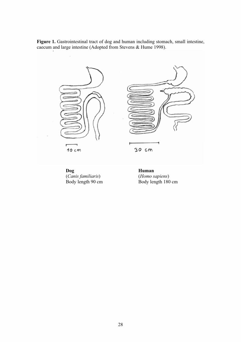
Since dogs are carnivorous the total length of their intestine in relation to the body
length is somewhat shorter and the motility slower than in humans (Figure 1). As a
whole the digestive tract of dog, however, resembles that of humans and the
physiology is similar in many ways. Like humans dogs utilise intestinal microbiota in
their physiology and both are homothermic mammals. The pH values in the different
compartments of the digestive tract are also comparable to those of human: the pH in
the dog stomach is 3, in duodenum and jejunum 6, in ileum 7.5, in colon 6.5 and in
feces 6.2 (Smith 1965).
The main cultivable bacterial groups and most common findings in humans and dogs
are the same including clostridia, bacteroides, streptococci, coliforms, enterococci,
lactobacilli and veillonellae with increasing counts towards the large intestine (Smith
1965, Davis et al. 1977, Benno et al. 1992b, Greetham et al. 2002, Buddington 2003,
Rinkinen et al. 2004). Most intestinal pathogens cause similar clinical symptoms in
dogs as in humans (C. difficile, multiresistant gram-negatives, enterococci,
staphylococci). Due to the ability of microbes to adapt and specify to niches with
unique characteristics there are differences between the bacterial species level
composition in different mammals. The most common intestinal dysfunction in dogs
is diarrhoea. Another common disturbance, small intestinal bacterial overgrowth,
composing of E. coli, enterococci and clostridia is associated with raised serum folate,
reduced serum vitamin B12 concentrations and altered mucosal permeability and
function (Batt et al. 1983, Rutgers et al. 1996).
28
Figure 1. Gastrointestinal tract of dog and human including stomach, small intestine, caecum and large intestine (Adopted from Stevens & Hume 1998).
Dog Human(Canis familiaris) (Homo sapiens)Body length 90 cm Body length 180 cm
29
4. Obtaining samples from the intestine
4.1. Getting representative samples from the upper gut
Studies of microbiota in vivo carry some limitating factors associated to the
complexity of the ecosystem leading to difficulties in identifying and determining the
functions of all members or subpopulations. These include the unknown dynamic
interactions within the microbiota and between microbes and host, inherent
differences in the microbiota between study subjects, natural variability of the
microbial strain composition with time, bacterial contamination during intervention
trials between controls or staff and study group, and differences in human and
experiment animal microbiota/physiology (Corpet 1987, Zoetendal et al. 2004).
Apart from general difficulties in analysing complex microbial populations, the
difficulty in studying the gut microbiota lies in the inaccessibility of the microbiota in
its natural habitat. Samples from intestinal contents from living healthy subjects
obtained without anaesthesia and fasting, preoperative antibiotic prophylaxis, or
laxatives/purgatives are simply not available. After any of these procedures the
microbiota is no more in its natural state and may have gone through vast
modification, and the sample size may also remain minimal. In addition, anaesthesia
slows down the peristaltis, which makes backflow possible, and subjects studied at
surgery have gastrointestinal dysfunctions and do not represent healthy individuals
(Borriello et al. 1978). Attempts to get samples from the intestine without surgery
include self-closing enterocapsules, tubes, endoscopes and, in animals, cannulation
(Hirtzmann & Reuter 1963, Shiner et al 1963, Gracey 1977, Hill 1996, Stotzer 1998).
Capsules that pass through the intestine or tube that remains in place allow bacteria to
multiply before the sample is processed. Invasive techniques going through other
segments of the digestive tract carry the risk of contamination on the way. Tubes may
also alter the intestinal motility and the position of the tube may be difficult to
determine (Donaldson 1964).
Thus most analyses of the intestinal contents or biopsies are from the caecum or colon
and originate from diseased persons (Gillian et al. 1992), sudden death victims
(Macfarlane 1992) or are focused on certain probiotics (Alander et al. 1997, Alander
30
et al. 1999, Johansson et al. 1993) or sulphate reducers (Zinkevich et al. 2000). Very
few studies have analysed the small intestinal microbiota of healthy subjects, and
limited data are thus available (Marteau et al. 2001).
Because of these difficulties in obtaining proper samples from different parts of the
intestinal tract, especially from the upper gut, and ease in collecting faecal samples,
faeces is often the chosen sample type in studies of the gut microbiota. However, the
faecal microbiota represents merely the distal colon, not even the proximal colon or
terminal ileum, and may not represent the upper intestinal microbiota or the metabolic
activities of it. Moreover, faecal predominant species tend to overgrow subdominant
bacteria and the selectivity of selective agar media may be poor which makes it very
difficult to gain isolates for further identification and to characterise the true bacterial
diversity in faecal samples. Molecular methods with higher sensitivity can be used to
detect the subdominant microbes but for further characterisation bacterial isolates
should be obtained by culture.
4.2. Jejunal fistula
To overcome the inaccessibility of the intestine, a dog fistula model was designed to
gain an easy access to jejunal chyme without causing pain to the animal or disturbing
the normal functions of the gut (Wilsson-Rahmberg & Johnsson 1997, Harmoinen et
al. 2001). The fistula is created using a 25 cm segment of the dog’s own intestine by
intussusception. The free ends of the intestine are connected by end-to-end
anastomosis, and the isolated intestinal segment is inserted by end-to-side anastomosis
to the intestine extending through abdominal cavity to connect the intestine to the skin
with proximal end of the segment facing the skin. Small intestinal content (jejunal
chyme) can be collected pre- or post-feeding without aspiration by inserting a silicon
tube through the nipple valve into the intestine. A nipple valve can be surgically
inserted to desired site at the small intestine. The closer the fistula is to the pylorus the
more running (liquid) the content is (Harmoinen 2004). Most productive samples are
obtained shortly after feeding (1-4 h) depending on the site of the fistula. Small
intestinal samples from permanent fistula operated dogs can be obtained without any
sedation/anaesthesia while the dog is fully active and standing. The fistula operation is
reported not to have any significant effect on the intestinal motility, measured as
31
transit percentage of barium-impregnated polyethylene spheres, or on the cultivable
microbiota (Harmoinen et al. 2001).
5. Analysing the microbiota
5.1. Microscopic methods
Microscopic methods can be valuable in analysing bacterial numbers (Kepner & Pratt
1994). However, microscopic counts do not necessarily correlate with the plate count.
Plate count detects only reproducible cells whereas with microscopy it is possible to
enumerate dead and/or viable cells depending on a stain applied (Breeuwer & Abee
2000). On the other hand, the clumping of cells and loss of cells after fixation during
staining may distort microscopic counts. Often microscopy is combined with
fluorescent in situ hybridisation (FISH), which enables the identification and
enumeration of selected bacteria (Amann et al. 1995).
5.2. Culture method
Traditionally culture method has been regarded as the golden standard for studying
bacteria. It is notable that it is the only way to get bacterial isolates for further studies.
The focus of the analysis of the intestinal microbiota has been on the enumeration and
identification of numerically predominant cultivable faecal species (Holdeman et al.
1976, Finegold et al. 1983, Moore & Moore 1995). However, up to 60-80% of the
mammal microbiota cannot be cultured and has not been characterised and identified
(Moore & Holdeman 1974, Wilson & Blitchington 1996, Suau et al. 1999). The
cultivable fraction is, however, relatively high compared to most other microbial
ecosystems such as environmental biofilms and activated-sludge flocks (Amann et al.
1995). Uncultured microbes fail to grow on artificial agar media used in the culture as
their suitable growth conditions, i.e. combination of nutrients, media, atmosphere, or
necessary interactions with the host cells or other microorganisms, are not known or
the cells are stressed or nonviable. In addition, the selective culture media used are
either not selective enough or are too selective supporting the growth of only part of
the microbes, and some microbes prevent the growth of others, and some are
misidentified (Nelson & George 1995, Hartemink & Rombouts 1999). Thus the
32
culture result may not always give a truly representative picture of the microbiota
studied. Culture method is also regarded as time-consuming and laborious. Despite
the limitations, culture methods have successfully provided us the basic knowledge of
clinically important microbes and intestinal ecosystem, and culture remains an
indispensable tool for clinical microbiology.
The identification of isolated bacteria has traditionally relied on morphological,
physiological and biochemical characteristics. This phenotypic characterisation based
on cellular and colonial morphology, growth requirements and characteristics,
fermentation profiles etc. has weaknesses connected to the poor reproducibility,
ambiguity because of plasticity of bacterial growth and poor discriminatory power
(McCartney 2002). Likewise, molecular-based genetic characterisation carries
limitations. Thus there is often need to combine phenotypic with genotypic
characterisation (polyphenic approach).
5.3. Molecular biological methods
Detection and identification
Alternatively, culture-independent molecular methods have been introduced and
notable amounts of DNA originating from uncultivable bacteria have been extracted,
analysed and classified. Molecular methods are recognised as a standard phylogenetic
classification tool (Stackebrandt et al. 1994). They may offer more rapid and reliable
identification than culture because the identification is based on the composition of
nucleic acids rather than on the genomic expression under given culture conditions,
and the DNA can originate from living or dead cells (Nissen & Dainty 1995, Tannock
et al. 2004). The applicability of molecular methods ranges from identification or
detection of single bacterial species to characterisation of compex microbiotas, and
molecular techniques have been applied in analysing evolutionary relatedness of
different bacteria, in bacterial diversity studies, enumeration of bacterial groups,
especially of extremely fastidious organisms, monitoring specific bacterial
populations, and identification of bacterial isolates (McCartney 2002).
Molecular methods are based mainly on detection of ribosomal RNA (rRNA) or
ribosomal DNA (rDNA; DNA encoding the rRNA). The probes for target DNA
33
sequences can be designed for detecting bacteria on different phylogenetic levels from
major genera or the group level to the species or even strain-specific level depending
on the type of the study ranging from gut ecology studies to tracking specific
probiotics or pathogens (Charteris et al. 1997, Franks et al. 1998). Highly conserved
regions of ribosome can be used for designing universal probes and different variable
regions for specific/targeted probes. Several thousands of 16S rDNA sequences
including numerous uncultured bacteria are freely available in genomic databanks.
The target DNA can be detected using various PCR-based methods or dot blot
hybridisation with specific synthetic oligonucleotide probes or using fixed bacterial
cells by fluorescent in situ hybridisation (FISH) combined with flow cytometry or
microscopic analysis (Amann et al. 1995, Wang et al. 1996, Wilson & Blitchington
1996, Lin et al. 1997). Quantitative PCR allows quantification of all DNA fragments
detected by PCR using specific controls of known quantity giving an estimate of the
number of target microorganisms in the sample (Doungudomdacha et al. 2001, Sanz
et al. 2004). Using multiplex PCR several target regions can be multiplied in single
reaction with all necessary primers (García et al. 1998).
Population fingerprinting
In general, fingerprinting techniques for studying bacterial communities within
ecosystem are based on PCR and generate profiles representing the sequence diversity
within the population (Zoetendal et al. 2004). Molecular fingerprinting techniques
allow rapid assessment of the predominant bacterial species present in a sample and
simultaneous analysis of multiple samples, and is well suited to studying changes in
individual microbial communities over time (Suchodolski et al. 2004). Studies using
rRNA are thought to reflect also the metabolic activity of bacteria; in hydridisation the
ribosomal number is thought to be comparable to the number of active cells (Felske et
al. 1997, Rigottier-Gois et al. 2003). TGGE (temperature gradient gel electrophoresis)
or DGGE (denaturing gradient gel electrophoresis) can be used for creating bacterial
profiles that can be compared using similarity percentages and dendrograms (Satokari
et al. 2003, Muyzer et al. 1993). TGGE has high sensitivity for detecting sequence
differences (Muyzer et al. 1993). The composition of individual rDNA genes can also
be sequenced and analysed using cloning libraries (Suau et al. 1999) or profiled using
probing grids, checkerboard hybridisation (O’Sullivan 1999) or DNA microarrays
34
(Wilson et al. 2002). Sequencing of single PCR clones is a laborious and expensive
procedure but allows immediate discrimination between bacterial species.
Like culture, molecular methods carry limitations such as PCR biases, laborious
processing at the species level, requirements for a clone library or sequence
information, semi-quantitative results, and expensive machinery and reagents
(Zoetendal et al. 2004). The biggest challenge in molecular methods lies in the
quantification of different microbial groups and determining their functionality,
activity and dynamics within the diverse microbial populations (Sghir et al. 2000,
McCartney 2002).
5.4. Molecular typing methods
Molecular methods are widely used for typing of cultured isolates. The most common
molecular typing methods are plasmid profiling, pulsed-field electrophoresis (PFGE),
multilocus enzyme electrophoresis (MLEE), ribotyping and random amplification of
polymorphic DNA (RAPD-PCR, AP-PCR) (O’Sullivan 2000, Tannock 2001). These
typing methods are applicable to numerous bacterial genera and can be used to
distinguish bacterial isolates and to determine their relatedness.
For epidemiologically related organisms PFGE has been proven a powerful
discriminating tool, especially in food-born outbreaks and nosocomial infections (Grif
et al. 1998, Barrett et al. 1994, Diekema et al. 1997). PFGE has high discriminatory
power and can differentiate e.g. between subtypes of O157:H7 associated with
specific outbreak investigations, though for large-scale screening of O157:H7 isolates
other fingerprinting methods such as repetitive extragenic palindromes (REP) and
Box primers (rep-PCR), amplified fragment length polymorphism (AFLP) and
ribotyping techniques are also applicable and less time consuming (Hahm et al. 2003).
In PFGE the bacterial DNA is digested with restriction enzyme(s), the obtained
fragments are separated electrophoretically and discrete banding patterns for each
isolate are analysed. Strains typed with PFGE using standardised methodology can be
compared internationally using clone libraries. The determination of relatedness of
non-related organisms with PFGE is more tricky and cannot use the same criteria as
for related organisms, since the relationship between restriction fragment banding
35
pattern and true genetic relatedness is poorly understood. In addition, PFGE includes
always subjective decisions such as the selection of restriction enzyme(s), reading the
restriction fragment banding pattern and choosing methods for analysing the data, and
the interpretation of the results (Davis et al. 2003, Singer et al. 2004). Distracting
variabilities in the patterns brought by transferable large plasmids can be eliminated
by excluding fragments smaller than 160 kb from the analysis (Guyot et al. 1999).
5.5. Indirect methods for analysing microbiota
The microbiota, including cultivable and uncultivable species, can also be studied by
indirect methods such as cellular fatty acid analysis (Peltonen et al. 1992), gas
production (El Oufir et al. 1996), fermentation end product analysis (Topping &
Clifton 2001) and enzyme activity profiling (Tannock 1999b, Gorbach & Bengt
1986). Analysis of microbial biochemical activities gives an insight to the
functionality of the microbiota without having to go through culture, isolation and
identification steps. The in vitro methods, such as continuous flow cultures or batch
fermentations, may not always be predictive of the actual in vivo situation, since the
complex and dynamic interactions between the gut and the microbiota are difficult to
mimic. The analysis of microbiota using bacterial metabolites is also complicated by
their unstability as they are used by other microbes or absorbed.
36
6. Antimicrobial agents
6.1. Classification of antimicrobial agents
Infectious diseases can be controlled by treating a disease with drugs (antimicrobial
therapy), or by preventing it by vaccines (immunisation) and healthy environment
(sanitation, hygiene, nutrition). The most important drugs for treating microbial
diseases are antimicrobial agents, representing one of the great triumphs of modern
medicine. Antimicrobial agents are chemical substances produced by microorganisms
which are active against other microorganisms. In prokaryotes cell wall synthesis,
protein synthesis (bacterial ribosome), nucleic acid synthesis and cell membrane
function are sufficiently different from those in human eukaryotic cells so that they
can be targets for antibacterial agents without inhibition of the equivalent mammalian
cell target (Mims et al. 1998). Many antimicrobial agents are natural microbial
metabolic products or derivates of them, chemically modified to improve their
properties. There are also totally synthetic antimicrobial agents. Some bacterial
species are inherently resistant to some classes of antimicrobials because they lack a
susceptible target, they are impermeable to the agent or they produce inactivating
enzyme (Brock et al. 2003). Antimicrobial agents can be divided on the basis of the
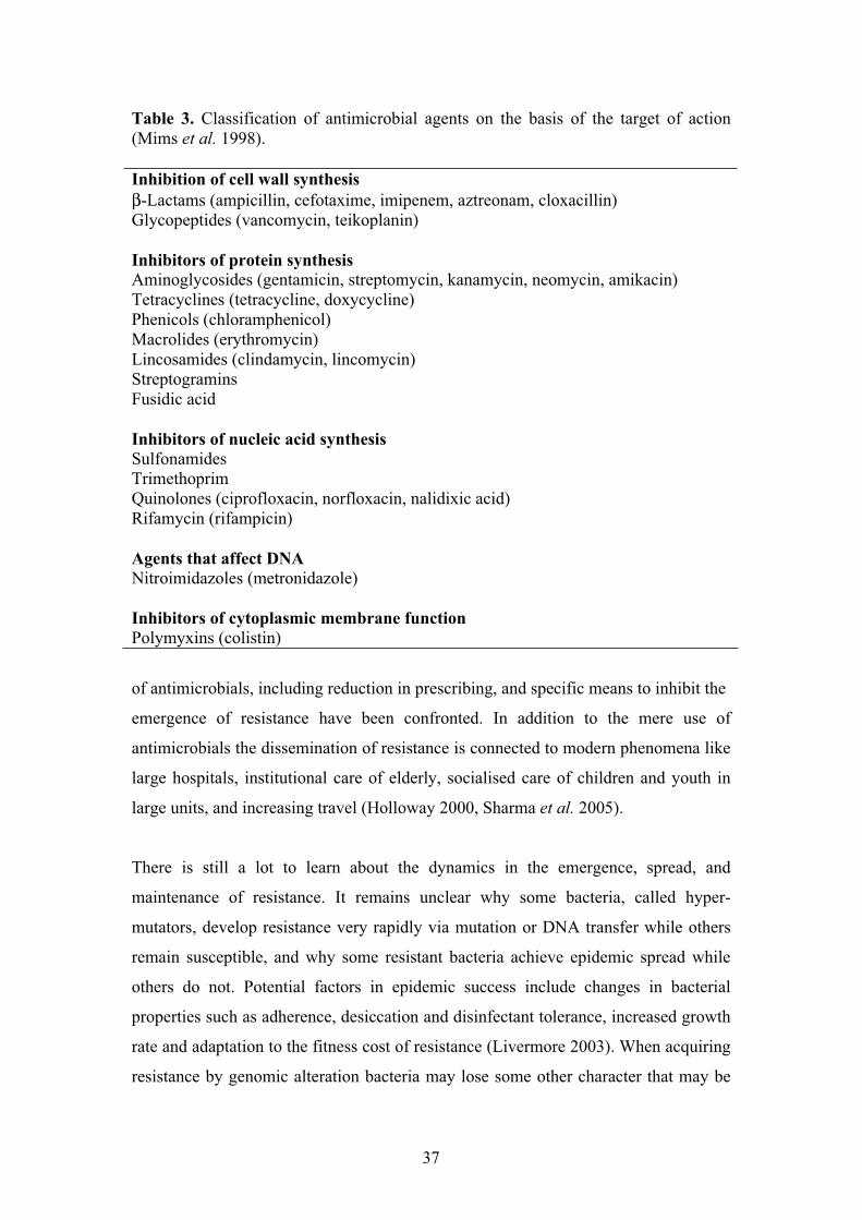
target site, chemical structure or function (Table 3).
6.2. Emergence of antimicrobial resistance
Antimicrobial resistance is a worldwide problem and numerous strategies to avoid the
emergence of resistance have been designed. New antimicrobials are being introduced
but as most of them are similar in structure to the previous ones, the emergence of
resistance is often only a matter of time. Despite of the development of a wide range
of different types of antimicrobial agents, extremely adaptable bacteria seem to
develop resistance to each new agent that comes along. Use of antimicrobials always
causes selective pressure leading to reduction in efficacy, and the huge overuse
induces bacteria to acquire resistance faster than expected (Livermore 2003).
Inappropriate use of antimicrobials brings potential risk to the patient as risks of
toxicity and opportunistic infection or superinfection, and to the community by
selection of resistant organisms and increased health care costs. Both appropriate use
37
Table 3. Classification of antimicrobial agents on the basis of the target of action (Mims et al. 1998).
Inhibition of cell wall synthesis β-Lactams (ampicillin, cefotaxime, imipenem, aztreonam, cloxacillin) Glycopeptides (vancomycin, teikoplanin)
Inhibitors of protein synthesis Aminoglycosides (gentamicin, streptomycin, kanamycin, neomycin, amikacin) Tetracyclines (tetracycline, doxycycline) Phenicols (chloramphenicol) Macrolides (erythromycin) Lincosamides (clindamycin, lincomycin) Streptogramins Fusidic acid
Inhibitors of nucleic acid synthesis Sulfonamides Trimethoprim Quinolones (ciprofloxacin, norfloxacin, nalidixic acid) Rifamycin (rifampicin)
Agents that affect DNA Nitroimidazoles (metronidazole)
Inhibitors of cytoplasmic membrane function Polymyxins (colistin)
of antimicrobials, including reduction in prescribing, and specific means to inhibit the
emergence of resistance have been confronted. In addition to the mere use of
antimicrobials the dissemination of resistance is connected to modern phenomena like
large hospitals, institutional care of elderly, socialised care of children and youth in
large units, and increasing travel (Holloway 2000, Sharma et al. 2005).
There is still a lot to learn about the dynamics in the emergence, spread, and
maintenance of resistance. It remains unclear why some bacteria, called hyper-
mutators, develop resistance very rapidly via mutation or DNA transfer while others
remain susceptible, and why some resistant bacteria achieve epidemic spread while
others do not. Potential factors in epidemic success include changes in bacterial
properties such as adherence, desiccation and disinfectant tolerance, increased growth
rate and adaptation to the fitness cost of resistance (Livermore 2003). When acquiring
resistance by genomic alteration bacteria may lose some other character that may be
38
valuable when the antimicrobial selective pressure is absent. This burden or losing
some feature is called fitness cost. Resistance mechanisms can be selected in both
pathogenic and non-pathogenic bacteria, as shown in E. coli (Sunde & Sorum 1999).
The largest reservoir of resistance has been thought to reside in the commensal gut
microbiota (Guyot et al. 1999, Gulay et al. 2000, McDonald et al. 2001).
Antimicrobial remains get into the intestine as residues that are not absorbed after oral
administration, by diffusion from the surrounding tissue, and in bile via enterohepatic
circulation (Nord & Heimdahl 1986b, Edlund & Nord 2000). After being enriched the
resistance determinants may transfer within or between different bacterial genera e.g.
between Bacteroides species and from gram-positive bacteria (Clostridium,
Streptococcus, Enterococcus) to gram-negative bacteria (Bacteroides), and between
bacteria originating from humans and animals (Nijsten et al. 1995, Shoemaker et al.
2001).
6.3. Mechanisms of resistance
As there are several mechanisms by which antibiotics influence bacteria, there are
also several mechanisms by which bacteria acquire resistance. Resistance may result
from a single random chromosomal mutation or a series of mutations leading to
synthesis of altered protein. Spontaneous mutants that have competitive advantage in
the presence of antibiotic pressure survive and multiply. Dissemination of resistance
is, however, more often the result of acquirence of transmissible plasmids,
transposons or gene cassettes in integrons carrying resistance genes, typically coding
for resistance to several unrelated classes of antimicrobials (Hall 1997, Poole 2001).
Mechanisms of resistance include target site alterations (lowered affinity), alterations
in the access to the target site (impermeability, efflux pumps, alternative metabolic
pathways) and inactivation of the drug (enzymes that modify or destroy the
antimicrobial) (Brock et al. 2003).
6.4. Adverse effects of antimicrobial agents
An ideal antimicrobial agent would provide selective decontamination of the
microbiota in a “gut friendly” manner, i.e. be active against pathogens and potentially
pathogenic microbes but not against those that preserve the colonisation resistance.
39
Unfortunately only few, if any, available antimicrobials meet these criteria (Vollaard
& Clasener 1994).
The use of antimicrobials leads to selection of resistant organisms among pre-existing
variants and may radically reduce the number of commensal anaerobes susceptible to
antimicrobials like Bifidobacterium, Bacteroides and Lactobacillus leading to
reduction in colonisation resistance. This opens new niches for the remaining resistant
opportunistic bacteria or transient pathogens that normally have restricted growth.
They may overgrow the intestine or move to another anatomical site where the host is
unable to tolerate the colonisation, and pathogenic microbes such as C. difficile,
vancomycin-resistant enterococci, multidrug-resistant enterobacteria and Candida
may proliferate (Berg 1996, Falk et al. 1998, Sullivan et al. 2001). In patients with C.
difficile-associated diarrhoea treated with metronidazole the proportion of facultative
bacteria was increased with higher enterobacterial and enterococcal counts but also
with greater diversity of clostridia and lactobacilli compared to healthy subjects
(Hopkins & Macfarlane 2002).
In addition to persisting infections and dissemination of resistant organisms,
antibiotics predispose patients to serious clinical implications such as gastrointestinal
disturbances and functional bowel symptoms, including diarrhoea, alterations in
fermentation processes and reduction of short-chain fatty acid production (Bergogne-
Berezin 2000, Sullivan et al. 2001, Maxwell et al. 2002). Antibiotic-associated
reduced carbohydrate metabolism in turn may result in osmotic diarrhoea, poor
absorption of fatty acids, water and electrolytes, altered bile acid balance and
cholesterol metabolism (Hashimoto et al. 1996, Bergogne-Berezin 2000).
Antimicrobials may also have allergic and toxic effects on the mucosa, and the
pharmacologic effect that macrolides have on intestinal motility may affect the
pathogenesis of antibiotic-associated diarrhoea (Bergogne-Berezin 2000). Antibiotics
may also trigger release of LPS (lipopolysaccharides) endotoxins from gram-negative
organisms with possible deleterious effects (Prins et al. 1985). A combination of oral
Candida albicans administration and Aspergillus fumigatus spore exposure has been
shown to induce allergic airway disease in mice treated with antibiotics
(cefoperazone) but not in untreated mice (Noverr et al. 2004). In clinical practise,
antimicrobial resistance increases mortality, morbidity and health care costs. If the
40
patient is not responding to the antimicrobial treatment, the length of hospital stay is
prolonged or repeated physician visits are needed, and secondary antimicrobials are
often more expensive than the first line choices (Livermore 2003).
Nosocomial infections
Cross-contamination of resistant bacteria between individuals is especially significant
in hospital environment where there is simultaneous selective pressure from several
antibiotics and pathogens with lowered antimicrobial susceptibility may be enriched.
In addition, a lot of patients with predisposing factors such as impaired
immunosystem are exposed (Blazquez et al. 2000). Both hospitalised individuals and
outpatiens are reported to frequently carry resistant bacteria regardless of antibiotic
usage (Levy et al. 1988). The most common organisms causing infections of the
urinary tract, surgical wounds, skin, respiratory and gastrointestinal tract, as well as
systemic infections are staphylococci including MRSA, enterococci including VRE,
E. coli, Klebsiella spp., Enterobacter spp., Pseudomonas spp., C. difficile and
Candida spp. (Schaberg et al. 1991, Farr 2002, Biedenbach et al. 2004, Hull & Beck
2004, D'Agata 2004). However, in many cases the infective strain originates from the
patient’s own microbiota, thus hygienic measures cannot alone completely prevent
nosocomial infections (Flynn et al. 1987), which are a widely acknowledged problem
in both human and animal care, and are known to contribute significantly to mortality
and health care costs. In Finnish patients over 50 000 nosocomial infections are
reported annually, contributing to 2000-5000 deaths (Ora 2005).
6.5. Use of antimicrobials in humans and animals
Common use of antimicrobials of human importance in cattle and pets may increase
the risk of transferring resistance from animal to human microbiota. Controversial
arguments about antibiotic use in animals are being debated (Piddock 1996, Cook
1997, Phillips et al. 2004, Guardabassi et al. 2004). Reductions in prescribing
antimicrobials at a national level have been shown to reduce the prevalence of
resistance within few years (Kristinsson 1997, Seppälä et al. 1997). However,
reduction in antimicrobial usage does not always lead to reduced resistance. Resistant
strains that are well adapted and carry minimal or no fitness cost because of the
resistance determinant may not be displaced for a long period of time after the
41
exposure to antimicrobial agent has ceased. Strains that acquire resistance plasmid
may initially grow more slowly than plasmid-free counterparts in the absence of
selection pressure but after repeated subculture the difference may diminish as shown
in tetracycline- and streptomycin-resistant E. coli and in tetracycline- and
erythromycin-resistant Bacteroides (Bouma & Lenski 1988, Lenski 1997, Shoemaker
et al. 2001).
6.6. β-Lactam antibiotics
One of the most important groups of antimicrobial agents is β-lactam antibiotics,
which are widely used in human and veterinary medicine (Livermore 1998). β-
Lactams account for 60% of antimicrobial consumption in Finland (National Medical
Statistics, www.nam.fi). Most commonly used β-lactams include penicillins
(ampicillin, piperacillin, cloxacillin), cephalosporins (cephalexin, cefuroxime,
ceftriaxone), cephamycins (cefoxitin) and carbapenems (imipenem, meropenem).
Their antimicrobial potential is based on their ability to interfere in the biosynthesis of
the peptidoglycan layer of the bacterial cell wall.
Ampicillin
Ampicillin (C16H19N3O4S) is relatively non-toxic and acid-stable and can be given
orally or parenterally, parenterally in sodium salt form. Absorption of ampicillin is
slower and more irregular than that of some of the esters (talampicillin) and analogs
(amoxicillin) developed from it. The serum half-life of ampicillin in normal adults is
0.7-1.4h (Neuvonen et al. 1994). Peak serum concentrations are obtained at about 2h
after oral administration and right after parenteral administration. After an oral dose
30% of ampicillin is exreted in urine within 6h, and after parenteral administration
75% (Kucers & Bennett 1987). Low amounts are excreted also in bile. Unexcreted
ampicillin is inactivated mainly in the liver (Kucers & Bennett 1987).
42
6.7. β-Lactamases
The most common mechanism of resistance to β-lactams is bacterial β-lactamases,
produced by a number of both gram-negative and gram-positive bacterial species. In
addition to clinically important enterobacteria, β-lactamase production has been
described in various anaerobic genera of the normal microbiota such as Bacteroides,
Fusobacterium and Clostridium (Nord 1986a). β-Lactamases inactivate the antibiotic
by hydrolysing the amide bond in the β-lactam ring. Numerous different β-
lactamases, both chromosomal and plasmid-mediated, have been described and they
have been classified into four main groups and several subgroups on the basis of their
function or structure (Livermore 1995, Bush et al. 1995, Bush & Jacoby 1997).
Although hyperproduction of wild type β-lactamase induces resistance, the major
contributors to resistance are thought to have arisen from single or multiple mutations
in the structural β-lactamase genes (Reguera et al. 1991, Henquell et al. 1995,
Belaaouaj et al. 1994, Brun et al. 1994). Resistance determinants are transferable and
are found within a wide variety of plasmids (Thomson et al. 1993). In hospital
environment ampicillin is considered a significant factor in maintaining selection
pressure for the most prevalent plasmid mediated β-lactamases in gram-negative
organisms (Burman et al. 1992).
The predominant β-lactamases in gram-negative bacteria are TEM-type β-lactamases,
coded by blaTEM genes, with TEM-1, TEM-2 and SHV-1 enzymes being the most
common ones (Brinas et al. 2002). In E. coli TEM-1 is the predominating β-lactamase
(Huovinen et al. 1988). These Ambler class A enzymes have traditionally been active
against a broad spectrum of penicillins but evolution and selective pressure have
conferred resistance to expanded-spectrum cephalosporins (extended-spectrum β-
lactamases; ESBLs) or to β-lactam-clavulanic acid combinations (inhibitor-resistant
TEM; IRT) or to both (complex mutant TEM; CMT) (Medeiros 1997, Sirot et al.
1997, Poirel et al. 2004).
43
6.8. Inhibition of β-lactam resistance
β-Lactamase inhibitors
A way to avoid the microbial breakdown of β-lactams is the use of β-lactamase
inhibitors simultaneously with β-lactams. β-Lactamase inhibitors such as clavulanic
acid, sulbactam and tazobactam contain β-lactam ring as substrates for the β-
lactamase to which the “suicide inhibitor” binds preventing the enzyme to destroy the
antibiotic. By themselves β-lactamase inhibitors have only little therapeutic value.
Bacteria are, however, acquiring mechanisms to overcome the action of many of these
inhibitors and a constant designing of new inhibitors is required.
Degradation of intestinal β-lactam residues
Another strategy to fight the emergence of resistance is intra-intestinal inactivation of
antimicrobials by degrading the leftover antibiotic accumulating in the intestine where
it triggers resistance and quantitative changes within the abundant microbial
population (de Vries-Hoepers et al. 1993, Van der Waaij & Nord 2000). By degrading
the antibiotic residue the selective pressure is abolished and the gut microbiota and
colonisation resistance remain unaffected during antimicrobial treatment.
One way to bring β-lactamase to the intestine is by administrating β-lactamase-
producing bacteria into the gut. Many intestinal microbes produce β-lactamase and
faecal preparations from healthy subjects have been shown to inactivate β-lactams
with individual variation (Veringa & van der Waaij 1984, Welling & Groen 1990).
Patients harbouring β-lactamase-producing intestinal bacteria have less β-lactam-
induced alterations in their microbiota (van der Waaij et al. 1986, Chachaty et al.
1992, Chachaty et al. 1993, Stark et al. 1995). In gnotobiotic mice detectable amounts
of β-lactamase and inhibition of ceftriaxone activity were produced in the intestine by
colonising the mice with a mixture of four β-lactamase-producing anaerobes
(Bacteroides thetaiotaomicron, Clostridium clostridioforme, B. uniformis E9, B.
uniformis V4E3) (Leonard et al. 1989).
Administrating the β-lactamase as a purified enzyme enables accurate screening and
evaluation of the effects of the administration providing knowledge of the dosage.
44
When given orally the enzyme should be in a form where it does not affect the
therapeutic effect of the target antimicrobial, thus it should be active only in the
intestine, and not be absorbed into the systemic circulation, and, on the other hand,
resist proteolysis and remain active in the intestine. This can be achieved in oral
administration by packing the enzyme in capsules that are targeted to release the
enzyme in the lower gastrointestinal tract and not in the stomach, in e.g. pH-based
manner. In the present study, we analysed the ability to inhibit antibiotic-induced
changes in the gut with a product with the above-mentioned characteristics, called
targeted recombinant oral β-lactamase (TRBL). TRBL has been shown to degrade
ampicillin in the jejunum of beagle dogs in a dose dependent manner but not to enter
the systemic circulation or affect the serum ampicillin levels after i.v. administration
of ampicillin (Harmoinen et al. 2003). A commercially available β-lactamase is in use
in animal husbandry to destroy penicillin residues in cow milk (Korycka-Dahl et al.
1985, Suomen eläinlääkkeet 2003)
6.9. Testing antimicrobial susceptibility
The antimicrobial susceptibility of bacteria can be determined in vitro by standardised
methods such as disk diffusion method and broth or agar dilution assay (NCCLS
2002). Disk diffusion method is convenient in screening of resistance phenotypes
(presence of the resistance genes) while dilution assay determines the precise level of
resistance. In disk diffusion method the size of growth-inhibiton zone around a filter
paper disk containing the antibiotic on agar plate determines the susceptibility of the
culture, and in antimicrobial dilution assay minimal inhibitory concentration (MIC) is
determined as inhibition of growth within a series of antibiotic dilutions in broth or on
agar. MIC can also be tested on agar using commercial E-test strips containing stable
increasing antibiotic gradient producing a growth-inhibition ellipse.
45
AIMS OF THE STUDY
The purpose of the study was to examine in detail the microbiota in the upper and
lower part of canine intestine. The samples were collected before, during and after
ampicillin treatment with or without simultaneous β-lactamase administration and
analysed using both traditional culture method and molecular methods. When
designing the study we hypothesised that (1) jejunal and faecal microbiota differ
significantly from each other, (2) ampicillin administration has dramatic effects on the
composition and counts of the microbiota and on the susceptibility of the coliforms,
(3) oral targeted β-lactamase inactivates intestinal ampicillin, inhibits the ampicillin-
induced changes and preserves colonisation resistance, (4) shifts in coliform
population can be followed using resistance data and PFGE (pulsed-field gel
electrophoresis) typing.
The specific aims of the study were to:
(I) Compare the cultivable canine microbiota in the jejunum to that in faeces.
(II) Analyse the ability of β-lactamase administration to prevent ampicillin-
induced emergence of resistance in dogs during ampicillin treatment
monitored by performing faecal bacterial culture combined to susceptibility
testing, performing TGGE (temperature gradient gel electrophoresis) for
dominant faecal bacterial groups and by determining the quantity of TEM-type
β-lactamase-producing faecal bacteria by PCR and by following ampicillin
levels in serum and jejunum.
(III) Characterise canine jejunal and faecal coliform populations, their species
variability and resistance patterns in the three treatment groups.
(IV) Determine the genetic relatedness of coliform populations in the three
treatment groups monitored by resistance patterns and PFGE typing.
46
MATERIALS AND METHODS
1. Study design
In the present study, healthy male laboratory beagles with permanent jejunal fistula
were used. The dogs were obtained from Harlan-Winkelmann Gmbh (Borchen,
Germany) and National Laboratory Animal Center (University of Kuopio, Finland).
Their age was between 1 and 3 years and weight between 12 and 19 kg. The dogs
were fistula operated and kept in the premises of the Department of Clinical
Veterinary Sciences (Faculty of Veterinary Medicine, University of Helsinki). An
intussuscepted nipple valve fistula was surgically inserted 170 cm distal to the pyrolus
(Harmoinen 2004). The dogs were kept individually, fed with commercial dog food
(Pedigree , Waltham , Masterfoods, Helsinki, Finland) and taken for walks in the
same premises. Coprophagy was avoided by cleaning faeces from the premise after
each dog had been taken out. The local ethics committee for animal experiment action
in Helsinki, Finland approved the study.
For a pilot study 6 beagles were randomised into 3 treatment groups, 2 in each,
receiving ampicillin and placebo, ampicillin and targeted recombinant β-lactamase
(TRBL) or only placebo. For the final study a new group of 18 beagles were similarly
randomised into 3 treatment groups, 6 in each, but instead of 20 mg/kg of ampicillin
for 10 days they received 40 mg/kg for 14 days (Table 1). The baseline samples were
collected on day 0 and the follow-up samples 2 weeks after the treatment period
(Table 2). Ampicillin sodium (A-PEN , Orion Pharma, Espoo, Finland) or placebo
(Natrosteril , Orion Pharma) was administered i.v. 4 times a day 30 min after feeding
and TRBL or placebo capsules were administered 20 and 27 min after feeding.
Because of the different dosing of the ampicillin, the pilot study samples were
excluded from the resistance studies (II, III, IV) but all baseline samples and all
consecutive samples from the dogs receiving only placebo were included in the
comparison study (I).
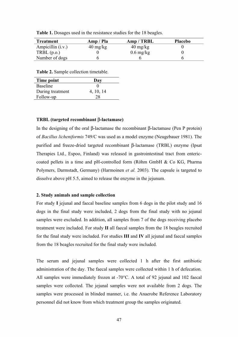
47
Table 1. Dosages used in the resistance studies for the 18 beagles.
Treatment Amp / Pla Amp / TRBL Placebo Ampicillin (i.v.) 40 mg/kg 40 mg/kg 0 TRBL (p.o.) 0 0.6 mg/kg 0 Number of dogs 6 6 6
Table 2. Sample collection timetable.
Time point Day Baseline 0 During treatment 4, 10, 14 Follow-up 28
TRBL (targeted recombinant β-lactamase)
In the designing of the oral β-lactamase the recombinant β-lactamase (Pen P protein)
of Bacillus licheniformis 749/C was used as a model enzyme (Neugebauer 1981). The
purified and freeze-dried targeted recombinant β-lactamase (TRBL) enzyme (Ipsat
Therapies Ltd., Espoo, Finland) was released in gastrointestinal tract from enteric-
coated pellets in a time and pH-controlled form (Röhm GmbH & Co KG, Pharma
Polymers, Darmstadt, Germany) (Harmoinen et al. 2003). The capsule is targeted to
dissolve above pH 5.5, aimed to release the enzyme in the jejunum.
2. Study animals and sample collection
For study I jejunal and faecal baseline samples from 6 dogs in the pilot study and 16
dogs in the final study were included, 2 dogs from the final study with no jejunal
samples were excluded. In addition, all samples from 7 of the dogs receiving placebo
treatment were included. For study II all faecal samples from the 18 beagles recruited
for the final study were included. For studies III and IV all jejunal and faecal samples
from the 18 beagles recruited for the final study were included.
The serum and jejunal samples were collected 1 h after the first antibiotic
administration of the day. The faecal samples were collected within 1 h of defecation.
All samples were immediately frozen at -70°C. A total of 92 jejunal and 102 faecal
samples were collected. The jejunal samples were not available from 2 dogs. The
samples were processed in blinded manner, i.e. the Anaerobe Reference Laboratory
personnel did not know from which treatment group the samples originated.
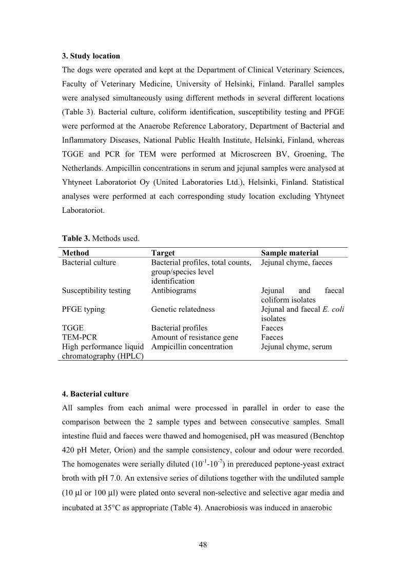
48
3. Study location
The dogs were operated and kept at the Department of Clinical Veterinary Sciences,
Faculty of Veterinary Medicine, University of Helsinki, Finland. Parallel samples
were analysed simultaneously using different methods in several different locations
(Table 3). Bacterial culture, coliform identification, susceptibility testing and PFGE
were performed at the Anaerobe Reference Laboratory, Department of Bacterial and
Inflammatory Diseases, National Public Health Institute, Helsinki, Finland, whereas
TGGE and PCR for TEM were performed at Microscreen BV, Groening, The
Netherlands. Ampicillin concentrations in serum and jejunal samples were analysed at
Yhtyneet Laboratoriot Oy (United Laboratories Ltd.), Helsinki, Finland. Statistical
analyses were performed at each corresponding study location excluding Yhtyneet
Laboratoriot.
Table 3. Methods used.
Method Target Sample material Bacterial culture Bacterial profiles, total counts,
group/species level identification
Jejunal chyme, faeces
Susceptibility testing Antibiograms Jejunal and faecal coliform isolates
PFGE typing Genetic relatedness Jejunal and faecal E. coliisolates
TGGE Bacterial profiles Faeces TEM-PCR Amount of resistance gene Faeces High performance liquid chromatography (HPLC)
Ampicillin concentration Jejunal chyme, serum
4. Bacterial culture
All samples from each animal were processed in parallel in order to ease the
comparison between the 2 sample types and between consecutive samples. Small
intestine fluid and faeces were thawed and homogenised, pH was measured (Benchtop
420 pH Meter, Orion) and the sample consistency, colour and odour were recorded.
The homogenates were serially diluted (10-1-10-7) in prereduced peptone-yeast extract
broth with pH 7.0. An extensive series of dilutions together with the undiluted sample
(10 µl or 100 µl) were plated onto several non-selective and selective agar media and
incubated at 35°C as appropriate (Table 4). Anaerobiosis was induced in anaerobic
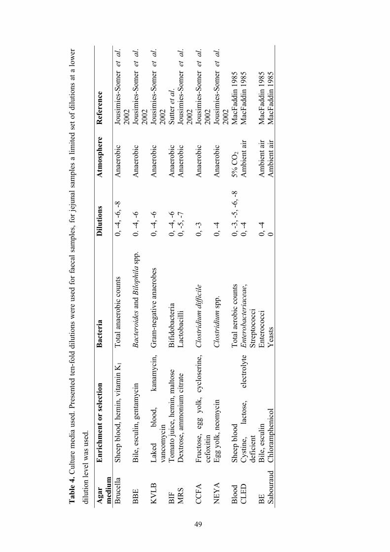
Tab
le 4
. Cul
ture
med
ia u
sed.
Pre
sent
ed te
n-fo
ld d
ilutio
ns w
ere
used
for f
aeca
l sam
ples
, for
jeju
nal s
ampl
es a
lim
ited
set o
f dilu
tions
at a
low
er
dilu
tion
leve
l was
use
d.
Aga
rm
ediu
mE
nric
hmen
t or
sele
ctio
n B
acte
ria
Dilu
tions
A
tmos
pher
e R
efer
ence
Bru
cella
Sh
eep
bloo
d, h
emin
, vita
min
K1
Tota
l ana
erob
ic c
ount
s 0,
-4, -
6, -8
A
naer
obic
Jo
usim
ies-
Som
er e
t al
.20
02B
BE
Bile
, esc
ulin
, gen
tam
ycin
Ba
cter
oide
s and
Bilo
phila
spp.
0.
-4, -
6 A
naer
obic
Jo
usim
ies-
Som
er e
t al
.20
02K
VLB
La
ked
bloo
d,
kana
myc
in,
vanc
omyc
in
Gra
m-n
egat
ive
anae
robe
s 0,
-4, -
6 A
naer
obic
Jo
usim
ies-
Som
er e
t al
.20
02B
IF
Tom
ato
juic
e, h
emin
, mal
tose
B
ifido
bact
eria
0,
-4, -
6 A
naer
obic
Su
tter e
t al.
MR
S D
extro
se, a
mm
oniu
m c
itrat
e La
ctob
acill
i 0,
-5, -
7 A
naer
obic
Jo
usim
ies-
Som
er e
t al
.20
02C
CFA
Fr
ucto
se,
egg
yolk
, cy
clos
erin
e,
cefo
xitin
C
lost
ridi
um d
iffic
ile
0, -3
A
naer
obic
Jo
usim
ies-
Som
er e
t al
.20
02N
EYA
Eg
g yo
lk, n
eom
ycin
C
lost
ridi
um sp
p.
0, -4
A
naer
obic
Jo
usim
ies-
Som
er e
t al
.20
02B
lood
Sh
eep
bloo
d To
tal a
erob
ic c
ount
s 0,
-3, -
5, -6
, -8
5% C
O2
Mac
Fadd
in 1
985
CLE
D
Cys
tine,
la
ctos
e,
elec
troly
te
defic
ient
En
tero
bact
eria
ceae
,St
rept
ococ
ci
0, -4
A
mbi
ent a
ir M
acFa
ddin
198
5
BE
Bile
, esc
ulin
En
tero
cocc
i0,
-4
Am
bien
t air
Mac
Fadd
in 1
985
Sabo
urau
d
Chl
oram
phen
icol
Y
east
s0
Am
bien
t air
Mac
Fadd
in 1
985
49
jars filled by the evacuation-replacement technique (Anoxomat WS8000, Mart B.V.,
Lichtenvoorde, Netherlands) with gas mixture (90% N2, 5% CO2, 5% H2). The exposure
time to air was minimized throughout the procedure with careful planning and
preparations, including the use of prereduced dilution broth and agar plates kept in
anaerobic conditions 18 h prior to their use.
The total counts and main groups of aerobic and anaerobic bacteria and yeasts were
enumerated (the detection limit 102 CFU/g), different colony morphotypes were recorded
with a stereomicroscope and isolated and identified by established methods. The isolates
were picked according to differing morphologies, not on the amount of colonies on the
plate or assumed importance of a finding. The identification scheme included
aerotolerance testing, gram staining, spot and screening tests (catalase, indole, nitrate,
lecitinase, lipase, pigment), special antimicrobial potency disk patterns (vancomycin,
kanamycin, colistin, sodium polyanethol sulfonate, penicillin, oxgall), biochemical tests
(carbohydrate fermentation, enzyme detection) and metabolic end-product analysis by
gas-liquid chromatography after peptone-yeast extract glucose broth culture (Jousimies-
Somer et al. 2002, Murray et al. 2003). The presence of C. difficile common antigen and
toxin A in faeces was determined with a commercial kit (Triage C. difficile Panel,
Biosite, San Diego, CA) according to the manufacturer’s instructions. For unclassified
clostridia resembling C. difficile, genes encoding 16S rRNA were amplified and
sequenced with the primers fD1 Mod and 533r by the method of Jalava et al. (1995) and
compared to the GenBank database. The microbial composition and counts between
different sampling days, different treatment groups and between corresponding jejunal
and faecal samples were compared.
5. Identification, susceptibility testing and PFGE typing of coliforms
From each sample ten colonies or, if less, as many as possible of coliformic colonies
(aerobic fermentative gram-negative rods) were isolated from Blood agar and CLED agar
plates with optimal growth, and sub-cultured for further identification (Murray et al.
2003) and susceptibility testing (NCCLS 2002). The isolates were picked at random but
all visibly different colonies were included to get a representative selection of the
50
51
coliform population. The resistant isolates may not present the most common
morphologic phenotype nor differ by e.g. lactose fermentation profile (Levy et al. 1988).
The species level identification of coliforms was performed with spot tests (catalase,
indole, oxidase), an individual diagnostic tablet of β-glucuronidase (Rosco, Taastrup,
Denmark) and with the Api 20E kit test (bioMérieux, Marcy l’Etoile, France).
For all isolated coliforms the resistance to several classes of antimicrobials including
penicillins (ampicillin 10 µg), carbapenems (meropenem 10 µg), tetracyclines
(tetracycline 30 µg), folate pathway inhibitors (trimethoprim 5 µg, trimethoprim/
sulfamethoxazole 24 µg), aminoglycosides (gentamicin 10 µg), quinolones (ciprofloxacin
5 µg), and cephems (cephalothin 30 µg, cefotaxime 30 µg) was tested (Oxoid,
Hampshire, England). The susceptibility was determined using the disk diffusion method
as recommended by the National Committee for Clinical Laboratory Standards (NCCLS
2002) on Müller Hinton agar inoculated with swabs dipped in bacterial suspension of 0.5
McFarland with 16-18 h incubation. E. coli ATCC 25922 was used as a control strain.
The selected E. coli isolates were typed using pulsed-field gel electrophoresis (PFGE) as
described by Saari et al. (2001). The selected strains presented all different resistance
patterns present in each dog at each time point. Bacterial suspension with A600 of 0.300
was done in TEN buffer and embedded in 1.8% agarose (InCert Agarose, BioWhittaker
Molecular Applications, Inc., Denmark) to prevent mechanical damage to the bacterial
chromosome, lysed with proteinase K (0.5 mg/ml) (Roche, Mannheim, Germany) in ES
buffer overnight at +50°C, washed to remove the protein digestion products, and the
DNA was digested at +37°C overnight with restriction endonuclease XbaI (New England
Biolabs, Beverly, MA) that cuts DNA infrequently. The blocks were run in 1% agarose
gel (Seakem Gold Agarose, BioWhittaker Molecular Applications, Inc., Denmark) in 0.5
x TBE buffer (6V/cm, 120°, 14°C, 21h, pulse times 5-40s) to resolve the restriction
fragments into different patterns of discrete bands. The electrophoretic apparatus switches
the direction of the current as programmed causing the fragments to reorientate between
movements. As the swiftness of the reorientation is dependent on the size of the
fragments this intensifies the separation of fragments with small difference in size. The
52
gels were stained with ethidium bromide (10µl EtBr / 200ml TBE, 30 min) and
photographed under UV-light (Alpha Imager, Alpha Innotech, San Leandro, CA). The
images were analysed using the Bionumerics version 3.0 cluster analysis with Dice
similarity indices (1% position tolerance, UPGMA) (Applied Maths BVBA, Austin,
Texas). The genetic relatedness of the strains based on PFGE profiles (fingerprint
patterns) was interpreted as recommended by Tenover et al. (1995).
6. TGGE (temperature gradient gel electrophoresis)
TGGE was used to assess the bacterial profile and diversity with a molecular approach,
based on the sequence variability of the V6 toV8 regions of the 16S rRNA. TGGE was
performed as described by Muyzer et al. (1993) using primers P16S968GCF and
P16S1401R. Briefly, 50 mg of faeces was homogenised and the bacterial DNA was
extracted. PCR reaction was performed using Invitrogen’s Taq DNA polymerase kit and
PTC-200 PCR system (MJ-Research). A temperature gradient was established in
polyacrylamide gel in parallel to an electric field. The gels were run (200 V, 60°C) in
1.5% (wt/vol) agarose gel using TGGE MAXI system (Biometra, Germany). The
amplified DNA fragments migrate differently according to their size and sequence
specific melting behaviour (thermal stability) to the point where the conformation of the
double stranded DNA changes. As the sequences in TGGE are all the same length the
fragments separate in sequence specific manner, and a profile reflecting the prominent
bacterial content of the sample is achieved. Fragments containing more A-T pairs are
more vulnerable to temperature than those containing C-G pairs. To prevent the double
stranded DNA to denature into single stranded DNA, a C-G rich segment is added to one
of the primers. The gels were silver stained with AgNO3 by the method of Cairns and
Murray (1994). The individually unique TGGE profiles obtained from the same
individual on different sampling days were compared using similarity percentages and
constructing dendrograms using GelCompar II software (Applied Maths, Belgium).
7. PCR for detecting TEM gene
The emergence of ampicillin resistance was evaluated also by performing quantitative
real-time PCR of the TEM β-lactamase gene by quantifying the percentage of TEM
53
producing bacteria of the total number of bacteria as proportions of DNA in the faecal
samples. The PCR was designed for V6-V8 region of the 16S rRNA gene using TemA,
TemB, TemE and TemH primers and SYBR green (Molecular Probes, AA Leiden,
Netherlands) as fluorogenic marker. The bacterial total count was set as 100% and the
amount of Tem sequences was compared to the total bacterial DNA. SYBR Green is a
higly sensitive (for low numbers of target DNA amplification product) fluorescent dye
specific for double-stranded DNA. During quantitative PCR reaction, SYBR Green dye
binds to the amplified double-stranded PCR products and emits a fluorescent signal.
Detection of this signal enables direct quantitation of amplified DNA by comparing the
fluorescence to known standards.
8. Measurement of ampicillin concentration
In addition to microbial analyses, the serum (quantification limit 1.0 µg /ml) and jejunal
(0.5 µg /ml) ampicillin levels were monitored in samples collected 1 h after the first
antibiotic administration of the day. The ampicillin analysis was performed using high
performance liquid chromatography (HPLC) by modification of the method of Vree et al.
(1978). HPLC analysis is based on the different migration rates of different compounds in
particular chromatographic conditions. A characteristic peak at expected retention time is
observed in a chromatograph for each compound in the injected sample. The
quantification of the compound the peak presents is calculated from the area under the
peak using standard calibration curve with known amounts of the compound.
The jejunal samples were frozen (-70°C) until analysis when they were thawed, mixed by
vortex and centrifuged (4°C, 1800 g, 15 min). The tubes were placed on ice and the
supernatant was transferred to a cooled glass beaker, diluted in cooled NaCl, filtered
trough 0.22 µm filters and frozen (-70°C) until analysed by HPLC. The serum samples
were centrifuged (1000g, 15 min) and frozen until analysed. A Hewlett Packard 1100
HPLC instrument with a diode-array detector was used as follows: Supelco Discovery RP
Amide C16 column, 2 mobile phases at a flow-rate of 1.0 ml/min (96 % phosphate
buffer, pH 6.0 and 4 % acetonitrile for quantitative determinations, 96 %
ammoniumacetate, pH 5.2 and 4 % acetonitrile for spectral identification), detection at
229 nm for ampicillin and 296 nm for internal standard (benzylpenicillin procain).
54
9. Statistical analysis
The statistical analyses for culture-based methods were performed using the SPSS 11.5
(SPSS, Chicago, IL, USA) and Epi Info 6 Statcalc (CDC, Atlanta, GA USA) software for
Windows. The pH values of the samples and number of isolates in the 2 sample types
were compared by the paired samples t-test and frequencies of different bacteria by
Statcalc single table tests. The differences between the bacterial counts were assessed by
the Wilcoxon signed rank test (non parametric). A P-value <0.05 was considered
statistically significant.
For the molecular methods the data analyses were performed with Statistical Analysis
System (SAS) software (version 8.1, 2000; SAS Institute Inc.). The PROC MIXED
program in SAS software was used to analyse the repeated measurements of the percent
similarities of the faecal flora determined by TGGE. The nonparametric analysis of
variance and ANOVA were used to compare the ampicillin concentrations and TEM gene
levels. A P-value < 0.01 was considered statistically significant.
55
RESULTS
1. Jejunal versus faecal cultivable microbiota (I)
The microbial findings of jejunal chyme and corresponding faecal samples were
compared. Jejunal fluid samples varied markedly in viscosity, texture, and color
(mucous/running, reddish/brown/greenish). The odor of the fluid was mild and often
slightly sweet. All samples were culture-positive: all growth in jejunal samples was
recorded and identified, while from faecal samples only bacterial groups in dilutions >10-
5—6 could usually be isolated and identified. Because of relatively low bacterial counts,
different colony types were technically easier to isolate from the jejunal than faecal
samples. Significant differences between the two sample types were found both
qualitatively and quantitatively. The total cultivable bacterial counts in the jejunum were
outnumbered by the counts in faeces (102-6 vs. 108-11 cfu/g, respectively). Notably,
bacterial counts in 9% (2/22) jejunal baseline samples reached 106 cfu/g and 27% (6/22)
reached 105 cfu/g with varied dominating species. Anaerobes covered a significantly
lower proportion of the total bacterial count in the jejunum compared to faeces (48% vs.
79%, respectively, P=0.004), and significantly less bacterial genera/species were found in
the jejunum than in faeces (7 vs. 12, respectively, P<0.001).
The proportions of different cultivable bacteria differed significantly between the jejunal
and faecal samples as presented in Figure 1 and 2. Altogether 25% of the bacteria found
in the jejunum were not detected in the corresponding faecal sample, or 45% of the faecal
findings in the jejunum. In addition, several microbes such as staphylococci, other
catalase-producing gram-positive cocci, non-fermentative gram-negative rods and yeasts
were more frequent in the jejunum than in faeces, and some bacteria (neisseria,
micrococci), most likely of orophraryngeal origin, were detected only in the jejunum.
Although gram-negative organisms had on average a higher prevalence in the faecal than
jejunal samples (53% vs. 28%), the proportion of gram-negative organisms of the total
count was higher in the jejunum than in faeces (28% vs 3%). The dominating cultivable
species (those with highest counts) were the same in the corresponding faecal and jejunal
sample in 5/22 (23%) dogs. In faeces, the dominating species were similar between
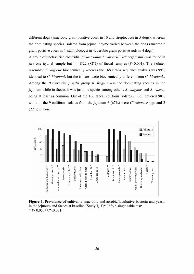
56
different dogs (anaerobic gram-positive cocci in 10 and streptococci in 5 dogs), whereas
the dominating species isolated from jejunal chyme varied between the dogs (anaerobic
gram-positive cocci in 4, staphylococci in 4, aerobic gram-positive rods in 4 dogs).
A group of unclassified clostridia (“Clostridium hiranonis- like” organisms) was found in
just one jejunal sample but in 18/22 (82%) of faecal samples (P<0.001). The isolates
resembled C. difficile biochemically whereas the 16S rRNA sequence analysis was 99%
identical to C. hiranonis but the isolates were biochemically different from C. hiranonis.
Among the Bacteroides fragilis group B. fragilis was the dominating species in the
jejunum while in faeces it was just one species among others, B. vulgatus and B. caccae
being at least as common. Out of the 166 faecal coliform isolates E. coli covered 98%
while of the 9 coliform isolates from the jejunum 6 (67%) were Citrobacter spp. and 2
(22%) E. coli.
0
20
40
60
80
100
Clo
strid
ia n
on-h
irano
nis
*
Gra
m-p
os c
occi
*
Bac
tero
ides
frag
ilis g
rp *
*
Fuso
bact
eria
*
C. h
irano
nis l
ike
**
Bifi
doba
cter
ia
Gra
m-n
eg ro
ds o
ther
Gra
m-p
os ro
ds o
ther
Lact
obac
illi
*
Gra
m-n
eg c
occi
Col
iform
s **
Stre
ptoc
occi
**
Gra
m-p
os ro
ds *
Ente
roco
cci
*
Stap
hylo
cocc
i
Gra
m-p
os c
occi
oth
er
Gra
m-n
eg ro
ds o
ther
Yea
sts
*
Gra
m-n
eg c
occi
Prev
alen
ce %
JejunumFaeces
Figure 1. Prevalence of cultivable anaerobic and aerobic/facultative bacteria and yeasts in the jejunum and faeces at baseline (Study I). Epi Info 6 single table test: * P<0.05, **P<0.001.
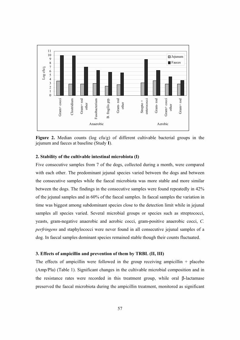
57
0123456789
1011
Gra
m+
cocc
i
Clo
strid
ium
Gra
m+
rod
othe
r
Fuso
bact
eriu
m
B. f
ragi
lis g
rp.
Gra
m- r
odot
her
Stre
pto
+en
tero
cocc
i
Gra
m- r
od
Gra
m+
cocc
iot
her
Gra
m+
rod
Anaerobic Aerobic
Log
cfu/
g
Jejunum
Faeces
Figure 2. Median counts (log cfu/g) of different cultivable bacterial groups in the jejunum and faeces at baseline (Study I).
2. Stability of the cultivable intestinal microbiota (I)
Five consecutive samples from 7 of the dogs, collected during a month, were compared
with each other. The predominant jejunal species varied between the dogs and between
the consecutive samples while the faecal microbiota was more stable and more similar
between the dogs. The findings in the consecutive samples were found repeatedly in 42%
of the jejunal samples and in 60% of the faecal samples. In faecal samples the variation in
time was biggest among subdominant species close to the detection limit while in jejunal
samples all species varied. Several microbial groups or species such as streptococci,
yeasts, gram-negative anaerobic and aerobic cocci, gram-positive anaerobic cocci, C.
perfringens and staphylococci were never found in all consecutive jejunal samples of a
dog. In faecal samples dominant species remained stable though their counts fluctuated.
3. Effects of ampicillin and prevention of them by TRBL (II, III)
The effects of ampicillin were followed in the group receiving ampicillin + placebo
(Amp/Pla) (Table 1). Significant changes in the cultivable microbial composition and in
the resistance rates were recorded in this treatment group, while oral β-lactamase
preserved the faecal microbiota during the ampicillin treatment, monitored as significant
58
differences between the groups receiving ampicillin + placebo and ampicillin + TRBL
(Amp/TRBL), and, on the other hand, no marked differences between Amp/TRBL and
placebo + placebo (Placebo) groups. The effects with similar differences between
treatment groups were found by all methods used: culture, susceptibility testing, TGGE
and TEM-PCR.
Table 1. The effects of ampicillin in the Amp / Pla group.
Method Effect detected in Amp/pla group Bacterial culture Altered bacterial profiles, decreased total counts Susceptibility testing Significant increase in ampicillin (multi)resistance TGGE Altered bacterial profiles (drop in similarity
percentage) TEM-PCR Increased amount of TEM resistance gene High performance liquid chromatography (HPLC)
High serum and jejunal ampicillin concentrations
3.1. Changes detected by culture (II, III)
During the intervention the total faecal count of cultivable aerobes decreased significantly
in the Amp/Pla group (P=0.003) whereas in the Amp/TRBL group the total counts
remained fairly stable. Although the decrease in the anaerobic total count in the Amp/Pla
group was not significant compared to the baseline, the anaerobic total count was
significantly lower than in the Amp/TRBL group during the treatment (P=0.041).
Decreased counts were recorded for streptococci, enterococci, clostridia and anaerobic
gram-positive cocci, while aerobic and anaerobic gram-negative rods increased their
proportion of the total count.
3.2. Microbial shifts detected by TGGE (II)
During the treatment the overall percentage similarity of the faecal microbiota,
determined by TGGE, dropped to a significantly lower level in the Amp/Pla group (38-
53%) compared to the Amp/TRBL and Placebo groups (78-85%) (P<0.001).
59
3.3. Ampicillin concentrations (II)
β-Lactamase had no effect on the ampicillin concentrations in serum; the concentrations
were similar in the groups receiving ampicillin and placebo or ampicillin and TRBL
(mean 17.7 vs. 18.0 µg/ml, respectively). No ampicillin was detected in the Placebo
group.
The ampicillin concentrations in the jejunum were significantly higher in the group
receiving ampicillin and placebo than in the group receiving ampicillin and TRBL (mean
26.3 vs. 1.5 µg/ml, respectively, P<0.0001). In the latter group β-lactamase had degraded
the intestinal antibiotic residue.
3.4. Change in TEM gene levels (II)
The TEM gene levels (determined as the mean percentage of TEM gene of the total
amount of DNA) were very low or undetectable in all dogs at baseline. During the
treatment the proportion of TEM gene reached a significantly higher level in the Amp/pla
group (>0.15%) than in the other groups (<0.025%) (P<0.001).
4. Antimicrobial resistance in coliforms (II, III, IV)
Altogether 982 (860 faecal, 122 jejunal) coliform isolates were collected, identified and
tested against 9 antimicrobials. E. coli covered 87% (106/122) of the jejunal and 99%
(855/860) of the fecal isolates. A total of 22 different resistance patterns, with 12 patterns
displaying ampicillin resistance, were identified. Each sample displayed 2-3 (ranging
between 1-6) different patterns. The 5 most common patterns covered 94% of the faecal
and 77% of the jejunal isolates.
4.1. Emergence of resistance in coliforms (II, III, IV)
An increase in ampicillin resistance (multi-resistance) was detected in all treatment
groups but the resistance rate was significantly higher in the Amp/Pla group than in the
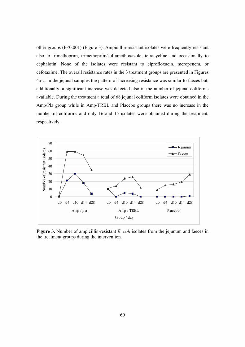
60
other groups (P<0.001) (Figure 3). Ampicillin-resistant isolates were frequently resistant
also to trimethoprim, trimethoprim/sulfamethoxazole, tetracycline and occasionally to
cephalotin. None of the isolates were resistant to ciprofloxacin, meropenem, or
cefotaxime. The overall resistance rates in the 3 treatment groups are presented in Figures
4a-c. In the jejunal samples the pattern of increasing resistance was similar to faeces but,
additionally, a significant increase was detected also in the number of jejunal coliforms
available. During the treatment a total of 68 jejunal coliform isolates were obtained in the
Amp/Pla group while in Amp/TRBL and Placebo groups there was no increase in the
number of coliforms and only 16 and 15 isolates were obtained during the treatment,
respectively.
0
10
20
30
40
50
60
70
d0 d4 d10 d14 d28 d0 d4 d10 d14 d28 d0 d4 d10 d14 d28
Amp / pla Amp / TRBL Placebo
Group / day
Num
ber o
f res
ista
nt is
olat
es
JejunumFaeces
Figure 3. Number of ampicillin-resistant E. coli isolates from the jejunum and faeces in the treatment groups during the intervention.
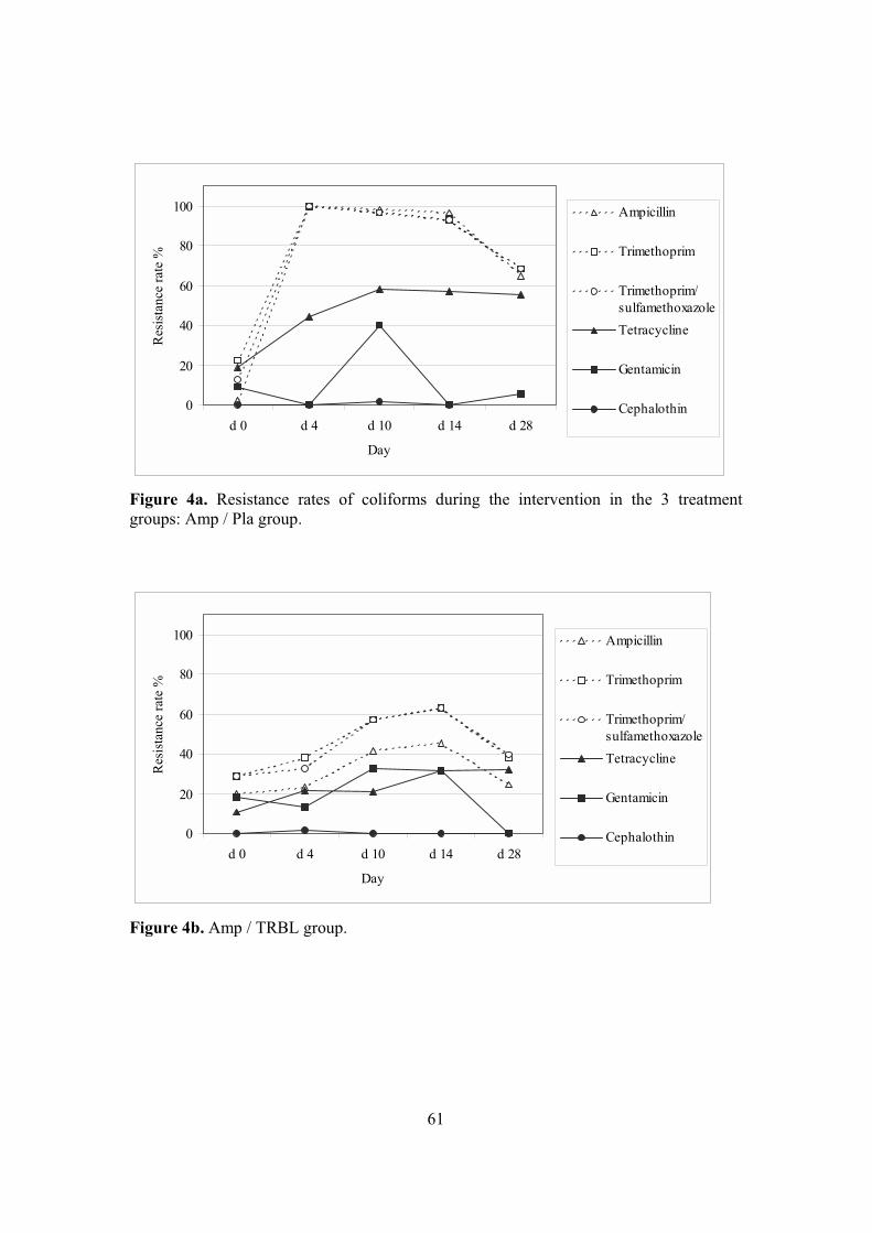
61
0
20
40
60
80
100
d 0 d 4 d 10 d 14 d 28
Day
Res
ista
nce
rate
%
Ampicillin
Trimethoprim
Trimethoprim/sulfamethoxazoleTetracycline
Gentamicin
Cephalothin
Figure 4a. Resistance rates of coliforms during the intervention in the 3 treatment groups: Amp / Pla group.
0
20
40
60
80
100
d 0 d 4 d 10 d 14 d 28
Day
Res
ista
nce
rate
%
Ampicillin
Trimethoprim
Trimethoprim/sulfamethoxazoleTetracycline
Gentamicin
Cephalothin
Figure 4b. Amp / TRBL group.
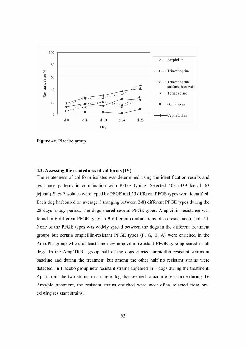
62
0
20
40
60
80
100
d 0 d 4 d 10 d 14 d 28
Day
Res
ista
nce
rate
%
Ampicillin
Trimethoprim
Trimethoprim/sulfamethoxazoleTetracycline
Gentamicin
Cephalothin
Figure 4c. Placebo group.
4.2. Assessing the relatedness of coliforms (IV) The relatedness of coliform isolates was determined using the identification results and
resistance patterns in combination with PFGE typing. Selected 402 (339 faecal, 63
jejunal) E. coli isolates were typed by PFGE and 25 different PFGE types were identified.
Each dog harboured on average 5 (ranging between 2-8) different PFGE types during the
28 days’ study period. The dogs shared several PFGE types. Ampicillin resistance was
found in 6 different PFGE types in 9 different combinations of co-resistance (Table 2).
None of the PFGE types was widely spread between the dogs in the different treatment
groups but certain ampicillin-resistant PFGE types (F, G, E, A) were enriched in the
Amp/Pla group where at least one new ampicillin-resistant PFGE type appeared in all
dogs. In the Amp/TRBL group half of the dogs carried ampicillin resistant strains at
baseline and during the treatment but among the other half no resistant strains were
detected. In Placebo group new resistant strains appeared in 3 dogs during the treatment.
Apart from the two strains in a single dog that seemed to acquire resistance during the
Amp/pla treatment, the resistant strains enriched were most often selected from pre-
existing resistant strains.
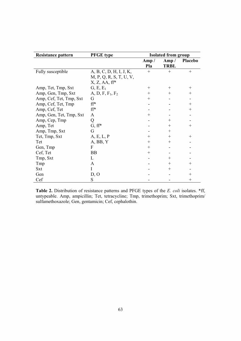
63
Resistance pattern PFGE type Isolated from group Amp /
PlaAmp / TRBL
Placebo
Fully susceptible A, B, C, D, H, I, J, K, M, P, Q, R, S, T, U, V, X, Z, AA, ff*
+ + +
Amp, Tet, Tmp, Sxt G, E, E1 + + + Amp, Gen, Tmp, Sxt A, D, F, F1, F2 + + + Amp, Cef, Tet, Tmp, Sxt G + - - Amp, Cef, Tet, Tmp ff* - - + Amp, Cef, Tet ff* - - + Amp, Gen, Tet, Tmp, Sxt A + - - Amp, Cep, Tmp Q - + - Amp, Tet G, ff* - + + Amp, Tmp, Sxt G - + Tet, Tmp, Sxt A, E, L, P + + + Tet A, BB, Y + + - Gen, Tmp F + - - Cef, Tet BB + - - Tmp, Sxt L - + - Tmp A - + + Sxt I - + - Gen D, O - - + Cef S - - +
Table 2. Distribution of resistance patterns and PFGE types of the E. coli isolates. *ff, untypeable. Amp, ampicillin; Tet, tetracycline; Tmp, trimethoprim; Sxt, trimethoprim/ sulfamethoxazole; Gen, gentamicin; Cef, cephalothin.
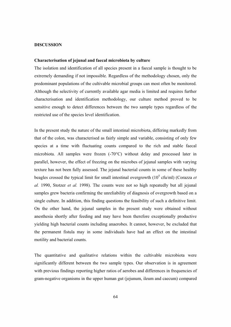
64
DISCUSSION
Characterisation of jejunal and faecal microbiota by culture
The isolation and identification of all species present in a faecal sample is thought to be
extremely demanding if not impossible. Regardless of the methodology chosen, only the
predominant populations of the cultivable microbial groups can most often be monitored.
Although the selectivity of currently available agar media is limited and requires further
characterisation and identification methodology, our culture method proved to be
sensitive enough to detect differences between the two sample types regardless of the
restricted use of the species level identification.
In the present study the nature of the small intestinal microbiota, differing markedly from
that of the colon, was characterised as fairly simple and variable, consisting of only few
species at a time with fluctuating counts compared to the rich and stable faecal
microbiota. All samples were frozen (-70°C) without delay and processed later in
parallel, however, the effect of freezing on the microbes of jejunal samples with varying
texture has not been fully assessed. The jejunal bacterial counts in some of these healthy
beagles crossed the typical limit for small intestinal overgrowth (105 cfu/ml) (Corazza et
al. 1990, Stotzer et al. 1998). The counts were not so high repeatedly but all jejunal
samples grew bacteria confirming the unreliability of diagnosis of overgrowth based on a
single culture. In addition, this finding questions the feasibility of such a definitive limit.
On the other hand, the jejunal samples in the present study were obtained without
anesthesia shortly after feeding and may have been therefore exceptionally productive
yielding high bacterial counts including anaerobes. It cannot, however, be excluded that
the permanent fistula may in some individuals have had an effect on the intestinal
motility and bacterial counts.
The quantitative and qualitative relations within the cultivable microbiota were
significantly different between the two sample types. Our observation is in agreement
with previous findings reporting higher ratios of aerobes and differences in frequencies of
gram-negative organisms in the upper human gut (jejunum, ileum and caecum) compared
65
to the colon and faeces (Gorbach et al. 1967, Justesen et al. 1984, Marteau et al. 2001).
Oropharyngeal microbes have been reported to dominate in jejunal biopsies of humans
(Sullivan et al. 2003), whereas E. coli and streptococci were the most common findings
in the jejunum of 35 mongrel dogs (Thompson et al. 1998). Higher or comparable counts
of fungi, staphylococci and lactobacilli have been reported in the human ileum than
faeces (Gorbach et al. 1967). Similarly we found staphylococci, yeasts and also other
catalase-producing cocci and non-fermentative gram-negative facultative rods more
frequently in the canine jejunum than in faeces but less lactobacilli. Additionally, some
species isolated from jejunal samples were undetected in faeces. Marteau et al. (2001)
reported facultative bacteria to be as numerous as anaerobes in the human caecum. This
seems to be true in the jejunum as well, as shown in the present stydy. B. fragilis proved
to be the most frequent species of the B. fragilis group in the canine small intestine but
not in faeces. In agreement with our observation, B. fragilis has been reported to be the
most frequent isolate from mucosal surfaces of humans but not in colon contents
(Namavar et al. 1989). In the present study the canine jejunal microbiota was also found
to be less stable than the faecal microbiota. The canine duodenal microbiota monitored by
DGGE profiles has been shown to differ more between individual dogs than between
samples from the same dog (Suchodolski et al. 2004). The canine fecal bacterial DGGE
profiles have individually unique and stable characteristic banding patterns (Simpson et
al. 2002).
It is noteworthy that we found no C. difficile in contrast to some other animal studies
(Marks et al. 2002, Buogo et al. 1995) but we detected instead a group of unclassified
clostridia resembling C. difficile as part of the dominating faecal microbiota. In addition
to strict anaerobes, facultative streptococci were among the dominating microbiota. Also,
we found bifidobacteria more often than lactobacilli (in 64% vs. 32% of the dogs,
respectively) while two recent studies on labrador and beagle dogs reported no
bifidobacteria but numerous lactobacilli (Buddington 2003, Greetham et al. 2002). The
lactic acid bacteria in fistulated dogs were recently characterised by Rinkinen et al.
(2004) with Streptococcus alactolyticus dominating, followed by Lactobacillus murinus
and L. reuteri.
66
Comparison of culture to molecular methods
Though molecular methods have brought new aspects to the phylogenetic and
quantitative relations of different bacteria in the gut, culture-based and molecular-based
profiling may not be contradictory and give often in general somewhat similar overview.
Culture-based analyses may, however, give higher qualitative and quantitative
fluctuations especially in the faecal microbiota compared to molecular methodology.
Similar to our study, the vast fluctuation in canine small intestinal microbiota compared
to faeces has been reported by both culture and DGGE reflecting genuine variation rather
than weakness in sampling or analysis methodology (Willard et al. 1994, Delles et al.
1994, Harmoinen et al. 2001, Suchodolski et al. 2004). We found by culture in the canine
faeces a relatively low mean proportion of Bacteroides (0.3%), a high proportion of
anaerobic gram-positive cocci (30%) and clostridia (37%), a relatively high proportion of
streptococci (19%) and relatively low proportion of bifidobacteria (4%) and
enterobacteria (1%). Culture negativity or limited number of microbial findings may not
always give an accurate picture of the number or consortia of microbes present. For
example, cultures and microscopic examinations of oesophagal washes and biopsies have
provided poor microbial yields but microbes in oesophageal biopsies detected by
sequence based broad-range 16S rDNA are by a large majority (82%) cultivable bacterial
species (Pajecki et al. 2002, Pei et al. 2004).
Ampicillin-induced changes
In the present study ampicillin was found to induce significant qualitative and
quantitative changes and emergence of multi-resistant coliform strains among the canine
intestinal microbiota. These phenomenona have been well documented for ampicillin and
several other antimicrobials also in previous human studies (Nord 1986a, Black et al.
1991, Garau et al. 1999, Shanahan et al. 1994, Huovinen et al. 1995, Dradenovich et al.
2004). Among human faecal enterobacteria E. coli is the main carrier of resistance
(Österblad et al. 2000). In the present study ampicillin resistance was most often
combined to trimethoprim and trimethoprim/sulfamethoxazole resistance in canine
strains. A similar combination of resistance occurs frequently, since transmissible
67
plasmids coding resistance to ampicillin and trimethoprim are common in human E. coli,
and selective pressure for either is likely to select resistance to both drugs (Amyes 1989,
Magee et al. 1999). As shown previously, the screening of coliform susceptibility is a
feasible and sensitive method to follow the steps in the emergence of resistance (Corpet
1993). In the present study, to intensify the resistance screening, 10 isolates representing
all colony morphotypes per sample were tested for resistance. Further, to overcome
biases due to the culture method, molecular methods were used to follow the changes in
resistance rates monitored as amounts of TEM gene. In addition, several samples during
the treatment were analysed.
Resistance within coliform populations
PFGE typing revealed a considerable genetic heterogeneity within the ampicillin-resistant
canine E. coli populations. Though digestion of DNA with more than one restriction
enzyme could have increased the discriminatory power of the PFGE method and
separated strains considered identical, the present analysis with one enzyme (XbaI) was
successful in discriminating numerous genetically different clones with varying
frequencies within a large number of related isolates in the three treatment groups. Dogs
shared several clones but no single resistant strain was widely spread. Since the resistant
strains were most often selected from the pre-existing microbiota or spread between dogs,
and were genetically heterogeneous, several resistant types co-existed. The selection of
resistant strains from the resident microbiota is thought to be more common than
acquirance of a new resistance determinant in strains previously susceptible (Guyot et al.
1999, McDonald et al. 2001) as was the case also in the present study.
Heterogeneity, constant shifts between strains within E. coli populations and sharing of
common types has been reported in humans, dogs and cattle (Levy et al. 1988, Avery et
al. 2004, Akiba et al. 1999, Münnich & Lübke-Becker 2004, Dradenovich et al. 2004).
The appeared resistant strains replaced the PFGE types present at baseline in the dogs
receiving ampicillin without β-lactamase but not in the other two groups.
68
Prevention of ampicillin-induced changes by TRBL
Orally administered β-lactamase did not affect the ampicillin level in serum but degraded
ampicillin in the intestine and prevented the ampicillin-induced changes in the intestinal
microbiota. Colonisation resistance was reduced and multi-resistant coliforms colonised
the jejunum only in dogs treated with ampicillin without β-lactamase but not in dogs
treated with ampicillin with β-lactamase. The slight increase in resistance rates detected
in the Amp/TRBL and Placebo groups could be due to the cross-contamination and
spread of resistance determinants from the dogs in the Amp/pla group, since the dogs
were not totally isolated from each other. Other possible sources of resistance
determinants include food, environment, kennel facilities and personnel as reported also
in other studies (Corpet 1987, Avery et al. 2004, de Graef 2004). Concurrent
administration of antibiotic and β-lactamase could provide a new strategy to limit the
emergence of resistance and spread of nosocomial pathogens, and reduce antibiotic-
associated diarrhoea. In addition to ampicillin, β-lactamase has shown to be applicaple to
inhibit piperacillin-induced changes (Stiefel et al. 2003).
Commensal microbes gradually overgrow the resistant strains when the selection pressure
is absent (Trott et al. 2004). The restoration of the microbiota may, however, be slower
than previously thought. The antibiotic-associated alterations in the microbiota can be
detected for months or even up to 3 years after a single antibiotic course (Maxwell et al.
2002, Mangin et al. 1994, Sjölund et al. 2003). Antimicrobial resistance determinants are
known to be transferable among the commensal microbiota (Gulay et al. 2000, Yates et
al. 2004). In the present study, the microbiota had not recovered to the baseline
configuration 2 weeks after the treatment. Although the bacterial counts and composition,
TGGE profiles and TEM gene proportions resumed levels close to the baseline, the
enriched or appeared ampicillin-resistant PFGE types in the Amp/pla and Placebo groups
were still present and the proportion of resistant isolates was higher than at baseline.
Future aspects
The next phase in studying combined ampicillin and β-lactamase administration would be
a study trial in humans. Further phases in developing means to prevent antimicrobial

69
resistance using β-lactamase could include development of new β-lactamases targeted to
other β-lactams.
70
KEY FINDINGS AND CONCLUSIONS
To protect the indigenous microbiota in a safe and convenient manner the composition
and functions of the microbiota should be well known and understood, and the desired
changes, or their absense, as well as their magnitude and dimensions should be closely
monitored with suitable methodology. The present study was carried out in order to
develop the understanding of the jejunal microbial composition and stability, and to
specify the dissimilarities between the jejunal and faecal microbiota. Another main focus
of the study was to evaluate the ability of enzymatic inactivation of intestinal
antimicrobial residues to inhibit antibiotic-induced adverse effects on the microbiota.
The key findings were:
I Significant microbial differences between the jejunal and faecal findings were
recorded in regard to the bacterial counts, species composition, species variability,
stability, relations of gram-negative to gram-positive organisms and relations of aerobic
to anaerobic microbes.
II Ampicillin-induced changes among the intestinal microbiota were inhibited to a
significant extent by simultaneous oral β-lactamase administration.
III Ampicillin treatment without simultaneous β-lactamase administration induced
the emergence of multi-resistant strains displaying numerous combinations of co-
resistance and significantly increased the colonisation of the jejunum with coliforms. The
increase in resistance was markedly inhibited and the jejunal colonisation resistance was
preserved when ampicillin treatment was combined to β-lactamase.
IV The emergence of resistance among intestinal coliforms was due to selection
and/or dissemination of several genetically different strains rather than acquirance of
resistance in previously susceptible strains or spread of single or few resistant strains
between the dogs in different treatment groups.
71
Conclusions
The small intestinal microbiota has individually unique features in regard to counts,
species composition and variability of the microbes, differing markedly from those of
faeces. Orally administered β-lactamase was shown to have potential in clinical use by
inhibiting the emergence of resistance and preserving the microbiota during antimicrobial
therapy. β-Lactamase can be beneficial in inhibiting the emergence and enrichment of
resistant strains and in maintaining the colonisation resistance in jejunum during
ampicillin-induced selection pressure.
72
ACKNOWLEDGEMENTS
This study was carried out at the Anaerobe Reference Laboratory, National Public Health
Institute, Helsinki, Finland. I thank Professor Pekka Puska and Professor Jussi Huttunen,
the present and former Head of the Institute for providing excellent working facilities.
I wish to express my gratitude to my supervisor Professor Pentti Huovinen for supportive
and instructive guidance, for clarifying the big pictures for me and also for kind-hearted
care for my well being during the process. To my other supervisor Docent Eija Könönen I
wish to express my warm thanks for her valuable advice and appreciation especially for
her expertise and efficiency in preparing scientific papers.
This study was done in cooperation with Ipsat Therapies Ltd. and I am indebted to all the
skilful and kind people there, especially Kai Lindevall, Lauri Jalkanen, Pertti Koski, Nina
Wickstrand, Marion Carson and Tuula Heinonen.
I am deeply grateful to all the people involved in this study at the Department of Clinical
Veterinary Sciences, University of Helsinki, especially to my co-authors Professor Elias
Westermarck, Jaana Harmoinen, Matti Heikkilä and Rafael Frias. Special thanks go to
Jaana for explicit and fruitful exchange of information and co-operation.
I wish to thank my co-authors Merja Rautio, Arja Kanervo-Nordström and Terhi Virtanen
and colleagues Anne Bryk, Anne Rinta-Opas and Marja Piekkola at the KTL Anaerobe
Reference Laboratory. This study was teamwork and could not have been done without
your excellent and fluent collaboration. Merja is also acknowledged for her advise,
empathy and common sense. I also thank Ani Sarpi, Mari Hyvönen, Nanna Väyrynen,
Susan Nyfors and Mervi Latva-Aho from KTL for support and friendship. The warm,
caring and cheerful atmosphere at the laboratory has turned colleagues to friends and
made working enjoyable.

73
I also thank my co-authors Michel van der Rest, Päivi Rajala-Schultz and Curtis Donskey
for collaboration.
The pre-reviewers of my thesis, Professor Charlotta Edlund and Docent Maria Saarela,
are gratefully acknowledged for valuable suggestions to improve the thesis.
I am grateful to all my dear old friends, especially Russe, Sari, Kukka, Ahti, Teemu and
Joge for enjoyable moments and reminding me that there is life outside science, and
friends from student years, Kaisa Saloniemi and Shea Beasley, for listening and
understanding the triumphs and troubles of this study.
My sincere thanks are due to my family for support and interest in my work, especially to
my parents Aulikki and Pertti for encouraging finding my own strengths and showing that
learning is a life long process. The warmest and most loving thanks are due to my life
companion Tuomas. His caring and involved attitude and inventive mind have been vital
for me during the years.
I owe my deepest gratitude to the late Professor Hannele “Haba” Jousimies-Somer, who
taught me what science is about and showed how much fun it can be. Her knowledge,
enthusiasm, conviction and support had a decisive effect on me to carry on doing research
after she was gone.
Helsinki, May 2005
74
REFERENCES
Akiba M, Sameshima T, Nakazawa M. The shift of genetic subtypes of Escherichia coli O157:H7 isolates from cattle. Epidemiol Infect 1999;122:343-346. Alander M, Korpela R, Saxelin M, Vilpponen-Salmela T, Mattila-Sandholm T, von Wright A.
Recovery of Lactobacillus rhamnosus GG from human colonic biopsies. Lett Appl Microbiol 1997;24:361-364.
Alander M, Satokari R, Korpela R, Saxelin M, Vilpponen-Salmela T, Mattila-Sandholm T, von Wright A. Persistence of colonization of human colonic mucosa by a probiotic strain, Lactobacillus rhamnosus GG, after oral consumption. Appl Environ Microbiol 1999;65:351-354.
Amann RI, Ludwig W, Schleifer KH. Phylogenetic identification and in situ detection of individual microbial cells without cultivation. Microbiol Rev 1995;59:143-169.
Amyes SG. The success of plasmid-encoded resistance genes in clinical bacteria. An examination of plasmid-mediated ampicillin and trimethoprim resistance genes and their resistance mechanisms. J Med Microbiol 1989;28:73-83.
Avery SM, Liebana E, Hutchison ML, Buncic S. Pulsed field gel electrophoresis of related Escherichia coli O157 isolates associated with beef cattle and comparison with unrelated isolates from animals, meats and humans. Int J Food Microbiol 2004;92:161-169.
Barrett TJ, Lior H, Green JH, Khakhria R, Wells JG, Bell BP, Greene KD, Lewis J, Griffin PM. Laboratory investigation of a multistate food-borne outbreak of Escherichia coli O157:H7 by using pulsed-field gel electrophoresis and phage typing. J Clin Microbiol 1994;32:3013-3017.
Batt RM, Needham JR, Carter MW. Bacterial overgrowth associated with a naturally occurring enteropathy in the German shepherd dog. Res Vet Sci. 1983;35:42-46.
Belaaouaj A, Lapoumeroulie C, Canica MM, Vedel G, Nevot P, Krishnamoorthy R, Paul G. Nucleotide sequences of the genes coding for the TEM-like beta-lactamases IRT-1 and IRT-2 (formerly called TRI-1 and TRI-2). FEMS Microbiol Lett 1994;120:75-80.
Benno Y, Nakao H, Uchida K, Mitsuoka T. Impact of the advances in age on the gastrointestinal microflora of beagle dogs. J Vet Med Sci 1992a;54:703-706.
Benno Y, Nakao H, Uchida K, Mitsuoka T. Individual and seasonal variations in the composition of fecal microflora of beagle dogs. Bifidobact. Microfl 1992b;11:69-76.
Berg RD. The indigenous gastrointestinal microflora. Trends Microbiol 1996;4:430-435. Berg RD. Bacterial translocation from the gastrointestinal tract. Adv Exp Med Biol 1999;473:11-30. Bergogne-Berezin E. Treatment and prevention of antibiotic associated diarrhea. Int J Antimicrob
Agents 2000;16:521-526. Bhat P, Albert MJ, Rajan D, Ponniah J, Mathan V I, Baker SJ. Bacterial flora of the jejunum: a
comparison of luminal aspirate and mucosal biopsy. J Med Microbiol 1980;13:247-256. Biedenbach DJ, Moet GJ, Jones RN. Occurrence and antimicrobial resistance pattern comparisons
among bloodstream infection isolates from the SENTRY Antimicrobial Surveillance Program (1997-2002). Diagn Microbiol Infect Dis 2004;50:59-69.
Black F, Einarsson K, Lidbeck A, Orrhage K, Nord CE. Effect of lactic acid producing bacteria on the human intestinal microflora during ampicillin treatment. Scand Infect Dis 1991;23:247-254.
Blazquez J, Morosini MI, Negri MC, Baquero F. Selection of naturally occurring extended-spectrum TEM beta-lactamase variants by fluctuating beta-lactam pressure. Antimicrob Agents Chemother 2000;44:2182-2184.
Blottiere HM, Buecher B, Galmiche JP, Cherbut C. Molecular analysis of the effect of short-chain fatty acids on intestinal cell proliferation. Proc Nutr Soc 2003;62:101-106.
Borriello P, Hudson M, Hill M. Investigation of the gastrointestinal bacterial flora. Clin Gastroenterol 1978;7:329-349.
Bouma JE, Lenski RE. Evolution of a bacteria/plasmid association. Nature 1988;335:351-352. Brandtzaeg P, Halstensen TS, Kett K, Krajci P, Kvale D, Rognum TO, Scott H, Sollid LM.
Immunobiology and immunopathology of human gut mucosa: humoral immunity and intraepithelial lymphocytes. Gastroenterology 1989;97:1562-1584.
Breeuwer P, Abee T. Assessment of viability of microorganisms employing fluorescence techniques. Int J Food Microbiol 2000;55:193-200.
75
Brinas L, Zarazaga M, Saenz Y, Ruiz-Larrea F, Torres C. Beta-lactamases in ampicillin-resistant Escherichia coli isolates from foods, humans, and healthy animals. Antimicrob Agents Chemother 2002;46:3156-3163.
Brock TD, Madigan MT, Martinko JM, Parker J. Biology of Microorganisms 10th ed. Prentice-Hall International, New Jersey, USA, 2003.
Brook I, Frazier EH. Significant recovery of nonsporulating anaerobic rods from clinical specimens. Clin Infect Dis 1993;16:476-480.
Brun T, Peduzzi J, Canica MM, Paul G, Nevot P, Barthelemy M, Labia R. Characterization and amino acid sequence of IRT-4, a novel TEM-type enzyme with a decreased susceptibility to beta-lactamase inhibitors. FEMS Microbiol Lett 1994;120:111-117.
Buddington RK. Postnatal changes in bacterial populations in the gastrointestinal tract of dogs. Am J Vet Res 2003;64:646-651.
von Buenau R, Jaekel L, Schubotz E, Schwarz S, Stroff T, Krueger M. Escherichia coli Strain Nissle 1917: Significant Reduction of Neonatal Calf Diarrhea. J Dairy Sci 2005;88:317-323.
Buogo C, Burnens AP, Perrin J, Nicolet J. Presence of Campylobacter spp., Clostridium difficile, C.perfringens and salmonellae in litters of puppies and in adult dogs in a shelter. Schweiz Arch Tierheilkd 1995;137:165-171.
Burman LG, Haeggman S, Kuistila M, Tullus K, Huovinen P. Epidemiology of plasmid-mediated beta- lactamases in enterobacteria Swedish neonatal wards and relation to antimicrobial therapy. Antimicrob Agents Chemother 1992;36:989-992.
Bush K, Jacoby GA, Medeiros AA. A functional classification scheme for beta-lactamases and its correlation with molecular structure. Antimicrob Agents Chemother 1995;39:1211-1233.
Bush K, Jacoby G. Nomenclature of TEM beta-lactamases. J Antimicrob Chemother 1997;39:1-3. Bäckhed F, Ding H, Wang T, Hooper LV, Koh GY, Nagy A, Semenkovich CF, Gordon JI. The gut
microbiota as an environmental factor that regulates fat storage. Proc Natl Acad Sci USA 2004;101:15718-15723.
Carratala J, Fernandez-Sevilla A, Tubau F, Callis M, Gudiol F. Emergence of quinolone-resistant Escherichia coli bacteremia in neutropenic patients with cancer who have received prophylactic norfloxacin. Clin Infect Dis 1995;20:557-560; discussion 561-563.
Castagliuolo I, Riegler MF, Valenick L, LaMont JT, Pothoulakis C. Saccharomyces boulardii proteaseinhibits the effects of Clostridium difficile toxins A and B in human colonic mucosa. Infect Immun 1999;67:302-307.
Chachaty E, Depitre C, Mario N, Bourneix C, Saulnier P, Corthier G, Andremont A. Presence of Clostridium difficile and antibiotic and beta-lactamase activities in feces of volunteers treated with oral cefixime, oral cefpodoxime proxetil, or placebo. Antimicrob Agents Chemother 1992;36:2009-2013.
Chachaty E, Bourneix C, Renard S, Bonnay M, Andremont A. Shedding of Clostridium difficile, fecal beta-lactamase activity, and gastrointestinal symptoms in 51 volunteers treated with oral cefixime. Antimicrob Agents Chemother 1993;37:1432-1435.
Charteris WP, Kelly PM, Morelli L, Collins JK. Selective detection, enumeration and identification of potentially probiotic Lactobacillus and Bifidobacterium species in mixed bacterial populations. Int J Food Microbiol 1997;35:1-27.
Cook RR. Antimicrobial resistance--use in veterinary and human medicine. J Antimicrob Chemother 1997;39:435.
Corazza GR, Menozzi MG, Strocchi A, Rasciti L, Vaira D, Lecchini R, Avanzini P, Chezzi C, Gasbarrini G. The diagnosis of small bowel bacterial overgrowth. Reliability of jejunal culture and inadequacy of breath hydrogen testing. Gastroenterol 1990;98:302-309.
Corpet DE. Antibiotic residues and drug resistance in human intestinal flora. Antimicrob Agents Chemother 1987;31:587-593.
Corpet DE. An evaluation of methods to assess the effect of antimicrobial residues on the human gut flora. Vet Microbiol 1993;35:199-212.
Cummings JH, Macfarlane GT. The control and consequences of bacterial fermentation in the human colon. J Appl Bacteriol 1991;70:443-459.
D'Agata EM. Rapidly rising prevalence of nosocomial multidrug-resistant, Gram-negative bacilli: a 9- year surveillance study. Infect Control Hosp Epidemiol 2004;25:842-846.
Davis CP, Cleven D, Balish E, Yale CE. Bacterial association in the gastrointestinal tract of beagle
76
dogs. Appl Environ Microbiol 1977;34:194-206.Davis MA, Hancock DD, Besser TE, Call DR. Evaluation of pulsed-field gel electrophoresis as a tool
for determining the degree of genetic relatedness between strains of Escherichia coli O157:H7. J Clin Microbiol 2003;41:1843-1849.
De Graef EM, Decostere A, Devriese LA, Haesebrouck F. Antibiotic resistance among fecal indicator bacteria from healthy individually owned and kennel dogs. Microb Drug Resist 2004;10:65-69.
Deitsch KW, Moxon ER, Wellems TE. Shared themes of antigenic variation and virulence in bacterial, protozoal, and fungal infections. Microbiol Mol Biol Rev 1997;61:281-293.
Delles EK, Willard MD, Simpson RB, Fossum TW, Slater M, Kolp D, Lees GE, Helman R, Reinhart G. Comparison of species and numbers of bacteria in concurrently cultured samples of proximal small intestinal fluid and endoscopically obtained duodenal mucosa in dogs with intestinal bacterial overgrowth. Am J Vet Res 1994;55:957-964.
Diekema DJ, Barr J, Boyken LD, Buschelman BJ, Jones RN, Pfaller MA, Herwaldt LA. A cluster of serious Escherichia coli infections in a neonatal intensive-care unit. Infect Control Hosp Epidemiol 1997;18:774-776.
Donaldson RM Jr. Normal bacterial populations of intewstine and their relation to intestinal function. New Engl J Med 1964;270:938-946, 994-1000, 1050-1056.
Doungudomdacha S, Rawlinson A, Walsh TF, Douglas CW. Effect of non-surgical periodontal treatment on clinical parameters and the numbers of Porphyromonas gingivalis, Prevotella
intermedia and Actinobacillus actinomycetemcomitans at adult periodontitis sites. J Clin Periodontol. 2001;28:437-445.
Drasar DR, Mill MJ. Human intestinal flora. Academic Press, London, 1975. Drazenovich N, Ling GV, Foley J. Molecular investigation of Escherichia coli strains associated with
apparently persistent urinary tract infection in dogs. J Vet Intern Med 2004;18:301-306.Edlund C, Nord CE. Effect on the human normal microflora of oral antibiotics for treatment of urinary
tract infections. J Antimicrob Chemother 2000;46(Suppl 1):41-48; discussion 63-65.Eisenstein BI, Zaleznik DF. Enterobacteriaceae, p. 2294-2310. In: G. L. Mandell, J. E. Bennett, and R.
Dolin (ed.), Principles and practice of infectious diseases, 5th ed. Churchill Livingstone, Philadelphia, PA, USA, 2000.
El Oufir L, Flourie B, Bruley des Varannes S, Barry JL, Cloarec D, Bornet F, Galmiche JP. Relations between transit time, fermentation products, and hydrogen consuming flora in healthy humans. Gut 1996;38:870-877.
Falk PG, Hooper LV, Midtvedt T, Gordon JI. Creating and maintaining the gastrointestinal ecosystem: what we know and need to know from gnotobiology. Microbiol Mol Biol Rev 1998;62:1157-1170.
Farr BM. Prevention and control of nosocomial infections. Business briefing: global health care issue. 2002;3:37-41.
Favier CF, Vaughan EE, de Vos WM, Akkermans ADL. Molecular monitoring of succession of bacterial communities in human neonates. Appl Environ Microbiol 2002;68:219-226.
Felske A, Rheims H, Wolterink A, Stackebrandt E, Akkermans AD. Ribosome analysis reveals prominent activity of an uncultured member of the class Actinobacteria in grassland soils. Microbiology 1997;143:2983-2989.
Finegold SM, Sutter VL, Mathisen GE. Normal indigenous intestinal flora, p. 3-31. In D. J. Hentges (ed.), Human intestinal microbiota in health and disease. Academic Press, New York, USA, 1983.
Flynn DM, Weinstein RA, Nathan C, Gaston MA, Kabins SA. Patients' endogenous flora as the source of "nosocomial" Enterobacter in cardiac surgery. J Infect Dis 1987;156:363-368.
Franks AH, Harmsen HJ, Raangs GC, Jansen GJ, Schut F, Welling GW. Variations of bacterial populations in human feces measured by fluorescent in situ hybridization with group-specific 16S rRNA-targeted oligonucleotide probes. Appl Environ Microbiol 1998;64:3336-3345.
Garau J, Xercavins M, Rodriguez-Carballeira M, Gomez-Vera JR, Coll I, Vidal D, Llovet T, Ruiz- Bremon A. Emergence and dissemination of quinolone-resistant Escherichia coli in the community. Antimicrob Agents Chemother 1999;43:2736-2741.
Garcia L, Tercero JC, Legido B, Ramos JA, Alemany J, Sanz M. Rapid detection of Actinobacillus actinomycetemcomitans, Prevotella intermedia and Porphyromona gingivalis by multiplex PCR. J Periodontal Res 1998;33:59-64.
Gillian Hartley M, Hudson MJ, Swarbrick ET, Hill MJ, Gent AE, Hellier MD, Grace RH. 1992. The
77
rectal mucosa-associated microflora in patients with ulcerative colitis. J Med Microbiol 1992;36:96-103.
Goldin BR, Swenson L, Dwyer J, Sexton M, Gorbach SL. Effect of diet and Lactobacillus acidophilussupplements on human fecal bacterial enzymes. J Natl Cancer Inst 1980;64:255-261.
Gorbach SL, Plaut AG, Nahas L, Weinstein L, Spanknebel G, Levitan R. Studies of intestinal microflora. II. Microorganisms of the small intestine and their relations to oral and fecal flora. Gastroenterology 1967;53:856-867.
Gorbach SL, Nahas L, Lerner PI, Weinstein L. Effects of diet, age, and periodic sampling on numbers of faecal microorganisms in man. Gastroenterology 1967;53:845-855.
Gorbach SL. Bengt E. Gustafsson memorial lecture. Function of the normal human microflora. Scand J Infect Dis Suppl 1986;49:17-30.
Gordon HA, Pesti L. The gnotobiotic animal as a tool in the study of host microbial relationships. Bacteriol Rev 1971;35:390-429.
Gracey M, Suharjono, Sunoto. Use of a simple duodenal capsule to study upper intestinal microflora. Arch Dis Child 1977;52:74-76.
Gracey M. The contaminated small bowel syndrome, p. 495-513. In: Hentges DJ (ed), Human intestinal microflora in health and disease. Academic Press, New York, USA, 1983.
Greetham HL, Giffard C, Hutson RA, Collins MD, Gibson GR. Bacteriology of the Labrador gut: a cultural and genotypic approach. J Appl Microbiol 2002;93:640-646.
Grif K, Karch H, Schneider C, Daschner FD, Beutin L, Cheasty T, Smith H, Rowe B, Dierich MP, Allerberger F. Comparative study of five different techniques for epidemiological typing of Escherichia coli O157. Diagn Microbiol Infect Dis 1998;32:165-176.
Guardabassi L, Schwarz S, Lloyd DH. Pet animals as reservoirs of antimicrobial-resistant bacteria. J Antimicrob Chemother 2004;54:321-332.
Guarner F, Malagelada JR. Gut flora in health and disease. Lancet 2003;361:512-519. Gulay Z, Bicmen M, Amyes SG, Yulug N. Beta-lactamase patterns and betalactam/clavulanic acid
resistance in Escherichia coli isolated from fecal samples from healthy volunteers. J Chemother 2000;12:208-215.
Guyot A, Barrett SP, Threlfall EJ, Hampton MD, Cheasty T. Molecular epidemiology of multi-resistant Escherichia coli. J Hosp Infect 1999;43:39-48.
Hahm BK, Maldonado Y, Schreiber E, Bhunia AK, Nakatsu CH. Subtyping of foodborne and environmental isolates of Escherichia coli by multiplex-PCR, rep-PCR, PFGE, ribotyping and AFLP. J Microbiol Methods. 2003;53:387-399.
Hall RM. Mobile gene cassettes and integrons: moving antibiotic resistance genes in gram-negative bacteria. Ciba Found Symp. 1997;207:192-202; discussion 202-205.
Hamilton I, Worsley BW, Cobden I, Cooke EM, Shoesmith JG, Axon AT. Simultaneous culture of saliva and jejunal aspirate in the investigation of small bowel bacterial overgrowth. Gut 1982;23:847-853.
Harmoinen J, Mättö J, Rinkinen ML, Wilsson-Rahmberg M, Westermarck E. Permanent jejunal fistula: promising method for obtaining small intestinal chyme without disturbing intestinal function. Comp Med 2001;51:252-256.
Harmoinen J, Vaali K, Koski P, Syrjänen K, Laitinen O, Lindevall K, Westermarck E. Enzymic degradation of a beta-lactam antibiotic, ampicillin, in the gut: a novel treatment modality. J Antimicrob Chemother 2003;51:361-365.
Harmoinen J. A novel enzymatic therapy, targeted recombinant β-lactamase, in the prevention of antibiotic-induced adverse effects on gut microbiota. Academic dissertation. Faculty of Veterinary Medicine, University of Helsinki, 2004.
Hartemink R, Rombouts FM. Comparison of media for the detection of bifidobacteria, lactobacilli and total anaerobes from faecal samples. J Microbiol Methods 1999;36:181-192.
Hashimoto S, Igimi H, Uchida K, Satoh T, Benno Y, Takeuchi N. Effects of beta-lactam antibiotics on intestinal microflora and bile acid metabolism in rats. Lipids 1996;31:601-609.
Henquell C, Chanal C, Sirot D, Labia R, Sirot J. Molecular characterization of nine different types of mutants among 107 inhibitor-resistant TEM beta-lactamases from clinical isolates of Escherichia coli. Antimicrob Agents Chemother 1995;39:427-430.
Hentges DJ. The anaerobic microflora of the human body. Clin Infect Dis 1993;16(Suppl 4):S175-180. Hill RC, Ellison GW, Burrows CF, Bauer JE, Carbia B. Ileal cannulation and associated complications
78
in dogs. Lab Anim Sci 1996;46:77-80. Hirtzmann M, Reuter G. Klinische Erfahrungen mit einer neuen, automatisch gesteuterten Kapsel zur
Gewinnung von Darminhalt und bakteriologische Untersuchungen des Inhalts hoherer Darmabschnitte. Klin Prax 1963;35:1408-1412.
Holdeman LV, Good IJ, Moore WE. Human fecal flora: variation in bacterial composition within individuals and a possible effect of emotional stress. Appl Environ Microbiol 1976;3:359-375.
Holloway K. Antimicrobial resistance: the facts. Essential Drug Monitor, WHO 2000;28-29:7-8. Holzapfel WH, Haberer P, Snel J, Schillinger U, Huis in't Veld JH. Overview of gut flora and
probiotics. Int J Food Microbiol 1998;41:85-101. Hopkins MJ, Sharp R, Macfarlane GT. Age and disease related changes in intestinal bacterial
populations assessed by cell culture, 16S rRNA abundance, and community cellular fatty acid profiles. Gut 2001;48:198-205.
Hopkins MJ, Macfarlane GT. Changes in predominant bacterial populations in human faeces with age and with Clostridium difficile infection. J Med Microbiol 2002;51:448-454.
Hooper LV, Gordon JI. Commensal host-bacterial relationships in the gut. Science 2001;292:1115- 1118.
Hooper LV, Midtvedt T, Gordon JI. How host-microbial interactions shape the nutrient environment of the mammalian intestine. Annu Rev Nutr 2002;22:283-307.
Horie H, Kanazawa K, Okada M, Narushima S, Itoh K, Terada A. Effects of intestinal bacteria on the development of colonic neoplasm: an experimental study. Eur J Cancer Prev 1999;8:237-245.
Hull MW, Beck PL. Clostridium difficile-associated colitis. Can Fam Physician 2004;50:1536-1540, 1543-1545.
Huovinen S, Huovinen P, Jacoby GA. Detection of plasmid-mediated beta-lactamases with DNA probes. Antimicrob Agents Chemother 1988;32:175-179.
Huovinen P, Sundstrom L, Swedberg G, Skold O. Minireview. Trimethoprim and sulfonamide resistance. Antimicrob Agents Chemother 1995;39:279-289.
Huovinen P. Bacteriotherapy: the time has come. BMJ 2001;323:353-354. Johansson M-L, Molin G, Jeppsson B, Nobaek S, Ahrné S, Bengmark S. Administration of different
Lactobacillus strains in fermented oatmeal soup: in vivo colonization of human intestinal mucosa and effect on the indigenous flora. Appl Environ Microbiol 1993;59:15-20.
Jousimies-Somer H, Summanen P, Citron DM, Baron EJ, Wexler HM, Finegold SM. Wadsworth-KTL Anaerobic bacteriology manual 6th ed. Star Publishing, Belmont, CA, USA, 2002.
Justesen T, Nielsen OH, Jacobsen IE, Lave J, Rasmussen SN. The normal cultivable microflora in upper jejunal fluid in healthy adults. Scand J Gastroenterol 1984;19:279-282.
Kepner RL Jr, Pratt JR. Use of fluorochromes for direct enumeration of total bacteria in environmental samples: past and present. Microbiol Rev 1994;58:603-615.
Korycka-Dahl M, Richardson T, Bradley RL Jr. Use of microbial beta-lactamase to destroy penicillin added to milk. J Dairy Sci 1985;68:1910-1916.
Krause R, Schwab E, Bachhiesl D, Daxbock F, Wenisch C, Krejs GJ, Reisinger EC. Role of Candidain antibiotic-associated diarrhea. J Infect Dis 2001;184:1065-1069.
Kristinsson KG. Effect of antimicrobial use and other risk factors on antimicrobial resistance in pneumococci. Microb Drug Resist 1997;3:117-123.
Kruis W, Fric P, Pokrotnieks J, Lukas M, Fixa B, Kascak M, Kamm MA, Weismueller J, Beglinger C, Stolte M, Wolff C, Schulze J. Maintaining remission of ulcerative colitis with the probiotic Escherichia coli Nissle 1917 is as effective as with standard mesalazine. Gut 2004;53:1617-1623.
Kucers A, Bennett N McK. The use of antibiotics 4th ed. William Heinimann Medical Books, London, UK, 1987.
Kuwahara T, Yamashita A, Hirakawa H, Nakayama H, Toh H, Okada N, Kuhara S, Hattori M, Hayashi T, Ohnishi Y. Genomic analysis of Bacteroides fragilis reveals extensive DNA inversions regulating cell surface adaptation. Proc Natl Acad Sci USA 2004;101:14919-1424.
Lee A, Gordon J, Lee CJ, Dubos R. The mouse intestinal microflora with emphasis on the strict anaerobes. J Exp Med 1971;133:339-352.
Lenski RE. The cost of antibiotic resistance--from the perspective of a bacterium. Ciba Found Symp 1997;207:131-140; discussion 141-151.
Leonard F, Andremont A, Leclerq B, Labia R, Tancrede C. Use of beta-lactamase-producing anaerobes
79
to prevent ceftriaxone from degrading intestinal resistance to colonization. J Infect Dis 1989;160:274-280.
Levy SB, Marshall B, Schluederberg S, Rowse D, Davis J. High frequency of antimicrobial resistance in human fecal flora. Antimicrob Agents Chemother 1988;32:1801-1806.
Lin C, Raskin L, Stahl DA. Microbial community structure in gastrointestinal tracts of domestic animals: comparative analyses using rRNA-targeted oligonucleotide probes FEMS Microbiol Ecol 1997;22:281-294.
Livermore DM. beta-Lactamases in laboratory and clinical resistance. Clin Microbiol Rev 1995;8:557- 584.
Livermore DM. Beta-lactamase-mediated resistance and opportunities for its control. J Antimicrob Chemother 1998;41(Suppl D):25-41.
Livermore DM. Bacterial resistance: origins, epidemiology, and impact. Clin Infect Dis 2003;36(Suppl 1):S11-23.
MacFaddin JF. Media for isolation-cultivation-identification-maintenance of medical bacteria, vol 1. Williams & Wilkins, Baltimore, MD, USA, 1985.
Macfarlane GT, Gibson GR, Cummings JH. Comparison of fermentation reactions in different regions of the human colon. J Appl Bacteriol 1992;72:57-64.
Macfarlane GT, Macfarlane S. Human colonic microbiota: ecology, physiology and metabolic potential of intestinal bacteria. Scand J Gastroenterol 1997;222(Suppl):3-9.
Mackie RI, Sghir A, Gaskins HR. Developmental microbial ecology of the neonatal gastrointestinal tract. Am J Clin Nutr 1999;69:1035S-1045S.
Mackowiak PA. The normal microbial flora. N Engl J Med 1982;307:83-93. Macpherson A, Khoo UY, Forgacs I, Philpott-Howard J, Bjarnason I. Mucosal antibodies in
inflammatory bowel disease are directed against intestinal bacteria. Gut 1996;38:365-375.Macpherson AJ, Gatto D, Sainsbury E, Harriman GR, Hengartner H, Zinkernagel RM. A primitive T
cell-independent mechanism of intestinal mucosal IgA responses to commensal bacteria. Science 2000;288:2222-2226.
Magee JT, Pritchard EL, Fitzgerald KA, Dunstan FD, Howard AJ. Antibiotic prescribing and antibiotic resistance in community practice: retrospective study, 1996-8. BMJ 1999;319:1239-1240.
Mangin I, Bourget N, Bouhnik Y, Bisetti N, Simonet JM, Decaris B. Identification of Bifidobacteriumstrains by rRNA gene restriction patterns. Appl Environ Microbiol 1994;60:1451-1458.
Marks SL, Kather EJ, Kass PH, Melli AC. Genotypic and phenotypic characterization of Clostridium perfringens and Clostridium difficile in diarrheic and healthy dogs. J Vet Intern Med 2002;16:533-540.
Marteau P, Rochart P, Dore J, Bera-Maillet C, Bernalier A, Corthier G. Comparative study of bacterial groups within the human cecal and fecal microbiota. Appl Environ Microbiol 2001;67:4939-4942.
Maslow JN, Whittam TS, Gilks CF, Wilson RA, Mulligan ME, Adams KS, Arbeit RD. Clonal relationships among bloodstream isolates of Escherichia coli. Infect Immun 1995;63:2409-2427.
Matsuki T, Watanabe K, Fujimoto J, Miyamoto Y, Takada T, Matsumoto K, Oyaizu H, Tanaka R. Development of 16S rRNA-gene-targeted group-specific primers for the detection and identification of predominant bacteria in human feces. Appl Environ Microbiol 2002;68:5445-5451.
Matthew M. Plasmid-mediated beta-lactamases of Gram-negative bacteria: properties and distribution. J Antimicrob Chemother 1979;5:349-358.
Maxwell PR, Rink E, Kumar D, Mendall MA. Antibiotics increase functional abdominal symptoms. Am J Gastroenterol 2002;97:104-108.
McCartney AL, Wenzhi W, Tannock GW. Molecular analysis of the composition of the bifidobacterial and lactobacillus microflora of humans. Appl Environ Microbiol 1996;62:4608-4613.
McCartney AL. Application of molecular biological methods for studying probiotics and the gut flora. Br J Nutr 2002;88(Suppl 1):S29-S37.
McDonald LC, Chen FJ, Lo HJ, Yin HC, Lu PL, Huang CH, Chen P, Lauderdale TL, Ho M. Emergence of reduced susceptibility and resistance to fluoroquinolones in Escherichia coli in Taiwan and contributions of distinct selective pressures. Antimicrob Agents Chemother 2001;45:3084-3091.
Medeiros AA. Evolution and dissemination of beta-lactamases accelerated by generations of beta- lactam antibiotics. Clin Infect Dis 1997;24(Suppl 1):S19-S45.

80
Midtvedt T. Monitoring the functional state of the microflora, p. 515 519. In: Hattori T, Ishida Y, Maruyama Y, Mortia RY, Uchida A (ed), Recent Advances in Microbial Ecology. Japan Sci Soc Press, Tokyo, Japan, 1989.
Mims C, Playfear J, Roitt I, Wakelin D, Williams R. Medical microbiology, 2nd ed. Mosby, London, UK, 1998.
Mitsuoka, T. Intestinal flora and aging. Nut Rev 1992;50:438-446. Miyauchi S, Gopal E, Fei YJ, Ganapathy V. Functional identification of SLC5A8, a tumor suppressor
down-regulated in colon cancer, as a Na(+)-coupled transporter for short-chain fatty acids. J Biol Chem 2004;279:13293-13296.
Moody FG, Haley-Russell D, Muncy DM. Intestinal transit and bacterial translocation in obstructive pancreatitis. Dig Dis Sci 1995;40:1798-1804.
Moore WE, Holdeman LV. Human fecal flora: the normal flora of 20 Japanese-Hawaiians. Appl Microbiol 1974;27:961 979.
Moore WE, Moore LH. Intestinal floras of populations that have a high risk of colon cancer. Appl Environ Microbiol 1995;61:3202-3207.
Münnich A, Lübke-Becker A. Escherichia coli infections in newborn puppies-clinical and epidemiological investigations. Theriogenology 2004;62:562-575.
Murray PR, Baron EJ, Jorgensen JH, Pfaller MA, Yolken RH. Manual of clinical microbiology, 8th ed.ASM Press, Washington DC, USA, 2003.
Muyzer G, de Waal EC, Uitterlinden AG. Profiling of complex microbial populations by denaturing gradient gel electrophoresis analysis of polymerase chain reaction-amplified genes coding for 16S rRNA. Appl Environ Microbiol 1993;59:695-700.
Namavar F, Theunissen EB, Verweij-Van Vught AM, Peerbooms PG, Bal M, Hoitsma HF, MacLaren DM. Epidemiology of the Bacteroides fragilis group in the colonic flora in 10 patients with colonic cancer. J Med Microbiol 1989;29:171-176.
National Committee for Clinical Laboratory Standards. Performance standards for antimicrobial susceptibility testing; twelfth informational supplement: Approved standard M2-A7- Disk diffusion, NCCLS, Wayne, PA, USA, 2000.
Nelson GM, George SE. Comparison of media for selection and enumeration of mouse fecal flora populations. J Microbiol Methods 1995;22:293-300.
Neuvonen PJ, Himberg J-J, Iisalo E, Mattila M.J, Ylitalo P. Kliininen farmakologia. Kandidaattikustannus Oy, Helsinki, Finland, 1994.
Nijsten R, London N, van den Bogaard A, Stobberingh E. In-vivo transfer of resistance plasmids in rat, human or pig-derived intestinal flora using a rat model. J Antimicrob Chemother 1995;36:975-985.
Nissen H, Dainty R. Comparison of the use of rRNA probes and conventional methods in identifying strains of Lactobacillus sake and Lactobacillus curvatus isolated from meat. Int J Food Microbiol 1995;25:311–315.
Nord CE, Kager L. The normal flora of the gastrointestinal tract. Neth J Med 1984;27:249-252. Nord CE. Mechanisms of beta-lactam resistance in anaerobic bacteria. Rev Infect Dis 1986a;8(Suppl
5):S543-548. Nord CE, Heimdahl A. Impact of orally administered antimicrobial agents on human oropharyngeal
and colonic microflora. J Antimicrob Chemother 1986b;18(Suppl C):159-164. Noverr MC, Noggle RM, Toews GB, Huffnagle GB. Role of antibiotics and fungal microbiota in
driving pulmonary allergic responses. Infect Immun 2004;72:4996-5003. Onoue M, Kado S, Sakaitani Y, Uchida K, Morotomi M. Specific species of intestinal bacteria
influence the induction of aberrant crypt foci by 1,2-dimethylhydrazine in rats. Cancer Lett 1997;113:179-186.
Ora J. Hyvällä hygienialla miljoonasäästöt. Suomen lääkärilehti 2005;10:1135. O’Sullivan DJ. Methods for analysis of the intestinal microflora. In Probiotics. A Critical Review, pp.
23–44. GW Tannock (ed). Horizon Scientific Press, Norfolk, UK, 1999. Pajecki D, Zilberstein B, dos Santos MA, Ubriaco JA, Quintanilha AG, Cecconello I, Gama-Rodrigues
J. Megaesophagus microbiota: a qualitative and quantitative analysis. J Gastrointest Surg 2002;6:723-729.
Pei Z, Bini EJ, Yang L, Zhou M, Francois F, Blaser MJ. Bacterial biota in the human distal esophagus. Proc Natl Acad Sci USA 2004;101(12):4250-4255.
81
Peltonen R, Ling WH, Hänninen O, Eerola E. An uncooked vegan diet shifts the profile of human fecal microflora: computerized analysis of direct stool sample gas-liquid chromatography profiles of bacterial cellular fatty acids. Appl Environ Microbiol 1992;58:3660-3666.
Phillips I, Casewell M, Cox T, De Groot B, Friis C, Jones R, Nightingale C, Preston R, Waddell J. Does the use of antibiotics in food animals pose a risk to human health? A critical review of published data. J Antimicrob Chemother 2004;53:28-52.
Piddock LJ. Does the use of antimicrobial agents in veterinary medicine and animal husbandry select antibiotic-resistant bacteria that infect man and compromise antimicrobial chemotherapy? J Antimicrob Chemother 1996;38:1-3.
Plaut AG, Gorbach SL, Nahas L, Weinstein L, Spanknebel G, Levitan R. Intestinal microflora III. The microbial flora of human small intestinal mucosa and fluids. Gastroenterology 1967;53:868-873.
Poirel L, Mammeri H, Nordmann P. TEM-121, a novel complex mutant of TEM-type beta-lactamase from Enterobacter aerogenes. Antimicrob Agents Chemother 2004;48:4528-4531.
Poole K. Multidrug efflux pumps and antimicrobial resistance in Pseudomonas aeruginosa and relatedorganisms. J Mol Microbiol Biotechnol. 2001;3:255-264.
Poxton IR, Brown R, Sawyerr A, Ferguson A. Mucosa-associated bacterial flora of the human colon. J Med Microbiol 1997;46:85-91.
Poxton IR. Clostridium difficile. J Med Microbiol 2005;54:97-100. Prins Jm, Kujper EJ, Mevissen MLCM, Speelman P, van Deventer SJH. Release of tumor necrosis
factoe alpha and interleukin 6 during antibiotic killing of Escherichia coli in whole blood: influence of antibiotic class, antibiotic concentration, and presence of septic serum. Infect Imm 1985;63:2236-2242.
Rastegar LA, Gold LF, Borderon JC, Laugier J, Lafont J-P. Implantation and in vivo antagonistic effects of antibiotic susceptible Escherichia coli strains administered to premature newborns. Biol Neonate 1990;56:73-78.
Reddy BS, Rivenson A. Inhibitory effect of Bifidobacterium longum on colon, mammary, and liver carcinogenesis induced by 2-amino-3-methylimidazo[4,5-f]quinoline, a food mutagen. Cancer Res 1993;53:3914-3918.
Reguera JA, Baquero F, Perez-Diaz JC, Martinez JL. Factors determining resistance to beta-lactam combined with beta-lactamase inhibitors in Escherichia coli. J Antimicrob Chemother 1991;27:569-575.
Rembacken BJ, Snelling AM, Hawkey PM, Chalmers DM, Axon AT. Non-pathogenic Escherichia coliversus mesalazine for the treatment of ulcerative colitis: a randomised trial. Lancet 1999;354:635-639.
Rigottier-Gois L, Le Bourhis A-G, Gramet G, Rochet V, Doré J. Fluorescent hybridisation combined with flow cytometry and hybridisation of total RNA to analyse the composition of microbial communities in human faeces using 16S rRNA probes. FEMS Microbiology Ecology 2003;43:237-245.
Rinkinen ML, Koort JM, Ouwehand AC, Westermarck E, Björkroth KJ. Streptococcus alactolyticus is the dominating culturable lactic acid bacterium species in canine jejunum and feces of four fistulated dogs. FEMS Microbiol Lett 2004;230:35-39.
Rutgers HC, Batt RM, Proud FJ, Sorensen SH, Elwood CM, Petrie G, Matthewman LA, Forster-van Hijfte MA, Boswood A, Entwistle M, Fensome RH. Intestinal permeability and function in dogs with small intestinal bacterial overgrowth. J Small Anim Pract. 1996;37:428-434.
Sanz M, Lau L, Herrera D, Morillo JM, Silva A. Methods of detection of Actinobacillus actinomycetemcomitans, Porphyromonas gingivalis and Tannerella forsythensis in periodontal microbiology, with special emphasis on advanced molecular techniques: a review. J Clin Periodontol. 2004;31:1034-1047.
Satokari RM, Vaughan EE, Smidt H, Saarela M, Mättö J, de Vos WM. Molecular approaches for the detection and identification of bifidobacteria and lactobacilli in the human gastrointestinal tract. Syst Appl Microbiol 2003;26:572-584.
Savage DC, Dubos R, Schaedler RW. The gastrointestinal epithelium and its autochthonous bacterial flora. J Exp Med 1968;127:67 76.
Savage DC. Microbial ecology of the gastrointestinal tract. Annu Rev Microbiol 1977;31:107-133. Schaberg DR, Culver DH, Gaynes RP. Major trends in the microbial etiology of nosocomial infection.
Am J Med 1991;91:72S-75S.
82
Seppälä H, Klaukka T, Vuopio-Varkila J, Muotiala A, Helenius H, Lager K, Huovinen P. The effect of changes in the consumption of macrolide antibiotics on erythromycin resistance in group A streptococci in Finland. Finnish Study Group for Antimicrobial Resistance. N Engl J Med 1997;337:441-446.
Sghir A, Gramet G, Suau A, Rochet V, Pochart P, Dore J. Quantification of bacterial groups within human fecal flora by oligonucleotide probe hybridization. Appl Environ Microbiol 2000;66:2263-2266.
Shanahan PM, Thomson CJ, Amyes SG. Beta-lactam resistance in aerobic faecal flora from general practice patients in the UK. Eur J Clin Microbiol Infect Dis 1994;13:760-763.
Sharma R, Sharma CL, Kapoor B. Antibacterial resistance: Current problems and possible solutions. Indian J Med Sci. 2005;59:120-129.
Shiner M, Waters TE, Gray JDA. Culture studies of the gastrointestinal tract with a newly devised capsule. Gastroenterology 1963;45:625-632.
Shoemaker NB, Vlamakis H, Hayes K, Salyers AA. Evidence for extensive resistance gene transfer among Bacteroides spp. and among Bacteroides and other genera in the human colon. Appl Environ Microbiol 2001;67:561-568.
Simon GL, Gorbach SL. Intestinal flora in health and disease. Gastroenterology 1984;86:174-193. Simon GL, Gorbach SL. The human intestinal microflora. Dig Dis Sci 1986;31(Suppl):147-162. Singer RS, Sischo WM, Carpenter TE. Exploration of biases that affect the interpretation of restriction
fragment patterns produced by pulsed-field gel electrophoresis. J Clin Microbiol 2004;42:5502-5511.
Singh J, Rivenson A, Tomita M, Shimamura S, Ishibashi N, Reddy BS. Bifidobacterium longum, a lactic acid-producing intestinal bacterium inhibits colon cancer and modulates the intermediate biomarkers of colon carcinogenesis. Carcinogenesis 1997;18:833-841.
Simpson JM, Martineau B, Jones WE, Ballam JM, Mackie RI. Characterization of fecal bacterial populations in canines: effects of age, breed and dietary fiber. Microb Ecol. 2002;44:186-197.
Sirot D, Recule C, Chaibi EB, Bret L, Croize J, Chanal-Claris C, Labia R, Sirot J. A complex mutant of TEM-1 beta-lactamase with mutations encountered in both IRT-4 and extended-spectrum TEM-15, produced by an Escherichia coli clinical isolate. Antimicrob Agents Chemother 1997;41:1322-1325.
Sjölund M, Wreiber K, Andersson DI, Blaser MJ, Engstrand L. Long-term persistence of resistant Enterococcus species after antibiotics to eradicate Helicobacter pylori. Ann Intern Med 2003;139:483-487.
Smith HW. Observations on the flora of the alimentary tract of animals and factors affecting its composition. J Path Bact 1965;89:95-122.
Smith EA, Macfarlane GT. Enumeration of human colonic bacteria producing phenolic and indolic compounds: effects of pH, carbohydrate availability and retention time on dissimilatory aromatic amino acid metabolism. J Appl Bacteriol 1996;81:288-302.
Sneath PHA, Mair NS, Sharpe ME, Holt JG. Bergey’s manual of systematic bacteriology, vol 2. Williams & Wilkins, Baltimore, MD, USA, 1986.
Stackebrandt E, Liesack W, Goebel BM. Bacterial diversity in a soil sample from a subtropical Australian environment as determined by 16S rDNA analysis. FASEB J 1993;7:232-236.
Stappenbeck TS, Hooper LV, Gordon JI. Developmental regulation of intestinal angiogenesis by indigenous microbes via Paneth cells. Proc Natl Acad Sci USA 2002;99:15451-1545.
Stark PL, Lee A, Parsonage BD. Colonization of the large bowel by Clostridium difficile in healthy infants: quantitative study. Infect Immun. 1982;35:895-899.
Stark C, Edlund C, Hedberg M, Nord CE. Induction of beta-lactamase production in the anaerobic microflora by cefoxitin. Clin Infect Dis 1995;20(Suppl 2):S350-S351.
Stevens CE, Hume ID. Contributions of microbes in vertebrate gastrointestinal tract to production and conservation of nutrients. Physiol Rev. 1998;78:393-427.
Stiefel U, Pultz NJ, Harmoinen J, Koski P, Lindevall K, Helfand MS, Donskey CJ. Oral administration of beta-lactamase preserves colonization resistance of piperacillin-treated mice. J Infect Dis 2003;188:1605-1609.
Strachan DP. Hay fever, hygiene, and household size. BMJ 1989;299:1259-1260. Stotzer PO, A Brandberg, and AF Kilander. Diagnosis of small intestinal bacterial overgrowth in
83
clinical praxis: a comparison of the culture of small bowel aspirate, duodenal biopsies and gastric aspirate. Hepatogastroenterol 1998;45:1018-1022.
Suau A, Bonnet R, Sutren M, Godon JJ, Gibson G, Collins MD, Doré J. Direct rDNA community analysis reveals a myriad of novel bacterial lineages within the human gut. Appl Environ Microbiol 1999;65:4799-4807.
Suchodolski JS, Ruaux CG, Steiner JM, Fetz K, Williams DA. Application of molecular fingerprinting for qualitative assessment of small-intestinal bacterial diversity in dogs. J Clin Microbiol. 2004;42:4702-8.
Sullivan Å, Edlund C, Nord CE. Effect of antimicrobial agents on the ecological balance of human microflora. Lancet Infect Dis 2001;1:101-114.
Sullivan Å, Törnblom H, Lindberg G, Hammarlund B, Palmgren A-C, Einarsson C, Nord CE. The microflora of the small bowel in health and disease. Anaerobe 2003;9:11-14.
Sunde M, Sorum H. Characterization of integrons in Escherichia coli of the normal intestinal flora of swine. Microb Drug Resist 1999;5:279-287.
Suomen eläinlääkkeet. pp 76. RT-Print Oy, Pieksämäki, Finland, 2003. Sutter VR, Citron DM, Edelstein MAC, Finegold SM. Wadsworth Anaerobic bacteriology manual 4th
ed. Star Publishing, Belmont, CA, USA, 1985. Swidsinski A, Ladhoff A, Pernthaler A, Swidsinski S, Loening-Baucke V, Ortner M, Weber J,
Hoffmann U, Schreiber S, Dietel M, Lochs H. Mucosal flora in inflammatory bowel disease. Gastroenterology 2002;122:44-54.
Tanaka K, Ishikawa H. Role of intestinal bacterial flora in oral tolerance induction. Histol Histopathol 2004;19:907-914.
Tannock GW. Analysis of the intestinal microflora: a renaissance. Antonie Van Leeuwenhoek 1999a;76:265-278.
Tannock GW. The normal microbiota: an introduction, p. 1-23. In: Tannock GW (ed). Probiotics: a critical review. Horizon Scientific Press, Wymondham, UK, 1999b.
Tannock GW. The bowel microflora: an important source of urinary tract pathogens. World J Urol. 1999c;17:339-344.
Tannock GW. Molecular assessment of intestinal microflora. Am J Clin Nutr 2001;73(2 Suppl):410S- 414S.
Tannock GW, Munro K, Bibiloni R, Simon MA, Hargreaves P, Gopal P, Harmsen H, Welling G. Impact of consumption of oligosaccharide-containing biscuits on the fecal microbiota of humans. Appl Environ Microbiol 2004;70:2129-2136.
Tenover FC, Arbeit RD, Goering RV, Mickelsen PA, Murray BE, Persing DH, Swaminathan B. Interpreting chromosomal DNA restriction patterns produced by pulsed-field gel electrophoresis: criteria for bacterial strain typing. J Clin Microbiol 1995;33:2233-2239.
Thomson CJ, Tait S, Jesudason MV, Amyes SG. Dissemination of the TEM-I beta-lactamase gene into disparate plasmids in faecal strains of Escherichia coli isolated in Vellore, south India. J Infect 1993;27:47-50.
Thompson JS, Quigley EM, Adrian TE. Qualitative changes in enteric flora and short-chain fatty acids after intestinal resection. Dig Dis Sci. 1998;43:624-631.
Toivanen P, Vaahtovuo J, Eerola E. Influence of major histocompatibility complex on bacterial composition of fecal flora. Infect Immun 2001;69:2372-2377.
Topping DL, Clifton PM. Short-chain fatty acids and human colonic function: roles of resistant starch and nonstarch polysaccharides. Physiol Rev 2001;81:1031-1064.
Tortorano AM, Peman J, Bernhardt H, Klingspor L, Kibbler CC, Faure O, Biraghi E, Canton E, Zimmermann K, Seaton S, Grillot R; ECMM Working Group on Candidaemia. Epidemiology of candidaemia in Europe: results of 28-month European Confederation of Medical Mycology (ECMM) hospital-based surveillance study. Eur J Clin Microbiol Infect Dis 2004;23:317-322.
Trott DJ, Filippich LJ, Bensink JC, Downs MT, McKenzie SE, Townsend KM, Moss SM, Chin JJ. Canine model for investigating the impact of oral enrofloxacin on commensal coliforms and colonization with multidrug-resistant Escherichia coli. J Med Microbiol 2004;53:439-443.
Van de Merwe JP, Stegeman JH, Hazenberg MP. The resident faecal flora is determined by genetic characteristics of the host. Implications for Crohn’s disease? Antonie Leewenhoek 1993;49:119-124.
Van Dijk JE. Morhology of the gut barrier. Eur J Compar Gastroenterol 1997;2:23-27.
84
Veringa EM, van der Waaij D. Biological inactivation by faeces of antimicrobial drugs applicable in selective decontamination of the digestive tract. J Antimicrob Chemother 1984;14:605-612.
Vollaard EJ, Clasener HA. Colonization resistance. Antimicrob Agents Chemother 1994;38:409-414. Vree TB, Hekster YA, Baars AM, Van der Kleijn E. Rapid determination of amoxycillin (clamoxyl)
and ampicillin (penbritin) in body fluids of many by means of high-performance liquid chromatography. J Chromatogr 1978;145:496-501.
de Vries-Hospers H, Jansen G, Tonk R, Oenema D, van der Waaij D. The in vitro inactivation of thirteen beta-lactam antibiotics by other mechanisms than adsorption to faecal substance. Infection 1993;21:127-130.
van der Waaij D, JM Berghuis-de Vries, JEC Lekkerkerk-van der Wees. Colonization resistance of the digestive tract in conventional and antibiotic-treated mice. J Hyg 1971;69:405-411.
van der Waaij D, de Vries-Hospers HG, Welling GW. The influence of antibiotics on gut colonization. J Antimicrob Chemother 1986;18(Suppl C):155-C158.
van der Waaij D, Nord CE. Development and persistence of multi-resistance to antibiotics in bacteria; an analysis and a new approach to this urgent problem. Int J Antimicrob Agents 2000;16:191-197.
Wachtershauser A, Stein J. Rationale for the luminal provision of butyrate in intestinal diseases. Eur J Nutr 2000;39:164-171.
Waldeck F. Funktionen des Magen-Darm-Kanals. In: Schmidt RF, Thews G. (ed). Physiologie des Menschen, 24 Aufl. Springer, Berlin, Germany, 1990.
Wang XD, Guo WD, Wang Q, Andersson R, Ekblad E, Soltesz V, Bengmark S. The association between enteric bacterial overgrowth and gastrointestinal motility after subtotal liver resection or portal vein obstruction in rats. Eur J Surg 1994;160:153-160.
Wang RF, Cao WW, Cerniglia CE. PCR detection and quantitation of predominant anaerobic bacteria in human and animal fecal samples. Appl Environ Microbiol 1996;62:1242-1247.
Welling GW, Groen G. Inactivation of amoxycillin, clavulanate and the combination by faecal preparations from volunteers. J Antimicrob Chemother 1990;25:301-302.
Wang X, Heazlewood SP, Krause DO, Florin TH. Molecular characterization of the microbial species that colonize human ileal and colonic mucosa by using 16S rDNA sequence analysis. J Appl Microbiol 2003;95:508-520.
Whittam TS, Wolfe ML, Wachsmuth IK, Orskov F, Orskov I, Wilson RA. Clonal relationship among Escherichia coli strains that cause hemorrhagic colitis and infantile diarrhea. Infect Immun 1993;61:1619-1629.
Willard MD, Simpson RB, Fossum TW, Cohen ND, Delles EK, Kolp DL, Carey DP, Reinhart GA. Characterization of naturally developing small intestinal bacterial overgrowth in 16 German shepherd dogs. J Am Vet Med Assoc 1994;204:1201-1206.
Wilson KH, Blitchington RB. Human colonic biota studied by ribosomal DNA sequence analysis. Appl Environ Microbiol 1996;7:2273-2278.
Wilson KH, Ikeda JS, Blitchington RB. Phylogenetic placement of community members of human colonic biota. Clin Infect Dis 1997;25(Suppl 2):S114-S116.
Wilson KH, Wilson WJ, Radosevich JL, DeSantis TZ, Viswanathan VS, Kuczmarski TA, Andersen GL. High-density microarray of small-subunit ribosomal DNA probes. Appl Environ Microbiol 2002;68:2535-2541.
Wilsson-Rahmberg M, Jonsson O. Method for long-term intestinal access in the dog. Lab Anim 1997;31:231-240.
Wold AE. The hygiene hypothesis revised: is the rising frequency of allergy due to changes in the intestinal flora? Allergy 1998;53(46 Suppl):20-25.
Wostmann BS, Larkin C, Moriarty A, Bruckner-Kardoss E. Dietary intake, energy metabolism, and excretory losses of adult male germfree Wistar rats. Lab Anim Sci 1983;33:46 50.
Xu J, Gordon JI. Inaugural Article: Honor thy symbionts. Proc Natl Acad Sci USA 2003;100:10452- 10459.
Yates CM, Pearce MC, Woolhouse ME, Amyes SG. High frequency transfer and horizontal spread of apramycin resistance in calf faecal Escherichia coli. J Antimicrob Chemother 2004;54:534-537.
Yi JH, Ni RY, Luo DD, Li SL. Intestinal flora translocation and overgrowth in upper gastrointestinal tract induced by hepatic failure. World J Gastroenterol 1999;5:327-329.
Zinkevich V, Beech IB. Screening of sulfate-reducing bacteria in colonoscopy samples from healthy and colitic human gut mucosa. FEMS Microbiol Ecol 2000;34:147-155.
85
Zoetendal EG, Akkermans AD, De Vos WM. Temperature gradient gel electrophoresis analysis of 16S rRNA from human fecal samples reveals stable and host-specific communities of active bacteria. Appl Environ Microbiol 1998;64:3854-3859.
Zoetendal EG, Akkermans AD, Akkermans-van Vliet WM, de Visser J, de Vos WM. The host genotype affects the bacterial community in the human gastrointestinal tract. Microb Ecol Health Dis 2001;13:129 134.
Zoetendal EG, von Wright A, Vilpponen-Salmela T, Ben-Amor K, Akkermans AD, de Vos WM. Mucosa-associated bacteria in the human gastrointestinal tract are uniformly distributed along the colon and differ from the community recovered from feces. Appl Environ Microbiol 2002;68:3401-3407.
Zoetendal EG, Collier CT, Koike S, Mackie RI, Gaskins HR. Molecular ecological analysis of the gastrointestinal microbiota: a review. J Nutr 2004;134:465-472.
Österblad M, Hakanen A, Manninen R, Leistevuo T, Peltonen R, Meurman O, Huovinen P, Kotilainen P. A between-species comparison of antimicrobial resistance in enterobacteria in fecal flora. Antimicrob Agents Chemother 2000;44:1479-1484.