ORIGINAL ARTICLES Antimicrobial susceptibility profile of ...
Brito-Arias et al. Organic and Medicinal Chemistry Letters 2014, 4:2http://www.orgmedchemlett.com/content/4/1/2
ORIGINAL ARTICLE Open Access
Synthesis of phenylazonaphtol-β-D-O-glycosides,evaluation as substrates for beta-glycosidaseactivity and molecular studiesMarco Brito-Arias1*, Carlos Aguilar-Lemus1, Pamela B Hurtado-Ponce1, Guadalupe Martínez-Barrón2 andMiguel Ibañez-Hernandez2
Abstract
Background: Phenylazonaphtol-β-D-O-glycosides are alternative substrates for the detection of enzymatic activity ofβ-glycosidases which are involved in various important processes. These azoic compounds are currently exploitedas prodrugs for colonic disease due the presence of β-glycosidase activity in the gut flora and therefore allowingthe release of the drug at the specific site.
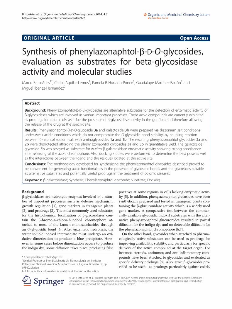
Results: Phenylazonaphtol-β-D-O-glucoside 3a and galactoside 3b were prepared via diazonium salt conditionsunder weak acidic conditions which do not compromise the O-glycosidic bond stability, by coupling reactionbetween 2-naphtol sodium salt with aminoglycosides 1a and 1b. The resulting phenylazonaphtol glycosides 2a and2b were deprotected affording the phenylazonaphtol glycosides 3a and 3b in quantitative yield. The galactosideglycoside 3b was assayed as substrate for in vitro β-galactosidase enzymatic activity showing strong absorbanceafter releasing of the azoic chromophore. Also, docking studies were performed to determine the best pose as wellas the interactions between the ligand and the residues located at the active site.
Conclusions: The methodology developed for synthesizing the phenylazonaphtol glycosides described proved tobe convenient for generating azoic functionalities in the presence of glycosidic bonds and the glycosides suitableas alternative substrates and potentially useful prodrugs in the treatment of colonic diseases.
Keywords: β-galactosidase; Synthesis; Phenylazonaphtol glycoside; Substrate; Docking
Backgroundβ-glycosidases are hydrolytic enzymes involved in a num-ber of important processes such as defense mechanism,growth regulation [1], gene markers in transgenic plants[2], and prodrugs [3]. The most commonly used substratesfor the histochemical localization of β-glycosidases con-tain the 5-bromo-4-chloro-3-indolyl chromophore at-tached to most of the known monosaccharides throughan O-glycosidic bond [4]. After enzymatic hydrolysis, thewater soluble indoxyl intermediate must undergo an oxi-dative dimerization to produce a blue precipitate. How-ever, in some cases before dimerization occurs to producethe indigo dye, some diffusion takes place, producing false
* Correspondence: [email protected] Profesional Interdisciplinaria de Biotecnología del InstitutoPolitécnico Nacional, Avenida Acueducto s/n La Laguna Ticomán DF cp07340, MexicoFull list of author information is available at the end of the article
© 2014 Brito-Arias et al.; licensee Springer. ThisAttribution License (http://creativecommons.orin any medium, provided the original work is p
positives at some regions in cells lacking enzymatic activ-ity [5]. In addition, phenylazonaphtol glucosides have beensynthetically prepared and tested in transgenic plants con-taining the β-glucuronidase activity which is a widely usedgene marker. A comparative test between the commer-cially available glycosidic indoxyl substrates with the alter-native phenylazonaphtol glucuronides resulted in partialdiffusion for the indigo dye and no detectable diffusion forthe phenylazonaphtol chromophore [6,7].On the other hand, glycosides when attached to pharma-
cologically active substances can be used as prodrugs forimproving availability, stability, and particularly for specificdelivery of the active compound at the target organ. Forinstance, steroids, antitumor, and anti-inflammatory com-pounds have been attached to glycosides and evaluated asspecific delivery prodrugs [8]. Also, azoic β-glycosides pro-vided to be useful as prodrugs particularly against colitis,
is an Open Access article distributed under the terms of the Creative Commonsg/licenses/by/2.0), which permits unrestricted use, distribution, and reproductionroperly credited.
Brito-Arias et al. Organic and Medicinal Chemistry Letters 2014, 4:2 Page 2 of 7http://www.orgmedchemlett.com/content/4/1/2
Crohn's disease, and colorectal cancer since the deliveryat colon level might be achieved by the action of azo-reductase or glycosidase present at the colonic micro-flora [9].Due the potential usefulness of phenylazonaphtol gly-
cosides in processes mentioned above, we developed amethodology for preparing phenylazonaphtol glycosidesunder weak acidic conditions and the resulting azoic gly-cosides evaluated as β-galactosidase substrates with po-tential application as prodrugs for colon diseases.
MethodsThe preparation of the phenylazonaphtol glycosides 3aand 3b was achieved according to Scheme 1 consistingin the coupling reaction between 2-naphtol in the so-dium salt form with 4-aminophenyl tetracetyl glycopyra-noside [10-12] 1a and 1b under modified diazonium saltcondition, which is mainly the use of acetic acid insteadof stronger acids, with sodium nitrite at 0°C to producethe diazonium salts which was condensed in situ with 2-naphtol sodium salt providing the corresponding pheny-lazonaphtol tetracetyl glucopyranoside 2a and 2b. Finalremoval of the acetate protecting groups under Zemplenconditions provided the target phenylazonaphtol glyco-sides 3a and 3b as a reddish solid in 60% yield.
SchemeThe beta galactosidase assay. β-galactosidase (2 mg; 2.1 U/mg) was suspended in 0.8 mL of buffer containing 0.06 MNa2HPO4, 0.04 M NaH2PO4, 0.01 M KCl, 0.001 M MgSO4,
O
OR3
R1
R3OOR3
O
NH2
R2
1a ; R1 = OAc, R2 = H, R3 = Ac1b ; R1 = H, R2 = OAc, R3 = Ac
O
i
ii
O
OH
R1
HOOH
O
R2
3a ; R1 = OH, R2 = H3b ; R1 = H, R2 = OH
Scheme 1 The preparation of the phenylazonaphtol glycosides 3a an
and 1 mM DTT, pH 7.0, and then incubated with glycoside3b (4 mg; 9.3 × 10⁻³ mM) in a mixture of 50 mM acetatebuffer with pH 5.0 (0.5 mL) and methanol (0.5 mL) at 37°Cfor 3 h. The reaction was stopped by adding 0.5 mL 1 MNa2CO3. The amount of the released phenylazonaphtolchromophore was measured spectrophotometrically at 410and 455 nm [13].
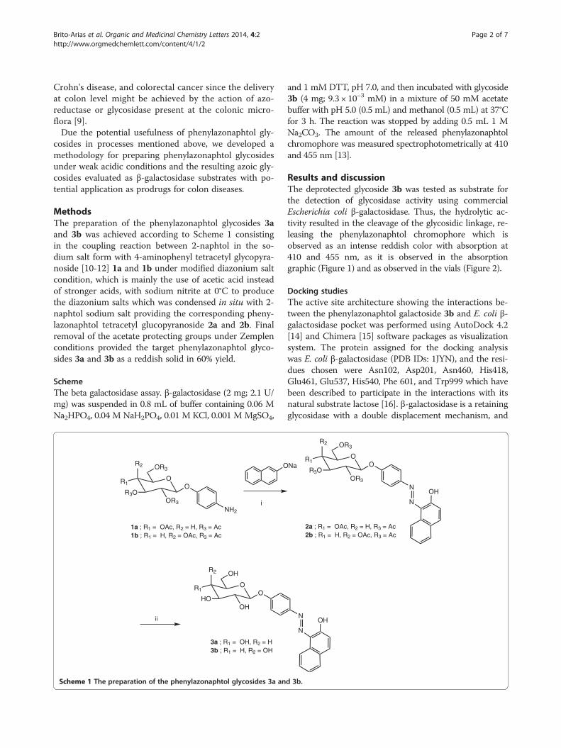
Results and discussionThe deprotected glycoside 3b was tested as substrate forthe detection of glycosidase activity using commercialEscherichia coli β-galactosidase. Thus, the hydrolytic ac-tivity resulted in the cleavage of the glycosidic linkage, re-leasing the phenylazonaphtol chromophore which isobserved as an intense reddish color with absorption at410 and 455 nm, as it is observed in the absorptiongraphic (Figure 1) and as observed in the vials (Figure 2).
Docking studiesThe active site architecture showing the interactions be-tween the phenylazonaphtol galactoside 3b and E. coli β-galactosidase pocket was performed using AutoDock 4.2[14] and Chimera [15] software packages as visualizationsystem. The protein assigned for the docking analysiswas E. coli β-galactosidase (PDB IDs: 1JYN), and the resi-dues chosen were Asn102, Asp201, Asn460, His418,Glu461, Glu537, His540, Phe 601, and Trp999 which havebeen described to participate in the interactions with itsnatural substrate lactose [16]. β-galactosidase is a retainingglycosidase with a double displacement mechanism, and
Na
N
N
OH
O
OR3
R1
R3OOR3
O
R2
2a ; R1 = OAc, R2 = H, R3 = Ac2b ; R1 = H, R2 = OAc, R3 = Ac
N
N
OH
d 3b.
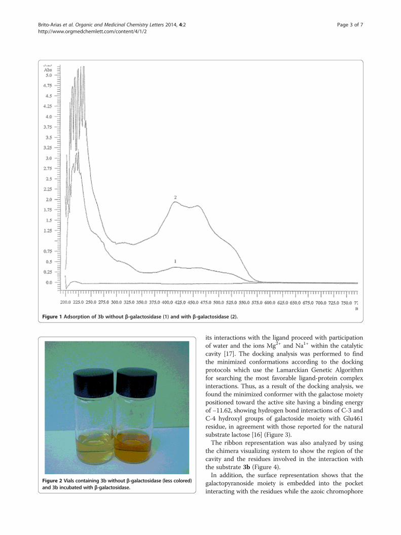
Figure 1 Adsorption of 3b without β-galactosidase (1) and with β-galactosidase (2).
Figure 2 Vials containing 3b without β-galactosidase (less colored)and 3b incubated with β-galactosidase.
Brito-Arias et al. Organic and Medicinal Chemistry Letters 2014, 4:2 Page 3 of 7http://www.orgmedchemlett.com/content/4/1/2
its interactions with the ligand proceed with participationof water and the ions Mg2+ and Na1+ within the catalyticcavity [17]. The docking analysis was performed to findthe minimized conformations according to the dockingprotocols which use the Lamarckian Genetic Algorithmfor searching the most favorable ligand-protein complexinteractions. Thus, as a result of the docking analysis, wefound the minimized conformer with the galactose moietypositioned toward the active site having a binding energyof −11.62, showing hydrogen bond interactions of C-3 andC-4 hydroxyl groups of galactoside moiety with Glu461residue, in agreement with those reported for the naturalsubstrate lactose [16] (Figure 3).The ribbon representation was also analyzed by using
the chimera visualizing system to show the region of thecavity and the residues involved in the interaction withthe substrate 3b (Figure 4).In addition, the surface representation shows that the
galactopyranoside moiety is embedded into the pocketinteracting with the residues while the azoic chromophore
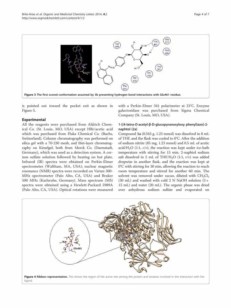
Figure 3 The first scored conformation assumed by 3b presenting hydrogen bond interactions with Glu461 residue.
Brito-Arias et al. Organic and Medicinal Chemistry Letters 2014, 4:2 Page 4 of 7http://www.orgmedchemlett.com/content/4/1/2
is pointed out toward the pocket exit as shown inFigure 5.
ExperimentalAll the reagents were purchased from Aldrich Chem-ical Co. (St. Louis, MO, USA) except HBr/acetic acidwhich was purchased from Fluka Chemical Co. (Buchs,Switzerland). Column chromatography was performed onsilica gel with a 70-230 mesh, and thin-layer chromatog-raphy on Kieselgel, both from Merck Co. (Darmstadt,Germany), which was used as a detection system. A cer-ium sulfate solution followed by heating on hot plate.Infrared (IR) spectra were obtained on Perkin-Elmerspectrometer (Waltham, MA, USA); nuclear magneticresonance (NMR) spectra were recorded on Varian 300-MHz spectrometer (Palo Alto, CA, USA) and Bruker500 MHz (Karlsruhe, Germany). Mass spectrum (MS)spectra were obtained using a Hewlett-Packard 5989A(Palo Alto, CA, USA). Optical rotations were measured
Figure 4 Ribbon representation. This shows the region of the active siteligand.
with a Perkin-Elmer 341 polarimeter at 23°C. Enzymegalactosidase was purchased from Sigma ChemicalCompany (St. Louis, MO, USA).
1-[(4-tetra-O-acetyl-β-D-glucopyranosyloxy phenyl)azo]-2-naphtol (2a)Compound 1a (0.543 g, 1.23 mmol) was dissolved in 8 mLof THF, and the flask was cooled to 0°C. After the additionof sodium nitrite (85 mg, 1.23 mmol) and 0.5 mL of aceticacid/H2O (1:1, v/v), the reaction was kept under ice-bathtemperature with stirring for 15 min. 2-naphtol sodiumsalt dissolved in 5 mL of THF/H2O (1:1, v/v) was addeddropwise in another flask, and the reaction was kept at0°C with stirring for 30 min, allowing the reaction to reachroom temperature and stirred for another 60 min. Thesolvent was removed under vacuo, diluted with CH2Cl2(30 mL) and washed with cold 2 N NaOH solution (3 ×15 mL) and water (20 mL). The organic phase was driedover anhydrous sodium sulfate and evaporated on
among the protein and residues involved in the interaction with the
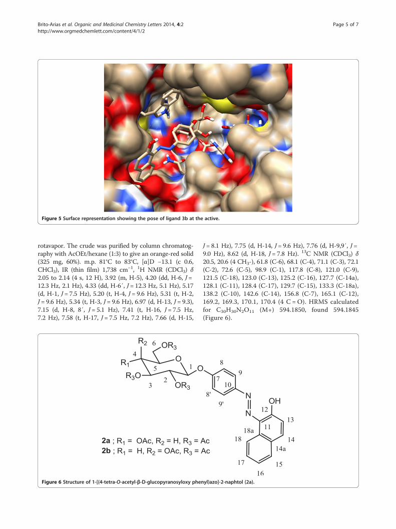
Figure 5 Surface representation showing the pose of ligand 3b at the active.
Brito-Arias et al. Organic and Medicinal Chemistry Letters 2014, 4:2 Page 5 of 7http://www.orgmedchemlett.com/content/4/1/2
rotavapor. The crude was purified by column chromatog-raphy with AcOEt/hexane (1:3) to give an orange-red solid(325 mg, 60%). m.p. 81°C to 83°C, [α]D −13.1 (c 0.6,CHCl3), IR (thin film) 1,738 cm−1, 1H NMR (CDCl3) δ2.05 to 2.14 (4 s, 12 H), 3.92 (m, H-5), 4.20 (dd, H-6, J =12.3 Hz, 2.1 Hz), 4.33 (dd, H-6′, J = 12.3 Hz, 5.1 Hz), 5.17(d, H-1, J = 7.5 Hz), 5.20 (t, H-4, J = 9.6 Hz), 5.31 (t, H-2,J = 9.6 Hz), 5.34 (t, H-3, J = 9.6 Hz), 6.97 (d, H-13, J = 9.3),7.15 (d, H-8, 8′, J = 5.1 Hz), 7.41 (t, H-16, J = 7.5 Hz,7.2 Hz), 7.58 (t, H-17, J = 7.5 Hz, 7.2 Hz), 7.66 (d, H-15,
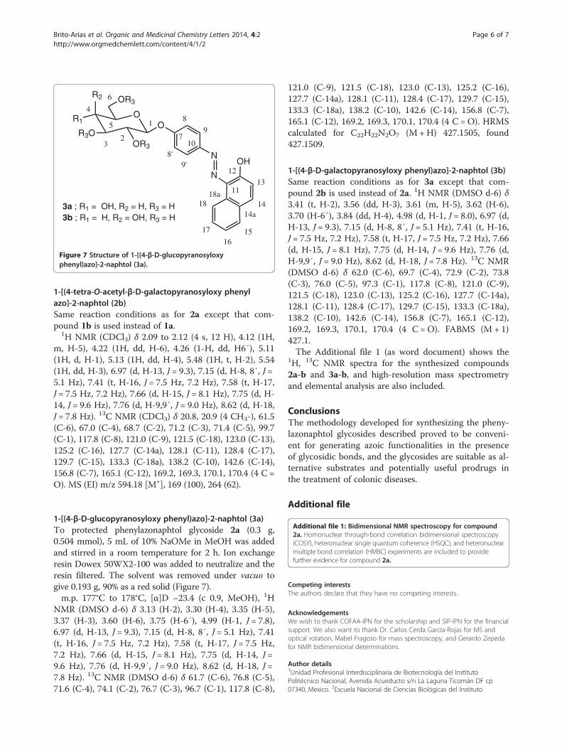
Figure 6 Structure of 1-[(4-tetra-O-acetyl-β-D-glucopyranosyloxy phen
J = 8.1 Hz), 7.75 (d, H-14, J = 9.6 Hz), 7.76 (d, H-9,9′, J =9.0 Hz), 8.62 (d, H-18, J = 7.8 Hz). 13C NMR (CDCl3) δ20.5, 20.6 (4 CH3-), 61.8 (C-6), 68.1 (C-4), 71.1 (C-3), 72.1(C-2), 72.6 (C-5), 98.9 (C-1), 117.8 (C-8), 121.0 (C-9),121.5 (C-18), 123.0 (C-13), 125.2 (C-16), 127.7 (C-14a),128.1 (C-11), 128.4 (C-17), 129.7 (C-15), 133.3 (C-18a),138.2 (C-10), 142.6 (C-14), 156.8 (C-7), 165.1 (C-12),169.2, 169.3, 170.1, 170.4 (4 C = O). HRMS calculatedfor C30H30N2O11 (M+) 594.1850, found 594.1845(Figure 6).
yl)azo]-2-naphtol (2a).
N
N
OH
O
OR3
R1
R3OOR3
O
R2
1
23
4
5
6
7
8
9
10
11
12
13
14
1516
17
18
8'9'
18a
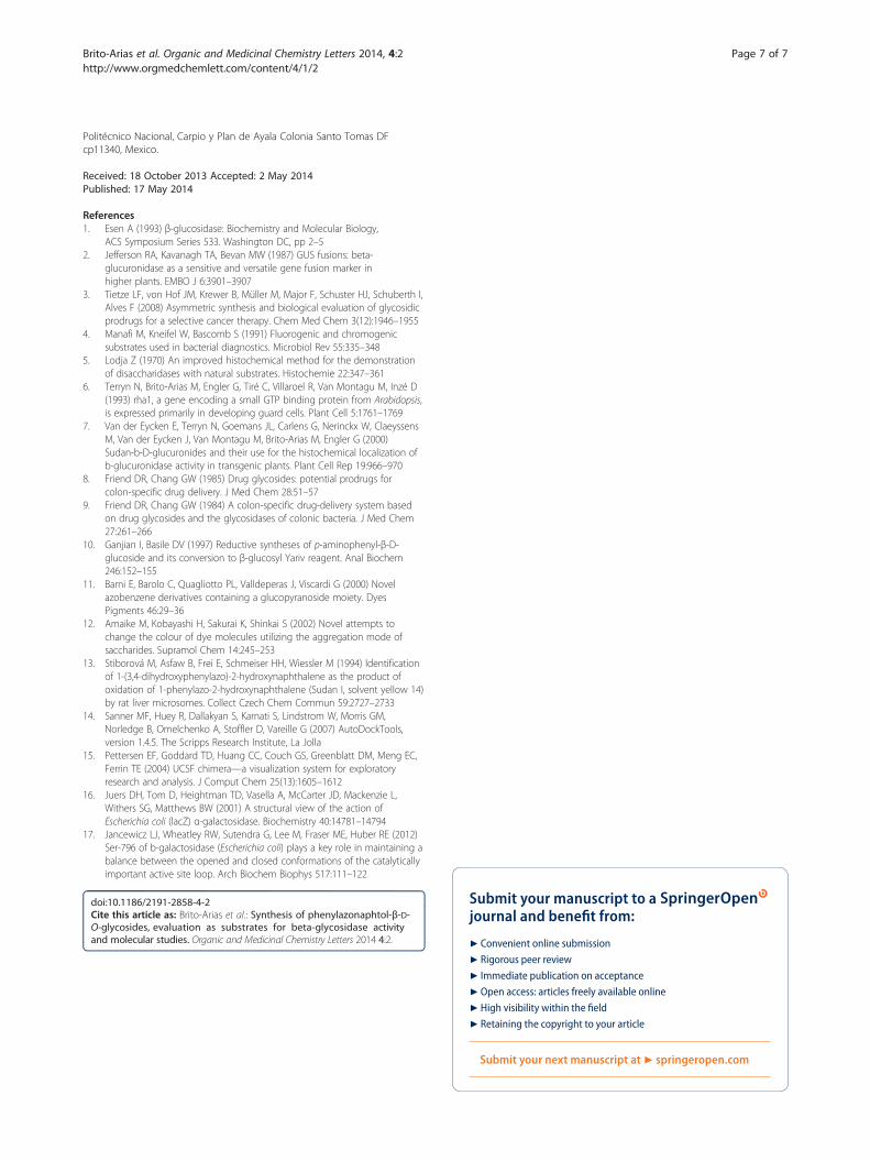
14a3a ; R1 = OH, R2 = H, R3 = H3b ; R1 = H, R2 = OH, R3 = H
Figure 7 Structure of 1-[(4-β-D-glucopyranosyloxyphenyl)azo]-2-naphtol (3a).
Brito-Arias et al. Organic and Medicinal Chemistry Letters 2014, 4:2 Page 6 of 7http://www.orgmedchemlett.com/content/4/1/2
1-[(4-tetra-O-acetyl-β-D-galactopyranosyloxy phenylazo]-2-naphtol (2b)Same reaction conditions as for 2a except that com-pound 1b is used instead of 1a.
1H NMR (CDCl3) δ 2.09 to 2.12 (4 s, 12 H), 4.12 (1H,m, H-5), 4.22 (1H, dd, H-6), 4.26 (1-H, dd, H6′), 5.11(1H, d, H-1), 5.13 (1H, dd, H-4), 5.48 (1H, t, H-2), 5.54(1H, dd, H-3), 6.97 (d, H-13, J = 9.3), 7.15 (d, H-8, 8′, J =5.1 Hz), 7.41 (t, H-16, J = 7.5 Hz, 7.2 Hz), 7.58 (t, H-17,J = 7.5 Hz, 7.2 Hz), 7.66 (d, H-15, J = 8.1 Hz), 7.75 (d, H-14, J = 9.6 Hz), 7.76 (d, H-9,9′, J = 9.0 Hz), 8.62 (d, H-18,J = 7.8 Hz). 13C NMR (CDCl3) δ 20.8, 20.9 (4 CH3-), 61.5(C-6), 67.0 (C-4), 68.7 (C-2), 71.2 (C-3), 71.4 (C-5), 99.7(C-1), 117.8 (C-8), 121.0 (C-9), 121.5 (C-18), 123.0 (C-13),125.2 (C-16), 127.7 (C-14a), 128.1 (C-11), 128.4 (C-17),129.7 (C-15), 133.3 (C-18a), 138.2 (C-10), 142.6 (C-14),156.8 (C-7), 165.1 (C-12), 169.2, 169.3, 170.1, 170.4 (4 C =O). MS (EI) m/z 594.18 [M+], 169 (100), 264 (62).
1-[(4-β-D-glucopyranosyloxy phenyl)azo]-2-naphtol (3a)To protected phenylazonaphtol glycoside 2a (0.3 g,0.504 mmol), 5 mL of 10% NaOMe in MeOH was addedand stirred in a room temperature for 2 h. Ion exchangeresin Dowex 50WX2-100 was added to neutralize and theresin filtered. The solvent was removed under vacuo togive 0.193 g, 90% as a red solid (Figure 7).m.p. 177°C to 178°C, [α]D −23.4 (c 0.9, MeOH), 1H
NMR (DMSO d-6) δ 3.13 (H-2), 3.30 (H-4), 3.35 (H-5),3.37 (H-3), 3.60 (H-6), 3.75 (H-6′), 4.99 (H-1, J = 7.8),6.97 (d, H-13, J = 9.3), 7.15 (d, H-8, 8′, J = 5.1 Hz), 7.41(t, H-16, J = 7.5 Hz, 7.2 Hz), 7.58 (t, H-17, J = 7.5 Hz,7.2 Hz), 7.66 (d, H-15, J = 8.1 Hz), 7.75 (d, H-14, J =9.6 Hz), 7.76 (d, H-9,9′, J = 9.0 Hz), 8.62 (d, H-18, J =7.8 Hz). 13C NMR (DMSO d-6) δ 61.7 (C-6), 76.8 (C-5),71.6 (C-4), 74.1 (C-2), 76.7 (C-3), 96.7 (C-1), 117.8 (C-8),
121.0 (C-9), 121.5 (C-18), 123.0 (C-13), 125.2 (C-16),127.7 (C-14a), 128.1 (C-11), 128.4 (C-17), 129.7 (C-15),133.3 (C-18a), 138.2 (C-10), 142.6 (C-14), 156.8 (C-7),165.1 (C-12), 169.2, 169.3, 170.1, 170.4 (4 C =O). HRMScalculated for C22H22N2O7 (M +H) 427.1505, found427.1509.
1-[(4-β-D-galactopyranosyloxy phenyl)azo]-2-naphtol (3b)Same reaction conditions as for 3a except that com-pound 2b is used instead of 2a. 1H NMR (DMSO d-6) δ3.41 (t, H-2), 3.56 (dd, H-3), 3.61 (m, H-5), 3.62 (H-6),3.70 (H-6′), 3.84 (dd, H-4), 4.98 (d, H-1, J = 8.0), 6.97 (d,H-13, J = 9.3), 7.15 (d, H-8, 8′, J = 5.1 Hz), 7.41 (t, H-16,J = 7.5 Hz, 7.2 Hz), 7.58 (t, H-17, J = 7.5 Hz, 7.2 Hz), 7.66(d, H-15, J = 8.1 Hz), 7.75 (d, H-14, J = 9.6 Hz), 7.76 (d,H-9,9′, J = 9.0 Hz), 8.62 (d, H-18, J = 7.8 Hz). 13C NMR(DMSO d-6) δ 62.0 (C-6), 69.7 (C-4), 72.9 (C-2), 73.8(C-3), 76.0 (C-5), 97.3 (C-1), 117.8 (C-8), 121.0 (C-9),121.5 (C-18), 123.0 (C-13), 125.2 (C-16), 127.7 (C-14a),128.1 (C-11), 128.4 (C-17), 129.7 (C-15), 133.3 (C-18a),138.2 (C-10), 142.6 (C-14), 156.8 (C-7), 165.1 (C-12),169.2, 169.3, 170.1, 170.4 (4 C =O). FABMS (M + 1)427.1.The Additional file 1 (as word document) shows the
1H, 13C NMR spectra for the synthesized compounds2a-b and 3a-b, and high-resolution mass spectrometryand elemental analysis are also included.
ConclusionsThe methodology developed for synthesizing the pheny-lazonaphtol glycosides described proved to be conveni-ent for generating azoic functionalities in the presenceof glycosidic bonds, and the glycosides are suitable as al-ternative substrates and potentially useful prodrugs inthe treatment of colonic diseases.
Additional file
Additional file 1: Bidimensional NMR spectroscopy for compound2a. Homonuclear through-bond correlation bidimensional spectroscopy(COSY), heteronuclear single quantum coherence (HSQC), and heteronuclearmultiple bond correlation (HMBC) experiments are included to providefurther evidence for compound 2a.
Competing interestsThe authors declare that they have no competing interests.
AcknowledgementsWe wish to thank COFAA-IPN for the scholarship and SIP-IPN for the financialsupport. We also want to thank Dr. Carlos Cerda Garcia-Rojas for MS andoptical rotation, Mabel Fragoso for mass spectroscopy, and Gerardo Zepedafor NMR bidimensional determinations.
Author details1Unidad Profesional Interdisciplinaria de Biotecnología del InstitutoPolitécnico Nacional, Avenida Acueducto s/n La Laguna Ticomán DF cp07340, Mexico. 2Escuela Nacional de Ciencias Biológicas del Instituto
Brito-Arias et al. Organic and Medicinal Chemistry Letters 2014, 4:2 Page 7 of 7http://www.orgmedchemlett.com/content/4/1/2
Politécnico Nacional, Carpio y Plan de Ayala Colonia Santo Tomas DFcp11340, Mexico.
Received: 18 October 2013 Accepted: 2 May 2014Published: 17 May 2014
References1. Esen A (1993) β-glucosidase: Biochemistry and Molecular Biology,
ACS Symposium Series 533. Washington DC, pp 2–52. Jefferson RA, Kavanagh TA, Bevan MW (1987) GUS fusions: beta-
glucuronidase as a sensitive and versatile gene fusion marker inhigher plants. EMBO J 6:3901–3907
3. Tietze LF, von Hof JM, Krewer B, Müller M, Major F, Schuster HJ, Schuberth I,Alves F (2008) Asymmetric synthesis and biological evaluation of glycosidicprodrugs for a selective cancer therapy. Chem Med Chem 3(12):1946–1955
4. Manafi M, Kneifel W, Bascomb S (1991) Fluorogenic and chromogenicsubstrates used in bacterial diagnostics. Microbiol Rev 55:335–348
5. Lodja Z (1970) An improved histochemical method for the demonstrationof disaccharidases with natural substrates. Histochemie 22:347–361
6. Terryn N, Brito-Arias M, Engler G, Tiré C, Villaroel R, Van Montagu M, Inzé D(1993) rha1, a gene encoding a small GTP binding protein from Arabidopsis,is expressed primarily in developing guard cells. Plant Cell 5:1761–1769
7. Van der Eycken E, Terryn N, Goemans JL, Carlens G, Nerinckx W, ClaeyssensM, Van der Eycken J, Van Montagu M, Brito-Arias M, Engler G (2000)Sudan-b-D-glucuronides and their use for the histochemical localization ofb-glucuronidase activity in transgenic plants. Plant Cell Rep 19:966–970
8. Friend DR, Chang GW (1985) Drug glycosides: potential prodrugs forcolon-specific drug delivery. J Med Chem 28:51–57
9. Friend DR, Chang GW (1984) A colon-specific drug-delivery system basedon drug glycosides and the glycosidases of colonic bacteria. J Med Chem27:261–266
10. Ganjian I, Basile DV (1997) Reductive syntheses of p-aminophenyl-β-D-glucoside and its conversion to β-glucosyl Yariv reagent. Anal Biochem246:152–155
11. Barni E, Barolo C, Quagliotto PL, Valldeperas J, Viscardi G (2000) Novelazobenzene derivatives containing a glucopyranoside moiety. DyesPigments 46:29–36
12. Amaike M, Kobayashi H, Sakurai K, Shinkai S (2002) Novel attempts tochange the colour of dye molecules utilizing the aggregation mode ofsaccharides. Supramol Chem 14:245–253
13. Stiborová M, Asfaw B, Frei E, Schmeiser HH, Wiessler M (1994) Identificationof 1-(3,4-dihydroxyphenylazo)-2-hydroxynaphthalene as the product ofoxidation of 1-phenylazo-2-hydroxynaphthalene (Sudan I, solvent yellow 14)by rat liver microsomes. Collect Czech Chem Commun 59:2727–2733
14. Sanner MF, Huey R, Dallakyan S, Karnati S, Lindstrom W, Morris GM,Norledge B, Omelchenko A, Stoffler D, Vareille G (2007) AutoDockTools,version 1.4.5. The Scripps Research Institute, La Jolla
15. Pettersen EF, Goddard TD, Huang CC, Couch GS, Greenblatt DM, Meng EC,Ferrin TE (2004) UCSF chimera—a visualization system for exploratoryresearch and analysis. J Comput Chem 25(13):1605–1612
16. Juers DH, Tom D, Heightman TD, Vasella A, McCarter JD, Mackenzie L,Withers SG, Matthews BW (2001) A structural view of the action ofEscherichia coli (lacZ) α-galactosidase. Biochemistry 40:14781–14794
17. Jancewicz LJ, Wheatley RW, Sutendra G, Lee M, Fraser ME, Huber RE (2012)Ser-796 of b-galactosidase (Escherichia coli) plays a key role in maintaining abalance between the opened and closed conformations of the catalyticallyimportant active site loop. Arch Biochem Biophys 517:111–122
doi:10.1186/2191-2858-4-2Cite this article as: Brito-Arias et al.: Synthesis of phenylazonaphtol-β-D-O-glycosides, evaluation as substrates for beta-glycosidase activityand molecular studies. Organic and Medicinal Chemistry Letters 2014 4:2.
Submit your manuscript to a journal and benefi t from:
7 Convenient online submission
7 Rigorous peer review
7 Immediate publication on acceptance
7 Open access: articles freely available online
7 High visibility within the fi eld
7 Retaining the copyright to your article
Submit your next manuscript at 7 springeropen.com


![Nootaree Niljianskul HHS Public Access Shaolin Zhu, and ... · attractive as starting materials for the synthesis of α-aminosilanes.[10] The intermolecular hydroamination of vinylsilanes](https://static.fdocument.org/doc/165x107/5ee1d576ad6a402d666c91d2/nootaree-niljianskul-hhs-public-access-shaolin-zhu-and-attractive-as-starting.jpg)


![ORIGINAL ARTICLE Open Access β-Keto esters from ketones ...tory and antiphlogistic properties. Especially, a pyrazolone derivative (edaravone) [3] acts as a radical scavenger to interrupt](https://static.fdocument.org/doc/165x107/608fba6ac49a6d7592273fd2/original-article-open-access-keto-esters-from-ketones-tory-and-antiphlogistic.jpg)