Original article: DEVELOPMENT OF A NEW RUTIN · PDF filedata the inhibitory concentration...
14
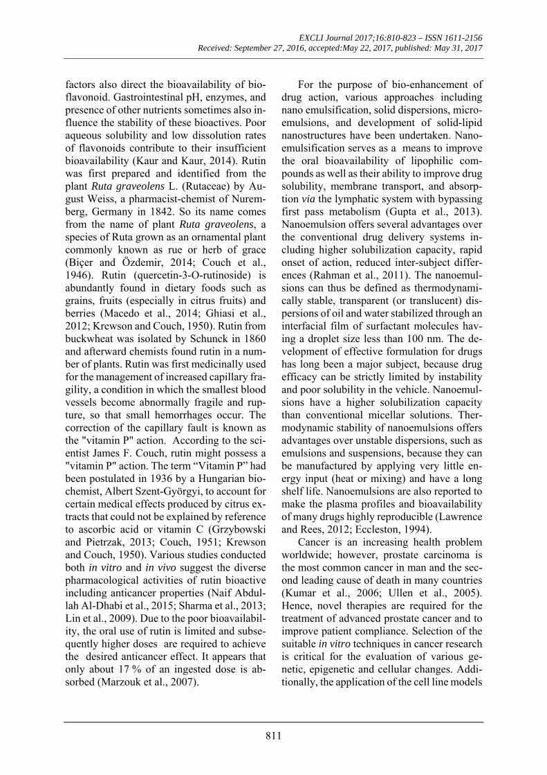
EXCLI Journal 2017;16:810-823 – ISSN 1611-2156 Received: September 27, 2016, accepted:May 22, 2017, published: May 31, 2017 810 Original article: DEVELOPMENT OF A NEW RUTIN NANOEMULSION AND ITS APPLICATION ON PROSTATE CARCINOMA PC3 CELL LINE Mohammad Ahmad 1 , Sahabjada 2 , Juber Akhtar 1 , Arshad Hussain 3 , Badaruddeen 1 , Md Arshad 2 , Anuradha Mishra 1* 1 Herbal Bioactive Research Laboratory, Faculty of Pharmacy, Integral University, Lucknow, India 2 Molecular Endocrinology Laboratory, Department of Zoology, University of Lucknow, Lucknow, India 3 College of Pharmacy, King Khalid University, Abha, KSA. Formerly, Faculty of Pharmacy, Integral University, Lucknow, India * corresponding author: Dr. Anuradha Mishra, Department of Pharmacology, Faculty of Pharmacy, Integral University, Lucknow-226026 (U.P.) India. E-mail: [email protected] http://dx.doi.org/10.17179/excli2016-668 This is an Open Access article distributed under the terms of the Creative Commons Attribution License (http://creativecommons.org/licenses/by/4.0/). ABSTRACT Biological effects of rutin bioactive are limited due to its poor oral bioavailability and its degradation in aqueous environments. For the purpose of bioenhancement, different nanoemulsion systems of rutin were developed by aqueous titration method using water as dispersion media. The nanoemulsion systems were characterized for sur- face morphology, droplet size, polydispersity index, zeta potential, in vitro release profile and the formulations were optimized. The anticancer potential of optimized nanoemulsion was evaluated by cells viability (MTT) assay, nuclear condensation, and ROS activity using human prostate cancer (PC3) cell line. On the basis of cell viability data the inhibitory concentration (IC 50 ) value for optimized nanoemulsion formulation on PC3 cancer cells was found to be 11.8 μM. Fluorescent microscopic analysis and intracellular ROS generation demonstrated significant ROS induction that might lead to triggering the apoptosis pathway. In conclusion, developed nanoemulsion dis- played significant efficacy against prostate carcinoma cells. Keywords: rutin, nanoemulsion, water titration method, cell viability, prostate cancer, reactive oxygen species INTRODUCTION Cancer chemoprevention using bioactive compounds has attracted increasing attention in recent years (Liu, 2013; Surh, 2003). Bio- active compounds with anticancer properties alone or in combinations with other potent synthetic molecules are used to prevent or re- verse the processes of carcinogenesis, further- more to minimize the undesirable side effects, which are commonly associated with current therapies (Pan and Ho, 2008). Several bio-fla- vonoids found to be effective in in vitro stud- ies at a particular concentration, often exhibit lower responses at a higher concentration as revealed by in vivo studies (Scalbert et al., 2005; Amidon et al., 1995). Many factors such as gastric residence time, permeability, and solubility within the gut and some other
Transcript of Original article: DEVELOPMENT OF A NEW RUTIN · PDF filedata the inhibitory concentration...
EXCLI Journal 2017;16:810-823 – ISSN 1611-2156 Received: September 27, 2016, accepted:May 22, 2017, published: May 31, 2017
810
Original article:
DEVELOPMENT OF A NEW RUTIN NANOEMULSION AND ITS APPLICATION ON PROSTATE CARCINOMA PC3 CELL LINE
Mohammad Ahmad1, Sahabjada2, Juber Akhtar1, Arshad Hussain3, Badaruddeen1, Md Arshad2, Anuradha Mishra1*
1 Herbal Bioactive Research Laboratory, Faculty of Pharmacy, Integral University,
Lucknow, India 2 Molecular Endocrinology Laboratory, Department of Zoology, University of Lucknow,
Lucknow, India 3 College of Pharmacy, King Khalid University, Abha, KSA. Formerly, Faculty of
Pharmacy, Integral University, Lucknow, India * corresponding author: Dr. Anuradha Mishra, Department of Pharmacology,
Faculty of Pharmacy, Integral University, Lucknow-226026 (U.P.) India. E-mail: [email protected]
http://dx.doi.org/10.17179/excli2016-668
This is an Open Access article distributed under the terms of the Creative Commons Attribution License (http://creativecommons.org/licenses/by/4.0/).
ABSTRACT
Biological effects of rutin bioactive are limited due to its poor oral bioavailability and its degradation in aqueous environments. For the purpose of bioenhancement, different nanoemulsion systems of rutin were developed by aqueous titration method using water as dispersion media. The nanoemulsion systems were characterized for sur-face morphology, droplet size, polydispersity index, zeta potential, in vitro release profile and the formulations were optimized. The anticancer potential of optimized nanoemulsion was evaluated by cells viability (MTT) assay, nuclear condensation, and ROS activity using human prostate cancer (PC3) cell line. On the basis of cell viability data the inhibitory concentration (IC50) value for optimized nanoemulsion formulation on PC3 cancer cells was found to be 11.8 μM. Fluorescent microscopic analysis and intracellular ROS generation demonstrated significant ROS induction that might lead to triggering the apoptosis pathway. In conclusion, developed nanoemulsion dis-played significant efficacy against prostate carcinoma cells. Keywords: rutin, nanoemulsion, water titration method, cell viability, prostate cancer, reactive oxygen species
INTRODUCTION
Cancer chemoprevention using bioactive compounds has attracted increasing attention in recent years (Liu, 2013; Surh, 2003). Bio-active compounds with anticancer properties alone or in combinations with other potent synthetic molecules are used to prevent or re-verse the processes of carcinogenesis, further-more to minimize the undesirable side effects,
which are commonly associated with current therapies (Pan and Ho, 2008). Several bio-fla-vonoids found to be effective in in vitro stud-ies at a particular concentration, often exhibit lower responses at a higher concentration as revealed by in vivo studies (Scalbert et al., 2005; Amidon et al., 1995). Many factors such as gastric residence time, permeability, and solubility within the gut and some other
EXCLI Journal 2017;16:810-823 – ISSN 1611-2156 Received: September 27, 2016, accepted:May 22, 2017, published: May 31, 2017
811
factors also direct the bioavailability of bio-flavonoid. Gastrointestinal pH, enzymes, and presence of other nutrients sometimes also in-fluence the stability of these bioactives. Poor aqueous solubility and low dissolution rates of flavonoids contribute to their insufficient bioavailability (Kaur and Kaur, 2014). Rutin was first prepared and identified from the plant Ruta graveolens L. (Rutaceae) by Au-gust Weiss, a pharmacist-chemist of Nurem-berg, Germany in 1842. So its name comes from the name of plant Ruta graveolens, a species of Ruta grown as an ornamental plant commonly known as rue or herb of grace (Biçer and Özdemir, 2014; Couch et al., 1946). Rutin (quercetin-3-O-rutinoside) is abundantly found in dietary foods such as grains, fruits (especially in citrus fruits) and berries (Macedo et al., 2014; Ghiasi et al., 2012; Krewson and Couch, 1950). Rutin from buckwheat was isolated by Schunck in 1860 and afterward chemists found rutin in a num-ber of plants. Rutin was first medicinally used for the management of increased capillary fra-gility, a condition in which the smallest blood vessels become abnormally fragile and rup-ture, so that small hemorrhages occur. The correction of the capillary fault is known as the "vitamin P" action. According to the sci-entist James F. Couch, rutin might possess a "vitamin P" action. The term “Vitamin P” had been postulated in 1936 by a Hungarian bio-chemist, Albert Szent-Györgyi, to account for certain medical effects produced by citrus ex-tracts that could not be explained by reference to ascorbic acid or vitamin C (Grzybowski and Pietrzak, 2013; Couch, 1951; Krewson and Couch, 1950). Various studies conducted both in vitro and in vivo suggest the diverse pharmacological activities of rutin bioactive including anticancer properties (Naif Abdul-lah Al-Dhabi et al., 2015; Sharma et al., 2013; Lin et al., 2009). Due to the poor bioavailabil-ity, the oral use of rutin is limited and subse-quently higher doses are required to achieve the desired anticancer effect. It appears that only about 17 % of an ingested dose is ab-sorbed (Marzouk et al., 2007).
For the purpose of bio-enhancement of drug action, various approaches including nano emulsification, solid dispersions, micro-emulsions, and development of solid-lipid nanostructures have been undertaken. Nano-emulsification serves as a means to improve the oral bioavailability of lipophilic com-pounds as well as their ability to improve drug solubility, membrane transport, and absorp-tion via the lymphatic system with bypassing first pass metabolism (Gupta et al., 2013). Nanoemulsion offers several advantages over the conventional drug delivery systems in-cluding higher solubilization capacity, rapid onset of action, reduced inter-subject differ-ences (Rahman et al., 2011). The nanoemul-sions can thus be defined as thermodynami-cally stable, transparent (or translucent) dis-persions of oil and water stabilized through an interfacial film of surfactant molecules hav-ing a droplet size less than 100 nm. The de-velopment of effective formulation for drugs has long been a major subject, because drug efficacy can be strictly limited by instability and poor solubility in the vehicle. Nanoemul-sions have a higher solubilization capacity than conventional micellar solutions. Ther-modynamic stability of nanoemulsions offers advantages over unstable dispersions, such as emulsions and suspensions, because they can be manufactured by applying very little en-ergy input (heat or mixing) and have a long shelf life. Nanoemulsions are also reported to make the plasma profiles and bioavailability of many drugs highly reproducible (Lawrence and Rees, 2012; Eccleston, 1994).
Cancer is an increasing health problem worldwide; however, prostate carcinoma is the most common cancer in man and the sec-ond leading cause of death in many countries (Kumar et al., 2006; Ullen et al., 2005). Hence, novel therapies are required for the treatment of advanced prostate cancer and to improve patient compliance. Selection of the suitable in vitro techniques in cancer research is critical for the evaluation of various ge-netic, epigenetic and cellular changes. Addi-tionally, the application of the cell line models
EXCLI Journal 2017;16:810-823 – ISSN 1611-2156 Received: September 27, 2016, accepted:May 22, 2017, published: May 31, 2017
812
already exists as a part of the ongoing re-search on cancer therapy (Louzada et al., 2012). Cell line models are very useful tools for the measurement of mediators such as pro-liferation deregulation, apoptosis and cancer progression (Vargo-Gogola and Rosen, 2007). To date there are 4 well studied Human Prostate Cancer cell lines. They are DU145, PC3, LNCaP and TSU-PnI. Out of all these PC3 cell line is easily available and shows most similar progression as that of human prostate cancer. The PC3 and DU145 human prostate cancer cell lines are representative of the earlier type-I ADI prostate cancers. PC3, but not DU145 cells retain the coregulators needed for AR tumor suppressor ability of an-drogen receptor (Litvinov et al., 2006).
In the present study we selected PC3 cell line of human prostate carcinoma to evaluate the antiproliferative potential of the devel-oped rutin nanoemulsion.
MATERIALS AND METHODS
Chemicals and reagents Propylene glycol monocaprylate (Capryol
90) and caprylocaproyl macrogol-8-glyceride (Labrasol) (Gattefosse, Gennevilliers, France) were gift samples from Colorcon Asia (Mumbai, India), while propylene glycol monocaprylic ester (Sefsol 218) and Kolli-phor RH-40 were a gift sample from Nikko Chemicals (Tokyo, Japan) and BASF, Mum-bai, respectively. Diethylenemonoglycol ether (Carbitol), Polyethylene Glycol (PEG-200, 400, 600), Isopropyl myristate (IPM), glycerol triacetate (Triacetin), methanol (HPLC-Grade), and Distilled Water were pur-chased from E-Merck (Mumbai, India). Poly-oxyethylene sorbitan monolaurate (Tween-20), polyoxy ethylene sorbitan monostearate (Tween-60), polyoxyethylene sorbitan mono-oleate (Tween-80), ethanol, isopropyl alcohol were procured from S.D. Fine Chemicals (Mumbai, India). Fluorescent dye 2,7-dichlo-rodihydrofluorescein diacetate (DCFH-DA) and 4',6'-diamidino-2 phenylindole (DAPI) were purchased from Sigma Aldrich, USA. Eagle’s minimal essential medium (MEM),
fetal bovine serum (FBS), MTT (3 (4,5-dime-thylthiazol-2-yl)-2,5-diphenyltetrazolium bromide) dye and antibiotic solutions were purchased from Himedia, India. Rutin (97 %) bioactives were purchased from Sigma-Al-drich Chemical Company (St. Louis, Mis-souri). All the reagents used for study were of analytical grade.
Cell line and culture
Human prostate adenocarcinoma (PC3) cell line was procured from National Centre for Cell Sciences (NCCS), Pune, India. PC3 cells were maintained in MEM medium sup-plemented with 2.0 mM L-glutamine, 1.5 g/l NaHCO3, 0.1 mM non-essential amino acids, 1.0 mM sodium pyruvate and 10 % (v/v) FBS. Cells were incubated at 37 °C and 5 % CO2 incubator.
Selection of excipients on the basis of solubility
To find out appropriate oils, surfactants and co-surfactant as components of nanoemulsion system with high loading ca-pacity is based on the solubility of poorly sol-uble drug in oils, surfactants, and co-surfac-tants, screening of component such as oils (middle chain, long chain and synthetic tri-glycerides) Triacetin, Isopropyl myristate, Capryol-90, Sefsol 218, Olive oil including surfactants Tween-80, Labrasol, Kolliphor-HS15, Kolliphor-RH40 and co-surfactants such as Transcutol-P, Carbitol, Polyethylene glycol-200, 400 and 600. An excess amount of drug was added in the oils and surfactants as well as co-surfactant and kept in isothermal shaker for 72 h at 25 ± 2 °C temperature to reach equilibrium. Finally samples were re-moved from the shaker and centrifuged at 3000 rpm for 15 min. The supernatant was taken and filtered through a 0.45 µm mem-brane filter to remove the remaining insoluble drugs. Concentration of rutin was determined by HPLC. All the excipients selected for for-mulation were under the GRAS (Generally Regarded as Safe) category. Finally excipi-ents were selected as formulation components
EXCLI Journal 2017;16:810-823 – ISSN 1611-2156 Received: September 27, 2016, accepted:May 22, 2017, published: May 31, 2017
813
on the basis of highest solubility of rutin in different excipient (Wang et al., 2009).
Method of preparation of nanoemulsion
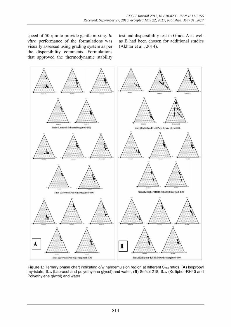
Nanoemulsion systems were developed using phase titration method keeping water as dispersion media. Pseudoternary phase dia-grams were constructed to represent the best ratio of oil, surfactant/co-surfactant (Smix), and water. Surfactant and co-surfactant was mixed (Smix) in different volume ratios (1:0, 1:1, 1:2, 1:3, 2:1, 3:1, etc). These Smix ratios were chosen to reflect increasing concentra-tions of co-surfactant with respect to surfac-tant and increasing concentrations of surfac-tant with respect to co-surfactant for detailed study of the phase diagrams in the nanoemul-sion formation. Mixture of oil with Smix was prepared at different ratios (e.g. 10:0, 9:1, 8:2, 7:3, 6:4, 5:5, 4:6, 3:7, 2:8, 1:9, 0:10) into dif-ferent vials. A small amount of water in 5 % (w/w) increments was added into the vials. Following each water addition the mixture in vials was centrifuged for 2 to 3 minute and in-cubated at 25 °C for 48 h with gentle shaking. The resulting mixtures were evaluated by vis-ual and microscopy observation. For phase di-agram the nanoemulsion is the region of clear and isotropic solution. The physical state of the nanoemulsion is marked on the phase dia-grams with one axis representing aqueous phase, the second representing oil and the third representing a mixture of surfactant and co-surfactant (Smix) at fixed weight ratios (Smix ratio). The nanoemulsion area in each phase diagram is plotted and the wider region indicated the better self nanoemulsifying effi-ciency (Singh et al., 2008).
Construction of pseudoternary phase diagrams
Pseudoternary phase diagrams were drawn for determining Smix ratio and its ratio with oil phase in emulsion system which gives the nanoemulsion region (Figure 1). On the basis of solubility and compatibility of ru-tin with various excipients, Kolliphor RH-40/Labrasol//PEG-200, 400, 600 was selected as the surfactant, PEG-200, 400, 600 as co-
surfactant and Sefsol218/Isopropyl myrsitate were selected as the oil phase. For every phase diagram, oil and accurate Smix ratio was mixed in volume ratios ranging from 1:9 to 9:1 to ob-tain sixteen different combinations like 1:9, 1:8, 1:7, 1:6, 1:5 1:4, 1:3.5, 1:3, 3:7, 1:2, 4:6, 5:5, 6:4, 7:3, 8:2 and 9:1 and titrated with wa-ter for constructing zone of nanoemulsion. Smix ratios 1:0, 1:1, 2:1, 1:2 and 3:1 were se-lected from above experiments. Concentra-tions of all the excipients used were within permissible limits recommended for oral us-age.
Development of drug containing formulations
Nanoemulsion system was developed by taking best suited optimized concentrations of oil phase (Sefsol 218), surfactant (Kolliphor RH-40), and co-surfactant (PEG-400) was se-lected from ternary phase diagrams and this system was then loaded with excess amount of rutin (10 mg/ml). Another system compris-ing the oil phase (Isopropyl myristate), sur-factant (Labrasol) and co-surfactant (PEG-600) was selected from ternary phase dia-grams and this system was then loaded with excess amount of rutin (10 mg/ml). These were subsequently sonicated for 10 minutes on a bath sonicator.
Measurement of drug contents in nanoemulsion
Finally six formulations were selected and the best suitable formulations amongst them were analyzed for the drug contents from nanoemulsion formulations that were ex-tracted in methanol. The solutions were fil-tered, using Whatman filter paper of 0.45 µm pore size and analyzed for the drug contents through HPLC.
Characterization of optimized formulation dispersibility test
One ml of each nanoemulsion formulation was added to 500 mL of distilled water and 0.1 N HCl separately at a temperature of 37 °C in a standard USP XXII dissolution ap-paratus. The dissolution paddle rotated at a
EXCLI Journal 2017;16:810-823 – ISSN 1611-2156 Received: September 27, 2016, accepted:May 22, 2017, published: May 31, 2017
814
speed of 50 rpm to provide gentle mixing. In vitro performance of the formulations was visually assessed using grading system as per the dispersibility comments. Formulations that approved the thermodynamic stability
test and dispersibility test in Grade A as well as B had been chosen for additional studies (Akhtar et al., 2014).
Figure 1: Ternary phase chart indicating o/w nanoemulsion region at different Smix ratios. (A) Isopropyl myristate, Smix (Labrasol and polyethylene glycol) and water, (B) Sefsol 218, Smix (Kolliphor-RH40 and Polyethylene glycol) and water
EXCLI Journal 2017;16:810-823 – ISSN 1611-2156 Received: September 27, 2016, accepted:May 22, 2017, published: May 31, 2017
815
Thermodynamic stability studies Selected formulations were subjected to
different thermodynamic stability tests to as-sess their physical stability. For centrifugation test, a definite volume of formulations were diluted in (1:10-1:100 ratios) with aqueous medium and centrifuged at 15,000 rpm for 15 minutes, then the formulation was observed visually for the phase separation. After that formulations were subjected to freeze thaw cycles between −21 °C and +25 °C, with for-mulation storage at each temperature for not less than 48 h. Those formulations found to be thermodynamically stable were selected for further characterization (Shafiq-un-Nabi et al., 2007).
Percentage transmittance
Percentage transmittance of the prepared nanoemulsion was determined spectrophoto-metrically after 100 times dilution with meth-anol using Shimadzu UV-visible spectropho-tometer keeping distilled water as a blank.
Droplet size and zeta potential analysis
The droplet size and the size distribution of the prepared nanoemulsion systems were determined using Zetasizer (Nano ZS, Mal-vern Instruments, U.K). For the purpose of particle size different dilution were made and each sample was analyzed in triplicate at a temperature, 20 °C and refractive index, 1.4. Zeta potential was also measured by photon correlation spectroscopy. The prepared nano-emulsions with and without dilution were an-alyzed using double distilled water.
Transmission electron microscopy (TEM)
Details on the morphology and other structural features of the nanoemulsion for-mulations were viewed using Transmission Electron Microscope (TEM), Model EM-410 LS facility available at USIF, AMU, Aligarh (India). Prior to the analysis, the samples were diluted at 10-100 times with water and ap-plied on the grids which were stained with 2 % (w/v) phosphotungstic acid for 30 s and then grids were observed after drying, using
combination of bright field imaging at in-creasing magnification to reveal the form and size of nanoemulsion droplets (Shafiq-un-Nabi et al., 2007).
Viscosity
Viscosity of the selected formulations were determined at 25 ± 0.5 °C by Brookfield Viscometer DV III ultra V6.0 RV cone and plate rheometer (Brookfield Engineering La-boratories, Middleboro, MA) (Shafiq-un-Nabi et al., 2007).
In vitro drug release study
Comparative in vitro release profile of the optimized nanoemulsion and the pure drug suspension in phosphate buffer (pH 6.8), 0.1 N HCl (pH 1.2) and distilled water were stud-ied using dialysis bag techniques at 100 rpm rotational speed. The temperature was main-tained at 37 ± 0.5 °C. Drug release was car-ried out by placing 1 ml of nanoemulsion in treated dialysis bag (MWCO 14,000 g/mole, Sigma, USA). 0.5 mL aliquots was withdrawn at pre-determined time intervals (0, 0.25, 0.5, 0.75, 1, 1.25, 1.5, 1.75, 2, 2.25, 2.5, 2.75, 3, 4, 6, 8, 12, 16, 20, and 24 h) and same volume was replenished with fresh dissolution media to maintain the sink condition. The samples withdrawn were filtered using 0.45 µm filter paper and the drug was analyzed by reported HPLC method (Shafiq-un-Nabi et al., 2007).
In vitro cell viability assay
To detect cell viability after treatment with rutin nanoemulsion, approximately 1x10⁴ cells/well of PC3 were seeded in 100 μl complete culture medium in 96-well culture plate and incubated overnight in humidified air. Stock was prepared in phosphate buffer saline (PBS) and diluted into culture medium to the desired concentrations 2, 5, 10, 15 and 20 μM, then added to the wells. After 24 h of incubation period, 10 μl of MTT (5 mg/ml in PBS) reagent was added and re-incubated at 37 °C until purple formazan crystals devel-oped. Formazan blue crystals were dissolved in 100 μl of DMSO and read at 540 nm using microplate ELISA reader (BIORAD 680,
EXCLI Journal 2017;16:810-823 – ISSN 1611-2156 Received: September 27, 2016, accepted:May 22, 2017, published: May 31, 2017
816
USA). The plot of percent cell viability versus nanoemulsion concentrations was used to cal-culate the concentration lethal to 50 % of the cells (IC50). The cellular morphological changes were observed under inverted phase contrast microscopy (Nikon ECLIPSE Ti-S, Japan) (Siddiqui and Arshad, 2014).
Intracellular ROS activity
Intracellular ROS generation was ana-lyzed by using fluorescence microscopic im-aging technique (Siddiqui et al., 2015). Cells (1×10⁴ per well) were exposed at two effec-tive concentrations i.e. 5 and 10 μM of rutin nanoemulsion for 12 h. Subsequently, cells were incubated with DCFH-DA (10 mM) at 37 °C for 30 min and washed with PBS. Intra-cellular fluorescence intensity of cells was visualized by inverted fluorescent microscope (Nikon ECLIPSE Ti-S, Japan). For quantita-tive fluorometric analysis, cells (1×10⁴ per well) were seeded and treated with nanoemul-sion in 96-well black bottom culture plate. Fluorescence intensity was measured with a multiwell microplate reader (Synergy H1 Hy-brid Multi-Mode Microplate Reader, BioTek, USA) at an excitation wavelength of 485 nm and at an emission wavelength of 528 nm. Data were expressed as percentage of fluores-cence intensity relative to the control wells.
Apoptotic effect of formulation using DAPI stain
Fluorescent nuclear dye was used to ana-lyze the apoptotic effect of rutin nanoemul-sion. PC3 cells (1×10⁵ cells per well) were seeded in 24-well culture plate overnight and treated with rutin nanoemulsion for 24 h. Fol-lowing incubation period, cells were washed and fixed in 4 % paraformaldehyde for 15 min followed by permeabilization with permea-bilizing buffer (3 % paraformaldehyde and 0.5 % Triton X-100) for 10 min. After stain-ing with DAPI dye (50 µg/ml), images of con-densed nuclei undergoing apoptosis were cap-tured with an inverted fluorescent microscope (Nikon ECLIPSE Ti-S, Japan). Apoptosis was quantitated by morphological changes of
nuclei with approximately 500 cells/well rep-resenting one sample (Siddiqui et al., 2015).
Statistical analysis
The results were expressed as mean ± SD and were analyzed statistically (graph pad prism for Windows, version 5) using one-way analysis of variance (ANOVA) followed by Tukey’s test and considered statistically sig-nificant when < 0.05.
RESULTS AND DISCUSSION
Preparation and characterization of nanoemulsion
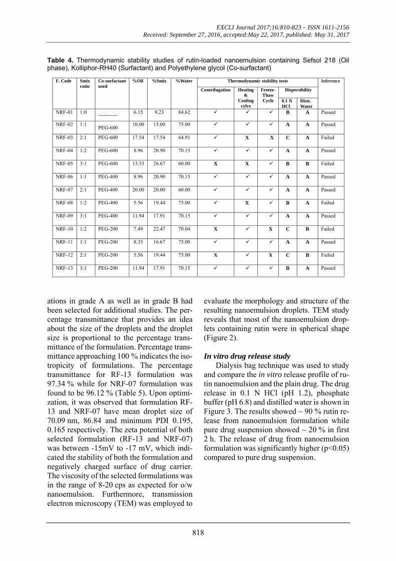
Pseudoternary phase diagrams were con-structed to represent the concentration of oils, surfactant and co-surfactant used for the de-velopment of appropriate formulation (Shafiq-un-Nabi et al., 2007). Solubility of Rutin in Isopropyl myristate, Sefsol 218 was highest among all the oils screened (Table 1). On the basis of solubility study Labrasol and Kolliphor-RH40 were selected as surfactant whereas polyethelene glycol 600 and poly-ethelene glycol-400 as co-surfactant respec-tively. Rutin nanoemulsion system (RF-13) contains Isopropyl myristate (IPM) as oil phase with Smix comprised of Labrasol and PEG-600 whereas another rutin nanoemul-sion system (NRF-07) has Sefsol 218 as the oil phase with Smix comprising Kolliphor-RH40 as surfactant and PEG-400 as co-sur-factant. Nanoemulsion systems of these opti-mized components were evaluated for the dis-persibility, thermodynamic stability, percent-age transmittance and droplet size analysis (Table 2). Thermodynamically stable systems are formed at a particular concentration of oil, surfactant and water, without any kinetic in-stability and phase separation (Table 3 and Table 4). In order to develop an oral nano-emulsion formulation, dispersibility studies were of great importance. Dispersibility tests were done to evaluate the dispersion effi-ciency and the stability of nanoemulsion sys-tems in the gastrointestinal fluids. On the ba-sis of dispersibility assessment, those formul-
EXCLI Journal 2017;16:810-823 – ISSN 1611-2156 Received: September 27, 2016, accepted:May 22, 2017, published: May 31, 2017
817
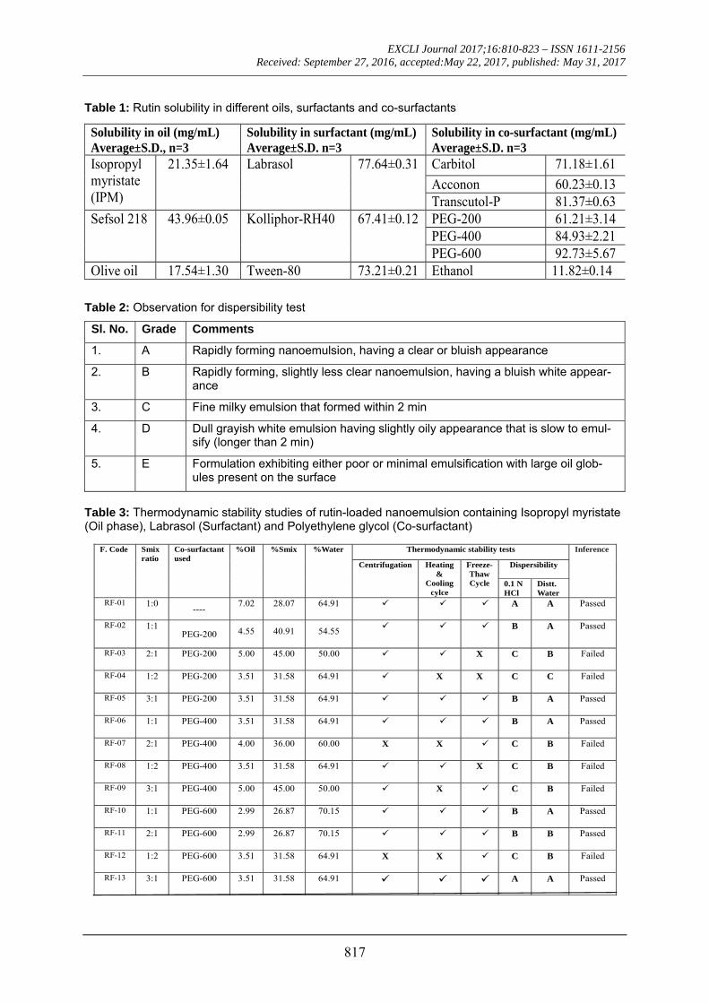
Table 1: Rutin solubility in different oils, surfactants and co-surfactants
Table 2: Observation for dispersibility test
Sl. No. Grade Comments
1. A Rapidly forming nanoemulsion, having a clear or bluish appearance
2. B Rapidly forming, slightly less clear nanoemulsion, having a bluish white appear-ance
3. C Fine milky emulsion that formed within 2 min
4. D Dull grayish white emulsion having slightly oily appearance that is slow to emul-sify (longer than 2 min)
5. E Formulation exhibiting either poor or minimal emulsification with large oil glob-ules present on the surface
Table 3: Thermodynamic stability studies of rutin-loaded nanoemulsion containing Isopropyl myristate (Oil phase), Labrasol (Surfactant) and Polyethylene glycol (Co-surfactant)
F. Code Smix ratio
Co-surfactant used
%Oil
%Smix
%Water Thermodynamic stability tests Inference
Centrifugation
Heating&
Cooling cylce
Freeze-Thaw Cycle
Dispersibility
0.1 N HCl
Distt. Water
RF-01 1:0 ----
7.02 28.07 64.91 A A Passed
RF-02 1:1 PEG-200 4.55 40.91 54.55
B A Passed
RF-03 2:1 PEG-200 5.00 45.00 50.00 X C B Failed
RF-04 1:2 PEG-200 3.51 31.58 64.91 X X C C Failed
RF-05 3:1 PEG-200 3.51 31.58 64.91 B A Passed
RF-06 1:1 PEG-400 3.51 31.58 64.91 B A Passed
RF-07 2:1 PEG-400 4.00 36.00 60.00 X X C B Failed
RF-08 1:2 PEG-400 3.51 31.58 64.91 X C B Failed
RF-09 3:1 PEG-400 5.00 45.00 50.00 X C B Failed
RF-10 1:1 PEG-600 2.99 26.87 70.15 B A Passed
RF-11 2:1 PEG-600 2.99 26.87 70.15 B B Passed
RF-12 1:2 PEG-600 3.51 31.58 64.91 X X C B Failed
RF-13 3:1 PEG-600 3.51 31.58 64.91 A A Passed
Solubility in oil (mg/mL) Average±S.D., n=3
Solubility in surfactant (mg/mL) Average±S.D. n=3
Solubility in co-surfactant (mg/mL) Average±S.D. n=3
Isopropyl myristate (IPM)
21.35±1.64
Labrasol 77.64±0.31 Carbitol 71.18±1.61
Acconon 60.23±0.13 Transcutol-P 81.37±0.63
Sefsol 218 43.96±0.05
Kolliphor-RH40 67.41±0.12 PEG-200 61.21±3.14 PEG-400 84.93±2.21 PEG-600 92.73±5.67
Olive oil 17.54±1.30 Tween-80 73.21±0.21 Ethanol 11.82±0.14
EXCLI Journal 2017;16:810-823 – ISSN 1611-2156 Received: September 27, 2016, accepted:May 22, 2017, published: May 31, 2017
818
Table 4. Thermodynamic stability studies of rutin-loaded nanoemulsion containing Sefsol 218 (Oil phase), Kolliphor-RH40 (Surfactant) and Polyethylene glycol (Co-surfactant)
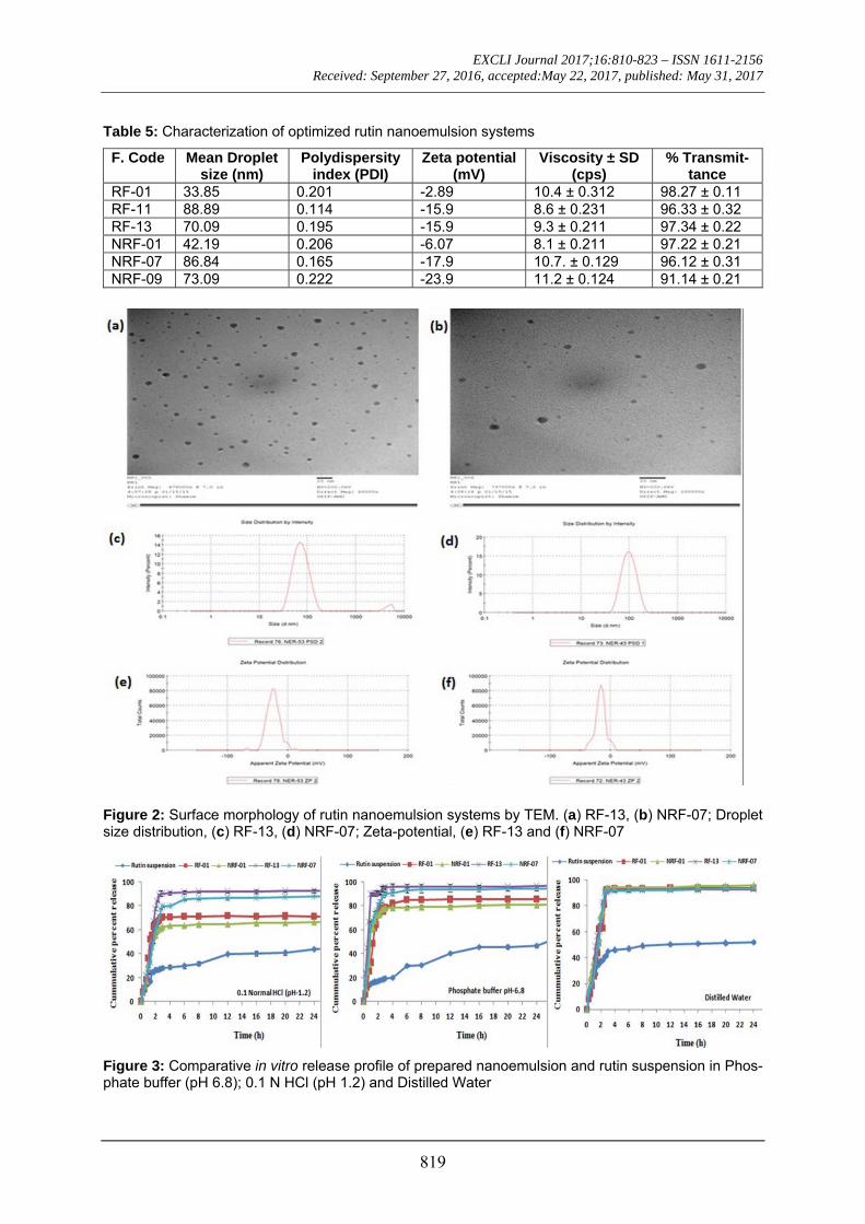
ations in grade A as well as in grade B had been selected for additional studies. The per-centage transmittance that provides an idea about the size of the droplets and the droplet size is proportional to the percentage trans-mittance of the formulation. Percentage trans-mittance approaching 100 % indicates the iso-tropicity of formulations. The percentage transmittance for RF-13 formulation was 97.34 % while for NRF-07 formulation was found to be 96.12 % (Table 5). Upon optimi-zation, it was observed that formulation RF-13 and NRF-07 have mean droplet size of 70.09 nm, 86.84 and minimum PDI 0.195, 0.165 respectively. The zeta potential of both selected formulation (RF-13 and NRF-07) was between -15mV to -17 mV, which indi-cated the stability of both the formulation and negatively charged surface of drug carrier. The viscosity of the selected formulations was in the range of 8-20 cps as expected for o/w nanoemulsion. Furthermore, transmission electron microscopy (TEM) was employed to
evaluate the morphology and structure of the resulting nanoemulsion droplets. TEM study reveals that most of the nanoemulsion drop-lets containing rutin were in spherical shape (Figure 2).
In vitro drug release study
Dialysis bag technique was used to study and compare the in vitro release profile of ru-tin nanoemulsion and the plain drug. The drug release in 0.1 N HCl (pH 1.2), phosphate buffer (pH 6.8) and distilled water is shown in Figure 3. The results showed ~ 90 % rutin re-lease from nanoemulsion formulation while pure drug suspension showed ~ 20 % in first 2 h. The release of drug from nanoemulsion formulation was significantly higher (p<0.05) compared to pure drug suspension.
F. Code Smix ratio
Co-surfactant used
%Oil
%Smix
%Water Thermodynamic stability tests Inference
Centrifugation
Heating&
Cooling cylce
Freeze-Thaw Cycle
Dispersibility
0.1 N HCl
Distt. Water
NRF-01 1:0 -----------
6.15 9.23 84.62 B A Passed
NRF-02 1:1 PEG-600
10.00 15.00 75.00 A A Passed
NRF-03 2:1 PEG-600 17.54 17.54 64.91 X X C A Failed
NRF-04 1:2 PEG-600 8.96 20.90 70.15 A A Passed
NRF-05 3:1 PEG-600 13.33 26.67 60.00 X X B B Failed
NRF-06 1:1 PEG-400 8.96 20.90 70.15 A A Passed
NRF-07 2:1 PEG-400 20.00 20.00 60.00 A A Passed
NRF-08 1:2 PEG-400 5.56 19.44 75.00 X B A Failed
NRF-09 3:1 PEG-400 11.94 17.91 70.15 A A Passed
NRF-10 1:2 PEG-200 7.49 22.47 70.04 X X C B Failed
NRF-11 1:1 PEG-200 8.33 16.67 75.00 A A Passed
NRF-12 2:1 PEG-200 5.56 19.44 75.00 X X C B Failed
NRF-13 3:1 PEG-200 11.94 17.91 70.15 B A Passed
EXCLI Journal 2017;16:810-823 – ISSN 1611-2156 Received: September 27, 2016, accepted:May 22, 2017, published: May 31, 2017
819
Table 5: Characterization of optimized rutin nanoemulsion systems
F. Code Mean Droplet size (nm)
Polydispersity index (PDI)
Zeta potential (mV)
Viscosity ± SD (cps)
% Transmit-tance
RF-01 33.85 0.201 -2.89 10.4 ± 0.312 98.27 ± 0.11 RF-11 88.89 0.114 -15.9 8.6 ± 0.231 96.33 ± 0.32 RF-13 70.09 0.195 -15.9 9.3 ± 0.211 97.34 ± 0.22 NRF-01 42.19 0.206 -6.07 8.1 ± 0.211 97.22 ± 0.21 NRF-07 86.84 0.165 -17.9 10.7. ± 0.129 96.12 ± 0.31 NRF-09 73.09 0.222 -23.9 11.2 ± 0.124 91.14 ± 0.21
Figure 2: Surface morphology of rutin nanoemulsion systems by TEM. (a) RF-13, (b) NRF-07; Droplet size distribution, (c) RF-13, (d) NRF-07; Zeta-potential, (e) RF-13 and (f) NRF-07
Figure 3: Comparative in vitro release profile of prepared nanoemulsion and rutin suspension in Phos-phate buffer (pH 6.8); 0.1 N HCl (pH 1.2) and Distilled Water
EXCLI Journal 2017;16:810-823 – ISSN 1611-2156 Received: September 27, 2016, accepted:May 22, 2017, published: May 31, 2017
820
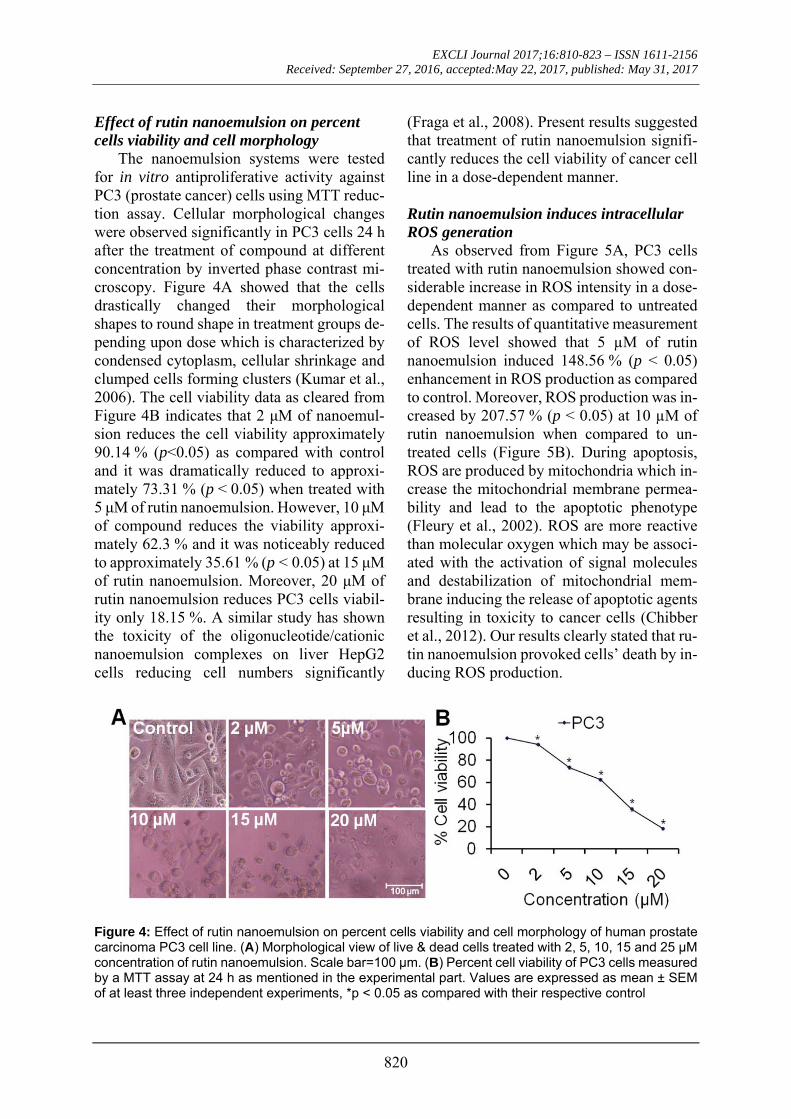
Effect of rutin nanoemulsion on percent cells viability and cell morphology
The nanoemulsion systems were tested for in vitro antiproliferative activity against PC3 (prostate cancer) cells using MTT reduc-tion assay. Cellular morphological changes were observed significantly in PC3 cells 24 h after the treatment of compound at different concentration by inverted phase contrast mi-croscopy. Figure 4A showed that the cells drastically changed their morphological shapes to round shape in treatment groups de-pending upon dose which is characterized by condensed cytoplasm, cellular shrinkage and clumped cells forming clusters (Kumar et al., 2006). The cell viability data as cleared from Figure 4B indicates that 2 μM of nanoemul-sion reduces the cell viability approximately 90.14 % (p<0.05) as compared with control and it was dramatically reduced to approxi-mately 73.31 % (p < 0.05) when treated with 5 μM of rutin nanoemulsion. However, 10 μM of compound reduces the viability approxi-mately 62.3 % and it was noticeably reduced to approximately 35.61 % (p < 0.05) at 15 μM of rutin nanoemulsion. Moreover, 20 μM of rutin nanoemulsion reduces PC3 cells viabil-ity only 18.15 %. A similar study has shown the toxicity of the oligonucleotide/cationic nanoemulsion complexes on liver HepG2 cells reducing cell numbers significantly
(Fraga et al., 2008). Present results suggested that treatment of rutin nanoemulsion signifi-cantly reduces the cell viability of cancer cell line in a dose-dependent manner.
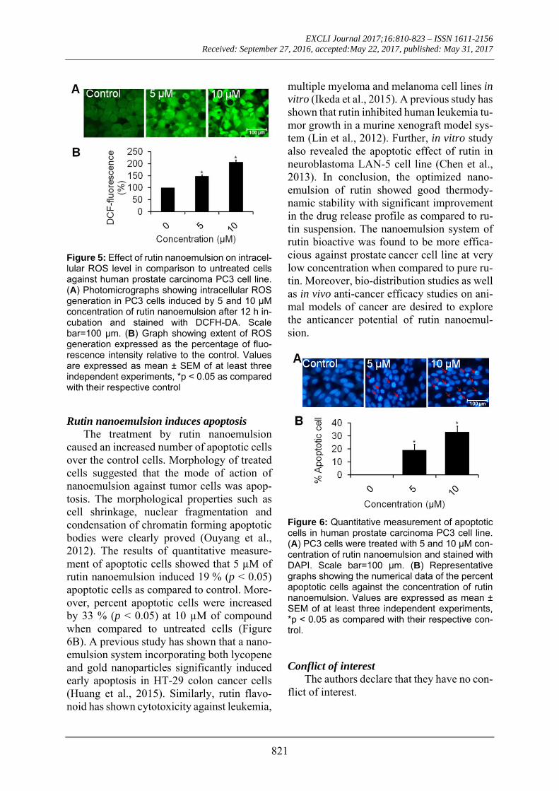
Rutin nanoemulsion induces intracellular ROS generation
As observed from Figure 5A, PC3 cells treated with rutin nanoemulsion showed con-siderable increase in ROS intensity in a dose-dependent manner as compared to untreated cells. The results of quantitative measurement of ROS level showed that 5 µM of rutin nanoemulsion induced 148.56 % (p < 0.05) enhancement in ROS production as compared to control. Moreover, ROS production was in-creased by 207.57 % (p < 0.05) at 10 µM of rutin nanoemulsion when compared to un-treated cells (Figure 5B). During apoptosis, ROS are produced by mitochondria which in-crease the mitochondrial membrane permea-bility and lead to the apoptotic phenotype (Fleury et al., 2002). ROS are more reactive than molecular oxygen which may be associ-ated with the activation of signal molecules and destabilization of mitochondrial mem-brane inducing the release of apoptotic agents resulting in toxicity to cancer cells (Chibber et al., 2012). Our results clearly stated that ru-tin nanoemulsion provoked cells’ death by in-ducing ROS production.
Figure 4: Effect of rutin nanoemulsion on percent cells viability and cell morphology of human prostate carcinoma PC3 cell line. (A) Morphological view of live & dead cells treated with 2, 5, 10, 15 and 25 µM concentration of rutin nanoemulsion. Scale bar=100 μm. (B) Percent cell viability of PC3 cells measured by a MTT assay at 24 h as mentioned in the experimental part. Values are expressed as mean ± SEM of at least three independent experiments, *p < 0.05 as compared with their respective control
EXCLI Journal 2017;16:810-823 – ISSN 1611-2156 Received: September 27, 2016, accepted:May 22, 2017, published: May 31, 2017
821
Figure 5: Effect of rutin nanoemulsion on intracel-lular ROS level in comparison to untreated cells against human prostate carcinoma PC3 cell line. (A) Photomicrographs showing intracellular ROS generation in PC3 cells induced by 5 and 10 µM concentration of rutin nanoemulsion after 12 h in-cubation and stained with DCFH-DA. Scale bar=100 μm. (B) Graph showing extent of ROS generation expressed as the percentage of fluo-rescence intensity relative to the control. Values are expressed as mean ± SEM of at least three independent experiments, *p < 0.05 as compared with their respective control Rutin nanoemulsion induces apoptosis
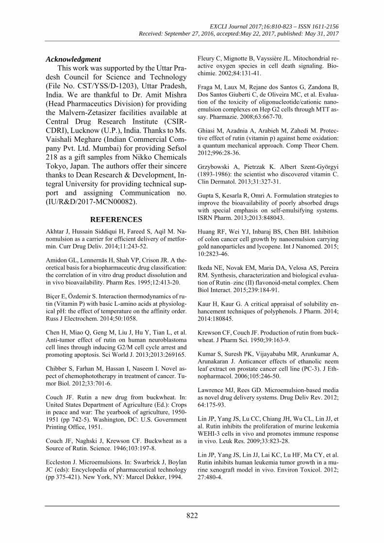
The treatment by rutin nanoemulsion caused an increased number of apoptotic cells over the control cells. Morphology of treated cells suggested that the mode of action of nanoemulsion against tumor cells was apop-tosis. The morphological properties such as cell shrinkage, nuclear fragmentation and condensation of chromatin forming apoptotic bodies were clearly proved (Ouyang et al., 2012). The results of quantitative measure-ment of apoptotic cells showed that 5 µM of rutin nanoemulsion induced 19 % (p < 0.05) apoptotic cells as compared to control. More-over, percent apoptotic cells were increased by 33 % (p < 0.05) at 10 µM of compound when compared to untreated cells (Figure 6B). A previous study has shown that a nano-emulsion system incorporating both lycopene and gold nanoparticles significantly induced early apoptosis in HT-29 colon cancer cells (Huang et al., 2015). Similarly, rutin flavo-noid has shown cytotoxicity against leukemia,
multiple myeloma and melanoma cell lines in vitro (Ikeda et al., 2015). A previous study has shown that rutin inhibited human leukemia tu-mor growth in a murine xenograft model sys-tem (Lin et al., 2012). Further, in vitro study also revealed the apoptotic effect of rutin in neuroblastoma LAN-5 cell line (Chen et al., 2013). In conclusion, the optimized nano-emulsion of rutin showed good thermody-namic stability with significant improvement in the drug release profile as compared to ru-tin suspension. The nanoemulsion system of rutin bioactive was found to be more effica-cious against prostate cancer cell line at very low concentration when compared to pure ru-tin. Moreover, bio-distribution studies as well as in vivo anti-cancer efficacy studies on ani-mal models of cancer are desired to explore the anticancer potential of rutin nanoemul-sion.
Figure 6: Quantitative measurement of apoptotic cells in human prostate carcinoma PC3 cell line. (A) PC3 cells were treated with 5 and 10 µM con-centration of rutin nanoemulsion and stained with DAPI. Scale bar=100 μm. (B) Representative graphs showing the numerical data of the percent apoptotic cells against the concentration of rutin nanoemulsion. Values are expressed as mean ± SEM of at least three independent experiments, *p < 0.05 as compared with their respective con-trol.
Conflict of interest
The authors declare that they have no con-flict of interest.
EXCLI Journal 2017;16:810-823 – ISSN 1611-2156 Received: September 27, 2016, accepted:May 22, 2017, published: May 31, 2017
822
Acknowledgment This work was supported by the Uttar Pra-
desh Council for Science and Technology (File No. CST/YSS/D-1203), Uttar Pradesh, India. We are thankful to Dr. Amit Mishra (Head Pharmaceutics Division) for providing the Malvern-Zetasizer facilities available at Central Drug Research Institute (CSIR-CDRI), Lucknow (U.P.), India. Thanks to Ms. Vaishali Meghare (Indian Commercial Com-pany Pvt. Ltd. Mumbai) for providing Sefsol 218 as a gift samples from Nikko Chemicals Tokyo, Japan. The authors offer their sincere thanks to Dean Research & Development, In-tegral University for providing technical sup-port and assigning Communication no. (IU/R&D/2017-MCN00082).
REFERENCES
Akhtar J, Hussain Siddiqui H, Fareed S, Aqil M. Na-nomulsion as a carrier for efficient delivery of metfor-min. Curr Drug Deliv. 2014;11:243-52.
Amidon GL, Lennernäs H, Shah VP, Crison JR. A the-oretical basis for a biopharmaceutic drug classification: the correlation of in vitro drug product dissolution and in vivo bioavailability. Pharm Res. 1995;12:413-20.
Biçer E, Özdemir S. Interaction thermodynamics of ru-tin (Vitamin P) with basic L-amino acids at physiolog-ical pH: the effect of temperature on the affinity order. Russ J Electrochem. 2014;50:1058.
Chen H, Miao Q, Geng M, Liu J, Hu Y, Tian L, et al. Anti-tumor effect of rutin on human neuroblastoma cell lines through inducing G2/M cell cycle arrest and promoting apoptosis. Sci World J. 2013;2013:269165.
Chibber S, Farhan M, Hassan I, Naseem I. Novel as-pect of chemophototherapy in treatment of cancer. Tu-mor Biol. 2012;33:701-6.
Couch JF. Rutin a new drug from buckwheat. In: United States Department of Agriculture (Ed.): Crops in peace and war: The yearbook of agriculture, 1950-1951 (pp 742-5). Washington, DC: U.S. Government Printing Office, 1951.
Couch JF, Naghski J, Krewson CF. Buckwheat as a Source of Rutin. Science. 1946;103:197-8.
Eccleston J. Microemulsions. In: Swarbrick J, Boylan JC (eds): Encyclopedia of pharmaceutical technology (pp 375-421). New York, NY: Marcel Dekker, 1994.
Fleury C, Mignotte B, Vayssière JL. Mitochondrial re-active oxygen species in cell death signaling. Bio-chimie. 2002;84:131-41.
Fraga M, Laux M, Rejane dos Santos G, Zandona B, Dos Santos Giuberti C, de Oliveira MC, et al. Evalua-tion of the toxicity of oligonucleotide/cationic nano-emulsion complexes on Hep G2 cells through MTT as-say. Pharmazie. 2008;63:667-70.
Ghiasi M, Azadnia A, Arabieh M, Zahedi M. Protec-tive effect of rutin (vitamin p) against heme oxidation: a quantum mechanical approach. Comp Theor Chem. 2012;996:28-36.
Grzybowski A, Pietrzak K. Albert Szent-Györgyi (1893-1986): the scientist who discovered vitamin C. Clin Dermatol. 2013;31:327-31.
Gupta S, Kesarla R, Omri A. Formulation strategies to improve the bioavailability of poorly absorbed drugs with special emphasis on self-emulsifying systems. ISRN Pharm. 2013;2013:848043.
Huang RF, Wei YJ, Inbaraj BS, Chen BH. Inhibition of colon cancer cell growth by nanoemulsion carrying gold nanoparticles and lycopene. Int J Nanomed. 2015; 10:2823-46.
Ikeda NE, Novak EM, Maria DA, Velosa AS, Pereira RM. Synthesis, characterization and biological evalua-tion of Rutin–zinc (II) flavonoid-metal complex. Chem Biol Interact. 2015;239:184-91.
Kaur H, Kaur G. A critical appraisal of solubility en-hancement techniques of polyphenols. J Pharm. 2014; 2014:180845.
Krewson CF, Couch JF. Production of rutin from buck-wheat. J Pharm Sci. 1950;39:163-9.
Kumar S, Suresh PK, Vijayababu MR, Arunkumar A, Arunakaran J. Anticancer effects of ethanolic neem leaf extract on prostate cancer cell line (PC-3). J Eth-nopharmacol. 2006;105:246-50.
Lawrence MJ, Rees GD. Microemulsion-based media as novel drug delivery systems. Drug Deliv Rev. 2012; 64:175-93.
Lin JP, Yang JS, Lu CC, Chiang JH, Wu CL, Lin JJ, et al. Rutin inhibits the proliferation of murine leukemia WEHI-3 cells in vivo and promotes immune response in vivo. Leuk Res. 2009;33:823-28.
Lin JP, Yang JS, Lin JJ, Lai KC, Lu HF, Ma CY, et al. Rutin inhibits human leukemia tumor growth in a mu-rine xenograft model in vivo. Environ Toxicol. 2012; 27:480-4.
EXCLI Journal 2017;16:810-823 – ISSN 1611-2156 Received: September 27, 2016, accepted:May 22, 2017, published: May 31, 2017
823
Litvinov IV, Antony L, Dalrymple SL, Becker R, Cheng L, Isaacs JT. PC3, but not DU145, human pros-tate cancer cells retain the coregulators required for tu-mor suppressor ability of androgen receptor. Prostate. 2006;66:1329-38.
Liu RH. Dietary bioactive compounds and their health implications. J Food Sci. 2013;78:18-25.
Louzada S, Adega F, Chaves R. Defining the sister rat mammary tumor cell lines HH-16 cl.2/1 and HH-16.cl.4 as an in vitro cell model for Erbb2. PloS one. 2012;7(1):e29923.
Macedo AS, Quelhas S, Silva AM, Souto EB. Nanoemulsions for delivery of flavonoids: formulation and in vitro release of rutin as model drug. Pharm Dev Technol. 2014;19:677-80.
Marzouk MS, Soliman FM, Shehata IA, Rabee M, Fawzy GA. Flavonoids and biological activities of Jus-siaea repens. Nat Prod Res. 2007;21:436-43.
Naif Abdullah Al-Dhabi NA, Arasu MV, Park CH, Park SU. An up-to-date review of rutin and its biolog-ical and pharmacological activities. EXCLI J. 2015; 14:59–63.
Ouyang L, Shi Z, Zhao S, Wang FT, Zhou TT, Liu B, et al. Programmed cell death pathways in cancer: a re-view of apoptosis, autophagy and programmed necro-sis. Cell Prolif. 2012;45:487-98.
Pan MH, Ho CT. Chemopreventive effects of natural dietary compounds on cancer development. Chem Soc Rev. 2008;37:2558–74.
Rahman A, Harwansh R, Mirza A, Hussain S, Hussain A. Oral lipid based drug delivery system: formulation, characterization and application: a review. Curr Drug Deliv. 2011;8:330-45.
Scalbert A, Manach C, Morand C, Rémésy C, Jiménez L. Dietary polyphenols and the prevention of diseases. Crit Rev Food Sci Nutr. 2005;45:287-306.
Schunck E. On the yellow colouring matters obtained from the leaves of Polygonum fagoeyrum, or common buckwheat. Mem Proc Manchester Lit Phil Soc Series 2. 1860;15:122-9.
Shafiq-un-Nabi S, Shakeel F, Talegaonkar S, Ali J, Ba-boota S, Ahuja A, et al. Formulation development and optimization using nanoemulsion technique: a tech-nical note. AAPS Pharm Sci Tech. 2007;18:E12-7.
Sharma S, Ali A, Ali J, Sahni JK, Baboota S. Rutin: therapeutic potential and recent advances in drug de-livery. Expert Opin Investig Drugs. 2013;22:1063-79.
Siddiqui S, Arshad M. Osteogenic potential of punica granatum through matrix mineralization, cell cycle progression and runx2 gene expression in primary rat osteoblasts. Daru. 2014;22:72.
Siddiqui S, Ahmad E, Gupta M, Rawat V, Shivnath N, Banerjee M, et al. Cissus quadrangularis Linn exerts dose‐dependent biphasic effects: osteogenic and anti‐proliferative, through modulating ROS, cell cycle and Runx2 gene expression in primary rat osteoblasts. Cell Prolif. 2015;48:443-54.
Singh AK, Chaurasiya A, Singh M, Upadhyay SC, Mukherjee R, Khar RK. Exemestane loaded self-mi-croemulsifying drug delivery system (SMEDDS): de-velopment and optimization. AAPS Pharm Sci Tech. 2008;9:628-34.
Surh YJ. Cancer chemoprevention with dietary phyto-chemicals. Nat Rev Cancer. 2003;3:768-80.
Ullén A, Lennartsson L, Harmenberg U, Lennernäs B, Majumder K, Holmberg AR, et al. Prostate cancer cell lines lack amplification: overexpression of HER2. Acta Oncol. 2005;44:490-5.
Vargo-Gogola T, Rosen JM. Modelling breast cancer: one size does not fit all. Nat Rev Cancer. 2007;7:659-72.
Wang L, Dong J, Chen J, Eastoe J, Li X. Design and optimization of a new self-nanoemulsifying drug deliv-ery system. J Colloid Interface Sci. 2009;330:443-8.